demystifying abnormal pupils - academy of vision care cet 53 the size of the pupil is determined by...
TRANSCRIPT

22/0
2/13
CET
52
CET CONTINUING EDUCATION & TRAINING
1 CET POINT
My AcademyA unique online resource, offering personalised education to meet individual needs and interests.
Visit www.optical.org for all the information about Enhanced CET requirements
xxxx
Demystifying abnormal pupilsTina Kipioti, MD, FRCSEd
About the authorTina Kipioti is a consultant ophthalmologist at Midland Eye Institute, specialising in children’s eye conditions, and squint surgery in both children and adults. She became a consultant paediatric ophthalmologist and squint surgeon in 2006 at the Heart of England NHS Foundation Trust. She qualified from the University of Athens and completed her post-graduate ophthalmology training in Chester, York, Leeds, Bradford, Hull and Athens. Ms Kipioti is also an honorary consultant at Birmingham Children’s Hospital and honorary senior lecturer at Aston University, Birmingham.
Course code C-30503 O | Deadline: March 22, 2013Learning objectives Be able to assess pupil reactions, including shape and size, to differentiate normal from abnormal (Group 3, 3.1.9)Be able to assess signs and symptoms of neurological significance to make a differential diagnosis and manage appropriately (Group 6, 6.1.14)
The importance of pupil examination lies in the fact that a few simple and inexpensive clinical tests can provide effective diagnosis of a variety of diseases of the anterior visual pathways (afferent) and the autonomic nervous system, and unveil sight- or even life-threatening conditions. This article describes an effective approach to determining the underlying cause, and appropriate management, of pupils which are misbehaving.
Sponsored by
Figure 2, page 54

22/0
2/13
CET
53
The size of the pupil is determined by many factors, including:• Afferent input from the retina, optic nerve,
and subsequent anterior visual pathways (chiasm, optic tract and midbrain pathways)
• Central processing in the brainstem• The balance of ‘tone’ in the autonomic
nervous system arising from the sphincter muscle, which is under parasympathetic innervation (resulting in miosis), and the dilator muscle, which is under sympathetic innervation (resulting in mydriasis)
• Local factors within the muscles and anatomy of the iris.
Any disturbance to these structures might result in abnormal size, shape or reactivity of the pupil. There are a number of other influences on pupil size. These include:• Alertness and emotions of the individual
(larger pupils occur in fear, surprise, action and pain. Psychic influences act via the limbic system on the hypothalamus)
• Accommodation (causes miosis)• Age (pupils are often larger in adolescents
and middle-age patients than in very young and old patients)
• Gender (pupils tend to be larger in women than in men)
• Refractive error (myopes usually have larger pupils than hypermetropes)
• Colour (pupils are usually larger in blue irides than brown irides).
As a result, under constant lighting conditions, there is significant variation in pupil size from moment to moment (hippus) and from person to person. Moreover, 10-20% of the population naturally have pupils which are not the same size as each other (physiological anisocoria – see later).
Examining the pupilsHistory and observationWhen a patient enters your consulting room, it is always important to observe their eyes, adnexa and face for any features which are unusual. Pupil size anomalies (anisocoria) can be detected readily by simple observation, and is easier in patients with light (for example blue) irides. The presence of lid ptosis might also indicate a neurological problem (for instance Horner’s syndrome – Figure 1 – or partial third nerve palsy).
Where anisocoria is suspected, it is
important to ask the patient if they are aware of this and if they have any associated symptoms (visual or neurological), since this can provide a clue to the cause; for example headache, numbness, blurred vision or visual field loss (possible orbital apex tumour), transient visual loss (internal carotid artery dissection) or diplopia (possible partial third nerve palsy). Previous trauma (possible traumatic mydriasis or neck trauma), surgery or medical conditions (for instance cancer) should also be elicited for clues to possible causes.
Clinical examinationIn the first instance, the practitioner will need to determine the size of the anisocoria. This will involve noting the shape and size of the pupils in ambient bright light and in dim light, with the patient fixating in the distance – measure with a ruler in millimetres (typical range of pupil sizes being 1-8 mm). If you think there is size asymmetry, it can be beneficial to observe the red retinal reflex of both eyes simultaneously with an ophthalmoscope light held at a distance of about 60cm, and directed over both eyes with the large aperture stop; even a subtle difference will then become more apparent.
The most important assessment is that of the pupillary reflexes, as described in detail below. Other clinical investigations which should also be conducted and considered to aid differential diagnosis include visual acuity, ocular motility, colour vision and visual fields to confrontation (perform automated perimetry if a field defect is detected, and if there is decreased VA, ocular motility anomalies and/or anomalous colour vision). Slit lamp examination will help to identify any localised structural anomalies (for example, note if pupils are oval, irregular or peaked and if there are unusual movements
such as vermiform movements). Also perform an assessment of iris transillumination to assess for segmental or diffuse iris atrophy, while corneal and facial sensation to light touch can help to determine the possibility of a neurological cause.
Assessing pupillary reflexesThere are three reflexes to specifically test for as described below.Light-reflex test: This assesses the integrity of the pupillary light reflex pathway. Perform in dim light and have the patient fixating a distant target. Shine the light onto the right eye from the right side of the patient and onto the left eye from the left side of the patient. Record whether there is a direct pupillary response (the pupil constricts when the light is shone on it) and a consensual response (the other pupil constricts when the light is shone onto one eye). Responses should be brisk, simultaneous and equal. If either or both are absent, record which eye and which response is affected.Swinging flashlight test: This is one of the most important objective tests for the detection and quantification of abnormalities of the retina, optic nerve, optic chiasm or optic tract. It compares the afferent input from these structures in each eye. Since the pupillary light reflex represents the sum of the entire neuronal input, damage anywhere along this portion of the visual pathway reduces the amplitude of pupil movement in response to a light stimulus. Thus, the practitioner can establish any asymmetrical damage between the two eyes by a simple comparison of how well the pupil constricts to a light shone into one eye compared with the same light shone into the other eye. It is performed in the same conditions as the light
Find out when CET points will be uploaded to the GOC at http://www.optometry.co.uk/cet/upload-dates | For the latest CET visit www.optometry.co.uk/cet
Sponsored by
Figure 1 Right Horner’s syndrome
22/0
2/13
CET
54
reflex test. Once the light has been shone onto one eye, it is then moved swiftly to the other eye and then rhythmically returned back and forth (stopping on each eye for about three seconds). When the light is shone onto one pupil, it should constrict or stay the same size. If it dilates, this means that the direct light reflex is weaker than the consensual reflex (which will have been produced by withdrawing the light from the unaffected eye), in that eye. This is known as a relative afferent pupillary defect (RAPD).Near-reflex test: This assesses the miosis component of near fixation. It is performed in normal ambient light, and the patient is instructed to look at a distant target. A near accommodative target is then placed in front of the patient and brisk pupil constriction should be observed due to the induced accommodation. Where the patient has a significantly better near pupillary reflex than a light pupillary reflex, this is known as near-light dissociation.
CET CONTINUING EDUCATION & TRAINING
1 CET POINT
Interpreting abnormal pupil size AnisocoriaThis is the most common pupillary abnormality and refers to unequal pupil sizes. The most important question to answer in a patient with anisocoria is whether both pupils are normally reactive to light. If so, then there could be physiological anisocoria or a lesion affecting the sympathetic pathway, because pupillary constriction controlled by the parasympathetic pathway is intact (the size of anisocoria will increase in darker light conditions). If one (or both) pupils are poorly reactive to light, this localises the lesion to the parasympathetic pathway (brainstem, third nerve, ciliary ganglion, short ciliary nerves and iris sphincter); in this scenario, the anisocoria increases in bright light conditions.
Anisocoria with normally reactive pupilsIn this instance, the differential diagnosis is
My AcademyA unique online resource, offering personalised education to meet individual needs and interests.
between physiological (simple) anisocoria and Horner’s syndrome.Physiological anisocoria: This is present in 10-20% of population but may be very subtle. The anisocoria varies from day-to-day or even hour-to-hour, and it may reverse. It is less apparent in bright light. The patient is likely to report that the anisocoria has been present since birth and they will be asymptomatic. No further action is required.Horner’s syndrome: This results from denervation of the sympathetic supply to the eye. It presents with pupil miosis and characteristic “dilatation lag” on the affected side. There will also be ipsilateral ptosis of the upper lid and slight elevation of the lower lid (resulting in a reduced palpebral aperture, which gives the appearance of apparent enophthalmos). Heterochromia iridis is common in congenital Horner’s syndrome (Figure 1).
A good pharmacological test for diagnosis of Horner’s syndrome is through instillation of topical cocaine 4% drops, which dilates normal pupils by increasing the basal sympathetic tone, but makes little difference to the pupil in Horner’s syndrome.
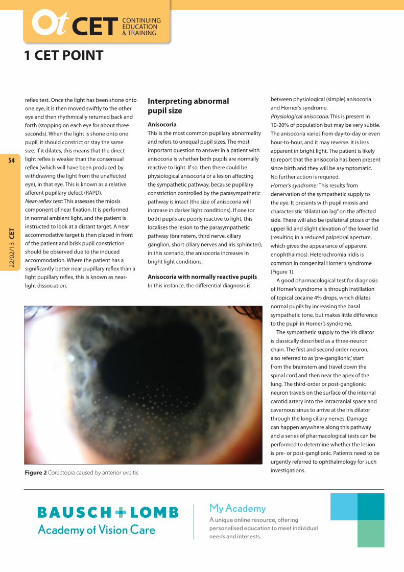
The sympathetic supply to the iris dilator is classically described as a three-neuron chain. The first and second order neuron, also referred to as ‘pre-ganglionic,’ start from the brainstem and travel down the spinal cord and then near the apex of the lung. The third-order or post-ganglionic neuron travels on the surface of the internal carotid artery into the intracranial space and cavernous sinus to arrive at the iris dilator through the long ciliary nerves. Damage can happen anywhere along this pathway and a series of pharmacological tests can be performed to determine whether the lesion is pre- or post-ganglionic. Patients need to be urgently referred to ophthalmology for such investigations.Figure 2 Corectopia caused by anterior uveitis

Anisocoria with one pupil which reacts poorly to lightIn this instance, the differential diagnosis is between iris sphincter damage, pharmacologic blockade, Adie’s tonic pupil, third cranial nerve palsy, and intermittent unilateral pupillary mydriasis (parasympathetic form).Iris sphincter damage (traumatic mydriasis): This results in an abnormal pupil shape. There is likely to be physical damage to the iris evident (including transillumination defects) due to previous Herpes zoster infection or ischaemic insults. Active disease will require emergency referral to ophthalmology.Pharmacologic blockade: This can be caused by miotic or dilating drugs (topical absorption). The affected pupil(s) is fixed and dilated or fixed and constricted. The anomaly cannot be overcome with topical agents (pilocarpine 1% or tropicamide 1%, respectively).Adie’s tonic pupil: This results from denervation of the post-ganglionic supply to the sphincter pupillae. There is a dilated pupil with sluggish or no reaction to light and near reflex. There is slow re-dilatation after constriction. Vermiform (worm-like) movements might be seen in
the iris. It occurs unilaterally in 80% of cases, in otherwise healthy patients (typically young adults, especially women). There may be sudden onset symptoms such as photophobia and blurring of vision, or it might be asymptomatic as an incidental finding. Denervation hypersensitivity is indicated when the pupil constricts upon instillation of weak miotic drops (for example pilocarpine 0.1%). Routine referral for diagnosis is warranted, although there is no treatment. Over months to years, the pupil becomes miotic, still with a sluggish, sectoral or no reaction to light, but a normal near reflex. At this stage, the affected pupil can end up smaller than that of the fellow eye. The condition is often associated with reduced deep tendon reflexes, especially in the knee and ankle (Adie’s syndrome).Third cranial nerve palsy: If this results in pupil involvement, it is a sign of intracranial aneurysm or tumour. Pain associated with pupil-involving third nerve palsy is a neurosurgical emergency and should be referred to eye casualty. In isolated cases, the pupil is never widely dilated and unresponsive. Other signs to look for are external ophthalmoplegia (vertical deviation
and exotropia in the side of the dilated pupil) inducing diplopia and upper lid ptosis. However, there may be evidence of aberrant regeneration, where there has been previous third cranial nerve damage, in which case as the patient looks down, their lid moves up.Intermittent unilateral pupillary mydriasis (parasympathetic form): This results in one pupil transiently becoming dilated and poorly reactive to light with absent accommodation. It might be associated with migraine (or history of migraines) or be an isolated event. It usually occurs in young adults and lasts 15 minutes to several hours.
Interpreting abnormal pupil shapeThis can be a result of congenital or acquired structural abnormalities. Aniridia results from a genetic mutation, affects both eyes and is associated with glaucoma and a number of systemic abnormalities. Polycoria (multiple pupils) can also be congenital, or a result of trauma, segmental iris atrophy or inflammation. Corectopia (or ectopic pupil), is similarly either congenital or acquired, as a result of penetrating trauma, surgery, iridodialysis or inflammation.
22/0
2/13
CET
55
Find out when CET points will be uploaded to the GOC at http://www.optometry.co.uk/cet/upload-dates | For the latest CET visit www.optometry.co.uk/cet
Sponsored by
Figure 3 Schematic to aid differential diagnosis of abnormal pupils

22/0
2/13
CET
56
For example, uveitis can cause synechiae, which refers to adhesions between the lens and the iris (posterior synechiae) or the iris and the peripheral cornea (peripheral anterior synechiae), and will give rise to an abnormally shaped pupil (Figure 2).
Iris coloboma is characterised by a unilateral or bilateral, partial inferior iris defect from birth. There may be other associated colobomatous defects in the eye (choroid, retina, optic nerve, lens and eyelid). Colobomas are the result of an incomplete closure of the foetal fissure during embryogenesis.
Other medical conditions which can affect pupil shape include acute angle closure glaucoma and Herpes Zoster infections, which cause ischaemic damage. Surgical interventions such as sphincterectomy (dissection of the sphincter), peripheral iridectomy, or vitreous prolapse in cataract surgery complicated by posterior capsule rupture, can also affect the shape of the pupil. Patients who are taking the prostate medication Tamsulosin run the risk of developing ‘floppy iris syndrome’, with much higher risk of intraoperative iris damage and resulting irregular pupil shape, from cataract surgery.
Recent onset pathological causes of abnormal pupil shape (for example uveitis, angle closure glaucoma and traumatic injury) should be referred to ophthalmology as an emergency.
CET CONTINUING EDUCATION & TRAINING
1 CET POINT
Interpreting abnormal pupil reactionsLight reflex test If neither pupil reacts when the light is shone onto the affected eye but both pupils react normally when the fellow eye is stimulated, this is known as an amaurotic pupil. It can only result from severe optic nerve damage (for instance transection) and the patient will be blind in that eye. If this is of recent onset, emergency referral to ophthalmology is warranted.
Swinging flashlight testIf a RAPD is observed (also known as a Marcus Gunn pupil), it indicates asymmetrical (bilateral symmetrical damage does not produce RAPD) retinal, optic nerve or optic tract damage, although post-chiasmic lesions rarely produce a clinically detectable RAPD. Similarly, it is rare for pre-retinal disease to be so severe as to produce RAPD. Media opacities scatter light, but usually do not significantly diminish the total afferent drive to the pupil light reflex. As a general rule, the presence of RAPD signifies retinal or optic nerve disease and requires urgent referral to ophthalmology. Very asymmetrical glaucoma can produce RAPD, making it a very useful clinical test when confronted with asymmetric optic disc cupping or pallor. The amount of RAPD is correlated, to a large extent, with the amount of asymmetry of visual field deficit between the two eyes.
Near-reflex testIf the light reaction seems a little weak, the
My AcademyA unique online resource, offering personalised education to meet individual needs and interests.
practitioner must check whether the pupils constrict better to a near reflex than to light. Light-near dissociation can be caused by severe loss of afferent light input to both eyes, caused by damage to the retina or optic nerve pathways, or samage at the tectum of the midbrain, causing loss of pretectal light input to the Edinger-Westphal nucleus (part of the parasympathetic chain of pupillary innervation). This can be caused by an infection, such as tertiary syphilis (Argyll-Robertson pupils), or compressive lesion (pinealoma). Light-near dissociation is also part of the Parinaud dorsal midbrain syndrome caused by such tumours.
Adie’s pupil is also an example of light-near dissociation, while aberrant third nerve regeneration (aberrant re-innervation of the iris sphincter by accommodative neurons or medial rectus neurons) can cause the pupil to constrict in attempted adduction. Medical causes of a tonic pupil and light-near dissociation include diabetes (especially after extensive laser pan-retinal photocoagulation), amyloidosis, alcoholism and myotonic dystrophy.
SummaryFigure 3 provides a useful guide to aid differential diagnosis of pupil anomalies. Examination of the pupil is an important objective clinical test because, although the patient might be asymptomatic, a pupil abnormality might be the only sign of sight- or life-threatening dysfunction in the eye or brain and can have a profound effect on visual function.
MORE INFORMATION References Visit www.optometry.co.uk/clinical, click on the article title and then on ‘references’ to download.
Exam Questions Under the new Enhanced CET rules of the GOC, MCQs for this exam appear online at http://www.optometry.co.uk/cet/exams. Please
complete online by midnight on March 22, 2013. You will be unable to submit exams after this date. Answers will be published on www.optometry.
co.uk/cet/exam-archive and CET points will be uploaded to the GOC on April 1, 2013. You will then need to log into your CET portfolio by clicking on
“MyGOC” on the GOC website (www.optical.org) to confirm your points.
Reflective learning Having completed this CET exam, consider whether you feel more confident in your clinical skills – how will you change the way you
practice? How will you use this information to improve your work for patient benefit?