demonstration chlamydial rna dna culture-negative · viscidi,4thomasc. quinn,1'6 and hugh r....
TRANSCRIPT

INFECTION AND IMMUNITY, May 1992, p. 2040-20470019-9567/92/052040-08$02.00/0Copyright C 1992, American Society for Microbiology
Demonstration of Chlamydial RNA and DNA duringa Culture-Negative State
STEVEN M. HOLLAND,2* ALAN P. HUDSON,3 LINDA BOBO,4 JUDITH A. WHITTUM-HUDSON,SRAPHAEL P. VISCIDI,4 THOMAS C. QUINN,1'6 AND HUGH R. TAYLOR7t
Divisions of Infectious Diseases' and Pediatric Infectious Diseases4 and The Dana Centerfor Preventive Ophthalmology,7Johns Hopkins Hospital, and Immunology Laboratories, The Wilmer Institute, Johns Hopkins School of
Medicine,5 Baltimore, Maryland 21205; Laboratories ofHost Defenses2 and Immunoregulation,6National Institutes of Health, Bethesda, Maryland 20892; and Department ofMicrobiology and
Immunology, The Medical College ofPennsylvania, Philadelphia, Pennsylvania 191299
Received 13 December 1991/Accepted 2 March 1992
Trachoma is a common blinding disease of humans caused by ocular infections with Chlamydia trachomatis.The cynomolgus monkey is a valuable primate model for the detection, pathobiology, and treatment of thisinfection. We have used this model system to compare the relative ability of tissue culture, direct fluorescencecytology, a modified polymerase chain reaction, and RNA blotting to detect C. trachomatis following primaryinfection and reinfection over 34 weeks. Six cynomolgus monkeys were given a primary ocular chlamydiainfection, and 20 weeks later they were reinoculated with the same organism. All animals showed briskinflammatory responses to the primary infection and milder inflammatory reactions to reinfection. All fourdiagnostic techniques detected chlamydia at 1 week after primary infection, but both nucleic acid detectionmethods suggested that organisms were present longer after primary infection than did either tissue culture ordirect fluorescence cytology (16 weeks for RNA blotting versus 12 weeks for tissue culture). Followingreinoculation at 20 weeks, the period of C. trachomatis detection by tissue culture or direct fluorescencecytology (4 weeks) was much shorter than after primary infection. In contrast, nucleic acid detection waspositive for up to 5 weeks longer than tissue culture or direct fluorescence cytology. Both polymerase chainreaction and RNA blotting, which involved no amplification step, indicated the presence of organisms duringthe culture-negative period. These data suggest that live chlamydiae may remain at a site of infection andproduce inflammation beyond the time at which standard microbiological techniques are able to detect them.
Chlamydia trachomatis is the cause of trachoma andremains the leading cause of infectious blindness in theworld. There is experimental and natural evidence thatrepeated reinfections are required for maintenance of theinflammatory state which in turn leads to scarring andeventually blindness (7, 22). However, despite clear epide-miologic and experimental association, the organism cannotbe identified by either tissue culture or immunocytologicaltechniques in over 20% of cases, even in the presence ofsevere clinical inflammatory trachoma (26, 28). The discrep-ancy between the presence of active inflammation and theability to demonstrate the inciting agent may have manycauses. It is possible that after initial infection the organismis cleared and the persistent clinical response is a residualdelayed-type hypersensitivity directed against chlamydiacomponents (11, 23, 30). Alternatively, viable organismsmay be present but unable to replicate in culture because ofblocking antibody or host-elaborated metabolic inhibitors,such as cytokines (15, 31-33).The chlamydial life cycle is divided between the infec-
tious, metabolically inactive extracellular elementary body(EB) and the metabolically active, replicating, noninfectiousreticulate body which exists in an intracellular inclusionbody (17). The dependence of tissue culture detection onviable EBs on one hand and the dependence of direct antigendetection on the expression of specific chlamydial gene
* Corresponding author.t Present address: Department of Ophthalmology, University of
Melbourne, East Melbourne, Australia.
products and their recognition by antibody (e.g., major outermembrane protein and lipopolysaccharide) on the other mayconstitute a gap in chlamydia detection (19). Factors whichaffect the rate of EB production, alter the level of expressionof certain genes, or block antigenic sites could therefore alterthe efficiency of detection under different circumstances.These factors, which inhibit detection, could permit aninapparent phase of infection which has been inaccessible toprevious diagnostic techniques.
Recently, nucleic acid-based detection systems have beendeveloped for chlamydiae and other pathogens. The poly-merase chain reaction (PCR) has been used to amplify C.trachomatis DNA in tissue culture and patient specimens (2,6, 8, 9, 13, 14). By adaptation to an enzyme-linked immuno-sorbent assay format we have been able to use the PCR fordetection and quantitation of infectious agents, including C.trachomatis (1, 2). Cloning of the C. trachomatis L2 serovarrRNA operons has provided the basis for a sensitive andspecific system for the detection of chlamydia RNA in tissueculture and clinical specimens (5).
In order to examine the possible mechanisms of C. tra-chomatis pathogenesis and to compare these newer molec-ular methods for detection of chlamydiae, we evaluated PCRand RNA detection methods for diagnosis of C. trachomatisin a monkey model of ocular infection in comparison withstandard tissue culture and direct fluorescent antibody(DFA) techniques. This animal system has been valuable indissecting the natural history of chlamydia infection and theimportance of repeated inoculations to the development ofdisease (22, 28). The ability to control the number and timeof inoculations and to readily evaluate the clinical response
2040
Vol. 60, No. 5
on February 15, 2019 by guest
http://iai.asm.org/
Dow
nloaded from
CHLAMYDIAL RNA AND DNA DETECTION 2041
as well as to monitor the laboratory aspects of infection atfrequent intervals made this model ideal for this comparativestudy.
MATERUILS AND METHODS
Animals. Six 3-year-old cynomolgus monkeys were re-ceived from Charles River Primate Imports Co. (New York,N.Y.) and were maintained and treated in accordance withNational Institutes of Health guidelines. For these experi-ments, the animals were kept in P3 Horsefall cages, airtightstainless steel cabinets with self-contained ventilation andfiltration of entering and exiting air. Initially these animalswere used in a study of a putative chlamydial vaccine inwhich three monkeys (D-5, D-9, and 1955) received 200 ,ug ofa recombinant chlamydial major outer membrane proteinpeptide in cholera toxin adjuvant orally 6 weeks beforeinoculation followed by oral and topical ocular boosting 4and 2 weeks before inoculation. The other three monkeys(D-7, D-8, and D-9) received the adjuvant alone on the sameschedule. Since there was no apparent humoral or cellularimmune effect of these immunizations, the data have beenpooled for all animals except where indicated.
Inoculations. C. trachomatis serovar C (strain TW-3) wasgrown, concentrated, and titered as described in "Labora-tory methods." All examinations and inoculations wereperformed under ketamine hydrochloride anesthesia. Theconjunctivae were examined and cultured before inocula-tion. At week 0 of this study, 5 x 10³ infection-forming unitsof C. trachomatis serovar C (strain TW-3) in 20 ,u wasinstilled into each conjunctival sac. At week 20 the animalswere reinoculated with 5 x 10³ infection-forming units of C.trachomatis serovar C (strain TW-3) in 20 ,ul into bothconjunctival sacs to assess whether they had developedresistance to further infection following recovery from theinitial challenge. Weeks 0 through 19 constitute period I, andweeks 20 through 34 constitute period II.
Collection of clinical data and specimens. Clinical examina-tion and specimen collection were performed over 34 weeks.During anesthesia the conjunctivae were examined for evi-dence of chlamydia infection by using a Kowa hand-held slitlamp (Parke, Davis & Co., Morris Plains, N.J.). Photographsof the conjunctivae were taken with a 35-mm camera with amacroscopic lens. The clinical response of each eye wasscored for a number of signs that were combined as theclinical disease score (21, 22, 25). Briefly, this score sums thefollicular response in the bulbar, limbal, superior tarsal, andsuperior fornix conjunctivae; the presence of hyperemia orinjection of the bulbar, superior tarsal, and superior fornixconjunctivae; and ocular discharge. Conjunctival culturesand smears for immunofluorescent staining were taken withsterile Dacron swabs (MicroTrak Collection Kit; Syva, PaloAlto, Calif.). Culture swabs were placed directly into 1 ml ofminimal essential medium (GIBCO) containing 10% fetal calfserum, 0.06% glucose, gentamicin (10 ,g/ml), vancomycin(50 ,ug/ml), and nystatin (10.5 ,ug/ml) and stored on wet icefor less than 60 min before being taken to the laboratory.Slides for DFA staining were allowed to air dry, fixed withmethanol for 5 min, and stored at -20°C until use.Specimens for PCR analysis were collected onto sterile
Dacron swabs (Scientific Products, McGaw Park, Ill.) fromthe upper and lower conjunctivae. Swabs were placed im-mediately into Dulbecco's phosphate-buffered saline con-taining 2% fetal bovine serum, gentamicin (5 ,ug/ml), vanco-mycin (12.5 ,ug/ml), and nystatin (12.5 p,g/ml). Specimenswere vortexed and stored at -70°C until use. Specimens for
chlamydia RNA detection were collected in the same man-ner as for PCR. Swabs were placed immediately into phenolbuffered with sterile-filtered 0.05 M Tris HCl, pH 7.4, 0.002M EDTA, 0.15 M NaCl, and 0.5% sodium dodecyl sulfate(SDS), mixed by vortexing for 30 s, and stored at roomtemperature.Laboratory methods. Tissue culture was performed in
96-well microdilution plates by using McCoy cell monolay-ers pretreated with 30 ,ug of DEAE-dextran per ml inphosphate-buffered saline with 0.01% CaCI2 for 30 min at37°C. Culture specimens were vortexed, and 100 RI ofmaterial was inoculated into each of four wells. Plates werecentrifuged at 1,100 x g at 27°C for 1 h and then incubatedfor 30 min at 37°C. The supernatant fluid was aspirated andreplaced with 200 pI of minimal essential medium with 10%fetal calf serum, 0.06% glucose, cycloheximide (1 pug/ml),gentamicin (10 pug/ml), vancomycin (10 ,ug/ml), and nystatin(10.5 ,ug/ml). After incubation at 37°C in 5% CO2 for 48 h,two wells of the primary plate were processed for immuno-fluorescent staining and two wells were scraped and passedto two wells on a secondary plate. The secondary plate wasincubated for 48 h and then stained. Wells were washed oncewith phosphate-buffered saline and fixed with ethanol for 5min. Wells were washed twice with distilled water andstained with 25 pI of fluorescein-labeled anti-C. trachomatismonoclonal antibody with Evans blue counterstain (Micro-Trak Chlamydia Direct Reagent; Syva Co.) for 30 min at37°C in a humidified chamber. After aspiration of the stain,wells were washed twice with deionized water. Mountingfluid (Syva Co.) was then added to each well, and a 5-mm-diameter circular coverslip was placed on top. Culture plateswere inverted and read by using a transmitted fluorescencemicroscope at a magnification of x500. Chlamydial inclu-sions were identified by their apple-green staining. Each wellwas carefully examined at a magnification of x500. If morethan 20 inclusions were seen, the number of inclusions in 15randomly selected high-power fields was taken and multi-plied by 120 to give the approximate number of inclusionsper well.
Slides of conjunctival smears were allowed to come toroom temperature and washed with methanol to clear resid-ual water. Slides were dried and stained with 30 pI offluorescein-labeled antichlamydial monoclonal antibodywith Evans blue counterstain (MicroTrak Chlamydia CultureConfirmation Reagent; Syva Co.) for 30 min in a humidifiedchamber. Slides were rinsed in deionized water, air dried,and covered with coverslips. The DFA slides were readusing transmitted or epifluorescence microscopy, and EBswere enumerated.A gene amplification method that combined the PCR with
an enzyme immunoassay of the PCR products was used todetect a conserved region of the major outer membraneprotein gene of C. trachomatis (1, 2, 20). In brief, specimensfor PCR were thawed and vortexed. Two-hundred-microliteraliquots were detergent lysed, proteinase K digested, mi-crodialyzed to remove sodium, and denatured by boiling.Fifty microliters of sample was used for DNA amplificationwith 0.5 puM (each) primers (Ct.0005, 5'-GATAGCCAGCACAAAGAGAGCTAA-3', and Ct.06, 5'-CTIT]GT''1TECGACCGTGTF[TIGCAAACAGATGTGAA-3'), 0.2 mM (each)dATP, dCTP, dGTP, and TTP; 0.01 M Tris, pH 8.3; 0.05 MKCI; 0.0025 M MgCl2; 0.01% gelatin; and 1.5 U of Taqpolymerase (Amersham, Arlington Heights, Ill.). The reac-tion was performed in a 100-pA total volume overlaid withmineral oil in a thermocycler (Perkin-Elmer Cetus, Norwalk,
VOL. 60, 1992
on February 15, 2019 by guest
http://iai.asm.org/
Dow
nloaded from
2042 HOLLAND ET AL.
Conn.) with denaturation at 94°C for 1 min, annealing at 55°Cfor 1 min, and extension at 72°C for 1 min for 30 cycles.The T7 RNA polymerase promoter-tagged probe was
generated with a set of primers (Ct.03T7, 5'-TTAATACGACTCACTATAGGGTCTGCTTCCTCCTTGCAAGCAAGTCTGCC-3', and Ct.04, 5'-CAGCATGCGTATGGGTTACTATGG-3') nested with respect to the sample amplificationprimers by using purified C. trachomatis L2 as a template.Approximately 500 ng of the T7-tagged PCR product was invitro transcribed in the presence of biotin 11-UTP (Enzo,New York, N.Y.) by the T7 RNA polymerase. ResidualDNA template, primers, and unincorporated biotin 11-UTPwere removed from the final probe by digestion and chro-matography.
Fifty microliters of the specimen PCR mixture was mixedwith 50 ,ul of a 1:200 dilution of the biotinylated RNA probein 0.6 M NaCI-0.06 M Na citrate (pH 7.0)-0.02 M N-2-hydroxyethylpiperazine-N'-2-ethanesulfonic acid (HEPES)(pH 7.4)-0.004 M EDTA-0.5% SDS. After the mix wasboiled for 3 min, maintained at 78°C for 1 h, and slowlycooled to ambient temperature, 10 ,Ju of 10% Triton X-100was added. Fifty-microliter aliquots were transferred induplicate to the wells of a black microdilution plate whichwas precoated with polyclonal goat antibiotin antibody (Sig-ma Chemical Co., St. Louis, Mo.). After incubation the platewas washed and reacted with the Fab' fragment of a 1-D-galactosidase-conjugated monoclonal antibody againstRNA-DNA hybrids (3). After further incubation, the platewas again washed and reacted with the substrate 4-methyl-umbelliferyl P-D-galactoside. The fluorogenic product, meth-ylumbelliferone, was measured at 450 nm after excitation at365 nm. A sample was scored positive if its mean fluores-cence exceeded that of a naive monkey specimen control by3 standard deviations.The recent cloning and characterization of the L2 (434)
rrnA operons and flanking sequence provided an opportunityto take advantage of the high levels of RNA present duringchlamydia replication for development of a detection system(5). In brief, total sample RNA was extracted from thebuffered phenol transport medium and cleared of DNA andprotein (16). RNA was applied to 0.45-,um-pore-size nitro-cellulose membranes by a slot blotting apparatus at 1 ,ug oftotal RNA per slot. Samples from naive monkeys and yeastwere included as negative controls, and L2 (434) RNA wasused as a positive control on each blot. Probes were pre-pared, eluted from gels, nick translated, and used at 3 x 10⁶cpm per hybridization reaction in an 8-ml total volume.Hybridization was carried out at high stringency in 50%formamide-containing buffer (5, 16, 29). Blots were visual-ized by autoradiography and quantitated by area underdensitometry curves by using Gaussian best-fit computeranalysis. Negative control values were subtracted fromexperimental areas to give the areas plotted. Each pointrepresents the average value of samples from both eyes of agiven animal on a given day.
RESULTS
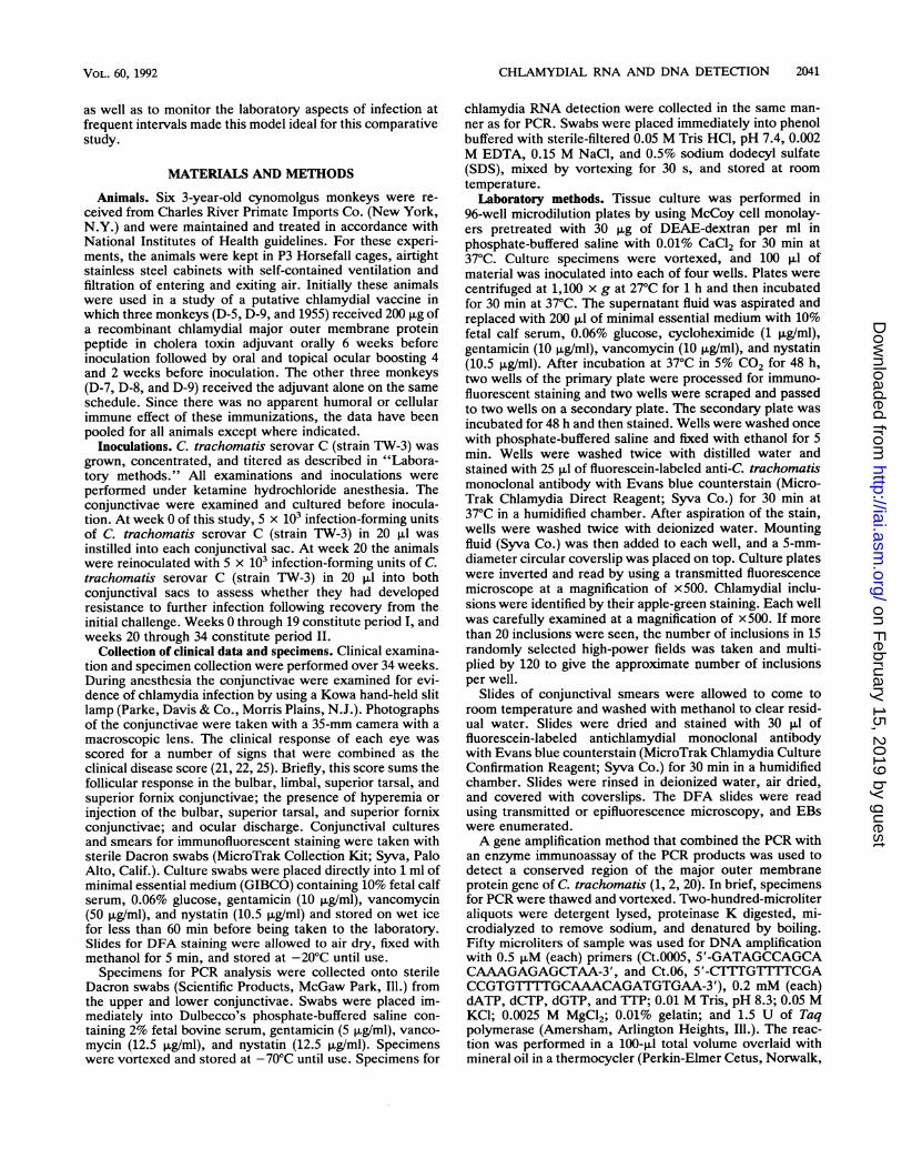
Clinical responses. The ocular clinical responses to inocu-lation are summarized in Fig. 1A and are given for eachanimal separately in Fig. 2. The primary inoculation causeda rapid inflammatory and follicular response in all animals,with the maxima appearing between weeks 2 and 6. Clinicaldisease was largely resolved by week 8 in three animals (D-5,D-6, and D-9) but remained somewhat unresolved in theothers (D-7, D-8, and 1955).
The clinical response to the second inoculation was milderand briefer than the response to the first inoculation. Themagnitude of the response to the second challenge was onlyone-third of that to the first inoculation. One animal hadvirtually no response to the second inoculation (D-8). Twoanimals had active clinical disease in period II withoutappreciable tissue culture or DFA recovery (D-7 and 1955),whereas one (D-8) had detectable EBs and inclusions with-out appreciable clinical disease. These results are in agree-ment with previous studies showing the presence of clinicaldisease in the absence of tissue culture and attenuation of theocular clinical response to secondary challenge with thesame serovar (26, 28).
Culture and DFA staining. Preinoculation cultures andconjunctival smears were negative in all animals. The num-bers of inclusions seen in tissue culture before and followinginoculation are summarized in Fig. 1A and presented foreach individual animal separately in Fig. 2. Samples becamemarkedly positive in all animals 1 week after inoculation.After the first week, numbers of inclusions and EBs tendedto decline rapidly and in parallel (EB data not shown).
Following the second inoculation at week 20, the numbersof inclusions and EBs were less than after the first inocula-tion. Only four of the six monkeys developed positivecultures after the second inoculation. All six animals werepositive by DFA, although two (D-7 and 1955) had only oneEB detected at week 33 and no inclusions seen on culturefollowing the second inoculation. DFA was markedly posi-tive in most animals at week 21, 1 week after the secondinoculation. In contrast, tissue culture did not becomepositive until week 22 or 23, 2 to 3 weeks after the secondinoculation. The animals that did have recoverable organ-isms cleared them much more rapidly than following the firstinoculation. Only one animal (D-6) was positive by tissueculture and DFA at week 24, and this animal had a secondburst of EBs at week 30. There was overall agreementbetween tissue culture and DFA, although DFA was positiveearlier and more often in period II (data not shown). Thefinding of reduced numbers of organisms, delayed growth intissue culture, and a shortened period of tissue culturepositivity is in agreement with previous studies and corre-lates with development of partial host immunity to theinfecting organism (26, 28).PCR. The results of the PCR as applied to the samples
following the first and second inoculations are summarizedin Fig. 1B and are given for each animal separately in Fig. 2.In period I, following the first inoculation, PCR becamepositive by the first week and remained positive until week14. PCR values for three animals (D-5, D-9, and 1955)developed at or near their maximum PCR fluorescence unitsat week 1, whereas values for the other animals did not peakuntil weeks 3 (D-7 and D-8) and 6 (D-6). PCR values tendedto fall off relatively rapidly over the weeks immediatelyfollowing the peak. However, four animals had secondarypeaks in period I at week 8 (1955), week 10 (D-7 and D-8),and week 12 (D-9). One animal had a positive PCR at week16 (D-7), but all were negative by the time of reinoculation atweek 20.
In period II, following the second inoculation, the maxi-mum peak was roughly the same as in period I. This mayreflect saturation of the assay, since the reaction is not linearat higher concentrations (1). All animals developed peakPCR fluorescence units for period II at week 21, the weekimmediately following inoculation. The decline in PCR sig-nal was faster than in period I. Three animals had significantsecondary peaks in period II at weeks 24 (D-7), 28 (D-8), and
INFECT. IMMUN.
on February 15, 2019 by guest
http://iai.asm.org/
Dow
nloaded from

CHLAMYDIAL RNA AND DNA DETECTION 2043
A-*0-- log IFU
E CDS
L11
C0 2 4 6 8 10 12 14 16 18 20 22 24 26 28 30 32 34
25
20a)00
cn._n
15 CD
10 -a.0
5
O0
Weeks
25
20C C
t 15
a).20D c= e 10
OZcLI _
5
BRNA x 200
* PCRx100
0 2 4 6 8 10 12 14 16 18 20 22 24 26 28 30 32 34
Weekst
FIG. 1. (A) Clinical responses and microbiologic results following primary infection and reinfection with C. trachomatis serovar C (strainTW-3). The mean ocular clinical disease score (CDS) and mean number of inclusions seen in tissue culture (IFU) for six cynomolgus monkeysare shown. Animals were reinfected at 20 weeks (arrow). (B) Detection of chlamydial RNA and DNA following primary infection andreinfection with C. trachomatis serovar C (strain TW-3). The mean PCR fluorescence units and the RNA densitometry area units for sixcynomolgus monkeys are shown. Raw unit values are obtained by multiplying the PCR values by 100 and multiplying the RNA values by 200.Use of these factors allows for representation of these data on the same scale. Animals were reinfected at 20 weeks (arrow).
30 (D-6). By week 26, 6 weeks after the second inoculation,five of the animals were still PCR positive, by week 28 threeanimals were still PCR positive, and by week 32 two animalswere still PCR positive. By week 34, 14 weeks after thesecond inoculation, all animals were negative by PCR.
Discordances between PCR and tissue culture or DFAoccurred in all animals. These discordances, in general, werethat PCR registered the presence of chlamydia DNA whiletissue culture and DFA were negative. There were fourinstances of tissue culture and DFA detection exceedingPCR detection, all in period I. In contrast, there were 26
instances of PCR detection exceeding tissue culture or DFAdetection, 19 of which were in period II.RNA blotting. The results obtained by RNA blotting in
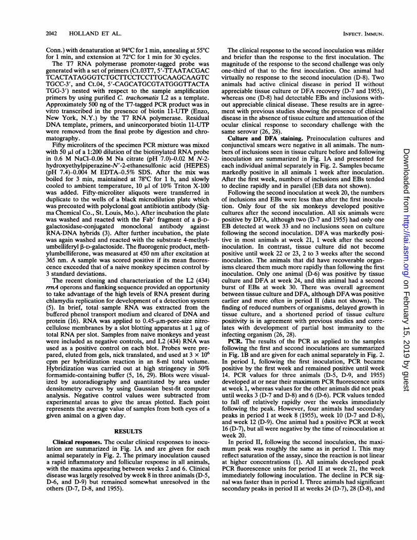
periods I and II are summarized in Fig. 1B and are given foreach animal separately in Fig. 2. RNA units rose rapidlyfollowing the first inoculation and peaked at weeks 1 (D-7), 2(D-5, D-9, and 1955), and 3 (D-6 and D-8). Following thepeak values, the overall trend in the RNA scores in period Iwas downward, but no animal returned to baseline RNAscores until week 20. One animal's RNA score increasedthrough period I following the initial decline (D-9). In ani-
VOL. 60, 1992
3
2
I
on February 15, 2019 by guest
http://iai.asm.org/
Dow
nloaded from

2044 HOLLAND ET AL.
D5
3
2
0
Weeks /
D6
2
0 2 4 6 8 10 12 14 16 18 20 22 24 26 28 30 32 3
Weeks /
25
C/)
O._10 C
5 >,
a)
CD)
25 a)
0
20 <zI=
15
0
10 CO
5 DCDa)
4 ca)C.)
C/)
m-C25
D7 o
20 l
a:
3 15
3
10
2~~~~~~~~~~~~~~~~
Weeks
D8
6 8 10 12 14 16 18 20 22 24 26 28 30 32
Weeks \
D9LL
LL
8'
0 2 4 6 8 10 12 14 16 18 20 22 24 26 28 30 32
Weeks /
Weeks '
FIG. 2. Clinical, microbiologic, PCR, and RNA blotting results following primary infection and reinfection with C. trachomatis serovar
C (strain TW-3). The ocular clinical disease score (CDS), log of the number of inclusions seen in tissue culture (IFU), PCR fluorescence units,and RNA densitometry area units are shown for each of six cynomolgus monkeys. Raw unit values are obtained by multiplying the PCRvalues by 100 and multiplying the RNA values by 200. Use of these factors allows for representation of these data on the same scale. Animalswere reinfected at 20 weeks (arrow).
mals D-7 (week 10) and D-8 (week 12) secondary peaks ofRNA score in period I corresponded to bursts of tissueculture or DFA activity.The RNA units in period II were lower than in period I.
Values rose to their period II maxima at weeks 21 (1955), 22(D-6 and D-7), and 24 (D-5, D-8, and D-9). The decline
toward baseline values was more rapid than in period I, withall animals returning to the baseline by week 30, 10 weeksafter the second inoculation. In contrast, RNA was stillreadily detectable 16 weeks after the primary inoculation.RNA levels were declining at the end of period I butrebounded after reinoculation. The rapid clearance of RNA
LL
8'
D
LL
8'
25
20
15Ce
10 CD
5 >%
O E4 0
CD)cn25 ()Q
20 <zcc
15
0
10 co
5 DCDa)
Oc
(n
0
CDa)
250
20 L
15 0
L-
INFECT. IMMUN.
1
1
on February 15, 2019 by guest
http://iai.asm.org/
Dow
nloaded from
CHLAMYDIAL RNA AND DNA DETECTION 2045
in period II roughly correlated with the shorter period ofculture positivity and clinical inflammation.Discordance between RNA detection and tissue culture
and DFA occurred in all animals. In general, RNA blottingsuggested the presence of organisms more often than didtissue culture or DFA. As seen in comparing Fig. 1A and B,RNA blotting remained positive, although decreasing,throughout period I. At the same time, culture and DFAbecame negative from weeks 14 to 20. The clinical scoreremained somewhat elevated over the same period. Inperiod II RNA became undetectable at week 30. In contrast,tissue culture and DFA became negative at week 26. Noanimal lacked detectable RNA in period II. However, onlyfour animals were culture positive. RNA blotting suggestedthe presence of chlamydiae more often than PCR. Specifi-cally, in the latter portion of period I there were points atwhich RNA units were elevated but PCR fluorescence unitswere negative, but there were no points at which PCR waspositive and RNA units were not elevated. In period II therewas broad agreement between PCR and RNA blotting.Discordances in D-6 at week 30 and D-8 week 32 consisted ofpositive PCR without corroborative RNA detection. Sincethere is no amplification step involved in the RNA isolationand hybridization used here, it is unlikely that systematiccontamination played a significant role in our RNA experi-ments.
DISCUSSION
The precise mechanism by which C. trachomatis causesthe development of ocular and genitourinary scarring andsubsequent dysfunction is still elusive. It is clear that re-peated bouts of infection are required, but it is unclearwhether these may result from a relapse of autochthonousinfection as well as by exogenous reinfection (7, 23). It isthought that the final common insult is a delayed hypersen-sitivity response to chlamydial antigen(s) (11, 23, 30). Whileinoculation with nonviable EBs fails to reproduce the inflam-matory response seen with infectious organisms (23, 30), theinflammatory response has been shown to be due to a 57-kDaTriton X-100-solubilized C. trachomatis heat shock protein(HSP-60) antigen. This antigen is present only in extractsfrom live organisms and can elicit an appropriate inflamma-tory response in the absence of other chlamydial antigens(10, 24). The dependence of the characteristic ocular inflam-mation on this factor produced only by living chlamydiae hassuggested that the continued presence of live organisms isnecessary for the development of trachoma (11, 23, 30).Many studies have shown the persistence of clinical diseasein the absence of detectable infection, but the cause of thispersistent clinical disease has been unclear (26-28). Nucleicacid detection data presented here indicate that demonstra-ble infection with C. trachomatis may persist well beyondthe period indicated by cell culture or DFA. Furthermore, inthe setting of reinfection, which is thought to play so criticala role in the development of blinding disease, the disparitybetween tissue culture or DFA and nucleic acid-baseddetection is quite pronounced. In period I PCR and RNAdetection were positive from 0 to 2 weeks longer than tissueculture and DFA, whereas in period II PCR and RNAdetection were positive from 2 to 5 weeks longer than tissueculture and DFA.The PCR has made great strides in improving the sensi-
tivity of microbial detection and diagnosis. However, in anamplification-dependent system like PCR, even slight con-tamination of samples before amplification can lead to falsely
positive results. Therefore, in addition to the exercise ofrigorous precautions and controls, the parallel use of chla-mydia rRNA hybridization has provided an important in-dependent corroboration. RNA isolation and storage aremore likely to result in RNA degradation and a resultantunderestimation of chlamydiae present than to result inan overestimation. Furthermore, since the RNA prepara-tion procedures we used involved no amplification step,there was little chance for systematic contamination ofspecimens.Three animals had marked discordance in period II among
tissue culture or DFA, clinical score, and nucleic aciddetection. In animals D-7 and 1955 brisk clinical responseswere essentially independent of tissue culture or DFA pos-itivity. During these periods, congruent with the clinicalresponse, both nucleic acid detection systems were positive.However, in animal D-8, both nucleic acid detection systemswere positive for at least 5 weeks beyond any detectableclinical response in period II.There are several possible explanations for the inability to
culture chlamydiae or demonstrate organisms by DFA attimes when nucleic acids were easily detectable. It is possi-ble that the nucleic acid systems were detecting only deadorganisms or organisms which had already been phagocy-tosed. PCR as performed here on DNA cannot distinguishliving from dead organisms. However, the detection of RNAby blotting suggests the persistence of living chlamydiae.RNA is an extremely labile moiety and would have beenunlikely to survive intact in dead organisms for weeks in thesetting of ongoing inflammation.
Factors generated in the course of the clinical responsemight inhibit infectivity and therefore culturability. The hostresponse to C. trachomatis includes the elaboration ofantibodies, interleukin-1, gamma interferon, and tumor ne-crosis factor (15, 28, 31-33). These factors may alter themetabolic capacity, surface receptor expression, or infectiv-ity of C. trachomatis without affecting nucleic acid detec-tion. It has been shown in tissue culture that gamma inter-feron inhibits maturation of the reticulate body to theinfectious EB (18). The mechanism of this inhibition proba-bly includes depletion of tryptophan, which is required forchlamydial growth (4). Chlamydiae which are stressed in thisway and by other factors have been shown to release the57-kDa HSP-60 into the supernatant (12). Therefore, in thesetting of a host response, which includes the elaboration ofcytokines, chlamydial maturation may be inhibited while theproduction and release of the inflammatory 57-kDa HSP-60may persist. In this way the infectious chlamydial burdenmight fall while the organism was still present and continuingto elicit inflammation. These organisms would still containDNA and high levels of RNA and therefore be detectable bythese techniques. This might be a cellular mechanism forlatent or persistent infection.
It is possible that there were just too few EBs available atcertain time points to establish an identifiable infection.However, the RNA and PCR data are roughly quantitative,and the period II data from animals D-6 and D-8 suggest highlevels of nucleic acid in the specimens. Previous studieshave shown that there can be a slight random variation inDFA recovery between specimen swabs taken at 5 min apart(27). In our study, the likelihood that sampling error couldaccount for the disparity seen between nucleic acid andtissue culture or DFA results is remote, given the frequencyof the finding and its nonrandom nature.The observation that there is a significant disparity be-
tween culture- and DFA-based detection systems and nu-
VOL. 60, 1992
on February 15, 2019 by guest
http://iai.asm.org/
Dow
nloaded from

2046 HOLLAND ET AL.
cleic acid detection systems is not surprising. Several clini-cal studies have suggested that PCR might be more sensitivefor diagnosis of C. trachomatis infection than other tech-niques (1, 2, 13). However, the question of false-positiveresults has been difficult to parry. The parallel application oftissue culture, direct fluorescence microscopy, PCR, andRNA blotting to multiple replicate samples obtained longi-tudinally over 34 weeks during the evolution of both primaryand secondary ocular infections has given us the chance tocarefully compare these modalities. We find that C. tracho-matis DNA and RNA are detectable in clinical ocularspecimens weeks beyond a time at which either tissueculture or DFA is able to diagnose infection. These findingscorrelate with the persistence of ocular clinical responsebeyond the time of culture demonstration of organisms.This disparity between nucleic acid- and culture- or DFA-based detection is more pronounced in the sensitized ani-mal than in the immunologically naive animal. The demon-stration of organisms by PCR and RNA blotting in the settingof culture-negative inflammation suggests that the presenceof unculturable but live organisms may be important inthe maintenance or prolongation of the inflammatory condi-tion and the pathogenesis of trachoma. These data extendthe presumed duration of active infection beyond thatsuggested previously by tissue culture and DFA and indi-cate that there are host-dependent factors in the immuneanimal which are inhibiting tissue culture and DFA demon-stration of infection. Furthermore, these findings suggestthat the "gold standard" of tissue culture may need to bereassessed.
ACKNOWLEDGMENTS
We gratefully acknowledge Charlotte A. Gaydos for stimulatingdiscussions, Beatriz Munoz for statistical advice, and Vivian Velezand Lori DeJong for technical assistance.
REFERENCES
1. Bobo, L., F. Coutlee, R. H. Yolken, T. Quinn, and R. P. Viscidi.1990. Diagnosis of Chlamydia trachomatis cervical infection bydetection of amplified DNA with an enzyme immunoassay. J.Clin. Microbiol. 28:1968-1973.
2. Bobo, L., B. Munoz, R. Viscidi, T. Quinn, H. Mkocha, and S.West. 1991. Diagnosis of Chiamydia trachomatis eye infectionin Tanzania by polymerase chain reaction/enzyme immunoas-say. Lancet 338:847-850.
3. Boguslawski, S. J., D. E. Smith, M. A. Michalak, K. E. Michel-son, C. 0. Yehle, W. L. Pattersom, and R. J. Carrico. 1986.Characterization of monoclonal antibody to DNA-RNA and itsapplication to immunodetection of hybrids. J. Immunol. Meth-ods 89:123-130.
4. Byrne, G. I., L. K. Lehmann, and G. J. Landry. 1986. Inductionof tryptophan catabolism is the mechanism for gamma-interfer-on-mediated inhibition of intracellular Chlamydia psittaci repli-cation in T24 cells. Infect. Immun. 53:347-351.
5. Cheema, M. A., H. R. Schumacher, and A. P. Hudson. 1991.RNA-directed molecular hybridization screening: evidence forinapparent chlamydial infection. Am. J. Med. Sci. 302:261-268.
6. Dutilh, B., C. Bebear, P. Rodriguez, A. Vekris, J. Bonnet, andM. Garret. 1989. Specific amplification of a DNA sequencecommon to all Chlamydia trachomatis serovars using the poly-merase chain reaction. Res. Microbiol. 140:7-16.
7. Grayston, J. T., S.-P. Wang, and L.-J. Yeh. 1985. Importance ofreinfection in the pathogenesis of trachoma. Rev. Infect. Dis.7:717-725.
8. Griffais, R., and M. Thibon. 1989. Detection of Chlamydiatrachomatis by the polymerase chain reaction. Res. Microbiol.140:139-141.
9. Holland, S. M., C. A. Gaydos, and T. C. Quinn. 1990. Detectionand differentiation of Chlamydia trachomatis, Chlamydia psitt-aci, and Chlamydia pneumoniae by DNA amplification. J.Infect. Dis. 162:984-987.
10. Morrison, R. P., R. J. Belland, K. Lyng, and H. D. Caldwell.1989. Chlamydial disease pathogenesis: the 57 kD chlamydialhypersensitivity antigen is a stress response protein. J. Exp.Med. 170:1271-1283.
11. Morrison, R. P., K. Lyng, and H. D. Caldwell. 1989. Chlamydialdisease pathogenesis: ocular hypersensitivity elicited by a ge-nus-specific 57-kD protein. J. Exp. Med. 169:663-675.
12. Morrison, R. P., D. S. Manning, and H. D. Caldwell. Immunol-ogy of Chlamydia trachomatis infections: immunoprotectiveand immunopathogenic responses. Adv. Host Def. Mech., inpress.
13. Ostergaard, L., S. Birkelund, and G. Christiansen. 1990. Use ofpolymerase chain reaction for detection of Chlamydia tracho-matis. J. Clin. Microbiol. 28:1254-1260.
14. Pollard, D. R., S. D. TIyler, C. W. Ng, and K. R. Rozee. 1989. Apolymerase chain reaction (PCR) protocol for the specific de-tection of Chlamydia spp. Mol. Cell. Probes 3:383-389.
15. Rothermel, C. D., J. Schachter, P. Lavrich, E. C. Lipsitz, and T.Francus. 1989. Chlamydia trachomatis-induced production ofinterleukin-1 by human monocytes. Infect. Immun. 57:2705-2711.
16. Sambrook, J., E. F. Fritsch, and T. Maniatis. 1989. Molecularcloning: a laboratory manual, 2nd ed. Cold Spring HarborLaboratory, Cold Spring Harbor, N.Y.
17. Schacter, J., and H. D. Caldwell. 1980. Chlamydiae. Annu. Rev.Microbiol. 34:285-309.
18. Shemer, Y., and I. Sarov. 1985. Inhibition of growth of Chla-mydia trachomatis by human gamma interferon. Infect. Immun.53:347-351.
19. Stamm, W. E. 1988. Diagnosis of Chlamydia trachomatis geni-tourinary infections. Ann. Intern. Med. 108:710-717.
20. Stephens, R. S., R. Sanchez-Pescador, E. A. Wagar, C. Inouye,and M. S. Urdea. 1987. Diversity of Chlamydia trachomatismajor outer membrane protein genes. J. Bacteriol. 169:3879-3885.
21. Taylor, H. R. 1990. Development of immunity to ocular chla-mydial infection. Am. J. Trop. Med. Hyg. 42:358-364.
22. Taylor, H. R., S. L. Johnson, R. A. Prendergast, J. Schachter,C. R. Dawson, and A. M. Silverstein. 1982. An animal model oftrachoma. II. The importance of repeated reinfection. Invest.Ophthalmol. Vis. Sci. 23:507-515.
23. Taylor, H. R., S. L. Johnson, J. Schachter, H. D. Caldwell, andR. A. Prendergast. 1987. Pathogenesis of trachoma: the stimulusfor inflammation. J. Immunol. 138:3023-3027.
24. Taylor, H. R., I. W. Maclean, R. C. Brunham, S. Pal, and J.Whittum-Hudson. 1990. Chlamydial heat shock proteins andtrachoma. Infect. Immun. 58:3061-3063.
25. Taylor, H. R., R. A. Prendergast, C. R. Dawson, J. Schachter,and A. M. Silverstein. 1981. An animal model for cicatrizingtrachoma. Invest. Ophthalmol. Vis. Sci. 21:422-433.
26. Taylor, H. R., P. A. Rapoza, S. West, S. Johnson, B. Munoz, S.Katala, and B. B. 0. Mmbaga. 1989. The epidemiology ofinfection in trachoma. Invest. Ophthalmol. Vis. Sci. 30:1823-1833.
27. Taylor, H. R., J. A. Siler, H. A. Mkocha, B. Munoz, V. Velez,L. Dejong, and S. West. 1991. Longitudinal study of the mi-crobiology of endemic trachoma. J. Clin. Microbiol. 29:1593-1595.
28. Taylor, H. R., E. Young, A. B. MacDonald, J. Schachter, andR. A. Prendergast. 1987. Oral immunization against chlamydialeye infection. Invest. Ophthalmol. Vis. Sci. 28:249-258.
29. Thomas, P. S. 1980. Hybridization of denatured RNA and smallDNA fragments transferred to nitrocellulose. Proc. Natl. Acad.Sci. USA 77:5201-5205.
30. Watkins, N. G., W. J. Hadlow, A. B. Moos, and H. D. Caldwell.1986. Ocular delayed hypersensitivity: a pathogenetic mecha-nism of chlamydial conjunctivitis in guinea pigs. Proc. Natl.Acad. Sci. USA 83:7480-7484.
INFECT. IMMUN.
on February 15, 2019 by guest
http://iai.asm.org/
Dow
nloaded from

CHLAMYDIAL RNA AND DNA DETECTION 2047
31. Williams, D. M., G. I. Byrne, B. Grubbs, T. J. Marshal, and J.Schachter. 1988. Role in vivo for gamma interferon in control ofpneumonia caused by Chlamydia trachomatis in mice. Infect.Immun. 56:3004-3006.
32. Williams, D. M., D. M. Magee, L. F. Bonewald, J. G. Smith,C. A. Bleicker, G. I. Byrne, and J. Schachter. 1990. A role for
tumor necrosis factor alpha in host defense against Chlamydiatrachomatis. Infect. Immun. 58:1572-1576.
33. Zhong, G., E. M. Peterson, C. W. Czarniecki, R. D. Schreiber,and L. M. de la Maza. 1989. Role of endogenous gammainterferon in host defense against Chlamydia trachomatis infec-tions. Infect. Immun. 57:152-157.
VOL. 60, 1992
on February 15, 2019 by guest
http://iai.asm.org/
Dow
nloaded from