delphinidin 3- o -(2- o -β- d -glucopyranosyl-α- l -arabinopyranoside): a novel anthocyanin...
TRANSCRIPT

Delphinidin3-O-(2-O-â-D-Glucopyranosyl- r-L-arabinopyranoside): A Novel
Anthocyanin Identified in Beluga Black Lentils
GARY R. TAKEOKA,*,† LAN T. DAO,† HIROTOSHI TAMURA,‡ AND
LESLIE A. HARDEN†
Western Regional Research Center, Agricultural Research Service, U.S. Department of Agriculture,800 Buchanan Street, Albany, California 94710, and Department of Biochemistry and Food Science,
Kagawa University, 2393 Miki-cho, Kagawa 761-0795, Japan
A major anthocyanin was isolated from the acidified methanolic extract of Beluga black lentils byXAD7 column chromatography and preparative high-performance liquid chromatography. By meansof electrospray ionization mass spectrometry and one- and two-dimensional nuclear magneticresonance spectroscopy, its structure was determined to be delphinidin 3-O-(2-O-â-D-glucopyranosyl-R-L-arabinopyranoside).
KEYWORDS: Anthocyanins; black lentils ( Lens culinaris ); 1D and 2D NMR; delphinidin derivative
INTRODUCTION
Lentils (Lens culinaris) are small annual legumes of the peafamily (Leguminosae). They are one of the oldest cropscultivated by humans, domesticated around 8000 BC in theFertile Crescent (1). The lens-shaped edible seeds are mainlyused in soups and are a good source of protein, dietary fiber,folate, iron, and phosphorus. Flour made from lentils is glutenfree and may be added to cereal flour to make breads, cakes,and baby foods. The seed coat color can be clear, green, tan,gray, brown, or black while the cotyledon is yellow, red, orgreen. The main market types are red and green, which togetheraccount for an estimated 95% of the world’s lentil production(2). U.S. lentil production, occurring in the states of Washington,Idaho, Montana, and North Dakota, totaled about 234 millionlbs. in 2002-2003 (2), mainly with green and brown types.Because of the possible health benefits of anthocyanins (3), wedecided to investigate their presence in the less utilized Belugablack lentil.
The subepidermal pigment in lentil stem was reported to havedelphinidin as the aglycon (4). Lentil seeds were reported tocontain a compound that resembled a diglycosyl derivative ofdelphinidin (5). Because of its low concentration (about 60 ppm),the authors were unable to fully characterize its structure.Slinkard and Bhatty (6) reported that stems of seedlings andplants of the large-seeded (Chilean type) lentil variety, Laird,are pigmented with anthocyanins. Anthocyanins are locatedessentially in their seed coat, which constitutes about 8-11%of the entire lentil seed (7). The goal of this work was to
elucidate the structure of the major anthocyanin(s) in Belugablack lentils.
EXPERIMENTAL PROCEDURES
Materials. Beluga black lentils were obtained from George F. Brockeand Sons, Inc. (Kendrick, ID). Concord grape puree was obtained fromthe Milne Fruit Products, Inc. (Prosser, WA). Methylâ-L-arabino-pyranoside was purchased from Wako Pure Chemical Industries, Ltd.(Osaka, Japan). Solvents were of high-performance liquid chromatog-raphy (HPLC) spectroquality grade unless otherwise stated.
Extraction Method. Lentils (1.26 kg) were ground to a powder witha UDY Cyclone sample mill (Fort Collins, CO). The powder was mixedwith methanol containing 3% HCl (2.8 L), and the resulting mixturewas allowed to stand overnight in a refrigerator. The mixture wasfiltered through a Whatman #4 filter (24.0 cm) using a Buchner funnel.The powder was mixed with methanol containing 0.3% HCl (1.8 L).The mixture was allowed to stand overnight in a refrigerator and wasthen filtered as above. The combined extract was concentrated to ca.100 mL with a rotary evaporator. Trifluoroacetic acid (TFA), a weakeracid than HCl, is preferred for the extraction of anthocyanins since itis less likely to cause pigment degradation, particularly during theconcentration step (8). We did notice anthocyanin degradation,especially during concentration of the methanol extract containing 3%HCl. Subsequent extractions were done using TFA as the acid.
Amberlite XAD7 Column Chromatography. Amberlite XAD7resin (600 mL; 20-60 mesh [wet]; average diameter, 90 Å; porevolume, 1.14 mL/g) was conditioned in the following way: The resinwas mixed with methanol (2 L), and the upper layer was decanted toremove the white colored solution. This procedure was repeated with1 L of methanol. The resin was packed into a glass column (4.0 cmi.d. × 80 cm; resin volume, 1.0 L) and eluted with 4 L of acetone, 1L of methanol, 1 L of methanol containing 0.3% HCl, 1 L of 50:50(v/v) methanol:water containing 0.3% HCl, and 1.5 L of 0.3% HCl.The anthocyanin extract was diluted two times in purified water andapplied to the column. The column was eluted with 3 L of 0.3% HClto remove polar constituents. The column was subsequently eluted in
* To whom correspondence should be addressed. Tel: 510-559-5668.Fax: 510-559-5828. E-mail: [email protected].
† U.S. Department of Agriculture.‡ Kagawa University.
4932 J. Agric. Food Chem. 2005, 53, 4932−4937
10.1021/jf040493h CCC: $30.25 © 2005 American Chemical SocietyPublished on Web 05/12/2005

a stepwise fashion with 10:90 (v/v) methanol:water containing 0.3%HCl, 20:80 (v/v) methanol:water containing 0.3% HCl, 30:70 (v/v)methanol:water containing 0.3% HCl, 40:60 (v/v) methanol:watercontaining 0.3% HCl, 50:50 (v/v) methanol:water containing 1% TFA,and 60:40 (v/v) methanol:water containing 1% TFA.
Thin-Layer Chromatography (TLC). Precoated RP-18 F254s TLCplates (Merck; layer thickness, 0.25 mm) were used with a solventsystem of acetic acid:acetonitrile:phosphoric acid:water (10:12.5:1.5:76) to evaluate the XAD7 column fractions.
Analytical HPLC. A Hewlett-Packard 1100 Series HPLC systemconsisting of an 1100 Series quaternary pump, an 1100 Series vacuumdegasser, a manual injector (model 7725i, Rheodyne, Rohnert Park,CA), equipped with a 20µL sample loop, an 1100 Series thermostatedcolumn compartment, and an 1100 Series diode array detector was used.The instrument was controlled and data were processed by an HPChemStation for LC 3D (Rev. A.08.03 [847]). The analytical columnwas an ODS/B (250 mm× 4.6 mm i.d., 5µm, 100 Å; KeystoneScientific Inc., Bellefonte, PA) protected by a Supelguard LC-18-DB(Supelco, Inc., Bellefonte, PA) guard column. Solvent A consisted of10% aqueous formic acid, and solvent B was formic acid/water/methanol (10:40:50 v/v). Separations were performed with a lineargradient of 40-80% solvent B over a period of 50 min at a flow rateof 1.0 mL/min and detection at 525 nm. Sample volumes of 50µLwere used for injection.
Preparative HPLC. The preparative HPLC system consisted ofGilson model 305 and 306 pumps, a Gilson 806 manometric module,a Gilson 811C dynamic mixer (Gilson Medical Electronics, Middleton,WI), a manual injector (Rheodyne model 7125) fitted with a 200µLsample loop, and a Gilson model 112 UV detector equipped with apreparative flow cell. A C18 reversed phase Dynamax preparativecolumn (250 mm× 21.4 mm i.d., 8µm, 100 Å; Varian, Inc., WalnutCreek, CA) was coupled directly to a guard column (50 mm× 21.4mm i.d.) containing the same packing material. The mobile phase wasthe same as used for analytical HPLC. Separations were performedwith a linear gradient of 50-80% solvent B over a period of 15 minat a flow rate of 8 mL/min. The detector was set at 525 nm. The majoranthocyanin was isolated, and its purity was checked by analyticalHPLC.
Acid Hydrolysis of Anthocyanins. Concord grape puree and thepurified lentil anthocyanin were evaporated to near dryness by rotaryevaporation and then redissolved in 10% aqueous formic acid. Twomilliliters of the sample was heated with 2 mL of 2 N HCl in a boilingwater bath for 60 min and then cooled in an ice bath. Shorter heatingtimes were also used to do controlled acid hydrolysis (8). Thehydrolysate was adsorbed on a C18 Bond Elut cartridge (3 cm3/500mg sorbent; Varian, Inc.) previously conditioned with methanolfollowed by water (9). The sugar fraction was eluted from the cartridgewith 0.1% HCl, concentrated to dryness, and redissolved in ethanol/water (50:50 v/v). The anthocyanidins remaining in the cartridge wereeluted with 0.1% HCl/MeOH and analyzed by HPLC promptly due tothe instability of anthocyanidins (10, 11).
Identification of Carbohydrates by TLC. Fifteen microliters ofthe sugar fraction was spotted on a Whatman K5 silica gel (AlltechAssociates, Deerfield, IL) TLC plate (20 cm× 20 cm, activated for60 min at 105°C) along with carbohydrate standards. After developmentwith 2-propanol/acetone/0.1 M lactic acid (4:4:2 v/v), the plate wasair-dried and the sugars were visualized by spraying with aniline/diphenylamine/acetone/80% phosphoric acid (4 mL:4 g:200 mL:30 mL)and heating for 30 min at 105°C (12).
Identification of Anthocyanidins. Because of the limited availabilityof anthocyanidin standards, concord grape puree was used as a sourceof five of the most common anthocyanidins with the exception ofpelargonidin (13). The HPLC elution order is as follows: dephinidin,cyanidin, petunidin, pelargonidin, peonidin, and malvidin (14, 15).
Preparation of Methyloxime-Trimethylsilyl (MO-TMS) Deriva-tives of Carbohydrates for Gas Chromatography (GC) and GC/MS Analyses. Carbohydrate samples were isolated using C18 SPEcartridges using the same procedure as for analyses of sugars by TLC.A mixture of dry pyridine (Mallinckrodt, St. Louis, MO) and 1 mg ofO-methylhydroxylamine-HCl (TCI America, Portland, OR) was addedto 1 mg of each carbohydrate sample and standard. Carbohydrate
standards were obtained from Sigma Chemical Co. (St. Louis, MO).The solutions were mixed well, heated for 2 h at 40°C, and allowedto stand overnight at room temperature. Pyridine was removed in astream of nitrogen while the sample was heated to 40°C. When thesolution approached dryness, two drops of benzene was added toazeotropically remove the last trace of water formed in the oximationreaction. One hundred microliters of a 99:1 mixture of N,O-bis-(trimethyl)trifluoroacetamide plus trimethylchlorosilane (Sylon BFT,Supelco, Inc.) was added to each sample. The mixtures were shakenwell, heated for 2 h at 40°C, and allowed to stand overnight at roomtemperature. This procedure is a modification of the method of Laineand Sweeley (16, 17) for the preparation of MO-TMS derivatives ofaldoses, partially methylated aldoses, deoxyaldoses, and ketoses.
Capillary GC. An HP 5890 Series II gas chromatograph equippedwith a flame ionization detector was used. A DB-1 fused silica capillarycolumn (60 m× 0.32 mm i.d.;df ) 0.25µm; J&W Scientific, Folsom,CA) was employed. The linear velocity of the helium carrier gas was35 cm/s (30°C). Split injection was used (1:25). The oven temperaturewas programmed from 30 (4 min isothermal) to 110°C at 8 C/minand then to 200°C at 2°C/min (final time, 35 min). The injector anddetector temperatures were 190 and 290°C, respectively. The instrumentwas controlled and data were processed by an HP ChemStation (Rev.A.06.01 [403]).
Capillary GC/MS. An HP 6890 Plus gas chromatograph equippedwith a split/splitless injector was coupled to an HP 5973 mass selectivedetector. The instrument was controlled and data were processed byan HP Enhanced ChemStation (G1701CA version C.00.00 21 Dec.1999). A 60 m× 0.25 mm i.d. (df ) 0.25 µm) DB-1 fused silicacapillary column was used with the following temperature program:30 (4 min isothermal) to 110°C at 8 C/min and then to 200°C at 2°C/min (final time, 40 min). Helium carrier gas was employed at acolumn headpressure of 22 psi. The injector temperature was 190°C,and the transfer line temperature was 200°C. The ion source andquadrupole temperatures were 170 and 130°C, respectively.
Figure 1. HPLC chromatogram (detection at 525 nm) of Beluga blacklentil anthocyanins. Conditions: column, Keystone ODS/B 250 mm × 4.6mm i.d.; solvent A, 10% formic acid; solvent B, formic acid/methanol/water (10:40:50 v/v); linear gradient from 40 to 80% solvent B in 50 min.Peak 1, delphinidin 3-O-(2-O-â-D-glucopyranosyl-R-L-arabinopyranoside).
Figure 2. HPLC chromatogram (detection at 525 nm) of Beluga blacklentil anthocyanidin (aglycon). Conditions were as in Figure 1. Peak 1,delphinidin.
Anthocyanins Identified in Beluga Black Lentils J. Agric. Food Chem., Vol. 53, No. 12, 2005 4933

Electrospray Ionization MS/MS. A hybrid quadrupole time-of-flight mass spectrometer, API QSTAR Pulsar (Applied Biosystems/PE Sciex, Foster City, CA) equipped with a nanoelectrospray ion source(Protana Engineering, now known as Proxeon Biosystems, Odense M,Denmark) was used. The sample was diluted in 50:50 (v/v) methanol:water with 0.1% TFA and sprayed with a nanospray emitter (ProxeonBiosystems). The instrument was operated in positive ion mode witha spray voltage of 1100 V. The collision gas was nitrogen, and thecollision energy was 20 eV. The scan range for product ions in TOFwas 100.0000-1000.0000m/z.
NMR Spectroscopy.1H NMR spectra of the anthocyanin in TFA-d1:DMSO-d6 (10:90) at 40°C and TFA:methanol-d4 (5:95) at 23°Cwere recorded on a JEOL-400 NMR spectrometer (400 MHz, JEOL,
Tokyo, Japan) using tetramethylsilane as an internal reference whereas13C NMR spectra were run at 100 MHz.
RESULTS AND DISCUSSION
The major anthocyanin was eluted from the XAD7 columnwith the solvent mixture of 50:50 (v/v) methanol:water contain-ing 1% TFA. The partially purified anthocyanin (120 mg) wassubjected to further cleanup using preparative HPLC.
HPLC Separation of Black Lentil Anthocyanins andAnthocyanidins. HPLC of black lentil extract revealed onemajor anthocyanin with a retention time of 6.1 min (Figure 1).
Figure 3. Gas chromatograms of the MO-TMS derivatives of the sugars released by hydrolysis of the major lentil anthocyanin (top) and D-xylose,L-arabinose, and D-glucose standards (bottom). The numbers (I) represent Kovats indices. The conditions are described in the Experimental Procedures.
Figure 4. Mass spectra of the MO-TMS derivatives of one of the sugars released by hydrolysis of the major anthocyanin (top) and the L-arabinosestandard (bottom). The conditions are described in the Experimental Procedures.
4934 J. Agric. Food Chem., Vol. 53, No. 12, 2005 Takeoka et al.
The UV-vis on-line spectrum using a diode array HPLCdetector showed that the anthocyanin had itsλmax in the 525nm region. The anthocyanin was not acylated with aromaticacids as evidenced by the absence of peaks in the 300-350 nmregion (18). Acid hydrolysis of the purified lentil anthocyaninshowed one anthocyanidin with a retention time of 16.1 min(Figure 2). Tentative identification of the anthocyanidin wasachieved by analyzing anthocyanidins obtained from naturalsources. The anthocyanidin from lentil had a retention timeconsistent with that of delphinidin.
Identification of Arabinose and Glucose from the AcidHydrolysis of a Black Lentil Anthocyanin . The major antho-cyanin was isolated by preparative HPLC and subjected to acidhydrolysis to liberate an anthocyanidin and carbohydrates. Thecarbohydrates were isolated from the other hydrolysis productsby SPE. TLC of carbohydrate samples and standards showedone blue spot with anRf value of 0.45, identical with that ofthe D-glucose standard. Another spot (purple color) with anRf
value of 0.53 could not be identified with certainty since theL-arabinose andD-xylose standards had nearly identicalRf
values. The carbohydrates in the hydrolysate were convertedto MO-TMS derivatives and analyzed by capillary GC and GC/MS. The GC chromatogram showed two major peaks withKovats indices of 1702 and 1951, which had identical retentionindices as derivatizedL-arabinose andD-glucose, respectively(Figure 3). The first peak had a shoulder on its leading edgethat had a nearly identical mass spectrum as the main peak.
The unresolved peaks are believed to be the syn and anti isomersof the O-methyloxime (16, 17) of L-arabinose. There was aminor peak with a Kova´ts index of 1969 that eluted after thesecond major peak (I ) 1951). These two peaks had identicalretention indices and peak area ratios as derivatizedD-glucosestandard. The two peaks that had nearly identical mass spectrawere again believed to be the syn and anti isomers of theO-methyloxime ofD-glucose. The mass spectra of the MO-TMSderivatives of the carbohydrates liberated from the isolatedanthocyanin closely matched those of theL-arabinose (Figure4) andD-glucose standards.
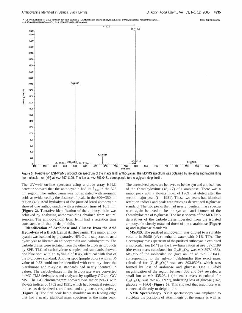
MS/MS. The purified anthocyanin was diluted to a suitablevolume in 50:50 (v/v) methanol:water with 0.1% TFA. Theelectrospray mass spectrum of the purified anthocyanin exhibiteda molecular ion [M+] as the flavylium cation atm/z 597.1199(the exact mass calculated for C26H29O16 was m/z 597.1456).MS/MS of the molecular ion gave an ion atm/z 303.0431corresponding to the aglycon delphinidin (the exact masscalculated for [C15H11O7]+ was m/z 303.0505), which wasformed by loss of arabinose and glucose. One 100-foldmagnification of the region between 303 and 597 revealed asmall ion at m/z 435.0841 (the exact mass calculated forC20H19O11 wasm/z 435.0927), indicating loss of glucose (162,glucose- H2O) (Figure 5). This showed that arabinose wasconnected directly to delphinidin.
NMR Spectroscopy.NMR spectroscopy was employed toelucidate the positions of attachments of the sugars as well as
Figure 5. Positive ion ESI-MS/MS product ion spectrum of the major lentil anthocyanin. The MS/MS spectrum was obtained by isolating and fragmentingthe molecular ion [M+] at m/z 597.1199. The ion at m/z 303.0431 corresponds to the aglycon delphinidin.
Anthocyanins Identified in Beluga Black Lentils J. Agric. Food Chem., Vol. 53, No. 12, 2005 4935

their conformations. The presence of delphinidin was confirmedby the observation of one signal belonging to the B-ring protonsat 7.79 ppm (Table 1). The signals at C-4, C-6, and C-8 werein accord with our previous studies of delphindin 3-glucoside(19). A characteristic coupling constant of 2 Hz was observedfor the meta coupling between the H-6 and the H-8 protons(observed with 5% TFA in CD3OD at 23°C). The glycosidicprotons were found as doublets downfield of the other sugarprotons in the region from 4.4 to 5.6 ppm. The attachment ofthe arabinose to the C-3 position of delphinidin was indicatedby the nuclear Overhauser effect (NOE) observed with H-1′′of arabinose and H-4 of delphinidin. For pyranoses, a couplingconstant of 7.5 Hz indicates a trans dihedral angle for H-1 andH-2, a â-linkage, while a coupling constant of less than 3 Hzindicates a gauche dihedral angle, anR-linkage. The H-1′′′ ofglucose had a coupling constant of 7.8 Hz, which indicated theâ-form. 1H coupling constants of the other three proton signalson the same sugar had values greater than 8.5 Hz (JH-2′′′ ) 8.5,JH-3′′′ ) 9.0, andJH-4′′′ ) 8.8), indicating the presence of theglucopyranose form (evidence of all trans configuration in thoseproton signals). The carbon signals were similar to those ofmethyl-D-glucopyranoside (20). The position of attachmentbetween glucose and arabinose was established by observationof an NOE between H-2′′ of arabinose with H-1′′′ of glucose.The remaining proton and carbon signals are consistent withthat of anO-R-L-arabinopyranoside (21-23) with the exceptionof C-2′′, which was shifted significantly downfield due to theglycosylation at C-2′′. An HMBC spectrum found correlationbetween H-2′′ and C-3′′ and between H-5′′ and C-4′′. Asopposed to glucose, which occurs in theD-form, arabinoseoccurs in theL-form. With L-arabinose,R-linkages shouldexhibit larger coupling constants (because of axial-axialcoupling between protons) thanâ-linkages. An intermediatecoupling constant of 4.2 Hz was observed with a solvent of10:90 TFA:DMSO at 40°C while a higher coupling constantof 5.1 Hz was seen with a solvent of 5:95 TFA:methanol at 23°C. The values of 4.2 and 5.1 Hz for the anomeric proton wereconsistent with anR-linkage. A coupling constant of 5.1 Hzwas reported for the anomeric proton of quercetin 3-O-R-L-arabinopyranoside using DMSO as the solvent (21). In addition,
we found a smaller coupling constant of 2 Hz for the anomericproton of methylâ-L-arabinopyranoside using a solvent of10:90 TFA:DMSO at 40°C. On the basis of the NMR data,the major anthocyanin was determined to be delphinidin 3-O-(2-O-â-D-glucopyranosyl-R-L-arabinopyranoside) (Figure 6).
LITERATURE CITED
(1) Vaughan, J. G.; Geissler, C.The New Oxford Book of FoodPlants; Oxford University Press Inc.: New York, 1997; pp 46-47.
(2) Agriculture and Agri-Food Canada Bi-weekly bulletin. Lentils/fababeans.2004, 17 (8), 1-4.
(3) Kong, J.-M.; Chia, L.-S.; Goh, N.-K.; Chia, T.-F.; Brouillard,R. Analysis and biological activities of anthocyanins.Phy-tochemistry2003, 64, 923-933.
(4) Nozzolillo, C. A survey of anthocyanin pigments in seedlinglegumes.Can. J. Bot. 1973, 51, 911-915.
(5) D’Arcy, A.; Jay, M. Les flavanoides des graines deLensculinaris. Phytochemistry1978, 17, 826-827.
(6) Slinkard, A. E.; Bhatty, R. S. Laird lentil.Can. J. Plant Sci.1979, 59, 503-504.
(7) Dueas, M.; Sun, B.; Hernndez, T.; Estrella, I.; Spranger, M. I.Proanthocyanidin composition in the seed coat of lentils (Lensculinaris L.). J. Agric. Food Chem. 2003, 51, 7999-8004.
(8) Strack, D.; Wray, V. Anthocyanins. InMethods in PlantBiochemistry; Harborne, J. B., Ed.; Academic Press: London,1989; Vol. 1, pp 325-356.
(9) Dao, L. T.; Takeoka, G. R.; Edwards, R. H.; Berrios, J. De J.Improved method for the stabilization of anthocyanidins.J. Agric.Food Chem. 1998, 46, 3564-3569.
(10) Harborne, J. B.ComparatiVe Biochemistry of the FlaVonoids;Academic Press: London, 1967.
(11) Iacobucci, G. A.; Sweeny, J. G. The chemistry of anthocyanins,anthocyanidins and related flavylium salts.Tetrahedron1983,39, 3005-3038.
(12) Hansen, S. A. Thin-layer chromatographic method for identifica-tion of mono-, di-, and trisaccharides.J. Chromatogr. 1975, 107,224-226.
(13) Hong, V.; Wrolstad, R. E. Cranberry juice composition.J. Assoc.Off. Anal. Chem. 1986, 69, 199-207.
(14) Karppa, J.; Kallio, H.; Peltonen, I.; Linko, R. Anthocyanins ofcrowberry,Empetrum nigrumcoll. J. Food Sci. 1984, 49, 634-636.
(15) Wilkinson, M.; Sweeny, J. G.; Iacobucci, G. A. High-pressureliquid chromatography of anthocyanins.J. Chromatogr. 1977,132, 349-351.
(16) Laine, R. A.; Sweeley, C. C. Analysis of trimethylsilylO-methyloximes of carbohydrates by combined gas-liquid chro-matography-mass spectrometry.Anal. Biochem. 1971, 43, 533-538.
(17) Laine, R. A.; Sweeley, C. C.O-methyl oximes of sugars. Analysisas O-trimethylsilyl derivatives by gas-liquid chromatographyand mass spectrometry.Carbohydr. Res. 1973, 27, 199-213.
(18) Andersen, M. Chromatographic separation of anthocyanins inCowberry (Lingonberry)VacciniumVites-IdaeaL. J. Food Sci.1985, 50, 1230-1232.
Table 1. 1H and 13C NMR Data for Delphinidin3-O-(2-O-â-D-Glucopyranosyl-R-L-arabinopyranoside) in 10%TFA−DMSO at 40 °C
1H NMR (ppm) 13C NMR (ppm)
delphinidin4 8.67 s 132.96 6.71 bs 102.58 6.82 bs 94.02′ 7.79 s 111.86′ 7.79 s 111.8
O-R-arabinopyranoside1 5.59 d (J ) 4.2 Hz) 99.282 4.18 t (J ) 5.4 Hz) 78.803 3.85 m 69.724 3.85 m 65.075a 3.75 dd (J ) 5.6, 11.0 Hz) 63.355b 3.59 dd (J ) 2.2, 11.0 Hz) 63.35
O-â-glucopyranosyl1 4.42 d (J ) 7.8 Hz) 104.172 2.93 t (J ) 8.5 Hz) 74.023 3.09 t (J ) 9.0 Hz) 76.594 3.01 t (J ) 8.8 Hz) 69.425 2.83 m 76.286a 3.20 dd (J ) 4.4, 11.5 Hz) 60.316b 3.15 dd (J ) 2.2, 11.5 Hz) 60.31
Figure 6. Structure of delphinidin 3-O-(2-O-â-D-glucopyranosyl-R-L-arabinopyranoside).
4936 J. Agric. Food Chem., Vol. 53, No. 12, 2005 Takeoka et al.
(19) Takeoka, G. R.; Dao, L. T.; Full, G. H.; Wong, R. Y.; Harden,L. A.; Edwards, R. H.; Berrios, J. De J. Characterization of blackbean (PhaseolusVulgarisL.) anthocyanins.J. Agric. Food Chem.1997, 45, 3395-3400.
(20) Agrawal, P. NMR spectroscopy in the structural elucidation ofoligosaccharides and glycosides.Phytochemistry1992, 31,3307-3330.
(21) Fraisse, D.; Heitz, A.; Carnat, A.; Carnat, A.-P.; Lamaison, J.-L. Quercetin 3-arabinopyranoside, a major flavonoid compoundfrom Alchemilla xanthochlora. Fitoterapia2000, 71, 463-464.
(22) Markham, K. R.; Ternai, B.; Stanley, R.; Geiger, H.; Mabry, T.J. Carbon-13 NMR studies of flavonoids. III. Naturally occurringflavonoid glycosides and their acylated derivatives.Tetrahedron1978, 34, 1389-1397.
(23) Pachaly, P.; Klein, M. Inhaltsstoffe vonAndromeda polifolia.Planta Med. 1987, 53, 442-444.
Received for review December 23, 2004. Revised manuscript receivedApril 4, 2005. Accepted April 7, 2005.
JF040493H
Anthocyanins Identified in Beluga Black Lentils J. Agric. Food Chem., Vol. 53, No. 12, 2005 4937