de novo point mutations in patients diagnosed with ataxic ... · pdf filede novo point...
TRANSCRIPT

De novo point mutations in patients diagnosedwith ataxic cerebral palsy
Ricardo Parolin Schnekenberg,1,2 Emma M. Perkins,3 Jack W. Miller,4 Wayne I. L. Davies,4,5,6
Maria Cristina D’Adamo,6 Mauro Pessia,6,7Katherine A. Fawcett,8 David Sims,8
Elodie Gillard,4 Karl Hudspith,4 Paul Skehel,3 Jonathan Williams,9 Mary O’Regan,10
Sandeep Jayawant,11 Rosalind Jefferson,12 Sarah Hughes,12 Andrea Lustenberger,13
Jiannis Ragoussis,1,† Mandy Jackson,3 Stephen J. Tucker14,15 and Andrea H. Nemeth4,16
Dedicated to the memory of Dr John Tolmie who tragically died during the preparation of this manuscript.
†Present address: McGill University and Genome Quebec Innovation Centre, McGill University, Montreal, Canada.
Cerebral palsy is a sporadic disorder with multiple likely aetiologies, but frequently considered to be caused by birth asphyxia.
Genetic investigations are rarely performed in patients with cerebral palsy and there is little proven evidence of genetic causes. As
part of a large project investigating children with ataxia, we identified four patients in our cohort with a diagnosis of ataxic
cerebral palsy. They were investigated using either targeted next generation sequencing or trio-based exome sequencing and were
found to have mutations in three different genes, KCNC3, ITPR1 and SPTBN2. All the mutations were de novo and associated
with increased paternal age. The mutations were shown to be pathogenic using a combination of bioinformatics analysis and
in vitro model systems. This work is the first to report that the ataxic subtype of cerebral palsy can be caused by de novo
dominant point mutations, which explains the sporadic nature of these cases. We conclude that at least some subtypes of cerebral
palsy may be caused by de novo genetic mutations and patients with a clinical diagnosis of cerebral palsy should be genetically
investigated before causation is ascribed to perinatal asphyxia or other aetiologies.
1 Wellcome Trust Centre for Human Genetics, University of Oxford, OX3 7BN, UK2 Universidade Positivo, School of Medicine, Rua Parigot de Souza 5300, 81280-330, Curitiba, Brazil3 Centre for Integrative Physiology, Euan MacDonald Centre for Motor Neurone Disease Research, University of Edinburgh,
Edinburgh, UK4 Nuffield Department of Clinical Neurosciences, University of Oxford, Oxford OX3 9DU5 School of Animal Biology, University of Western Australia, Perth, Australia6 Section of Physiology & Biochemistry, Department of Experimental Medicine, School of Medicine & Surgery, University of
Perugia, P.le Gambuli 1, Edificio D, Piano 106132 San Sisto, Perugia, Italy7 Department of Neural and Behavioral Sciences, Pennsylvania State University College of Medicine, Hershey, PA 17033-0850,
USA8 CGAT Programme, MRC Functional Genomics Unit, Department of Physiology, Anatomy and Genetics, University of Oxford,
South Parks Road, Oxford, OX1 3PT, UK9 Oxford Medical Genetics Laboratories, Churchill Hospital, Oxford, OX3 7LJ, UK
10 Fraser of Allander Neurosciences Unit, Royal Hospital for Sick Children, Glasgow G3 8SJ, UK11 Department of Paediatrics, Oxford University Hospitals NHS Trust, Oxford, OX3 9DU, UK12 Department of Paediatrics, Royal Berkshire Foundation Trust Hospital, Reading, UK13 Department of Neuropaediatrics, Development and Rehabilitation, University Children’s Hospital, Inselspital, Bern, Switzerland14 Clarendon Laboratory, Department of Physics, University of Oxford, OX1 3PU, UK15 OXION Initiative in Ion Channels and Disease, University of Oxford, OX1 3PT, UK
doi:10.1093/brain/awv117 BRAIN 2015: Page 1 of 16 | 1
Received November 17, 2014. Revised February 2, 2015. Accepted February 25, 2015.
� The Author (2015). Published by Oxford University Press on behalf of the Guarantors of Brain.
This is an Open Access article distributed under the terms of the Creative Commons Attribution License (http://creativecommons.org/licenses/by/4.0/), which permits unrestricted reuse,
distribution, and reproduction in any medium, provided the original work is properly cited.
Brain Advance Access published May 16, 2015by guest on June 8, 2015
Dow
nloaded from

16 Department of Clinical Genetics, Churchill Hospital, Oxford University Hospitals NHS Trust, Oxford, OX3 7LJ, UK
Correspondence to: Dr Andrea H. Nemeth,
Nuffield Department of Clinical Neurosciences,
6th Floor West Wing,
John Radcliffe Hospital,
Oxford, OX3 9DU, UK
E-mail: [email protected]
Correspondence may also be addressed to: Dr Mandy Jackson, Centre for Integrative Physiology, The University of Edinburgh, Hugh
Robson Building, George Square, Edinburgh, EH8 9XD. E-mail: [email protected]
Dr Stephen J. Tucker, Clarendon Laboratory, Department of Physics, University of Oxford, Parks Road, Oxford, OX1 3PU.
E-mail: [email protected]
Keywords: cerebral palsy; ataxia; de novo; intellectual disability
Abbreviation: SNP = single nucleotide polymorphism
IntroductionThe cerebral palsies are defined as a group of permanent
disorders of movement and posture that are attributed to
non-progressive disturbances that occurred in the develop-
ing foetal or infant brain (Rosenbaum et al., 2007). They
are classified into clinical subtypes including spastic, dyski-
netic, dystonic and ataxic groups, but there may be add-
itional disturbances of cognition, communication and
behaviour. Some patients have brain imaging abnormal-
ities, such as focal infarction, brain malformations and
periventricular leukomalacia; however, not all children
with cerebral palsy have abnormal brain imaging and
determining the aetiology in such cases is particularly chal-
lenging (O’Shea, 2008; Reid et al., 2013).
Cerebral palsy is a common disorder affecting approxi-
mately 1 in 500 of the population in Western industrialized
nations (Oskoui et al., 2013), but despite being so
common, a proven cause is not always found. Although
recent studies have suggested that only 10–20% of cerebral
palsy cases can be explained by birth asphyxia (Nelson,
2003; Colver et al., 2014), there remains a widespread
belief that obstetric misadventure or even negligence is
the main cause of cerebral palsy, and many parents with
children diagnosed with cerebral palsy enquire about the
possibility of damage during the delivery process.
Obstetrics therefore has one of the highest rates of litiga-
tion within the medical profession and one of the highest
rates of outcome favouring the plaintiff (Jena et al., 2011).
Indeed, an internet search for the term ‘cerebral palsy’ pro-
vides dozens of legal services for claims of negligence and
as a result of this climate of litigation in developed coun-
tries the Caesarian section rate (and its complications) has
soared in the last 40 years (Nelson, 2003).
Despite major improvements in obstetric and perinatal
healthcare, the prevalence of cerebral palsy has remained
stubbornly stable (Nelson, 2003), suggesting that intrinsic
biological factors rather than extrinsic causes may be an
explanation. Genetic factors have been suggested in the
context of consanguinity (Moreno-De-Luca et al., 2011,
2012; Kruer et al., 2013), implicating autosomal recessive
mutations, but this still fails to explain the frequency of
sporadic cases without affected siblings. A recent study of
cerebral palsy found a higher rate of rare inherited copy-
number variations in cerebral palsy, but the functional sig-
nificance of these variants was uncertain (McMichael et al.,
2013).
In the study presented here, four children were identified
who had a working diagnosis of sporadic ataxic cerebral
palsy and were found to have de novo mutations in three
different genes: KCNC3, which encodes a voltage-gated
potassium channel (Kv3.3); ITPR1, which encodes the re-
ceptor for inositol 1,4,5-trisphosphate (IP3R); and
SPTBN2, which encodes b-III spectrin. The likely patho-
genicity of the identified mutations was confirmed by
using bioinformatics and electrophysiology. This study
has significant implications for our understanding of cere-
bral palsy including its investigation, classification and
aetiology.
Materials and methods
Genetic studies
Patients were recruited from throughout the UK andSwitzerland and consent for participation in the study was ob-tained according to the Declaration of Helsinki (WMA, 1997)and approved by the Central Oxford Research EthicsCommittee and the Research and Development Department ofthe Oxford Radcliffe Hospitals NHS Trust, Oxford. All patientsor their parents provided written consent for the study. Tencases with a congenital cerebellar ataxia were analysed, thefirst using targeted capture and next generation sequencing,and the remaining cases were analysed using exome sequencing.
Targeted sequencing in Case 1
The exonic and 25 base pairs (bp) intronic flanking sequencesof 118 genes known to be associated with ataxia in humans or
2 | BRAIN 2015: Page 2 of 16 R. P. Schnekenberg et al.
by guest on June 8, 2015D
ownloaded from

good candidate genes based on function were captured usingAgilent SureSelect enrichment and sequenced on the IlluminaGAII high-throughput sequencing platform as previouslydescribed (Nemeth et al., 2013) (Supplementary material).Sequence reads were aligned to the human reference genome(GRCh37/hg19) and single-nucleotide variants (SNVs) andsmall indels (insertion and deletions) were identified.Sequence variants were annotated using the Ensembl VariantEffect Predictor (VEP) tool (release 62, April 2011) (Fliceket al., 2011). For each variant, the VEP predicts potential func-tional consequences (e.g. non-synonymous coding, splice site,intronic effects) and checks if the variant is already present inthe single nucleotide polymorphism database (dbSNP, Build132) (Sherry et al., 2001). All variants were filtered using apreviously reported algorithm (Shanks et al., 2013a) andanalysed using standard pathogenicity prediction programsincluding PolyPhen-2 (Adzhubei et al., 2010), SIFT (Kumaret al., 2009), MutPred (Li et al., 2009) and the mutation in-terpretation software Alamut (http://www.interactive-biosoftware.com/). Sanger sequencing was used to confirm variantsand parentage by analysing six informative single nucleotidepolymorphisms (SNPs) identified as part of the sequencinganalysis.
Next-generation exome sequencing
Instead of targeted capture and sequencing, exome capture wasperformed in Cases 2–4 plus their parents (‘Trios 2–4’) usingthe SureSelect Human All Exon kit v5 (Agilent Technologies)and 100 bp paired-end sequencing performed on the IlluminaHiSeq 2000 platform. At least 92% of the target region wascovered at �20. Reads were quality trimmed using theFASTX-Toolkit v0.0.13 and then aligned to the 1000Genomes version of the human reference genome(human_g1k_v37) using the Burrows-Wheeler Aligner (BWA)v0.7.5a. Likely PCR duplicates were removed using Picardv1.106 and BAM files were processed using the GenomeAnalysis Toolkit (GATK) v2.7.2 software. Single nucleotidevariants and indels were jointly called within each trio usingthe GATK HaplotypeCaller and annotated using SnpEff v3.3and SnpSift (Cingolani et al., 2012). De novo mutations wereselected using the Genome Analysis Toolkit Select Variantswalker according to previously described criteria (Epi4KConsortium and Epilepsy Phenome/Genome Project, 2013)(Allen et al., 2013). Two additional filters were imposed toselect for putative de novo variants present at 50.1% fre-quency in 1000 Genomes individuals and the exome variantserver (http://evs.gs.washington.edu/EVS/) and predicted bySnpEff to have a moderate or high impact. Variants were visu-ally inspected using the Integrative Genomics Viewer (IGV)(Thorvaldsdottir et al., 2013) and validated by Sanger sequen-cing. Nucleotide conservation was estimated using theGenomic Evolutionary Rate Profiling (GERP) (scores:�12.3 = least conserved to 6.17 = most conserved) (Cooperet al., 2005) and PhyloP, which compares the probability ofobserved substitutions under the hypothesis of neutral evolu-tionary rate: positive scores suggest constraint (conservation)(Pollard et al., 2010). Effects of amino acid changes were ana-lysed using SIFT (probability of being pathogenic: 0 = highest;1 = lowest) (Adzhubei et al., 2010) and PolyPhen-2 (probabil-ity of being pathogenic: 0 = lowest; 1 = highest) (Sim et al.,2012).
Confirmation of parentage usingexome sequencing data
For Cases 2–4, analysis of parentage was performed using
86 000 exonic Hapmap SNPs, which were genotyped from
the exome sequencing data using GATK HaplotypeCaller(McKenna et al., 2010). The non-reference discordance
rate (NDR) was calculated by comparing genotype
calls for each pair-wise combination of individuals usingvcf-compare from the VCFtools suite of software (Danecek
et al., 2011). A heatmap depicting NDR values from all
combinations was plotted using the ggplot2 package
(Wickham, 2009) within the R statistical computingenvironment (R Core Team (2014). R: A language and
environment for statistical computing. R Foundation for
Statistical Computing, Vienna, Austria. URL http://www.R-project.org/). Parent-child segregation of SNPs was also inves-
tigated for 43000 SNPs where one parent was homozygous
non-reference and the other homozygous reference (probandexpected to be heterozygous in 499% of cases) and
415 000 SNPs where both parents were homozygous non-ref-
erence (proband expected to be homozygous in 499% of
cases).
Kv3.3 (KCNC3) electrophysiologicalanalysis
To examine the functional effects of the T428I mutation in
the voltage-gated potassium channel Kv3.3, the coding region
of a sequence-verified human Kv3.3 (KCNC3) cDNA (Source
Bioscience) was subcloned into the pBF oocyte expressionvector prior to the introduction of the c.1283C4T mutation
by SPLICE-based site-directed mutagenesis (Davies et al.,2007). Messenger RNA encoding either wild-type or mutantKv3.3 was then transcribed in vitro and injected into Xenopuslaevis oocytes for electrophysiological analysis using a stand-
ard two-electrode voltage clamp protocol as previouslydescribed (D’Adamo et al., 1998; Imbrici et al., 2000, 2006).
Briefly, whole-cell currents for either wild-type or mutant
channels were evoked by 200 ms depolarizing commands to
+ 60 mV from a holding potential of –80 mV. For analysis ofthe heterozygous state, oocytes were co-injected with wild-type
and T428I mutant mRNAs (1 ng wild-type and 1 ng T428I)
and compared with oocytes injected with either only wild-typemRNA (2 ng) or T428I mutant mRNA (2 ng). Data are
presented as the mean � SEM of 20–50 cells. Statistical
significance was determined using an unpaired Student’s t-test (**P50.001). Relative activation rates were recorded at
several voltages and fitted with a single exponential function.
The relevant time constants were then calculated and plotted
as a function of the test pulse (data are presented as themean � SEM of eight cells). The structural homology model
of the fourth transmembrane segment (S4 voltage sensor
domain), showing the relative position of the T428I mutationwas created using the published crystal structure of the related
Kv1.2/KCNA2 (potassium voltage-gated channel subfamily A
member 2) channel as a template. Within this region, Kv1.2exhibits 480% sequence identity with Kv3.3 (Long et al.,2005).
De novo mutations in ataxic cerebral palsy BRAIN 2015: Page 3 of 16 | 3
by guest on June 8, 2015D
ownloaded from
b-III spectrin (SPTBN2)electrophysiology
Wild-type and R480W mutant rat b-III spectrin cDNA se-quences were myc-tagged and cloned into the pRK5 mamma-lian expression vector. The missense mutation was introducedby site-directed mutagenesis using the pRK5-myc-tagged b-IIIspectrin as template. Primary rat hippocampal neuronal cul-tures were prepared from embryonic Day 18 Sprague-Dawleyembryos, transfected and current recordings made as previ-ously described (Clarkson et al., 2014). Briefly, cells were co-transfected with constructs expressing wild-type b-III spectrinand GFP or the R480W mutant and DsRed as it was previ-ously shown that the different fluorophores had no effect onsodium currents when co-expressed or expressed alone(Clarkson et al., 2014). Following transfection the cells weremixed and plated together so that recordings could be madefrom untransfected, wild-type b-III spectrin transfected andR480W mutant transfected cells maintained under identicalculture conditions.
Results
Clinical summary
Cases 1–4 were diagnosed with ataxic cerebral palsy based
on standard criteria (Smithers-Sheedy et al., 2014), which
included the presence of an early ataxic motor disorder
with no clinical evidence of a neurodegenerative disorder.
All the children were born at term and there were no ob-
vious risk factors for cerebral palsy such as prematurity,
low birth weight, infections and hypoglycaemia. No other
identifiable causes were found, including neurometabolic
disorders. Having identified four cases with de novo muta-
tions we reviewed all the ataxia cases in our cohort and
identified six additional children who had congenital cere-
bellar ataxia and who had been analysed using exome
sequencing, but who did not have a specific diagnostic
label of ataxic cerebral palsy. Case 5 had no brain imaging
abnormalities and most closely resembles the other cases in
our series. Case 6 had cerebellar vermis hypoplasia and
Case 7 had global cerebellar hypoplasia (vermis and cere-
bellar hemispheres), both these cases were clinically stable.
Cases 8 and 9 developed clinical regression by the age of 5
and Case 10 had additional cataracts and an affected sib-
ling. Cases 8–10 would not fulfil the criteria for ataxic
cerebral palsy because of the clinical regression and syn-
dromic features and will be presented elsewhere. Table 1
summarizes the clinical details of Cases 1–7 and Fig. 1
shows brain imaging of Cases 2–4. The mutations identi-
fied in Cases 1–4 were de novo, with parentage confirmed
using SNP analysis (Fig. 2 and Supplementary Table 1).
Analysis of the raw exome data for Cases 2–4 did not
identify any low level somatic mosaicism of the mutant
alleles in either parent. No mutations have been found in
Cases 5 and 6 and a putative new gene had been found in
Case 7, which is currently under investigation and will be
presented elsewhere.
Case 1: T428I in KCNC3
Case 1 was reviewed locally in a specialist paediatric neu-
rodisability centre. He is the second child of healthy
non-consanguineous parents, with no family history of
neurological disorders. He was born at term, weighing
6 lbs 5 oz (2860 g), following an uneventful pregnancy
and normal vaginal delivery. He was noted to have early
motor delay of sitting, crawling and standing, associated
with some delay in cognitive abilities compared with his
peers. Investigations included creatine kinase, thyroid func-
tion tests, urinary amino and organic acids, chromosomes,
genetic testing for Fragile X syndrome and a brain MRI, all
of which were normal. After multiple paediatric neurology
reviews he was diagnosed as having ataxic cerebral palsy
and provided with intensive physiotherapy and occupa-
tional therapy support. Further developmental assessments
confirmed both the motor abnormalities, with marked dis-
ability due to ataxia, in addition to some less prominent
cognitive difficulties requiring special support at school.
Non-verbal IQ testing gave a score of 90, on the low end
of the normal range.
Three non-synonymous changes were identified using tar-
geted capture and sequencing of 57 known ataxia genes. Of
these, only one, a previously unreported KCNC3 variant
c.1283C4T, p.T428I, was predicted to be functionally
damaging based on bioinformatic analysis including
amino acid conservation (Fig. 3A and Table 1). Bi-
directional Sanger sequencing confirmed the next-genera-
tion sequencing findings (Fig. 3B) and analyses of other
rare SNPs in the proband and his parents were consistent
with the stated parentage (Fig. 2), confirming that the mu-
tation arose de novo in the patient.
Mutations in KCNC3 have previously been reported to
underlie autosomal dominant spinocerebellar ataxia type
13 (SCA13) (Waters et al., 2006), although some variants
of uncertain pathogenic significance have also been re-
ported (Figueroa et al., 2011). Therefore, we further inves-
tigated the pathogenicity of the T428I variant by
electrophysiological analysis of wild-type and mutant
Kv3.3 potassium channel currents expressed in a heterol-
ogous system.
The threonine residue at position 428 is a highly con-
served amino acid located at the base of the positively
charged S4 voltage-sensor helix of the Kv3.3 potassium
channel, a position that has previously been implicated in
the control of Kv channel activity (Fig. 3C) (Ledwell and
Aldrich, 1999; Pathak et al., 2005). Examination of the
functional properties of the T428I mutant channel in com-
parison with wild-type Kv3.3 clearly demonstrated that the
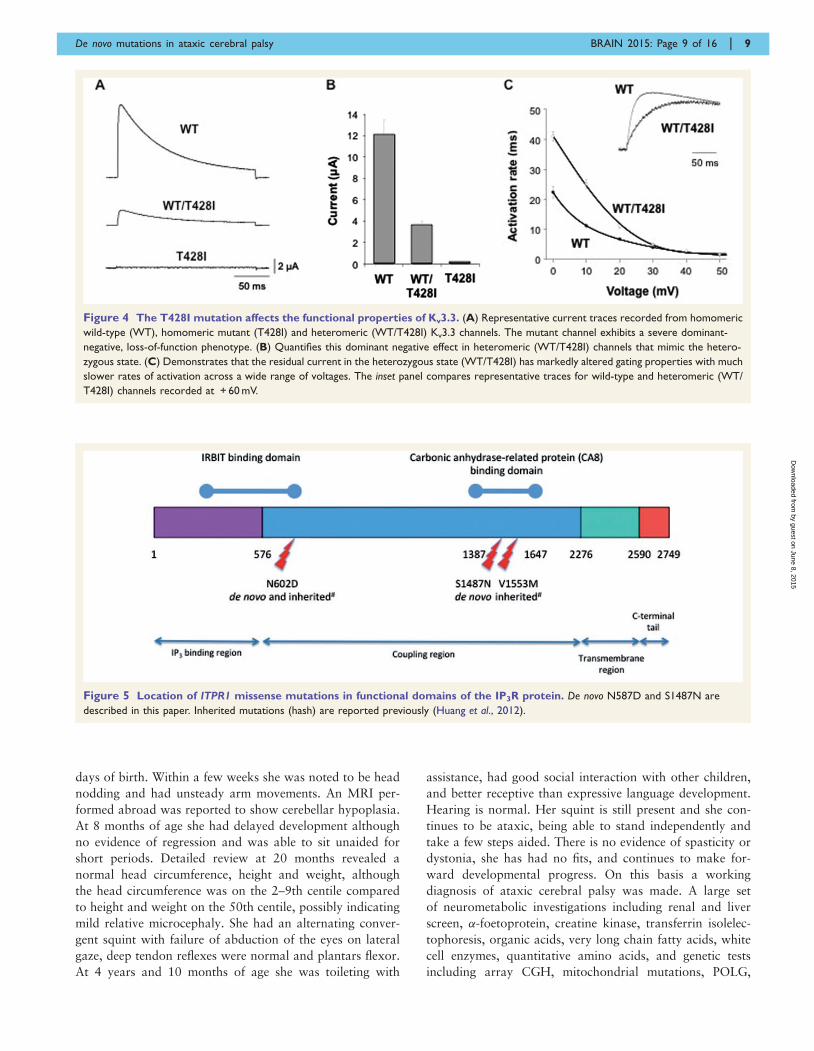
T428I mutant is non-functional (Fig. 4). Moreover, when
co-expressed with the wild-type subunit (to mimic the het-
erozygous state) the T428I mutation exerted a severe dom-
inant negative loss-of-function phenotype, reducing overall
4 | BRAIN 2015: Page 4 of 16 R. P. Schnekenberg et al.
by guest on June 8, 2015D
ownloaded from
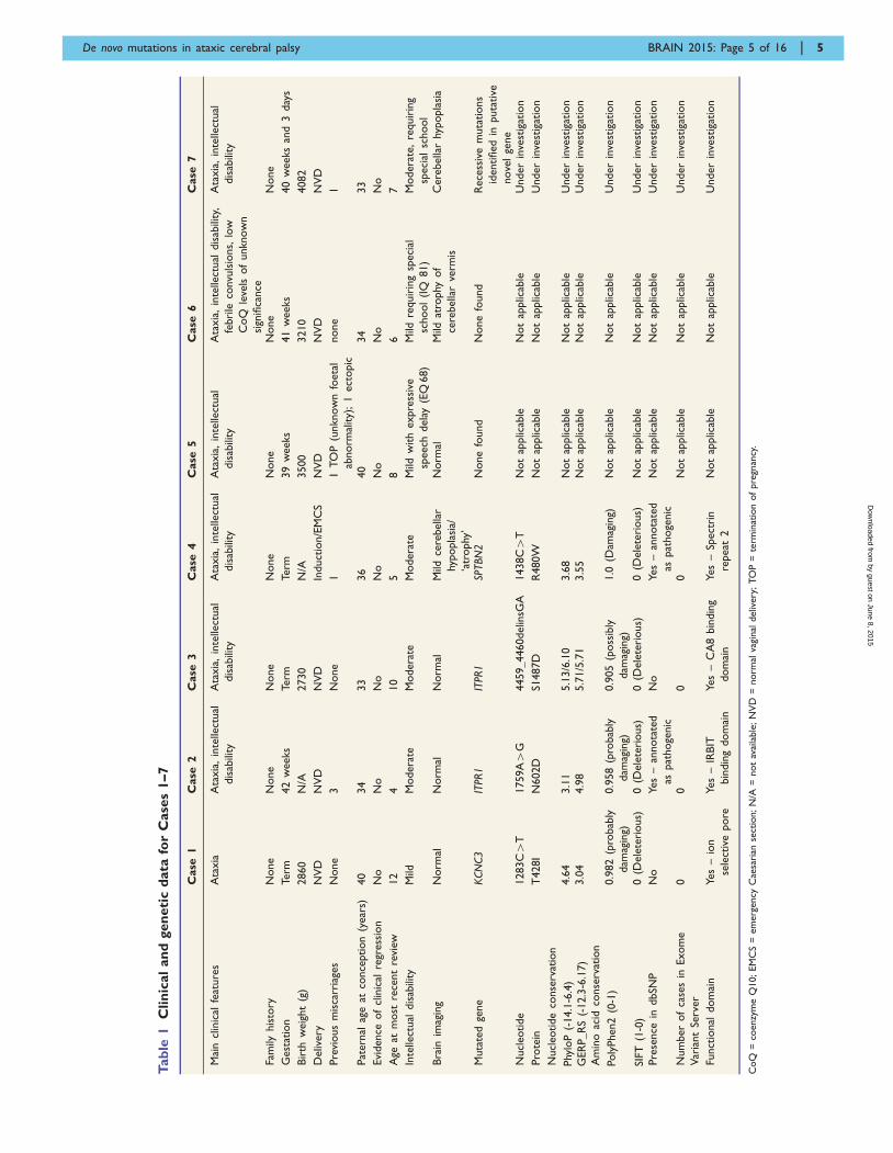
Tab
le1
Clin
ical
an
dgen
eti
cd
ata
for
Case
s1–7
Case
1C
ase
2C
ase
3C
ase
4C
ase
5C
ase
6C
ase
7
Mai
ncl
inic
alfe
ature
sA
taxia
Ata
xia
,in
telle
ctual
dis
abili
ty
Ata
xia
,in
telle
ctual
dis
abili
ty
Ata
xia
,in
telle
ctual
dis
abili
ty
Ata
xia
,in
telle
ctual
dis
abili
ty
Ata
xia
,in
telle
ctual
dis
abili
ty,
febri
leco
nvu
lsio
ns,
low
CoQ
leve
lsof
unknow
n
sign
ifica
nce
Ata
xia
,in
telle
ctual
dis
abili
ty
Fam
ilyhis
tory
None
None
None
None
None
None
None
Gest
atio
nTe
rm42
weeks
Term
Term
39
weeks
41
weeks
40
weeks
and
3day
s
Bir
thw
eig
ht
(g)
2860
N/A
2730
N/A
3500
3210
4082
Deliv
ery
NV
DN
VD
NV
DIn
duct
ion/E
MC
SN
VD
NV
DN
VD
Pre
vious
mis
carr
iage
sN
one
3N
one
11
TO
P(u
nknow
nfo
eta
l
abnorm
ality)
;1
ect
opic
none
1
Pat
ern
alag
eat
conce
ption
(year
s)40
34
33
36
40
34
33
Evi
dence
of
clin
ical
regr
ess
ion
No
No
No
No
No
No
No
Age
atm
ost
rece
nt
revi
ew12
410
58
67
Inte
llect
ual
dis
abili
tyM
ildM
odera
teM
odera
teM
odera
teM
ildw
ith
expre
ssiv
e
speech
dela
y(E
Q68)
Mild
requir
ing
speci
al
school
(IQ
81)
Modera
te,
requir
ing
speci
alsc
hool
Bra
inim
agin
gN
orm
alN
orm
alN
orm
alM
ildce
rebella
r
hypopla
sia/
‘atr
ophy
’
Norm
alM
ildat
rophy
of
cere
bella
rve
rmis
Cere
bella
rhy
popla
sia
Muta
ted
gene
KCN
C3
ITPR
1IT
PR1
SPTBN
2N
one
found
None
found
Rece
ssiv
em
uta
tions
identified
inputa
tive
nove
lge
ne
Nucl
eotide
1283C4
T1759A4
G4459_4460delin
sGA
1438C4
TN
ot
applic
able
Not
applic
able
Under
inve
stig
atio
n
Pro
tein
T428I
N602D
S1487D
R480W
Not
applic
able
Not
applic
able
Under
inve
stig
atio
n
Nucl
eotide
conse
rvat
ion
Phy
loP
(-14.1
-6.4
)
GER
P_R
S(-
12.3
-6.1
7)
4.6
4
3.0
4
3.1
1
4.9
8
5.1
3/6
.10
5.7
1/5
.71
3.6
8
3.5
5
Not
applic
able
Not
applic
able
Not
applic
able
Not
applic
able
Under
inve
stig
atio
n
Under
inve
stig
atio
n
Am
ino
acid
conse
rvat
ion
Poly
Phen2
(0-1
)
SIFT
(1-0
)
0.9
82
(pro
bab
ly
dam
agin
g)
0(D
ele
teri
ous)
0.9
58
(pro
bab
ly
dam
agin
g)
0(D
ele
teri
ous)
0.9
05
(poss
ibly
dam
agin
g)
0(D
ele
teri
ous)
1.0
(Dam
agin
g)
0(D
ele
teri
ous)
Not
applic
able
Not
applic
able
Not
applic
able
Not
applic
able
Under
inve
stig
atio
n
Under
inve
stig
atio
n
Pre
sence
indbSN
PN
oYes
–an
nota
ted
aspat
hoge
nic
No
Yes
–an
nota
ted
aspat
hoge
nic
Not
applic
able
Not
applic
able
Under
inve
stig
atio
n
Num
ber
of
case
sin
Exom
e
Var
iant
Serv
er
00
00
Not
applic
able
Not
applic
able
Under
inve
stig
atio
n
Funct
ional
dom
ain
Yes
–io
n
sele
ctiv
epore
Yes
–IR
BIT
bin
din
gdom
ain
Yes
–C
A8
bin
din
g
dom
ain
Yes
–Sp
ect
rin
repeat
2
Not
applic
able
Not
applic
able
Under
inve
stig
atio
n
CoQ
=co
enzy
me
Q10;EM
CS
=em
erg
ency
Cae
sari
anse
ctio
n;N
/A=
not
avai
lable
;N
VD
=norm
alva
ginal
deliv
ery
;TO
P=
term
inat
ion
of
pre
gnan
cy.
De novo mutations in ataxic cerebral palsy BRAIN 2015: Page 5 of 16 | 5
by guest on June 8, 2015D
ownloaded from
channel activity to below 50% of that seen with the wild-
type channel alone (Fig. 4B). Importantly, we also observed
that the remaining heteromeric channels displayed remark-
ably slower activation kinetics (Fig. 4C). These results
clearly demonstrate that the T428I mutation has a physio-
logically relevant damaging effect on Kv3.3 channel
function.
Case 2: N602D in ITPR1
Case 2 is the only child of non-consanguineous parents
who was born following difficulties with conception and
a single miscarriage. Birth weight was normal and head
circumference at birth on the 25th centile. The child was
noted to have delayed head control at 6 months, with head
nodding and rotatory nystagmus. The abnormal eye move-
ments and head nodding settled and subsequently delay
was noted in acquiring developmental milestones.
Multiple metabolic investigations were performed but no
cause was found. A brain MRI was normal (Fig. 1A). At
age 3 she was still not walking independently. Examination
revealed hypotonia and ataxia but no long-tract signs. Her
head circumference had dropped to the 3–10th centile. She
was socially very interactive and could use at least 15
words and will receive special educational support. At
almost 4 years old she is making good forward develop-
mental progress; clinical examination reveals ongoing
ataxia, she is able to weight-bear, but is not taking steps
and has mildly brisk reflexes with tight achilles tendons. In
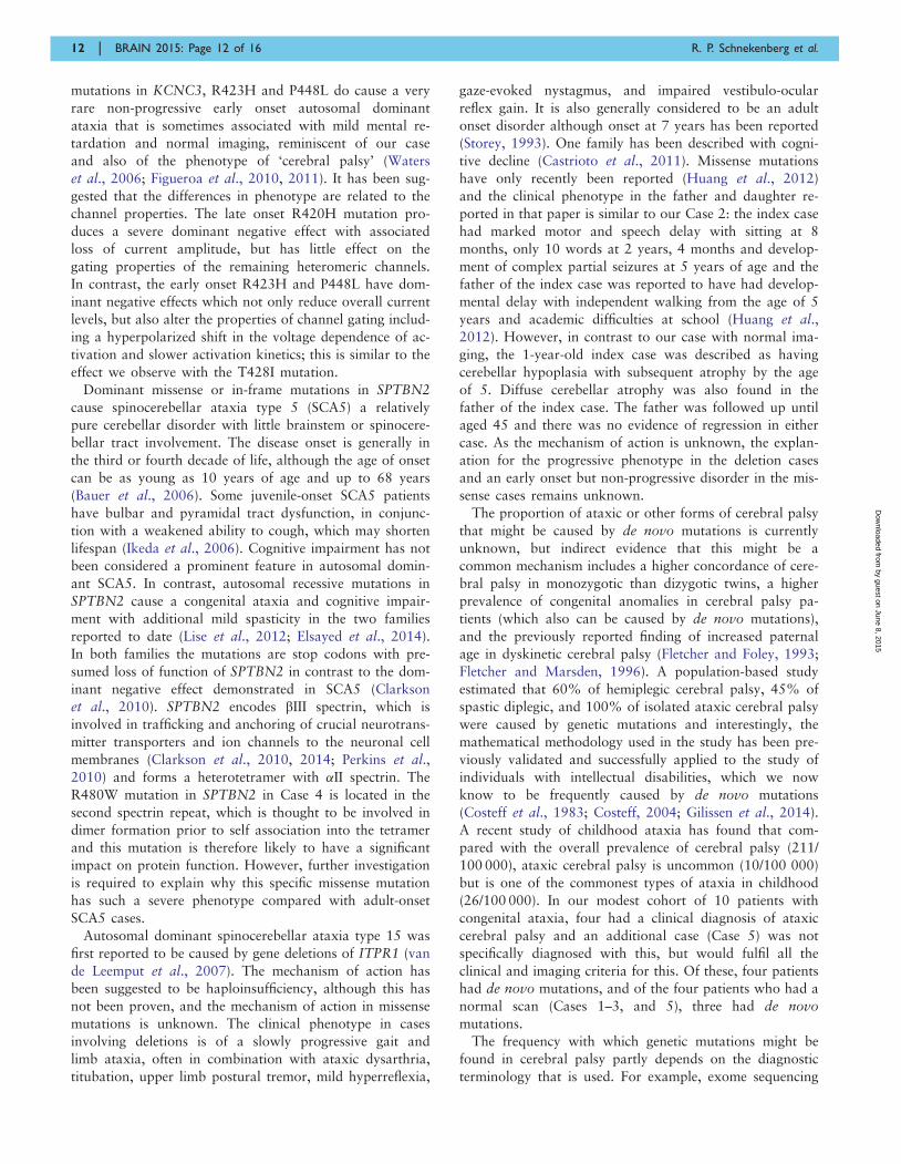
this patient a de novo mutation c.1759A4G, p. N602D
was detected in ITPR1 (Fig. 5). Partial gene deletions (usu-
ally encompassing the 5’ region) of ITPR1 are known to
cause spinocerebellar ataxia type 15 (SCA15) (van de
Leemput et al., 2007) and more recently two missense mu-
tations, N602D and V1553M, were reported in two
families with autosomal dominant non-progressive cerebel-
lar ataxia (Huang et al., 2012). Residue N602 is located in
the IRBIT (IP3R Binding protein released with Inositol
1,4,5-Trisphosphate) binding domain of the ITPR1 gene
product, the IP3 receptor (IP3R), but little else is known
of how this missense mutation causes ataxia. IP3R is a re-
ceptor for inositol 1,4,5-trisphosphate (IP3), which is gen-
erated by the hydrolysis of the membrane lipid
phosphatidylinositol 4,5-bisphosphate (PIP2). IP3 binds to
the IP3R receptor, which is an intracellular ligand-gated
calcium ion (Ca2 + ) release channel localized in the endo-
plasmic reticulum. IP3Rs are involved in numerous pro-
cesses, such as dorsoventral axis formation, synaptic
plasticity, neural circuit formation and neuronal dendrite
formation (Mikoshiba et al., 1994; Mikoshiba, 2011;
Huang et al., 2012). IP3R is also modulated by a variety
of other proteins including IRBIT, which is a non-enzym-
atic homologue of S-adenosylhomocysteine hydrolase.
IRBIT modulates IP3R activation by competing with IP3
at the IP3 binding site, thereby reducing the receptor’s sen-
sitivity to IP3 and reducing Ca2 + release (Ando et al.,
2006). The location of the N602D mutation, its evolution-
ary conservation (Table 1) and the previous report of an
identical mutation in an autosomal dominant cerebellar
ataxia family strongly support this mutation being
pathogenic.
Case 3: S1487D in ITPR1
Case 3 is the middle child of non-consanguineous parents;
the other two children are well. Pregnancy was notable for
an increased nuchal thickness, but amniocentesis revealed a
normal karyotype. He was born at term by normal vaginal
delivery and required no resuscitation. He sat at 12
months, crawled at 13–14 months and walked a few
steps before falling at 2 years 5 months. Height, weight
and head circumference were all on the 9th–25th centiles.
At 4 years he could walk unaided but continued to fall
frequently, he had shaky handwriting and could speak in
simple sentence structures but had good social interaction.
A brain MRI including the cerebellum was normal
(Fig. 1B). A complex deletion-insertion in ITPR1 was iden-
tified in this patient at nucleotides c.4459_4460delinsGA,
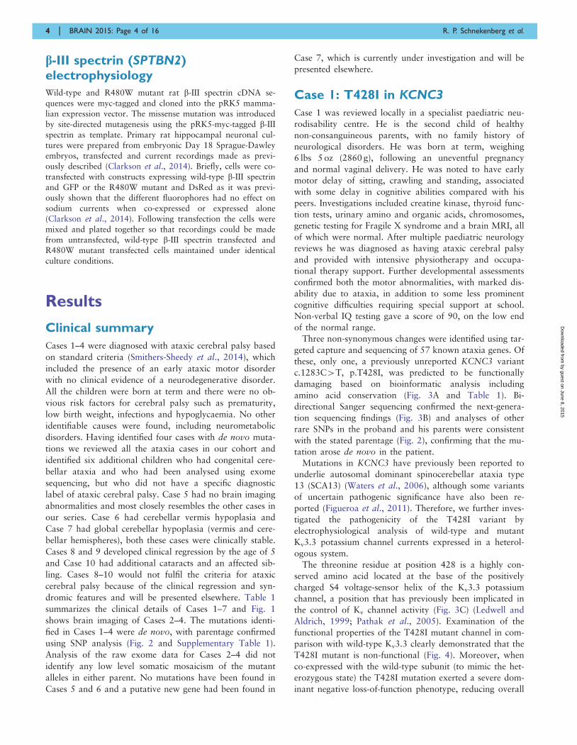
Figure 1 MRI of the brains of Cases 2, 3 and 4. (A and B) Cases 2 and 3 showing a normal brain MRI. (C) Case 4 brain MRI shows a small
cerebellum, with increased spacing of the cerebellar folia and an enlarged fourth ventricle. This was reported to be cerebellar atrophy, rather than
hypoplasia.
6 | BRAIN 2015: Page 6 of 16 R. P. Schnekenberg et al.
by guest on June 8, 2015D
ownloaded from

Figure 2 Confirmation of parentage in Cases 1–4. (A) Sequences of rare SNPs in parents and affected of Case 1 showing consistency with
parentage: genes, variant and genomic location (hg19) are shown. (B) The non-reference discordance rate (NDR) over 86 000 exonic Hapmap
SNPs for Cases 2, 3 and 4. Related individuals show lower discordance (yellow) than unrelated individuals (blue/purple). This analysis confirms
that the probands in the study are genetically related to both parents and that parents are not genetically related to each other.
De novo mutations in ataxic cerebral palsy BRAIN 2015: Page 7 of 16 | 7
by guest on June 8, 2015D
ownloaded from
resulting in a missense change p.S1487D. Both nucleotides
involved in the mutation are evolutionarily conserved and
S1487 is located at a highly conserved position in the CA8
(carbonic anhydrase related protein 8) binding domain of
ITPR1 (Hirota et al., 2003) (Fig. 5 and Table 1) suggesting
that this mutation is indeed pathogenic, although at this
stage it can only be classified as a ‘possible’ mutation.
Similar to IRBIT, CA8 is also a non-enzymatic competitor
of IP3 that binds to IP3R and interestingly, recessive muta-
tions in CA8 cause a congenital ataxia with intellectual
disability, possibly acting by disinhibiting the interaction
between IP3 and the IP3R, allowing extra Ca2 + to be
released from IP3-sensitive stores (Turkmen et al., 2009;
Kaya et al., 2011)
Case 4: R480W in SPTBN2
Case 4 is the only child of non-consanguineous parents of
Mediterranean origin. She was born at term following a
normal pregnancy. Delivery was by induction and emer-
gency caesarean section for foetal distress, but she was
well following delivery and discharged home within a few
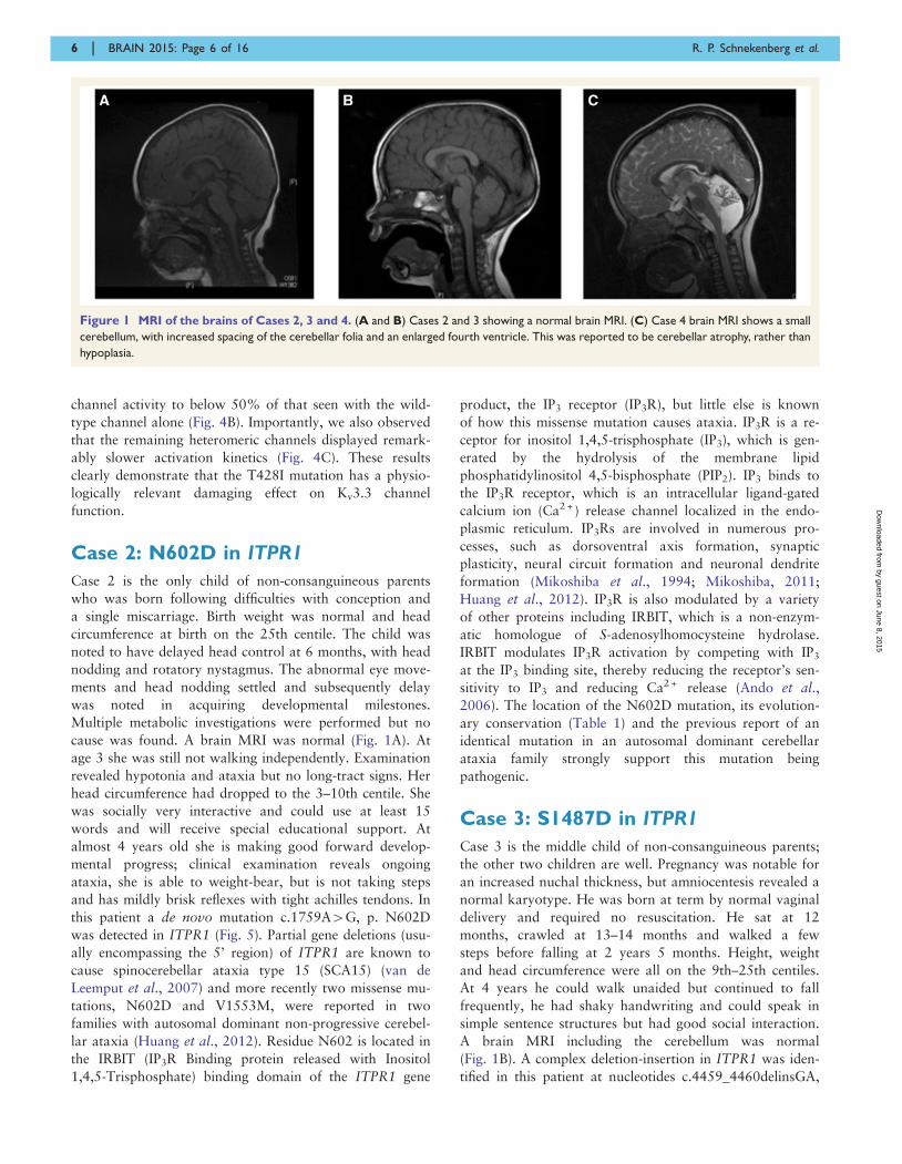
Figure 3 A novel de novo mutation predicted to affect the S4 voltage-sensor of Kv3.3. (A) The high degree of amino acid conser-
vation (asterisk) in the voltage-sensor S4 helix and S4-S4 linker region of human Kv3.3 and related species. This region is also highly conserved in
the paralogous channels KCNC1 (Kv3.1) and KCNC2 (Kv3.2). Threonine 428 in KCNC3 (Kv3.3) is highlighted in grey and is absolutely conserved
between species. (B) Sanger sequencing of the patient and parents to show that the heterozygous mutation is de novo. (C) A structural model of
this region in Kv3.3 with the predicted location of the T428I mutation. The conserved voltage-sensing arginine and lysine residues are also shown.
8 | BRAIN 2015: Page 8 of 16 R. P. Schnekenberg et al.
by guest on June 8, 2015D
ownloaded from
days of birth. Within a few weeks she was noted to be head
nodding and had unsteady arm movements. An MRI per-
formed abroad was reported to show cerebellar hypoplasia.
At 8 months of age she had delayed development although
no evidence of regression and was able to sit unaided for
short periods. Detailed review at 20 months revealed a
normal head circumference, height and weight, although
the head circumference was on the 2–9th centile compared
to height and weight on the 50th centile, possibly indicating
mild relative microcephaly. She had an alternating conver-
gent squint with failure of abduction of the eyes on lateral
gaze, deep tendon reflexes were normal and plantars flexor.
At 4 years and 10 months of age she was toileting with
assistance, had good social interaction with other children,
and better receptive than expressive language development.
Hearing is normal. Her squint is still present and she con-
tinues to be ataxic, being able to stand independently and
take a few steps aided. There is no evidence of spasticity or
dystonia, she has had no fits, and continues to make for-
ward developmental progress. On this basis a working
diagnosis of ataxic cerebral palsy was made. A large set
of neurometabolic investigations including renal and liver
screen, �-foetoprotein, creatine kinase, transferrin isolelec-
tophoresis, organic acids, very long chain fatty acids, white
cell enzymes, quantitative amino acids, and genetic tests
including array CGH, mitochondrial mutations, POLG,
Figure 4 The T428I mutation affects the functional properties of Kv3.3. (A) Representative current traces recorded from homomeric
wild-type (WT), homomeric mutant (T428I) and heteromeric (WT/T428I) Kv3.3 channels. The mutant channel exhibits a severe dominant-
negative, loss-of-function phenotype. (B) Quantifies this dominant negative effect in heteromeric (WT/T428I) channels that mimic the hetero-
zygous state. (C) Demonstrates that the residual current in the heterozygous state (WT/T428I) has markedly altered gating properties with much
slower rates of activation across a wide range of voltages. The inset panel compares representative traces for wild-type and heteromeric (WT/
T428I) channels recorded at + 60 mV.
Figure 5 Location of ITPR1 missense mutations in functional domains of the IP3R protein. De novo N587D and S1487N are
described in this paper. Inherited mutations (hash) are reported previously (Huang et al., 2012).
De novo mutations in ataxic cerebral palsy BRAIN 2015: Page 9 of 16 | 9
by guest on June 8, 2015D
ownloaded from

Friedreichs ataxia, and PLA2G6 were normal. Brain MRI
was reported to show cerebellar atrophy (Fig. 1C). Exome
sequencing revealed a point mutation c.1438C4T,
p.R480W in SPTBN2 in this patient. The nucleotide is
conserved, as is the amino acid at this position (Table 1)
and the mutation is predicted as highly likely to be
damaging.
The same mutation has been reported in another spor-
adic case of early onset cerebellar ataxia with developmen-
tal delay; however in that case no segregation analysis was
performed, but the parents were unrelated to each other
and healthy, suggesting that the mutation may also have
arisen de novo (Jacob et al., 2013). This initial case report
did not provide any data on the pathogenicity of the mu-
tation, either from segregation or functional analysis. We
have previously shown that expression of wild-type bIII
spectrin (encoded by SPTBN2) can enhance voltage-gated
sodium channel currents in cultured hippocampal neurons
(Clarkson et al., 2014) in contrast to mutant b-III spectrin.
We therefore investigated the effect of this mutation on
voltage-gated sodium channel currents in cultured hippo-
campal neurons and show that sodium currents in the
presence of R480W bIII spectrin are lower compared to
wild-type bIII spectrin (Fig. 6) providing further evidence
for the pathogenicity of this mutation.
DiscussionThis study is the first to report de novo mutations in pa-
tients with a diagnostic label of cerebral palsy and has im-
portant implications for our understanding of this complex
condition. Although cerebral palsy is known to be an um-
brella diagnostic term with likely multiple causes, the aeti-
ology is often unknown. Our finding of de novo mutations
provides an explanation for many of the seemingly puzzling
features that have been described in the past including the
sporadic nature, the highly variable phenotype and the as-
sociation with increased paternal age (Fletcher and Foley,
1993).
From a clinical perspective, the diagnostic criteria for
cerebral palsy were recently and comprehensively reviewed
to assist clinicians. These include: a disorder of movement
or posture of central origin, a disorder of motor function,
no evidence of clinical regression, latest assessment at 4
years of age or older, absence of syndrome/brain anom-
aly/chromosomal anomaly and no evidence of generalized
hypotonia. If the patient fulfils these criteria and, in add-
ition, has ataxia then the diagnosis is ataxic cerebral palsy,
although the authors note that differentiating a neurode-
generative cerebellar ataxia can be difficult and recommend
investigations to exclude this (Smithers-Sheedy et al., 2014).
Three of our cases clearly fulfilled the clinical criteria for
ataxia cerebral palsy as there was no clinical or imaging
evidence of regression and there were no syndromic fea-
tures. However, Case 4 illustrates the complexity of
making this diagnosis in young children when based on
clinical and radiological features. This patient was born
following an emergency Caesarian section for foetal distress
and in an era before imaging, the aetiology might have
been ascribed to ‘birth injury’. The clinical picture in this
case is of a stable cerebellar ataxia, rather than a neurode-
generative disorder and a diagnosis of ataxic cerebral palsy
falls within the current definition. Although the imaging
shows ‘cerebellar atrophy’ (defined as a cerebellum with
initially normal structures in a posterior fossa of normal
size, which displays enlarged interfolial spaces in compari-
son with the foliae secondary to the loss of cerebellar
tissue) (Poretti et al., 2008) the lack of clinical regression
is not in keeping with a neurodegenerative disorder. Our
patient is still young but the previously reported case in the
literature reached 12 years of age without evidence of a
neurodegenerative disorder, suggesting that this is indeed
a clinically stable condition in childhood. Interestingly,
there are other cases in the literature with apparent cere-
bellar atrophy who do not have evidence of clinical regres-
sion either and in whom autosomal recessive genetic
mutations have been found (Yapici and Eraksoy, 2005;
Poretti et al., 2008; Burns et al., 2014). Case 4 certainly
warrants specific follow-up for evidence of clinical deteri-
oration and long-term follow-up in patients with these
newly defined genetic disorders should greatly assist in
determining the prognosis. The difficulty of resolving the
apparent contradiction between a clinically stable disorder
with cerebellar imaging suggestive of atrophy is also illu-
strated in the case of ITPR1 mutations where there do not
appear to be consistent genotype–imaging correlations.
N602D in ITPR1 has been reported to cause cerebellar
hypoplasia and/or atrophy with long term clinical stability
(Huang et al., 2012) but the same de novo variant in our
Case 2 is associated with normal imaging.
The cases presented here also illustrate the overlap be-
tween the diagnostic categories of ataxic cerebral palsy and
non-progressive cerebellar ataxia. The latter group of dis-
orders was described in detail prior to the advent of next-
generation sequencing (Steinlin, 1998; Steinlin et al., 1998)
and include patients with or without cerebellar hypoplasia
(as defined in Poretti et al., 2008). Therefore some of these
cases would also fulfil the criteria for a diagnosis of ataxic
cerebral palsy. The largest group of patients with non-
progressive cerebellar ataxia were those of unknown her-
edity and unknown aetiology (Steinlin, 1998). The clinical
features included early hypotonia and motor developmental
delay, with occasional spasticity and dystonia. The majority
had intellectual disability but the degree of the cognitive
impairment could not be predicted by the degree of
ataxia or the imaging findings.
A finding of specific interest is that nearly all of our pa-
tients also had some degree of intellectual disability. The
presence of intellectual disability in cerebral palsy is well
known and is estimated to be in the region of 35–50%,
therefore the finding of intellectual disability in the majority
of our cases of ataxic cerebral palsy is perhaps not surpris-
ing. However, there have been no previous reports
10 | BRAIN 2015: Page 10 of 16 R. P. Schnekenberg et al.
by guest on June 8, 2015D
ownloaded from

specifically investigating the association of intellectual dis-
ability with ataxic cerebral palsy, and the rate of intellec-
tual disability is higher than might be expected from the
overall prevalence rate in cerebral palsy. Although the as-
sociation between congenital ataxia and intellectual disabil-
ity has not been emphasized in the literature, recently it has
been described in several cases of genetically confirmed
congenital ataxia associated with cerebellar atrophy or
hypoplasia (Ozcelik et al., 2008; Turkmen et al., 2009;
Doi et al., 2011; Bourassa et al., 2012). The neural basis
underlying this observation remains uncertain. The concept
of the ‘cerebellar cognitive affective syndrome’ has been put
forward (Schmahmann and Sherman, 1997) and proposes
that the cerebellum has intrinsic cognitive functions.
However, in some genetic disorders there is evidence that
the genes responsible for cerebellar development are also
important in development of the cerebral cortex (and/or
hippocampus) (Lise et al., 2012). Further work is required
to elucidate the neural basis of this association.
The similarity in clinical phenotypes between our cases
suggests that there may be shared molecular mechanisms of
pathogenicity. In the case of KCNC3 and SPTBN2, we
have shown that the mutations behave as dominant
negatives and both of these affect channel function, directly
or indirectly. The mechanism of action of missense point
mutations in ITPR1 causing ataxia is unknown, but given
the effect of IP3R in modulating calcium release it is pos-
sible that these also cause dominant negative effects, which
further studies will need to elucidate. It is also possible that
these three genes affect converging cerebellar neurodevelop-
mental pathways (Marzban et al., 2014). Genes involved in
cerebellar development may also involve cerebral develop-
ment (Vieira et al., 2010; Martinez et al., 2013) and pro-
tein/regulatory networks involved in intellectual disability
have been identified in which there is an overlap with pro-
teins affecting cerebellar development (Najmabadi et al.,
2011). Whether this hypothesis can be extended to other
subtypes of cerebral palsy is unknown, although the ex-
tremely wide phenotypic spectrum of cerebral palsy sug-
gests that multiple genes and pathways are likely to be
involved.
The clinical phenotype of our patients is unusual for
many autosomal dominant spinocerebellar ataxias.
Mutations such as R420H in KCNC3 cause autosomal
dominant spinocerebellar ataxia, type 13 (SCA13) with a
late onset, progressive disorder. However, two other
Figure 6 Peak sodium current enhanced less by R480W than wild-type b-III spectrin. (A) Sodium current traces from representative
cells evoked with a series of 50 ms depolorizations from a holding potential of �90 mV to potentials ranging from �80 to + 20 mV in 10 mV
increments (stimulus protocol shown at bottom). (B) Sodium current peak at �10 mV normalized to control cells cultured at same time. (C)
Current-voltage relationships for control, wild-type (WT) and R480W with current amplitude normalized to peak value. All data are presented as
the mean � SEM (n = 5–9 cells from each of three independent cultures; P5 0.05).
De novo mutations in ataxic cerebral palsy BRAIN 2015: Page 11 of 16 | 11
by guest on June 8, 2015D
ownloaded from
mutations in KCNC3, R423H and P448L do cause a very
rare non-progressive early onset autosomal dominant
ataxia that is sometimes associated with mild mental re-
tardation and normal imaging, reminiscent of our case
and also of the phenotype of ‘cerebral palsy’ (Waters
et al., 2006; Figueroa et al., 2010, 2011). It has been sug-
gested that the differences in phenotype are related to the
channel properties. The late onset R420H mutation pro-
duces a severe dominant negative effect with associated
loss of current amplitude, but has little effect on the
gating properties of the remaining heteromeric channels.
In contrast, the early onset R423H and P448L have dom-
inant negative effects which not only reduce overall current
levels, but also alter the properties of channel gating includ-
ing a hyperpolarized shift in the voltage dependence of ac-
tivation and slower activation kinetics; this is similar to the
effect we observe with the T428I mutation.
Dominant missense or in-frame mutations in SPTBN2
cause spinocerebellar ataxia type 5 (SCA5) a relatively
pure cerebellar disorder with little brainstem or spinocere-
bellar tract involvement. The disease onset is generally in
the third or fourth decade of life, although the age of onset
can be as young as 10 years of age and up to 68 years
(Bauer et al., 2006). Some juvenile-onset SCA5 patients
have bulbar and pyramidal tract dysfunction, in conjunc-
tion with a weakened ability to cough, which may shorten
lifespan (Ikeda et al., 2006). Cognitive impairment has not
been considered a prominent feature in autosomal domin-
ant SCA5. In contrast, autosomal recessive mutations in
SPTBN2 cause a congenital ataxia and cognitive impair-
ment with additional mild spasticity in the two families
reported to date (Lise et al., 2012; Elsayed et al., 2014).
In both families the mutations are stop codons with pre-
sumed loss of function of SPTBN2 in contrast to the dom-
inant negative effect demonstrated in SCA5 (Clarkson
et al., 2010). SPTBN2 encodes bIII spectrin, which is
involved in trafficking and anchoring of crucial neurotrans-
mitter transporters and ion channels to the neuronal cell
membranes (Clarkson et al., 2010, 2014; Perkins et al.,
2010) and forms a heterotetramer with �II spectrin. The
R480W mutation in SPTBN2 in Case 4 is located in the
second spectrin repeat, which is thought to be involved in
dimer formation prior to self association into the tetramer
and this mutation is therefore likely to have a significant
impact on protein function. However, further investigation
is required to explain why this specific missense mutation
has such a severe phenotype compared with adult-onset
SCA5 cases.
Autosomal dominant spinocerebellar ataxia type 15 was
first reported to be caused by gene deletions of ITPR1 (van
de Leemput et al., 2007). The mechanism of action has
been suggested to be haploinsufficiency, although this has
not been proven, and the mechanism of action in missense
mutations is unknown. The clinical phenotype in cases
involving deletions is of a slowly progressive gait and
limb ataxia, often in combination with ataxic dysarthria,
titubation, upper limb postural tremor, mild hyperreflexia,
gaze-evoked nystagmus, and impaired vestibulo-ocular
reflex gain. It is also generally considered to be an adult
onset disorder although onset at 7 years has been reported
(Storey, 1993). One family has been described with cogni-
tive decline (Castrioto et al., 2011). Missense mutations
have only recently been reported (Huang et al., 2012)
and the clinical phenotype in the father and daughter re-
ported in that paper is similar to our Case 2: the index case
had marked motor and speech delay with sitting at 8
months, only 10 words at 2 years, 4 months and develop-
ment of complex partial seizures at 5 years of age and the
father of the index case was reported to have had develop-
mental delay with independent walking from the age of 5
years and academic difficulties at school (Huang et al.,
2012). However, in contrast to our case with normal ima-
ging, the 1-year-old index case was described as having
cerebellar hypoplasia with subsequent atrophy by the age
of 5. Diffuse cerebellar atrophy was also found in the
father of the index case. The father was followed up until
aged 45 and there was no evidence of regression in either
case. As the mechanism of action is unknown, the explan-
ation for the progressive phenotype in the deletion cases
and an early onset but non-progressive disorder in the mis-
sense cases remains unknown.
The proportion of ataxic or other forms of cerebral palsy
that might be caused by de novo mutations is currently
unknown, but indirect evidence that this might be a
common mechanism includes a higher concordance of cere-
bral palsy in monozygotic than dizygotic twins, a higher
prevalence of congenital anomalies in cerebral palsy pa-
tients (which also can be caused by de novo mutations),
and the previously reported finding of increased paternal
age in dyskinetic cerebral palsy (Fletcher and Foley, 1993;
Fletcher and Marsden, 1996). A population-based study
estimated that 60% of hemiplegic cerebral palsy, 45% of
spastic diplegic, and 100% of isolated ataxic cerebral palsy
were caused by genetic mutations and interestingly, the
mathematical methodology used in the study has been pre-
viously validated and successfully applied to the study of
individuals with intellectual disabilities, which we now
know to be frequently caused by de novo mutations
(Costeff et al., 1983; Costeff, 2004; Gilissen et al., 2014).
A recent study of childhood ataxia has found that com-
pared with the overall prevalence of cerebral palsy (211/
100 000), ataxic cerebral palsy is uncommon (10/100 000)
but is one of the commonest types of ataxia in childhood
(26/100 000). In our modest cohort of 10 patients with
congenital ataxia, four had a clinical diagnosis of ataxic
cerebral palsy and an additional case (Case 5) was not
specifically diagnosed with this, but would fulfil all the
clinical and imaging criteria for this. Of these, four patients
had de novo mutations, and of the four patients who had a
normal scan (Cases 1–3, and 5), three had de novo
mutations.
The frequency with which genetic mutations might be
found in cerebral palsy partly depends on the diagnostic
terminology that is used. For example, exome sequencing
12 | BRAIN 2015: Page 12 of 16 R. P. Schnekenberg et al.
by guest on June 8, 2015D
ownloaded from

has been reported in cases of ‘cerebral palsy-like encephal-
opathy’ or ‘masqueraders of cerebral palsy’ and although
most examples are autosomal recessive disorders a few de
novo mutations have been described (Rainier et al., 2006;
Lee et al., 2014; Srivastava et al., 2014; Yonekawa et al.,
2014). Some of these patients would not fulfil the strict
criteria for cerebral palsy but do reflect the challenges clin-
icians often face in finding an appropriate diagnostic term
for young children with highly variable clinical features of
unknown aetiology and with an unknown prognosis.
On average each person is thought to harbour �75 de
novo point mutations in their genome, but the majority are
in non-coding regions and are unlikely to have deleterious
functional consequences. In fact, on average, humans are
expected to have only one functionally significant de novo
point mutation in the exome (Veltman and Brunner, 2012),
making the identification of de novo mutations the most
likely cause of our patient’s motor and intellectual impair-
ments. However, the likelihood that de novo mutations are
disease-causing can only be determined when such genetic
changes are both bioinformatically and functionally tested
(Davies et al., 2012; Shanks et al., 2013b). Thus, we also
provide additional direct experimental evidence for the
pathogenicity of several of these mutations by electro-
physiological analysis of the mutant proteins. In Cases 1,
2 and 4 the combination of bioinformatics and electro-
physiology combined with previous reports provide com-
pelling evidence that these variants are pathogenic. In
Case 3, the de novo mutation has not previously been re-
ported. The evidence in favour of pathogenicity are that the
nucleotide conservation is high, it is predicted as deleterious
by SIFT and possibly damaging by PolyPhen-2, in common
with the other mutations reported here it is not present in
EVS, the variant is located in a functional domain and no
other possible variants were identified on exome sequen-
cing. Nevertheless, caution is necessary and at this stage
we would classify this as a possible mutation.
Cerebral palsy is a relatively common disorder, but de
novo mutations are rare genetic events. This paradox can
be explained by the reciprocal relationship between the size
of the ‘mutational target’ (i.e. the cumulative size of gene
loci in which a single large-effect mutation may cause the
phenotype) and the frequency of a disease caused by de
novo mutations (Veltman and Brunner, 2012). In the case
of cerebral palsy the ‘mutational target’ is likely to include
a huge number of neurodevelopmental genes in which in-
dividually rare single de novo mutations can lead to an
overall high frequency of cerebral palsy within the popula-
tion. The de novo origin of these cerebral palsy cases ex-
plains the lack of family history in parents or siblings and,
therefore, the lack of a readily apparent genetic cause. This
fact may also apply to other cases of cerebral palsy where
there are no obvious genetic risks and the risk to siblings of
the index case is comparatively low. Advanced paternal age
at conception is also associated with de novo mutations (de
Ligt et al., 2013) and the rate of paternal mutations is
estimated to increase by 4.28% per year, which
corresponds to doubling every 16.5 years and an increase
of two mutations per year (Kong et al., 2012). As much as
20 years ago it was proposed that sporadic cerebral palsy
could be associated with such a mechanism (Fletcher and
Foley, 1993) and it is therefore interesting to note that the
fathers of these four cases all ranged from 34–40 years old
at conception.
Our data suggest that cerebral palsy is on a diagnostic
and genetic continuum with intellectual disability, disorders
of motor development and possibly autistic spectrum dis-
order, with these latter conditions already known to be
caused by de novo mutations. Although, hypoxic ischaemic
encephalopathy is still likely to explain some cases of cere-
bral palsy, the identification of genetic causes suggest that
this may be less frequent than previously thought. When
foetal monitoring was first introduced hypoxic ischaemic
encephalopathy was considered to account for up to half
of cerebral palsy cases, but this was not supported by sub-
sequent studies and current evidence suggests that only 10–
15% of cerebral palsy is caused by birth asphyxia (Nelson,
1988; Graham et al., 2008). There are known risk factors
such as low birth weight, prematurity, maternal infections
and multiple gestations, but in these cases the cause is rela-
tively obvious. For example maternal fever in labour is
associated with an increased risk of cerebral palsy
(Grether and Nelson, 1997). However, in many cases,
including those presented here, no obvious risk factors
were identified and given the implications of this diagnosis
to an individual and family, searching for the causation
remains a priority. This suggests that great care should be
taken in using the term cerebral palsy without extensive
investigations to determine aetiology. Indeed in the genomic
era, being able to redefine cases according to genetic caus-
ation may be extremely useful, allowing for better
classifications of cerebral palsy, which in turn should
assist management and understanding of prognosis. The
identification of de novo mutations, which may only have
a modest effect on reproductive fitness, also has important
implications for genetic counselling of patients, as the risk
to the offspring of such individuals will be 50%,
whereas the risk to the parents of having further affected
children is comparatively low and dependent on the (cur-
rently unknown) germline mosaicism rate. Even in cases
where there are obvious perinatal risk factors, an
underlying genetic mutation still may be present and a
thorough search for de novo mutations is now clearly war-
ranted before the cause can be ascribed to obstetric
misadventure.
The identification of specific, proven pathogenic muta-
tions suggests that a useful way of investigating cerebral
palsy patients could be using family based whole-exome
or genome sequencing, which efficiently identifies de novomutations in parents and child trio samples (de Ligt et al.,
2012, 2013). Exome and whole genome sequencing has
revolutionized our ability to investigate patients with rare
and complex conditions and its use in clinical practice has
been extensively reviewed (Miyatake and Matsumoto,
De novo mutations in ataxic cerebral palsy BRAIN 2015: Page 13 of 16 | 13
by guest on June 8, 2015D
ownloaded from

2014; Schnekenberg and Nemeth, 2014). Although not yet
routinely available from all genetic services, there are in-
novative efforts to test the utility of whole genome sequen-
cing in clinical practice (e.g. http://www.genomicsengland.
co.uk/) and whole genome sequencing may yet uncover
additional types of de novo mutation. Our study has re-
ported point mutations that are comparatively straightfor-
ward to detect, but smaller copy number and structural
variants (which can be much harder to detect using stand-
ard technologies) also may be detectable using whole
genome sequencing as recently shown in intellectual disabil-
ity (Gilissen et al., 2014). Recent large scale studies have
particularly demonstrated its efficacy for the identification
of de novo mutations in developmental disorders that were
hitherto intractable using standard genetic investigations
(Wright et al., 2014).
In summary, we now provide the first clear evidence that
some cases of ataxic cerebral palsy can be caused by
de novo point mutations and suggest that this finding
may be relevant to other subtypes of cerebral
palsy as well. The diagnostic overlap between ataxic cere-
bral palsy, non-progressive cerebellar ataxia and other con-
ditions such as intellectual disability and autistic
spectrum disorder raises important questions about the
use of the diagnostic term ‘cerebral palsy’, its relationship
to perinatal injury and the effect of presumed causation on
obstetric practice. Although further work is required to
expand the spectrum of mutations causing cerebral palsy,
our observations should prompt a major re-evaluation of
the aetiology and management of this devastating
condition.
FundingThis work was supported by CNPq (National Council for
Scientific and Technological Development), Brazil, to
R.P.S.; by the Wellcome Trust (093077) to M.J. and E.P.,
part of an Australian Research Council (ARC) grants
awarded to W.I.L.D. (Future Fellowship, FT110100176;
Discovery Project, DP140102117); grants awarded by
Telethon Italy, MIUR-PRIN, Ministry of Health
(GGP11188, 20108WT59Y_004 and GR-2009-1580433)
to M.P.; KF and DS are funded by the Medical Research
Council (UK) Computational Genomics Analysis and
Training programme (G1000902). Wellcome Trust Grant
075491/Z/04 to J.R.; grants from the Wellcome Trust to
S.J.T. (WT084655MA); and awards to A.H.N. from Ataxia
UK, Action Medical Research, the Thomas Smith Charity,
the Oxford Partnership Comprehensive Biomedical
Research Centre funded by the Department of Health
National Institute of Health Research (NIHR) Biomedical
Research Centre Programme and the Thames Valley
Dementias and Neurodegenerative Diseases Research
Network (DeNDRoN), UK.
Supplementary materialSupplementary material is available at Brain online.
ReferencesAdzhubei IA, Schmidt S, Peshkin L, Ramensky VE, Gerasimova A,
Bork P, et al. A method and server for predicting damaging missensemutations. Nat Methods 2010; 7: 248–9.
Allen AS, Berkovic SF, Cossette P, Delanty N, Dlugos D, Eichler EE,
et al. De novo mutations in epileptic encephalopathies. Nature 2013;
501: 217–21.
Ando H, Mizutani A, Kiefer H, Tsuzurugi D, Michikawa T,Mikoshiba K. IRBIT suppresses IP3 receptor activity by competing
with IP3 for the common binding site on the IP3 receptor. Mol Cell
2006; 22: 795–806.
Bauer P, Schols L, Riess O. Spectrin mutations in spinocerebellar
ataxia (SCA). Bioessays 2006; 28: 785–7.Bourassa CV, Meijer IA, Merner ND, Grewal KK, Stefanelli MG,
Hodgkinson K, et al. VAMP1 mutation causes dominant hereditary
spastic ataxia in Newfoundland families. Am J Hum Genet 2012;
91: 548–52.Burns R, Majczenko K, Xu J, Peng W, Yapici Z, Dowling JJ, et al.
Homozygous splice mutation in CWF19L1 in a Turkish family with
recessive ataxia syndrome. Neurology 2014; 83: 2175–82.
Castrioto A, Prontera P, Di Gregorio E, Rossi V, Parnetti L, Rossi A,
et al. A novel spinocerebellar ataxia type 15 family with involuntary
movements and cognitive decline. Eur J Neurol 2011; 18: 1263–5.Cingolani P, Patel VM, Coon M, Nguyen T, Land SJ, Ruden DM,
et al. Using Drosophila melanogaster as a Model for Genotoxic
Chemical Mutational Studies with a New Program, SnpSift. Front
Genet 2012; 3: 35.Clarkson YL, Gillespie T, Perkins EM, Lyndon AR, Jackson M. Beta-
III spectrin mutation L253P associated with spinocerebellar ataxia
type 5 interferes with binding to Arp1 and protein trafficking from
the Golgi. Hum Mol Genet 2010; 19: 3634–41.
Clarkson YL, Perkins EM, Cairncross CJ, Lyndon AR, Skehel PA,Jackson M. beta-III spectrin underpins ankyrin R function in
Purkinje cell dendritic trees: protein complex critical for sodium
channel activity is impaired by SCA5-associated mutations. Hum
Mol Genet 2014; 23: 3875–82.
Colver A, Fairhurst C, Pharoah PO. Cerebral palsy. Lancet 2014; 383:
1240–9.Cooper GM, Stone EA, Asimenos G, Green ED, Batzoglou S, Sidow A.
Distribution and intensity of constraint in mammalian genomic se-
quence. Genome Res 2005; 15: 901–13.
Costeff H, Cohen BE, Weller L. Relative importance of genetic andnongenetic etiologies in idiopathic mental retardation: estimates
based on analysis of medical histories. Ann Hum Genet 1983; 47:
83–93.
Costeff H. Estimated frequency of genetic and nongenetic causes of
congenital idiopathic cerebral palsy in west Sweden. Ann HumGenet 2004; 68: 515–20.
D’Adamo MC, Liu Z, Adelman JP, Maylie J, Pessia M. Episodic
ataxia type-1 mutations in the hKv1.1 cytoplasmic pore region
alter the gating properties of the channel. EMBO J 1998; 17:
1200–7.Danecek P, Auton A, Abecasis G, Albers CA, Banks E, DePristo MA,
et al. The variant call format and VCFtools. Bioinformatics 2011;
27: 2156–8.
Davies WI, Downes SM, Fu JK, Shanks ME, Copley RR, Lise S, et al.
Next-generation sequencing in health-care delivery: lessons from thefunctional analysis of rhodopsin. Genet Med 2012; 14: 891–9.
Davies WL, Carvalho LS, Hunt DM. SPLICE: a technique for gener-
ating in vitro spliced coding sequences from genomic DNA.
BioTechniques 2007; 43: 785–9.
14 | BRAIN 2015: Page 14 of 16 R. P. Schnekenberg et al.
by guest on June 8, 2015D
ownloaded from

de Ligt J, Willemsen MH, van Bon BW, Kleefstra T, Yntema HG,
Kroes T, et al. Diagnostic exome sequencing in persons with
severe intellectual disability. N Engl J Med 2012; 367: 1921–9.
de Ligt J, Veltman JA, Vissers, LE, Point mutations as a source of de
novo genetic disease. Curr Opin Genet Dev 2013; 23: 257–63.
Doi H, Yoshida K, Yasuda T, Fukuda M, Fukuda Y, Morita H, et al.
Exome sequencing reveals a homozygous SYT14 mutation in adult-
onset, autosomal-recessive spinocerebellar ataxia with psychomotor
retardation. Am J Hum Genet 2011; 89: 320–7.
Elsayed SM, Heller R, Thoenes M, Zaki MS, Swan D, Elsobky E,
et al. Autosomal dominant SCA5 and autosomal recessive infantile
SCA are allelic conditions resulting from SPTBN2 mutations. Eur J
Hum Genet 2014; 22: 286–8.
Figueroa KP, Minassian NA, Stevanin G, Waters M, Garibyan V,
Forlani S, et al. KCNC3: phenotype, mutations, channel
biophysics-a study of 260 familial ataxia patients. Hum Mutat
2010; 31: 191–6.
Figueroa KP, Waters MF, Garibyan V, Bird TD, Gomez CM, Ranum
LP, et al. Frequency of KCNC3 DNA variants as causes of spino-
cerebellar ataxia 13 (SCA13). PloS One 2011; 6: e17811.
Fletcher NA, Foley J, Parental age, genetic mutation, and cerebral
palsy. J Med Genet 1993; 30: 44–6.
Fletcher NA, Marsden CD, Dyskinetic cerebral palsy: a clinical and
genetic study. Dev Med Child Neurol 1996; 38: 873–80.
Flicek P, Amode MR, Barrell D, Beal K, Brent S, Chen Y, et al.
Ensembl 2011. Nucleic Acids Res 2011; 39: D800–6.Gilissen C, Hehir-Kwa JY, Thung DT, van de Vorst M, van Bon BW,
Willemsen MH, et al. Genome sequencing identifies major causes of
severe intellectual disability. Nature 2014; 511: 344–7.Graham EM, Ruis KA, Hartman AL, Northington FJ, Fox HE. A
systematic review of the role of intrapartum hypoxia-ischemia in
the causation of neonatal encephalopathy. Am J Obstet Gynecol
2008; 199: 587–95.
Grether JK, Nelson KB. Maternal infection and cerebral palsy in in-
fants of normal birth weight. JAMA 1997; 278: 207–11.Hirota J, Ando H, Hamada K, Mikoshiba K. Carbonic anhydrase-
related protein is a novel binding protein for inositol 1,4,5-trispho-
sphate receptor type 1. Biochem J 2003; 372: 435–41.Huang L, Chardon JW, Carter MT, Friend KL, Dudding TE,
Schwartzentruber J, et al. Missense mutations in ITPR1 cause auto-
somal dominant congenital nonprogressive spinocerebellar ataxia.
Orphanet J Rare Dis 2012; 7: 67.Ikeda Y, Dick KA, Weatherspoon MR, Gincel D, Armbrust KR,
Dalton JC, et al. Spectrin mutations cause spinocerebellar ataxia
type 5. Nat Genet 2006; 38: 184–90.Imbrici P, Tucker SJ, D’Adamo MC, Pessia M. Role of receptor pro-
tein tyrosine phosphatase alpha (RPTPalpha) and tyrosine phos-
phorylation in the serotonergic inhibition of voltage-dependent
potassium channels. Pflugers Arch 2000; 441: 257–62.
Imbrici P, D’Adamo MC, Kullmann DM, Pessia M. Episodic
ataxia type 1 mutations in the KCNA1 gene impair the fast inacti-
vation properties of the human potassium channels Kv1.4-1.1/
Kvbeta1.1 and Kv1.4-1.1/Kvbeta1.2. Eur J Neurosci 2006; 24:
3073–83.
Jacob FD, Ho ES, Martinez-Ojeda M, Darras BT, Khwaja OS. Case of
infantile onset spinocerebellar ataxia type 5. J Child Neurol 2013;
28: 1292–5.
Jena AB, Seabury S, Lakdawalla D, Chandra A. Malpractice risk ac-
cording to physician specialty. N Engl J Med 2011; 365: 629–36.
Kaya N, Aldhalaan H, Al-Younes B, Colak D, Shuaib T, Al-Mohaileb
F, et al. Phenotypical spectrum of cerebellar ataxia associated with a
novel mutation in the CA8 gene, encoding carbonic anhydrase (CA)
VIII. Am J Med Genet Part B Neuropsychiatr Genet 2011; 156B:
826–34.
Kong A, Frigge ML, Masson G, Besenbacher S, Sulem P, Magnusson
G, et al. Rate of de novo mutations and the importance of father’s
age to disease risk. Nature 2012; 488: 471–5.
Kruer MC, Jepperson T, Dutta S, Steiner RD, Cottenie E, Sanford L,
et al. Mutations in gamma adducin are associated with inherited
cerebral palsy. Ann Neurol 2013; 74: 805–14.
Kumar P, Henikoff S, Ng PC. Predicting the effects of coding non-
synonymous variants on protein function using the SIFT algorithm.
Nat Protoc 2009; 4: 1073–81.
Ledwell JL, Aldrich RW. Mutations in the S4 region isolate the final
voltage-dependent cooperative step in potassium channel activation.
J Gen Physiol 1999; 113: 389–414.
Lee RW, Poretti A, Cohen JS, Levey E, Gwynn H, Johnston MV, et al.
A diagnostic approach for cerebral palsy in the genomic era.
Neuromolecular Med 2014; 16: 821–44.
Li B, Krishnan VG, Mort ME, Xin F, Kamati KK, Cooper DN, et al.
Automated inference of molecular mechanisms of disease from
amino acid substitutions. Bioinformatics 2009; 25: 2744–50.
Lise S, Clarkson Y, Perkins E, Kwasniewska A, Sadighi Akha E,
Schnekenberg RP, et al. Recessive mutations in SPTBN2 implicate
beta-III spectrin in both cognitive and motor development. PLoS
Genet 2012; 8: e1003074.
Long SB, Campbell EB, Mackinnon R. Crystal structure of a mamma-
lian voltage-dependent Shaker family K + channel. Science 2005;
309: 897–903.
Martinez S, Andreu A, Mecklenburg N, Echevarria D. Cellular and
molecular basis of cerebellar development. Front Neuroanat 2013;
7: 18.
Marzban H, Del Bigio MR, Alizadeh J, Ghavami S, Zachariah RM,
Rastegar M. Cellular commitment in the developing cerebellum.
Front Cell Neurosci 2014; 8: 450.
McKenna A, Hanna M, Banks E, Sivachenko A, Cibulskis K,
Kernytsky A, et al. The Genome Analysis Toolkit: a MapReduce
framework for analyzing next-generation DNA sequencing data.
Genome Res 2010; 20: 1297–303.
McMichael G, Girirajan S, Moreno-De-Luca A, Gecz J, Shard C,
Nguyen LS, et al. Rare copy number variation in cerebral palsy.
Eur J Hum Genet 2013; 22: 40–5.
Mikoshiba K, Furuichi T, Miyawaki A. Structure and function of IP3
receptors. Semin Cell Biol 1994; 5: 273–81.
Mikoshiba K. Role of IP(3) receptor in development. Cell Calcium
2011; 49: 331–40.Miyatake S, Matsumoto N. Genetics: clinical exome sequencing in
neurology practice. Nat Rev Neurol 2014; 10: 676–8.Moreno-De-Luca A, Helmers SL, Mao H, Burns TG, Melton AM,
Schmidt KR, et al. Adaptor protein complex-4 (AP-4) deficiency
causes a novel autosomal recessive cerebral palsy syndrome with
microcephaly and intellectual disability. J Med Genet 2011; 48:
141–4.
Moreno-De-Luca A, Ledbetter DH, Martin CL. Genetic [corrected]
insights into the causes and classification of [corrected] cerebral
palsies. Lancet Neurol 2012; 11: 283–92.
Najmabadi H, Hu H, Garshasbi M, Zemojtel T, Abedini SS, Chen W,
et al. Deep sequencing reveals 50 novel genes for recessive cognitive
disorders. Nature 2011; 478: 57–63.
Nelson KB. What proportion of cerebral palsy is related to birth as-
phyxia? J Pediatr 1988; 112: 572–4.Nelson KB. Can we prevent cerebral palsy? N Engl J Med 2003; 349:
1765–9.
Nemeth, AH, Kwasniewska AC, Lise S, Parolin Schnekenberg R,
Becker EB, Bera KD, et al. Next generation sequencing for molecular
diagnosis of neurological disorders using ataxias as a model. Brain
2013; 136: 3106–18.O’Shea TM. Diagnosis, treatment, and prevention of cerebral palsy.
Clin Obstet Gynecol 2008; 51: 816–28.
Oskoui M, Coutinho F, Dykeman J, Jette N, Pringsheim T. An update
on the prevalence of cerebral palsy: a systematic review and meta-
analysis. Dev Med Child Neurol 2013; 55: 509–19.
Ozcelik T, Akarsu N, Uz E, Caglayan S, Gulsuner S, Onat OE, et al.
Mutations in the very low-density lipoprotein receptor VLDLR
De novo mutations in ataxic cerebral palsy BRAIN 2015: Page 15 of 16 | 15
by guest on June 8, 2015D
ownloaded from

cause cerebellar hypoplasia and quadrupedal locomotion in humans.
Proc Natl Acad Sci USA 2008; 105: 4232–6.
Pathak M, Kurtz L, Tombola F, Isacoff E. The cooperative voltage
sensor motion that gates a potassium channel. J Gen Physiol 2005;
125: 57–69.
Perkins EM, Clarkson YL, Sabatier N, Longhurst DM, Millward CP,
Jack J, et al. Loss of beta-III spectrin leads to Purkinje
cell dysfunction recapitulating the behavior and neuropathology of
spinocerebellar ataxia type 5 in humans. J Neurosci 2010; 30:
4857–67.
Pollard KS, Hubisz MJ, Rosenbloom KR, Siepel A. Detection of non-
neutral substitution rates on mammalian phylogenies. Genome Res
2010; 20: 110–21.
Poretti A, Wolf NI, Boltshauser E. Differential diagnosis of cerebellar
atrophy in childhood. Eur J Paediatr Neurol 2008; 12: 155–67.
Rainier S, Sher C, Reish O, Thomas D, Fink JK. De novo occurrence
of novel SPG3A/atlastin mutation presenting as cerebral palsy. Arch
Neurol 2006; 63: 445–7.
Reid SM, Dagia CD, Ditchfield MR, Carlin JB, Reddihough DS.
Population-based studies of brain imaging patterns in cerebral
palsy. Dev Med Child Neurol 2013; 56: 222–32.
Rosenbaum P, Paneth N, Leviton A, Goldstein M, Bax M, Damiano
D, Dan B, et al. A report: the definition and classification of cere-
bral palsy April 2006. Dev Med Child Neurol Suppl 2007; 109: 8–
14.
Schmahmann JD, Sherman JC. Cerebellar cognitive affective syn-
drome. Int Rev Neurobiol 1997; 41: 433–40.
Schnekenberg RP, Nemeth AH. Next-generation sequencing in child-
hood disorders. Arch Dis Child 2014; 99: 284–90.Shanks ME, Downes SM, Copley RR, Lise S, Broxholme J, Hudspith
KA, et al. Next-generation sequencing (NGS) as a diagnostic tool for
retinal degeneration reveals a much higher detection rate in early-
onset disease. Eur J Hum Genet 2013a; 21: 1031.
Shanks ME, Downes SM, Copley RR, Lise S, Broxholme J,
Hudspith KA, et al. Next-generation sequencing (NGS) as a
diagnostic tool for retinal degeneration reveals a much higher detec-
tion rate in early-onset disease. Eur J Hum Genet 2013b; 21: 274–
80.
Sherry ST, Ward MH, Kholodov M, Baker J, Phan L, Smigielski EM,
et al. dbSNP: the NCBI database of genetic variation. Nucleic Acids
Res 2001; 29: 308–11.
Sim NL, Kumar P, Hu J, Henikoff S, Schneider G, Ng PC. SIFT web
server: predicting effects of amino acid substitutions on proteins.
Nucleic Acids Res 2012; 40: W452–7.
Smithers-Sheedy H, Badawi N, Blair E, Cans C, Himmelmann K,
Krageloh-Mann I, McIntyre S, et al. What constitutes cerebral
palsy in the twenty-first century? Developmental medicine and
child neurology 2014; 56: 323–8.
Srivastava S, Cohen JS, Vernon H, Baranano K, McClellan R, JamalL, et al. Clinical whole exome sequencing in child neurology prac-
tice. Ann Neurol 2014; 76: 473–83.
Steinlin M. Non-progressive congenital ataxias. Brain Dev 1998; 20:
199–208.Steinlin M, Zangger B, Boltshauser E. Non-progressive congenital
ataxia with or without cerebellar hypoplasia: a review of 34 sub-
jects. Dev Med Child Neurol 1998; 40: 148–54.
Storey E. Spinocerebellar Ataxia Type 15. In: Pagon RA, Adam, MP,Ardinger HH, Bird TD, Dolan CR, Fong CT, et al., editor.
GeneReviews(R) [Internet]. Seattle, WA: University of Washington;
1993. Available from: http://www.ncbi.nlm.nih.gov/books/NBK1116/Thorvaldsdottir H, Robinson JT, Mesirov JP. Integrative Genomics
Viewer (IGV): high-performance genomics data visualization and
exploration. Brief Bioinform 2013; 14: 178–92.
Turkmen S, Guo G, Garshasbi M, Hoffmann K, Alshalah AJ,Mischung C, et al. CA8 mutations cause a novel syndrome charac-
terized by ataxia and mild mental retardation with predisposition to
quadrupedal gait. PLoS Genet 2009; 5: e1000487.
van de Leemput J, Chandran J, Knight MA, Holtzclaw LA, Scholz S,Cookson MR, et al. Deletion at ITPR1 underlies ataxia in mice
and spinocerebellar ataxia 15 in humans. PLoS Genet 2007; 3:
e108.
Veltman JA, Brunner HG. De novo mutations in human genetic dis-ease. Nat Rev Genet 2012; 13: 565–75.
Vieira C, Pombero A, Garcia-Lopez R, Gimeno L, Echevarria D,
Martinez S. Molecular mechanisms controlling brain development:an overview of neuroepithelial secondary organizers. Int J Dev Biol
2010; 54: 7–20.
Waters MF, Minassian NA, Stevanin G, Figueroa KP, Bannister JP,
Nolte D, et al. Mutations in voltage-gated potassium channelKCNC3 cause degenerative and developmental central nervous
system phenotypes. Nat Genet 2006; 38: 447–51.
Wickham H. ggplot2: elegant graphics for data analysis. New York;
London: Springer; 2009.WMA. World Medical Association declaration of Helsinki.
Recommendations guiding physicians in biomedical research invol-
ving human subjects. JAMA 1997; 277: 925–6.Wright CF, Fitzgerald TW, Jones WD, Clayton S, McRae JF, van
Kogelenberg M, et al. Genetic diagnosis of developmental disorders
in the DDD study: a scalable analysis of genome-wide research data.
Lancet 2014 Advance Access published on December 16, 2014, doi:10.1016/S0140-6736(14)61705-0.
Yapici Z, Eraksoy M. Non-progressive congenital ataxia with
cerebellar hypoplasia in three families. Acta Paediatr 2005; 94:
248–53.Yonekawa T, Oya Y, Higuchi Y, Hashiguchi A, Takashima H, Sugai
K, et al. Extremely severe complicated spastic paraplegia 3A with
neonatal onset. Pediatr Neurol 2014; 51: 726–9.
16 | BRAIN 2015: Page 16 of 16 R. P. Schnekenberg et al.
by guest on June 8, 2015D
ownloaded from