david k.c. cooper hhs public access (1) burcin ekser(2
TRANSCRIPT
The role of genetically-engineered pigs in xenotransplantation research
David K.C. Cooper(1), Burcin Ekser(2), Jagdeece Ramsoondar(3), Carol Phelps(3), and David Ayares(3)
(1)Thomas E. Starzl Transplantation Institute, University of Pittsburgh, Pittsburgh, PA
(2)Transplant Division, Department of Surgery, Indiana University School of Medicine, Indianapolis, IN
(3)Revivicor, Blacksburg, VA, USA
Abstract
There is a critical shortage in the number of deceased human organs that become available for
purposes of clinical transplantation. This problem might be resolved by the transplantation or
organs from pigs genetically-engineered to protect them from the human immune response. The
pathobiological barriers to successful pig organ transplantation in primates include activation of
the innate and adaptive immune systems, coagulation dysregulation, and inflammation. Genetic
engineering of the pig as an organ source has increased the survival of the transplanted pig heart,
kidney, islet and corneal graft in nonhuman primates (NHP) from minutes to months or
occasionally years. Genetic engineering may also contribute to any physiological barriers that
might be identified as well as to reducing the risks of transfer of a potentially infectious micro-
organism with the organ. There are now an estimated 40 or more genetic alterations that have been
carried out in pigs, with some pigs expressing 5 or 6 manipulations. With the new technology now
available, it will become increasingly common for a pig to express even more genetic
manipulations, and these could be tested in the pig-to-NHP models to assess their efficacy and
benefit. It is therefore likely that clinical trials of pig kidney, heart, and islet transplantation will
become feasible in the near future.
Keywords
Pig; genetically-engineered; Nonhuman primate Xenotransplantation; islets Xenotransplantation; organs
Correspondence to: David K.C. Cooper MD, PhD, Thomas E. Starzl Transplantation Institute, Starzl Biomedical Science Tower, W1543, University of Pittsburgh, 200 Lothrop Street, Pittsburgh, PA 15261, USA, Tel:412-383-6961; Fax:412-624-1172, [email protected].
Conflicts of interest: DKCC and BE have no conflict of interest. JR, CP, and DA are employees of Revivicor, Inc.
Authors’ ContributionsDKCC wrote the manuscript; and BE, JR, CP, and DA edited the final version.
HHS Public AccessAuthor manuscriptJ Pathol. Author manuscript; available in PMC 2017 January 01.
Published in final edited form as:J Pathol. 2016 January ; 238(2): 288–299. doi:10.1002/path.4635.
Author M
anuscriptA
uthor Manuscript
Author M
anuscriptA
uthor Manuscript

Introduction
The current issue of this journal is directed towards animal models of human disease. This
present review is not strictly on this topic, but it is in the closely-related field of genetically-
engineering a large animal to overcome pathobiological barriers that currently prevent cross-
species organ or cell transplantation.
There is a critical shortage in the number of deceased human organs that become available
for purposes of clinical transplantation. For example, in the USA alone, there are currently
more than 124,000 patients waiting for transplant and yet only approximately 28,000 organ
transplants are carried annually using organs from deceased donors. Despite enormous
efforts over the past 50 years, this problem has not been resolved, and in fact is increasing.
This is despite the fact that transplant surgeons have liberalized their criteria for selection of
the donor; they now use organs from what are termed ‘high risk’, ‘marginal’, or ‘extended
criteria’ deceased donors, which are organs from less-than-ideal donors that would not have
been used a few years ago. Furthermore, organs from living donors are increasingly being
used. With regard to kidney donation, the risk to the living donor is small, though there are
some small long-term detrimental sequelae. With regard to donors of partial livers, there is a
significant risk of both mortality and morbidity. We are, therefore, putting healthy altruistic
humans at some risk in an effort to provide organs for those with end-stage organ failure [1].
Background
Throughout the 20th century, there were attempts to use animals as sources of organs for
clinical transplantation [2,3]. These generally involved non-human primates (NHPs),
although a few efforts were made using non-primate mammals. In these latter cases, graft
survival was extremely short, often measured in minutes, rather than hours or days.
Transplantation between immunologically ‘discordant’ species, e.g., pig-to-human, therefore
represents a major immunological barrier. However, it was soon realized that NHPs would
not be the ideal source of organs or cells for clinical transplantation. Therefore, during the
latter part of the 20th century, attention was directed towards using the pig as the potential
source of organs and cells. The pig has a number of advantages in this respect although the
immunological barriers are far greater than those associated with NHP species.
Without the genetic-engineering of pigs to protect their organs and cells from the primate’s
immune response, little progress would have been made. The transplantation of organs from
wild-type (genetically-unmodified) pigs into NHPs (or even humans) resulted in rapid
antibody-dependent complement-mediated rejection (hyperacute rejection) (Figures 1 and
2), similar to that which occurs when an ABO-incompatible allograft is transplanted
between humans. In allotransplantation, hyperacute rejection may also result when the
recipient is sensitized to donor human leukocyte antigens (HLA, i.e., has a high level of
panel-reactive antibodies [PRA]). The standard pharmacologic immunosuppressive therapy
administered to prolong allograft survival has no or little effect in protecting pig organs from
hyperacute rejection.
As the identity of the antigens on the vascular endothelium of the pig organ was unknown in
the 1980s, efforts were made to prolong graft survival by removing all immunoglobulins
Cooper et al. Page 2
J Pathol. Author manuscript; available in PMC 2017 January 01.
Author M
anuscriptA
uthor Manuscript
Author M
anuscriptA
uthor Manuscript
(which included anti-pig antibodies) from the recipient blood by plasmapheresis [4]. The
temporary removal of immunoglobulins (antibodies) directed to ABO blood group antigens
had indeed proved successful in allowing permanent ABO-incompatible allograft survival
[5–7], and so it was hoped that this approach might also suffice in xenotransplantation [8,9].
As the pathobiological differences between pig and primate are so much more complex than
between ABO-incompatible primates, this proved not to be the case. The continuing
production of anti-pig antibodies resulted in graft destruction (a form of rejection variously
termed acute humoral xenograft rejection [AHXR], delayed xenograft rejection, or acute
vascular rejection).
Gal antigen expression in pigs
A major step forward was taken when the major carbohydrate antigen on pig vascular
endothelial cells, against which humans have anti-pig antibodies, was identified as
galactose-α1,3-galactose (Gal) (reviewed in [10]). This resulted in efforts to remove
specifically anti-Gal antibodies from the potential recipient using either immunoadsorption
with immunoaffinity columns of synthetic Gal oligosaccharides or by the intravenous
infusion of the Gal oligosaccharides (which were then bound by anti-Gal antibodies, and the
resulting antigen-antibody complexes excreted) (reviewed in [11] and [12]). These
approaches proved no more successful than plasmapheresis. Although delaying graft
rejection, they were not associated with truly prolonged graft survival even when combined
with immunosuppressive therapy that prevented a T cell-dependent elicited antibody
response.
Pig genetic engineering to control human complement-mediated injury
Attention then turned to genetic engineering to try to overcome these barriers. The first
approach was directed towards protecting the graft from the effects of primate complement.
When the complement cascade is activated in humans, for example in response to the
presence of pathogenic bacteria in the blood, complement activation generally does not
damage the host’s tissues. This is because of the expression of human complement-
regulatory proteins (CRPs) on the surface of vascular endothelial cells. Pigs have similar
CRPs, but these are relatively inefficient at protecting the pig cells from human
complement-mediated injury [13]. It was therefore suggested that pigs should be
genetically-manipulated to introduce transgenes for human CRPs, of which there are several
(e.g., CD46, CD55, CD59).
At least two independent groups made this original suggestion [14,15], and the first pig
expressing a human CRP (CD55, decay-accelerating factor), produced by microinjection of
DNA directly into the pronucleus of a fertilized egg, was born in the early 1990s (Table 1)
[16–19]. The U.K. group of White and his colleagues performed numerous studies in the
pig-to-baboon or pig-to-monkey model and demonstrated that the expression of CD55
prolonged pig heart and kidney graft survival to days or weeks (reviewed in [11]). This was
the first demonstration of the benefit of genetically-engineering the pig in overcoming the
barriers of xenotransplantation. However, this manipulation alone did not enable truly long-
term graft survival.
Cooper et al. Page 3
J Pathol. Author manuscript; available in PMC 2017 January 01.
Author M
anuscriptA
uthor Manuscript
Author M
anuscriptA
uthor Manuscript
Pig genetic engineering to delete Gal antigens
When it became possible to delete a gene from a pig, which was not possible until nuclear
transfer (cloning) technology was introduced [20,21], efforts were made to delete the gene
for the enzyme that attaches the terminal Gal saccharides to the underlying carbohydrates on
the pig vascular endothelium [22–24]. Nuclear transfer was combined with homologous
recombination technology, and the resulting α1,3-galactosyltransferase gene-knockout
(GTKO) pigs were then used as sources of organs for experimental transplantation in NHPs.
In baboons selected for low levels of the remaining anti-pig antibodies (so-called anti-
nonGal antibodies), the transplantation of GTKO pig hearts and kidneys prolonged graft
survival significantly [25–30].
The transplantation of organs from GTKO/hCRP pigs had a further beneficial effect over
GTKO or CRP alone [31–33].
Coagulation dysregulation
However, vascular endothelial cell activation by anti-nonGal antibodies (the antigenic
targets of which were unknown) (or by anti-Gal antibodies), complement deposition, and the
activation of the endothelium by innate immune cells (e.g., neutrophils, monocytes,
macrophages) resulted in the development of a thrombotic microangiopathy and graft failure
(Figure 3) [25,30,34]. This was a result of fibrin deposition and platelet aggregation in the
vessels of the graft, which, when advanced (i.e., when all the recipient coagulation factors
had been exhausted) could result in a consumptive coagulopathy. This lead to spontaneous
bleeding (e.g., in the gastrointestinal tract) and could be fatal [25,35–37]. Excision of the
graft reversed this process, confirming that it was the changes taking place in the graft that
was the source of the problem.
This development of thrombotic microangiopathy from vascular endothelial activation was
enhanced by molecular incompatibilities in the coagulation systems between pig and
primate. For example, pig tissue factor pathway inhibitor does not successfully inhibit
primate factor Xa, pig thrombomodulin does not catalyze primate protein C, and pig von
Willebrand factor is associated with excessive primate platelet aggregation. The mechanism
is complicated in that not only do the activated porcine endothelial cells express high levels
of tissue factor (a procoagulant molecule) and increased tissue factor activity, but direct
exposure of primate platelets and monocytes to porcine endothelial cells results in increased
tissue factor activity on these primate structures also [37]. Coagulation-anticoagulation
dysregulation of relevance to xenotransplantation has been discussed previously by several
authors [38–40].
Pig genetic engineering to correct coagulation dysfunction
It was therefore determined that the pig should be genetically-engineered to protect it from
these coagulation discrepancies [41]. As with CRPs, efforts were made to introduce
transgenes of human coagulation regulatory proteins (e.g., thrombomodulin, endothelial
protein C receptor, tissue factor pathway inhibitor, CD39, CD73) [42–45]. An alternative
approach is to reduce expression of tissue factor by small interfering RNA [46].
Cooper et al. Page 4
J Pathol. Author manuscript; available in PMC 2017 January 01.
Author M
anuscriptA
uthor Manuscript
Author M
anuscriptA
uthor Manuscript

Multi-gene transgenic pigs have been produced that express not only GTKO and CRPs, but
multiple anticoagulant transgenes, including various combinations of thrombomodulin,
endothelial protein C receptor, tissue factor pathway inhibitor, and CD39 [41]. The
expression of one or more human coagulation-regulatory proteins increases graft survival
further. For example, in pig heterotopic heart transplantation (i.e., non-life-supporting
grafts), grafts have beat strongly for more than one or even two years [47–49]. After
orthotopic heart transplantation, where the pig heart actually replaces the NHP heart and is
therefore entirely responsible for support of the circulation, recipient survival has been
approximately 2 months (but the demise of the recipient has more commonly been related to
complications in the management of the animal than to graft failure) (reviewed in [12]).
With pig life-supporting kidney transplantation, survival has been for >6 months in at least
one recipient animal [50,51].
McGregor and his colleagues have demonstrated that increased immunosuppressive therapy,
thus reducing the immune activation of the pig vascular endothelium, is at least as effective
as the administration of anticoagulant agents in delaying the development of thrombotic
microangiopathy [52–54]. This adds support to the conclusion that the development of this
complication is primarily related to immune activation and injury of the endothelium.
Nevertheless, since at present immune activation of the vascular endothelial cells cannot be
totally prevented, reducing the problem by correcting the coagulation dysfunction between
pig and primate provides an alternative approach towards a solution and may even be
essential when there is no immune response.
NonGal antigen expression in pigs
Efforts have been made to identify the structure of nonGal antigens. It has been known since
the 1990s that pigs express N-glycolylneuraminic acid (NeuGc), which is not expressed in
humans, although it is expressed in all apes and Old World monkeys [55]. As humans do not
express NeuGc, they make anti-NeuGc antibodies, just as they make anti-Gal antibodies
(reviewed in [56]). It has therefore been predicted for many years that expression of NeuGc
might be a factor when clinical xenotransplantation is carried out even if the donor organs
are from GTKO pigs. However, NeuGc does not play a role in the experimental laboratory
model as both pigs and NHPs express this saccharide, and therefore anti-NeuGc antibodies
are not involved in the immune response.
Recently, pigs that express neither Gal nor NeuGc have been produced [57,58]. In vitro
studies using human serum have demonstrated that there is reduced human antibody binding
to cells from these GTKO/NeuGcKO pigs compared to binding to GTKO pig cells [59]. It is
therefore likely to be important to utilize organs from pigs that express neither Gal nor
NeuGc (i.e., GTKO/NeuGcKO pigs) for clinical organ xenotransplantation.
A second nonGal antigenic target for primate anti-pig antibodies has recently been
identified, β1,4 N-acetylgalactosaminyltransferase [60]. Baboons have pre-formed
antibodies to this glycan, as do most humans [59]. There is preliminary evidence that the
absence of this antigen on pig cells also reduces human serum antibody binding [59]. As this
antigen (like Gal) is not expressed in NHPs, the pig-to-NHP model will be valuable in
Cooper et al. Page 5
J Pathol. Author manuscript; available in PMC 2017 January 01.
Author M
anuscriptA
uthor Manuscript
Author M
anuscriptA
uthor Manuscript

determining the relative importance of its effect. This pig antigen may also need to be
deleted in pigs used as organ-sources in clinical trials of xenotransplantation. Tector’s group
has produced pigs that express neither Gal, NeuGc, nor β1,4 N-
acetylgalactosaminyltransferase [59]. In vitro evidence therefore suggests that pigs that do
not express NeuGc or Gal (and possibly β1,4 N-acetylgalactosaminyltransferase), but do
express one or more human CRPs and one or more human coagulation-regulatory proteins
that (AQ: as intended?) may be beneficial for clinical xenotransplantation.
Pig genetic engineering to suppress the inflammatory response
There are other genetic modifications that may provide further benefit. For example, there is
increasing evidence that a primate inflammatory response to pig grafts is playing a
significant role in graft failure, a condition that Ezzelarab has called a ‘systemic
inflammatory response in xenograft recipients’ (SIXR) [61,62]. It is known that
interleukin-6 (IL-6) plays a significant role in this response, but other cytokines and
chemokines are also involved. It may therefore be beneficial to develop pigs that are
transgenic for one or more human anti-inflammatory gene(s), e.g., hemeoxygenase-1 or
A20. Transgenic pigs expressing either A20 or HO1 are available [63–65], and these have
recently become available on a GTKO/hCRP background (Ayares D, unpublished). While
there is preliminary evidence, from ex vivo organ perfusion experiments, of a functional
effect of these transgenes, it has not yet been clarified in vivo.
The adaptive (T cell) immune response
Whatever genetically-engineered pig is used as the organ source, the NHP recipient will
develop a T cell-dependent (adaptive) immune response that will lead to graft infiltration
with T cells and other immune cells as well as a major increase in elicited anti-pig (largely
IgG) antibodies. This response will inevitably result in graft rejection [66]. Therefore, an
effective immunosuppressive regimen has to be administered to the recipient animal to
prevent this response. The standard pharmacologic immunosuppressive agents, for example
tacrolimus, cyclosporine, mycophenolate mofetil, rapamycin, even when employed in
combinations that are effective in controlling allotransplant rejection, are not particularly
successful in xenotransplantation, unless given in high doses that are inevitably associated
with an increase in complications, particularly infection [67,68]. Newer agents that prevent
T cell costimulation (and are therefore known as costimulation blockade agents) have
proven more successful.
The initial agent (directed towards blocking the CD40/CD154 costimulation pathway) was
an anti-CD154 monoclonal antibody (mAb), first introduced into xenotransplantation studies
by Buhler and colleagues in 2000 [69]. This proved particularly effective in preventing an
elicited antibody response, but has been shown to be associated with some thrombogenic
effects (both in xenotransplantation and allotransplantation) and therefore may not be
available for clinical use. An alternative is an anti-CD40 mAb, that also inhibits the CD40/
CD154 costimulation pathway, and this has been associated with excellent results,
particularly by the groups of Mohiuddin [47–49] and Iwase [51,70].
Cooper et al. Page 6
J Pathol. Author manuscript; available in PMC 2017 January 01.
Author M
anuscriptA
uthor Manuscript
Author M
anuscriptA
uthor Manuscript

An alternative costimulation pathway, namely the B7/CD28 pathway, can be blocked by
commercially-available agents, such as CTLA4-Ig (abatacept) and a modified version
(belatacept), but this has proved less successful. Belatacept prevents a T cell proliferative
response to a pig graft in vivo, but it does not prevent an elicited antibody response [50,70–
72]. Ezzelarab has suggested that other mechanisms of inducing sensitization may be
involved [61,73]. There may be a case for blockade of both the CD40/CD154 and B7/CD28
pathways [70].
The less systemic immunosuppressive therapy that is administered to the recipient, then the
less likelihood there will be of major complications related to this therapy. Attention is
therefore being directed to protect the graft from the T cell response by genetic manipulation
of the organ-source pig.
Pig genetic engineering to suppress the adaptive immune response
Surprisingly, but fortunately, genetic engineering of the organ-source pigs directed towards
protection from the innate response in some cases also reduces the adaptive immune
response [74]. Deletion of Gal antigens [75] or expression of a human CRP [76,77]
significantly reduces the in vitro T cell response to pig cells, though the nature of the
experiments is such that it is difficult to measure this in vivo.
The gene for CTLA4-Ig has been introduced into the pig; this was carried out successfully
by Phelps and colleagues in 2009 [78] (AQ: please check your meaning has been retained).
These pigs expressed CTLA4-Ig to such an extent that the levels of soluble CTLA4-Ig in the
blood were on occasions many times higher than the therapeutic levels required after
administration of the agent to a patient with an allograft. The genetic manipulation,
therefore, had been extremely successful. However, once weaned from the sow, and thus no
longer receiving immunoglobulin in the milk, these piglets became immunocompromised
and susceptible to infection, precluding their survival to an age at which they could
reproduce.
Since then, methods to express the transgene only in specific cell types have been
developed, and pigs have been produced that express CTLA4-Ig only in the pancreatic islet
beta cells [79,80], vascular endothelial cells, or in certain neuronal cells (that have been used
to try to correct Parkinson-like condition in monkeys [81,82]. Whether expression confined
to the endothelial cells of an organ graft or to the beta cells of the islets will be sufficient to
protect the organ or islets from the adaptive immune response remains uncertain, although it
is likely to have some beneficial effect and may allow reduced systemic immunosuppressive
therapy to be administered. It is difficult to determine this in the current animal models of
xenotransplantation.
An alternative approach has been to introduce a dominant-negative mutant major
histocompatibility complex (MHC) class II transactivator gene that results in reduced
expression of swine leukocyte antigen (SLA) class II on the vascular endothelium of the pig
organs [83]. This has been clearly demonstrated to reduce the T cell response in vitro [84],
and there is a report indicating a modest effect in vivo [72]. This would therefore hopefully
allow a reduced intensity of systemic immunosuppressive therapy.
Cooper et al. Page 7
J Pathol. Author manuscript; available in PMC 2017 January 01.
Author M
anuscriptA
uthor Manuscript
Author M
anuscriptA
uthor Manuscript

Deletion by knockout of MHC class I expression in the pig has recently been achieved and
its effect is being explored [85].
Over-expression of human CD47 in transgenic pigs, with potential for ‘immune cloaking’
that prevents macrophage activation and phagocytosis of CD47 transgenic pig cells, is also
being tested [86]. It is possible that peripheral tolerance can be induced in the host if mixed
hematopoietic chimaerism can be established using CD47 transgenic pig progenitor cells.
Three-gene transgenic pigs (GTKO/CD46/CD47) have been generated that robustly express
both CD46 and CD47 for use in ex vivo lung perfusion and in vivo lung transplantation NHP
models (Phelps C, unpublished), as well as in pig-to-NHP studies of tolerance induction.
Natural killer cells also play a role in xenograft rejection [87–91], and here again genetic
engineering of the pig (to express HLA-E and/or G and/or Cw3) may prevent this [92–99].
Pig genetic engineering to correct physiological incompatibilities
If problems related to the immune response can be completely resolved, then attention can
be directed to whether pig organs will function normally in primate hosts. There have been
few studies in this respect [100,101]. These indicate that some problems initially perceived
to be related to physiological differences between pig and primate, were in fact related to the
effects of the immune response. When the immune response had been adequately controlled,
organ function remained normal.
For example, early pig kidney transplants were associated with high levels of proteinuria
that resulted in hypoalbuminemia [27,100]. When protection from the immune response was
increased by the genetic engineering of the pig, this complication was greatly reduced or not
seen. The fact that heterotopic pig hearts have functioned well in NHPs for >12 months, and
life-supporting hearts for almost 2 months, suggests that pig myocardial function will not be
compromised in a primate host.
With regard to genetically-engineered pig islet transplantation in diabetic NHPs,
normoglycemia has been maintained for >1 year, confirming that pig insulin (which has
only one amino acid difference from human insulin, and was used therapeutically in diabetic
patients for decades) will be sufficient to correct diabetes after clinical islet
xenotransplantation [79,102].
Pig livers have only functioned in NHPs for periods of a few days (largely associated with
specific coagulation problems that have been identified after pig liver transplantation,
particularly relating to the phagocytosis of the host platelets by the pig vascular endothelial
cells and Kupffer cells in the graft [103–105]. These problems are also being addressed by
genetic engineering of the pigs [106,107]. However, observations have been made that
indicate that pig coagulation factors are at least partially functional in NHPs [108].
Nevertheless, it is unlikely that all of the myriad products of a pig liver will be able to fulfil
the requirements of a primate. This problem can also be resolved by genetic engineering of
the pig. For example, if pig albumin is found to be inadequate in a human host, then the pig
could be genetically manipulated to produce human albumin.
Cooper et al. Page 8
J Pathol. Author manuscript; available in PMC 2017 January 01.
Author M
anuscriptA
uthor Manuscript
Author M
anuscriptA
uthor Manuscript

Pig genetic engineering to ensure the safety of xenotransplantation
One final area that has received considerable attention over a number of years is the
potential risk of transferring a porcine virus or micro-organism to the recipient of the graft
and, of greater importance, the potential for that micro-organism to be transferred to close
contacts of the recipient, e.g., family members, medical or nursing staff. This could provide
a potential risk to the community, particularly as humans may not have a natural immunity
to this microorganism. The organ-source pigs will be housed under isolation conditions and
will be monitored closely, and so it will be known if they are carrying any potentially
infectious micro-organisms that could cause problems in the transplant recipient. It should
be noted that this is often not the case in allotransplantation when viruses or micro-
organisms are knowingly transferred (such as cytomegalovirus and Epstein-Barr virus), and
on occasions may unknowingly be transferred, for example human immunodeficiency
(HIV), West Nile, or rabies viruses.
Expert opinion is that, by breeding, housing, and management of the donor pigs in
designated pathogen-free (DPF) indoor facilities, it should be possible to prevent all
significant pathogenic micro-organisms from being transferred with the pig organ, though
endogenous retroviruses may be one exception [109–111].
The genome of every human cell contains viruses and virus particles known as human
endogenous retroviruses (HERVs). It is not believed that these have any pathogenic effect
and do not play any role in diseases that develop in humans. Similarly, pig genomes contain
porcine endogenous retroviruses (PERVs), which are also thought not to be associated with
any disease process in pigs. Concern has been raised, however, that the transfer of PERVs to
humans might be pathogenic, or that recombination between PERVs and HERVs may give
rise to a new virus that may be pathogenic [111,112]. Current opinion is that PERVs are not
likely to be pathogenic to humans, but it would be possible to prevent activation of these
viruses by genetic engineering of the pig if it were deemed necessary. For example, PERV
activation can be suppressed by small interfering RNA technology [113–115].
The present status of pig genetic engineering for xenotransplantation
Ten years ago, developing a pig with a single genetic modification was a prolonged process,
involving either pronuclear injection to add genes or homologous recombination for gene-
knockout. Techniques by which this can be achieved more rapidly, including the addition of
co-expressed strings of transgenes using multi-cistronic 2A technology, have been
introduced [41].
Recently, new methodologies have been developed using gene editing nuclease technology,
and it is now possible to produce pigs with multiple genetic modifications simultaneously
(Table 1) [116,117]. The techniques of zinc finger nucleases (ZFNs) [118–120],
transcription activator-like effector nucleases (TALENS) [121,122] and genome editing by
RNA-guided endonucleases (also known as clustered regularly interspaced short
palindromic repeat [CRISPR]-Cas [CRISPR-associated]) significantly increase gene-editing
efficiency.
Cooper et al. Page 9
J Pathol. Author manuscript; available in PMC 2017 January 01.
Author M
anuscriptA
uthor Manuscript
Author M
anuscriptA
uthor Manuscript

CRISPRs are short, directly-repeating nucleotide sequences that alternate with small unique
DNA fragments acquired from invading bacteriophages or plasmids, and are transcribed into
target-specific RNA. The CRISPRs hybridize and form a complex with Cas9 nuclease that
recognizes and cleaves foreign genetic material matching the CRISPR-derived RNA [123].
This complex CRISPR/Cas9 system has been developed into an elegant genome editing tool
that allows the rapid and efficient production of pigs with multiple genetic modifications
[116,124–126], which is having an impact on developing new pigs for xenotransplantation.
Recent studies by Tector’s group have shown that it is possible to produce pigs in which
there are variable genetic manipulations demonstrated in different members of the litter
[58,59]. These piglets may demonstrate 1, 2, or 3 genetic manipulations, thus providing a
range of piglets from the same sow that can be tested in vitro and in vivo. These new
technologies have increased the speed at which pigs with multiple genetic manipulations can
be produced, and have also reduced the costs. It is likely, therefore, that more rapid progress
will be achieved in the genetic manipulation of pigs for the specific purposes of
xenotransplantation.
However, the ability to introduce multiple genetic manipulations simultaneously could lead
to increased difficulty in determining exactly which manipulation is essential and which
superfluous. Obviously, if more than one manipulation has been introduced, it may be
difficult to determine which genetic alteration promoted a beneficial outcome following
transplantation of a pig organ into a NHP. Furthermore, some manipulations have already
been shown to be lethal or detrimental to the pig’s health, and it remains unknown whether
there will be a maximum number of manipulations that can be tolerated by a pig.
There are now an estimated 40 or more genetic manipulations that have been carried out in
pigs (Table 2), with some pigs expressing 5 or 6 manipulations [70,79,80]. With the
technology now available, it will become increasingly common for a pig to express even
more genetic manipulations, and these could be tested in the pig-to-NHP models to assess
their efficacy and benefit [127]. It is therefore very likely that progress will be rapid during
the next few years, and clinical trials of pig kidney, heart, and islet transplantation will
become feasible.
Clinical trials of xenotransplantation
There are already clinical trials taking place of encapsulated pig islet transplantation [128]
and of pig corneal transplantation. In the former case, results have to date been
disappointing, but pig corneal transplantation has shown encouraging results [129]. Organ
transplantation will follow when there is sufficient evidence in NHPs of consistent survival
of the recipients of a life-supporting graft for periods of 6 or 12 months in the absence of
complications from over-intensive immunosuppression therapy. Consideration is already
being given to the selection of potential patients for these early clinical trials.
For example, patients awaiting kidney allotransplants who have a high level of anti-HLA
antibody (with panel reactive antibodies [PRA] that may be 100%, indicating that it would
be exceedingly difficult to find a human donor against whom there will not be a high risk of
hyperacute rejection) may be suitable candidates for an initial clinical trial of pig kidney
Cooper et al. Page 10
J Pathol. Author manuscript; available in PMC 2017 January 01.
Author M
anuscriptA
uthor Manuscript
Author M
anuscriptA
uthor Manuscript

transplantation. Current limited evidence is that patients with a high PRA are at no greater
risk of rejecting a pig graft than patients with a low level or no PRA [130–132].
Furthermore, there is limited evidence that, even if a patient with a pig graft became
sensitized to pig antigens, this would not preclude him or her from undergoing a subsequent
allotransplant if a suitable donor could be found [132–134].
Other potential patients include those who might be ‘bridged’ by a pig heart while they
await a human heart, although left ventricular assist devices can support a patient for several
months or even 1–2 years. In the case of patients with fulminant liver failure, however, there
is no similar device to support them and they might benefit from bridging with a pig liver (if
the coagulation problems referred to above can be overcome). In an emergency, a human
liver can often become available within days, and bridging with a pig organ would be short-
term, but potentially life-saving.
Alternative approaches to resolving the shortage of deceased human organs for
transplantation include regenerative medicine approaches, stem cell technology, and
blastocyst complementation, which have been discussed elsewhere (135). None of these
approaches has yet reached the stage of testing in NHPs, and so remain less advanced than
xenotransplantation. Mechanical assist devices to support or replace a failing heart,
however, are in a much more advanced state than xenotransplantation (135), though a
biological heart will have some advantages.
Comment
In 1982, our senior colleague, Thomas Starzl, one of the great pioneers of
allotransplantation, wrote, “History tells us that procedures which were inconceivable
yesterday, and are barely achievable today, often become routine tomorrow.” We believe
that, with the novel genetically-engineered pigs becoming available to us, clinical pig organ
and cell transplantation, inconceivable only a few years ago, and barely achievable today,
will become routine tomorrow. Indeed, allotransplantation will eventually become of
historic interest only.
Acknowledgments
Work on xenotransplantation in the Thomas E. Starzl Transplantation Institute at the University of Pittsburgh has been supported in part by NIH grants U01 AI068642, R21 AI074844, and U19 AI090959, and by Sponsored Research Agreements between the University of Pittsburgh and Revivicor, Inc., Blacksburg, VA.
Abbreviations
Gal galactose-α1,3-galactose
GTKO α1,3-galactosyltransferase gene-knockout
HERV human endogenous retrovirus
NeuGc N-glycolylneuraminic acid
NeuGcKO NeuGc gene-knockout
Cooper et al. Page 11
J Pathol. Author manuscript; available in PMC 2017 January 01.
Author M
anuscriptA
uthor Manuscript
Author M
anuscriptA
uthor Manuscript

NHP nonhuman primate
PERV porcine endogenous retrovirus
References
1. Cooper DK. The case for xenotransplantation. Clin Transplant. 2015; 29:288–293. [PubMed: 25728841]
2. Taniguchi S, Cooper DK. Clinical xenotransplantation: past, present and future. Ann R Coll Surg Engl. 1997; 79:13–19. [PubMed: 9038490]
3. Cooper DK. A brief history of cross-species organ transplantation. Proc (Bayl Univ Med Cent). 2012; 25:49–57. [PubMed: 22275786]
4. Alexandre, GPJ.; Gianello, P.; Latinne, D., et al. Plasmapheresis and splenectomy in experimental renal xenotransplantation Xenograft 25. In: Hardy, M., editor. Excerpta Medica. Amsterdam, New York, Oxford: 1989. p. 259-266.
5. Alexandre GP, De Bruyere M, Squifflet JP, et al. Human ABO-incompatible living donor renal homografts. Neth J Med. 1985; 28:231–234. [PubMed: 3892322]
6. Alexandre GP, Squifflet JP, De Bruyere M, et al. Present experiences in a series of 26 ABO-incompatible living donor renal allografts. Transplant Proc. 1987; 19:4538–4542. [PubMed: 3321614]
7. Cooper DK, Ye Y, Niekrasz M, et al. Specific intravenous carbohydrate therapy. A new concept in inhibiting antibody-mediated rejection--experience with ABO-incompatible cardiac allografting in the baboon. Transplantation. 1993; 56:769–777. [PubMed: 8212194]
8. Alexandre GP. From ABO-incompatible human kidney transplantation to xenotransplantation. Xenotransplantation. 2004; 11:233–236. [PubMed: 15099202]
9. Stussi G, West L, Cooper DK, et al. ABO-incompatible allotransplantation as a basis for clinical xenotransplantation. Xenotransplantation. 2006; 13:390–399. [PubMed: 16925662]
10. Kobayashi T, Cooper DK. Anti-Gal, alpha-Gal epitopes, and xenotransplantation. Subcell Biochem. 1999; 32:229–257. [PubMed: 10391998]
11. Lambrigts D, Sachs DH, Cooper DK. Discordant organ xenotransplantation in primates: world experience and current status. Transplantation. 1998; 66:547–561. [PubMed: 9753331]
12. Cooper DK, Satyananda V, Ekser B, et al. Progress in pig-to-nonhuman primate transplantation models (1998–2013): a comprehensive review of the literature. Xenotransplantation. 2014; 21:397–419. [PubMed: 25176336]
13. Atkinson JP, Oglesby TJ, White D, et al. Separation of self from non-self in the complement system: a role for membrane cofactor protein and decay accelerating factor. Clin Exp Immunol. 1991; 86(Suppl 1):27–30. [PubMed: 1718640]
14. Dalmasso AP, Vercellotti GM, Platt JL, et al. Inhibition of complement-mediated endothelial cell cytotoxicity by decay-accelerating factor. Potential for prevention of xenograft hyperacute rejection. Transplantation. 1991; 52:530–533. [PubMed: 1716798]
15. White DJ, Oglesby T, Liszewski MK, et al. Expression of human decay accelerating factor or membrane cofactor protein genes on mouse cells inhibits lysis by human complement. Transpl Int. 1992; 5(Suppl 1):S648–650. [PubMed: 14621899]
16. White DJG, Langford GA, Cozzi E, et al. Production of pigs trangenic for human DAF: a strategy for xenotransplantation. Xenotransplantation. 1995; 2:213–217.
17. Rosengard AM, Cary NR, Langford GA, et al. Tissue expression of human complement inhibitor, decay-accelerating factor, in transgenic pigs. A potential approach for preventing xenograft rejection. Transplantation. 1995; 59:1325–1333. [PubMed: 7539168]
18. Cozzi E, White DJ. The generation of transgenic pigs as potential organ donors for humans. Nat Med. 1995; 1:964–966. [PubMed: 7585226]
19. Cozzi, E.; Yannoutsos, N.; Langford, GA., et al. Effects of transgenic expression of human decay accelerating factor on the inhibition of hyperacute rejection of pig organs. In: Cooper, D.; Kemp,
Cooper et al. Page 12
J Pathol. Author manuscript; available in PMC 2017 January 01.
Author M
anuscriptA
uthor Manuscript
Author M
anuscriptA
uthor Manuscript

E.; Platt, JL.; White, DJG., editors. Xenotransplantation - the transplantation of organs and tissues betwen species. 2. Springer; Berlin: 1997. p. 665-682.
20. Campbell KH, McWhir J, Ritchie WA, et al. Sheep cloned by nuclear transfer from a cultured cell line. Nature. 1996; 380:64–66. [PubMed: 8598906]
21. Polejaeva IA, Chen SH, Vaught TD, et al. Cloned pigs produced by nuclear transfer from adult somatic cells. Nature. 2000; 407:86–90. [PubMed: 10993078]
22. Cooper DK, Koren E, Oriol R. Genetically engineered pigs. Lancet. 1993; 342:682–683. [PubMed: 8103167]
23. Phelps CJ, Koike C, Vaught TD, et al. Production of alpha 1,3- galactosyltransferase-deficient pigs. Science. 2003; 299:411–414. [PubMed: 12493821]
24. Kolber-Simonds D, Lai L, Watt SR, et al. Production of alpha-1,3-galactosyltransferase null pigs by means of nuclear transfer with fibroblasts bearing loss of heterozygosity mutations. Proc Natl Acad Sci U S A. 2004; 101:7335–7340. [PubMed: 15123792]
25. Kuwaki K, Tseng YL, Dor FJ, et al. Heart transplantation in baboons using alpha1,3-galactosyltransferase gene-knockout pigs as donors: initial experience. Nat Med. 2005; 11:29–31. [PubMed: 15619628]
26. Tseng YL, Kuwaki K, Dor FJ, et al. alpha1,3-Galactosyltransferase gene-knockout pig heart transplantation in baboons with survival approaching 6 months. Transplantation. 2005; 80:1493–1500. [PubMed: 16340796]
27. Yamada K, Yazawa K, Shimizu A, et al. Marked prolongation of porcine renal xenograft survival in baboons through the use of alpha1,3-galactosyltransferase gene-knockout donors and the cotransplantation of vascularized thymic tissue. Nat Med. 2005; 11:32–34. [PubMed: 15619627]
28. Hisashi Y, Yamada K, Kuwaki K, et al. Rejection of cardiac xenografts transplanted from alpha1,3-galactosyltransferase gene-knockout (GalT-KO) pigs to baboons. Am J Transplant. 2008; 8:2516–2526. [PubMed: 19032222]
29. Shimizu A, Yamada K, Yamamoto S, et al. Thrombotic microangiopathic glomerulopathy in human decay accelerating factor-transgenic swine-to-baboon kidney xenografts. J Am Soc Nephrol. 2005; 16:2732–2745. [PubMed: 16049072]
30. Shimizu A, Hisashi Y, Kuwaki K, et al. Thrombotic microangiopathy associated with humoral rejection of cardiac xenografts from alpha1,3-galactosyltransferase gene-knockout pigs in baboons. Am J Pathol. 2008; 172:1471–1481. [PubMed: 18467706]
31. McGregor CG, Ricci D, Miyagi N, et al. Human CD55 expression blocks hyperacute rejection and restricts complement activation in Gal knockout cardiac xenografts. Transplantation. 2012; 93:686–692. [PubMed: 22391577]
32. Azimzadeh A, Kelishadi S, Ezzelarab M, et al. Early graft failure of GTKO pig organs in baboons is reduced in hCPRP expression. Xenotransplantation. 2009; 16:356. (Abstract).
33. Azimzadeh A, Kelishadi S, Ezzelarab MB, et al. Early graft failure of GalTKO pig organs in baboons is reduced by expression of a human complement-regulatory protein. Xenotransplantation. 2015 In press.
34. Houser SL, Kuwaki K, Knosalla C, et al. Thrombotic microangiopathy and graft arteriopathy in pig hearts following transplantation into baboons. Xenotransplantation. 2004; 11:416–425. [PubMed: 15303978]
35. Buhler L, Basker M, Alwayn IP, et al. Coagulation and thrombotic disorders associated with pig organ and hematopoietic cell transplantation in nonhuman primates. Transplantation. 2000; 70:1323–1331. [PubMed: 11087147]
36. Cowan PJ, Aminian A, Barlow H, et al. Renal xenografts from triple-transgenic pigs are not hyperacutely rejected but cause coagulopathy in non-immunosuppressed baboons. Transplantation. 2000; 69:2504–2515. [PubMed: 10910270]
37. Lin CC, Ezzelarab M, Shapiro R, et al. Recipient tissue factor expression is associated with consumptive coagulopathy in pig-to-primate kidney xenotransplantation. Am J Transplant. 2010; 10:1556–1568. [PubMed: 20642682]
38. Robson SC, Cooper DK, d’Apice AJ. Disordered regulation of coagulation and platelet activation in xenotransplantation. Xenotransplantation. 2000; 7:166–176. [PubMed: 11021661]
Cooper et al. Page 13
J Pathol. Author manuscript; available in PMC 2017 January 01.
Author M
anuscriptA
uthor Manuscript
Author M
anuscriptA
uthor Manuscript

39. Cowan PJ, Roussel JC, d’Apice AJ. The vascular and coagulation issues in xenotransplantation. Curr Opin Organ Transplant. 2009; 14:161–167. [PubMed: 19469030]
40. Cowan PJ, Robson SC, d’Apice AJ. Controlling coagulation dysregulation in xenotransplantation. Curr Opin Organ Transplant. 2011; 16:214–221. [PubMed: 21415824]
41. Ayares D, Vaught T, Ball S, et al. Genetic engineering of source pigs for xenotransplantation: progress and prospects. Xenotransplantation. 2013; 20:361. (Abstract 408).
42. Roussel JC, Moran CJ, Salvaris EJ, et al. Pig thrombomodulin binds human thrombin but is a poor cofactor for activation of human protein C and TAFI. Am J Transplant. 2008; 8:1101–1112. [PubMed: 18444940]
43. Petersen B, Ramackers W, Tiede A, et al. Pigs transgenic for human thrombomodulin have elevated production of activated protein C. Xenotransplantation. 2009; 16:486–495. [PubMed: 20042048]
44. Miwa Y, Yamamoto K, Onishi A, et al. Potential value of human thrombomodulin and DAF expression for coagulation control in pig-to-human xenotransplantation. Xenotransplantation. 2010; 17:26–37. [PubMed: 20149186]
45. Lee KF, Salvaris EJ, Roussel JC, et al. Recombinant pig TFPI efficiently regulates human tissue factor pathways. Xenotransplantation. 2008; 15:191–197. [PubMed: 18611227]
46. Ahrens HE, Petersen B, Herrmann D, et al. siRNA mediated knockdown of tissue factor expression in pigs for xenotransplantation. Am J Transplant. 2015; 15:1407–1414. [PubMed: 25808638]
47. Mohiuddin MM, Singh AK, Corcoran PC, et al. Genetically engineered pigs and target-specific immunomodulation provide significant graft survival and hope for clinical cardiac xenotransplantation. J Thorac Cardiovasc Surg. 2014; 148:1106–1113. discussion 1113–1104. [PubMed: 24998698]
48. Mohiuddin MM, Singh AK, Corcoran PC, et al. Role of anti-CD40 antibody-mediated costimulation blockade on non-Gal antibody production and heterotopic cardiac xenograft survival in a GTKO. hCD46Tg pig-to-baboon model. Xenotransplantation. 2014; 21:35–45. [PubMed: 24164510]
49. Mohiuddin MM, Singh AK, Corcoran PC, et al. One-year heterotopic cardiac xenograft survival in a pig to baboon model. Am J Transplant. 2014; 14:488–489. [PubMed: 24330419]
50. Higginbotham L, Mathews D, Breeden CA, et al. Pre-transplant antibody screening and anti-CD154 costimulation blockade promote long-term xenograft survival in a pig-to-primate kidney transplant model. Xenotransplantation. 2015; 22:221–230. [PubMed: 25847130]
51. Iwase H, Liu H, Wijkstrom M, et al. Pig kidney graft survival in a baboon for 136 days: longest life-supporting organ graft survival to date. Xenotransplantation. 2015 In press.
52. Schirmer JM, Fass DN, Byrne GW, et al. Effective antiplatelet therapy does not prolong transgenic pig to baboon cardiac xenograft survival. Xenotransplantation. 2004; 11:436–443. [PubMed: 15303980]
53. Byrne GW, Schirmer JM, Fass DN, et al. Warfarin or low-molecular-weight heparin therapy does not prolong pig-to-primate cardiac xenograft function. Am J Transplant. 2005; 5:1011–1020. [PubMed: 15816881]
54. Byrne GW, Davies WR, Oi K, et al. Increased immunosuppression, not anticoagulation, extends cardiac xenograft survival. Transplantation. 2006; 82:1787–1791. [PubMed: 17198277]
55. Bouhours D, Pourcel C, Bouhours JE. Simultaneous expression by porcine aorta endothelial cells of glycosphingolipids bearing the major epitope for human xenoreactive antibodies (Gal alpha 1–3Gal), blood group H determinant and N-glycolylneuraminic acid. Glycoconj J. 1996; 13:947–953. [PubMed: 8981086]
56. Padler-Karavani V, Varki A. Potential impact of the non-human sialic acid N- glycolylneuraminic acid on transplant rejection risk. Xenotransplantation. 2011; 18:1–5. [PubMed: 21342282]
57. Lutz AJ, Li P, Estrada JL, et al. Double knockout pigs deficient in N-glycolylneuraminic acid and galactose alpha-1,3-galactose reduce the humoral barrier to xenotransplantation. Xenotransplantation. 2013; 20:27–35. [PubMed: 23384142]
58. Li P, Estrada JL, Burlak C, et al. Efficient generation of genetically distinct pigs in a single pregnancy using multiplexed single-guide RNA and carbohydrate selection. Xenotransplantation. 2015; 22:20–31. [PubMed: 25178170]
Cooper et al. Page 14
J Pathol. Author manuscript; available in PMC 2017 January 01.
Author M
anuscriptA
uthor Manuscript
Author M
anuscriptA
uthor Manuscript

59. Estrada JL, Martens G, Li P, et al. Evaluation of human and non-human primate antibody binding to pig cells lacking GGTA1/CMAH/beta4GalNT2 genes. Xenotransplantation. 2015; 22:194–202. [PubMed: 25728481]
60. Byrne GW, Du Z, Stalboerger P, et al. Cloning and expression of porcine beta1,4 N-acetylgalactosaminyl transferase encoding a new xenoreactive antigen. Xenotransplantation. 2014; 21:543–554. [PubMed: 25176027]
61. Ezzelarab MB, Ekser B, Azimzadeh A, et al. Systemic inflammation in xenograft recipients precedes activation of coagulation. Xenotransplantation. 2015; 22:32–47. [PubMed: 25209710]
62. Iwase H, Ekser B, Zhou H, et al. Further evidence for a sustained systemic inflammatory response in xenograft recipients (SIXR). 2015 Submitted.
63. Oropeza M, Petersen B, Carnwath JW, et al. Transgenic expression of the human A20 gene in cloned pigs provides protection against apoptotic and inflammatory stimuli. Xenotransplantation. 2009; 16:522–534. [PubMed: 20042052]
64. Petersen B, Lucas-Hahn A, Lemme E, et al. Generation and characterization of pigs transgenic for human hemeoxygenase-1 (hHO-1). Xenotransplantation. 2010; 17:102–103.
65. Petersen B, Ramackers W, Lucas-Hahn A, et al. Transgenic expression of human heme oxygenase-1 in pigs confers resistance against xenograft rejection during ex vivo perfusion of porcine kidneys. Xenotransplantation. 2011; 18:355–368. [PubMed: 22168142]
66. Chen G, Qian H, Starzl T, et al. Acute rejection is associated with antibodies to non-Gal antigens in baboons using Gal-knockout pig kidneys. Nat Med. 2005; 11:1295–1298. [PubMed: 16311604]
67. McGregor CG, Davies WR, Oi K, et al. Cardiac xenotransplantation: recent preclinical progress with 3-month median survival. J Thorac Cardiovasc Surg. 2005; 130:844–851. [PubMed: 16153938]
68. Teotia SS, Walker RC, Schirmer JM, et al. Prevention, detection, and management of early bacterial and fungal infections in a preclinical cardiac xenotransplantation model that achieves prolonged survival. Xenotransplantation. 2005; 12:127–133. [PubMed: 15693843]
69. Buhler L, Awwad M, Basker M, et al. High-dose porcine hematopoietic cell transplantation combined with CD40 ligand blockade in baboons prevents an induced anti-pig humoral response. Transplantation. 2000; 69:2296–2304. [PubMed: 10868629]
70. Iwase H, Ekser B, Satyananda V, et al. Pig-to-baboon heart transplantation - first experience with pigs transgenic for human thrombomodulin and comparison of three costimulation blockade-based regimens. Xenotransplantation. 2015; 22:211–220. [PubMed: 25847282]
71. Ezzelarab MB, Ekser B, Echeverri G, et al. Costimulation blockade in pig artery patch xenotransplantation - a simple model to monitor the adaptive immune response in nonhuman primates. Xenotransplantation. 2012; 19:221–232. [PubMed: 22909135]
72. Iwase H, Satyananda V, Zhou H, et al. Initial in vivo experience of pig artery patch transplantation in baboons using mutant MHC (CIITA-DN) pigs. Transpl Immunol. 2015; 32:99–108. [PubMed: 25687023]
73. Ezzelarab C, Ayares D, Cooper DK, et al. Human T-cell proliferation in response to thrombin-activated GTKO pig endothelial cells. Xenotransplantation. 2012; 19:311–316. [PubMed: 22970807]
74. Satyananda V, Hara H, Ezzelarab MB, et al. New concepts of immune modulation in xenotransplantation. Transplantation. 2013; 96:937–945. [PubMed: 23851935]
75. Wilhite T, Ezzelarab C, Hara H, et al. The effect of Gal expression on pig cells on the human T-cell xenoresponse. Xenotransplantation. 2012; 19:56–63. [PubMed: 22360754]
76. Ezzelarab M, Ezzelarab C, Wilhite T, et al. Genetically-modified pig mesenchymal stromal cells: xenoantigenicity and effect on human T-cell xenoresponses. Xenotransplantation. 2011; 18:183–195. [PubMed: 21696448]
77. Ezzelarab MB, Ayares D, Cooper DKC. Transgenic expression of human CD46 reduces primate T cell responses to pig cells in vitro. Xenotransplantation. 2015 (Submitted).
78. Phelps CJ, Ball SF, Vaught TD, et al. Production and characterization of transgenic pigs expressing porcine CTLA4-Ig. Xenotransplantation. 2009; 16:477–485. [PubMed: 20042047]
79. Bottino R, Wijkstrom M, van der Windt DJ, et al. Pig-to-monkey islet xenotransplantation using multi-transgenic pigs. Am J Transplant. 2014; 14:2275–2287. [PubMed: 25220221]
Cooper et al. Page 15
J Pathol. Author manuscript; available in PMC 2017 January 01.
Author M
anuscriptA
uthor Manuscript
Author M
anuscriptA
uthor Manuscript

80. Wijkstrom M, Bottino R, Iwase H, et al. Glucose metabolism in pigs expressing human genes under an insulin promoter. Xenotransplantation. 2015; 22:70–79. [PubMed: 25382150]
81. Martin C, Plat M, Nerriere-Daguin V, et al. Transgenic expression of CTLA4-Ig by fetal pig neurons for xenotransplantation. Transgenic Res. 2005; 14:373–384. [PubMed: 16201404]
82. Leveque X, Cozzi E, Naveilhan P, et al. Intracerebral xenotransplantation: recent findings and perspectives for local immunosuppression. Curr Opin Organ Transplant. 2011; 16:190–194. [PubMed: 21415822]
83. Phelps, C.; Dai, Y.; Ball, S., et al. Production of transgenic pigs with down-regulation of SLA Class II on a GTKO/CD46 genetic background. 12th Congress of the International Xenotransplantation Association; Osaka, Japan. 2013; Abstract 262
84. Hara H, Witt W, Crossley T, et al. Human dominant-negative class II transactivator transgenic pigs - effect on the human anti-pig T-cell immune response and immune status. Immunology. 2013; 140:39–46. [PubMed: 23566228]
85. Reyes LM, Estrada JL, Wang ZY, et al. Creating class I MHC-null pigs using guide RNA and the Cas9 endonuclease. J Immunol. 2014; 193:5751–5757. [PubMed: 25339675]
86. Tena A, Kurtz J, Leonard DA, et al. Transgenic expression of human CD47 markedly increases engraftment in a murine model of pig-to-human hematopoietic cell transplantation. Am J Transplant. 2014; 14:2713–2722. [PubMed: 25278264]
87. Inverardi L, Clissi B, Stolzer AL, et al. Human natural killer lymphocytes directly recognize evolutionarily conserved oligosaccharide ligands expressed by xenogeneic tissues. Transplantation. 1997; 63:1318–1330. [PubMed: 9158028]
88. Baumann BC, Forte P, Hawley RJ, et al. Lack of galactose-alpha-1,3-galactose expression on porcine endothelial cells prevents complement-induced lysis but not direct xenogeneic NK cytotoxicity. J Immunol. 2004; 172:6460–6467. [PubMed: 15128838]
89. Rieben R, Seebach JD. Xenograft rejection: IgG1, complement and NK cells team up to activate and destroy the endothelium. Trends Immunol. 2005; 26:2–5. [PubMed: 15629401]
90. Horvath-Arcidiacono JA, Porter CM, Bloom ET. Human NK cells can lyse porcine endothelial cells independent of their expression of Galalpha(1,3)-Gal and killing is enhanced by activation of either effector or target cells. Xenotransplantation. 2006; 13:318–327. [PubMed: 16768725]
91. Kennett SB, Porter CM, Horvath-Arcidiacono JA, et al. Characterization of baboon NK cells and their xenogeneic activity. Xenotransplantation. 2010; 17:288–299. [PubMed: 20723201]
92. Seebach JD, Comrack C, Germana S, et al. HLA-Cw3 expression on porcine endothelial cells protects against xenogeneic cytotoxicity mediated by a subset of human NK cells. J Immunol. 1997; 159:3655–3661. [PubMed: 9317166]
93. Dorling A, Monk NJ, Lechler RI. HLA-G inhibits the transendothelial migration of human NK cells. Eur J Immunol. 2000; 30:586–593. [PubMed: 10671215]
94. Forte P, Pazmany L, Matter-Reissmann UB, et al. HLA-G inhibits rolling adhesion of activated human NK cells on porcine endothelial cells. J Immunol. 2001; 167:6002–6008. [PubMed: 11698480]
95. Crew MD. Play it in E or G: utilization of HLA-E and -G in xenotransplantation. Xenotransplantation. 2007; 14:198–207. [PubMed: 17489859]
96. Weiss EH, Lilienfeld BG, Muller S, et al. HLA-E/human beta2-microglobulin transgenic pigs: protection against xenogeneic human anti-pig natural killer cell cytotoxicity. Transplantation. 2009; 87:35–43. [PubMed: 19136889]
97. Maeda A, Kawamura T, Ueno T, et al. The suppression of inflammatory macrophage-mediated cytotoxicity and proinflammatory cytokine production by transgenic expression of HLA-E. Transpl Immunol. 2013; 29:76–81. [PubMed: 23994719]
98. Esquivel EL, Maeda A, Eguchi H, et al. Suppression of human macrophage-mediated cytotoxicity by transgenic swine endothelial cell expression of HLA-G. Transpl Immunol. 2015; 32:109–115. [PubMed: 25559170]
99. Forte P, Baumann BC, Weiss EH, et al. HLA-E expression on porcine cells: protection from human NK cytotoxicity depends on peptide loading. Am J Transplant. 2005; 5:2085–2093. [PubMed: 16095487]
Cooper et al. Page 16
J Pathol. Author manuscript; available in PMC 2017 January 01.
Author M
anuscriptA
uthor Manuscript
Author M
anuscriptA
uthor Manuscript

100. Soin B, Smith KG, Zaidi A, et al. Physiological aspects of pig-to-primate renal xenotransplantation. Kidney Int. 2001; 60:1592–1597. [PubMed: 11576378]
101. Ibrahim Z, Busch J, Awwad M, et al. Selected physiologic compatibilities and incompatibilities between human and porcine organ systems. Xenotransplantation. 2006; 13:488–499. [PubMed: 17059572]
102. van der Windt DJ, Bottino R, Casu A, et al. Long-term controlled normoglycemia in diabetic non-human primates after transplantation with hCD46 transgenic porcine islets. Am J Transplant. 2009; 9:2716–2726. [PubMed: 19845582]
103. Ekser B, Long C, Echeverri GJ, et al. Impact of thrombocytopenia on survival of baboons with genetically modified pig liver transplants: clinical relevance. Am J Transplant. 2010; 10:273–285. [PubMed: 20041862]
104. Burlak C, Paris LL, Chihara RK, et al. The fate of human platelets perfused through the pig liver: implications for xenotransplantation. Xenotransplantation. 2010; 17:350–361. [PubMed: 20955292]
105. Ezzelarab M, Ekser B, Gridelli B, et al. Thrombocytopenia after pig-to-baboon liver xenotransplantation: where do platelets go? Xenotransplantation. 2011; 18:320–327. [PubMed: 22168139]
106. Burlak C, Paris LL, Lutz AJ, et al. Reduced binding of human antibodies to cells from GGTA1/CMAH KO pigs. Am J Transplant. 2014; 14:1895–1900. [PubMed: 24909344]
107. Paris LL, Estrada JL, Li P, et al. Reduced human platelet uptake by pig livers deficient in the asialoglycoprotein receptor 1 protein. Xenotransplantation. 2015; 22:203–210. [PubMed: 25728617]
108. Ekser B, Echeverri GJ, Hassett AC, et al. Hepatic function after genetically engineered pig liver transplantation in baboons. Transplantation. 2010; 90:483–493. [PubMed: 20606605]
109. Onions D, Cooper DK, Alexander TJ, et al. An approach to the control of disease transmission in pig-to-human xenotransplantation. Xenotransplantation. 2000; 7:143–155. [PubMed: 10961299]
110. Fishman JA, Patience C. Xenotransplantation: infectious risk revisited. Am J Transplant. 2004; 4:1383–1390. [PubMed: 15307825]
111. Denner J, Tonjes RR. Infection barriers to successful xenotransplantation focusing on porcine endogenous retroviruses. Clin Microbiol Rev. 2012; 25:318–343. [PubMed: 22491774]
112. Smith DM. Endogenous retroviruses in xenografts. N Engl J Med. 1993; 328:142–143. [PubMed: 8416436]
113. Dieckhoff B, Karlas A, Hofmann A, et al. Inhibition of porcine endogenous retroviruses (PERVs) in primary porcine cells by RNA interference using lentiviral vectors. Arch Virol. 2007; 152:629–634. [PubMed: 17106623]
114. Dieckhoff B, Petersen B, Kues WA, et al. Knockdown of porcine endogenous retrovirus (PERV) expression by PERV-specific shRNA in transgenic pigs. Xenotransplantation. 2008; 15:36–45. [PubMed: 18333912]
115. Ramsoondar J, Vaught T, Ball S, et al. Production of transgenic pigs that express porcine endogenous retrovirus small interfering RNAs. Xenotransplantation. 2009; 16:164–180. [PubMed: 19566656]
116. Hsu PD, Lander ES, Zhang F. Development and applications of CRISPR-Cas9 for genome engineering. Cell. 2014; 157:1262–1278. [PubMed: 24906146]
117. Kim H, Kim JS. A guide to genome engineering with programmable nucleases. Nat Rev Genet. 2014; 15:321–334. [PubMed: 24690881]
118. Bibikova M, Golic M, Golic KG, et al. Targeted chromosomal cleavage and mutagenesis in Drosophila using zinc-finger nucleases. Genetics. 2002; 161:1169–1175. [PubMed: 12136019]
119. Urnov FD, Rebar EJ, Holmes MC, et al. Genome editing with engineered zinc finger nucleases. Nat Rev Genet. 2010; 11:636–646. [PubMed: 20717154]
120. Hauschild J, Petersen B, Santiago Y, et al. Efficient generation of a biallelic knockout in pigs using zinc-finger nucleases. Proc Natl Acad Sci U S A. 2011; 108:12013–12017. [PubMed: 21730124]
121. Tesson L, Usal C, Menoret S, et al. Knockout rats generated by embryo microinjection of TALENs. Nat Biotechnol. 2011; 29:695–696. [PubMed: 21822240]
Cooper et al. Page 17
J Pathol. Author manuscript; available in PMC 2017 January 01.
Author M
anuscriptA
uthor Manuscript
Author M
anuscriptA
uthor Manuscript

122. Galli C, Perota A, Brunetti D, et al. Genetic engineering including superseding microinjection: new ways to make GM pigs. Xenotransplantation. 2010; 17:397–410. [PubMed: 21158942]
123. Seruggia D, Montoliu L. The new CRISPR-Cas system: RNA-guided genome engineering to efficiently produce any desired genetic alteration in animals. Transgenic Res. 2014; 23:707–716. [PubMed: 25092533]
124. Doudna JA, Charpentier E. Genome editing. The new frontier of genome engineering with CRISPR-Cas9. Science. 2014; 346:1258096. [PubMed: 25430774]
125. Ni W, Qiao J, Hu S, et al. Efficient gene knockout in goats using CRISPR/Cas9 system. PLoS One. 2014; 9:e106718. [PubMed: 25188313]
126. Mali P, Esvelt KM, Church GM. Cas9 as a versatile tool for engineering biology. Nat Methods. 2013; 10:957–963. [PubMed: 24076990]
127. Cooper DK, Ekser B, Burlak C, et al. Clinical lung xenotransplantation--what donor genetic modifications may be necessary? Xenotransplantation. 2012; 19:144–158. [PubMed: 22702466]
128. Wynyard S, Nathu D, Garkavenko O, et al. Microbiological safety of the first clinical pig islet xenotransplantation trial in New Zealand. Xenotransplantation. 2014 May 7. Epub ahead of print. 10.1111/xen.12102
129. Zhang MC, Liu X, Jin Y, et al. Lamellar keratoplasty treatment of fungal corneal ulcers with acellular porcine corneal stroma. Am J Transplant. 2015; 15:1068–1075. [PubMed: 25762108]
130. Hara H, Ezzelarab M, Rood PP, et al. Allosensitized humans are at no greater risk of humoral rejection of GT-KO pig organs than other humans. Xenotransplantation. 2006; 13:357–365. [PubMed: 16768729]
131. Wong BS, Yamada K, Okumi M, et al. Allosensitization does not increase the risk of xenoreactivity to alpha1,3-galactosyltransferase gene-knockout miniature swine in patients on transplantation waiting lists. Transplantation. 2006; 82:314–319. [PubMed: 16906027]
132. Cooper DK, Tseng YL, Saidman SL. Alloantibody and xenoantibody cross- reactivity in transplantation. Transplantation. 2004; 77:1–5. [PubMed: 14724427]
133. Ye Y, Luo Y, Kobayashi T, et al. Secondary organ allografting after a primary “bridging” xenotransplant. Transplantation. 1995; 60:19–22. [PubMed: 7624938]
134. Baertschiger RM, Dor FJ, Prabharasuth D, et al. Absence of humoral and cellular alloreactivity in baboons sensitized to pig antigens. Xenotransplantation. 2004; 11:27–32. [PubMed: 14962290]
135. Cooper DKC, Bottino R. Recent advances in understanding xenotransplantation: implications for the clinic. Expert Rev Clin Immunol. 2015 In press.
Cooper et al. Page 18
J Pathol. Author manuscript; available in PMC 2017 January 01.
Author M
anuscriptA
uthor Manuscript
Author M
anuscriptA
uthor Manuscript

Figure 1. Macroscopic appearance of a wild-type pig kidney immediately after transplantation and
reperfusion in a baboon (A) and 10 minutes later when hyperacute rejection had occurred
(B).
Cooper et al. Page 19
J Pathol. Author manuscript; available in PMC 2017 January 01.
Author M
anuscriptA
uthor Manuscript
Author M
anuscriptA
uthor Manuscript

Figure 2. Histopathology of hyperacute rejection in a wild-type pig heart graft. Complement-mediated
injury associated with the binding of baboon natural preformed anti-pig antibodies to
antigens expressed on the vascular endothelium of the pig organ results in intravascular
thrombosis and interstitial haemorrhage. Acute humoral xenograft rejection, a delayed
antibody-mediated response, is often, but not always, associated with the production of
elicited antibodies, and has a similar histopathological appearance but possibly with the
presence of rather more innate immune cells, such as macrophages and neutrophils.
Reproduced with permission from Byrne GW, et al. Xenotransplantation 2013;20:292–307
(AQ : ensure that this reference is cited in sequence in the References list and the correct
citation [number] used in this legend.)
Cooper et al. Page 20
J Pathol. Author manuscript; available in PMC 2017 January 01.
Author M
anuscriptA
uthor Manuscript
Author M
anuscriptA
uthor Manuscript

Figure 3. Thrombotic microangiopathy in a GTKO pig heart graft. Occlusion of small vessels by
fibrin deposition and platelet aggregation results in ischemic injury with replacement
fibrosis.
Cooper et al. Page 21
J Pathol. Author manuscript; available in PMC 2017 January 01.
Author M
anuscriptA
uthor Manuscript
Author M
anuscriptA
uthor Manuscript
Author M
anuscriptA
uthor Manuscript
Author M
anuscriptA
uthor Manuscript
Cooper et al. Page 22
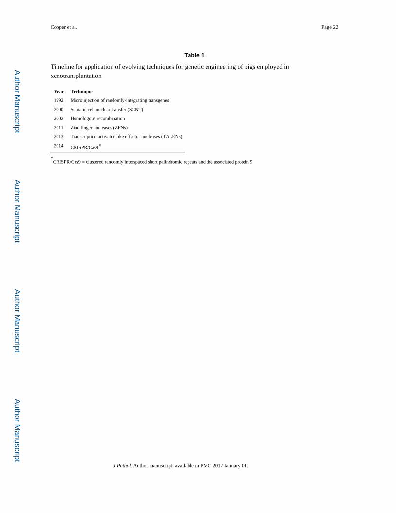
Table 1
Timeline for application of evolving techniques for genetic engineering of pigs employed in
xenotransplantation
Year Technique
1992 Microinjection of randomly-integrating transgenes
2000 Somatic cell nuclear transfer (SCNT)
2002 Homologous recombination
2011 Zinc finger nucleases (ZFNs)
2013 Transcription activator-like effector nucleases (TALENs)
2014 CRISPR/Cas9*
*CRISPR/Cas9 = clustered randomly interspaced short palindromic repeats and the associated protein 9
J Pathol. Author manuscript; available in PMC 2017 January 01.

Author M
anuscriptA
uthor Manuscript
Author M
anuscriptA
uthor Manuscript
Cooper et al. Page 23
Table 2
Selected genetically-modified pigs currently available for xenotransplantation research
Complement regulation by human complement-regulatory gene expression
CD46 (membrane cofactor protein)
CD55 (decay-accelerating factor)
CD59 (protectin or membrane inhibitor of reactive lysis)
Gal or nonGal antigen ‘masking’ or deletion
Human H-transferase gene expression (expression of blood type O antigen)
Endo-beta-galactosidase C (reduction of Gal antigen expression)
α1,3-galactosyltransferase gene-knockout (GTKO)
Cytidine monophosphate-N-acetylneuraminic acid hydroxylase (CMAH) gene-knockout (NeuGcKO)
β4GalNT2 (β1,4 N-acetylgalactosaminyltransferase) gene-knockout (β4GalNT2KO)
Suppression of cellular immune response by gene expression or downregulation
CIITA-DN (MHC class II transactivator knockdown, resulting in swine leukocyte antigen class II knockdown)
Class I MHC-knockout (MHC-IKO)
HLA-E/human β2-microglobulin (inhibits human natural killer cell cytotoxicity)
Human FAS ligand (CD95L)
Human GnT-III (N-acetylglucosaminyltransferase III) gene
Porcine CTLA4-Ig (Cytotoxic T-Lymphocyte Antigen 4 or CD152)
Human TRAIL (tumor necrosis factor-alpha-related apoptosis-inducing ligand)
Anticoagulation and anti-inflammatory gene expression or deletion
von Willebrand factor (vWF)-deficient (natural mutant)
Human tissue factor pathway inhibitor (TFPI)
Human thrombomodulin
Human endothelial protein C receptor (EPCR)
Human CD39 (ectonucleoside triphosphate diphosphohydrolase-1)
Anticoagulation, anti-inflammatory, and anti-apoptotic gene expression
Human A20 (tumor necrosis factor-alpha-induced protein 3)
Human heme oxygenase-1 (HO-1)
Human CD47 (species-specific interaction with SIRP-α inhibits phagocytosis)
Porcine asialoglycoprotein receptor 1 gene-knockout (ASGR1-KO) (decreases platelet phagocytosis)
Human signal regulatory protein α (SIRPα) (decreases platelet phagocytosis by ‘self’ recognition)
Prevention of porcine endogenous retrovirus (PERV) activation
PERV siRNA
J Pathol. Author manuscript; available in PMC 2017 January 01.