danish brain research laboratories meeting aarhus 29...
TRANSCRIPT

14th Annual OAK MeetingDanish Brain Research Laboratories Meeting
Aarhus 29 May 2015
Aarhus UniversityMerete Barker Auditorium

w w w . c f i n . a u . d k / O A K - 2 0 1 5
14th Annual OAK MeetingDanish Brain Research Laboratories Meeting
P R O G R A M
Friday 29 May 2015
10:00 Arrival & registration at Merete Barker Auditorium, Aarhus University10:15-10:30 Welcome by Arne Møller
Session 1: In-vivo Neuroimaging (Chair: Anne M. Landau & Kim Ryun Drasbek)
10:30-10:45 Ali Khalidan Vibholm: Preclinical in-vivo imaging of activated NMDA receptor ion channels with the novel radioligand [18F]-GE179
10:45-11:00 Athanasios Metaxas: PET imaging of the NMDA receptor using [18F]PK209
11:00-11:15 Andreas N. Glud: Parkinson’s disease models of abnormal protein aggregation in the Göttingen minipig CNS
11:15-11:30 Jenny-Ann Phan: Alpha synuclein model of Parkinson’s disease displays early synaptic disruption
11:30-11:45 Janne Vejlby: Endocannabinoid modulation of noradrenaline release
11:45-12:00 Majken Borup Thomsen: Increased receptor density of α2 adrenoceptors and GABAA α5 receptors in limbic brain regions in the domoic acid rat model of epilepsy
12.00-13.00 L U N C H B R E A K
Session 2: Animal models (Chair: Flemming Fryd Johansen)
13:00-13:15 Charlotte Havelund Nykjær: Stereological estimation of the brain white matter in Multiple System Atrophy
13:15-13:30 Jonas Folke: Deregulation of Wnt pathway in the prefrontal cortex from Alzheimer’s disease brains
13:30-13:45 Anders Malmendal: Insights into Alzheimer’s disease from NMR metabolomics of Aβ-expressing Drosophila
13:45-14:00 Simone Krog : Cognitive Assessment of ‘Depressed’ Rats Using Translational Touchscreen Tasks
14:00-14:15 Louise Lind Holm and Tuva Åsatun Solheim : Pharmacologically induced hypothermia in stroke treatment- a search for potential drugs
14:15-14:45 C O F F E E B R E A K

14th Annual OAK MeetingDanish Brain Research Laboratories Meeting
P R O G R A M
Friday 29 May 2015
Session 3: Regulation of neuronal processes (Chair: Kate Lykke Lambertsen)
14:45-15:00 Ditte Gry Ellman: The role of neuronal nuclear factor-kappa B after moderate spinal cord injury
15:00-15:15 Alba Manresa: Transmigration assay as an in vitro model to study the role of the small GTPase RhoA in the migration of the immune cells into the CNS during experimental autoimmune encephalomyelitis (EAE)
15:15-15:30 Reza M. H. Khorooshi: Endogenous Type I interferon in the central nervous system and neuroinflammation
15:30-15:45 Simon Mølgaard Jensen: SorCS2 in the development of inhibitory neuronal circuits
15:45-16:00 Jibrin Danladi: Structural and functional aspects of subiculum and the effect of sorCS2
16:00-16:15 Vibeke Bay: Remote ischemic conditioning: Evaluating the protective effect of miRNAs in a stroke model
16:15 - 16:30 Mette Sloth Larsen: Circulating miRNA signaling underlying the beneficial effects of blood flow restricted exercise
16:45 Hotel Check-in, AUH Patient Hotel, Nørrebrogade18:30 D i n n e r a t H o t e l S c a n d i c A a r h u s C i t y

w w w . c f i n . a u . d k / O A K - 2 0 1 5

14th Annual OAK MeetingDanish Brain Research Laboratories Meeting
A B S T R A C T S
Ali Khalidan Vibholm Preclinical in-vivo imaging of activated NMDA receptor ion channels with the novel radioligand 18F-GE179
Danish Neuroscience Centre, Nuclear Medicine and PET-Centre, Aarhus University and University Hospital
Background: NMDA receptor ion channels (NMDAr-IC) play a key role in excitatory synaptic transmission and synaptic plasticity. Overactive NMDAr-IC may result in neuronal epileptogenesis and seizure. Localising epileptic foci can be difficult and imprecise. Imaging with Positron Emission Tomography (PET), using the novel radioligand 18F-GE179, which selectively binds to NMDAr-IC, may lead to better detection and understanding of altered NMDAr-IC.
Purpose: To validate 18F-GE179 in rat and pig models of brain stimulation as a prelude to human studies.
Objective: Image activated (NMDAr-IC) with 18F-GE179 in rats and pigs during focal electrical stimulation.
Methods: Stereotactic implantation of unilateral tripolar electrode in rats and Deep Brain Stimulator (DBS) implantation in pigs. Seizure is induced in rats by repetitive electrical stimulation. Electrical discharge is induced during acute DBS stimulation in pigs.
Results: Preliminary rat data shows global increase in retention of 18F-GE179 after unilateral stimulation. Scan results in pig show a lateralised focal 18F-GE179 signal induced by DBS suggesting an intensity-dependent retention.
Conclusion: In-vivo PET imaging of activated NMDAr-IC with 18F-GE179 could be beneficial in the study of refractory focal epilepsy and aid the pre-surgical evaluation of patients. Future studies include 18F-GE179 PET in patients with refractory epilepsy in order to detect overactive NMDAr-IC and thereby their epileptic foci.

14th Annual OAK MeetingDanish Brain Research Laboratories Meeting
A B S T R A C T S
Athanasios Metaxas PET imaging of the NMDA receptor using [18F]PK209
University of Southern Denmark, Department of Neurobiology, Odense
Objectives: The N-methyl-D-Aspartate receptor (NMDAr) is an ionotropic glutamate receptor with key roles in physiological and pathological brain processes. The current study employed positron emission tomography (PET) to examine whether the N,N’-diaryl guanidine derivative [18F]PK-209 can visualise NMDArs in the rhesus monkey brain.
Methods: Dynamic PET scans were acquired from three rhesus monkeys over 120 min, at baseline and after the acute administration of dizocilpine (MK-801, 0.3mg/kg; n=3/condition). Continuous and discrete arterial blood samples were manually obtained, to generate metabolite-corrected input functions. Parametric volume-of-distribution (VT) images were obtained by Logan analysis. The selectivity profile of PK-209 was assessed in-vitro, on a broad screen of 79 targets.
Results: PK-209 was at least 50-fold more selective for NMDArs over all other targets examined. At baseline, prolonged retention of radioactivity was observed in NMDAr-rich cortical regions relative to the cerebellum. Pretreatment with MK-801 reduced the VT of [18F]PK-209 compared with baseline in two of three subjects. The rate of radioligand metabolism was high, both at baseline and after MK-801 administration.
Conclusion: PK-209 targets the intrachannel site with high selectivity. Imaging of the NMDAr is feasible with [18F]PK-209, despite its fast metabolism. Further in-vivo evaluation in humans is warranted.
Support: This work was supported by CTMM LeARN, work package 02N-101-01.

14th Annual OAK MeetingDanish Brain Research Laboratories Meeting
A B S T R A C T S
Glud, Andreas Nørgaard 1, 2, Landau, Annie M3, Lillethorup, Thea P3, Landeck, Natalie4, Johnsen, Erik5,
Bjarkam, Carsten R.2, Brooks, David3, Kirik, Deniz5, Sørensen, Jens Christian H.1 Parkinson’s Disease Models of Abnormal Protein Aggregation in the Göttingen Minipig CNS
1 Department of Neurosurgery, Aarhus University Hospital, 2 Department of Neurosurgery, Aalborg University Hospital, 3 Department of Nuclear Medicine and PET Centre, Aarhus University Hospital,4 Department of Experimental Medical Science, Lund University5 Department of Neurology, Aalborg University Hospital,
Background:Parkinson’s disease(PD) animal models are important translational steps toward clinical applications. The Göttingen minipig(GM) has a large gyrencephalic brain (6x5x4cm) that can be examined using conventional scanning modalities. Preclinical neuromodulatory devices can be evaluated in GM models. aSYN is a known pathological protein in PD. Aggregation or overexpression occurs in human PD and has been used to induce PD in other animal models.
We aim to develop and validate novel models of PD in the GM based on abnormal protein aggregation.
Using human intended MRI-guided stereotaxic neurosurgery, we are inducing a PD-syndrome in GM via overexpression of mutated human aSYN using viral vectors or intracerebroventricular administration of an ubiquitin proteasome system inhibitor.
The resulting pathological changes are quantified by neurological tests, PET-imaging of monaminergic function and neuroinflammation, HPLC and post mortem histological evaluation for aSYN and neuroinflammatory markers.Data from ongoing studies with the different models demonstrate gait disturbances, bradykinesia, deficits in monoaminergic neurotransmission, alterations in the aSYN levels in CNS and histological signs of PD including aSYN accumulation.
We predict that these animal models will be beneficial in the understanding of pathological mechanisms of human PD and in the testing of novel therapeutic strategies.
14th Annual OAK MeetingDanish Brain Research Laboratories Meeting
A B S T R A C T S
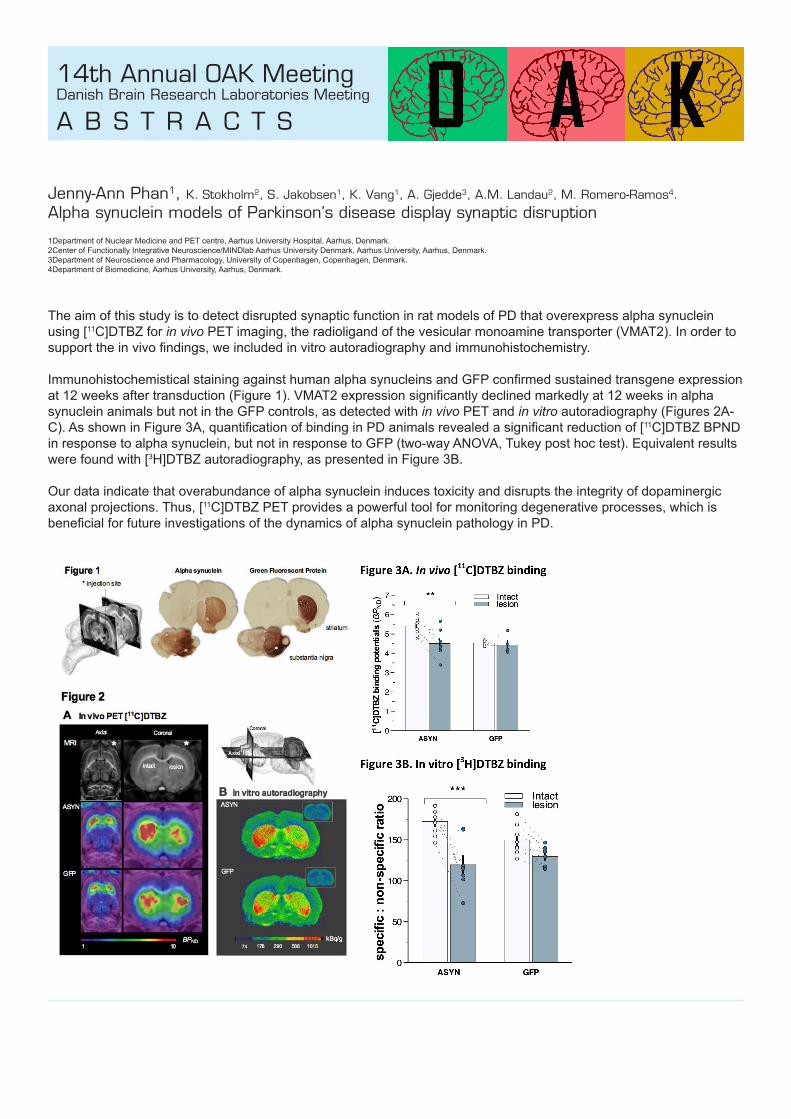
Jenny-Ann Phan1, K. Stokholm2, S. Jakobsen1, K. Vang1, A. Gjedde3, A.M. Landau2, M. Romero-Ramos4.
Alpha synuclein models of Parkinson’s disease display synaptic disruption
1Department of Nuclear Medicine and PET centre, Aarhus University Hospital, Aarhus, Denmark.2Center of Functionally Integrative Neuroscience/MINDlab Aarhus University Denmark, Aarhus University, Aarhus, Denmark.3Department of Neuroscience and Pharmacology, University of Copenhagen, Copenhagen, Denmark.4Department of Biomedicine, Aarhus University, Aarhus, Denmark.
The aim of this study is to detect disrupted synaptic function in rat models of PD that overexpress alpha synuclein using [11C]DTBZ for in vivo PET imaging, the radioligand of the vesicular monoamine transporter (VMAT2). In order to support the in vivo findings, we included in vitro autoradiography and immunohistochemistry.
Immunohistochemistical staining against human alpha synucleins and GFP confirmed sustained transgene expression at 12 weeks after transduction (Figure 1). VMAT2 expression significantly declined markedly at 12 weeks in alpha synuclein animals but not in the GFP controls, as detected with in vivo PET and in vitro autoradiography (Figures 2A-C). As shown in Figure 3A, quantification of binding in PD animals revealed a significant reduction of [11C]DTBZ BPND in response to alpha synuclein, but not in response to GFP (two-way ANOVA, Tukey post hoc test). Equivalent results were found with [3H]DTBZ autoradiography, as presented in Figure 3B.
Our data indicate that overabundance of alpha synuclein induces toxicity and disrupts the integrity of dopaminergic axonal projections. Thus, [11C]DTBZ PET provides a powerful tool for monitoring degenerative processes, which is beneficial for future investigations of the dynamics of alpha synuclein pathology in PD.

14th Annual OAK MeetingDanish Brain Research Laboratories Meeting
A B S T R A C T S
Adjmal Nahimi1, Janne Vejlby1, Anne M. Landau1,3, Steen Jakobsen1, Mette Simonsen1, Albert Gjedde2, and Michael Wintherdahl1
Endocannabinoid modulation of noradrenaline release1. Department of Nuclear Medicine and PET Centre, Aarhus University Hospitals, Denmark 2. Department of Neuroscience and Pharmacology, University of Copenhagen, Denmark 3. Centre for Psychiatric Research, Aarhus University Hospitals, Denmark
Introduction: Inhibition of fatty acid amide hydrolase (FAAH), which increases the concentration of endogenous cannabinoids, has anxiolytic and antidepressant effects in animals. These beneficial effects of FAAH are poorly understood. Here, we test the hypothesis that FAAH-induced increase in CB1 receptor activity results in cortical and subcortical noradrenaline release and displacement of [11C]yohimbine.
Methods: Female Sprague Dawley rats (N=8) rats received 90 minute dynamic positron-emission tomography (PET) in baseline and challenge sessions with [11C]yohimbine, 60 minutes after treatment with URB597 (0.03mg/kg, i.v). PET images were registered to a normalized rat brain atlas (Px rat (W. Schiffer)-T2) using the PMOD image analysis software, and time activity curves were extracted for volumes of interest. We then calculated the volumes of distribution (VT) by means of Logan graphical analysis (Logan et al., 1990). Data are presented as mean (± standard deviations).
Results: Administration of URB597 significantly reduced [11C]yohimbine VT in all VOIs compared to baseline. URB597 challenge reduced [11C]Yohimbine VT from 1.9 (±0.8) to 0.9 (±0.3) in striatum, from 1.5 (±0.5) to 0.7 (±0.2) in cortex, from 1.9 (±0.6) to 0.9 (±0.2) in hippocampus, from 2.0 (±0.6) to 0.9 (±0.3) in thalamus, from 1.9 (±0.7) to 0.8 (±0.3) in n. accumbens, and from 1.5 (±0.5) to 0.7 (±0.2) in amygdala.
Conclusion: URB597 challenge reduced [11C]yohimbine VT in all brain areas examined. The finding is consistent with the hypothesis that antidepressant and anxiolytic effects of FAAH-inhibtion with URB597 are mediated by noradrenaline release in brain.

14th Annual OAK MeetingDanish Brain Research Laboratories Meeting
A B S T R A C T S
Majken B. Thomsen1, Thea P. Lillethorup1, Gregers Wegener2, R. Andrew Tasker2,3, Anne M. Landau1
Increased receptor density of α2 adrenoceptors and GABAA α5 receptors in limbic brain regions in the domoic acid rat model of epilepsy
1. Department of Nuclear Medicine and PET Center, Aarhus University and Hospital, Denmark, 2. Translational Neuropsychiatry Unit, Aarhus University, Denmark, 3. Department of Biomedical Sciences, University of PEI, Canada
Background: The presymptomatic events involved in epilepsy remain elusive but represent a chance to understand disease development and stop the pathogenic processes leading to chronic epilepsy. Previous studies have found increased levels of α2 adrenoceptors and decreased levels of glutamic acid decarboxylase, a catalyst of the decarboxylation of glutamate to GABA.
Methods: Male Sprague-Dawley rats (N=3) were injected (s.c.) daily from postnatal day 8-14 with saline or sub-convulsive doses of the glutamate agonist DOM (20µg/kg). At ~120 days of age the rats were decapitated. The brains were removed, fresh frozen and cut into 20 µM thick slices. Autoradiography was performed using tracers of the α5 subtype of the GABAA receptor ([11C]Ro15-4513) and the α2 adrenoceptors ([3H]RX821002) to determine the binding in limbic brain regions.
Results: The binding of postsynaptic GABA receptors was significantly increased in the dorsal hippocampus and basolateral amygdala of the DOM rats. A trend towards an increase in the density of α2 adrenoceptors was found throughout the limbic system of the DOM rats compared to controls.
Conclusion: Although preliminary, the increase in postsynaptic GABA receptor concentrations in DOM rats may represent a compensatory up-regulation in response to reduced GABAergic input. Noradrenaline reduces neuronal excitability. Elevated receptor expression could be a protective mechanism that attempts to compensate for the lowered seizure threshold caused by DOM.

14th Annual OAK MeetingDanish Brain Research Laboratories Meeting
A B S T R A C T S
Charlotte Havelund Nykjær Stereological estimation of the brain white matter in Multiple System Atrophy
Research Laboratory for Stereology and Neuroscience, Bispebjerg-Frederiksberg Hospital, Copenhagen
Multiple system atrophy (MSA) is a progressive neurodegenerative disorder with adult onset and a life expectancy of 6 years after debut. The hallmark lesion of MSA is glial cytoplasmic inclusions (GCI), primarily located in the oligodendrocytes. Along with GCI, demyelination, gliosis and degeneration of oligodendrocytes have been reported in MSA. Despite the severity and frequency of pathological changes in the white matter, only few MSA studies have focused on this particular region. In order to establish the extent of involvement of the white matter in MSA, we have used stereology to quantify the total number of neurons and glial cells in brains from 10 patients with MSA and 11 control subjects. We found a significant loss of neurons and an increased number of microglia in MSA brains compared to controls. No significant difference of astrocytes was found between the two groups. Surprisingly, we did not find a loss of oligodendrocytes in MSA brains, which indicates that loss of oligodendrocytes is not a widespread and pronounced neuropathological feature of MSA. This is in contrast to previous literature. However, other studies have demonstrated potential repair mechanisms in GCI-bearing oligodendroglia. In sum, this suggests that the aggregation of GCIs may not directly cause oligodendrocyte loss but only cellular dysfunction. This could be important for the understanding of the pathogenesis behind the neurodegenerative processes that cause the death of the MSA patients.

14th Annual OAK MeetingDanish Brain Research Laboratories Meeting
A B S T R A C T S
Jonas Folke Deregulation of Wnt pathway in the prefrontal cortex from Alzheimer’s disease brains
Research Laboratory for Stereology and Neuroscience, Bispebjerg Hospital
Alzheimer’s disease (AD) is a neurodegenerative disease with progressive dementia accompanied by three main structural changes in the brain: diffuse loss of neurons: intracellular protein deposits termed neurofibrillary tangles (NFT) and extracellular protein deposits termed amyloid plaques, surrounded by dystrophic neurites.
Wnt genes encode a family of secreted glycoproteins that play a vital role as intracellular signaling molecules both during neural development and in the mature nervous system. A role of Wnt proteins have been established in a number of diverse processes including embryonic patterning, regulation of cell proliferation, polarity and fate determination. Furthermore, there are also indications suggesting that Wnt signaling may play a role in synaptic modulation and plasticity. The canonical Wnt pathway negatively regulates glycogen synthase kinase-3 beta (GSK3β) activity. Previous work has established that hyperactive GSK3β is, linked to both “sporadic” and “genetic” forms of AD, suggesting a crucial role of GSK3β in AD pathogenesis. Deregulation of Wnt signaling, has been seen in hippocampus from AD patients. We hypothesize that loss of function of Wnt signaling components may lead to the onset and to the development of the major molecular hallmarks of AD. The aim of this project is characterize the Wnt signaling pathway at the transcriptional and translational levels in prefrontal cortex of Alzheimer’s disease brains in comparison to Non-demented controls.

14th Annual OAK MeetingDanish Brain Research Laboratories Meeting
A B S T R A C T S
Anders Malmendal Insights into Alzheimer’s disease from NMR metabolomics of Aβ-expressing Drosophila
Department of Biomedical Science, University of Copenhagen
The ability of our cells and tissues to retain proteins in their proper native conformation is impaired with age. The consequences of the proteostatic collapse in old age include a number of very common neurodegenerative disorders, the most prevalent of which is Alzheimer’s disease (AD), which is characterised microscopically by the accumulation of two distinct protein amyloid deposits, extracellular neuritic plaques of the amyloid β peptide (Aβ), and the intracellular tangles composed of the tau protein.
The use of a Drosophila Alzheimer’s disease (AD) model with its short life cycle and low maintenance costs allowed us to study the metabolite responses across all ages and to different Aβ variants, all while avoiding vertebrate models.Both variants that affect performance and aging–mimicking AD toxicity, and those that affects neither, show distinct effects at the metabolite level, and the toxic effects were detected several days before symptoms were detected.
The results provide insight into the organism’s protective response and to the toxic effects including several markers of oxidative stress.
14th Annual OAK MeetingDanish Brain Research Laboratories Meeting
A B S T R A C T S
Simone Krog Cognitive Assessments of ‘Depressed’ Rats Using Translational Touchscreen Tasks
Translational Neuropsychiatry Unit, Department of Clinical Medicine, Aarhus University
Major depressive disorder is a leading cause for disability worldwide. Well established and validated animal models are crucial for development and verification of antidepressant treatment strategies.
Objectives: To develop and optimize an animal model of depression to investigate treatment strategies and cognitive functions. We investigated: 1) the eligibility of Long Evans (LE) rats in the chronic mild stress model (CMS) of depression, 2) if CMS impairs cognitive performance in LE rats, and 3) if Wistar or LE rat strains are more suitable for cognitive assessment using a touchscreen operant platform.
Methods: We exposed LE rats to CMS and used the sucrose consumption test (SCT) as readout of depressive-like state. Social interaction, y-maze, elevated plus-maze and open field were used for behavioral phenotyping. CMS LE rats, LE controls and Wistar controls were tested in the touchscreen. Pairwise discrimination, reversal task and retention were used to assess visual discrimination, stimulus-reward association learning and memory consolidation.Results: Preliminary data revealed a significant difference of cognitive performance in the touchscreen tasks between LE and Wistar control rats. In the CMS paradigm, LE rats proved to be equally susceptible to stress exposure as Wistar rats shown with the SCT and behavioral tests.
Conclusion: LE rats are preferred over Wistar rats when using touchscreen based cognitive tests. LE rats were susceptible to the CMS paradigme. We will use LE rats in upcoming studies on cognitive function in depression.

14th Annual OAK MeetingDanish Brain Research Laboratories Meeting
A B S T R A C T S
Louise Lind Holm and Tuva Åsatun Solheim Pharmacologically induced hypothermia in stroke treatment - a search for potential drugs
University of Copenhagen
Therapeutic hypothermia has been shown to have neuroprotectiv effects and is a promising treatment for patients with ischemic stroke. An overweight of clinical trials researching this area utilise forced hypothermia. These methods have disadvantages, leading to a growing interest in the possibilities of pharmacologically induced hypothermia. The focus of this project is to investigate potential drugs for their hypothermic qualities and their relevant contraindications and adverse effects.
The data was summarized in a scheme, and the drugs were graded. Five drugs were chosen as the ones showing most potential.
Ipsapirone has been shown to induce hypothermia in humans, but with approval only for clinical trials, knowledge about adverse effects and contra indications is lacking. The same problems apply for 2-deoxuglucose -a fascinating drug, with central and peripheral hypothermic effects, and a wide therapeutic index, though studies show a possible effect on blood pressure.
Ibuprofen is a well-known drug with few adverse effects, but has only showed hypothermic effects in extreme doses and children.
Though animal research on cannabinoid-induced hypothermia is promising, the question remains if the results can be reconstructed in humans. The complex mechanisms of action make the specificity of the effects debateable.
Causing minor temperature reduction, paracetamol may not be enough to give the desired neuroprotective effect. A combination of paracetamol and a different hypothermia-inducing drug has been proposed.

14th Annual OAK MeetingDanish Brain Research Laboratories Meeting
A B S T R A C T S
Ditte Gry Ellman1, Hans Gram Novrup1, Valerie Bracchi-Ricard2,3, John Bethea2,3, Karin Lykke-Hartmann4, Roberta Brambilla3 , Kate Lykke Lambertsen1
The role of neuronal nuclear factor-kappa B and tumor necrosis factor after moderate spinal cord injury
1. Neurobiology Research, Institute of Molecular Medicine, University of Southern Denmark, Odense, Denmark, 2. Department of Biology, Drexel University, Philadelphia, PA, USA, and 3. The Miami Project to Cure Paralysis, University of Miami Miller School of Medicine, Miami, FL, USA 4. Aarhus Institute of Advanced Studies and Department of Biomedicine, Aarhus University, Denmark.
The purpose of the present project is to study the significance of nuclear factor-kappa B (NF-κB) signalling in neurons on the secondary phase of tissue loss observed after moderate spinal cord injury (SCI). Our hypothesis is that inhibition of neuronal NF-κB signalling will result in a reduced inflammatory response leading to decreased secondary damage and increased functional recovery after SCI. The hypothesis will be tested using Syn1CreIKK2fl/fl mice and their respective littermates (IKK2fl/fl).
Mice expressing reduced levels of NF-κB in neurons display a normal appearance and behavioral phenotype. Evaluation of the g-ratio (fiberdiameter/axon diameter) of axons in the thoracic spinal cord using electron microscopy revealed a significant difference in the g-ratio in fibers with a diameter of 1.5-2.0 µm. Long term survival studies have revealed a significant better functional recovery 3 and 7 days after SCI, when using Basso Mouse Scale, in Syn1CreIKK2fl/fl mice compared to IKK2fl/fl mice, a finding which was not present at later time points (14 days and onwards).
These findings suggest that neuronal NF-κB plays a role in the acute phase after SCI, but not at later stages. Further analyses, using Q-PCR analysis and western blotting, will hopefully reveal possible mechanisms for this difference early after SCI.

14th Annual OAK MeetingDanish Brain Research Laboratories Meeting
A B S T R A C T S
Alba Manresa Transmigration assay as an in-vitro model to study the role of the small GTPase RhoA in the migration of the immune cells into the CNS during experimental autoimmune encephalomyelitis (EAE)
Biomedical Institute, Dept. of Molecular Pathology, BRIC
Aim: T-cell migration into the CNS via diapedesis is one of the key steps in the development of multiple sclerosis (MS). Furthermore, it is a step required for the induction of experimental autoimmune encephalomyelitis (EAE) and studies have shown that the small GTPase RhoA is involved in leukocyte adhesion and migration, implying that RhoA or downstream effectors may be suitable drug targets in MS (Rougerie & Delon, 2012). We aim to investigate whether the lack of RhoA in T-cells has an impact in their ability to migrate through the blood-brain barrier (BBB) into the CNS.
Materials and methods: Endothelial cells (ECs) bEnd.3 were cultured in monolayer in gelatin-coated transwell inserts. The ECs were stimulated with IL-1β prior the start of the migration assay. In parallel, the lymphocytes EL-4 were stimulated with PMA/Ionomycin. The activated lymphocytes are added to the upper chamber of the well and media with the chemokine CCL-19 is added to the lower chamber. After 4h of incubation, the cells that migrated to the through the endothelial monolayer to the lower chamber are quantified manually.
Preliminary results: Our primary data suggest that the activation of the endothelial cells is necessary for the migration of the lymphocytes through the endothelial monolayer. The presence of the chemokine CCL-19 enhances the migration of the lymphocytes. In its absence lymphocytes are still able to migrate through an activated endothelial monolayer, although to a lesser extent.
14th Annual OAK MeetingDanish Brain Research Laboratories Meeting
A B S T R A C T S
Reza Khorooshi, Marlene Mørch, Thomas Holm, Carsten Tue Berg, Ruthe Truong Dieu, Dina Dræby, Shohreh Issazadeh-Navikas, Siegfried Weiss, Stefan Lienenklaus and Trevor Owens
Endogenous Type I interferon in the central nervous system and neuroinflammation
Department of Neurobiology Research, Institute of Molecular Medicine, University of Southern Denmark, Odense
Type I interferon (IFN-α/β) acts through a common receptor (IFNAR) and have anti-inflammatory effects. IFN-β is used to treat multiple sclerosis (MS) and is effective against experimental autoimmune encephalomyelitis (EAE), an animal model for MS. The therapeutic benefit of Type I IFN produced in the CNS remains to be established. The aim of this study was to examine whether experimentally induced CNS-endogenous Type I IFN influences EAE. Using IFN-β reporter mice, we showed that administration of polyinosinic–polycytidylic acid (poly I:C), a potent inducer of IFN-β, into the cerebrospinal fluid induced increased leukocyte numbers and transient upregulation of IFN-β in CD45/CD11b-positive cells located in the meninges and choroid plexus, as well as enhanced IFN-β expression by parenchymal microglial cells. Intrathecal injection of poly I:C to mice showing first symptoms of EAE substantially increased the normal disease-associated expression of IFN-α/β and IL-10 in CNS, and disease worsening was prevented for as long as type I IFN was expressed. In contrast, there was no therapeutic effect on EAE in poly I:C-treated IFNAR1-deficient mice. These results show that Type I IFN induced within the CNS can play a protective role in EAE and highlight the role of endogenous type I IFN in mediating neuroprotection.

14th Annual OAK MeetingDanish Brain Research Laboratories Meeting
A B S T R A C T S
Simon Mølgaard Jensen1,2, Simon Bøggild1,2, Maj Ulrichsen1, Abdelrahman Alabsi2, Anders Nykjaer1,2, Simon Glerup1, Jens R Nyengaard1,2
SorCS2 is essential for postnatal maturation of the hippocampal GABAergic system
1. The Lundbeck Foundation Research Center MIND, Department of Biomedicine, Aarhus University, 8000 C Aarhus, Denmark2. Stereology and Electron Microscopy Laboratory, Centre for Stochastic Geometry and Advanced Bioimaging, Department of Clinical Medicine, Aarhus University, 8000 C Aarhus, Denmark
Balancing neurotrophic cues is critical for the proper formation of GABAergic interneuron circuits in the hippocampus. During development, Brain-derived neurotrophic factor (BDNF) plays an important role for the formation of GABAergic interneuron circuits in the hippocampus. Here, we identify SorCS2 as an important mediator of BDNF responses in GABAergic interneurons during postnatal development. Expression of SorCS2 is absent in the embryonic brain but peaks around postnatal day 10, the only time point where GABAergic interneurons also express SorCS2. BDNF is important during postnatal development for the formation of the hippocampus, including neurite branching and synapse formation. SorCS2 deficiency in mice caused reduced neurite branching and synapse formation in cultured hippocampal interneurons. Furthermore, hippocampal tissue lysates showed a marked reduction in expression levels of the GABAA receptor subunit α2 in mice deficient of SorCS2. Accordingly, adult SorCS2-/- mice showed a reduced number of inhibitory synapses and an increased susceptibility to pentylenetetrazole (PTZ) induced seizures as well as reduced amplitude of gamma oscillations in hippocampal CA3 and CA1. In conclusion, we have demonstrated that SorCS2 is important during BDNF stimulation and this is critical for the differentiation of the hippocampal GABAergic system.
14th Annual OAK MeetingDanish Brain Research Laboratories Meeting
A B S T R A C T S
Jibrin Danladi1,2, Simon Mølgaard Jensen1,2, Anders Nykjaer1,2, Simon Glerup1, Jens R Nyengaard1,2
Structural and functional aspects of subiculum and the effect of sorCS2
1. The Lundbeck Foundation Research Center MIND, Department of Biomedicine, Aarhus University, 8000 C Aarhus, Denmark2. Stereology and Electron Microscopy Laboratory, Centre for Stochastic Geometry and Advanced Bioimaging, Department of Clinical Medicine, Aarhus University, 8000 C Aarhus, Denmark
Structural and functional understanding of subiculum is lacking. Morphological and neurophysiological diversity of differing neuronal types within the subiculum is a largely unexplored, as are the mechanisms of feed-forward, feedback and lateral inhibition of intrinsic and extrinsic subicular projections. Therefore, a more holistic research approach is needed to understand subiculum and its relation to neurodegenerative disorders. The Vps10p-domain (Vps10p-D) family of receptors comprises sortilin, sorLA, and sorCS1-3. SorCS2 (Sortilin-related VPS10-domain containing receptor family) is found to be highly expressed throughout the developing and mature brain. SorCS2 belongs to the category of leucine-rich repeat transmembrane proteins, members of which many are involved in axon guidance, including Slit, Slitrk, the Nogo receptors, and TrkA, -B, and -C. In these proteins, the leucine-rich repeat domains create a free moving structural context for protein-protein interactions. The biological mechanism via which sorCS2 regulates neuronal growth and survival remains unclear although, it has been reported that sorCS2 regulates the wiring of dopaminergic neurons which is processed into apoptotic two chain receptors in peripheral glia. SorCS2 is also proposed to be genetically linked to the development of schizophrenia. This project will aimed at; (1) Using light microscopy, confocal microscopy, electron microscopy, immunohistochemistry and stereology, mouse subiculum is defined and characterized structurally and the effect of lack of sorCS2 examined. (2) The use of anterograde and retrograde tracers with tissue clearing technique will display synaptic connections inside and outside of subiculum (especially hippocampus and ventral tegmentum area as well as other brain regions) and the effect of lack of sorCS2 will be investigated. (3) Using optogenetics, it will be attempted to turn off or on the Pyramidal cells within subiculum by injecting NpHR genes using a recombinant adeno-associated virus and afterwards the function of subiculum is studied by testing the behaviour of the animals. We hypothesized that, Subicular layers volume, number of pyramidal neurons, spindle-shaped and molecular cells, pyramidal dendrites (length, branches, filament length and area mean dendritic branch length) and spine density and number will be different in wt mice compared to sorCS2 KO mice. There will be a difference in synaptic contacts coming into subiculum and out from subiculum between wt and sorCS2 KO mice. The pyramidal cells within subiculum will be directly or indirectly involved in memory and learning, therefore we expect that mice without functioning pyramidal neurons will perform worse in memory and learning related behavioural tests.

14th Annual OAK MeetingDanish Brain Research Laboratories Meeting
A B S T R A C T S
Vibeke Bay, Brian Hansen, Jørgen Kjems, Leif Østergaard & Kim Ryun Drasbek Remote ischemic conditioning - evaluating the protective effect of miRNAs in a stroke model
CFIN, Aarhus University
Stroke is one of the leading causes of death and disability, thereby posing a huge financial burden to society, signifying the need for better and preferably cheaper treatment. A treatment procedure named conditioning has shown promising results by reducing infarct sizes and improving outcome in both animal and clinical studies. Specifically, the term conditioning refers to the induction of short periods of controlled undamaging hypoxia that protect tissue against prolonged ischemia.
The underlying protective mechanism remains unknown, but it is shown to be transferable via blood within and among species. MicroRNAs (miRNAs), small nucleotide sequences targeting mRNA, are secreted from cells in a controlled manner, and can thereby function as mediators in distant cell-to-cell communication. Thus, miRNAs are proposed to be the blood borne effectors of the protection produced by conditioning.
Comparing miRNA expression levels, analyzed by next-generation sequencing (NGS), in human plasma before and after conditioning revealed 14 miRNAs with significantly changed expression levels of which some have functional associations to hypoxia and ischemia. Using MRI, the direct effect of these miRNAs on infarct size will be evaluated in a MCAO rat model followed by 48 hours of reperfusion and brain fixation.
In all, this will lead to new insights in miRNA function and potentially new ischemic treatment, where specific miRNAs can be synthesized, packaged and delivered by injection.

14th Annual OAK MeetingDanish Brain Research Laboratories Meeting
A B S T R A C T S
Mette Sloth Larsen, Vibeke Bay, Kristian Vissing, Jørgen Kjems, Kim Ryun Drasbek Circulating miRNA signaling underlying the beneficial effects of blood flow restricted exercise
CFIN
MircoRNAs (miRNAs) are small non-coding nucleotide sequences that act as posttranscriptional regulators by targeting messenger RNA. miRNAs play a large role in gene regulation and thereby cell function as an estimated one third of all human genes are targeted by miRNAs. In the blood, miRNAs are found in circulating exosomes, which are small, secreted membrane vesicles from endogen origin that can play a role in distant cell-cell communication. As miRNA expression changes during disease, miRNAs have a great potential as diagnostic biomarkers.
Remote ischemic conditioning (RIC) refers to short periods of undamaging ischemia that protect the tissue e.g. brain or heart against prolonged ischemia as seen in both animal studies and human studies. Similar protective effects have been seen after high intensity training. An interesting training form that is evaluated in rehabilitation name occlusion training or blood flow restricted (BFR) training could evoke the same molecular signals as RIC.
During my master thesis I will analyse miRNAs from isolated plasma exosomes obtained from a human BFR training study performed at Institute for Sport Science. Next generation sequencing will be used to identify differences in both known and novel miRNAs due to BFR. Identification of RIC associated miRNAs suggests that the molecular mechanism underlying BFR and RIC is shared to some extends. In the future miRNA expression profiles might be used as a fingerprint for hypoxia and as a diagnostic tool.

w w w . c f i n . a u . d k / O A K - 2 0 1 5
14th Annual OAK MeetingDanish Brain Research Laboratories Meeting
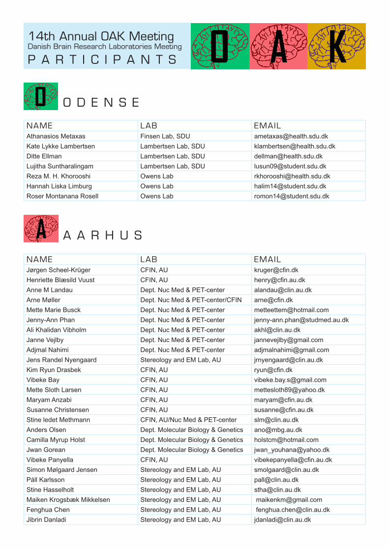
P A R T I C I P A N T S
O D E N S E
A A R H U S
NAME LAB EMAILAthanasios Metaxas Finsen Lab, SDU [email protected] Lykke Lambertsen Lambertsen Lab, SDU [email protected] Ellman Lambertsen Lab, SDU [email protected] Suntharalingam Lambertsen Lab, SDU [email protected] M. H. Khorooshi Owens Lab [email protected] Liska Limburg Owens Lab [email protected] Montanana Rosell Owens Lab [email protected]
NAME LAB EMAILJørgen Scheel-Krüger CFIN, AU [email protected] Blæsild Vuust CFIN, AU [email protected] M Landau Dept. Nuc Med & PET-center [email protected] Møller Dept. Nuc Med & PET-center/CFIN [email protected] Mette Marie Busck Dept. Nuc Med & PET-center [email protected] Jenny-Ann Phan Dept. Nuc Med & PET-center [email protected] Ali Khalidan Vibholm Dept. Nuc Med & PET-center [email protected] Janne Vejlby Dept. Nuc Med & PET-center [email protected] Nahimi Dept. Nuc Med & PET-center [email protected] Randel Nyengaard Stereology and EM Lab, AU [email protected] Ryun Drasbek CFIN, AU [email protected] Bay CFIN, AU [email protected] Sloth Larsen CFIN, AU [email protected] Anzabi CFIN, AU [email protected] Christensen CFIN, AU [email protected] ledet Methmann CFIN, AU/Nuc Med & PET-center [email protected] Olsen Dept. Molecular Biology & Genetics [email protected] Myrup Holst Dept. Molecular Biology & Genetics [email protected] Gorean Dept. Molecular Biology & Genetics [email protected] Panyella CFIN, AU [email protected] Mølgaard Jensen Stereology and EM Lab, AU [email protected]áll Karlsson Stereology and EM Lab, AU [email protected] Hasselholt Stereology and EM Lab, AU [email protected] Krogsbæk Mikkelsen Stereology and EM Lab, AU [email protected] Chen Stereology and EM Lab, AU [email protected] Danladi Stereology and EM Lab, AU [email protected]

14th Annual OAK MeetingDanish Brain Research Laboratories Meeting
P A R T I C I P A N T S
A A R H U S
NAME LAB EMAILAbd EL-Rahman AL-ABSI Stereology and EM Lab, AU [email protected]şe Ikinci Stereology and EM Lab, AU [email protected] H. Rafati Stereology and EM Lab, AU [email protected] Ardalan Stereology and EM Lab, AU [email protected] Borup Thomsen Dept. Nuc Med & PET-center [email protected] Pinholt Lillethorup Dept. Nuc Med & PET-center [email protected] Nørgaard Glud PET/CENSE [email protected] Wiborg TNU / AU [email protected] Krog TNU / AU [email protected] Karina Christensen TNU / AU [email protected]
C O P E N H A G E N
NAME LAB EMAILAlbert Gjedde Neuroscience & Pharmacology, KU [email protected] Fryd BRIC & Biomedical Institute, KU [email protected] Bente Pakkenberg Stereology & Neuroscience, BBH [email protected] Charlotte Havelund Nykjær Pakkenberg - N-Lab [email protected]
Tomasz Brudek Pakkenberg - N-Lab [email protected] Lind Alemu Pakkenberg - N-Lab [email protected] Folke Pakkenberg - N-Lab [email protected] Rydbirk Pakkenberg - N-Lab [email protected] Fomsgaard Pakkenberg - N-Lab [email protected] El-Sayed Pakkenberg - N-Lab [email protected] Malmendal Biomedical Inst, KU [email protected] Manresa BRIC & Biomedical Institute, KU [email protected] Lind Holm BRIC & Biomedical Institute, KU [email protected] Åsatun Solheim BRIC & Biomedical Institute, KU [email protected]
Huilin Hong IGDB, Beijing [email protected] Yin IBP, Beijing [email protected] Tan IPCAS, Beijing [email protected] Wang IBP, Beijing [email protected] Sun IBP, Beijing [email protected] Zhong IBP, Beijing [email protected] Liu IPCAS, Beijing [email protected]
S i n o D a n i s h C e n t e r s t u d e n t s

14th Annual OAK MeetingDanish Brain Research Laboratories Meeting
D I N N E R
Friday 29 May 2015 at 19:30 the OAK Meeting speakers and senior/organizing participants will have a dinner.
The dinner will take place at the Hotel Scandic Aarhus City in the centre of Aarhus.
Hotel Scandic Aarhus City is located at:Østergade 108000 Aarhus C.

14th Annual OAK MeetingDanish Brain Research Laboratories Meeting
S P O N S O R S
Sponsor:BioNordika Denmark A/SMarielundvej 48 1.DK-2730 Herlev Tel: +45 3956 2000 Email: [email protected] Web: https://www.bionordika.dk/
Sponsor:Hotel Scandic Aarhus CityØstergade 10 8000 Aarhus C.
Tel: +45 89 31 81 00Email: [email protected]: http://www.scandichotels.dk/
Sponsor:Center of Functionally Integrative Neuroscience (CFIN) Aarhus Sygehus, Nørrebrogade 44, 10G 8000 Aarhus C.
Tel: +45 7846 4398Fax: +45 7846 4400Web: http://www.cfin.au.dk/
Dept. of Nuclear Medicine & PET-Center Aarhus University Hospital, Nørrebrogade 44, 10G8000 Aarhus C.
Tel: +45 7846 3019Fax: +45 7846 3020Web: http://www.en.auh.dk/

w w w . c f i n . a u . d k / O A K - 2 0 1 5