danio rerio embryo as a model for study abortifacient effects using ananas comosuss
TRANSCRIPT

www.wjpps.com Vol 3, Issue 7, 2014.
1463
Ramakrishna et al. World Journal of Pharmacy and Pharmaceutical Sciences
DANIO RERIO EMBRYO AS A MODEL FOR STUDY
ABORTIFACIENT EFFECTS USING ANANAS COMOSUS
Abhinava J V1, Srinivas Raju2, Guruprasad R*
Durga Femto Technologies and Research, #22,4th main, 4th cross, Chamarajpet, Bangalore-
560018, India.
ABSTRACT
Danio rerio is one of the best model organisms to study various kinds
of disorders. This study was conducted by treating Danio rerio
embryos with a well-known plant having abortifacient activity like
Ananas comosus. The ethanol and chloroform extract were subjected to
the embryos at different post fertilization periods and this includes
1cell stage, blastula, gastrula and organogenesis. The effects of the
different concentrations were studied at different time intervals. From
the statistical analysis for time with concentration, the blastula stage
embryos show the most significant effect P (F≥0.8753) = 0.05. As
embryos develop, they get resistance against both extracts and the
survival rate increases. Prolonged exposure to the embryos shows
death in all stages. This experiment demonstrates that Danio rerio is a suitable model
organism for the study of abortifacient activity.
Key Words: abortifacient, Danio rerio, Ananas comosus, model organism, embryogenesis.
INTRODUCTION
Increasing population is one of the critical things in developing countries like India. India is
one of the nations which adopted the family planning program during the year 1950. Today
controlling population becomes necessary, and for this the taking up of abortifacient
compound is common1. Thus the study of the abortifacient compounds from different sources
is very important; at present the screening of abortifacient activity is done by using different
model organisms.
Article Received on 07 May 2014, Revised on 27 May 2014, Accepted on 23 June 2014
*Correspondence for Author
Dr. Guruprasad
Ramakrishna
DurgaFemto Technologies and
Research, #22,4th main, 4th
cross, Chamarajpet, Bangalore,
India.
WWOORRLLDD JJOOUURRNNAALL OOFF PPHHAARRMMAACCYY AANNDD PPHHAARRMMAACCEEUUTTIICCAALL SSCCIIEENNCCEESS SSJJIIFF IImmppaacctt FFaaccttoorr 22..778866
VVoolluummee 33,, IIssssuuee 77,, 11446633--11447722.. RReesseeaarrcchh Article IISSSSNN 2278 – 4357

www.wjpps.com Vol 3, Issue 7, 2014.
1464
Ramakrishna et al. World Journal of Pharmacy and Pharmaceutical Sciences
The benzene extract of Achyranthesasperawas used to determine the abortifacient effect in
rabbits2. The unripe fruits of Carica papaya were used to determine the abortifacient activity
in rats3. In Guinea pigs the alkaloid called as vasicine shows the abortifacient effects4.
Ananas comosus is one of the known plants having abortifacien effect in India5. The steroids,
which are present in the Ananas comosus extract, show significant abortifacient in mice6, 7.
Throughout the study abortifacient effects of different plants were carried out on different
model organisms (mice, rat, rabbit, guineapigs, etc…), the maintenance of the organisms is
difficult when compared to aquatic organisms like zebrafish (Daniorerio).
Danio rerio is one of the best model organisms in current years8, 9. It is already proven that
80% of the Danio rerio genome matches the human genome10. Developments of all chordate
embryos are similar to human embryogenesis. The complete development of the embryo
from the single cell stage to larval stage requires up to 72hrs after post fertilization period.
The Danio rerio embryos are all transparent11.Due to the transparency of the embryos the
changes in the development of embryos can easily be studied and it not possible in higher
organism12, 13. The death of the embryos caused by the abortifacient compounds shows the
abortifacient activity of that compound. Thus adopting Danio rerio embryo for studying
abortifacient activity gives a faster and viable result than other organisms like tadpoles, mice,
rabbits, monkeys, etc…
MATERIALS AND METHODS
Preparation of extracts
The soxhelet apparatus was used to prepare Chloroform and Ethanol extracts of plant
material. The different concentrations (10, 20, 30, 40 and 50 mg/ml) were prepared using
double distilled water. The crude extract was prepared by homogenizing the plant materials.
Collection of embryos
The zebrafish (wild type) were maintained at standard lab condition. The embryos were
collected using natural spawning and they were incubated at 28.50c11.
Embryonic study
The crude Ananas comosus extract was added to petri-plates containing fertilized Danio rerio
embryo and observed at intervals of 1hr.

www.wjpps.com Vol 3, Issue 7, 2014.
1465
Ramakrishna et al. World Journal of Pharmacy and Pharmaceutical Sciences
The embryos with four different stages includes: 1cell stage (0-1hr post fertilization),
Blastula stage (2.25-5.25hr post fertilization), Gastrula stage (5.25-10hr post fertilization) and
Organogenesis stage (21-48hr post fertilization) were taken along with the different
concentrations of 0, 10, 20, 30, 40 & 50mg/ml of plant extract with distilled water and
incubated at 28.50c. Five embryos were taken in every plate for analysis. The readings were
noted every hour and the results were tabulated.
Statistical analysis
The death of the embryos against the concentration and time were taken for the analysis of
variance (ANOVA) and it was presented as mean ± SEM in different stages. From the data,
one way ANOVA analysis was done and F-value and P- values were calculated and analyzed
with the standard values. The comparative analysis of all stages with ethanol and chloroform
extract was presented as mean ± SEM.
RESULTS AND DISCUSSION
Embryonic study
Analysis of crude extract
The addition of crude extract shows the drastic change in the Danio rerio embryo
development and finally these extracts shows the death of the embryos within 1hr. The
microscopic image of the embryos with plant extracts along with the control was shown in
the figure 1.
Fig. 1: microscopic image of embryos with crude extract, showing the lyses of the
embryo (Right) and control having 5hr PF (Left).
Analysis of plant extract
The effect of Ananas comosus extracts was analyzed at four distinct stages and these include:
www.wjpps.com Vol 3, Issue 7, 2014.
1466
Ramakrishna et al. World Journal of Pharmacy and Pharmaceutical Sciences
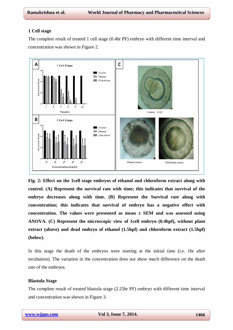
1 Cell stage
The complete result of treated 1 cell stage (0.4hr PF) embryo with different time interval and
concentration was shown in Figure 2.
Fig. 2: Effect on the 1cell stage embryos of ethanol and chloroform extract along with
control. (A) Represent the survival rate with time; this indicates that survival of the
embryo decreases along with time. (B) Represent the Survival rate along with
concentration; this indicates that survival of embryo has a negative effect with
concentration. The values were presented as mean ± SEM and was assessed using
ANOVA. (C) Represent the microscopic view of 1cell embryo (0.4hpf), without plant
extract (above) and dead embryo of ethanol (1.5hpf) and chloroform extract (1.5hpf)
(below).
In this stage the death of the embryos were starting at the initial time (i.e. 1hr after
incubation). The variation in the concentration does not show much difference on the death
rate of the embryos.
Blastula Stage
The complete result of treated blastula stage (2.25hr PF) embryo with different time interval
and concentration was shown in Figure 3.

www.wjpps.com Vol 3, Issue 7, 2014.
1467
Ramakrishna et al. World Journal of Pharmacy and Pharmaceutical Sciences
Fig. 3: Effect on the blastula stage embryos of ethanol and chloroform extract along
with control. (A) Represent the Survival rate with time; this indicates that survival of
the embryo decreases along with time. (B) Represent the Survival rate along with
Concentration; this indicates that survival of embryo has a negative effect with
Concentration. The values were presented as mean ± SEM and were assessed using
ANOVA. (C) Represent the image of early blastula (2.25hpf) and late blastula (3.25hpf)
stage embryo without plant extract (above) and dead embryo of ethanol (4.5hpf) and
chloroform extract (3.5hpf) (below).
At this stage only the chloroform extract starts death at first hour and ethanol extract starts
death after second hour of plant extract addition. The concentration does not vary the embryo
death rate.
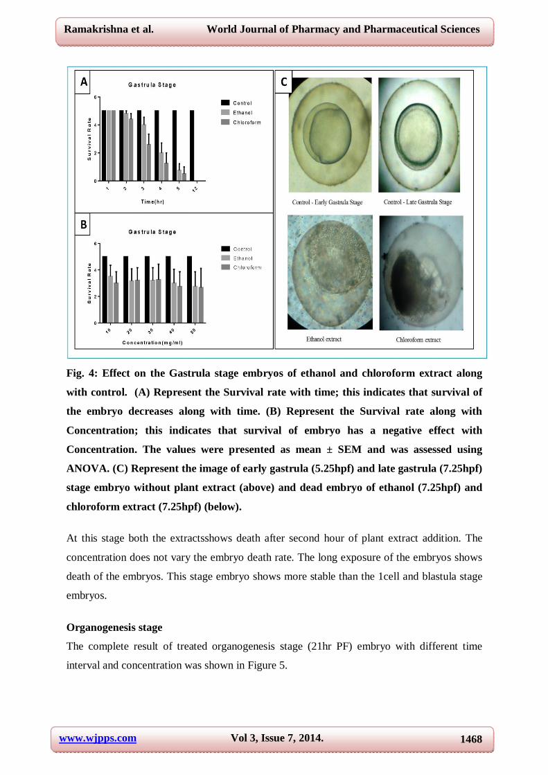
Gastrula stage
The complete result of treated gastrula stage (5.25hr PF) embryo with different time interval
and concentration was shown in Figure 4.
www.wjpps.com Vol 3, Issue 7, 2014.
1468
Ramakrishna et al. World Journal of Pharmacy and Pharmaceutical Sciences
Fig. 4: Effect on the Gastrula stage embryos of ethanol and chloroform extract along
with control. (A) Represent the Survival rate with time; this indicates that survival of
the embryo decreases along with time. (B) Represent the Survival rate along with
Concentration; this indicates that survival of embryo has a negative effect with
Concentration. The values were presented as mean ± SEM and was assessed using
ANOVA. (C) Represent the image of early gastrula (5.25hpf) and late gastrula (7.25hpf)
stage embryo without plant extract (above) and dead embryo of ethanol (7.25hpf) and
chloroform extract (7.25hpf) (below).
At this stage both the extractsshows death after second hour of plant extract addition. The
concentration does not vary the embryo death rate. The long exposure of the embryos shows
death of the embryos. This stage embryo shows more stable than the 1cell and blastula stage
embryos.
Organogenesis stage
The complete result of treated organogenesis stage (21hr PF) embryo with different time
interval and concentration was shown in Figure 5.

www.wjpps.com Vol 3, Issue 7, 2014.
1469
Ramakrishna et al. World Journal of Pharmacy and Pharmaceutical Sciences
Fig. 5: Effect on the Organogenesis stage embryos of ethanol and chloroform extract
along with control. (A) Represent the Survival rate with time; this indicates that
survival of the embryo decreases only at prolonged time intervals. (B) Represent the
Survival rate along with Concentration; this indicates that survival of the embryo not
shows much effect with Concentration. The values were presented as mean ± SEM and
were assessed using ANOVA. (C) Represent the image of early Organogenesis (21hpf)
and late Organogenesis (30hpf) stage embryo without plant extract (above) and dead
embryo of ethanol (26hpf) and chloroform extract (25hrpf) (below).
At this stage the long exposure of the plant extracts shows the death of the embryos. This
stage embryo develops more resistance than the 1hr, blastulaand gastrula stage embryos on
the plant extracts.
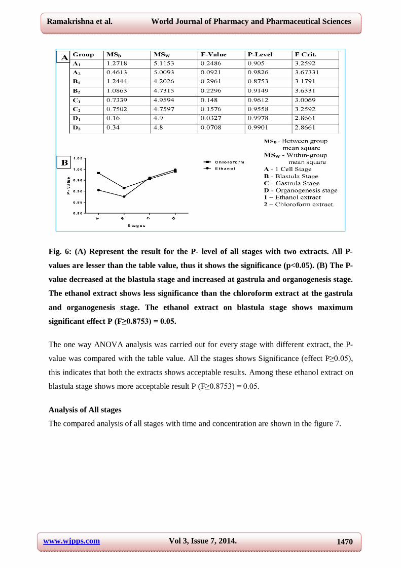
One way - ANOVA analysis
The one way ANOVA analysis is done by considering both time and concentration at a time
for different stages with different extracts. This data was subjected to a one way analysis and
the detailed results are shown in Figure 6.
www.wjpps.com Vol 3, Issue 7, 2014.
1470
Ramakrishna et al. World Journal of Pharmacy and Pharmaceutical Sciences
Fig. 6: (A) Represent the result for the P- level of all stages with two extracts. All P-
values are lesser than the table value, thus it shows the significance (p<0.05). (B) The P-
value decreased at the blastula stage and increased at gastrula and organogenesis stage.
The ethanol extract shows less significance than the chloroform extract at the gastrula
and organogenesis stage. The ethanol extract on blastula stage shows maximum
significant effect P (F≥0.8753) = 0.05.
The one way ANOVA analysis was carried out for every stage with different extract, the P-
value was compared with the table value. All the stages shows Significance (effect P≥0.05),
this indicates that both the extracts shows acceptable results. Among these ethanol extract on
blastula stage shows more acceptable result P (F≥0.8753) = 0.05.
Analysis of All stages
The compared analysis of all stages with time and concentration are shown in the figure 7.

www.wjpps.com Vol 3, Issue 7, 2014.
1471
Ramakrishna et al. World Journal of Pharmacy and Pharmaceutical Sciences
Fig. 7: (A) Represent the effect of time on different stages of embryos. (B) Showing the effect of concentration on different stages of embryos. In both cases the organogenesis stage embryo shows more tolerance to the extracts. The comparison studies were carried out between the different stages with time and
concentration. This study indicates that the development of embryo will increase the
resistance against the extract, thus organogenesis stage embryo has more resistance than the
gastrula stage, blastula stage and 1cell stage.Irrespective of the stages the long lime exposure
of the embryos shows death.
CONCLUSION
By using the Danio rerio embryo the abortifacient effects can be studied with different
parameters like time of exposure, concentration and stages. This experiment shows the better
efficacy in the blastula stage and this helps in designing herbal abortifacient drug. Thus the
Danio rerio is best model organism for studying abortifacient activity than the other higher
organisms like mice, rat, rabbit, etc.
ACKNOWLEDGEMENTS
The authors would grateful to the Mrs. Deepa, Department of Statistics and teaching staff,
Department of Biotechnology, Government Science College Bangalore, for there valuable
statistical assistance and there guidance.
REFERENCE
1. Gediya Shweta, Ribadiya Chetna, Soni Jinkal1, Shah Nancy, Jain Hitesh. Herbal Plants
Used as Contraceptives. International Journal of Current Pharmaceutical Review and
Research 2011; Volume 2, Issue1, February – April.

www.wjpps.com Vol 3, Issue 7, 2014.
1472
Ramakrishna et al. World Journal of Pharmacy and Pharmaceutical Sciences
2. Prakash, A. O. and Mathur, R. Effect of ArtobotrysodoratissimusLinn. extracts on oestrus
cycle, body weight and uterine weight in albinorats. Indian J. Exp. Biol 1997; 15, 1038.
3. Gopalkrishnan, M. and Rajasekharasetty, M. R. Effect of Papaya (Carica papaya linn) on
pregnancy and estrous cycle in albino rats of wistar strain. Indian J. Physiol, Pharmac
1978; 22, 66.
4. Gupta, O. P., Anand, K. K., Ray Ghatak, B. J. and Atal, C. K. Vasicine, Alkaloid of
Adhatodavasica, Indian J. Exp. Biol 1978; 16, 1075.
5. Manjunath, B.L. The wealth of India—raw materials. Council of Scientific and Industrial
Research, 75-77, New Delhi 1, (1948).
6. Pakrashi A and Basak B. Abortifacient effect of steroids from Ananas comosus and their
analogues on mice, J. Reprod. Fert 1976; 46, 461-462.
7. Pakrashi, Anita and Chakrabarty, Smritinath. Antifertility effects of the steroid 5 –
stigmastane – 3B, 5, 6B – triol – 3 – monobenzoate. Contraception 1979;23, 315.
8. Fishman, M.C. Genomics. Zebrafish—the canonical vertebrate. Science 2001; 294, 1290–
1291.
9. Christian Lawrence. The husbandry of zebrafish (Daniorerio): A review, Science direct
Aquaculture 2007; 269, 1–20.
10. Bradley Barbazuk. W, Ian Korf, Candy Kadavi, Joshua Heyen, Stephanie Tate, Edmund
Wun, Joseph A. Bedell, John D. McPherson, and Stephen L. Johnson, The Syntenic
Relationship of the Zebrafish and Human Genomes, Genome Research 1351.
11. Westerfield, M., The Zebrafish Book. A Guide for the Laboratory Use of Zebrafish
(Daniorerio), 3rd edition. University of Oregon Press, Eugene, OR. 385 pp, 1995.
12. Eisen, J S. Zebra fish make a big splash. Cell 87 1996; 969-977.
13. Fishman, M C. zebra fish genetics: The enigma of arrival. Proc Natl AcadSci USA 96
1999; 10554-10556.