d yn a m ic a n d s ta tic in te ra c tio n s b e tw e e n...
TRANSCRIPT

Dynamic and Static Interactions betweenp120 Catenin and E-Cadherin Regulatethe Stability of Cell-Cell AdhesionNoboru Ishiyama,1 Seung-Hye Lee,3 Shuang Liu,1 Guang-Yao Li,1 Matthew J. Smith,1 Louis F. Reichardt,3
and Mitsuhiko Ikura1,2,*1Division of Signaling Biology, Ontario Cancer Institute2Department of Medical Biophysics, University of TorontoMaRS Toronto Medical Discovery Tower, Room 4-804, 101 College Street, Toronto, ON M5G 1L7, Canada3Department of Physiology, University of California, San Francisco, San Francisco, CA 94158, USA*Correspondence: [email protected] 10.1016/j.cell.2010.01.017
SUMMARY
The association of p120 catenin (p120) with the juxta-membrane domain (JMD) of the cadherin cyto-plasmic tail is critical for the surface stability ofcadherin-catenin cell-cell adhesion complexes.Here, we present the crystal structure of p120 iso-form 4A in complex with the JMD core region(JMDcore) of E-cadherin. The p120 armadillo repeatdomain contains modular binding pockets that arecomplementary to electrostatic and hydrophobicproperties of the JMDcore. Single-residue mutationswithin the JMDcore-binding site of p120 abolishedits interaction with E- and N-cadherins in vitro andin cultured cells. These mutations of p120 enabledus to clearly differentiate between N-cadherin-dependent and -independent steps of neuronaldendritic spine morphogenesis crucial for synapsedevelopment. NMR studies revealed that p120 regu-lates the stability of cadherin-mediated cell-celladhesion by associating with the majority of theJMD, including residues implicated in clathrin-medi-ated endocytosis and Hakai-dependent ubiquitina-tion of E-cadherin, through its discrete ‘‘dynamic’’and ‘‘static’’ binding sites.
INTRODUCTION
p120 catenin (p120) is an armadillo (ARM) repeat-containingprotein that, along with the classical cadherins, b-catenin anda-catenin, plays a crucial role in the regulation of cell-cell adhe-sion at adherens junctions (AJs) (Daniel and Reynolds, 1995;Nishimura and Takeichi, 2009; Reynolds et al., 1992, 1994).The classical cadherins mediate Ca2+-dependent homophilicadhesion via trans-dimerization of cadherin ectodomainsbetween adjacent cells (Shapiro and Weis, 2009) and by havingthe cadherin cytoplasmic tail participate in cytoskeleton reorga-
nization and intracellular signaling through catenins (Nishimuraand Takeichi, 2009; Yamada and Nelson, 2007). b-catenin asso-ciates with the catenin-binding domain (CBD) and functionallylinks cadherins with the actin cytoskeleton through a-catenin(Yamada et al., 2005), while p120 is responsible for stabilizingcadherin-catenin complexes at the cell surface by interactingwith the juxtamembrane domain (JMD; Figure 1A) (Davis et al.,2003; Ireton et al., 2002; Lampugnani et al., 1997; Thoresonet al., 2000; Xiao et al., 2003). The cadherin JMD has beenimplicated in various processes, including cadherin clusteringand adhesive strengthening (Yap et al., 1998), promotion ofaxon outgrowth (Riehl et al., 1996), and suppression of cellmotility (Chen et al., 1997).Previous studies have shown that uncoupling of the p120-
JMD interaction or a reduction in p120 protein levels in culturedcells significantly increases levels of cadherin internalization andreduces the amount of cadherin available for cell-cell adhesion(Davis et al., 2003; Ireton et al., 2002; Miyashita and Ozawa,2007b; Perez-Moreno et al., 2006; Xiao et al., 2003), therebyimplicating p120 as a gatekeeper of cadherin turnover in verte-brates (Peifer and Yap, 2003). The significance of the p120-JMD interaction is underscored by the fact that the JMD is highlyconserved among classical (type I), type II, and some inverte-brate cadherins (Nollet et al., 2000). Prior work by Thoresonet al. (2000) has determined that the JMD core region (JMDcore;residues 758–775 in mouse E-cadherin; Figure 1A) is crucial forthe p120-JMD interaction. Conversely, p120 associates withthe cadherin JMD through a central ARM repeat domain (ARMdomain; Figure 1B) (Daniel and Reynolds, 1995). The p120ARM domain is well conserved from fly to human (>40% primarysequence identity [Myster et al., 2003]) and is flanked by anN-terminal regulatory region (NTR) and a C-terminal tail region(CTR).The loss of E-cadherin expression, a major hallmark of tumor
malignancy (Hanahan and Weinberg, 2000), is induced by avariety of factors, including transcriptional regulation, mutation,and aberrant cadherin internalization (Mosesson et al., 2008).Consistent with the critical role of p120 in the regulation ofcadherin turnover, the loss, downregulation, or mislocalizationof p120 in tumors has been linked to poor prognoses
Cell 141, 117–128, April 2, 2010 ª2010 Elsevier Inc. 117

(Thoreson and Reynolds, 2002). The major route of E-cadherininternalization appears to be clathrin-mediated endocytosisinvolving a noncanonical endocytic dileucine motif (LL motif)(Miyashita and Ozawa, 2007a, 2007b). In addition, ubiquitina-tion-dependent endocytosis of E-cadherin induced by the E3ubiquitin ligase Hakai (Fujita et al., 2002) and presenilin-1/g-sec-retase-mediated cleavage of E-cadherin (Marambaud et al.,2002) have been implicated in the depletion of E-cadherin fromthe cell surface. As the endocytic LL motif (residues 743–744)and tyrosine phosphorylation sites (residues 755–756) involvedin recruitment of Hakai are both located near the JMDcore
(Figure 1A), the association of p120 with the JMD is proposedto sterically hinder endocytic machinery and Hakai from associ-
ating with cadherin (Fujita et al., 2002; Miyashita and Ozawa,2007b). However, the mechanisms through which p120 regu-lates cadherin endocytosis have remained elusive because ofa lack of detailed knowledge of the p120-JMD interaction. More-over, a novel missense germline mutation within the JMD ofhuman E-cadherin (R749W, Arg751 in mouse; Figure 1A) linkedto hereditary diffuse gastric cancer (HDGC) was recently discov-ered and shown to have impaired cell adhesive function in vitro(Kaurah et al., 2007). This raises the possibility that such muta-tions might disrupt the p120-E-cadherin interaction, therebycontributing to an increased risk for diffuse gastric cancer.In the present study, we have determined the crystal structure
of p120 in complex with the JMDcore peptide at 2.4 A resolution.
R R R - - - T V V K E P L L P P D D D T R D N V Y Y Y D E E G G G E E D Q D - F D L S Q L H R G L D A R P ER R R - - - A V V K E P L L P P E D D T R D N V Y Y Y D E E G G G E E D Q D - F D L S Q L H R G L D A R P EK R R D K E R Q A K Q L L I D P E D D V R D N I L K Y D E E G G G E E D Q D - Y D L S Q L Q Q P D T V E P DK R K - - - K V V K E P L L L P E D D T R D N I F Y Y G E E G G G E E D Q D - Y D L S Q L H R G L D S R P DR L R - - - K Q A R A - H G K S V P E I H E Q L V T Y D E E G G G E M D T T S Y D V S V L N S V R R G G A KK - Q - - - K N G - W - H E K D I D D I R E T I I N Y E D E G G G E R D T D - Y D L N V L R T Q P F Y E E KR R S - - - P G A F E - N V R P E E M N R D N L R Q Y G V E G G G E A D N D Q Y S M A G L R K P V M P L D T
EC1 EC2 EC3 EC4 EC5JMD
TCBD
E-cadherin
734
Cancer-associated human E-cadherin mutations
R732Q
JMDext (736-781)783
884157
R749W E763X E781DFrame shiftFrame shift
734732
74462313521106
mEcadhEcad
747hNcadxCcadhVEcadDEcadHMR-1
78378179979367413931150
JMDcore (758-775)* **
A
p120
PKP1
JMDcore
FENTR 324 MIGEEVPSDQYYWAPLAQHERGSLASLDSLRKGGPPPPNWRQPE
R1 368 LPEVIA-MLGF-----------------RLDAVKSNAAAYLQHLCY
R2 396 RNDKVKTDVRKL-KGIPVLVG-LLDH-----------------PKKEVHLGACGALKNISF
R3 438 GRDQDNKIAIKNC-DGVPALVR-LLR-K---------------ARDMDLTEVITGTLWNLSS
R4 482 HDSIKMEIVDH--ALHALTDEVIIPHSGWEREPNEDCKPRHIEWESVLTNTAGCLRNVSS
R5 540 ERSEARRKLRECDGLVDALIF-IVQAEIG-----------QKDSDSKLVENCVCLLRNLSY
Ins 589 QVHREIPQAERYQEAAPNVANNTG--[PHAASCFGAKKGKGKKPIEDPANDTVDFPKR]--TSP
R6 647 ARGYELLF-QPEVVRIYIS-LLKES----------------KTPAILEASAGAIQNLCA
R7 688 GRWTYGRYIRSALR-QEKALSAIAD-LLTN-----------------EHERVVKAASGALRNLAV
R8 734 DARNKELIGKH--AIPNLVK-NLP---------GGQQNSSWNFSEDTVISILNTINEVIA
R9 782 ENLEAAKKLRETQ-GIEKLVL-INKS---------------GNRSEKEVRAAALVLQTIWG
CTR 826 YKELRKPLEKEGWKKSDFQVNLNNASRSQSSHSYDDSTLPLIDRNQKSDKKPDREEIQMSNMGSN
CTR 891 TKSLDNNYSTPNERGDHNKTLDRSGDLGDMEPLKGTTPLMQKI 933
H1 H2 H3
p120 AR
M dom
ain
R1H2 R1H3
R2H2 R2H3R2H1
R3H2 R3H3R3H1
R4H2 R4H3R4H1
R5H2 R5H3R5H1
R6H2 R6H3R6H1
R7H2 R7H3R7H1
R8H2 R8H3
R9H2 R9H3R9H1
Cadherin, KaisoRhoAFerPLEKHA7
Binding PartnersB
1A
4A Ins
A
1 2 3 4 5 6 7 8 91 933
A
1 2 3 4 5 6 7 8 9324 933
613-643
1ABCCoil A B
1 2 3 4 5 6 7 8 91 968
1 55 102 324
Cp120
D
p120
R1H3
R2H3
R3H3
R4H3
NTR
R5H3
R6H3
R8H3
R7H3
R9H3CTR
JMDcore
N
C
Ins
C
-catenin+CBD (1I7W+1I7X)
-catenin(1DOW+1H6G)
Cadherin EC1-5 (1L3W)
cell surface
cytoplasm
p120catenin+JMD
Cadherin-Catenin
Complex
90¡
ARM domainNTR CTR
Figure 1. E-Cadherin JMDcore Interactswith p120 ARM Repeats 1–5(A) Schematic representation of E-cadherin.
E-cadherin consists of the extracellular cadherin
domains 1–5 (EC1–5), the transmembrane region
(T) and the cytoplasmic tail, which contains the
JMD and CBD. The JMDext and the JMDcore used
in this study are indicated.Multiple sequence align-
ment of seven cadherin JMD sequences is shown
(mEcad, mouse E-cadherin; hEcad, human E-cad-
herin; hNcad, human N-cadherin; xCcad, frog
C-cadherin; hVEcad, human VE-cadherin; DEcad,
fly DE-cadherin; HMR-1, nematode HMR-1). Iden-
tical residues are highlighted in black boxes, and
residues conserved in at least four cadherins are
shown in yellow. * indicates the endocytic LLmotif.
** indicates tyrosine phosphorylation sites.
(B) Schematic representation of p120. p120
consists of the central ARM domain (ARM repeats
are indicated in red) flanked by the NTR and CTR.
Variousp120 isoforms result from four transcription
start sites (residue 1, 55, 102, 324) and three alter-
natively splicedexons (A,B, andC). Isoforms1ABC
and1Acontain a coiled-coil domain (Coil)within the
NTR. p120-4ADIns was created by deletion of resi-
dues 613–643 in the insert region of isoform 4A.
Binding partners of p120 and their binding sites
are shown (Cadherin [Daniel and Reynolds, 1995;
Ireton et al., 2002], Kaiso [Daniel and Reynolds,
1999], RhoA [Castano et al., 2007; Yanagisawa
et al., 2008], PLEKHA7 [Meng et al., 2008], and
Fer kinase [Lee et al., 2008; Xu et al., 2004]).
(C) Crystal structure of the p120-4ADIns/JMDcore
complex. p120 contains nine ARM repeats (R1–9)
with each repeat consisting of three a helices (H1,
H2, and H3), except for R1 and R8 (H3 helices
from ARM R1–9 are shown in different colors).
The modified insert region (Ins), loops between
R4H2 and R4H3, NTR, and CTR are partially disor-
dered (dashed lines). The JMDcore is shown in
magenta (stick and space filling representation).
See Table S1 for structure determination and
refinement statistics. The ‘‘head-to-tail’’ intercom-
plex contact site observed in both crystal forms is
shown in Figure S1.
(D) Model of the cadherin-catenin complex.
(E) Primary sequence and secondary structure of p120-4ADIns. The primary sequence of the ARM domain is aligned according to H1–3 helices to highlight
the sequence conservation (residues shaded gray). Disordered regions are shown as dashed lines, and gaps are indicated by light gray line above the sequence.
Square brackets indicate the deleted residues from the insert region (residues 613–643). The JMDcore-binding site residues are indicated by magenta
underscore.
(F) Superposition of the p120-4ADIns /JMDcore complex and PKP1 ARM domain (PDB code 1XM9).
118 Cell 141, 117–128, April 2, 2010 ª2010 Elsevier Inc.

Structure-based mutagenesis studies in both cultured epithelialand neuronal cells demonstrated that single-residue mutationswithin the JMDcore-binding site of p120 is sufficient to uncouplep120 from E- and N-cadherins and to delineate multifunctionalroles of p120 in cell-cell adhesion and other signaling processes.Nuclear magnetic resonance (NMR) data revealed that p120regulates the stability of cadherin-mediated cell-cell adhesionby associating with themajority of the E-cadherin JMD, includingthe endocytic LL motif and tyrosine-phosphorylation sites,through discrete ‘‘dynamic’’ and ‘‘static’’ binding sites. Inaddition, we propose a model for a p120-dependent cadherinclustering mechanism, which involves the JMD-induced oligo-merization of p120.
RESULTS AND DISCUSSION
Overall Structure of the p120-4ADIns/JMDcore ComplexTo better understand how the interaction between p120 and theJMDmodulates the stability of cadherin in cell-cell adhesion, wesubjected variants of p120 to cocrystallization with JMD frag-ments. We successfully crystallized a modified form of humanp120 isoform 4A (p120-4ADIns; Figure 1B) in complex with themouse E-cadherin JMDcore fragment (mouse and human haveidentical primary sequences; Figure 1A). p120-4ADIns wasproduced by deletion of residues 613–643 from the insert region(residues 589–648), which resulted in noticeably higher proteinstability and crystallizability. The three-dimensional structure ofp120-4ADIns/JMDcore complex was determined in two crystalforms at 2.4 A (form I) and 3.0 A (form II) resolution (Table S1available online). The following characterization of the complexis based mainly on form I, as the complex structures were nearlyidentical in both forms except for an increased disorderedness inthe C terminus of p120 in form II.The crystal structure of the p120-4ADIns/JMDcore complex
provides the first opportunity to examine the three-dimensionalarchitecture of p120, as well as the binding interface betweenp120 and the cadherin JMD (Figure 1C). p120-4ADIns consistsof a central arch-shaped ARM domain (residues 368–825)accompanied by largely disordered NTR (residues 324–367)and CTR (residues 826–933). The bound JMDcore peptide isstretched over the N-terminal half of the p120 ARM domain inthe opposite orientation (Figure 1C), resembling the orientationof the CBD bound to the ARM domain of b-catenin (Huber andWeis, 2001). The p120 ARM domain is similar to that of plakophi-lin-1 (PKP1) (Choi and Weis, 2005), as it contains nine ARMrepeats (R1–9) and a partially unstructured insert region (Figures1C and 1E). They can be superposed with a root mean squaredistance (RMSD) of 1.2 A over 324 Ca atoms (Figure 1F). Whilethe previously determined PKP1 structure is limited to the ARMdomain (Choi and Weis, 2005), p120-4ADIns contains uniquestructural features beyond the ARMdomain; the last ten residuesof the NTR of p120 wrap over the C terminus of cadherinJMDcore, and the first 21 residues of the CTR of p120 formtwo a helices that fold over the hydrophobic surface of ARMR9 (Figure 1C). Though other isoforms contain longer NTR,CTR, and insert region compared to p120-4A, the structures ofARM domain, NTR, and CTR revealed here are likely conservedin all p120 isoforms.
The p120-4ADIns/JMDcore complex structure has allowed us tobring together the three-dimensional structures of C-cadherinectodomain (PDB code 1L3W) (Boggon et al., 2002), the b-cate-nin/CBD complex (PDB codes 1I7W and 1I7X) (Huber and Weis,2001), and a-catenin fragments (PDB codes 1DOW and 1H6G)(Pokutta and Weis, 2000; Yang et al., 2001), finally revealingthe multimeric arrangement of the cadherin-catenin complex inits entirety (Figure 1D). As these complexes are expected tocluster to mediate cell-cell adhesion (Yap et al., 1998), the pres-ence of JMD-bound p120 (105 A 3 55 A 3 40 A) in a compactspace between the plasma membrane and the b-catenin/CBDcomplex is likely to restrict other proteins (e.g., endocyticmachinery and Hakai) from gaining access to the JMD(Figure 1D). Although JMD residues 734–757 were not ac-counted for in our p120/JMD complex structure, the C terminusof the JMDcore (His775) is only separated by eight residues fromthe N terminus of the CBD (Val784) from the b-catenin/CBDcomplex (Huber and Weis, 2001). This places the N terminus ofp120 in close proximity to the ARM domain of b-catenin, whichis consistent with the role of p120 in recruiting Fer kinase throughits NTR to modulate the cadherin-b-catenin interaction (Leeet al., 2008; Xu et al., 2004).
E-Cadherin JMDcore Interacts with p120 ARMRepeats 1–5The p120-JMDcore interface buries approximately 2400 A2 oftotal solvent accessible surface area, and this interface can befurther divided into two regions on the basis of different typesof intermolecular interactions (Figure 2A). The first regioninvolves extensive electrostatic interactions between theN-terminal acidic region (residues 758–766) of the JMDcore andthe p120 basic ARM groove formed along the third helix (H3) ofARM R1–5 (Figure 2B). Five salt bridges are formed betweenacidic residues (Asp758, Glu759, Glu760, Glu764, and Glu765)from the cadherin JMDcore and basic residues (Lys401,Lys433, Lys444, Lys486, and Lys574) from p120 (Figure 2C). Inaddition, the triple glycine motif (Gly761, Gly762, and Gly763)of the JMDcore forms a turn that fits into a trough formed byPhe437, Trp477, and Asn478 of p120. The backbone of theJMDcore forms hydrogen bonds with p120 residues, Asn434,Asn478, and Asn536 (Figure 2C). Several p120 residues involvedin this interface are conservedwithin p120ARMR2–7 (Figure 1E):the first turn of H1 helices from ARM R2–5 contain conservedbasic residues, including Lys401, Lys444, and Lys486, and thelast turn of H3 helices from ARM R2–7 contain conserved aspar-agines, including Asn434, Asn478, and Asn536. In contrast, thesecond region of the p120-4ADIns/JMDcore interface involvesmainly hydrophobic interactions between the C-terminal anchorregion of the JMDcore (residues 767–775) and the N terminusof p120 (Figure 2B). The anchor region of the JMDcore forms asingle 310-helix turn that is wedged between ARM R1 and theNTR of p120 (Figure 2D). Consequently, the side chain ofLeu774 from the JMDcore is buried or ‘‘anchored’’ into a hydro-phobic pocket formed between H2 and H3 helices of ARM R1,involving Pro366, Val371, Met374, Val382, Ala386, and Tyr389(Figures 2B and 2D). Given that the triple glycine motif with twoadjoining glutamates (EGGGE) and the anchoring leucine arestrictly conserved in the JMDcore from fly DE-cadherin to human
Cell 141, 117–128, April 2, 2010 ª2010 Elsevier Inc. 119

E-cadherin (Figure 1A), we propose that both electrostatic andhydrophobic interactions are equally important for the p120-JMDcore interaction.
Aside from interactions within the complex, several residuesfrom the anchor region of the JMDcore and the NTR of p120 areinvolved in intercomplex association with the ARM domainfrom an adjacent p120molecule in the crystal (Figure S1). Intrigu-ingly, in both crystal forms, p120-4ADIns/JMDcore complexesassemble into head-to-tail oligomers through this interface,and its periodicity (!60 A) is similar to the spacing observed inthe ordered array of cadherins along the desmosomal midlineof human epidermis (Al-Amoudi et al., 2007). Certainly, onemust exercise caution when distinguishing protein-proteincontacts observed in the crystal to be specific or from a packingcontact, and, moreover, neither p120 nor the cadherin cyto-plasmic tail alone self-associates in yeast two-hybrid and blotoverlay assays, respectively (Daniel and Reynolds, 1995; Yapet al., 1998). We thus employed other approaches to examinethe potential JMD-induced oligomerization of p120, includingdynamic light-scattering and chemical crosslinking analyses ofp120 wild-type (WT) and a W363A mutant (which shouldcompromise the intercomplex interface). Unfortunately, theseexperiments were hampered by the aggregation-prone natureof p120. Analytical ultracentrifugation analysis of p120 at a lowprotein concentration (%1.0 mg/ml), however, indicated that
R1
R2
H775
L774
Q773
S772
L771
D770
F769
D768
D766
Q767 H392
W363Y389
N385
V382
A386
M374
V371P366
P361 P360
R364N362
Q391
D
D758
E759
E760
G761
G762
G763
E764
E765
D766
K574
W477N536
K486
K444
F437
G438
N478
K401
K433
N434
H392 R1
R2
R3
R4CA
Hydrophobic Pocket
Basic ARMGroove
Anc
hor
regi
on
B
L774
K486
R584R545
N585
N536
E540
K444
K401R396
Y395
H392
Y389
Q365
K433
F437
K574
W477
W363
R535
Aci
dic
regi
on
E764
JMDcore
Figure 2. The JMDcore-Binding SiteConsists of the Basic ARM Groove andHydrophobic Binding Pocket(A) Overview of the JMDcore-binding site of p120-
4ADIns (same orientation as Figure 1C). The
JMDcore (red) is bound to the basic ARM groove
(blue) and the N-terminal hydrophobic pocket
(green).
(B) The surface electrostatic potential of the
JMDcore-binding site of p120 with positively and
negatively charged regions in blue and red,
respectively. 2Fo-Fc electron density map (green
mesh; contoured at sA = 1.5) of the JMDcore
(orange) is shown.
(C) The electrostatic interface between p120 and
the JMDcore. The JMDcore N-terminal acidic region
(red; residues 758–766) forms extensive electro-
static interaction with the p120 basic ARM groove
(blue). Red spheres represent water molecules.
(D) The hydrophobic interface between p120 and
the JMDcore. The side chain of Leu774 from the
C-terminal anchor region of JMDcore (red; residues
767–775) is buried into the p120 hydrophobic
pocket. Deletion of residues 613–643 did not
cause any detrimental effect on the structure of
p120 or the interaction between p120 and the
E-cadherin JMD (see Figure S2).
both p120 WT and W363A are mostly‘‘monomeric’’ in the presence of JMDpeptide (data not shown). Nonetheless,the JMD-induced oligomerization ofp120 observed in the crystals providesa basis for further examining a possible
model for cadherin clustering, a phenomenon presumablyfacilitated by the cadherin JMD and p120 during maturation ofcell-cell junctions (Anastasiadis and Reynolds, 2000; Thoresonet al., 2000; Yap et al., 1997; Yap et al., 1998).
p120 Residues Lys401 and Asn478 Are Essentialfor Interaction with E-CadherinTo confirm the significance of the crystallographically deter-mined JMD-binding site of p120, we performed a pulldownassay with glutathione S-transferase-fused JMD residues747–781 (GST-JMD) and p120-4ADIns containing single-residuemutations within the JMDcore-binding site (K401M, K444M,W477A, or N478A) or a mutation (W363A) in the NTR that is notexpected to affect the p120-JMD interaction (Figure 3A). Circulardichroism (CD) analyses show that the structural integrity ofp120-4ADIns was not affected by the partial deletion of the insertregion or by the single-residue mutations (Figure S2A). Tightassociation of p120-4ADIns WTwith GST-JMD (Figure 3A), equiv-alent to that observed for p120-4A (Figure S2B), indicates thatresidues 613–643, which contain a RhoGTPase-binding site(residues 622–628) (Yanagisawa et al., 2008), are not requiredfor the interaction between p120 and the JMD. In contrast, alter-ation of the trough structure (W477A or N478A) or the eliminationof a basic residue (K401M or K444M) in the p120 ARM groovewas sufficient to disrupt the interaction, though some residual
120 Cell 141, 117–128, April 2, 2010 ª2010 Elsevier Inc.

association was observed with the W477A mutant. In addition,deletion of the C-terminal JMD residues 776–781 locatedoutside of the JMDcore also abolished the interaction. Theseobservations confirm that the electrostatic interface betweenthe p120 basic ARM groove and the acidic region of the JMDcore
are crucial for the p120-JMD interaction and reveal that tightassociation between p120 and the JMD requires a series ofbinding pockets that include additional interactions outside ofthe core region.To further analyze the JMDcore-binding site of p120 in the
context of cell-cell adhesion, we tested the ability of thesep120 mutants to colocalize with E-cadherin at AJs by transientlyexpressing p120 isoform 1A (Figure 1B) fused to a monomericred fluorescent protein (p120-1A-mRFP) in p120-downregulatedMadin-Darby canine kidney (MDCK) and MCF-7 cells(Figure 3B). In both cell types, the p120 WT as well as theK444M and W477A mutants colocalized with endogenousE-cadherin at AJs (Figure 3C). The observed association of theK444M mutant with the E-cadherin JMD in cells appears to betoo weak to be detected by the in vitro pulldown assay, but isstrong enough to localize endogenous E-cadherin at AJs. Incontrast, p120 with either the K401M or N478A mutationsresulted in complete loss of the p120-E-cadherin interaction
and significantly reduced expression of E-cadherin at the cellperimeter (Figure 3C) (Davis et al., 2003). The K401M or N478Amutants also failed to colocalize with endogenous E-cadherinwhen MDCK and MCF-7 cells expressing endogenous p120were transfected with a vector expressing p120-1A-mRFP(Figure S3). Thus, our data strongly suggest that the newly re-vealed p120-JMD interface is crucial for colocalization of p120with E-cadherin at AJs, and a minimal alteration within theJMD-binding site of p120 is sufficient to uncouple p120 fromE-cadherin.
Uncoupling of the p120-N-Cadherin Interactionfrom p120-Rho-GTPase-Mediated PathwaysThe dynamic regulation of cell-cell adhesion by p120 is not solelydependent on its interaction with the JMD, but also involves theassociation of p120 with numerous binding partners (Figure 1B).In neurons, synapse development and formation are highlydependent on the coordination of p120-dependent signalingprocesses, including N-cadherin retention, RhoA inhibition,and Fer-dependent dephosphorylation of b-catenin (Arikkathand Reichardt, 2008; Elia et al., 2006; Lee et al., 2008).Prior work using p120 deletion mutants, DARM-R3 and D622–
628, has demonstrated that the maturation of the dendritic spine
p120Input
GSHElution
p120
p120
GST-JMD!C
WT
K401
M
K444
M
W47
7A
N47
8A
W36
3A
GST-JMD + + + + + +GST
+
+
WT
WT
A
B
C
p120
E-cad
Merge
WT W477A N478AK401M K444M W363A
p120-downregulated MCF-7
p120
E-cad
Merge
WT W477A N478AK401M K444M W363A
p120-downregulated MDCK
p120
MCF-7+
shRNA
MDCK+
shRNA
contr
ol
p120
i-A
p120
i-B
contr
ol
p120
i-1
p120
i-2
E-cad
Tubulin
Figure 3. p120 Residues Lys401 and Asn478 Are Necessary for the Colocalization of p120 with E-Cadherin at AJs(A) GST-JMD pulldown assay with p120-4ADIns. GST-JMD (residues 747–781), GST-JMDDC (residues 747–775), or GST was used to perform pulldown assay
with p120-4ADIns WT or mutants (K401M, K444M, W477A, N478A, or W363A). W363A mutation did not affect the p120-JMD interaction since the side chain
of Trp363 is not part of the JMD-binding site.
(B) Lysates from MCF-7 or MDCK cells stably expressing shRNA (controls transfected with scrambled shRNA; p120i-A and p120i-B are p120-downregulated
monoclonal cell lines in MCF-7; p120i-1 and p120i-2 are p120-downregulated monoclonal cell lines in MDCK) were analyzed by western blotting for levels of
p120 and E-cadherin (E-cad). Tubulin levels were used as a loading control.
(C) p120-downregulatedMCF-7 cells (p120i-A) or p120-downregulatedMDCK cells (p120i-1) were transfected with amammalian expression vector encoding for
p120-1A-mRFP WT or mutants (same as in A).
See also Figure S3 for the expression of p120-1A-mRFP WT or mutants in MDCK and MCF-7 cells expressing endogenous p120.
Cell 141, 117–128, April 2, 2010 ª2010 Elsevier Inc. 121

head width (Figure S4) requires a proper p120-N-cadherin inter-action and the accumulation of spine density and growth of spinelength require the p120 regulation of Rho-GTPases (Elia et al.,2006). However, creation of p120 DARM-R3 mutant involvesremoval of more than 40 residues from the ARM domain, whichcould have a detrimental effect on the structure and function ofp120. Since we have confirmed that single-residue mutants ofp120 (K401M, K444M, and N478A) are well folded and unable
to tightly associate with E-cadherin (Figure 3A), we decided tofurther test the role of p120 binding to cadherin in neuronaldendritic spine formation by expressing these mutants in hippo-campal neurons cultured from p120flox/flox mice lacking p120.The inability of these p120 mutants to interact with N-cadherinwas confirmed by coimmunoprecipitation of N-cadherin andp120 variants coexpressed in 293T cells (Figure 4A). When Crerecombinase expression was used to acutely knock down
Figure 4. p120 Mutants Show Reduced Affinity for N-Cadherin, and Reduced Dendritic Spine Head Width in Neurons Lacking p120(A) With transfected 293T cells, the interactions of N-cadherin with Flag-p120-1A (wild-type, K401M, K444M, or N478A) were examined in anti-Flag immunopre-
cipitates by anti-N-cadherin immunoblotting. As expression controls, N-cadherin levels were examined in total cell lysates.
(B–G) Hippocampal neurons from postnatal day 0 p120flox/flox (p120fl/fl) mice were transfected at day 10 with the indicated constructs (Cre, p120 mutants)
together with GFP and fixed at day 14. Representative images of transfected neurons are shown at lowmagnification (upper panels) and highmagnification (lower
panels).
(H–J) Densities of dendritic spines (H), mean spine length (I), and mean spine head width (J) were quantified. The description of dendritic spine is shown in
Figure S4. Spines from three independent hippocampal cultures were measured. For (H), n = 14 (Vec), 15 (Cre), 18 (Cre+p120), 9 (Cre+K401M), 10 (Cre+K444M),
and 12 (Cre+N478A). For (I) and (J), n = 460 (Vec), 358 (Cre), 635 (Cre+p120), 343 (Cre+K401M), 355 (Cre+K444M), and 413 (Cre+N478A). The scale bars represent
20 mm (lowmagnification) and 5 mm (high magnification). #, p < 0.05 versus Vec. *, p < 0.05 versus Cre. Error bars represent the standard error of the mean (SEM).
See Table S2 for Student’s t test results.
122 Cell 141, 117–128, April 2, 2010 ª2010 Elsevier Inc.
p120 expression at the stage of spinemorphogenesis in culturedneurons from p120flox/flox mice, the p120-deficient spines ex-hibited reductions in spine density, length, and head widthcompared to the control neurons (Figures 4B, 4C, 4H, 4I, and4J). Expression of p120 WT, K401M, K444M, or N478A inp120-deficient neurons resulted in restoration of spine densityand length (Figures 4B–4I). However, the reduced width of spinehead observed in p120-deficient neurons was only restored byexpression of p120 WT, not by the p120 mutants (Figures 4B–4G and 4J). These observations confirm the necessity of ap120-N-cadherin interaction during spine head maturation (Eliaet al., 2006) but further illuminate the multifunctional roles ofp120 by demonstrating that single-residue mutations in p120(K401M, K444M, and N478A) can promote dendritic spinemorphogenesis with nearly normal spine density and length,presumably via Rho-GTPase signaling, as previously described(Elia et al., 2006). In future experiments, it will be interesting todetermine whether p120 and N-cadherin-dependent promotionof spine head maturation requires cadherin clustering mediatedvia the proposed p120/JMD-dependent mechanism (Figure S1).
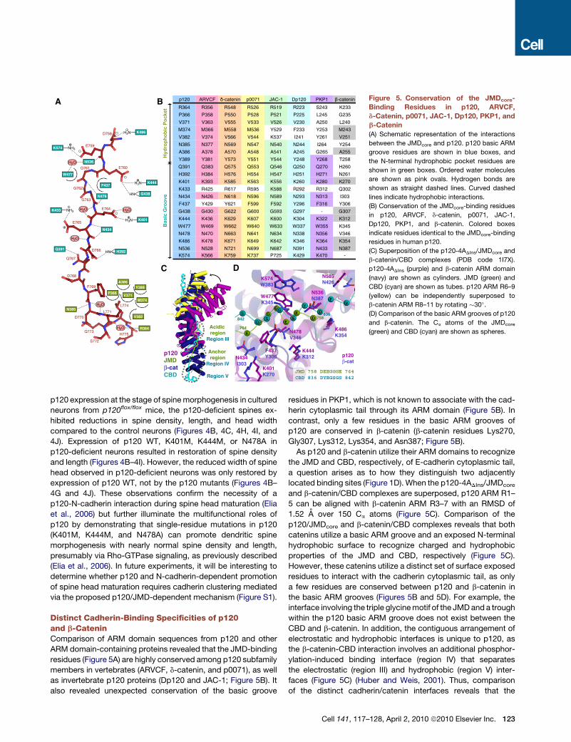
Distinct Cadherin-Binding Specificities of p120and b-CateninComparison of ARM domain sequences from p120 and otherARM domain-containing proteins revealed that the JMD-bindingresidues (Figure 5A) are highly conserved among p120 subfamilymembers in vertebrates (ARVCF, d-catenin, and p0071), as wellas invertebrate p120 proteins (Dp120 and JAC-1; Figure 5B). Italso revealed unexpected conservation of the basic groove
residues in PKP1, which is not known to associate with the cad-herin cytoplasmic tail through its ARM domain (Figure 5B). Incontrast, only a few residues in the basic ARM grooves ofp120 are conserved in b-catenin (b-catenin residues Lys270,Gly307, Lys312, Lys354, and Asn387; Figure 5B).As p120 and b-catenin utilize their ARM domains to recognize
the JMD and CBD, respectively, of E-cadherin cytoplasmic tail,a question arises as to how they distinguish two adjacentlylocated binding sites (Figure 1D). When the p120-4ADIns/JMDcore
and b-catenin/CBD complexes are superposed, p120 ARM R1–5 can be aligned with b-catenin ARM R3–7 with an RMSD of1.52 A over 150 Ca atoms (Figure 5C). Comparison of thep120/JMDcore and b-catenin/CBD complexes reveals that bothcatenins utilize a basic ARM groove and an exposed N-terminalhydrophobic surface to recognize charged and hydrophobicproperties of the JMD and CBD, respectively (Figure 5C).However, these catenins utilize a distinct set of surface exposedresidues to interact with the cadherin cytoplasmic tail, as onlya few residues are conserved between p120 and b-catenin inthe basic ARM grooves (Figures 5B and 5D). For example, theinterface involving the triple glycinemotif of the JMDand a troughwithin the p120 basic ARM groove does not exist between theCBD and b-catenin. In addition, the contiguous arrangement ofelectrostatic and hydrophobic interfaces is unique to p120, asthe b-catenin-CBD interaction involves an additional phosphor-ylation-induced binding interface (region IV) that separatesthe electrostatic (region III) and hydrophobic (region V) inter-faces (Figure 5C) (Huber and Weis, 2001). Thus, comparisonof the distinct cadherin/catenin interfaces reveals that the
p120 ARVCF -catenin p0071 JAC-1 Dp120 PKP1 -catenin
R364 R356 R548 R526 R519 R223 S243 K233
P366 P358 P550 P528 P521 P225 L245 G235
V371 V363 V555 V533 V526 V230 A250 L240
M374 M366 M558 M536 Y529 F233 Y253 M243
V382 V374 V566 V544 K537 I241 Y261 V251
N385 N377 N569 N547 N540 N244 I264 Y254
A386 A378 A570 A548 A541 A245 G265 A255
Y389 Y381 Y573 Y551 Y544 Y248 Y268 T258
Q391 Q383 Q575 Q553 Q546 Q250 Q270 H260
H392 H384 H576 H554 H547 H251 H271 N261
K401 K393 K585 K563 K556 K260 K280 K270
K433 R425 R617 R595 K588 R292 R312 Q302
N434 N426 N618 N596 N589 N293 N313 I303
F437 Y429 Y621 F599 F592 Y296 F316 Y306
G438 G430 G622 G600 G593 G297 - G307
K444 K436 K629 K607 K600 K304 K322 K312
W477 W469 W662 W640 W633 W337 W355 K345
N478 N470 N663 N641 N634 N338 N356 V346
K486 K478 K671 K649 K642 K346 K364 K354
N536 N528 N721 N699 N687 N391 N433 N387K574 K566 K759 K737 P725 K429 K470 -
Hy
dro
ph
ob
ic P
oc
ke
tB
as
ic G
ro
ov
e
B
C
D
K444K312
Acidicregion
Region III
K486K354
p120!-cat
F437Y306
N478V346
N536N387
N585N426
W477K345
K574W383
K401K270
N434I303
DY
VF
EE
ES
S
E
G
G
G
758
764
836842
Anchorregion
Region IV
Region VJMD 758 DEEGGGE 764CBD 836 DYEGSGS 842
D
p120JMD!-catCBD
A
K444
W477
K574 NH3
H3N
G438HN
F437
E759
E760G761
G762
G763
E764
E765
D766
D768
F769
D770L771
S772
Q773
L774
H775
Q767
D758 K486H3N
N478
N536
K401H3N
K433 NH3
N434
Q391H392HN
N385
V382
P366
M374V371
A386Y389
H2O
H2O H2O
H2O
H2O
H2O R364
Figure 5. Conservation of the JMDcore-Binding Residues in p120, ARVCF,d-Catenin, p0071, JAC-1, Dp120, PKP1, andb-Catenin(A) Schematic representation of the interactions
between the JMDcore and p120. p120 basic ARM
groove residues are shown in blue boxes, and
the N-terminal hydrophobic pocket residues are
shown in green boxes. Ordered water molecules
are shown as pink ovals. Hydrogen bonds are
shown as straight dashed lines. Curved dashed
lines indicate hydrophobic interactions.
(B) Conservation of the JMDcore-binding residues
in p120, ARVCF, d-catenin, p0071, JAC-1,
Dp120, PKP1, and b-catenin. Colored boxes
indicate residues identical to the JMDcore-binding
residues in human p120.
(C) Superposition of the p120-4ADIns/JMDcore and
b-catenin/CBD complexes (PDB code 1I7X).
p120-4ADIns (purple) and b-catenin ARM domain
(navy) are shown as cylinders. JMD (green) and
CBD (cyan) are shown as tubes. p120 ARM R6–9
(yellow) can be independently superposed to
b-catenin ARM R8–11 by rotating !30".
(D) Comparison of the basic ARM grooves of p120
and b-catenin. The Ca atoms of the JMDcore
(green) and CBD (cyan) are shown as spheres.
Cell 141, 117–128, April 2, 2010 ª2010 Elsevier Inc. 123

ligand-specific binding pockets, as well as the spatial arrange-ment of modular binding pockets within the ARM domains,determine distinct substrate specificities of p120 and b-cateninfor E-cadherin.
Dynamic and Static Binding Interfacesbetween p120 and the JMDOur structural and mutagenesis data explain the basis for theJMDcore mutations often used to uncouple p120 from E-cadherin(Thoreson et al., 2000). As an example, alanine substitution of thetriple glycine motif (761GGG763/AAA) or the succeeding acidicresidues (764EED766/AAA) either indirectly or directly preventsthe formation of salt bridges with lysines from p120 (Lys401and Lys433; Figure 5A). However, we could not provide anyinsight on the involvement of JMD residues 776–781 in thep120 interaction suggested by the pulldown assay (Figure 3A),prompting us to investigate the significance of regions outsideof the JMDcore. More specifically, we examined the involvementof the endocytic LLmotif (residues 743–744), tyrosine phosphor-ylation sites (residues 755–756), and Arg751 (a HDGC mutationsite) in the direct association with p120.
Isothermal titration calorimetry (ITC) determined that p120-4ADIns shows affinity in the submicromolar range for both theJMDext peptide (residues 736–781; Kd = 0.6 mM; Figure 6A) andthe peptide without the N-terminal 11 residues (JMDDN; residues747–781; Kd = 0.9 mM). In contrast, p120-4ADIns shows signifi-cantly reduced affinity toward the JMDcore (Kd = !40 mM), indi-cating that the residues flanking the N and C termini of theJMDcore (residues 747–757 and 776–781) are necessary for tightbinding with p120. We therefore performed NMR titrations of15N-labeled JMDext with unlabeled p120-4ADIns to map thep120-binding site within the JMD (Figures 6B–6H). The narrowrange of 1H chemical shifts observed for the JMDext indicatedthat the peptide in solution is unstructured (Figure S5), which isconsistent with the previously observed property of the cadherincytoplasmic tail (Huber et al., 2001). On the basis of observedsignal broadening and chemical shift differences between freeand bound states (DdB-F) in the 1H-15N HSQC spectra of JMDext,we determined that three sections within the JMDext showeddistinct modes of interaction with p120-4ADIns (Figure 6B): resi-dues 736–740 (section I) are in the fast exchange regime (smallDdB-F) reflecting the N-terminal disordered region participatingin weak association; residues 741–746 (section II) are in theslow-to-intermediate exchange regime (medium DdB-F) reflect-ing this proline-rich segment (containing the endocytic LL motif)participating in medium-strength association; and residues747–781 (section III) are in the slow exchange regime (largeDdB-F) reflecting this large segment of JMDext (containingArg751, the tyrosine phosphorylation sites, the core region andthe C-terminal residues 776–781) participating in strong associ-ation. On the basis of these observations, we have defined theJMD residues 747–781 as a ‘‘static’’ p120-binding site mainlyresponsible for the specific interaction between p120 and E-cad-herin, and the JMD residues 736–746 as a ‘‘dynamic’’ p120-binding site where the intermediate-to-weak interaction withp120 provides moderate masking of the LL motif from the endo-cytic machinery proteins (see below). The orientation of theJMDcore within the complex structure and an extensive basic
ARM groove formed by ARM R1–7 (Figures 1C and 6I) suggestthat a basic patch formed by ARM R5–7 is likely to interactwith the JMD residues preceding the core region (residues736–757; Figure 6B). The proposed interaction between thep120 ARM R1–7 and the JMDext is consistent with the previousstudies by Ireton et al. (2002), which showed that individual dele-tion of ARM repeats 1–6 (repeat number based on our structure)abolished binding of p120 to the cadherin JMD.
Dynamic p120-JMD Interaction Determines the Fateof CadherinThe present study has revealed that there are two types of p120-JMD interfaces, dynamic and static in binding nature. Wepropose that p120 contributes to the regulation of cadherin-mediated cell-cell adhesion by protecting the endocytic LL motifthrough the ‘‘dynamic’’ p120-JMD interface, while its associationwith E-cadherin is stabilized through the ‘‘static’’ p120-JMDinterface (Figure 7). Although the ‘‘dynamic’’ interface does notsignificantly contribute to the affinity between p120 and theJMD (Figure 6A), the propensity of the LL motif to be releasedfrom the ‘‘dynamic’’ interface of JMD-bound p120 providesopportunities for clathrin adaptor complexes, such as AP2, tocompetitively bind to the LL motif and initiate the endocytosisof cadherins (Figure 7). Interestingly, the affinity between p120-4ADIns and the JMDext peptide is similar to that reported forAP2 and an LL motif containing peptide (Kd = 0.85 mM) (Kellyet al., 2008). This suggests that the stability of E-cadherin at AJsis dependent on an equilibrium of cadherin retention promotedby p120, and cadherin internalization induced by the endocyticmachinery. Further destabilization of the ‘‘dynamic’’ p120-JMDinterface due to phosphorylation of p120 (Bauer et al., 1998) orallosterically increased affinity between endocytic proteins andthe LL motif (Kelly et al., 2008) could lead to reduced levels ofE-cadherin at the cell surface. Alternatively, cadherin internaliza-tion could also be induced by destabilization of the ‘‘static’’p120-JMD interface. Src-dependent phosphorylation of theE-cadherin JMD at Tyr755 and Tyr756 within the ‘‘static’’ inter-face could either hinder association of p120 with the JMD and/or promote association of Hakai with the JMD, consequentlydisplacing p120 from the cadherin-catenin complex at the AJ(Figure 7) (Fujita et al., 2002). The HDGC-associated R749Wmutation of human E-cadherin (Kaurah et al., 2007) would alsobe expected to interfere with the stable binding of p120 withthe JMD as a result of a change within the ‘‘static’’ interface.Taken together, our structural data provide insights into thesignificance of the p120-regulated stability of cadherin-catenincomplexes in AJ formation, in neuronal synapse development,and in increased risks of diffuse gastric cancer.
EXPERIMENTAL PROCEDURES
Protein Expression and PurificationRecombinant p120 proteins and the JMD peptides used in this study were
individually expressed as GST-fusion proteins in Escherichia coli and purified
with the glutathione-sepharose resin (GE Healthcare). Subsequently, GST was
cleaved off by thrombin and the cleaved protein (p120-4ADIns or JMD) was
further purified by size-exclusion chromatography. The purified proteins were
exchanged into protein storage buffer (50 mM Tris-HCl [pH 7.0], 150 mM NaCl,
1mMTCEP-HCl). See theExtendedExperimental Procedures for further details.
124 Cell 141, 117–128, April 2, 2010 ª2010 Elsevier Inc.

Crystallization and Data CollectionCrystals of the p120-4ADIns/JMDcore complex were grown at 293 K by
vapor diffusion. The protein solution, containing p120-4ADIns (20 mg/ml) and
four molar excess of the JMDcore peptide, was mixed with an equal volume
of the reservoir solution containing 0.1 M Bis-Tris (pH 5.6), 1.8 M (NH4)2SO4,
and 3% methanol for form I crystals, or 0.1 M HEPES (pH 7.4) and 8% (w/v)
PEG-8000 for form II crystals. Diffraction data were collected at the
Advanced Photon Source beamline 19BM and processed with HKL2000
(Otwinowski and Minor, 1997). See the Extended Experimental Procedures
for further details. Statistics pertaining to the diffraction data are presented
in Table S1.
Structure Determination and RefinementThe structure solution for the p120-4ADIns/JMDcore complex structure was
solved bymolecular replacement with the PKP1ARMdomain used as a search
model. Successive rounds of manual model building and refinement were
performed to refine the models of p120-4ADIns and the JMDcore. Refine-
ment statistics are presented in Table S1. See the Extended Experimental
740 750745 755 760 765 770 775 780RTVVKEPL LPPDDDTRDNVYYYDEEG GG EEDQDFDLSQLHRGLDAR
A B
JMDcore
p120-JMD
Interfaces
[p120]/[JMD]
0 1.5
II. Slow-to-Intermediate
Exchange
Chemical ShiftPerturbation
I. Fast Exchange
III. Slow Exchange
Static
binding site
Dynamic
binding site
N
p120 ARM repeats
C R1R2R4R6R9 R8 R3R5R70.0 0.5 1.0 1.5 2.0 2.5
-12
-10
-8
-6
-4
-2
0-0.7
-0.6
-0.5
-0.4
-0.3
-0.2
-0.1
0.0
0.1-10 0 10 20 30 40 50 60 70 80 90 100 110 120
Time (min)
Molar Ratio (JMD/p120)
JMDextJMD!NJMDcore
Kd (µM)0.60.9
~40.0
Dynamic &Static Binding
Sites
90°
JMD
Asp758
R7H3R6H3
R5H3
R8H3R9H3
I
A780
G777
G763G762
G761
E741aE741b L744c
L744aL744b
Y755
K740
C
L743aL743b
D749E759
1H (ppm)
ED HGF
8.43 8.43 8.31 8.19 8.40 8.05
126.2124.0
122.1 124.1 109.4124.5
15
N (
pp
m)
Figure 6. Dynamic and Static Binding of p120 with the JMD of E-Cadherin(A) Calorimetric titration analysis of the p120-4ADIns-JMD interaction. p120-4ADIns binds to the JMDext with Kd of 0.6 mM and the stoichiometry of 1.1 mole
JMD/mole p120-4ADIns. In comparison, p120-4ADIns interacts with the JMDDN and the JMDcore with Kd of 0.9 mM and !40 mM, respectively.
(B–H) NMR titration analysis of 15N-labeled JMDext peptide binding to unlabeled p120-4ADIns.1H-15N HSQC spectra of 15N-JMDext were collected at titration
steps corresponding to the [p120]/[JMD] molar ratio of 0 (100% peak height not shown in B, black in C–H), 0.5 (blue), 1 (red), and 1.5 (green). Complete backbone
amide assignment of 15N-JMDext is shown in Figure S5. Normalized peak heights of 15N-JMDext from the HSQC spectra are plotted against the JMDext residues
736–781 (B). On the basis of the NMR behavior of JMD residues during titration, the JMDext was divided into three sections: I, II, and III (colored in pink, yellow, and
blue, respectively). In section I, peaks corresponding to residues 736–740 were gradually shifted from the free state (black peak) to the p120-bound state (green
peak), indicating a fast exchange regime on a NMR time scale (e.g., K740 in C). Section II consists of residues 741–746 and peaks corresponding to the free and
p120-bound states of Glu741 (E741a and E741b in D), Leu743 (L743a and L743b in E), and Leu744 (L744a and L744b in F) were separately observed during
titration, indicating a slow-to-intermediate exchange regime. In section III, peaks corresponding to the free state of residues 747–781 (e.g., D749 in E, E759
in E, Y755 in F, G761 in G, G762 in G, G763 in G, G777 in G, and A780 in H) experienced extensive signal broadening, while peaks corresponding to the
p120-bound state were undetected by NMR (the p120-4ADIns/JMDext complex is !70 kDa), indicating a slow exchange regime. An additional peak for
Leu744 also experienced extensive broadening during titration (L744c in F). On the basis of these observations, we defined that JMD residues 736–746 constitute
the ‘‘dynamic’’ p120-binding site and JMD residues 747–781 constitute the ‘‘static’’ p120-binding site. The JMDcore residues are shown as bold letters. The yellow
box indicates the endocytic LL motif. The green box indicates Arg751 (the R749Wmutation site in human E-cadherin). The blue box indicates tyrosine phosphor-
ylation sites.
(I) The basic ARM groove formed by the p120 ARM R5–7 involves R5H3, R6H3, and R7H3 helices (indicated by the dashed lines in the close-up view).
Cell 141, 117–128, April 2, 2010 ª2010 Elsevier Inc. 125

Procedures for further details. Molecular graphics representations were
prepared with PyMOL (DeLano Scientific).
GST-Pulldown AssaysGST-fusion protein (GST-JMD [residues 747–781], GST-JMDDC [residues
747–775], or GST) was bound to the glutathione-sepharose beads, mixed
with purified p120-4ADIns (WT or mutant), and nonbound material was washed
away from the beads. The bound protein mixture was eluted with a buffer
containing 10 mM reduced glutathione, resolved by SDS-PAGE, and analyzed
by Coomassie staining.
Circular DichroismData were collected in 1 nm increments (20 nm/min) with 0.01 or 0.1 cm
pathlength cuvette on a Jasco J-815 CD Spectrometer (Jasco) at 293 K.
Mammalian Cell Culture, Transfection, and ImmunofluorescenceStable p120-knockdownmonoclonal cell lines of MDCK andMCF-7 cells were
established through the use of a vector expressing short hairpin RNA (shRNA)
directed against endogenous p120 and a puromycin resistance gene. For
fluorescence microscopy, cells grown to 80% confluency were transfected
with a modified pcDNA3.1 vector containing p120-1A-mRFP and further incu-
bated for 24 hr. Immunostaining of the fixed, pretreated cells was performed
by incubation with appropriate primary and secondary antibodies for 1 hr at
293 K. Anti-E-cadherin mouse monoclonal (BD Transduction Laboratories)
and goat anti-mouse Alexa 488 (Invitrogen) were used as primary and
secondary antibodies. See the Extended Experimental Procedures for further
details.
Immunoprecipitation, Immunoblotting, Primary HippocampalCulture, Gene Transfer, Imaging, and Statistical Analysis293T cells were transfected with vector or Flag-tagged p120 constructs
together with N-cadherin using Fugene-6 (Roche Applied Science) and lysed
3 days after transfection. Lysates were clarified by centrifugation, incubated
with anti-Flag antibody, and immunoprecipitated with Protein A Sepharose
CL-4B (GE Healthcare). The immunoprecipitates were fractionated by SDS-
PAGE and analyzed by immunoblotting.
Dissociated hippocampal neurons were prepared from postnatal day 0 (P0)
p120flox/flox mice as described previously (Elia et al., 2006), using procedures
approved by the University of California, San Francisco, committee on Animal
Research. Cells were transfected with a vector containing p120-1A at day 10,
and fixed for confocal imaging at day 14. Nine to 15 neurons from three
different cultures were examined per each condition. Student’s t tests were
performed to assess statistical significance (Table S2). See the Extended
Experimental Procedures for further details.
Isothermal Titration CalorimetryCalorimetric titrations were carried out on a Microcal VP-ITC titration calorim-
eter (MicroCal) at 293 K. A protein solution (14–50 mM) was titrated with a
peptide solution (160–500 mM) in 26 steps of 10 ml. Data were analyzed
with a one-site binding model with the Origin software package supplied by
MicroCal.
Internalization
Cadherin
Turnover
LYG
L
YP
G
HakaiAP
!cat
"cat
Synthesis Degradation Degradation
Ubiquitination
Clathrin-
dependent
Endocytosis
Hakai-
dependent
Ubiquitination
F-actin
Retention
p120
RhoA
p120
Dynamic Site Static Site
Ecad
Cadherin Clustering
LY G
PLY G
Figure 7. Scheme of p120-Dependent Regulation of the Stability of Cadherin-Catenin Complexes at the Cell SurfaceThe cadherin-catenin complex consists of E-cadherin (Ecad), p120, b-catenin (bcat), and a-catenin (acat). p120 associates with the cadherin JMD, which
contains the endocytic LL motif (L), tyrosine-phosphorylation sites (Y), and triple glycine motif (G), through the dynamic (red) and static (blue) binding sites.
Adaptor proteins (AP) and Hakai recognize the exposed LL motif and phosphorylated (P) tyrosines, respectively, to facilitate endocytic processing of
cadherin-catenin complexes. Cadherin clustering is likely to require a high local concentration of cadherin-catenin complexes engaged in trans-dimerization
and involve synergistic interactions occurring on both sides of the plasma membrane: the JMD-induced p120 oligomerization occurring on the cytoplasmic
side and the cis-interaction of trans-dimerized cadherin ectodomains occurring on the extracellular side.
126 Cell 141, 117–128, April 2, 2010 ª2010 Elsevier Inc.

NMR SpectroscopyBackbone assignment of the 15N/13C labeled JMDext was accomplished with
a set of standard 3D triple-resonance experiments. 1H-15N HSQC spectra
were collected during titration of 15N-JMD with unlabeled p120-4ADIns. See
the Extended Experimental Procedures for further details.
Sequence AlignmentMultiple sequence alignment was performed with T-Coffee (Notredame et al.,
2000). See the Extended Experimental Procedures for further details.
ACCESSION NUMBERS
Atomic coordinates and structure factors for the structures reported in this
work have been deposited in the Protein Data Bank under PDB codes 3L6X
and 3L6Y.
SUPPLEMENTAL INFORMATION
Supplemental Information includes Extended Experimental Procedures, five
figures, and two tables and can be found with this article online at doi:
10.1016/j.cell.2010.01.017.
ACKNOWLEDGMENTS
Weare grateful toM. Takeichi for valuable discussion and encouragement. We
thank P.B. Stathopulos for assistance with circular dichroism spectroscopy
and fluorescence microscopy and for critical reading of the manuscript. We
thank S. Oliveri for assistance with crystallization. We are grateful to
P.L. Howell and Y. Lobsanov for assistance with crystal screening. We are
grateful to the staff of the APS beamline 19BM for help with data collection.
We thank S. Ng for assistancewith site-directedmutagenesis and biochemical
experiments in 293T cells. This work was supported by the grants from the
Canadian Cancer Society (M.I.) and the Simons Foundation (L.F.R.). N.I. is
a recipient of a Canadian Institutes of Health Research postdoctoral fellow-
ship. S.-H.L. was supported by a Larry L. Hillblom Foundation fellowship.
M.I. holds a Canada Research Chair in Cancer Structural Biology.
Received: October 29, 2009
Revised: December 23, 2009
Accepted: January 8, 2010
Published: April 1, 2010
REFERENCES
Al-Amoudi, A., Dıez, D.C., Betts, M.J., and Frangakis, A.S. (2007). The
molecular architecture of cadherins in native epidermal desmosomes. Nature
450, 832–837.
Anastasiadis, P.Z., and Reynolds, A.B. (2000). The p120 catenin family:
complex roles in adhesion, signaling and cancer. J. Cell Sci. 113, 1319–1334.
Arikkath, J., and Reichardt, L.F. (2008). Cadherins and catenins at synapses:
roles in synaptogenesis and synaptic plasticity. Trends Neurosci. 31, 487–494.
Bauer, A., Lickert, H., Kemler, R., and Stappert, J. (1998). Modification of the
E-cadherin-catenin complex in mitotic Madin-Darby canine kidney epithelial
cells. J. Biol. Chem. 273, 28314–28321.
Boggon, T.J., Murray, J., Chappuis-Flament, S., Wong, E., Gumbiner, B.M.,
and Shapiro, L. (2002). C-cadherin ectodomain structure and implications
for cell adhesion mechanisms. Science 296, 1308–1313.
Castano, J., Solanas, G., Casagolda, D., Raurell, I., Villagrasa, P., Bustelo,
X.R., Garcıa de Herreros, A., and Dunach, M. (2007). Specific phosphorylation
of p120-catenin regulatory domain differently modulates its binding to RhoA.
Mol. Cell. Biol. 27, 1745–1757.
Chen, H., Paradies, N.E., Fedor-Chaiken, M., and Brackenbury, R. (1997).
E-cadherin mediates adhesion and suppresses cell motility via distinct
mechanisms. J. Cell Sci. 110, 345–356.
Choi, H.J., and Weis, W.I. (2005). Structure of the armadillo repeat domain of
plakophilin 1. J. Mol. Biol. 346, 367–376.
Daniel, J.M., and Reynolds, A.B. (1995). The tyrosine kinase substrate
p120cas binds directly to E-cadherin but not to the adenomatous polyposis
coli protein or a-catenin. Mol. Cell. Biol. 15, 4819–4824.
Daniel, J.M., and Reynolds, A.B. (1999). The catenin p120(ctn) interacts
with Kaiso, a novel BTB/POZ domain zinc finger transcription factor.
Mol. Cell. Biol. 19, 3614–3623.
Davis, M.A., Ireton, R.C., and Reynolds, A.B. (2003). A core function for
p120-catenin in cadherin turnover. J. Cell Biol. 163, 525–534.
Elia, L.P., Yamamoto, M., Zang, K., and Reichardt, L.F. (2006). p120 catenin
regulates dendritic spine and synapse development through Rho-family
GTPases and cadherins. Neuron 51, 43–56.
Fujita, Y., Krause, G., Scheffner, M., Zechner, D., Leddy, H.E., Behrens, J.,
Sommer, T., and Birchmeier, W. (2002). Hakai, a c-Cbl-like protein, ubiquiti-
nates and induces endocytosis of the E-cadherin complex. Nat. Cell Biol. 4,
222–231.
Hanahan, D., and Weinberg, R.A. (2000). The hallmarks of cancer. Cell 100,
57–70.
Huber, A.H., and Weis, W.I. (2001). The structure of the b-catenin/E-cadherin
complex and the molecular basis of diverse ligand recognition by b-catenin.
Cell 105, 391–402.
Huber, A.H., Stewart, D.B., Laurents, D.V., Nelson,W.J., andWeis,W.I. (2001).
The cadherin cytoplasmic domain is unstructured in the absence of b-catenin.
A possible mechanism for regulating cadherin turnover. J. Biol. Chem. 276,
12301–12309.
Ireton, R.C., Davis, M.A., van Hengel, J., Mariner, D.J., Barnes, K., Thoreson,
M.A., Anastasiadis, P.Z., Matrisian, L., Bundy, L.M., Sealy, L., et al. (2002).
A novel role for p120 catenin in E-cadherin function. J. Cell Biol. 159, 465–476.
Kaurah, P., MacMillan, A., Boyd, N., Senz, J., De Luca, A., Chun, N., Suriano,
G., Zaor, S., Van Manen, L., Gilpin, C., et al. (2007). Founder and recurrent
CDH1 mutations in families with hereditary diffuse gastric cancer. JAMA
297, 2360–2372.
Kelly, B.T., McCoy, A.J., Spate, K., Miller, S.E., Evans, P.R., Honing, S., and
Owen, D.J. (2008). A structural explanation for the binding of endocytic
dileucine motifs by the AP2 complex. Nature 456, 976–979.
Lampugnani, M.G., Corada, M., Andriopoulou, P., Esser, S., Risau, W., and
Dejana, E. (1997). Cell confluence regulates tyrosine phosphorylation of
adherens junction components in endothelial cells. J. Cell Sci. 110, 2065–
2077.
Lee, S.-H., Peng, I.-F., Ng, Y.G., Yanagisawa, M., Bamji, S.X., Elia, L.P.,
Balsamo, J., Lilien, J., Anastasiadis, P.Z., Ullian, E.M., and Reichardt, L.F.
(2008). Synapses are regulated by the cytoplasmic tyrosine kinase Fer in
a pathway mediated by p120catenin, Fer, SHP-2, and b-catenin. J. Cell Biol.
183, 893–908.
Marambaud, P., Shioi, J., Serban, G., Georgakopoulos, A., Sarner, S., Nagy,
V., Baki, L., Wen, P., Efthimiopoulos, S., Shao, Z., et al. (2002). A presenilin-
1/g-secretase cleavage releases the E-cadherin intracellular domain and
regulates disassembly of adherens junctions. EMBO J. 21, 1948–1956.
Meng, W., Mushika, Y., Ichii, T., and Takeichi, M. (2008). Anchorage of micro-
tubule minus ends to adherens junctions regulates epithelial cell-cell contacts.
Cell 135, 948–959.
Miyashita, Y., and Ozawa, M. (2007a). A dileucine motif in its cytoplasmic
domain directs b-catenin-uncoupled E-cadherin to the lysosome. J. Cell Sci.
120, 4395–4406.
Miyashita, Y., and Ozawa, M. (2007b). Increased internalization of p120-
uncoupled E-cadherin and a requirement for a dileucine motif in the
cytoplasmic domain for endocytosis of the protein. J. Biol. Chem. 282,
11540–11548.
Mosesson, Y., Mills, G.B., and Yarden, Y. (2008). Derailed endocytosis: an
emerging feature of cancer. Nat. Rev. Cancer 8, 835–850.
Cell 141, 117–128, April 2, 2010 ª2010 Elsevier Inc. 127

Myster, S.H., Cavallo, R., Anderson, C.T., Fox, D.T., and Peifer, M. (2003).
Drosophila p120catenin plays a supporting role in cell adhesion but is not an
essential adherens junction component. J. Cell Biol. 160, 433–449.
Nishimura, T., and Takeichi, M. (2009). Remodeling of the adherens junctions
during morphogenesis. Curr. Top. Dev. Biol. 89, 33–54.
Nollet, F., Kools, P., and van Roy, F. (2000). Phylogenetic analysis of the
cadherin superfamily allows identification of six major subfamilies besides
several solitary members. J. Mol. Biol. 299, 551–572.
Notredame, C., Higgins, D.G., and Heringa, J. (2000). T-Coffee: a novel
method for fast and accurate multiple sequence alignment. J. Mol. Biol. 302,
205–217.
Otwinowski, Z., and Minor, W. (1997). Processing of X-ray diffraction data
collected in oscillation mode. Methods Enzymol. 276, 307–326.
Peifer, M., and Yap, A.S. (2003). Traffic control: p120-catenin acts as a
gatekeeper to control the fate of classical cadherins in mammalian cells. J.
Cell Biol. 163, 437–440.
Perez-Moreno, M., Davis, M.A., Wong, E., Pasolli, H.A., Reynolds, A.B., and
Fuchs, E. (2006). p120-catenin mediates inflammatory responses in the skin.
Cell 124, 631–644.
Pokutta, S., andWeis, W.I. (2000). Structure of the dimerization and b-catenin-
binding region of a-catenin. Mol. Cell 5, 533–543.
Reynolds, A.B., Herbert, L., Cleveland, J.L., Berg, S.T., and Gaut, J.R. (1992).
p120, a novel substrate of protein tyrosine kinase receptors and of p60v-src, is
related to cadherin-binding factors b-catenin, plakoglobin and armadillo.
Oncogene 7, 2439–2445.
Reynolds, A.B., Daniel, J., McCrea, P.D., Wheelock, M.J., Wu, J., and Zhang,
Z. (1994). Identification of a new catenin: the tyrosine kinase substrate
p120cas associates with E-cadherin complexes. Mol. Cell. Biol. 14, 8333–
8342.
Riehl, R., Johnson, K., Bradley, R., Grunwald, G.B., Cornel, E., Lilienbaum, A.,
and Holt, C.E. (1996). Cadherin function is required for axon outgrowth in
retinal ganglion cells in vivo. Neuron 17, 837–848.
Shapiro, L., andWeis,W.I. (2009). Structure and biochemistry of cadherins and
catenins. Cold Spring Harb Perspect Biol 1, a003053.
Thoreson, M.A., and Reynolds, A.B. (2002). Altered expression of the catenin
p120 in human cancer: implications for tumor progression. Differentiation 70,
583–589.
Thoreson, M.A., Anastasiadis, P.Z., Daniel, J.M., Ireton, R.C., Wheelock, M.J.,
Johnson, K.R., Hummingbird, D.K., and Reynolds, A.B. (2000). Selective
uncoupling of p120(ctn) from E-cadherin disrupts strong adhesion. J. Cell
Biol. 148, 189–202.
Xiao, K., Allison, D.F., Buckley, K.M., Kottke, M.D., Vincent, P.A., Faundez, V.,
and Kowalczyk, A.P. (2003). Cellular levels of p120 catenin function as a set
point for cadherin expression levels in microvascular endothelial cells. J. Cell
Biol. 163, 535–545.
Xu, G., Craig, A.W., Greer, P., Miller, M., Anastasiadis, P.Z., Lilien, J., and
Balsamo, J. (2004). Continuous association of cadherin with b-catenin requires
the non-receptor tyrosine-kinase Fer. J. Cell Sci. 117, 3207–3219.
Yamada, S., and Nelson, W.J. (2007). Synapses: sites of cell recognition,
adhesion, and functional specification. Annu. Rev. Biochem. 76, 267–294.
Yamada, S., Pokutta, S., Drees, F., Weis, W.I., and Nelson, W.J. (2005).
Deconstructing the cadherin-catenin-actin complex. Cell 123, 889–901.
Yanagisawa, M., Huveldt, D., Kreinest, P., Lohse, C.M., Cheville, J.C., Parker,
A.S., Copland, J.A., and Anastasiadis, P.Z. (2008). A p120 catenin isoform
switch affects Rho activity, induces tumor cell invasion, and predicts meta-
static disease. J. Biol. Chem. 283, 18344–18354.
Yang, J., Dokurno, P., Tonks, N.K., and Barford, D. (2001). Crystal structure of
the M-fragment of a-catenin: implications for modulation of cell adhesion.
EMBO J. 20, 3645–3656.
Yap, A.S., Brieher, W.M., Pruschy, M., and Gumbiner, B.M. (1997). Lateral
clustering of the adhesive ectodomain: a fundamental determinant of cadherin
function. Curr. Biol. 7, 308–315.
Yap, A.S., Niessen, C.M., and Gumbiner, B.M. (1998). The juxtamembrane
region of the cadherin cytoplasmic tail supports lateral clustering, adhesive
strengthening, and interaction with p120ctn. J. Cell Biol. 141, 779–789.
128 Cell 141, 117–128, April 2, 2010 ª2010 Elsevier Inc.