d pustular psoriasis: the dawn of a new era ta a
TRANSCRIPT

Act
aDV
Act
aDV
Advan
ces
in d
erm
ato
logy a
nd v
en
ere
olo
gy
Acta
Derm
ato
-Ven
ere
olo
gic
a
Acta Derm Venereol 2020; 100: adv00034This is an open access article under the CC BY-NC license. www.medicaljournals.se/actaJournal Compilation © 2020 Acta Dermato-Venereologica.
doi: 10.2340/00015555-3388
REVIEW ARTICLE
Centenary theme section: PSORIASIS
SIGNIFICANCEPustular psoriasis defines a heterogeneous group of skin inflammatory diseases, which have in common the presen-ce of aseptic pustules. Genetically distinct from psoriasis vulgaris, they have been shown to be related to mutations in any of 3 genes of the skin immune system, respectively called IL36RN, CARD14 and AP1S3. These recent advan-ces have initiated the design of biological drugs specifically targeting key actors of inflammation in pustular psoriasis, with interleukin-36 inhibitors as the most advanced ex-ample of therapeutic development.
Pustular psoriasis is a clinically heterogeneous entity of different, orphan disease subtypes, among which the most clearly defined are generalized pustular pso-riasis, palmoplantar psoriasis, and acrodermatitis con-tinua of Hallopeau. Although phenotypically and gene-tically distinct from psoriasis vulgaris, these subtypes may be associated with plaque psoriasis lesions, es-tablishing the rationale for their inclusion in the pso-riasis spectrum. Unlike psoriasis, however, their ge-netic background is thought to be mainly monogenic, as shown by the recent identification of mutations in 3 different genes of the skin innate immune system; IL36RN, CARD14 and AP1S3. These major advances in the understanding of the disease pathogenesis have led to the design and ongoing development of tailored therapeutic approaches, which are highly necessary given the refractory nature of pustular psoriasis in re-sponse to most available antipsoriatic drugs.
Key words: pustular psoriasis; pustulosis; generalized pustular psoriasis; palmoplantar pustulosis; interleukin-36.
Accepted Nov 28; 2019; Epub ahead of print Jan 23, 2020
Acta Derm Venereol 2020; 100: adv00034.
Corr: Hervé Bachelez, Service de Dermatologie, Hôpital Saint-Louis, 1, avenue Claude Vellefaux, FR-75475 Paris cedex 10, France. E-mail: [email protected]
Psoriasis is a chronic inflammatory disease entity that includes different clinical phenotypes, of which the
so-called psoriasis vulgaris (PV) or plaque psoriasis variant is by far the most prevalent, representing approxi-mately 80% of cases, and resulting from the combination of a multigenic, complex genetic background with envi-ronmental triggers (1, 2). Aside from this most frequent clinical form of psoriasis, much rarer clinical phenotypes, all characterized by the presence of neutrophilic skin in-flammation with macroscopically visible, non-infectious or aseptic pustules, have been termed pustular psoriasis and, in some cases, psoriasis-related subphenotypes (2). Long-neglected, these phenotypes have attracted a lot of interest over the last 10 years due to major advances in the understanding of their pathogenic mechanisms, involving a major deregulation of skin innate immune responses, reflected at the histological level by an in-tense afflux of neutrophils and monocytes in the lesional dermis and epidermis (2). The current review integrates long-established clinical features with the more recently
re-defined disease subtypes classification and, more importantly, advances in physiopathological scenarios, which have already driven therapeutic innovations.
Pustular psoriasis consists of several clinical entities, of which the best defined are: (i) localized pustular psoriasis dominated by palmoplantar pustular psoriasis (PPPP), also called palmoplantar pustulosis (PPP); (ii) acrodermatitis continua of Hallopeau (ACH), which predominantly involves acral areas of the hands and/or feet; and (iii) generalized pustular psoriasis (GPP), a disseminated, severe and potentially life-threatening form of psoriasis. The question of whether these pustular skin disorders belong to the psoriasis spectrum has been debated for a long time. Their intersection and overlap with PV is reflected by their frequent coexistence in a given patient, and by some commonalities in their respective mechanistic models. In this sense, studies of pustular psoriasis have also been insightful regarding mechanisms of skin inflammation, both in physiology and for other skin-inflammatory diseases.
GENERALIZED PUSTULAR PSORIASIS
Clinical characteristics and diagnostic proceduresGPP, the most severe of all the psoriatic disease variants, is an orphan skin and multisystemic inflammatory disease characterized, in its typical forms, by intermittent flares or attacks with partial or complete remission in between (Fig. 1). Estimated prevalences of GPP are 0.0002% and 0.0007%, in France and Japan, respectively (3, 4). Each flare consists of the acute onset of a rapidly disseminating cutaneous eruption an extensive skin rash covered with aseptic pustules at an early stage, combined at some point
Pustular Psoriasis: The Dawn of a New EraHervé BACHELEZDepartment of Dermatology, AP-HP Hôpital Saint-Louis, Université de Paris, Paris, and Laboratory of Genetics of Skin Diseases, UMR INSERM U1163, Institut Imagine, Necker Hospital, Paris, France

Act
aDV
Act
aDV
Advan
ces
in d
erm
ato
logy a
nd v
en
ere
olo
gy
Acta
Derm
ato
-Ven
ere
olo
gic
a
H. Bachelez88
Theme issue: Psoriasis
with systemic symptoms, such as a variable degree of fever up to 40°C, and general malaise with fatigue. Other extracutaneous manifestations, such as polymyalgia and polyarthralgia, are common, and arthritis may occur (5, 6). Several subtypes of GPP have been defined depending both on disease course and clinical presentation: (i) the acute von Zumbusch type; (ii) GPP in pregnancy, pre-viously named impetigo herpetiformis; (iii) the annular GPP clinical subphenotype; and (iv) GPP associated with PV. GPP is an unpredictable disease; the spectrum of severity of attacks varies widely between patients and in any given patient, ranging from the absence of any systemic symptoms, which does not rule out diagnosis, to the presence of high fever or even life-threatening complications requiring admission to the intensive care unit (6). Biological test abnormalities typically consist of raised serum concentrations of inflammatory proteins, mainly C-reactive protein (CRP), peripheral blood hyper-leukocytosis with neutrophilia, and a high prevalence of liver test abnormalities, sometimes delayed with respect to the onset of ongoing GPP flare (7, 8). Extracutaneous manifestations of GPP, such as osteoarthritis, uveitis, acute respiratory distress syndrome, and cardiovascular aseptic shock (the last related to the massive release of in-flammatory cytokines), may occur at any stage during the disease course (7, 9–11). The reported high prevalence of liver test abnormalities during GPP attacks, mainly mild to moderate cholestasis and/or cytolysis, raised the hypothesis of aspecific liver/biliary involvement by the inflammatory process. This hypothesis was reinforced by results from magnetic resonance imaging (MRI) of the liver and biliary ducts, showing, in some patients, stric-tures alternating with dilatations of the principal and/or
accessory biliary ducts, and was definitively ascertained by histopathological analysis of liver biopsies showing innate immune cells, mainly neutrophils, infiltrating the epithelium of biliary ducts and the portal and periportal spaces (6). This last entity, which shares features of other types of inflammatory cholangitis, was further termed neutrophilic cholangitis, and seems to have a benign short- and medium-term evolutive profile, although additional follow-up studies are needed to investigate its long-term prognosis (8). More recently, cases of neutrophilic cholangitis have been reported in patients with localized pustular psoriasis, and in those with PV with or without psoriatic arthritis, raising the hypothesis that deregulation of the extracutaneous innate immune system is not exclusive to GPP, but may also be observed in more frequent psoriatic variants (12).
One very specific evolutive feature of GPP is the spontaneously self-remitting pattern of disease flares, at least in classical intermittent forms. Typically, this spontaneous remission of attacks happens in a matter of weeks following the onset of attacks, but there are cases in which chronic skin lesions persist in between attacks of GPP (9). Whatever the genetic background, the intermittent, acutely flaring course of GPP allowed triggering factors to be identified, the best known being infections, stress, corticosteroid treatment withdrawal, and pregnancy (5, 6, 13). Cases of GPP with onset during pregnancy, usually early during the third trimester, have been also termed impetigo herpetiformis, but they are now acknowledged as part of the GPP entity. Their prog-nosis may be severe both for the mother and the foetus, potentially leading to intrauterine growth restriction, miscarriage, or foetal death (14, 15). Therefore, GPP in pregnancy requires close monitoring of foetal viability. GPP in pregnancy should be considered as a serious, potentially life-threatening situation for both mother and foetus.
Physiopathology and prognosisInfectious respiratory viral triggers have been identified recently by multiplex PCR-based analysis of nasopha-ryngeal swabs in a small cohort of patients with different subtypes of psoriasis, including GPP (16). Interestingly, viral nucleic acids are potent agonists of the innate im-mune system, and can stimulate the release of inflam-matory cytokines operating in psoriasis pathogenesis, including interleukin-36 (IL-36) (16). The role of these viral triggers has been raised in several studies, and a striking observation is the short time interval between the infection and onset of GPP flare, in keeping with a potent stimulation of the innate immune system (9, 13, 16). The role of these infectious triggers raises the chal-lenge of immune intervention with immunosuppressants during GPP attacks with simultaneous infection, empha-sizing the need for effective therapeutic strategies with appealing infectious safety profiles. The deciphering of
Fig. 1. Skin lesions in patients with generalized pustular psoriasis. (A) A diffuse erythema is covered with confluent pustules, leading to formation of pustular lake, with superficial scaling at a later stage, in a patient free of IL36RN or CARD14 mutation. (B) Disseminated, separated pustules on an erythematous basis of the forearm of an adult patient with identified deficiency of IL36 receptor antagonist.
Act
aDV
Act
aDV
Advan
ces
in d
erm
ato
logy a
nd v
en
ere
olo
gy
Acta
Derm
ato
-Ven
ere
olo
gic
a
89Pustular psoriasis: the dawn of a new era
Theme issue: Psoriasis
the pathogenesis due to identification of causal genetic abnormalities, mainly mutations of the IL36RN gene encoding a regulator of the IL-36 inflammatory pathway in a subset of patients with GPP, established a strong rationale for the development of targeted therapies, a crucial breakthrough which is addressed below (13).
Mortality rates for recent cohorts of GPP are not available, but its life-threatening potential is acknow-ledged. Likewise, some skin and systemic signs and symptoms-based attempts to score the severity of GPP flares have been launched in Japan, paving the way for more specific, reproducible scoring tools, in a disease where PV-specific Psoriasis Area Severity Index (PASI) is not suitable, notably due to the absence of any indura-tion in GPP lesions (7).
Recent advances in the assessment of the severity of pustular psoriatic diseases are addressed below.
PALMOPLANTAR PUSTULAR PSORIASIS
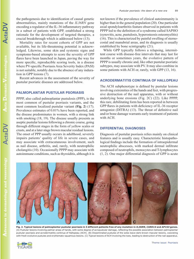
PPPP, also called palmoplantar pustulosis (PPP), is the most common of pustular psoriasis variants, and the most common localized pustular variant (Fig. 2) (17). Prevalence estimates of 0.01% have been reported, and the disease predominates in women, with a strong link with smoking (18, 19). The disease usually presents as aseptic pustular lesions following a chronic course, going through different stages in the form of yellow scales or crusts, and at a later stage brown macular residual lesions. The onset of PPP usually occurs in adulthood, severely impairs patients’ quality of life in severe cases, and may associate with extracutaneous involvement, such as nail disease, arthritis, and, rarely, with neutrophilic cholangitis (10). Occasionally PPPP may associate with autoimmune conditions, such as thyroiditis, although it is
not known if the prevalence of clinical autoimmunity is higher than in the general population (20). One particular axial spondyloarthritis feature observed in patients with PPPP led to the definition of a syndrome called SAPHO (synovitis, acne, pustulosis, hyperosteosis osteomyelitis) (16). This is characterized by painful swelling of sterno-costal and manubrial areas, and its diagnosis is usually established by bone scintigraphy (21).
While GPP typically follows a relapsing, intermit-tent course with disease-free intervals that may last for months or sometimes years, the evolutive pattern of PPPP is usually chronic and, like other pustular psoriatic subtypes, may associate with PV. It may also combine in some patients with ACH or, rarely, with GPP (13, 16).
ACRODERMATITIS CONTINUA OF HALLOPEAU
The ACH subphenotype is defined by pustular lesions involving extremities of the hands and feet, with progres-sive destruction of the nail apparatus, with or without underlying bone erosions (Fig. 2C) (22). Like PPPP, this rare, debilitating form has been reported in between GPP flares in patients with deficiency of IL-36 receptor antagonist (DITRA) (13). The threat of definitive nail and/or bone damage warrants early treatment of patients with ACH.
DIFFERENTIAL DIAGNOSES
Diagnosis of pustular psoriasis relies mainly on clinical features and is usually easy. Characteristic histopatho-logical findings include the formation of intraepidermal neutrophilic abscesses, with marked dermal infiltrate composed of neutrophils, monocytes and T-lymphocytes (1, 2). One major differential diagnosis of GPP is acute
Fig. 2. Typical lesions of palmoplantar pustular psoriasis in 3 different patients free of any mutation in IL36RN, CARD14 and AP1S3 genes. (A) Pustular lesions involving palmar areas of hands, with some degree of acropustular damage, reflecting the possible association between palmoplantar pustular psoriasis and acrodermatitis continua of Hallopeau (ACH). (B) Disseminated pustules of the soles leave dark-brown macular lesions, coexisting with fresh evolutive pustules and erythemato-squamous lesions. (C) Typical lesions of ACH involving the toes, leading to destruction of the nail apparatus.

Act
aDV
Act
aDV
Advan
ces
in d
erm
ato
logy a
nd v
en
ere
olo
gy
Acta
Derm
ato
-Ven
ere
olo
gic
a
H. Bachelez90
Theme issue: Psoriasis
exanthematous generalized pustular eruption (AGEP), the clinical signs and symptoms of which may be impos-sible to differentiate from GPP, but which is caused by drugs, notably by anti-infectious chemotherapy, such as pristinamycin and amoxicillin, but also other classes, such as non-steroidal anti-inflammatory drugs, among others (23, 24). The recent detection in patients with AGEP of mutations in IL36RN, sometimes identical to the ones identified in patients with GPP/DITRA, chal-lenges the current view of AGEP and GPP being separate entities (25).
GENETICS AND IMMUNOPATHOGENIC MECHANISMS
The extreme severity of these inflammatory pustular skin disorders, especially GPP, and the existence of Mende-lian familial cases, raised the hypothesis of a monogenic model, unlike most cases of PV. This monogenic model has been robustly established by the identification of ho-mozygous or composite heterozygous, loss-of-function mutations of the IL36RN gene, which encodes a negative regulator of the IL-36 pathway, which is involved in the limitation of the intensity of skin and systemic innate immune responses. Indeed, IL36RN mutations have been found in sporadic or familial cases of GPP in patients from different geographical territories worldwide (13, 26–32). These IL36RN mutations are more prevalent in patients with GPP without plaque psoriasis, and influence the age of disease onset (32). Mutations of IL36RN lead to major structural and functional impairments of its encoded protein, the IL36 receptor antagonist (IL36Ra), leading to increased inflammatory responses resulting from unrepressed interactions of the IL36 pathway ago-nists IL36α, IL36β and IL36γ with their receptor, and from subsequent uncontrolled activation of the transcrip-tion factor NFκB (13). This results in the massive release by keratinocytes, macrophages and dendritic cells, of se-veral inflammatory mediators including CXCL8, TNFα, IL1 and IL23 (33). Dysregulated activation of the IL-36 pathway has also been shown to trigger the expansion and activation of TH17 cells in GPP (34). So far, diffe-rent scale studies of cohorts from various geographical territories have reported various prevalences of IL36RN mutations, ranging from approximately 5% to 70%, while much lower prevalences have been observed in patients with PPPP, and no causal IL36RN mutation has been detected in patients with PV without pustular psoriasis (29–32, 34). An interesting finding has been the identifi-cation of identical IL36RN mutations across the different subtypes of pustular psoriasis (35). However, mutations leading to the absence of IL-36Ra protein expression are preferentially associated with the most severe entities of GPP and AGEP, while hypomorphous mutations seem to be more prevalent in PPPP and ACH (35). The major
breakthrough in the identification of causal mutations of the IL36RN gene has been instrumental in establishing without ambiguity the autoinflammatory nature of GPP, and led to the definition of a new entity called DITRA, which differs from the previously described deficiency of IL-1 receptor antagonist (DIRA) by the presence of striking lesions of joints and bones (13, 37, 38). Finally, although causal mutations of IL36RN have not been found in patients with PV, several studies have shown deregulation of the IL-36 pathway in PV lesions (39).
The 2 other genes associated with pustular psoriasis so far are CARD14 and AP1S3. Likewise, heterozygous gain-of-function mutations of CARD14 (caspase activa-ting recruitment domain, member 14), a gene expressed in keratinocytes the protein of which interacts with Bcl 10, a positive regulator of NFκB activation, has been shown to be primarily involved in autosomal dominant forms of PV and in some patients with pityriasis rubra pilaris (40–43). The Adaptor Related Protein Complex 1 subunit sigma 3 (AP1S3) gene has been also found to be heterozygously mutated in patients with different subty-pes of pustular psoriasis, mainly GPP and ACH, leading to structural and functional alterations of the protein, a member of the Adaptor Protein 1 (AP1) family, contri-buting to deregulation of skin innate immune responses (42, 44, 45). Likewise, it is notable that some patients have “digenic” features, e.g. a pattern characterized by mutations reported to be damaging in 2 of the 3 genes identified so far (32). Further identification of other genes, especially in GPP, will undoubtedly complement the current genetic map of pustular psoriasis, and is likely to greatly contribute to personalized therapeutic approaches.
THERAPEUTICS: TOWARDS PRECISION MEDICINE
The low prevalence of pustular psoriasis and the capri-cious course of the disease with unpredictable flaring frequency in many cases of GPP, explain the low level of scientific evidence regarding treatment efficacy. Indeed, although topical steroids and/or vitamin D derivatives, used as single agents or combined, or phototherapies are still used in mild forms of pustular skin disease with limited involved body surface area, pustular psoriasis often requires systemic therapy. In PPPP, cyclosporine has the highest level of evidence for efficacy, while there is weak or very weak evidence, respectively, for acitretin and methotrexate (46–48). More recently, randomized, placebo-controlled phase 3 clinical trials have been conducted in PPPP with secukinumab and guselkumab, targeted inhibitors of IL17A and IL23p19, respectively (49, 50). However, neither drug showed clinically re-levant superiority over placebo at the population level, suggesting that the IL23/IL17 pathway is not the major

Act
aDV
Act
aDV
Advan
ces
in d
erm
ato
logy a
nd v
en
ere
olo
gy
Acta
Derm
ato
-Ven
ere
olo
gic
a
91Pustular psoriasis: the dawn of a new era
Theme issue: Psoriasis
pathogenic axis in pustular disease (49, 50). Randomized clinical trials are currently being conducted in PPPP with inhibitors of cytokines of the IL-1 family, the most ad-vanced programme investigating the efficacy and safety of anakinra, the recombinant form of the IL-1 receptor antagonist, based on encouraging responses in isolated cases, including with ACH (51, 52).
There is even less available evidence in GPP, due to the previously exposed challenges, but also to the spon-taneously self-remitting evolutive pattern of acute GPP flare. Thus positive responses reported with conventional or biological drug interventions in the setting of retro-spective, or open-labelled prospective trials, should be considered with caution. Therefore, although high-dose steroids, cyclosporine, acitretin and apheresis have been promoted for severe acute flares, and although some bio-logics, such as IL17 inhibitors, have been approved for GPP in Japan, these interventions lack randomized con-trolled studies to assess the magnitude of their efficacy effect (53, 54). Furthermore, the efficacy of anakinra, the recombinant form of the IL-1 receptor antagonist, has been reported only in a case series of GPP with or without DITRA, reporting most often transient and partial responses (55, 56). These cases should be con-fronted with the outstanding efficacy of IL-1 inhibitors in patients with DIRA, emphasizing the fine specificity of pathogenic pathways across different monogenic au-toinflammatory syndromes of the skin (36). Therefore, the emerging development of specific inhibitors of the IL-36 pathway in GPP and PPPP is not surprising. The most advanced development investigates an anti-IL-36 receptor monoclonal antibody, which, administered as a single intravenous dose, proof-of-concept study in acute GPP, showed very encouraging results in 7 patients, only 3 of whom were carrying IL36RN mutations (57). Ongoing phase 2 and 3 studies will provide a more accurate picture of the efficacy and safety of this new targeted strategy.
CONCLUSION
Pustular psoriasis is a very challenging spectrum of auto-inflammatory skin diseases, with both clinical and genetic heterogeneity. However, the increasing collaboration between medical experts and scientists is encouraging in enabling the better nosological classification of subenti-ties, as well as the development of specific therapeutic strategies, approximately 100 years after the pioneering description of GPP by von Zumbusch (58).
ACKNOWLEDGEMENTSHB had paid activities as advisor, speaker or consultant for Abb-vie, Almirall, Amgen, Biocad, Boehringer-Ingelheim, Celgene, Eli-Lilly, Janssen, Leo Pharma, Mylan, Novartis, Pfizer, Sun Pharmaceuticals and UCB.
REFERENCES1. Nestle FO, Kaplan DH, Barker J. Psoriasis. N Engl J Med
2009; 361: 496–509.2. Naldi L, Gambini D. The clinical spectrum of psoriasis. Clin
Dermatol 2007; 25: 510–518.3. Augey F, Renaudier P, Nicolas JF. Generalized pustular pso-
riasis (Zumbusch): a French epidemiological survey. Eur J Dermatol 2006; 16: 669–673.
4. Ohkawara A, Yasuda H, Kobayashi H, Inaba Y, Ogawa H, Hashimoto I, et al. Generalized pustular psoriasis in Japan: two distinct groups formed by differences in symptoms and genetic background. Acta Derm Venereol 1996: 76: 68–71.
5. Baker H, Ryan TJ. Generalized pustular psoriasis. A clinical and epidemiological study of 104 cases. Br J Dermatol 1968; 80: 771–793.
6. Zelickson BD, Muller SA. Generalized pustular psoriasis. A review of 63 cases. Arch Dermatol 1991; 127: 1339–1345.
7. Umezawa Y, Ozawa A, Kawasima T, Shimizu H, Terui T, Ta-gami H, et al. Therapeutic guidelines for the treatment of generalized pustular psoriasis (GPP) based on a proposed classification of disease severity. Arch Dermatol Res 2003; 295: S43–S54.
8. Viguier M, Allez M, Zagdanski AM, Bertheau P, de Kerviler E, Rybojad M, et al. High frequency of cholestasis in generalized pustular psoriasis: evidence for neutrophilic involvement of the biliary tract. Hepatology 2004; 40: 452–458.
9. Ryan TJ, Baker H. The prognosis of generalized pustular psoriasis. Br J Dermatol 1971; 85: 407–411.
10. Kawana S, Nishiyama S. Pustular psoriasis and aseptic puru-lent arthritis: possible role of leukotrienes B4 and C4 in a flare of synovitis. Dermatology 1995; 190: 35–38.
11. Sadeh JS, Rudikoff D, Gordon ML, Bowden J, Goldman BD, Lebwohl M. Pustular and erythrodermic psoriasis complica-ted by acute respiratory distress syndrome. Arch Dermatol 1997; 133: 747–750.
12. Dieude P, Sbidian E, Viguier M, Zafrani E, de Bazelaire C, Da-widowicz K, et al. Neutrophilic cholangitis in psoriasis vulgaris and psoriatic arthritis. Br J Dermatol 2013; 168: 216–218.
13. Marrakchi S, Guigue P, Renshaw BR, Puel A, Pey XY, Fraitag S, et al. Interleukin-36-receptor antagonist deficiency and generalized pustular psoriasis. N Engl J Med 2011; 365: 620–628.
14. Oumeish OY, Parish JL. Impetigo herpetiformis. Clin Dermatol 2006; 24: 101–104.
15. Trivedi M, Vaughn AR, Murase JE. Pustular psoriasis of preg-nancy. Int J Womens Health 2018; 10: 109–115.
16. Sbidian E, Madrange M, Viguier M, Salmona M, Duchatelet S, Hovnanian A, et al. Respiratory virus infection triggers acute psoriasis flares across different clinical subtypes and genetic backgrounds. Br J Dermatol 2019; 181: 1304–1306.
17. Farley E, Masrour S, Mc Key J, Menter A. Palmoplantar pso-riasis: a phenotypical and clinical review with introduction of a new quality-of-life assessment tool. J Am Acad Dermatol 2009; 60: 1024–1031.
18. De Waal JC, van de Kerkhof PC. Pustulosis palmoplantaris is a disease distinct from psoriasis. J Dermatol Treat 2011; 22: 102–105.
19. Parisi R, Symmons DP, Griffiths CE, Ashcroft DM. Global epidemiology of psoriasis: a systematic review of incidence and prevalence. J Invest Dermatol 2013; 133: 377–385.
20. Kobayashi T, Naka W, Harada T, Nishikawa T. Association of the acral type of pustular psoriasis, Sjögren’s syndrome, systemic lupus erythematosus, and Hashimoto’s thyroiditis. J Dermatol 1995; 22: 125–128.
21. Nguyen MT, Borchers A, Selmi C, Naguwa SM, Cheema G, Gershwin ME. SAPHO syndrome. Semin Arthritis Rheum 2012; 42: 254–265.
22. Puig L, Barco D, Vilarrasa E, Alomar A. Treatment of acro-dermatitis continua of Hallopeau with TNF-blocking agents: case report and review. Dermatology 2010; 220: 154–158.
23. Sidoroff A, Halevy S, Bavinck JN, Vaillant L, Roujeau JC. Acute generalized exanthematous pustulosis (AGEP): a clinical reaction pattern. J Cutan Pathol 2001; 28: 113–119.
Act
aDV
Act
aDV
Advan
ces
in d
erm
ato
logy a
nd v
en
ere
olo
gy
Acta
Derm
ato
-Ven
ere
olo
gic
a
H. Bachelez92
Theme issue: Psoriasis
24. Sidoroff A, Dunant A, Viboud C, Halevy S, Bavinck JN, Naldi L, et al. Risk factors for acute generalized exanthematous pustulosis (AGEP) – results of a multinational case-control study (EuroSCAR). Br J Dermatol 2007; 157: 989–996.
25. Navarini AA, Valeyrie-Allanore L, Setta-Kaffetzi N, Barker JN, Capon F, Creamer D, et al. Rare variations in IL36RN in severe adverse drug reactions manifesting as acute gene-ralized exanthematous pustulosis. J Invest Dermatol 2013; 133: 1904–1907.
26. Onoufriadis A, Simpson MA, Pink AE, Di Meglio P, Smith CH, Pullabhatla V, et al. Mutations in IL36RN/IL1F5 are associated with the severe episodic inflammatory skin disease known as generalized pustular psoriasis. Am J Hum Genet 2011; 89: 432–437.
27. Farooq M, Nakai H, Fujimoto A, et al. Mutation analysis of the IL36RN gene in 14 Japanese patients with generalized pustular psoriasis. Hum Mutation 2013; 34: 176–183.
28. Sugiura K, Takemoto A, Yamaguchi M, Fujikawa H, Matsuy-ama A, Kariya N, et al. The majority of generalized pustular psoriasis without psoriasis vulgaris is caused by deficiency of interleukin-36 receptor antagonist. J Invest Dermatol 2013; 133: 2514–2521.
29. Wang TS, Chiu HY, Hong JB, Chan CC, Lin SJ, Tsai TF. Cor-relation of IL36RN mutation with different clinical features of pustular psoriasis in Chinese patients. Arch Dermatol Res 2016; 308: 55–63.
30. Tauber M, Bal E, Pei XY, Madrange M, Khelil A, Sahel H, et al. IL36RN Mutations affect protein expression and function: a basis for genotype-phenotype correlation in pustular di-seases. J Invest Dermatol 2016; 136: 1811–1819.
31. Hussain S, Berki DM, Choon SE, Burden AD, Allen MH, Aro-stegui JI, et al. IL36RN mutations define a severe autoin-flammatory phenotype of generalized pustular psoriasis. J Allergy Clin Immunol 2015; 135: 1067–1070.
32. Twelves S, Mostafa A, Dand N, Burri E, Farkas K, Wilson R, et al. Clinical and genetic differences between different pustular psoriasis subtypes. J All Clin Immunol 2019; 143: 1021–1026.
33. Johnston A, Xing X, Wolterink L, Barnes DH, Yin Z, Reingold L, et al. IL-1 and IL-36 are dominant cytokines in generalized pustular psoriasis. J All Clin Immunol 2017; 140: 109–120.
34. Setta-Kaffetzi N, Navarini AA, Patel VM, Pullabhatla V, Pink AE, Choon SE, et al. Rare pathogenic variants in IL36RN underlie a spectrum of psoriasis-associated pustular pheno-types. J Invest Dermatol 2013; 133: 1366–1369.
35. Arakawa A, Vollmer S, Besgen P, Galinski A, Summer B, Kawakami Y, et al. Unopposed IL36 activity promotes clonal CD4+ T-cell responses with IL17A production in generalized pustular psoriasis. J Invest Dermatol 2018; 138: 1338–1347.
36. Berki DM, Mahil, SK, Burden AD, Trembath RC, Smith CH, Capon F, et al. Loss of IL36RN function does not confer susceptibility to psoriasis vulgaris. J Invest Dermatol 2014; 134: 271–273.
37. Aksentijevich I, Masters SL, Ferguson PJ, Dancey P, Frenkel J, van Royen-Kerkhoff A, et al. An autoinflammatory disease with deficiency of the interleukin-1-receptor antagonist. N Engl J Med 2009; 360:2426–2437.
38. Reddy S, Jia S, Geoffrey R, Lorier R, Suchi M, Broeckel U, et al. An autoinflammatory disease due to homozygous deletion of the IL1RN locus. N Engl J Med 2009; 360: 2438–2444.
39. Tian S, Krueger JG, Li K, Jabbari A, Brodmerkel C, Lowes MA, et al. Meta-analysis derived (MAD) transcriptome of psoriasis defines the “core” pathogenesis of disease. PLoS One 2012; 7: e44274.
40. Berki DM, Liu L, Choon SE, Burden AD, Griffiths CEM, Nava-rini AA, et al. Activating CARD14 mutations are associated with generalized pustular psoriasis but rarely account for familial recurrence in psoriasis vulgaris. J Invest Dermatol 2015; 135: 2964–2970.
41. Mössner R, Frambach Y, Wilsmann-Theis D, Löhr S, Jacobi A, Weyergraf A, et al. Palmoplantar pustular psoriasis is associated with missense variants in CARD14, but not with loss-of-function mutations in IL36RN in European patients J
Invest Dermatol 2015; 135: 2538–2541.42. Jordan CT, Cao L, Robertson ED, Duan S, Helms CA, Nair RP,
et al. Rare and common variants in CARD14, encoding an epidermal regulator of NF-kappaB, in psoriasis. Am J Hum Genet 2012; 90: 796–808.
43. Fuchs-Telem D, Sarig O, van Steensel MA, Isakov O, Israeli S, Nousbeck J, et al. Familial pityriasis rubra pilaris is caused by mutations in CARD14. Am J Hum Genet 2012; 91: 163–170.
44. Setta-Kaffetzi N, Simpson MA, Navarini AA, Patel VM, Lu HC, Allen MH, et al. AP1S3 mutations are associated with pustular psoriasis and impaired Toll-like receptor 3 trafficking. Am J Hum Genet 2014; 94: 790–797.
45. Mahil SK, Twelves S, Farkas K, Setta-Kaffetzi N, Burden AD, Gach JE, et al. ap1s3 mutations cause skin autoinflamma-tion by disrupting keratinocyte autophagy and up-regulating IL-36 production. J Invest Dermatol 2016; 136: 2251–2259.
46. Marsland AM, Chalmers R, Hollis S, Leonardi-Bee J, Griffiths CE. Interventions for chronic palmoplantar pustulosis. Co-chrane Database Syst Rev 2006; 1:CD001433.
47. Robinson A, Van Vorhees AS, Hsu S, Korman NJ, Lebwohl MG, Bebo BF, et al. Treatment of pustular psoriasis: From the Medical Board of the National Psoriasis Foundation. J Am Acad Dermatol 2012; 67: 279–288.
48. Sevrain M, Richard MA, Barnetche T, Rouzaud M, Villani AP, Paul C, et al. Treatment for palmoplantar pustular psoriasis: systematic literature review, evidence-based recommen-dations and expert opinion. J Eur Acad Dermatol Venereol 2014; 28: 13–16.
49. Mrowietz U, Bachelez H, Burden AD, Rissler M, Sieder C, Orsenigo R, et al. Secukinumab for moderate-to-severe palmoplantar pustular psoriasis: results of the 2PRECISE study. J Am Acad Dermatol 2019; 80: 1344–1352.
50. Terui T, Kobayashi S, Okubo Y, Murakami M, Zheng R, Morishima H, et al. Efficacy and safety of guselkumab in Japanese patients with palmoplantaru pustulosis: a phase 3 randomized clinical trial. JAMA Dermatol 2019 Jul 3. [Epub ahead of print].
51. Cornelius V, Wilson R, Cro S, Barker J, Burden D, Griffiths CEM, et al. A small population, randomised, placebo-control-led trial to determine the efficacy of anakinra in the treatment of pustular psoriasis: study protocol for the APRICOT trial. Trials 2018; 19: 465.
52. Lutz V, Lipsker D. Acitretin- and tumor necrosis factor inhibitor-resistant acrodermatitis continua of Hallopeau re-sponsive to the interleukin 1 receptor antagonist anakinra. Arch Dermatol 2012; 148: 297–299.
53. Imafuku S, Honma M, Okubo Y, Komine M, Ohtsuki M, Morita A, et al. Efficacy and safety of secukinumab in patients with generalized pustular psoriasis: a 52-week analysis from phase III open-label multicenter Japanese study. J Dermatol 2016; 43: 1011–1017.
54. Sano S, Kubo H, Morishima H, Goto R, Zheng R, Nakagawa H. Guselkumab, a human interleukin-23 monoclonal antibody in Japanese patients with generalized pustular psoriasis and erythrodermic psoriasis: efficacy and safety analyses of a 52-week, phase 3, multicenter, open-label study. J Dermatol 2018; 45: 529–539.
55. Viguier M, Guigue P, Pages C, Smahi A, Bachelez H. Successful treatment of generalized pustular psoriasis with the inter-leukin-1-receptor antagonist anakinra: lack of correlation with IL1RN mutations. Ann Intern Med 2010; 153: 66–67.
56. Hüffmeier U, Wätzold M, Mohr J, Schön MP, Mössner R. Suc-cessful therapy with anakinra in a patient with generalized pustular psoriasis carrying IL36RN mutations. Br J Dermatol 2014; 170: 202–204.
57. Bachelez, Choon SE, Marrakchi S, Burden AD, Tsai TF, Morita A, et al. Inhibition of the interleukin-36 pathway for the treatment of generalized pustular psoriasis. N Engl J Med 2019; 380: 981–983.
58. Navarini AA, Burden AD, Capon F, Mrowietz U, Puig L, Köks S, et al. European consensus statement on phenotypes of pustular psoriasis. J Eur Acad Dermatol Venereol 2017; 31: 1792–1799.