d-aspartate exerts an opposing role upon age …1 d-aspartate exerts an opposing role upon...
TRANSCRIPT

1
D-aspartate exerts an opposing role upon age-dependent NMDAR-related
synaptic plasticity and memory decay
Francesco Errico1,*, Robert Nisticò2,3*, Antimo D’Aniello 4,*, Sandra Sivilia5, Michela Giustizieri2,
Francesco Napolitano1, Enza Topo4, Giorgio Bernardi 2,6, Laura Calzà5, Nicola B. Mercuri2,6,
Alessandro Usiello1,7,#
1Laboratory of Behavioural Neuroscience, CEINGE Biotecnologie Avanzate, Naples, Italy; 2
Centro Europeo per la Ricerca sul Cervello (CERC)/Fondazione Santa Lucia, Rome, Italy; 3Department of Pharmacobiology, University of Calabria and Experimental Neuropharmacology Center “Mondino-Tor Vergata,” Fondazione C. Mondino-IRCCS, Rome, Italy; 4 Department of Neurobiology, Stazione Zoologica “A. Dohrn”, Naples, Italy; 5 University of Bologna, Italy; 6Clinica Neurologica, Università Tor Vergata, Rome, Italy; 7Department of Health Science, University of Molise, Italy. * F. E., S. R. and A. D. contributed equally to this work.
#Corresponding author: Alessandro Usiello, CEINGE - Biotecnologie Avanzate, Via Comunale
Margherita 482, 80145 Naples, Italy. Tel. (+39 08) 13 73 78 99; Fax (+39 08) 13 73 78 08; Email:

2
In the present study, we demonstrated that D-aspartate acts as an in vitro and in vivo
neuromodulatory molecule upon hippocampal NMDAR transmission. Accordingly, we showed
that this D-amino acid, widely expressed during embryonic phase, was able to strongly influence
hippocampus-related functions at adulthood. Thus, while up-regulated levels of D-aspartate
increased LTP and spatial memory in four-month old adult mice, the prolonged deregulation of
this molecule in thirteen-month old animals induced a substantial acceleration of age-dependent
decay of synaptic plasticity and cognitive functions. Moreover, we highlighted a role for D-
aspartate in enhancing NMDAR-dependent synaptic plasticity through an inducible “turn-on/turn-
off-like mechanism”. Strikingly, we also showed that D-aspartate, when administered to aged
mice, strongly rescued their physiological synaptic decay and attenuated their cognitive
deterioration. In conclusion, our data suggest a tantalizing hypothesis for which this in-embryo-
occurring D-amino acid, might disclose plasticity windows in the aging brain.

3
INTRODUCTION
Cognitive processes are believed to depend on changes in synaptic efficacy in certain key brain
regions, including the hippocampus1. Among different forms of synaptic plasticity, hippocampal N-
methyl D-aspartate receptors (NMDARs)-dependent Long-Term Potentiation (LTP) has been
proposed as one of the most probable neuronal substrates underlying plastic changes associated
with spatial learning and memory1-4. Accordingly, antagonists of NMDARs blocked the induction
of LTP in hippocampal slices5-7 and impaired reference memory in rats8, 9. Similarly, synaptic
memory and cognition were influenced by genetically manipulated NMDARs. Indeed,
hippocampus-related synaptic plasticity and learning were improved in mice over-expressing NR2B
subunit of these receptors10. Intriguingly, up-regulation of this subunit has been also described
beneficial for improving cognitive functions in aged mice11. On the other hand, extensive evidence
suggests that the hippocampus is the main brain region sensitive and vulnerable to age-related loss
of functional synapses and NMDAR-mediated responses12. Age-related deficits in reference
memory have been explored across a wide variety of species, especially in rodents, where a decline
in the hippocampal ability to produce plastic synaptic changes has been documented during aging13-
15. In this respect, alterations of synaptic connectivity and plasticity are thought to be important
neurobiological determinants in causing the memory deterioration observed in old animals12, 16.
According to this view, aged rats exhibit reduced NMDAR binding sites, which correlate with their
impairments in synaptic plasticity and cognition17.
Although in the past free D-amino acids were considered to be involved only in the “bacterial
world”, recently, mounting evidence pointed out their neuromodulatory role in controlling neuronal
functions in mammals. Specifically, the D-amino acid D-serine, by acting as an endogenous co-
agonist at glicyne-binding-sites of NMDARs, has been shown to be able to rescue age-related
impairment of NMDAR-mediated synaptic potentials in old mice and rats18, 19. Besides D-serine,
today well characterized for its pharmacological properties and clinical implications in
schizophrenia treatment20, another D-amino acid, namely D-aspartate, occurs in the mammalian
4
brain. In contrast to the former, the central role of the latter remains, so far, an issue of controversy.
However, neuro-anatomical and biochemical analyses indicated that in both humans and rodents, D-
aspartate occurs at abundant levels in developing brain to decrease at low amounts during postnatal
life21, 22. This time-dependent regulation of D-aspartate brain content has been linked to the
concomitant postnatal expression of D-Aspartate Oxidase (DDO), the only enzyme known so far to
be responsible for its degradation23, 24. Moreover, in vitro studies pointed out that this D-amino acid
binds NMDARs25. However, its in vivo relevance in modulating glutamatergic neurotransmission
remains unclear, since the affinity of these receptors for D-aspartate is 10 times lower than for
glutamate. Based on these findings, in the present work, we challenged the hypothesis by which
“forcing” higher D-aspartate levels in aged animals, this in-embryo-occurring molecule might
disclose neuronal plasticity features aimed to reduce the physiological synaptic and cognitive
deterioration appearing during brain aging.

5
RESULTS
D-aspartate triggered NMDAR-dependent and NMDAR-independent currents.
The pharmacological features of D-aspartate were studied on CA1 pyramidal neurons. NMDA
induced a remarkable inward current (INMDA), which remained constant after repeated applications
of the agonist. Perfusion with NVP, a selective NR2A blocker, and cis-PPDA, a NR2C-D
antagonist, reversibly reduced INMDA to great extent (Fig. 1a,b). In the same neurons, INMDA was
largely blocked, in an irreversible manner, by Ro 25-6981, a selective NR2B antagonist (Fig. 1
a,b). Similarly, also D-aspartate-mediated inward currents (ID-Asp) were reduced by each NR2
selective antagonist (Fig. 1 a,b). Interestingly, while INMDA was totally blocked by the simultaneous
combination of all NR2 selective antagonists or by the non-competitive NMDAR antagonist,
MK801 (Fig. 1b), under the same conditions D-aspartate was still able to excite CA1 pyramidal
neurons (Fig. 1b). Next, we studied the calcium dynamics mediated by D-aspartate, performing
voltage-clamp recordings associated with microfluorometry with the use of a pipette solution filled
with the calcium-sensitive dye fura-2. As expected, puff applications of D-aspartate caused an
inward current that was associated with a transient increase of [Ca2+]i (Fig. 1c). This current was
largely, but not totally, antagonized in a reversible manner by a high concentration of D-AP5 or
irreversibly by MK801 (Fig. 1c). Finally, a further set of experiments conducted in the continuous
presence of MK801 showed that the residual ID-Asp remained constant even with the co-application
of the sodium-dependent glutamate/aspartate transporter inhibitor DL-TBOA (Fig. 1d). This current
is rather inhibited by switching the normal ACSF solution to one not containing calcium ions (Fig.
1d). Notably, chelation of intracellular Ca2+ with EGTA, blockade of all glutamate receptors with
CNQX and MCPG, and the use of non-specific Voltage Gated Calcium Channel (VGCC) blocker
cadmium chloride, all added to the superfusion medium, did not affect the remaining Excitatory
Post Synaptic Currents (EPSC) (Fig. 1d). Therefore, our data indicate that the action of D-aspartate
on CA1 pyramidal neurons occurs via Ca2+ currents through, at least, two alternative routes based
on NMDAR-dependent and NMDAR-independent pathways.

6
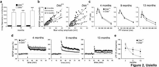
Lack of D-Aspartate Oxidase induced a dramatic acceleration of age-dependent decay of
hippocampal synaptic transmission and plasticity.
We have recently demonstrated that higher D-aspartate levels, in the hippocampus of two-month
old Ddo-/- mice, enhance the NMDAR-dependent form of LTP, while maintain synaptic
transmission unaltered26. Based on this study, here we determined the age-related effect of Ddo
gene ablation on synaptic transmission and plasticity in area CA1.
Firstly, we examined D-aspartate levels in the hippocampus of Ddo-/- animals and their control
littermates at 4, 9 and 13 months of age. According to the pivotal role of DDO in controlling D-
aspartate levels, HPLC analysis indicated a strong and persistent hippocampal up-regulation of this
D-amino acid content throughout all ages tested (Fig. 2a).
Next, we measured basal synaptic transmission on hippocampal slices from mutants and controls, at
the same ages at which we evaluated D-aspartate concentrations. At 4 months of age, Ddo+/+ and
Ddo-/- mice showed similar trendlines (Fig. 2b). Also at 9 months of age, both genotypes exhibited
comparable fEPSP slopes and fiber volley amplitudes at all stimulus intensities tested (Fig. 2b). In
contrast, thirteen-month old Ddo-/- animals had significantly reduced maximum fEPSPs, relative to
Ddo+/+ controls (Fig. 2b). Therefore, these results show that the age-dependent decline in fast
glutamatergic transmission is exacerbated in absence of DDO enzyme. We then investigated paired-
pulse facilitation (PPF), a measure of short-term plasticity. Interestingly, no differences in the
paired-pulse ratio between Ddo-/- and Ddo+/+ mice were observed in the three age-group tested (Fig.
2c). Finally, we explored the effects of deregulated levels of D-aspartate on age-related LTP at CA1
synapses. At 4 months of age, we showed a significant increase in the magnitude of LTP in Ddo-/-
animals, compared to control mice (Fig. 2d). In contrast, LTP was impaired either in nine-month
(Fig. 2d) and, even more severely, in thirteen-month old mutants, when compared to age-matched
controls (Fig. 2d). Interestingly, at 13 months of age, a physiological decay of LTP appeared also in

7
Ddo+/+ mice that, in any case, never reached the dramatic impairment observed in mutant CA1
synapses (Fig. 2e).
Ddo-/- mice displayed spatial memory enhancement or worsening in an age-dependent fashion.
As putative behavioural correlate of hippocampal NMDAR-dependent synaptic plasticity, we
investigated ontogenetic changes in learning and memory abilities of Ddo-/- mice, at 4, 9 and 13
months of age, in a reference memory task of the Morris water maze paradigm.
During acquisition phase, we found similar escape latencies between four-month old Ddo+/+ and
Ddo-/- animals (Fig. 3a). In order to evaluate their spatial memory formation, we performed two
retention tests, after a three-day (short) or five-day (long) training period. Interestingly, in probe 1,
knockout mice displayed a forward ability in remembering the correct location of the platform,
compared to controls (Fig. 3b). However, after a longer training exposure, both genotypes showed a
comparable bias spatial search in the target quadrant (Fig. 3c). Then, in order to analyze the ability
of the mice to erase previous spatial information and planning a new navigation strategy, we moved
the platform to the opposite position. Also in the reversal phase, both groups displayed similar
learning abilities (Fig. 3a). Moreover, the results in the retention tests confirmed in Ddo-/- mice an
improved spatial memory after a short training exposure, compared to controls (Fig. 3d). In fact,
although both groups showed a search preference for the new goal quadrant, Ddo-/- mice
significantly spent more time in this location, compared to controls. After additional training, both
genotypes showed comparable preferential search for the target quadrant (Fig. 3e).
Also at 9 months of age, both genotypes evidenced comparable spatial learning aptitudes in both
acquisition and reversal phases (Fig. 3f). Importantly, in probe 1, Ddo-/- group did not confirm
improved spatial memory, evidenced at 4 months of age (Fig. 3g), since the correct position of the
platform was acquired only after a longer training (Fig. 3h). Moreover, in the reversal phase, spatial
retention in the new goal quadrant occurred only in Ddo+/+ animals after five-day training exposure
(Fig. 3i,j), thus highlighting in mutants the occurrence of mild memory deterioration at this age.

8
The worsening of cognitive aptitudes of Ddo-/- animals was further evidenced in thirteen-month old
mice. While comparable spatial learning curves were observed for both genotypes during
acquisition (Fig. 3k), only Ddo+/+ animals showed preserved spatial memory abilities (Fig. 3l,m). In
the reversal phase, a prominent overall impairment in spatial learning and memory was observed in
mutants (Fig. 3k,n,o).
Alteration of hippocampal synaptic markers in Ddo-/- mice.
Here, we analysed whether synaptic and cognitive decline were mirrored by morphological
variations in the hippocampus of Ddo-/- mice. First, toluidine staining showed no distinguishing
abnormalities in the pyramidal cell layer and dentate gyrus (DG) of the hippocampus from Ddo+/+
and Ddo-/- mice, at any age. This was confirmed by the counting of Neu-N-IR profiles (data not
shown). A diffuse age-dependent increase in GFAP-IR (Fig. 4h,i,m,n,s) and astrocyte hypertrophy
(Fig. 4g,l), were observed in the CA3 subfield of the hippocampus, regardless genotype.
Nevertheless, in the same area, these changes were selectively associated with a reduced
immunostaining for the presynaptic marker synaptophysin in thirteen-month old Ddo-/- mice (Fig.
4s) and, interestingly, were anticipated by a decreased staining for the postsynaptic marker MAP2
in four-month old mutants (Fig. 4s). Synaptophysin significantly decreased also in knockout mice
in CA1/2 area, at both 4 and 13 months of age (Fig. 4o-s), and in DG of aged mice, compared to
respective controls (Fig. 4s). Similarly, a reduced pattern of staining for MAP2 was detected in
CA1/2 field of elderly Ddo-/- mice, compared to age-matched Ddo+/+ animals (Fig. 4a,b,d,e,s).
Remaining dendrites resulted irregular in shape and size (Fig. 4c,f). Overall, morphological
alterations found in the hippocampus of old mutants are consistent with the behavioural and
electrophysiological decline observed during aging.
D-aspartate induced a transient and reversible modulatory effect on NMDAR-dependent
LTP.
9
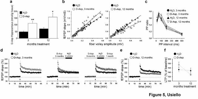
To study the in vitro and in vivo consequences associated to non-physiological increased levels of
D-aspartate, without interfering with Ddo gene expression, we recently developed a
pharmacological mouse model based on one-month oral administration of this D-amino acid26.
Taking advantage of this model, we further examined the neurochemical and electrophysiological
consequences of a more prolonged chronic administration. According to our previous report, we
found that D-aspartate oral treatment overall resulted in a significant increase of its hippocampal
content. However, despite the different time-exposure extent, we failed to detect gross differences
in D-aspartate amount following moderate (3 months) and long-term (12 months) delivery periods
(Fig. 5a).
After a three-month administration, C57BL/6J treated mice showed a higher I-O trend, although not
significant, compared to their untreated controls (Fig. 5b). Similarly, chronic one-year
administration of D-aspartate produced only a slight decrease in basal transmission. Indeed,
differently to what observed in mutants, this effect did not reach statistical significance (Fig. 5b),
demonstrating that even prolonged oral administration of this molecule does not perturb basal
synaptic responses. Also PPF was indistinguishable in all groups (Fig. 5c). Finally, we examined
the effects of higher D-aspartate levels on synaptic plasticity in area CA1. Notably, we showed that
three-month D-aspartate treatment induced a robust enhancement in LTP magnitude (Fig. 5d).
Interestingly, this plastic augmentation appeared to be regulated through an inducible mechanism,
since three-week withdrawal was able to reverse this potentiating effect that was, conversely, re-
instated following one-month D-aspartate re-administration (Fig. 5d). In contrast, hippocampal
slices from twelve-month chronically treated mice were dramatically unresponsive to the
conditioning train, compared to age-matched controls (Fig. 5e). Moreover, despite what was found
in Ddo+/+ mice, no signs of age-dependent LTP decay was detected in thirteen-month old untreated
C57BL/6J mice (Fig. 5f). These results mirrored, to a large extent, those obtained in Ddo-/- mice,
thus highlighting a common opposing time-dependent effect of chronic D-aspartate exposure on
hippocampal LTP. To summarize, our data clearly point out that D-aspartate is able to modulate
10
NMDAR-dependent LTP at CA1 synapses and, in turn, provide strong evidence about its role in
influencing age-dependent changes in synaptic plasticity.
Chronic D-aspartate oral administration in C57BL/6J mice improved spatial memory in
adulthood without perturbing cognitive functions in elderly phases.
Spatial learning and memory abilities in D-aspartate treated C57BL/6J mice were analyzed after its
three-month or twelve-month chronic oral administration, according to the same behavioural
protocol adopted for mutant animals. During acquisition and reversal phases, three-month D-
aspartate treatment did not produce any visible change in learning aptitudes of mice (Fig. 6a).
Importantly, after a short training, treated mice evidenced an improvement of spatial memory, as
indicated by longer search in the goal area, compared to controls (Fig. 6b). Nevertheless, such
slight difference between groups disappeared after a two-day additional training (Fig. 6c).
Similarly, during reversal phase, treated animals showed enhanced spatial memory skills, as
indicated by the longer time spent in the new goal quadrant in the probe 3, compared to non-treated
mice (Fig. 6d), while both groups were similarly able to remember the position of the new target
quadrant after five-day training (Fig. 6e).
Like the shorter treatment, also one-year D-aspartate administration did not modify spatial learning
performances of C57BL/6J mice (Fig. 6f). Nevertheless, this long-term treatment induced a lack of
the previous memory enhancement seen in three-month D-aspartate-treated mice. Indeed, no
differences were detected between D-aspartate-treated and untreated groups in each of the retention
tests performed (Fig. 6g-j).
D-aspartate rescued CA1 synaptic plasticity decline associated with aging.
We have previously shown that one-month oral administration of D-aspartate was able to strongly
increase LTP magnitude in young animals26. Here, we investigated whether such short-term
treatment might also be beneficial to counterbalance the age-related decay of hippocampus-

11
dependent functions. Since it is known that rodent females exhibit a faster rate of deterioration in
reference memory27, 28, we decided to utilize for this study twelve-month old C57BL/6J females.
Such females, treated for 1 month with D-aspartate, exhibited a significant increase of this D-amino
acid levels in their hippocampi, compared to untreated mice (Fig. 7a). Electrophysiological
experiments indicated no main differences in basal synaptic transmission and short-term plasticity,
as measured by I–O curves (Fig. 7b) and PPF (data not shown), respectively. In contrast, we found
a strong enhancement of LTP in treated mice, compared to both age-matched controls (Fig. 7c,d)
and two-month old C57BL/6J females (Fig. 7c,d). Moreover, according to gender differences
relative to age-related hippocampal functions, C57BL/6J females showed a pronounced age-
dependent LTP decay, not seen in males (Fig. 7f).
Finally, we explored spatial cognitive abilities of D-aspartate-treated aged females. Surprisingly, we
found that treated animals displayed a better spatial learning, compared to untreated mice (Fig. 7e).
In this regard, it is important to notice that the difference seen after the first training day exclusively
derives from the second daily session since, in the first test session, mice exhibited comparable
escape latencies (untreated: 84.25 ± 2.26 s; treated: 76.50 ± 4.21 s). However, according to a more
relevant age-dependent mnemonic decay likely due to hormonal influences, even after a five-day
training exposure, both treated and untreated mice did not remember the correct location of the
platform (Fig. 7f,g). During reversal phase, both groups showed a comparable learning profile (Fig.
7e). Nevertheless, a mild improvement was detected at days 4 and 5 in D-aspartate treated animals.
Conversely, no differences were found in the retention tests (Figure 7h,i).
12
DISCUSSION
While a relevant role for D-aspartate in controlling hormonal activity is well established29, its
involvement in central functions, so far, remains largely unclear. Indeed, although pharmacological
studies demonstrated that this D-amino acid binds NMDARs, its in vivo neuromodulatory action is
still actively debated. In accordance to our previous studies performed on single cells, in CA1
pyramidal neurons26, here we confirmed the existence of an inward current triggered by D-aspartate,
not fully suppressed by NMDAR antagonists. For the first time, we demonstrated that D-aspartate-
mediated excitatory currents are exclusively calcium-dependent, since they are abolished by
perfusion in a calcium-free medium. We were unable, so far, to identify any putative target, since
the chelation of intracellular calcium and the blockade of all glutamate receptors and VGCC never
affected this persistent inward current. Therefore, besides demonstrating a direct action on NMDAR
via interaction with each of NR2A-D subunits, we indicated that the physiological function of D-
aspartate is selectively mediated through a calcium-dependent signalling. Hence, we propose that
D-aspartate in the brain acts presumably through, at least, two different pathways based on
NMDAR-dependent and NMDAR-independent signalling. On the other hand, the proposed “dual”
mechanism of D-aspartate action is also supported by biochemical findings which pointed out a lack
of functional NMDARs in organs, like testis and adrenal glands, where high levels of this D-amino
acid were also detected21, 30.
The pharmacological properties of D-aspartate to act as an endogenous NMDAR agonist are in
favour of its ability in modulating, among others, synaptic plasticity occurring in the hippocampus
at CA1 synapses. Accordingly, we have recently demonstrated that non-physiological increase of
D-aspartate levels in the mouse, achieved either by generating animals with a targeted deletion of
Ddo gene or by treating mice with D-aspartate for 1 month, enhanced this NMDAR-dependent form
of LTP26. Based on this, we explored the consequences of deregulated levels of D-aspartate upon

13
hippocampus-related functions during brain aging. Remarkably, we showed that a constitutive
deregulated high level of D-aspartate in mutant hippocampi (roughly 15-fold, at each age tested) is
able to act as potent biphasic modulator of neuronal activity: potentiating synaptic memory in adult
mice (4 months), whilst dramatically accelerating age-dependent basal transmission and LTP decay
during maturity stages (9-13 months).
Notably, behavioral studies failed, so far, to reveal consistent learning and memory changes induced
by D-aspartate in adult animals26, 31. Therefore, in the present study, we carried out a more
“stringent” spatial cognitive demanding task. In these new setting conditions, a selective
enhancement of hippocampus-mediated reference memory was found in adult Ddo-/- mice compared
to their Ddo+/+ littermates. Interestingly, these in vivo data were confirmed in three-month D-
aspartate treated mice. Taken together, such findings are in accordance to those reported in
ddY/DAO- mutants32 and NR2B transgenic mice10 in which enhanced NMDAR-dependent synaptic
plasticity mirrored a spatial memory improvement. Moreover, according to Bach et al.33, our studies
indicated a good correspondence between the biphasic age-dependent LTP changes and spatial
memory abilities. In fact, while four-month old knockouts displayed enhanced spatial memory, in
nine- and, even more prominently, thirteen-month old Ddo-/- animals this improvement completely
disappeared and a significant worsening of cognitive abilities occurred, compared to their relative
age-matched controls.
We also showed that, when D-aspartate was administered under an intermittent regimen, this D-
amino acid triggered a transient and reversible enhancement of LTP at CA1 synapses through an
inducible “turn-on/turn-off-like mechanism”. Hence, these data further corroborated a role for D-
aspartate in regulating NMDAR signalling and, in turn, provided an attractive possibility when
considering its clinical potential.
Furthermore, similarly to aged Ddo-/- mice, long-term exposure of D-aspartate evocated a severe
decay of hippocampal synaptic plasticity. However, differently to the oldest Ddo-/- mice here tested

14
(13 months), one-year D-aspartate chronic administration in C57BL/6J animals failed to produce
deficits in basal transmission and reference memory.
Overall, our data strongly supported a neuromodulatory role for D-aspartate on glutamatergic
transmission and, in turn, showed that, according to its properties as NMDAR agonist, non-
physiological amounts of this molecule in the brain could unmask the opposite neuronal influences
exerted by this receptor34, 35. In fact, while physiological stimulation of NMDARs has a crucial
adaptive role in brain processes such as neuronal survival and synaptic plasticity, on the other hand,
their abnormal activation leads to deregulation of calcium homeostasis, responsible for loss of
synaptic memory and neurotoxicity34, 36, 37. Consistently, altered LTP has been widely documented in
hippocampal slices from several transgenic mouse models of Alzheimer Disease (AD)38-41 and in
senescence-accelerated mouse strains42. In this line, we showed that higher D-aspartate levels render
glutamatergic synapses more prone to plastic changes, in both four-month old genetic and
pharmacological animal models, while the prolonged activation of NMDARs in 9-13 month old
Ddo-/- mice and in long-term treated animals produced a strong accelerated rate of age-dependent
synaptic plasticity deterioration.
Nevertheless, our data pointed out also a direct involvement of the specific genetic background on
which genetic or pharmacological manipulation are studied43,44. In fact, C57BL/6J animals,
characterized to display very high performances in hippocampus-dependent functions, represent a
“difficult” mouse strain for easily detecting changes associated to aging45. Thus, even though D-
aspartate induced very strong changes on hippocampal LTP between three- and twelve-month
treated animals, surprisingly, only mild, if any, differences were found in their spatial memory
aptitudes.
On the contrary, based on the observations that C57BL/6J aged females manifested a more
prominent hippocampal cognitive27 and synaptic memory decline (Fig. 7c,d), compared to males,
we decided to test whether this embryonic D-amino acid may produce a “youth” influence on
neuronal plasticity in this more aging-sensitive gender. Remarkably, we showed that one-month
15
oral administration of D-aspartate to one-year old females conferred considerable stronger plastic
properties, relative to those observed in age-matched controls. The entity of this amelioration was
even more surprising comparing the level of LTP of these D-aspartate-treated mice to that observed
in two-month old untreated females. Moreover, this D-amino acid also slightly improved cognitive
abilities of old females. Taken together, these evidences further extended to aged brains the ability
of this endogenous D-amino acid to modulate NMDAR-dependent functions.
In conclusion, our data demonstrated an in vitro and in vivo neuromodulatory role for D-asparate in
modulating synaptic plasticity and cognition and, in turn, highlighted a crucial, yet unknown,
implication of DDO in controlling the age-related physiological decline of hippocampus-dependent
functions. In fact, while the lack of DDO in adult mice induced an enhancement of both LTP and
spatial memory, at maturity stages it determined a strong acceleration of aging processes, which
resembled the synaptic and behavioural features described in AD-like animal models38-41. In this line,
also alterations in synaptic markers MAP2 and synaptophysin found in old mutant hippocampi, are
consistent to those seen in APP-null mice46. Moreover, the in dissociable opposite age-dependent
electrophysiological and behavioural phenotypes seen in Ddo-/- mice, let retain these animals as a
peculiar model for studying in vivo the “Yin and Yang” consequences associated to an abnormal
NMDARs signalling34. Nevertheless, the lack of gross cognitive alterations after a long-term D-
aspartate chronic administration further supported a potential interest for this D-amino acid when
considering the beneficial effects of this molecule in attenuating schizophrenia-like symptoms
induced by amphetamine and MK801 challenges (FE and AU unpublished data).

16
METHODS
Animals
Mutant mice for Ddo gene were generated as previously described23. Four-, nine- and thirteen-
month (± 3 weeks) old male wild type (Ddo+/+) and knockout (Ddo-/-) mice were used in this study
and derived from mating of heterozygous (Ddo+/-) mice, back-crossed to F5 generation to C57BL/6J
strain47. Animals were genotyped by polymerase chain reaction according to Errico et al.23.
C57BL/6J male mice were used to test the effects of chronic three- and twelve-month oral
administration of D-aspartate in neurochemical, cognitive and electrophysiological studies. D-
aspartate was delivered in drinking water at the concentration of 20 mM to 45 days old mice until
they were used for experiments. Thirteen-month old C57BL/6J female mice were used to test the
effects of chronic one-month D-aspartate oral administration, delivered between the 12th and the
13th month of their life.
Mice were housed in groups (n = 4-5) in standard cages (29 x 17.5 x 12.5 cm) at constant
temperature (22° ± 1°C) and maintained on a 12/12 h light/dark cycle, with food and water ad
libitum. Experiments were conducted in conformity with protocols approved by the veterinary
department of the Italian Ministry of Health and in accordance to the ethical and safety rules and
guidelines for the use of animals in biomedical research provided by the relevant Italian laws and
European Union’s directives (n. 86/609/EC). All efforts were made to minimize the animal’s
suffering.
HPLC analysis
Mice were killed and the hippocampus dissected and stored at -80°C. The determination of D-
aspartate was performed by HPLC technique, based on the diastereomeric separation of D-aspartate
from the L-form and other L-amino acids, as previously described48. Data were analysed using
ANOVA or two-tailed Student’s t test and data are expressed as means ± standard error of the mean
(SEM).
Morris water maze
Morris water maze was performed according to a modified version of the protocol described by
Errico et al.26. The apparatus consisted of a circular pool (100 cm in diameter), surrounded by three-
dimensional visual cues, containing opaque water at 21°C (± 1°C) with a platform (8 cm in
diameter) submerged 1 cm beneath the water surface. Mice were gently handled 5 min per day for a
week before the experiment. The acquisition phase consisted of 2 sessions per day (3 h interval
between sessions) over a five-day period. Each session was composed of 4 trials with an inter-trial
17
interval of approximately 5 min. On day 6, the platform was moved to the opposite position and the
reversal learning was monitored for 5 additional days. Overall, 4 probe tests were performed (2
tasks per each training phase) in which animals were allowed to swim for 60 s in the absence of the
platform, in order to evaluate time-dependent memory retention of mice. During acquisition phase,
the first test (probe 1) was performed before starting the 4th day of training, while the second (probe
2) was conducted at day 6, before the first session of the reversal phase. During reversal phase, the
first retention task (probe 3) was done before starting the 9th day of training, while the second
(probe 4) was conducted at the end of the reversal phase, at day 11. Each retention test was
conducted about 18 h after the second session of the previous day. In both acquisition and reversal
phases, the time to reach the target was measured. In the probe tests, the percentage of time spent in
each quadrant was recorded. A computerized video tracking system (Videotrack, Viewpoint S.A.,
Champagne au Mont d’Or, France) was used for all mentioned parameters. In the acquisition phase,
the measure of the escape latency was used as dependent variable and data were examined using
two-way ANOVA (genotype or treatment x days) with repeated measures. Data obtained in the
probe trials were analysed by one-way ANOVA followed by appropriate post-hoc comparison. A
significance level of p < 0.05 was accepted as statistically significant in all the experiments
performed. All measures are expressed as mean ± SEM. All statistical analyses were performed
with StatView software (version 5.0.1.0; SAS Institute, Cary, NC).
Electrophysiology
Patch-clamp recordings and microfluorometric measurements.
Experiments aimed at investigating the effects of D-aspartate application were performed in whole-
cell path-clamp recordings on CA1 pyramidal neurons obtained from juvenile (thirteen- to fifteen-
day-old) Wistar rats. All recordings were obtained in voltage-clamp mode (at –60 mV holding
potential), in the continuous presence of TTX (1 µM) and nifedipine (10 µM). Vibratome-cut
parasagittal slices (300 μm) were prepared from hippocampi, incubated for one hour and then
transferred to a recording chamber submerged in a continuously flowing artificial cerebrospinal
fluid (30°C, 2-3 ml/min) gassed with 95% O2- 5% CO2. The composition of the control solution
was (in mM): 126 NaCl, 2.5 KCl, 1.2 MgCl2, 1.2 NaH2PO4, 2.4 CaCl2, 11 Glucose, 25 NaHCO3.
The recording chamber was mounted on the stage of an upright microscope (Axioscope FS, Carl
Zeiss) equipped for infrared video microscope (Hamamatsu, Japan) and video microfluorometry
(ImproVision, Coventry, UK). Borosilicate glass electrodes (3-4 MΩ) were filled with (in mM): K-
Gluconate 135; KCl 10; MgCl2 2; CaCl2 0.045; EGTA 0.1; HEPES 10; ATP 2; GTP 0.3 (pH 7.3,
with KOH). Data were acquired using pClamp and Axoscope software (Axon Instruments). For
18
microfluorometric measurements, neurons were filled with calcium-sensitive dye through the patch
pipette as described previously49. The fluorescent calcium indicator (fura-2 pentapotassium salt,
Molecular Probes) was excited via a 40x water immersion objective by illumination with light
provided by a 75-W Xenon lamp. Excitation light was band-pass-filtered alternatively at 340 or 380
nm whereas emission light passed a barrier filter (500 nm) and was detected by a CCD camera
(Photonic Science, Milham, UK). Time courses of fluorescence values, obtained at 340 and 380 nm,
were calculated over the cell soma (region of interest, ROI) and corrected for background
fluorescence (BK), measured from a region > 100 μm away from the soma. Changes in
fluorescence corresponding to calcium concentrations [Ca2+] are reported as ratio values and were
calculated from the following equation: R=(F340ROI-F340BK) / (F380ROI-F380BK), where F340
and F380 are the specific fluorescence values emitted by ROI and background at 340 and 380 nm
excitation wave-lenghts. Drugs were bath-applied by switching the solution to one containing
known concentrations of drugs. D-aspartate and NMDA were also applied via a patch pipette that
was positioned in close vicinity of the cell body and was connected to a pressure application system
(Picospritzer, 20–30 psi, 0.5–1 s).
Extracellular recordings.
Ddo-/- mice at different ages and mice chronically treated with D-aspartate (3 and 12 months) were
compared to their respective controls. Hippocampal slices (400 µm thick) were prepared from
halothane-anesthetized mice as described above. A bipolar nichrome wire stimulating electrode was
used to evoke field excitatory postsynaptic potentials (fEPSPs) at approximately 50% of the
maximal fEPSP amplitude in the Schaffer collateral/commissural fiber pathway. Stimulation pulses
were delivered at 0.02 Hz for a duration of 0.02 ms. Extracellular recordings of fEPSPs in the
stratum radiatum of the CA1 region were made using glass microelectrodes filled with ACSF (5–10
MΩ). I–O, paired-pulse facilitation (PPF) and baseline-normalized LTP fEPSP slope data, were
grouped according to age and genotype. LTP was induced by a HFS protocol (1 train, 100 Hz, 1 s)
and the effect of conditioning train was expressed as the mean (±SEM) percentage of baseline EPSP
slopes measured at 60 min after stimulation protocol. Significance of data were determined using
the Student’s t test and ANOVA. The p-values were always set at a significance level of less than
0.05.
Drugs
D-aspartate, D-(-)-2-amino-5-phosphonoeptanoic acid (AP5), 5S,10R-(+)-5-Methyl-10,11-dihydro-
5H-dibenzo[a,d]cyclohepten-5,10-imine maleate (MK801), cadmium chloride, L-β-threo-benzyl-
aspartate (DL-TBOA), Tetrodotoxin (TTX), Nifedipine, N-Methyl-D-aspartic acid (NMDA) from

19
Sigma-Aldrich, Milan, Italy; (2S*,3R*)-1-(Phenanthren-2-carbonyl)piperazine-2,3-dicarboxylic
acid (cis-PPDA), 6-Cyano-7-nitroquinoxaline-2,3-dione (CNQX), (aR,bS)-a-(4-Hydroxyphenyl)-b-
methyl-4-(phenylmethyl)-1 -piperidinepropanol maleate (Ro 25-6981) and MCPG, (RS)-a-Methyl-
4-carboxyphenylglycine (MCPG) from Tocris Cookson Ltd, Bristol, UK. NVP-AAM077 was a
generous gift from Novartis Pharma AG, Switzerland.
Histology
All animals were deeply anesthetized with Ketamine (Ketalar, Parke Davis, Italy) 10 mg/kg body
weight, i.p. (+ diazepam. 2 mg/kg i.m.) and perfused through the ascending aorta with Tyrode-Ca2+
free, pH 6.9, followed by 4% paraformaldehyde in Sorensen phosphate buffer 0.1 M pH 7.0. During
perfusion, animals were bathed in ice-cold water. The brains were then removed and immersed for
90 min in the same ice-cold fixative, before being rinsed for at least 48 h in 5% sucrose in 0.1M
phosphate buffer. Brains were frozen in CO2 and 14 μm thick coronal sections were then obtained
from the dorsal hippocampus at rostro-caudal level –1.58 to –1.94 from the bregma. For
immunofluorescence experiments, sections were first incubated in 0.1 M phosphate buffered saline
(PBS) at room temperature for 10-30 min, followed by incubation at 4°C for 24h in a humid
atmosphere with the primary antibodies diluted in PBS containing 0.3% Triton X-100, v/v (rabbit
polyclonal anti-MAP-2, Santa Cruz Biotecnology, INC; rabbit polyclonal anti-GFAP,
EuroDiagnostica; goat polyclonal anti-synaptophysin, Santa Cruz Biotecnology). After rinsing in
PBS, the sections were incubated at 37°C for 30 min in a humid atmosphere with the secondary
antisera RedTM X- and CyTM2- conjugated antisera (Jackson Immunoresearch West Grove PA).
Sections were then rinsed in PBS (as above) and mounted in glycerol containing 1,4-
phenylendiamine 0.1 g/l.
Images from tissues were taken by Olympus AX70-PROVIS microscope equipped with motorized
z-stage control and F-VIEW II CCD Camera and processed using Cell^P software (Soft Imaging
System, GmbH, Munster Germany). Three sections (bilaterally) from each hippocampus were taken
for analysis of each antigen. The hippocampus was divided into three distinct fields, which were
labeled as CA1/2, CA3 and DG. Quantification of the tissue staining was performed
morphometrically by the same investigator who was blinded to the experimental protocol. Shading
error correction was performed before measurements to correct for irregularities in illumination of
the microscopic fields. Background levels were equalized and the detection threshold was tested
and kept at the same level for all samples. MAP2 and GFAP immunostainings were estimated as
fraction area, whereas synaptophysin staining was examined as optical density. For each animal and

20
histochemical staining, multiple areas were measured and the results were averaged. More details
on the sampling areas and statistical analysis are included in the legend to the figure.

21
FIGURE LEGENDS
Figure 1. D-aspartate triggers NMDAR-dependent and NMDAR-independent inward currents on
CA1 pyramidal neurons. (a) Plot of EPSC amplitude vs time showing the effect of pressure applied
NMDA and D-aspartate on CA1 pyramidal neurons. NMDA-mediated inward current (INMDA 173.6
± 47.2 pA, p < 0.001, Student’s t test) was reversibly reduced to 66.47 ± 3.18% of control by NVP
(0.4 µM) (p < 0.001) and to 67.40 ± 3.43% of control by cis-PPDA (10 µM) (p < 0.001). Ro 25-
6981 (5 µM), inhibited INMDA amplitude to 66.99 ± 3.55% of control (p < 0.05) and this effect was
irreversible at the wash. Similarly, NVP, cis-PPDA and Ro 25-6981 reduced the inward current
triggered by D-aspartate (ID-Asp 260 ± 84.4 pA) to 66.68 ± 4.88% (p < 0.001), 65.79 ± 5.61% (p <
0.001) and 67.11 ± 4.62% (p < 0.001) of control, respectively. On top of each plot, traces are
shown, acquired at the times indicated by the corresponding letters in the plots. Bars indicate the
time duration of each compound. (b) Summary bar chart representing the pharmacological effects
of different NMDAR antagonists on the inhibition of current (expressed as mean percentage of
change ± SEM) triggered by pressure application of NMDA and D-aspartate on at least seven
separate cells. Notably, INMDA amplitude is totally blocked by the simultaneous application of the
three selective NR2 antagonists (98.98 ± 0.67% of control, p > 0.1) as well as MK801 (10 µM)
(99.54 ± 5.18% of control, p > 0.1). In contrast, the ID-Asp amplitude is largely, but not totally,
inhibited when blocking all NR2 subunits (80.65 ± 0.55% of control, p < 0.05) or by MK801 (82.43
± 5.92% of control, p < 0.05). (c) Time course of [Ca2+]i in response to a puff-application of D-
aspartate (2 μM) in the presence of D-AP5 (450 μM) and MK801 (10 μM). On top, corresponding
inward currents elicited by D-aspartate are shown. On the right, images are representative examples
of a CA1 neuron patch clamped with a pipette containing fura-2 under the three different
experimental conditions (scale bar, 20 µm). (d) Normalized pooled data (n = 9) of ID-Asp amplitude
against time, in response to perfusion with MK801 (10 μM), DL-TBOA (100 μM), ACSF calcium
free, CNQX (30 µM), MCPG (200 µM) and cadmium chloride (100 µM). On top, representative
raw traces are shown, acquired at the times indicated by the corresponding letters in the plot. Bars
indicate the time duration of each treatment.
Figure 2. Age-related synaptic deterioration in Ddo-/- mice (a) D-aspartate levels in the
hippocampus of Ddo+/+ and Ddo-/- mice were measured by HPLC at 4 (Ddo+/+, n = 6; Ddo-/-, n = 3),
9 (n = 3, per genotype) and 13 (n = 3, per genotype) months of age. At each age analysed, D-
aspartate amount in mutants was significantly higher, compared to their wild type counterpart,
although no differences were found within groups at different ages. *** = p < 0.0001, compared
22
with control groups (Student’s t test). Values are expressed as mean ± SEM. Genotypes are as
indicated. (b) Input/output (I/O) curves measured by plotting the fEPSP slopes and their
corresponding presynaptic fiber volley amplitudes at increasing stimulus strengths are shown for
Ddo-/- and Ddo+/+ mice at 4, 9 and 13 months of age. At 4 months of age, the trendlines for Ddo+/+
and Ddo-/- were: y = 2.30x, R2 =0.96 and y = 2.58x, R2 = 0.88, respectively (p > 0.1). I-O were
similar also at 9 months of age (y = 1.41x, R2 =0.91 for Ddo+/+, y = 1.37x, R2 = 0.88 for Ddo-/-, p >
0.1). Conversely, at 13 months of age, basal transmission was impaired in Ddo-/-, relative to Ddo+/+
(y = 1.10x, R2 =0.89 for Ddo+/+, y = 0.68x, R2 =0.81 for Ddo-/-, p < 0.05). Each data point is an
average of two recordings. (c) Paired-pulse facilitation is not altered between the two genotypes in
the three age groups under observation (p > 0.1, for all comparisons). The facilitation ratio (slope of
second EPSP/slope of first EPSP) was plotted as a function of interpulse interval, 20, 50, 100, 200
and 500 ms. For each group, the mean ± SEM is indicated. (d) Superimposed pooled data showing
the normalized changes in field potential slope (± SEM) in Ddo+/+ and Ddo-/- mice induced by HFS
protocol. fEPSP slopes were recorded and expressed as the percentage of the pre-tetanus baseline. A
stimulation intensity that evoked 50% of maximal fEPSP response was used. There was a
significant increase (p < 0.05) in the magnitude of LTP in four-month old mutants (n = 8),
compared to control mice (n = 6) (43.9 ± 10.3% above baseline in Ddo+/+, and 61.5 ± 3.3% in Ddo-/-
). At 9 months of age, this tendency was inverted and LTP values were 39.7 ± 9.3% above baseline
in control (n = 8), and 19.5 ± 2.5% in mutant slices (n = 7). Finally, fEPSP was potentiated to only
8.5 ± 12.5% in thirteen-month old Ddo-/- mice (n = 6), compared to 30.07 ± 10.4% in age-matched
Ddo+/+ animals (n = 7). At 9 and 13 months of age, the genotypic effect on LTP was always
significant (p < 0.001). The arrow represents the HFS used to induce potentiation (e) Summary
graph (mean ± SEM) showing genotype- and age-effects on the fEPSP slopes (% of baseline)
quantified 50-60 min after HFS (100 Hz for 1 s).
Figure 3. Memory abilities of Ddo-/- mice improve at adulthood but worsen during aging. (a) Four-
month old Ddo+/+ (n = 14) and Ddo-/- (n = 14) mice were trained for 5 consecutive days in a
submerged platform version of the Morris water maze (acquisition phase). Two-way ANOVA, with
escape latency as repeated measure, revealed a significant days effect [F (4, 104) =24.723, p <
0.0001], non significant genotype effect [F(1, 104) = 0.211, p = 0.6500] and genotype x days
interaction [F(4, 104) = 0.261, p = 0.9026]. Acquisition phase was followed by a five-day reversal
phase, in which the submerged platform was moved to the opposite position of the pool (as
indicated in the figure). Two-way ANOVA showed a significant days effect [F(4, 104)=5.147, p =
0.0008] but no differences between genotypes [F(1, 104) = 0.717, p = 0.4047] and no interaction

23
between genotype and days [F(4, 104) = 0.898, p = 0.4681]. (b) A 60 s transfer test was performed
after 3 days of training (probe 1). Ddo-/- animals spent significantly more time in the goal quadrant
(p < 0.05, compared to right and opposite, p < 0.01 compared to left; Fisher’s post-hoc comparison)
while Ddo+/+ mice did not show any preference for the target quadrant. (c) A second probe test was
carried out at the end of acquisition phase (probe 2). After a longer training, targeted spatial
searching was comparable between wild type and knockout mice (Ddo+/+: p < 0.01, compared to
others; Ddo-/-: p < 0.05, compared to opposite, p < 0.01, compared to right, p < 0.0001, compared to
left). (d) After a three-day reversal phase (probe 3), both Ddo+/+ and Ddo-/- animals displayed a
preference for the new goal quadrant (Ddo+/+: p < 0.05, compared to others; Ddo-/-: p < 0.01,
compared to old goal and right, p < 0.0001, compared to left) even if knockouts spent significantly
more time in this area, compared to wild type mice (p < 0.05; Student’s t test). (e) At the end of the
reversal phase (probe 4), both Ddo+/+ and Ddo-/- mice showed a comparable preference for the new
goal area (Ddo+/+: p < 0.05, compared to right and left, p < 0.01, compared to old goal; Ddo-/-: p <
0.05, compared to right, p < 0.01, compared to left and old goal). (f) At 9 months of age, Ddo+/+ (n
= 10) and Ddo-/- (n = 10) mice showed similar learning abilities both during acquisition [two-way
ANOVA with repeated measures: days effect, F(4, 72) = 35.729, p < 0.0001; genotype effect, F(1, 72) =
0.135, p = 0.7179; genotype x days interaction, F(4, 72) = 0.668, p = 0.6160] and reversal phase [two-
way ANOVA with repeated measures: days effect, F(4, 72) = 10.090, p < 0.0001; genotype effect, F(1,
72) = 0.045, p = 0.8352; genotype x days interaction, F(4, 72) = 0.270, p = 0.8963]. (g) In the first
retention test, wild type and mutant mice did not show preference for the goal quadrant while, (h)
after a longer training, both groups spent a significantly longer time in the proper quadrant (Ddo+/+:
p < 0.01, compared to others; Ddo-/-: p < 0.05, compared to right, p < 0.01, compared to left and
opposite). Similarly, (i) in the first retention test of the reversal phase, Ddo+/+ and Ddo-/- mice did
not properly discriminate among quadrants. On the other hand, (j) after a five-day reversal training,
only Ddo+/+ animals remembered the new targeted location of the platform (p < 0.01, compared to
right, p < 0.0001, compared to left and old goal). (k) At 13 months of age, both Ddo+/+ (n = 11) and
Ddo-/- (n = 9) mice showed similar acquisition. Two-way ANOVA revealed a significant days effect
[F(4, 72) = 8.534, p < 0.0001], non significant genotype effect [F(1, 72) = 4.641E-4, p = 0.9830] and
genotype x days interaction [F(4, 72) = 1.353, p = 0.2588]. During reversal phase, differently to
controls, Ddo-/- animals show altered spatial learning [two-way ANOVA: genotype x days
interaction, F (4, 72) = 3.129, p = 0.0198]. (l) In the transfer test, performed after 3 days of training,
both Ddo+/+ and Ddo-/- animals showed an unbiased spatial search. (m) In the probe 2, only Ddo+/+
mice acquired the information about the correct spatial location of the platform (p < 0.05, compared
to left, p < 0.01, compared to right and opposite). (n) After a three-day reversal training, mice from

24
both genotypes did not show searching preference for the new goal area. (o) Conversely, in the
probe 4, wild type, but not knockout mice, remembered the new spatial location of the platform (p <
0.05, compared to right, p < 0.01, compared to left and old goal). Escape time, expressed in
seconds, was used as dependent variable in the acquisition and reversal phases. Search quadrant,
expressed as percentage of time, was used as dependent variable in the probe tests. The dashed lines
in panels b-e, g-j and l-o indicate the chance level (25%) of search in the four quadrants. * = p <
0.05 in b, d, j and m; ** = p < 0.01 in o, between genotypes (Student’s t test). All values are
expressed as mean ± SEM. Genotypes are as indicated.
Figure 4. Hippocampal synaptic markers are reduced in Ddo-/- mice. Representative, confocal
images of MAP2 (a-f, o-r, green), NeuN (a, d, red), GFAP (g-n) and synaptophysin- (o-r, red)
immunostaining in the CA1/2 (a-f, o-r) and CA3 (g-n) hippocampal subfield in four- and thirteen-
month old Ddo+/+ and Ddo-/- mice. In old Ddo-/- mice, a severe reduction of the MAP2-positive
dendritic tree of pyramidal neurons is observed, compared to wild type (Ddo+/+: a, low power; b,
high power; Ddo-/-: d, low power; e, high power). Remaining dendrites are irregular in shape and
size (c: Ddo+/+; f: Ddo-/-). No significant variations in cell density (NeuN-positive nuclei) were
observed in the pyramidal layer of CA1/2, between genotypes (a: Ddo+/+; d: Ddo-/-). In both
genotypes, GFAP-IR increases in old (m: Ddo+/+, n: Ddo-/-) compared to young mice (h: Ddo+/+, i:
Ddo-/-), and high power confocal images of astrocytes indicate hypertrophy of these cells in old
animals, regardless genotypes (g: young, l: old; Ddo+/+). Synaptophysin, associated to synaptic
vesicles, depicts apical dendrites and soma of pyramidal neurons in CA1/2 hippocampal field of old
mice (o, p: Ddo+/+; q, r: Ddo-/-).
(s) The semiquantitative evaluation of the different immunostainings is shown in the column
graphs, where black bars refer to Ddo+/+ and white bars to Ddo-/- mice. The percentage area for
MAP2 and GFAP, and the optical density synaptophysin for young (4 months) and old (13 months)
mice are illustrated. Data are expressed as mean ± SEM of 4 animals/group. The value in each
animal derives from 6 measures on adjacent sections. The sampled areas were 15,000 μm2 for
MAP2 and GFAP; 60,000 μm2 for synaptophysin. One-way ANOVA and Tukey post-hoc test were
used for statistical analyses. Genotype differences are indicated by asterisks (* p < 0.05, ** p <
0.01, *** p < 0.0001) and age differences are indicated by hashes (# p < 0.05; ## p < 0.01).
Abbreviations: o: stratum oriens; p: stratum pyramidale; r: stratum radiatum; lm: stratum
lacunosum-moleculare; m: stratum molecular. Bars: a, d, 100 μm; b, e, 50 μm; c, f, 10 μm; h, i, m,
n, 100 μm; g, l, 10 μm; o, q, 20 μm; p, r, 10 μm.

25
Figure 5. Oral administration of D-aspartate modulates hippocampal synaptic functions at CA1
synapses. (a) D-aspartate levels were measured by HPLC in the hippocampus of C57BL/6J mice
treated for 3 (H2O, n = 7; D-aspartate, n = 6) or 12 months (H2O, n = 3; D-aspartate, n = 3) with a
20 mM D-aspartate solution. Shorter and longer chronic treatments started on 45 days old mice.
Both treatments significantly increased the hippocampal levels of the D-amino acid, compared to
respective controls. However, no differences appeared within groups at different ages. * = p < 0.05,
** = p < 0.01, compared with control groups (Student’s t test). Values are expressed as mean ±
SEM. Treatments are as indicated. (b) Pooled data (mean ± SEM) showing the relationship between
fEPSP slope and their corresponding presynaptic fiber volley amplitudes in C57BL/6J D-aspartate
treated vs untreated mice. Treated mice continuously drank 20 mM D-aspartate for 3 (left panel) or
12 months (right panel). A regression fit of fEPSP versus presynaptic fiber volley at a stimulating
intensity that produces 35% of the maximum response shows a similar pattern of response between
the two groups. The trendlines for I-O curves in three-month treated mice were: y = 2.13x, R2 =0.96
for treated vs y = 2.70x, R2 =0.91 for untreated animals (p > 0.1). The following trendlines were
observed following one year chronic treatment: y = 1.56, R2 =0.89 in treated vs y = 2.15x, R2 = 0.96
in untreated animals (p > 0.1). (c) Pooled data showing the paired-pulse ratio ± S.E.M., against the
paired-pulse interval following three- and twelve-month D-aspartate treatments vs respective H2O
groups. No significant differences in paired-pulse facilitation were observed between the two
treatments, at every tested interval (p > 0.1 for all comparisons). (d) Superimposed pooled data
showing the normalized changes in field potential slope (± SEM) induced by HFS in three-month
treated mice vs H2O (left panel). Sixty min after application of HFS, the magnitude of LTP was 52.9
± 10.1% above baseline in untreated (n = 8) but 74.5 ± 11.5% in three-month treated mice (n = 6) (p
< 0.001). This potentiating effect was transient (middle panel) since LTP returned to control levels
(45.1 ± 8.1% above baseline, n = 6, p > 0.05) following a three-week D-aspartate withdrawal, and
was re-inducible (right panel) after one-month re-administration (62.2 ± 5.9% above baseline, n = 5,
p < 0.001). (e) Superimposed pooled data showing the effects of continuous one-year D-aspartate
administration on NMDAR-dependent LTP. The degree of potentiation was 47.15 ± 11.9% above
baseline in untreated (n = 7) compared to 29.09 ± 2.8% in treated mice (n = 8) (p < 0.001). (f)
Summary graph (mean ± SEM) showing the modulatory effects of three- vs twelve-month treatment
on the fEPSP slopes (% of baseline) quantified 50-60 min after HFS.
Figure 6. Chronic D-aspartate treatment enhances spatial memory in C57BL/6J adult mice. (a)
C57BL/6J mice treated for 3 months with D-aspartate (n = 10) and age-matched untreated animals
(n = 10) were trained for 5 consecutive days in a submerged platform version of the Morris water

26
maze (acquisition phase). As shown in the figure, platform was subsequently moved to the opposite
location and mice further trained for 5 days (reversal phase). As indicated by two-way ANOVA
with repeated measures, treated and untreated mice displayed similar learning profiles, both during
acquisition [days effect, F(4, 72) = 29.186, p < 0.0001; treatment effect, F(1, 72) = 0.153, p = 0.7001;
genotype x days interaction, F(4, 72) = 0.545, p = 0.7034] and reversal phase [days effect, F(4, 72) =
39.970, p < 0.0001; treatment effect, F(1, 72) = 0.534, p = 0.4744; genotype x days interaction, F(4, 72)
= 1.408, p = 0.2398]. (b) In the first probe test performed after 3 days of training, both groups
preferentially searched the platform in the proper quadrant (untreated: p < 0.05, compared to others;
treated: p < 0.01, compared to opposite, p < 0.0001, compared to right and left; Fisher’s post-hoc
comparison) but treated mice spent significantly more time in this location (p < 0.05, between
genotypes; Student’s t test). (c) After a five-day training, both D-aspartate treated and untreated
animals similarly remembered the correct position of the platform (untreated: p < 0.05, compared to
others; treated: p < 0.05, compared to left, p < 0.01, compared to right, p < 0.0001, compared to
opposite). (d) Differently to untreated mice, after 3 days of reversal training, D-aspartate treated
mice were already able to discriminate the new goal quadrant from others (p < 0.05, compared to
left and old goal, p < 0.01, compared to right) while, (e) after 5 days, both groups displayed a
similar spatial search (untreated: p < 0.05, compared to others; treated: p < 0.05, compared to left
and old goal, p < 0.01, compared to right). (f) After twelve-month D-aspartate treatment, both
C57BL/6J treated (n = 11) and untreated (n = 10) mice similarly learned the correct location of the
platform, both during acquisition [days effect, F(4, 76) = 63.430, p < 0.0001; treatment effect, F(1, 76) =
1.264, p = 0.2748; genotype x days interaction, F(4, 76) = 0.630, p = 0.6424] and reversal phase [days
effect, F(4, 76) = 16.685, p < 0.0001; treatment effect, F(1, 76) = 0.031, p = 0.8618; genotype x days
interaction, F(4, 76) = 0.391, p = 0.8144]. (g) In the first probe test, both treated and untreated mice
were not able to remember the position of the platform. (h) However, after two additional days,
both D-aspartate- and H2O-treated animals spent significantly more time in the goal quadrant
(untreated: p < 0.05, compared to left, p < 0.01, compared to right and opposite; treated: p < 0.01,
compared to others). Similarly, (i) in the first probe test of the reversal phase, both groups displayed
a random search of the platform while, (j) in the second reference memory task, treated and
untreated animals showed a bias spatial search in the correct new goal quadrant (untreated: p <
0.05, compared to right, p < 0.01, compared to left and old goal; treated: p < 0.05, compared to right
and left, p < 0.01, compared to old goal). Escape time, expressed in seconds, was used as dependent
variable in the acquisition and reversal phases. Search quadrant, expressed as percentage of time,
was used as dependent variable in the probe tests. The dashed lines in panels b-e and g-j indicate

27
the chance level (25%) of search in the four quadrants. * = p < 0.05 in b and d, between treatments
(Student’s t test). All values are expressed as mean ± SEM. Treatments are as indicated.
Figure 7. Beneficial effects of D-aspartate in hippocampus-related functions during aging. All
experiments were carried out on female C57BL/6J mice. (a) D-aspartate levels were measured by
HPLC in the hippocampus of thirteen-month old C57BL/6J mice which drank H2O (n = 4) or a 20
mM D-aspartate solution (n = 3) between their 12th and 13th month of life. One-month D-aspartate
administration consistently increased the hippocampal levels of this D-amino acid, compared to the
control. ** = p < 0.01, compared to untreated mice (Student’s t test). Values are expressed as mean
± SEM. Treatments are as indicated. (b) Superimposed pooled data showing input-output curves in
two-month (n = 7), thirteen-month old (n = 6) untreated mice and in thirteen-month old D-aspartate
treated mice (n = 8). No differences were observed among the three groups under observation. (c)
Superimposed pooled data showing the normalized changes in field potential slope (± SEM)
induced by HFS in two-month old (58.07 ± 3.1% above baseline, n = 8) vs thirteen-month old
untreated mice (32.22 ± 6.5% above baseline, n = 6). A treatment with D-aspartate between the 12th
and the 13th month of life was able to reverse the potentiation decay observed in controls to 82.25 ±
4.6% above baseline (n = 8). (d) Summary graph (mean ± SEM) showing the fEPSP slopes (% of
baseline) quantified 50-60 min after HFS (100 Hz for 1 s) in the three groups under observation.
Notably, the degree of potentiation was significantly higher in treated vs age-matched controls (p <
0.001). Also the age-dependent effect on the decay of LTP in control animals was significant (p <
0.001). (e) Thirteen-month old untreated (n = 10) and age-matched D-aspartate treated animals (n =
10) were employed for 5 consecutive days in a submerged platform version of the Morris water
maze (acquisition phase). Platform was subsequently moved to the opposite location (as shown in
the figure) and mice further trained for 5 days (reversal phase). As revealed by two-way ANOVA
with repeated measures, both groups revealed a significant decrease in the latency to reach the
platform during the acquisition days [F(4, 72) = 31.347, p < 0.0001], even if treated mice displayed
improved learning abilities, compared to untreated animals [F(1, 72) = 7.774, p = 0.0121]. During
reversal phase, both untreated and one-month treated old mice evidenced similar learning abilities
[days effect, F(4, 72) = 13.232, p < 0.0001; treatment effect, F(1, 72) = 0.251, p = 0.6222; genotype x
days interaction, F(4, 72) = 1.850, p = 0.1287]. Nevertheless, D-aspartate treatment slightly
ameliorated learning abilities of mice at day 10 (p < 0.05). In the probe tests performed during
acquisition phase, however, treated and untreated mice did not display to remember the correct
location of the platform, both after a (f) shorter and a (g) longer training. During reversal phase, (h)
both groups displayed a random search of the platform after 3 days of training while, (i) after a

28
longer training period, both treated and untreated mice displayed a significant bias search in the
proper quadrant (treated and untreated: p < 0.05, compared to others). Escape time, expressed in
seconds, was used as dependent variable in the acquisition and reversal phases. Search quadrant,
expressed as percentage of time, was used as dependent variable in the probe tests. The dashed lines
in panels f-i indicate the chance level (25%) of search in the four quadrants. All values are
expressed as mean ± SEM. Treatments are as indicated.

29
ACKNOWLEDGEMENTS: We thank P. B. Imbimbo, G. Fisone, L. Giardino and M. Carta, for
helpful comments and discussions on the manuscript. We are also indebted to A. Giuliani, M.
Armogida, G. Ferrandino and S. Esposito for their excellent technical assistance.
We are also grateful to Mrs M. Hipwood and Mrs C. Melone for the preparation of the manuscript.
A. U. represents Mariano Scippacercola Foundation.
AUTHOR CONTRIBUTIONS
A.U. was the project leader and wrote the manuscript together with R.N. and F.E.
F. E. and F. N. performed behavioral studies (Fig. 3, Fig. 6, Fig. 7) and prepared animal models
used in this work. All electrophysiological experiments were carried out by R. N. (Fig. 2, Fig. 5,
Fig. 7), except those reported in Fig 1, performed by M. G.. HPLC experiments included in Fig. 2,
Fig. 5, Fig. 7 were performed by A. D. and E. T.. Morphological analysis showed in Fig. 4 was
performed by S. S. under the supervision of L. C. who prepared such figure.
N. B. M. and G. B. participated in the study design and revised the draft manuscript.

30
Bibliography
1. Bliss, T.V. & Collingridge, G.L. A synaptic model of memory: long-term potentiation in the
hippocampus. Nature 361, 31-39 (1993).
2. Doyere, V. & Laroche, S. Linear relationship between the maintenance of hippocampal
long-term potentiation and retention of an associative memory. Hippocampus 2, 39-48 (1992).
3. Lynch, M.A. Long-term potentiation and memory. Physiological reviews 84, 87-136 (2004).
4. Malenka, R.C. & Nicoll, R.A. Long-term potentiation--a decade of progress? Science (New
York, N.Y 285, 1870-1874 (1999).
5. Bashir, Z.I., Alford, S., Davies, S.N., Randall, A.D. & Collingridge, G.L. Long-term
potentiation of NMDA receptor-mediated synaptic transmission in the hippocampus. Nature 349,
156-158 (1991).
6. Collingridge, G. Synaptic plasticity. The role of NMDA receptors in learning and memory.
Nature 330, 604-605 (1987).
7. Harris, E.W., Ganong, A.H. & Cotman, C.W. Long-term potentiation in the hippocampus
involves activation of N-methyl-D-aspartate receptors. Brain research 323, 132-137 (1984).
8. Morris, R.G. Synaptic plasticity and learning: selective impairment of learning rats and
blockade of long-term potentiation in vivo by the N-methyl-D-aspartate receptor antagonist AP5. J
Neurosci 9, 3040-3057 (1989).
9. Morris, R.G., Hagan, J.J. & Rawlins, J.N. Allocentric spatial learning by
hippocampectomised rats: a further test of the "spatial mapping" and "working memory" theories of
hippocampal function. The Quarterly journal of experimental psychology 38, 365-395 (1986).
10. Tang, Y.P., et al. Genetic enhancement of learning and memory in mice. Nature 401, 63-69
(1999).
11. Cao, X., et al. Maintenance of superior learning and memory function in NR2B transgenic
mice during ageing. The European journal of neuroscience 25, 1815-1822 (2007).

31
12. Rosenzweig, E.S. & Barnes, C.A. Impact of aging on hippocampal function: plasticity,
network dynamics, and cognition. Progress in neurobiology 69, 143-179 (2003).
13. Barnes, C.A. Memory deficits associated with senescence: a neurophysiological and
behavioral study in the rat. Journal of comparative and physiological psychology 93, 74-104
(1979).
14. Rapp, P.R. & Amaral, D.G. Individual differences in the cognitive and neurobiological
consequences of normal aging. Trends in neurosciences 15, 340-345 (1992).
15. Rapp, P.R. & Gallagher, M. Preserved neuron number in the hippocampus of aged rats with
spatial learning deficits. Proceedings of the National Academy of Sciences of the United States of
America 93, 9926-9930 (1996).
16. Barnes, C.A. Long-term potentiation and the ageing brain. Philosophical transactions of the
Royal Society of London 358, 765-772 (2003).
17. Magnusson, K.R. Aging of glutamate receptors: correlations between binding and spatial
memory performance in mice. Mechanisms of ageing and development 104, 227-248 (1998).
18. Mothet, J.P., et al. A critical role for the glial-derived neuromodulator D-serine in the age-
related deficits of cellular mechanisms of learning and memory. Aging cell 5, 267-274 (2006).
19. Yang, S., Qiao, H., Wen, L., Zhou, W. & Zhang, Y. D-serine enhances impaired long-term
potentiation in CA1 subfield of hippocampal slices from aged senescence-accelerated mouse
prone/8. Neuroscience letters 379, 7-12 (2005).
20. Martineau, M., Baux, G. & Mothet, J.P. D-serine signalling in the brain: friend and foe.
Trends in neurosciences 29, 481-491 (2006).
21. Schell, M.J., Cooper, O.B. & Snyder, S.H. D-aspartate localizations imply neuronal and
neuroendocrine roles. Proceedings of the National Academy of Sciences of the United States of
America 94, 2013-2018 (1997).
22. Wolosker, H., D'Aniello, A. & Snyder, S.H. D-aspartate disposition in neuronal and
endocrine tissues: ontogeny, biosynthesis and release. Neuroscience 100, 183-189 (2000).

32
23. Errico, F., et al. A physiological mechanism to regulate D-aspartic acid and NMDA levels in
mammals revealed by D-aspartate oxidase deficient mice. Gene 374, 50-57 (2006).
24. Huang, A.S., et al. D-aspartate regulates melanocortin formation and function: behavioral
alterations in D-aspartate oxidase-deficient mice. J Neurosci 26, 2814-2819 (2006).
25. Olverman, H.J., Jones, A.W., Mewett, K.N. & Watkins, J.C. Structure/activity relations of
N-methyl-D-aspartate receptor ligands as studied by their inhibition of [3H]D-2-amino-5-
phosphonopentanoic acid binding in rat brain membranes. Neuroscience 26, 17-31 (1988).
26. Errico, F., et al. Increased levels of d-aspartate in the hippocampus enhance LTP but do not
facilitate cognitive flexibility. Molecular and cellular neurosciences 37, 236-246 (2008).
27. Frick, K.M., Burlingame, L.A., Arters, J.A. & Berger-Sweeney, J. Reference memory,
anxiety and estrous cyclicity in C57BL/6NIA mice are affected by age and sex. Neuroscience 95,
293-307 (2000).
28. Williams, C.L., Barnett, A.M. & Meck, W.H. Organizational effects of early gonadal
secretions on sexual differentiation in spatial memory. Behavioral neuroscience 104, 84-97 (1990).
29. D'Aniello, A. D-Aspartic acid: an endogenous amino acid with an important neuroendocrine
role. Brain research reviews 53, 215-234 (2007).
30. D'Aniello, A., et al. Involvement of D-aspartic acid in the synthesis of testosterone in rat
testes. Life sciences 59, 97-104 (1996).
31. Weil, Z.M., et al. Behavioural alterations in male mice lacking the gene for D-aspartate
oxidase. Behavioural brain research 171, 295-302 (2006).
32. Maekawa, M., Watanabe, M., Yamaguchi, S., Konno, R. & Hori, Y. Spatial learning and
long-term potentiation of mutant mice lacking D-amino-acid oxidase. Neuroscience research 53,
34-38 (2005).
33. Bach, M.E., et al. Age-related defects in spatial memory are correlated with defects in the
late phase of hippocampal long-term potentiation in vitro and are attenuated by drugs that enhance

33
the cAMP signaling pathway. Proceedings of the National Academy of Sciences of the United States
of America 96, 5280-5285 (1999).
34. Hardingham, G.E. & Bading, H. The Yin and Yang of NMDA receptor signalling. Trends in
neurosciences 26, 81-89 (2003).
35. Kemp, J.A. & McKernan, R.M. NMDA receptor pathways as drug targets. Nature
neuroscience 5 Suppl, 1039-1042 (2002).
36. Lipton, S.A. & Rosenberg, P.A. Excitatory amino acids as a final common pathway for
neurologic disorders. The New England journal of medicine 330, 613-622 (1994).
37. Malenka, R.C., et al. An essential role for postsynaptic calmodulin and protein kinase
activity in long-term potentiation. Nature 340, 554-557 (1989).
38. Dewachter, I., et al. Neuronal deficiency of presenilin 1 inhibits amyloid plaque formation
and corrects hippocampal long-term potentiation but not a cognitive defect of amyloid precursor
protein [V717I] transgenic mice. J Neurosci 22, 3445-3453 (2002).
39. Huang, Y. Apolipoprotein E and Alzheimer disease. Neurology 66, S79-85 (2006).
40. Jacobsen, J.S., et al. Early-onset behavioral and synaptic deficits in a mouse model of
Alzheimer's disease. Proceedings of the National Academy of Sciences of the United States of
America 103, 5161-5166 (2006).
41. Oddo, S., et al. Triple-transgenic model of Alzheimer's disease with plaques and tangles:
intracellular Abeta and synaptic dysfunction. Neuron 39, 409-421 (2003).
42. Katsuki, H., Ishihara, K., Shimada, A., Takeda, T. & Satoh, M. Age-related deterioration of
long-term potentiation in the CA3 and CA1 regions of hippocampal slices from the senescence-
accelerated mouse. Archives of gerontology and geriatrics 11, 77-83 (1990).
43. Gerlai, R. Gene-targeting studies of mammalian behavior: is it the mutation or the
background genotype? Trends in neurosciences 19, 177-181 (1996).
44. Gerlai, R. Hippocampal LTP and memory in mouse strains: is there evidence for a causal
relationship? Hippocampus 12, 657-666 (2002).
34
45. Nguyen, P.V. & Gerlai, R. Behavioural and physiological characterization of inbred mouse
strains: prospects for elucidating the molecular mechanisms of mammalian learning and memory.
Genes, brain, and behavior 1, 72-81 (2002).
46. Seabrook, G.R., et al. Mechanisms contributing to the deficits in hippocampal synaptic
plasticity in mice lacking amyloid precursor protein. Neuropharmacology 38, 349-359 (1999).
47. Mutant mice and neuroscience: recommendations concerning genetic background. Banbury
Conference on genetic background in mice. Neuron 19, 755-759 (1997).
48. D'Aniello, A., et al. Occurrence of D-aspartic acid and N-methyl-D-aspartic acid in rat
neuroendocrine tissues and their role in the modulation of luteinizing hormone and growth hormone
release. Faseb J 14, 699-714 (2000).
49. Guatteo, E., Mercuri, N.B., Bernardi, G. & Knopfel, T. Intracellular sodium and calcium
homeostasis during hypoxia in dopamine neurons of rat substantia nigra pars compacta. Journal of
neurophysiology 80, 2237-2243 (1998).