cytotoxicity, chemical stability, and surface properties
TRANSCRIPT
OR I G I N A L AR T I C L E
Cytotoxicity, chemical stability, and surface properties offerroelectric ceramics for biomaterials
Matias Acosta1 | Rainer Detsch2 | Alina Gr€unewald2 | Virginia Rojas1 |
Jan Schultheiß1 | Aleksandra Wajda3 | Robert W. Stark4,5 | Suman Narayan4,5 |
Maciej Sitarz3 | Jurij Koruza1 | Aldo R. Boccaccini2
1Institute of Materials Science,Department of Geo- and MaterialsScience, Technische Universit€atDarmstadt, Darmstadt, Germany2Institute of Biomaterials, Department ofMaterials Science and Engineering,Friedrich-Alexander-Universit€at Erlangen-N€urnberg, Erlangen, Germany3Faculty of Materials Science andCeramics, AGH University of Scienceand Technology, Cracow, Poland4Physics of Surfaces, Institute ofMaterials Science, Technische Universit€atDarmstadt, Darmstadt, Germany5Center of Smart Interfaces, TechnischeUniversit€at Darmstadt, Darmstadt,Germany
CorrespondenceAldoR. Boccaccini, Institute of Biomaterials,Department ofMaterials Science andEngineering, Friedrich-Alexander-Universit€atErlangen-Nürnberg Erlangen, Germany.Email: [email protected] Koruza, Jurij Koruza, Institute ofMaterialsScience, Department of Geo- andMaterialsScience, TechnischeUniversit€at Darmstadt,Darmstadt, Germany.Email: [email protected]
Funding informationNarodowe Centrum Nauki, Grant/AwardNumber: 2014/15/B/ST8/02827; DeutscheForschungsgemeinschaft, Grant/AwardNumber: Leibniz program under RO954/22-1, KO 5100/1-1
AbstractSurface chemistry and topo-physical properties determine the interactions of bio-
materials with their physiological environment. Ferroelectrics hold great promise
as the next generation of scaffolds for tissue repair since they feature tunable sur-
face electrical charges, piezoelectricity, and sensing capabilities. We investigate
the topography, wettability, chemical stability, and cytotoxicity in salient ferro-
electric systems such as (1�x) (Na1/2Bi1/2)TiO3–xBaTiO3, (1�x)Ba(Zr0.2Ti0.8)
O3�x(Ba0.7Ca0.3)TiO3, and Pb(Zr,Ti)O3 to test their suitability as biomaterials.
The lead-free ferroelectrics promote in vitro cell viability and proliferation to a
considerably high extent. 0.94 mol % (Na1/2Bi1/2)TiO3–0.06 mol% BaTiO3
showed the greatest potential leading to a cell viability of (149 � 30)% and DNA
synthesis of (299 � 85)% in comparison to the reference. Lead leaching from Pb
(Zr,Ti)O3 negatively affected the cultured cells. Wettability and chemical stability
are key factors that determine the cytotoxicity of ferroelectrics. These variables
have to be considered in the design of novel electroactive scaffolds based on fer-
roelectric ceramics.
KEYWORD S
biocompatible materials, cytotoxicity, ferroelectrics, mouse embryonic fibroblasts, piezoelectric
materials
1 | INTRODUCTION
Surface properties determine the adaptation of a biomaterialto the physiological environment and are thus essential fortheir proper function. Ferroelectrics are an important class
of electroactive biomaterials, owing to their capability tointeract with cells through electrical signals. Nonetheless,so far only a few works have addressed the interaction ofdifferent classes of ferroelectrics with physiological media.The interactions between the biomaterial surface and cells
Received: 30 May 2017 | Accepted: 22 August 2017
DOI: 10.1111/jace.15193
440 | © 2017 The American Ceramic Society wileyonlinelibrary.com/journal/jace J Am Ceram Soc. 2018;101:440–449.
determine, for example, morphology, proliferation, differen-tiation, and function. Variables such as surface chemistry,topography, wettability, and chemical stability have beentraditionally considered as crucial parameters that determinecell expressions.1-4 Thus, a careful analysis of these param-eters is mandatory for the design of biomaterials.
Surface electrical charges in biomaterials have also beenrecognized as an important parameter that determines cellactivity.5-7 This is a result of cellular endogenous electricaland transmembrane voltage potentials.8-12 For electroactivebiomaterials, such as piezoelectrics, the terms “smart bio-materials”9 or “the fourth generation of biomaterials”10
have been coined. These materials feature the unique capa-bility to interact with their physiological environmentthrough electrical signals to foment tissue regeneration andsense cellular responses. As electroactive materials, ferro-electrics constitute a sub-class of piezoelectric materials.
Ferroelectrics are characterized by a spontaneous polar-ization that manifests as surface electrical charges. They fea-ture extremely high piezoelectricity and sensing capabilitiesrendering that application of an electric field or mechanicalload can be used for tuning their surface electrical charges.Thus, due to their surface electrical charges and piezoelec-tricity, they can interact with cells electrically or via nanos-cale mechanical stimulation.11-13 Ferroelectrics neitherrequire external power sources to manifest surface electriccharges, nor electrodes, which is a clear advantage over otherelectroactive materials.11 Despite the enormous potential offerroelectrics as novel “smart biomaterials,” studies on theirtopo-physical properties, their chemistry, chemical stability,and cytotoxicity are still missing. This knowledge, however,is essential for theoretical and experimental works focusingon bioelectric signaling with ferroelectrics.
The most widely used ferroelectric material is Pb(Zr,Ti)O3 (PZT), because of its superior piezoelectric propertiesand continuous engineering efforts over more than 5 dec-ades.14 Despite its outstanding electromechanical proper-ties, major concerns regarding the toxicity of Pb15-17
triggered the development of lead-free ferroelectrics.18
Currently, the most thoroughly investigated lead-free ferro-electrics are BaTiO3 (BT)-based, (Na1/2Bi1/2)TiO3 (NBT)-based, and (K0.5Na0.5)NbO3 (KNN)-based materials.19 The(1�x)(Na1/2Bi1/2)TiO3–xBaTiO3 (NBT–x mol% BT) is themost intensively investigated system from the NBT-basedmaterials family since the discovery in 1991. It has highelectromechanical properties and can be sintered at rela-tively low temperatures between 1100°C and 1200°C.20
Although Bi is a heavy metal, studies in humans showedthat this chemical element does not seem to have detrimentaleffects on human health.21,22 The suitability of NBT-basedcompositions as biomaterials remains largely unknown. The(1�x)Ba(Zr0.2Ti0.8)O3�x(Ba0.7Ca0.3)TiO3 (BZT�x mol%BCT) is by far the most salient example from the family of
BT-based materials due to its outstanding piezoelectricity.23
Very recent works highlighted the potential applicability ofBZT-BCT as biocompatible nanogenerators24 and thinfilms,25 albeit no investigation of topo-physical features andchemical stability has been done. Several KNN-based mate-rials with remarkable piezoelectric properties have also beenreported,26 although their processing remains challenging27
and cytotoxicity of several compositions did not revealpromising results.28,29
Taking into account the perspectives on the develop-ment of ferroelectrics as electroactive biomaterials, weinvestigate topography, wettability, chemical stability, andcytotoxicity of salient ferroelectric systems. We focus ourexperiments on NBT–6BT, BZT–60BCT, and PZT. Weselected the lead-free compositions because their d33 val-ues are similar. We contrast these results with PZT inorder to get insights into the cytotoxicity caused by leadin classical piezoelectric ceramics. We demonstrate thatlead-free ferroelectrics hold great promise as biomaterialsbecause they promote mouse embryonic fibroblast (MEF)activity and DNA synthesis to a considerably highextent.
2 | EXPERIMENTAL PROCEDURE
2.1 | Materials
Three ferroelectric materials were selected for this investiga-tion based on their technological relevance. Namely, the 0.94(Na1/2 Bi1/2)TiO3–0.06BaTiO3, 0.40Ba(Zr0.2Ti0.8)O3–0.60(Ba0.7Ca0.3)TiO3, and commercially available soft PZT PIC151 (PI Ceramic GmbH, Germany). The lead-free ferro-electrics were synthesized via a solid state mixed oxide routewith raw chemicals from Alfa Aesar (Alfa Aesar GmbH &Co., Karlsruhe, Germany). The raw chemicals Bi2O3 (purity99.975%), Na2O3 (purity 99.5%), TiO2 (purity 99.6%),BaCO3 (purity 99.8%), CaCO3 (purity 99.5%), and ZrO2 (pu-rity 99.5%) were mixed according to the respective stoi-chiometry. The synthesis procedure of NBT–6BT calcinedpowders is described elsewhere.30 Calcination was done at800°C for 3 hours, with a heating rate of 5°C/min. The pro-cessing details for the BZT-60BCT powders can be foundelsewhere.31 Calcination was done at 1300°C for 2 hours.Calcined powders of both systems were pressed into greenbodies of 40 g with dimensions of (70 9 40 9 5) mm3. Theprocedure to compact such specimens has been describedpreviously.32 While no binder was used for NBT–6BT, 2 gof binder (5 wt% Polyvinyl butyral and 95 wt% ethanol)were required for the green bodies of BZT–60BCT. Greenbodies of NBT–6BT were initially shaped using 53 MPauniaxial pressure for 10 minutes (type 2SPW25, Walter NeffMaschinenbau, Karlsruhe, Germany), whereas for BZT–60BCT the initial shape was done using 6 MPa uniaxial
ACOSTA ET AL. | 441

pressure during 10 minutes (type 2SPW25, Walter NeffMaschinenbau, Karlsruhe, Germany). After the initial shap-ing, cold isostatic pressing at 300 MPa for 1.5 minutes wasapplied to the green bodies of both systems (KIP 100 E,Paul-Otto Weber GmbH, Germany). Sintering of NBT–6BTgreen bodies was done at 1100°C for 3 hours with a heatingrate of 5°C/min. For BZT–60BCT, the binder was burnedout at 600°C for 3 hours, with a heating rate of 0.5°C/min.Thereafter, sintering was done at 1500°C for 2 hours with aheating rate of 5°C/min. Following this procedure, all syn-thesized samples showed relative densities above 95% andgrain size and morphology as described elsewhere.30,31
Several disc-shaped samples of 0.5 mm thickness and10 mm diameter were drilled from each material system.After the drilling process, all specimens were ground using asemiautomatic grinding station with a diamond coated(15 lm diameter) grinding wheel (ZB 42T, Ziersch Ferti-gungstechnik GmbH & Co. KG, Germany). Thereafter, allsamples were cleaned in acetone in an ultrasonic bath. After-ward, all samples were annealed at 400°C to remove residualstresses that were induced by the grinding process.
2.2 | Surface topography
The nano-roughness of the samples was studied using aNanowizard 3 Bioscience (JPK Instruments AG, Germany).PPP-ZEIHR cantilevers (Nanosensors, NanoWorld AG,Germany) with a resonance frequency of about 130 kHzand a force constant of about 20 N/m were used to scanthe sample surfaces. Three identical samples were scannedat three different points each. Mean values and standarddeviation were calculated from these data. The investigatedarea was (1.5 9 1.5) lm² with a scanning rate of 0.4 Hz.The roughness parameters Ra (average roughness) and Rrms
(root mean square roughness) were calculated according tothe standard JIS B 0601: 1994. Micro-roughness was alsocharacterized in all samples. No substantial differencesbetween the samples roughness was measured, thus we donot discuss details on these measurements here.
2.3 | Wettability
The wettability of the piezoceramics was studied bymeans of static contact angle (CA) measurements (OCA20, Dataphysics Instruments GmbH, Germany). A dropletof 2 lL deionized water (resistivity 18.2 M�cm) wasplaced on the surface and the CA was derived usingYoungs-Laplace fitting to the drop shape. The mean valueof the water CA was obtained from at least five dropletson each sample.
The surface energy (SE) of the piezoceramics was deter-mined using three test liquids namely, water, diiodo-methane and ethylene glycol at room temperature. The
surface energy value was calculated according to Owens,Wendt, Rabel and Kaelble (OWRK) method which givesboth the disperse (SEdisperse) and the polar part (SEpolar) ofthe surface energy.
2.4 | Chemical stability
Prior to the chemical analysis of leaching ions in simulatedbody fluid (SBF), the samples were sterilized at 100°C. Theconcentration of the relevant cations for this work in SBF (fol-lowing indications as described in the next paragraph) were94.2 mmol�L�1 Na+, 3.84 mmol�L�1 K+, 1.18 mmol�L�1
Mg2+, and 1.88 mmol�L�1 Ca2+. The pH value of the SBFprior to soaking experiments was 7.40. Immersion in SBFwas done at 37°C for times of 1 hour (0.042 days), 24 hours(1 day), 240 hours (10 days), and 720 hours (30 days) usinga ratio of 0.2 g sample to 20 mL of solution.
The resulting solutions with leached ions were then ana-lyzed directly using an inductively coupled plasma mass spec-trometer (ICP-MS) (6100 Perkin Elmer PerkinElmer Inc.,United States) with a detection limit between 0.01 lg/L and100 g/L as well as with an inductively coupled plasmaoptical emission spectrometer (ICP-OES) (Plasm 40 PerkinElmer, PerkinElmer Inc., United States) with detection limitbetween 1 lg/L and 1000 g/L. ICP-MS was chosen for Ba,Bi, Pb, Ti, and Zr ions because of their very low concen-tration in the SBF, whereas ICP-OES was used to detectCa and Na cations because of their high concentration asconsequence of their inherent presence in the SBF. Theconcentration of ions in SBF was also measured for refer-ence. All the ICP measurements were performed in anaccredited laboratory following the standards PN-EN ISO11885:2009 for ICP-OES and PN-EN ISO 17294-2:2006for ICP-MS. For statistical purposes, measurements wereperformed on three different samples for each soaking time.The results are reported in the form of a mean value andstandard deviation.
2.5 | Cytotoxicity
Mouse embryonic fibroblast (MEF) cells were used to assessthe cell-surface interactions, as described elsewhere.33 MEFcells were seeded at a density of 100 000 cells/sample, cul-tured in Dulbecco’s modified Eagle’s medium (DMEM)(Gibco�, Thermo Fisher Scientific GmbH, Germany) supple-mented with 10% (v/v) fetal calf serum (FCS) (Sigma-AldrichGmbH, Germany) and 1% (v/v) antibiotic-antimycotic(Gibco�, Germany) and incubated in a humidified atmo-sphere of 95% relative humidity and 5% CO2, 37°C.
After an incubation period of 24 hours, MEF cells wereanalyzed for cell proliferation by quantifying the amount ofDNA synthesis, cell activity by assessing cell viability andcell morphology by scanning electron microscopy (SEM)
442 | ACOSTA ET AL.
(Auriga CrossBeam, Carl Zeiss Microscopy GmbH, Ger-many). The 5-bromo-20-deoxyuridine (BrdU) assay (RocheDiagnostics GmbH, Germany) is based on the detection ofBrdU incorporation into genomic DNA of proliferatingcells. Following incorporation, BrdU was then colorimetri-cally detected by an ELISA reader (Phomo, AnthosMikrosysteme GmbH, Germany).
The viability of seeded MEFs on the samples wasassessed through the enzymatic conversion of tetrazoliumsalt (WST-8 assay kit, Sigma-Aldrich GmbH, Germany)after 24 hours of cultivation. Culture media was completelyremoved from the incubated films and subsequently added1% (v/v) WST-8 assay kit containing culture medium andincubated for 2 hours. 100 lL of supernatant from each sam-ple was transferred into a well of a 96 well-plate and mea-sured the absorbance at 450 nm with a microplate reader(PHOmo, Autobio Labtec Instruments co. Ltd., China).
For the SEM investigation, the MEF cells were fixedwith a solution containing 3% (v/v) glutaraldehyde (Sigma-Aldrich GmbH, Germany) and 3% (v/v) paraformaldehyde(Sigma-Aldrich GmbH, Germany) in 0.2 mol�L�1 sodiumcacodylate buffer (pH 7.4) and rinsed three times withphosphate-buffered saline (PBS) solution. In a next step,the samples were incubated in a diluted ethanol series start-ing from a concentration of 30% up to 99.8%. Afterward,the samples were critical point dried (EM CPD300, LeicaCamera AG, Germany) and analyzed in SEM (AurigaCrossBeam, Carl Zeiss Microscopy GmbH, Germany).
2.5.1 | Statistics
For the cell viability studies, the results are presented usingthe mean value and standard deviation of four replicates ofeach sample type. All results were normalized to cell growthon the cell culture well-plate surface (REF = 100%). The dif-ferences in analysis parameters between the different samplesinvestigated were evaluated by one-way analysis of variance(ANOVA). The level of the statistical significance wasdefined at P < .05 (Origin 8.1G, Origin Lab Corporations,USA). The significance level was set as P < .05: not signifi-cant, P < .01: significant, and P < .001: highly significant.The Turkey test was used for comparing the mean values.
3 | RESULTS
3.1 | Surface topography
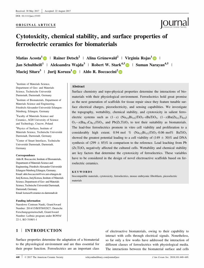
Figure 1A-C show the topographies obtained by atomicforce microscopy (AFM) for NBT–6BT, BZT–60BCT, andPZT, respectively. The sample surfaces were ground by asemiautomatic grinding station. The grinding procedurecaused characteristic micro-roughness for all samples withregular changes in topography in the range between 10 lm
and 20 lm (not shown). This micron-size topography isnot expected to alter cell adhesion, spreading, or activity toa great extent.34 Thus, we focus further on the investigationof the nano-roughness that has been generally recognizedas a major factor determining the cell activity.4
Figure 1 also shows the nano-roughness parametersused for the topography characterization that includes theaverage roughness (Ra) and root mean square roughness(Rrms). The average roughness values are all within thesame order of magnitude. The slight deviations betweenthe samples topographies can be attributed to their differenthardness.35,36 The root mean square roughness indicates lit-tle variations between the lead-free ferroelectrics (Fig-ure 1A-B), although PZT has a slightly lower root meansquare roughness.
3.2 | Wettability
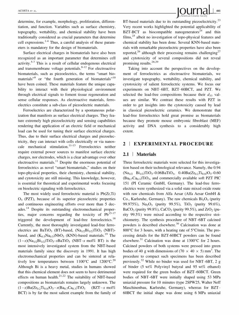
The contact angle (CA) of water at room temperature wasused to determine the wettability of the ferroelectric materi-als. The CA of water on NBT–6BT, BZT–60BZT, andPZT surfaces is displayed in Figure 2A-C, respectively.Despite the minor differences in the surface topography,the three system surfaces showed a clear difference in theirwettability.
The lead-free ferroelectrics have a hydrophilic nature.The NBT–6BT is the most hydrophilic system with aCA = (59 � 8)°. The BZT–60BCT is also hydrophilic andhas CA = (87 � 4)°. Considering the high CA of BZT–60BCT and the measurement uncertainty, it is clear thatthis material is quite close to being hydrophobic. PZT isthe only clear hydrophobic system, as indicated by aCA = (104 � 4)°.
The SE measurements indicated that NBT–6BT had thehighest SE value of 52 mJ/m2 with SEdisperse = 40.09 mJ/m2 and SEpolar = 11.69 mJ/m2. The BZT–60BCT exhibiteda SE of 46.2 mJ/m2, where the SEdisperse = 44.91 mJ/m2
and SEpolar = 1.25 mJ/m2. The PZT had the lowest SEvalue of 42.72 mJ/m2 with SEpolar � 0.
3.3 | Chemical stability
Figure 3 introduces the results of the chemical stabilityexperiment performed for all systems in SBF at 37°C. Fig-ure 3A presents the ion dissolution of Ba and Pb cations asa function of soaking time. Due to the relatively low con-centration of these cations in SBF, the measurements wereperformed by means of ICP-MS. After 30 days of soakingtime the concentrations of Bi, Ti, and Zr cations in theSBF were below the detection limit of the instrument.Thus, considering the detection limit of our experimentalsetup of 0.01 lg/L, we can assure a considerably low oreven negligible leaching of Bi, Ti, and Zr cations.
ACOSTA ET AL. | 443
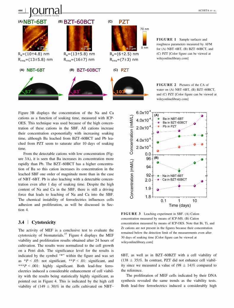
Figure 3B displays the concentration of the Na and Cacations as a function of soaking time, measured with ICP-OES. This technique was used because of the high concen-tration of these cations in the SBF. All cations increasetheir concentration exponentially with increasing soakingtime, although Ba leached from BZT–60BCT and Pb lea-ched from PZT seem to saturate after 10 days of soakingtime.
From the detectable cations with low concentration (Fig-ure 3A), it is seen that Ba increases its concentration morerapidly than Pb. The BZT–60BCT has a higher concentra-tion of Ba so this cation increases its concentration in theleached SBF one order of magnitude more than in the caseof NBT–6BT. Pb is also leaching with a detectable concen-tration even after 1 day of soaking time. Despite the highcontent of Na and Ca in the SBF, there is still a drivingforce that leads to leaching of Na and Ca into the SBF.The chemical instability of ferroelectrics influences cellsadhesion and proliferation, as will be discussed in Sec-tion 4.
3.4 | Cytotoxicity
The activity of MEF is a conclusive test to evaluate thecytotoxicity of biomaterials.37 Figure 4 displays the MEFviability and proliferation results obtained after 24 hours ofcultivation. The results were normalized to the cell growthon a Petri dish. The significance level for the results isindicated by the symbol “*” within the figure and was setas *P < .05: not significant, **P < .01: significant, and***P < .001: highly significant. Both lead-free ferro-electrics induced a considerable enhancement of cell viabil-ity with the results being statistically highly significant, aspointed out in Figure 4. This is indicated by the high cellviability of (149 � 30)% in the cells cultivated on NBT–
6BT, as well as in BZT–60BZT with a cell viability of(138 � 35)%. In contrast, PZT did not enhance cell viabil-ity since we measured a value of (98 � 14)% compared tothe reference.
The proliferation of MEF cells indicated by their DNAsynthesis revealed the same trends as the viability tests.Both lead-free ferroelectrics induced a considerably high
FIGURE 1 Sample surfaces androughness parameters measured by AFMfor (A) NBT–6BT, (B) BZT–60BCT, and(C) PZT [Color figure can be viewed atwileyonlinelibrary.com]
FIGURE 2 Pictures of the CA ofwater on (A) NBT–6BT, (B) BZT–60BCT,and (C) PZT [Color figure can be viewed atwileyonlinelibrary.com]
FIGURE 3 Leaching experiment in SBF. (A) Cationconcentration measured by means of ICP-MS. (B) Cationconcentration measured by means of ICP-OES. Note that Bi, Ti, andZr cations are not present in the figures because their concentrationremained below the detection limit of the measurements even after30 days of soaking time [Color figure can be viewed atwileyonlinelibrary.com]
444 | ACOSTA ET AL.

DNA synthesis, with an enhancement of (299 � 85)% inNBT–6BT and of (310 � 80)% in BZT–60BZT as com-pared to the reference. For PZT, the DNA synthesis activ-ity in MEF cells was much lower. A DNA synthesis of(115 � 25)% was measured for PZT as compared tothe reference. The results give a substantial indicationof the biocompatibility of the investigated lead-freeferroelectrics.
Figure 5 shows SEM micrographs for (A), (D) NBT–6BT; (B), (E) BZT–60BCT; and (C), (F) PZT surfaces after24 hours of MEF incubation. The MEF cells almost com-pletely cover the surface of the NBT–6BT and form a mul-tilayer structure (Figure 5A), thus it was not possible toanalyze single cells. The overall morphology of the cellmultilayer indicated a preferred growth orientation.
The cell density on BZT–60BCT (Figure 5B,E) waslower than on NBT–6BT. The MEF cells spread quite con-siderably but did not completely cover the materials’ sur-face (Figure 5B). Single cells could be detected with thepresence of filopodia or very thin extensions. Very recentlysimilar results have been reported for BZT–45BCT thinfilms.25 Even in the case of PZT (Figure 5C,F), MEF cellsspread extensively, although they covered a much smallersurface area in comparison to the other samples. The MEFcells on PZT were also much more flattened than in lead-free ferroelectrics. In this case, single cells characterized bya polygonal shape with filopodia or very thin extensionswere easily distinguishable. For BZT–60BCT and PZT, wedid not discern preferred growth orientation of the MEFcells.
4 | DISCUSSION
The cytotoxicity results indicate that that NBT–6BT offersthe best cell viability of (149 � 30)%, followed closely bythe cell viability of BZT–60BCT with (138 � 35)%. TheDNA synthesis of cells on NBT–6BT was (299 � 85)%,which is quite similar to the DNA synthesis of the cells onBZT–60BZT with (310 � 80)%. In contrast, cells on PZTfeature a much lower cell viability of (98 � 14)% andDNA synthesis of (115 � 25)%.
The nano-roughness did not differ much between thesamples. Considering the measurement errors, no relevantdiscrepancies were found for the Ra values. PZT, however,did show smaller Rrms values than lead-free ferroelectrics.The higher discrepancy between Rrms values than betweenRa values can be explained considering the calculation ofboth roughness parameters. From a mathematical point ofview, Ra assigns equal statistical weight to each roughnessmeasurement. In contrast, the Rrms emphasizes the extremeroughness values due to the square terms involved in itscalculation. The extremes of the nano-roughness
measurements of our samples tend to be in the order ofmicrons. Thus, they are directly linked to the grinding pro-cess. Since this micron-size topography is not expected toaffect cell behavior considerably,34 we will neglect theeffect of surface roughness in further discussion. We fur-ther justify this approximation taking into account that aprevious work indicated that, in materials with similarnano-roughness, rat osteoblasts adhesion, spreading, prolif-eration and differentiation were determined by the wettingcharacteristics rather than nano-topography.8 In otherwords, under similar nano-roughness, we expect that thewettability and chemical stability influence more consider-ably the cell viability and proliferation. We also note thatdue to the high and similar Young’s moduli of perovskiteferroelectric ceramics,14 we do not expect that elasticityplays a major role.
In materials with hydrophobic surfaces, cell adhesionand spreading are generally limited. In contrast, moderatelyhydrophilic or hydrophilic surfaces tend to favor cell adhe-sion and spreading.4,38-40 This was shown exemplary onsurface-treated polymers with human fibroblasts, humanMG63 osteosarcoma cells, Chinese hamster ovary, Chinesehamster fibroblast, and Chinese hamster endothelialcells.38-40 NBT–6BT is the most hydrophilic material inves-tigated here with a CA = (59 � 7.6)°. This CA is in thesame range that was previously highlighted to maximizecell adhesion and proliferation.39 This maximization of celladhesion and spreading can be reconciled taking intoaccount that moderately hydrophilic surfaces with CAbetween 60° and 65° maximize the adsorption of cell-adhe-sive proteins.41 The other lead-free ferroelectric, BZT–60BCT, features a CA = (87 � 4)° which is in the limit of
FIGURE 4 Cell viability and proliferation of MEF cells on REF,NBT–6BT, BZT–60BCT, and PZT after 24 h of cultivation,normalized to cell growth on Petri dishes.37 ***P < .001: highlysignificant [Color figure can be viewed at wileyonlinelibrary.com]
ACOSTA ET AL. | 445

hydrophobic surfaces. PZT has a hydrophobic surface withCA = (104 � 4)°. Therefore, wettability seems to be amajor factor determining the cell activity and proliferation.However, the effect of the surface chemistry must also betaken into account,40 especially considering that ions leachfrom the perovskite ferroelectric ceramics into aqueousenvironments as shown in Figure 3 and in the litera-ture.28,29,42-44
The chemical stability of surfaces in contact with biologi-cal media is extremely important since it can affect the cellactivity and proliferation.1,3,4,45 Our experiments with com-mercial PZT (PIC 151) indicate that the material is unstablein aqueous media. Figure 3 highlights that Pb is the mostleachable species of PIC 151 and other cations of this mate-rial remain below the detection limit of our device. Similarresults were obtained for other PZT-based materials .42-44 Pbleaching rates depend on the chemical composition of thematerial, sintering conditions, as well as the composition andpH value of the aqueous media.42-44 This is a major concernfor technological applications in contact with aqueous mediaor under atmosphere conditions such as ferroelectric ceram-ics implemented as biomaterials, sensors or actuators. It isalso a concern in other applications for which lead-contain-ing ceramics are currently under investigation such as per-ovskite solar cells.46,47
Mitochondrial dysfunction can be interpreted as a cyto-toxicity measure due to the crucial role of this organelle inmaintaining the cellular structure and function. Thus, it
directly affects the cell viability.48 Reduction of 50% in cellviability as compared to the reference can be well attribu-ted to cytotoxicity.49 We did not observe such a dramaticreduction of MEF cell viability on the cells cultivated24 hours on PZT. We attribute this to the limited leachingof Pb to the SBF after 24 hours (Figure 4) since MEF cellsgenerally display a quasi-instantaneous reactivity to theirenvironment.33 Despite that there is no clear cytotoxicity,the negative effect of Pb leached ions is corroborated tak-ing into account the much lower cell density observed onPZT as compared to the lead-free ferroelectrics (Figure 5).Experiments with Chinese hamsters and mouse fibroblastsalso indicated relatively low cell density and proliferationon PZT surfaces even after longer periods of incuba-tion.50,51 Moreover, it was also demonstrated that a consid-erable fraction of mouse fibroblasts has survival timeshorter than 4 days on PZT.51
Overall the cells on the lead-free ferroelectrics show goodadhesion, spreading, and mutual interconnections (Figure 5).On NBT–6BT even a MEF multilayer structure formed cov-ering the complete materials’ surface with a preferred growthorientation. Together with the high MEF cell activity andDNA synthesis, this implies that the material can be consid-ered as a promising biomaterial. At this point, it is worth ana-lyzing the potential human toxicity of other transitionmetals.52 To date, Bi does not seem to show detrimentaleffects to human health.21,22 Nonetheless, it should bepinpointed that cytotoxicity of the Bi(NO3)3 salt was
(A) (B) (C)
(D) (E) (F)
FIGURE 5 SEM-images of MEF cells on as-polished surface of NBT–6BT (A) and (D), BZT–60BCT (B) and (E), and PZT (C) and (F)after 24 h of incubation. (D-F) are images with higher magnification on the same material but from a different area [Color figure can be viewedat wileyonlinelibrary.com]
446 | ACOSTA ET AL.
demonstrated for a concentration higher than 1.20 9 10�2
mmol�L�1 after testing the viability of murine fibroblastsL929 and murine osteoblastic cells MC3T3-E1.52 The resultsof our ICP analysis indicate that Bi concentration in the SBFremains below 0.01 lg/L even after 30 days of soaking timeof NBT–6BT. Thus, at this concentration doses, Bi does notaffect cells activity or proliferation.
Ba is present in NBT–6BT and in BZT–60BCT. Forboth materials, we could detect leaching of Ba into theSBF. The concentration of Ba in the SBF was much higherfor BZT–60BCT than for NBT–6BT, which is expecteddue to the much higher content of Ba in the former mate-rial. This alkali earth metal is characterized by a low cyto-toxicity as corroborated by a study on the effect of BaCl2salt in the viability of murine fibroblasts L929 and murineosteoblastic cells MC3T3-E1. This work determined that aconcentration higher than 1.269 10-03 mol/L leads to cyto-toxicity.52 Moreover, no clear beneficial physiologicaleffects were reported so far for Ba cations.53 Thus, we donot expect that Ba is playing a major role in determining thecell viability or proliferation here. We note, however, thatsome studies on animals indicated that Ba could be incorpo-rated into the bone matrix replacing Ca albeit no detrimentaleffects on the bone integrity were reported.54
Na and Ca are present in bioactive glasses and areexpected to have a positive physiological effect.45,53 Someof their positive effects can include favoring human osteo-blast proliferation, differentiation, and extracellular matrixmineralization. We speculate that the high leaching rateand concentration of Na in NBT–6BT could actually favorthe cellular proliferation more positively than the muchlower Ca leaching rate of BZT–60BCT. More detailedexperiments are required to verify this hypothesis. The cor-relation between the wettability and cytotoxicity for theinvestigated materials, makes us believe that wettabilitymight be the major factor affecting cell viability, adhesion,and proliferation.
Another important aspect that affects cell activity andproliferation is the surface electrical charge. The surfacetopography, chemical composition, and crystal structuredetermine the wettability, surface energy, and surface elec-trical charges. For instance, the effect of the surfacecharges on the CA was demonstrated for differently polar-ized hydroxyapatite.55 Moreover, also for hydroxyapatite, itwas shown that surface energy and electrical charges havea mutual interdependence.56 Therefore, the wettability, sur-face energy, and surface electrical charges are clearly inter-dependent in electroactive materials.
From the results presented in Figure 1, it follows that wemay safely neglect the effect of the topography in the con-text discussed here. Moreover, all the materials in this workhave a perovskite crystal structure. Thus, in the lead-freeferroelectrics investigated here, the key parameters
responsible for the wettability are the surface energy (SE)and electrical charges. Since the materials have not beenpoled, no net macroscopic polarization should be expected.However, on the local cell level, electrical charges are cer-tainly present as consequence of the characteristic sponta-neous polarization of ferroelectrics. On a local scale, thegrain crystallographic orientation is responsible for deter-mining the spontaneous polarization and thus also the sur-face electrical charges of ferroelectrics. This, in turn, affectsthe wettability. In this study, we annealed all samples toavoid the influence of ferroelastically induced domain tex-turing that could result from residual stresses. The measure-ments of SE indicated that NBT–6BT had the highestSEpolar with a value of 11.69 mJ/m2. The SEpolar of theBZT–60BCT and PZT were 1.25 mJ/m2 and almost 0,respectively. This means that NBT–6BT has highest polarinteractions available as compared to other samples with lit-tle polar interactions. Thus, electrowetting could be the rea-son behind the tendency found on the wettability of thesystems. Namely, NBT–6BT is the most hydrophilic mate-rial, whereas PZT is the most hydrophobic one. The surfaceelectrical charges and thus the hydrophilic nature of theNBT–6BT seem to have a positive effect on cell adhesionand proliferation.57,58
5 | CONCLUSIONS
In this work, we discuss the potential of ferroelectricmaterials as biomaterials. We contrast lead-containing andlead-free ferroelectrics. Pb(Zr,Ti)O3 (PIC 151), which is acommercially relevant lead-based ferroelectric, was chosenas representative lead-containing material. Two promisinglead-free ferroelectrics, 0.94(Bi1/2Na1/2)TiO3–0.06BaTiO3
and 0.40Ba(Zr0.2Ti0.8)O3–0.60(Ba0.7Ca0.3)TiO3, were selectedas lead-free representative materials. We investigated thecell growth, proliferation, and viability. Among the threematerials, 0.94(Bi1/2Na1/2)TiO3–0.06BaTiO3 showed thebest performance since it promotes a high cell viability of(149 � 30)% and DNA synthesis of (299 � 85)% withrespect to the reference.
The materials were characterized in terms of chemicalstability, wettability, and surface roughness. All three ferro-electrics were chemically unstable in SBF, with the A-sitecations Pb, Na, Ba, or Ca being the only detectable speciesleaching after 30 days of immersion. Taking into accountthe cytotoxicity of lead, it is evident that Pb(Zr,Ti)O3 shouldnot be used in applications as a biomaterial or under envi-ronmental conditions. Despite this effect, our results high-light that the wetting angle and thus surface energydetermine the biocompatibility of the ferroelectrics. 0.94(Bi1/2Na1/2)TiO3–0.06BaTiO3 is moderately hydrophilicwith a contact angle of (59 � 8)° and has the highest polar
ACOSTA ET AL. | 447

surface energy of 11.69 mJ/m2, while both other materialswere hydrophobic and featured lower polar interactions.
Our results indicate that the complexity of the interac-tion of ferroelectrics and piezoelectrics with a physiologicalenvironment call for a better understanding before thesematerials can be used in biomedical applications. Corrobo-rating the evolution of primary cells and/or target cells onferroelectrics is certainly required. Further work towardsferroelectric biomaterials can also focus on the modificationof surface energy, surface electrical charges, and wettabilitythrough poling (ferroelectricity) or mechanical loading(piezoelectricity).
ACKNOWLEDGMENTS
MA acknowledges the support of the Leibniz programunder RO954/22-1. JK and JS acknowledge the support ofthe Deutsche Forschungsgemeinschaft (DFG), Grant Nr.KO 5100/1-1. AW and MS acknowledge the support of theNational Science Centre Poland under project 2014/15/B/ST8/02827.
ORCID
Matias Acosta http://orcid.org/0000-0001-9504-883XVirginia Rojas http://orcid.org/0000-0003-2283-229XRobert W. Stark http://orcid.org/0000-0001-8678-8449Jurij Koruza http://orcid.org/0000-0002-0258-6709Aldo R. Boccaccini http://orcid.org/0000-0002-7377-2955
REFERENCES
1. Ozdemir T, Higgins AM, Brown JL. Osteoinductive biomaterialgeometries for bone regenerative engineering. Curr Pharm Des.2013;19:1-10.
2. Menzies KL, Jones L. The impact of contact angle on the bio-compatibility of biomaterials. Optom Vis Sci. 2010;87:387-399.
3. von der Mark K, Park J. Engineering biocompatible implant sur-faces. Prog Mater Sci. 2013;58:327-381.
4. Bacakova L, Filova E, Parizek M, et al. Modulation of cell adhe-sion, proliferation and differentiation on materials designed forbody implants. Biotechnol Adv. 2011;29:739-767.
5. Kobayashi T, Nakamura S, Yamashita K. Enhanced osteobondingby negative surface charges of electrically polarized hydroxyap-atite. J Biomed Mater Res. 2001;57:477-484.
6. Tarafder S, Bodhak S, Bandyopadhyay A, et al. Effect of electri-cal polarization and composition of biphasic calcium phosphateson early stage osteoblast interactions. J Biomed Mater Res, PartB. 2011;97B:306-314.
7. Yamashita K, Oikawa N, Umegaki T. Acceleration and decelera-tion of bone-like crystal growth on ceramic hydroxyapatite byelectric poling. Chem Mater. 1996;8:2697-2700.
8. He J, Zhou W, Zhou X, et al. The anatase phase of nanotopogra-phy titania plays an important role on osteoblast cell morphologyand proliferation. J Mater Sci: Mater Med. 2008;19:3465-3472.
9. Balint R, Cassidy NJ, Cartmell SH. Conductive polymers:towards a smart biomaterial for tissue engineering. Acta Biomater.2014;10:2341-2353.
10. Ning C, Zhou L, Tan G. Fourth-generation biomedical materials.Mater Today. 2015;19:2-3.
11. Rajabi AH, Jaffe M, Arinzeh TL. Piezoelectric materials for tis-sue regeneration: a review. Acta Biomater. 2015;24:12-23.
12. Tofail SAM, Bauer J. Electrically polarized biomaterials. AdvMater. 2016;28:5470-5484.
13. Childs PG, Boyle CA, Pemberton GD, et al. Use of nanoscalemechanical stimulation for control and manipulation of cell beha-viour. Acta Biomater. 2016;34:159-168.
14. Jaffe B, Cook WR, Jaffe H. Piezoelectric Ceramics, 1st edn.India: Academic Press Limited; 1971.
15. Lyn PND. Lead toxicity, a review of the literature. Part I: expo-sure, evaluation, and treatment. Altern Med Rev. 2006;11:22.
16. Johnson FM. The genetic effects of environmental lead. MutatRes, Rev Mutat Res. 1998;410:123-140.
17. Garc�ıa-Lest�on J, M�endez J, P�asaro E, et al. Genotoxic effects oflead: an updated review. Environ Int. 2010;36:623-636.
18. R€odel J, Jo W, Seifert KTP, et al. Perspective on the develop-ment of lead-free piezoceramics. J Am Ceram Soc.2009;92:1153-1177.
19. R€odel J, Webber KG, Dittmer R, et al. Transferring lead-freepiezoelectric ceramics into application. J Eur Ceram Soc.2015;35:1659-1681.
20. Takenaka T, Maruyama K-I, Sakata K. Bi1/2Na1/2TiO3–BaTiO3
system for lead-free piezoelectric applications. Jpn J Appl Phys.1991;30:2236-2239.
21. Serfontein WJ, Mekel R, Bank S, et al. Bismuth toxicity in man- I. Bismuth blood and urine levels in patients after administra-tion of a bismuth protein complex (Bicitropeptide). Res CommunChem Pathol Pharmacol. 1979;26:383-389.
22. Rodilla V, Miles AT, Jenner W, et al. Exposure of culturedhuman proximal tubular cells to cadmium, mercury, zinc and bis-muth: toxicity and metallothionein induction. Chem-Biol Interact.1998;115:71-83.
23. Liu W, Ren X. Large piezoelectric effect in Pb-free ceramics.Phys Rev Lett. 2009;103:257602.
24. Yuan MM, Cheng L, Xu Q, et al. Biocompatible nanogeneratorsthrough high piezoelectric coefficient 0.5Ba(Zr0.2Ti0.8)O3–0.5(Ba0.7Ca0.3)TiO3 nanowires for in-vivo applications. Adv Mater.2014;26:7432-7437.
25. Scarisoreanu ND, Craciun F, Ion V, et al. Lead-free piezoelectric(Ba, Ca)(Zr, Ti)O3 thin films for biocompatible and flexibledevices. ACS Appl Mater Interfaces. 2017;9:266-278.
26. Li JF, Wang K, Zhu FY, et al. (K, Na)NbO3-based lead-freepiezoceramics: fundamental aspects, processing technologies, andremaining challenges. J Am Ceram Soc. 2013;96:3677-3696.
27. Mali�c B, Koruza J, Hre�s�cak J, et al. Sintering of lead-free piezo-electric sodium potassium niobate ceramics. Materials.2015;8:5449.
28. Yu S-W, Kuo S-T, Tuan W-H, et al. Ion release from three lead-free piezoelectric ceramics and their physical and cytotoxicitycharacteristics. Mater Lett. 2011;65:3522-3524.
448 | ACOSTA ET AL.

29. Yu S-W, Kuo S-T, Tuan W-H, et al. Cytotoxicity and degrada-tion behavior of potassium sodium niobate piezoelectric ceramics.Ceram Int. 2012;38:2845-2850.
30. Ayrikyan A, Rojas V, Molina-Luna L, et al. Enhancing elec-tromechanical properties of lead-free ferroelectrics with bilayerceramic/ceramic composites. IEEE Trans Ultrason FerroelectrFreq Control. 2015;62:997-1006.
31. Acosta M, Novak N, Jo W, et al. Relationship between elec-tromechanical properties and phase diagram in the Ba(Zr0.2Ti0.8)O3–x(Ba0.7Ca0.3)TiO3 lead-free piezoceramic. Acta Mater.2014;80:48-55.
32. V€ogler M, Acosta M, Brandt DRJ, et al. Temperature-dependentR-curve behavior of the lead-free ferroelectric 0.615Ba(Zr0.2Ti0.8)O3–0.385(Ba0.7Ca0.3)TiO3 ceramic. Eng Fract Mech. 2015;144:68-77.
33. Ponsot I, Pontikes Y, Baldi G, et al. Magnetic glass ceramics bysintering of borosilicate glass and inorganic waste. Materials.2014;7:5565-5580.
34. Lehnert D, Wehrle-Haller B, David C, et al. Cell behaviour onmicropatterned substrata: limits of extracellular matrix geometryfor spreading and adhesion. J Cell Sci. 2003;117:41.
35. Efe C, Yilmaz H, Tur YK, et al. Mechanical property characteri-zation of Na1/2Bi1/2TiO3–BaTiO3 ceramics. Int J Chem EngAppl. 2014;5:429-432.
36. Srinivas A, Krishnaiah RV, Niranjani VL, et al. Ferroelectric,piezoelectric and mechanical properties in lead free (0.5)Ba(Zr0.2Ti0.8)O3–(0.5)(Ba0.7Ca0.3)TiO3 electroceramics. Ceram Int.2015;41:1980-1985.
37. Amaral M, Gomes PS, Lopes MA, et al. Cytotoxicity evaluationof nanocrystalline diamond coatings by fibroblast cell cultures.Acta Biomater. 2009;5:755-763.
38. Faucheux N, Schweiss R, Lutzow K, et al. Self-assembled mono-layers with different terminating groups as model substrates forcell adhesion studies. Biomaterials. 2004;25:2721-2730.
39. Lee JH, Khang G, Lee JW, et al. Interaction of different types ofcells on polymer surfaces with wettability gradient. J ColloidInterface Sci. 1998;205:323-330.
40. Dowling DP, Miller IS, Ardhaoui M, et al. Effect of surface wet-tability and topography on the adhesion of osteosarcoma cells onplasma-modified polystyrene. J Biomater Appl. 2010;26:327-347.
41. Xu L-C, Siedlecki CA. Effects of surface wettability and contacttime on protein adhesion to biomaterial surfaces. Biomaterials.2007;28:3273-3283.
42. Kosec M, Malic B, Wolny W, et al. Effect of achemicallyaggressive environment on the electromechanical behaviourofmodified lead titanate ceramics. J. Korean Phys. Soc. 1998;32:S1163-S1166.
43. Foster WJ, Meen JK, Fox DA. The effect of physiologic aqueoussolutions on the perovskite material lead-lanthanum-zirconiumtitanate (PLZT): potential retinotoxicity. Cutan Ocul Toxicol.2013;32:18-22.
44. Bakari�c T, Budi�c B, Mali�c B, et al. The influence of pH depen-dent ion leaching on the processing of lead-zirconate-titanateceramics. J Eur Ceram Soc. 2015;35:2295-2302.
45. Jones JR. Review of bioactive glass: from Hench to hybrids. ActaBiomater. 2013;9:4457-4486.
46. Park N-G, Gr€atzel M, Miyasaka T, et al. Towards stable and com-mercially available perovskite solar cells. Nat Energy. 2016;1:16152.
47. Hailegnaw B, Kirmayer S, Edri E, et al. Rain on methylammo-nium lead iodide based perovskites: possible environmentaleffects of perovskite solar cells. J Phys Chem Lett. 2015;6:1543-1547.
48. Pollard TD, Earnshaw WC, Lippincott-Schwartz J. Cell Biology,Easy Reading. Saunders Elsevier, Philadelphia: SpektrumAkademischer Verlag; 2008.
49. Trevan JW. The error of determination of toxicity. Proc R SocLond B Biol Sci. 1927;101:483-513.
50. Sajin G, Petrescu D, Sajin M, et al. Interactions between biologi-cal media and piezoelectric ceramic in micromixing applications.Rom J Inf Sci Tech Adv Mater. 2007;10:279-289.
51. Sakai T, Hoshiai S, Nakamachi E. Biochemical compatibility ofPZT piezoelectric ceramics covered with titanium thin film. JOptoelectron Adv Mater. 2006;8:1435-1437.
52. Yamamoto A, Honma R, Sumita M. Cytotoxicity evaluation of43 metal salts using murine fibroblasts and osteoblastic cells. JBiomed Mater Res. 1998;39:331-340.
53. Hoppe A, Guldal NS, Boccaccini AR. A review of the biologicalresponse to ionic dissolution products from bioactive glasses andglass-ceramics. Biomaterials. 2011;32:2757-2774.
54. Bligh PH, Taylor DM. Comparative studies of metabolismof strontium and barium in rat. Biochem J. 1963;87:612-618.
55. Bodhak S, Bose S, Bandyopadhyay A. Role of surface chargeand wettability on early stage mineralization and bone cell–mate-rials interactions of polarized hydroxyapatite. Acta Biomater.2009;5:2178-2188.
56. Horiuchi N, Nakaguki S, Wada N, et al. Polarization-induced sur-face charges in hydroxyapatite ceramics. J Appl Phys.2014;116:014902.
57. Dames JE, Causton B, Bovell Y, et al. The migration of osteo-blasts over substrata of discrete surface charge. Biomaterials.1986;7:231-233.
58. Takeda H, Seki Y, Nakamura S, et al. Evaluation of electricalpolarizability and in vitro bioactivity of apatite Sr5(PO4)3OHdense ceramics. J Mater Chem. 2002;12:2490-2495.
How to cite this article: Acosta M, Detsch R,Gr€unewald A, et al. Cytotoxicity, chemical stability,and surface properties of ferroelectric ceramics forbiomaterials. J Am Ceram Soc. 2018;101:440–449.https://doi.org/10.1111/jace.15193
ACOSTA ET AL. | 449