cutaneous malignancies · non-melanoma skin cancers includes basal cell carcinomas (bcc) &...
TRANSCRIPT
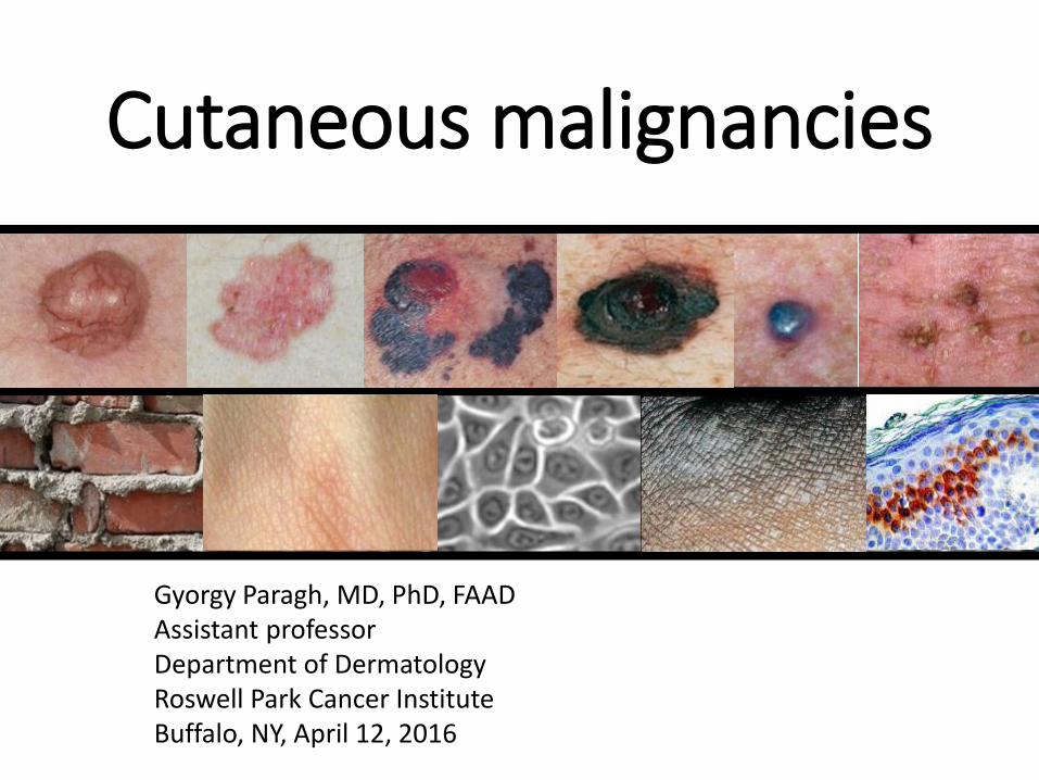
Cutaneous malignancies
Gyorgy Paragh, MD, PhD, FAAD Assistant professor Department of Dermatology Roswell Park Cancer Institute Buffalo, NY, April 12, 2016
Disclosure
• Unless otherwise noted the pictures and tables were borrowed from:
http://www.visualdx.com
http://www.dermnetnz.org/
Bolognia: Dermatology, Third Edition 2012, Elsevier

Learning objectives
• Introduction to the skin
• Introduction to epidermal carcinogenesis
• Epidemiology of common cutaneous malignancies
• Classification of common cutaneous malignancies
• Treatment of common cutaneous malignancies
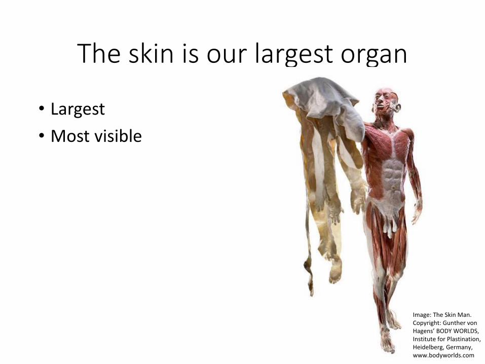
The skin is our largest organ
• Largest
• Most visible
Image: The Skin Man. Copyright: Gunther von Hagens’ BODY WORLDS, Institute for Plastination, Heidelberg, Germany, www.bodyworlds.com

Functions of the skin
• Maintaining internal homeostasis in light of variable external stimuli • Mechanical protection
• Regulates temperature
• Photoprotection
• Barrier against micro-organisms
• Metabolic function (vitamin D)
• Detects sensory stimuli
• Excretion
• Esthetic, psychosocial role
Scanning electro micrograph of the epidermis
http://science.nationalgeographic.com/science/enlarge/epidermis.html
Light microscopy of the epidermis H&E
http://microanatomy.net/skin/skin_and_mammary_glands.htm
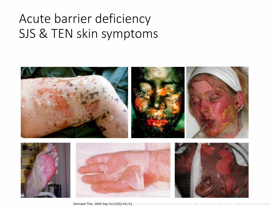
Acute barrier deficiency SJS & TEN skin symptoms
Dermatol Ther. 2009 Sep-Oct;22(5):441-51.; Thieme clinical companions: Dermatology; http://www.medscape.com/; & http://www.uptodate.com
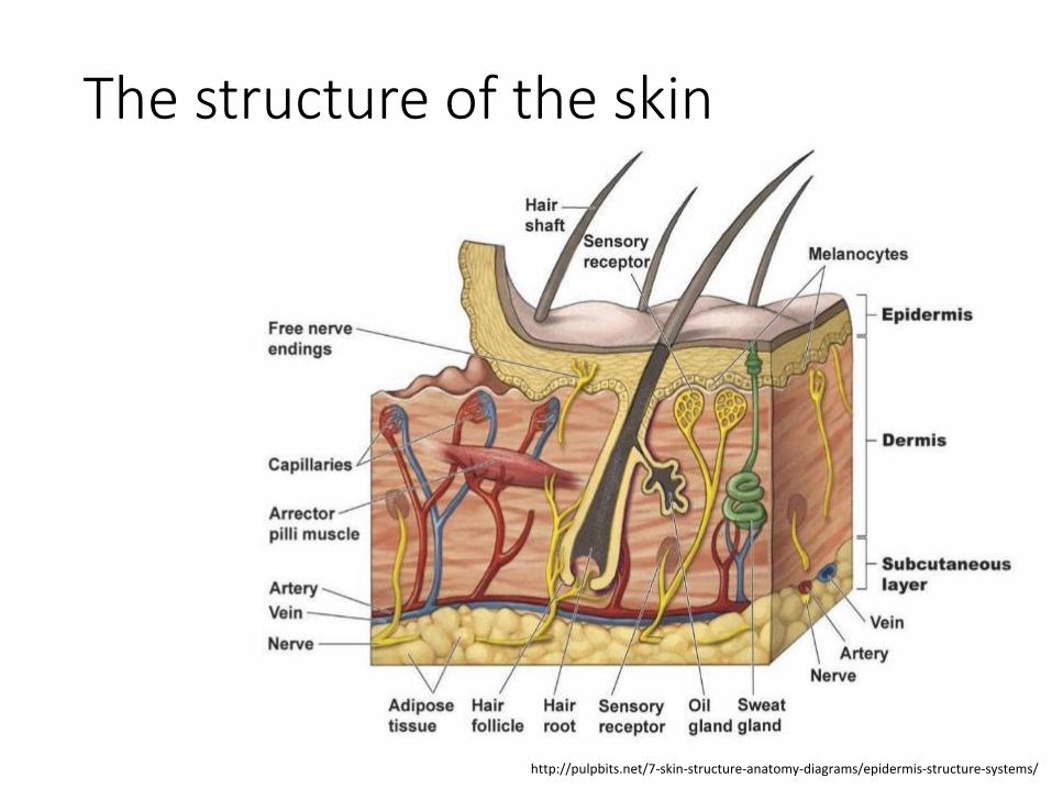
http://pulpbits.net/7-skin-structure-anatomy-diagrams/epidermis-structure-systems/
The structure of the skin
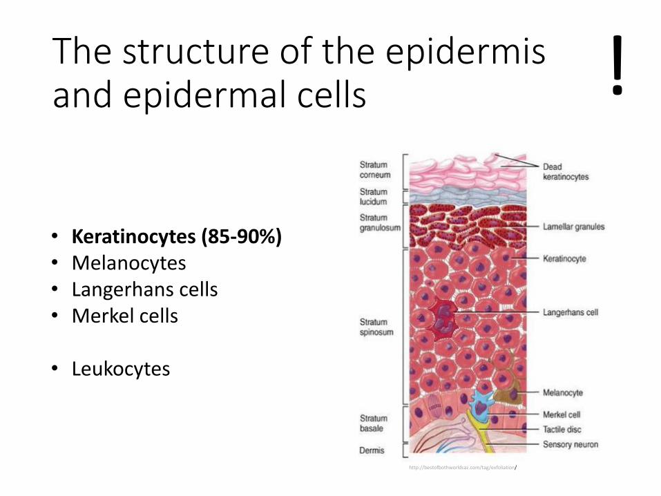
The structure of the epidermis and epidermal cells
• Keratinocytes (85-90%) • Melanocytes • Langerhans cells • Merkel cells
• Leukocytes
http://bestofbothworldsaz.com/tag/exfoliation/
!
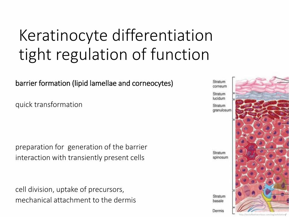
Keratinocyte differentiation tight regulation of function
http://bestofbothworldsaz.com/tag/exfoliation/
barrier formation (lipid lamellae and corneocytes)
quick transformation
preparation for generation of the barrier
interaction with transiently present cells
cell division, uptake of precursors,
mechanical attachment to the dermis
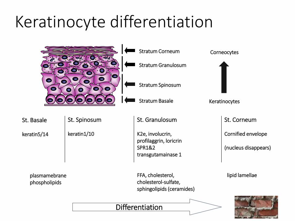
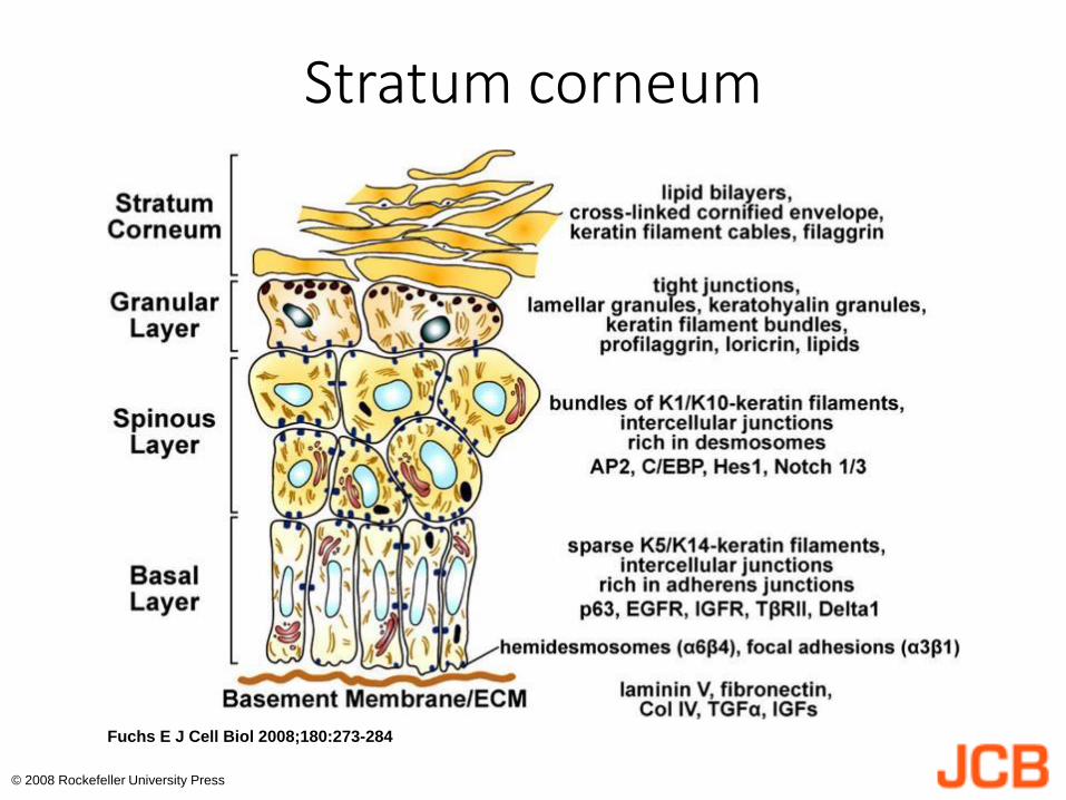
Stratum Granulosum
Stratum Spinosum
Stratum Basale
Stratum Corneum Corneocytes
Keratinocytes
St. Basale keratin5/14 plasmamebrane phospholipids
St. Spinosum keratin1/10
St. Granulosum K2e, involucrin, profilaggrin, loricrin SPR1&2 transgutamainase 1 FFA, cholesterol, cholesterol-sulfate, sphingolipids (ceramides)
St. Corneum Cornified envelope (nucleus disappears) lipid lamellae
Differentiation
Keratinocyte differentiation
Fuchs E J Cell Biol 2008;180:273-284
© 2008 Rockefeller University Press
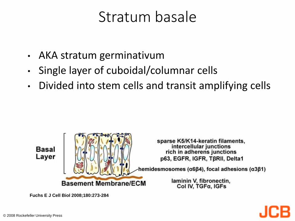
Stratum basale
• AKA stratum germinativum
• Single layer of cuboidal/columnar cells
• Divided into stem cells and transit amplifying cells
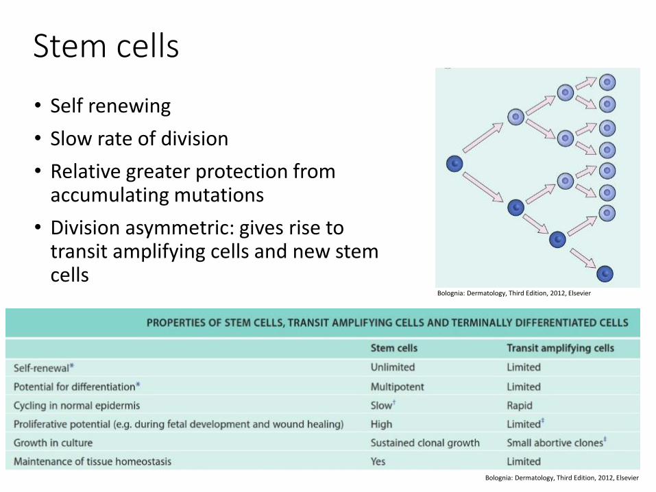
Stem cells
• Self renewing
• Slow rate of division
• Relative greater protection from accumulating mutations
• Division asymmetric: gives rise to transit amplifying cells and new stem cells
Bolognia: Dermatology, Third Edition, 2012, Elsevier
Bolognia: Dermatology, Third Edition, 2012, Elsevier
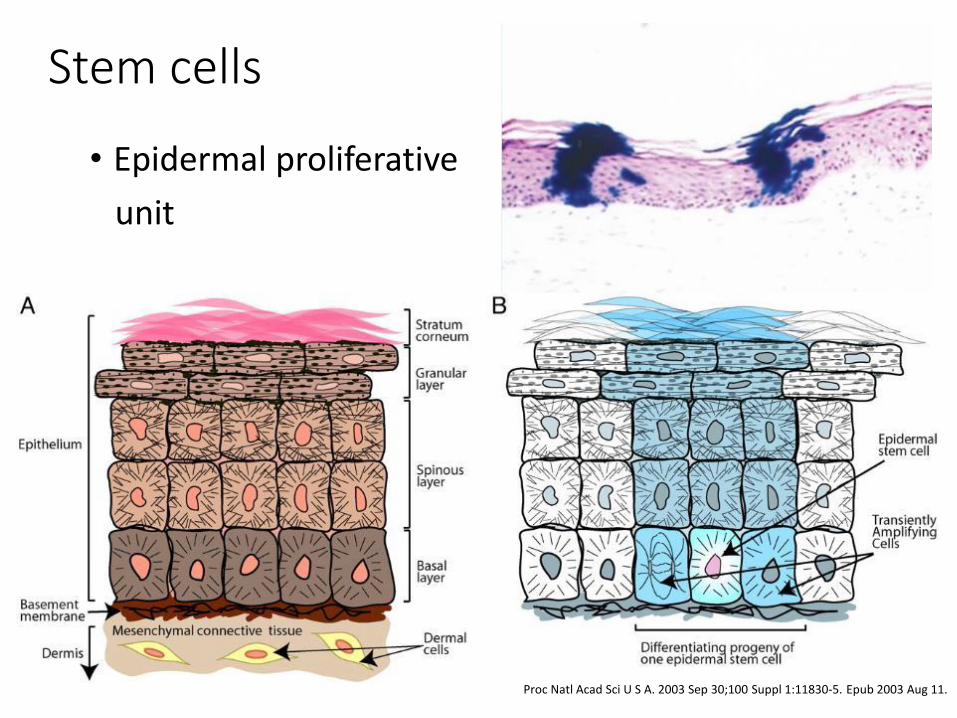
Stem cells
• Epidermal proliferative
unit
Proc Natl Acad Sci U S A. 2003 Sep 30;100 Suppl 1:11830-5. Epub 2003 Aug 11.
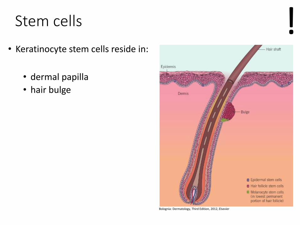
Stem cells
• Keratinocyte stem cells reside in:
• dermal papilla
• hair bulge
!
Bolognia: Dermatology, Third Edition, 2012, Elsevier
Fuchs E J Cell Biol 2008;180:273-284
© 2008 Rockefeller University Press
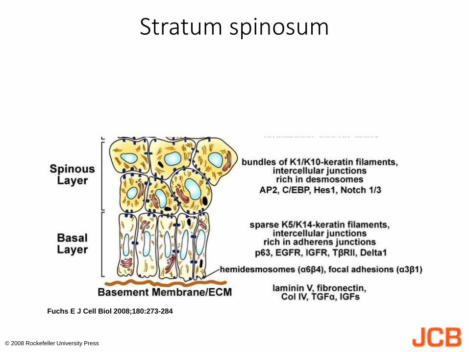
Stratum spinosum
Fuchs E J Cell Biol 2008;180:273-284
© 2008 Rockefeller University Press
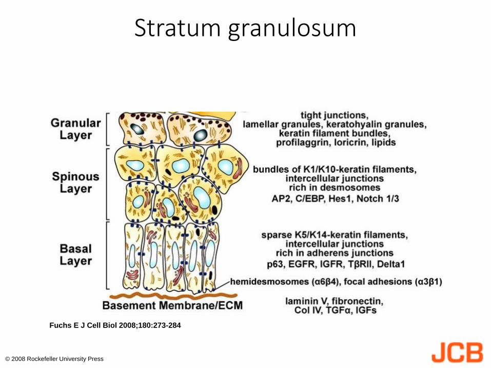
Stratum granulosum
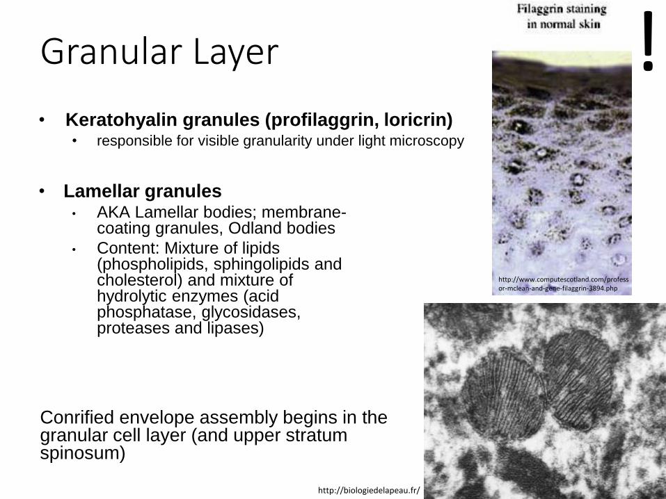
Granular Layer
• Keratohyalin granules (profilaggrin, loricrin) • responsible for visible granularity under light microscopy
• Lamellar granules • AKA Lamellar bodies; membrane-
coating granules, Odland bodies
• Content: Mixture of lipids (phospholipids, sphingolipids and cholesterol) and mixture of hydrolytic enzymes (acid phosphatase, glycosidases, proteases and lipases)
Conrified envelope assembly begins in the granular cell layer (and upper stratum spinosum)
http://www.computescotland.com/professor-mclean-and-gene-filaggrin-3894.php
http://biologiedelapeau.fr/
!
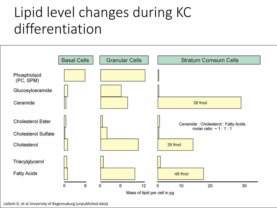
Lipid level changes during KC differentiation
Liebish G. et al University of Regensuburg (unpublished data)
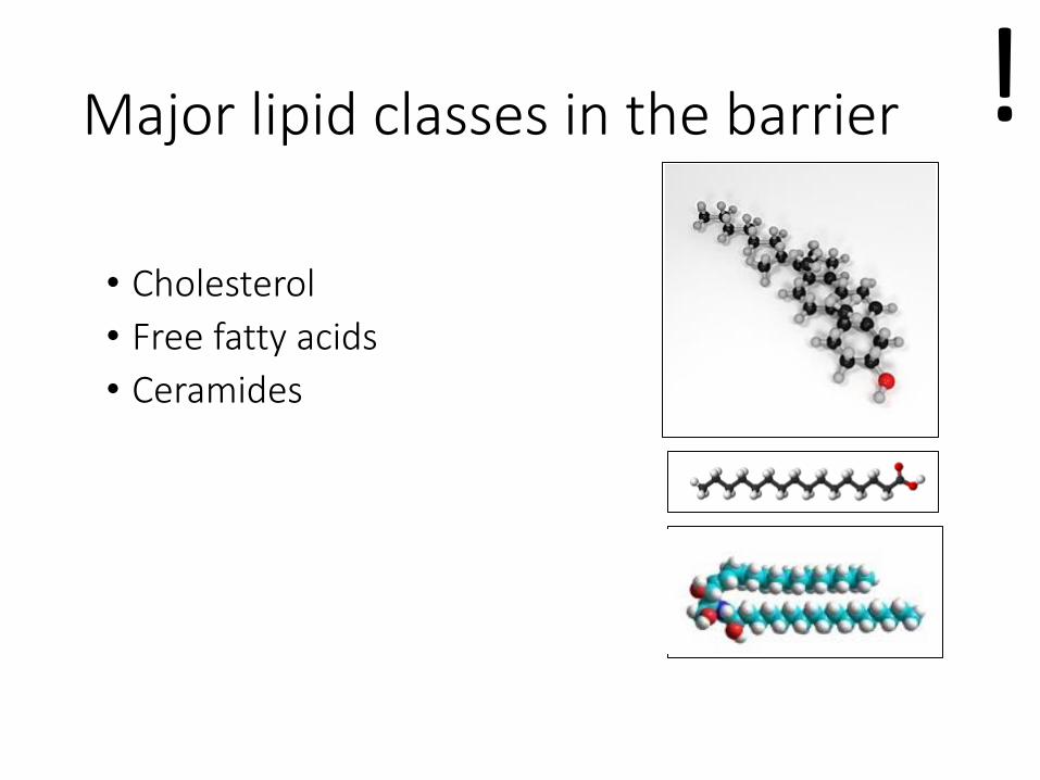
Major lipid classes in the barrier
• Cholesterol
• Free fatty acids
• Ceramides
!
Fuchs E J Cell Biol 2008;180:273-284
© 2008 Rockefeller University Press
Stratum corneum
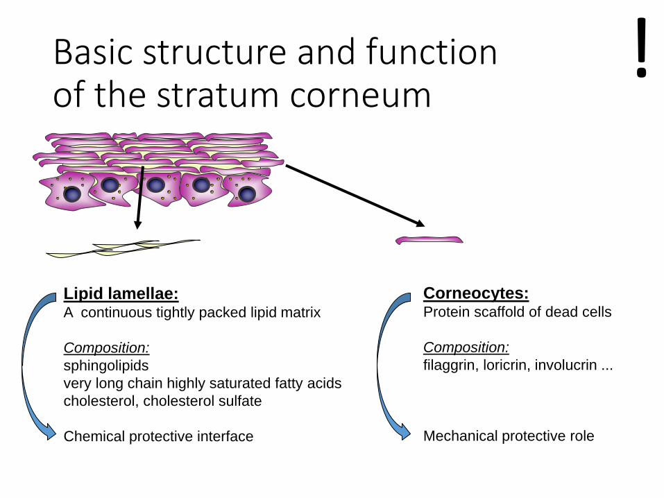
Corneocytes: Protein scaffold of dead cells
Composition:
filaggrin, loricrin, involucrin ...
Mechanical protective role
Lipid lamellae: A continuous tightly packed lipid matrix
Composition:
sphingolipids
very long chain highly saturated fatty acids
cholesterol, cholesterol sulfate
Chemical protective interface
Basic structure and function of the stratum corneum
!
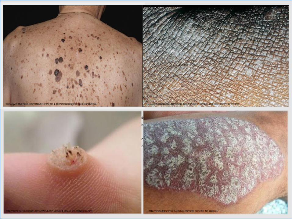
In many dermatological
diseases:
deregulation of normal KC
differentiation.
http://doctorcayoo.blogspot.com/2009/08/skin-verruca-5-veruka-and-malignancy.html http://www.drgranny.com/2011/03/06/home-remedies-for-psoriasis/
http://www.studyblue.com/notes/note/n/week-1-dermatological-pathology/deck/1330400 http://www.medicinenet.com/image-collection/lichen_simplex_chronicus_picture/picture.htm
Outline
• Non-melanoma skin cancer • Epidemiology • Risk factors • UV radiation • Photocarcinogenesis • Non-melanoma skin cancer
• Basal cell carcinoma • Squamous cell carcinoma
• Melanoma • Melanocyte biology • Epidemiology • Melanoma subtypes • Therapy
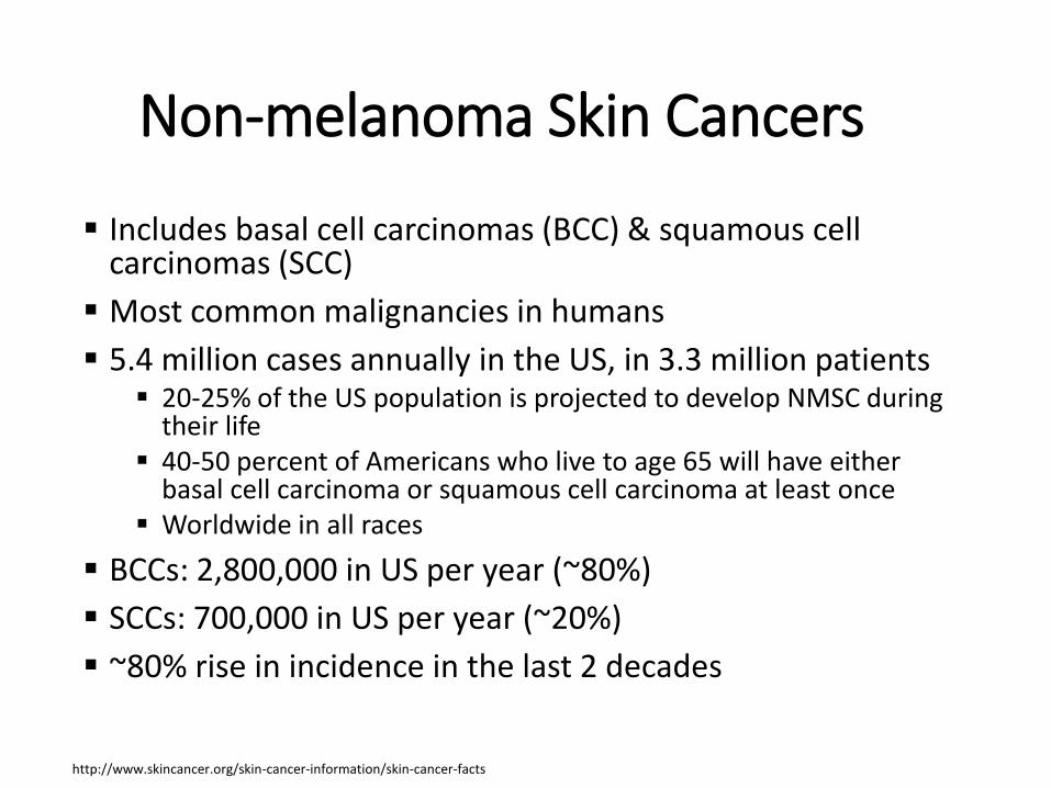
Non-melanoma Skin Cancers
Includes basal cell carcinomas (BCC) & squamous cell carcinomas (SCC)
Most common malignancies in humans
5.4 million cases annually in the US, in 3.3 million patients 20-25% of the US population is projected to develop NMSC during
their life 40-50 percent of Americans who live to age 65 will have either
basal cell carcinoma or squamous cell carcinoma at least once Worldwide in all races
BCCs: 2,800,000 in US per year (~80%)
SCCs: 700,000 in US per year (~20%)
~80% rise in incidence in the last 2 decades
http://www.skincancer.org/skin-cancer-information/skin-cancer-facts
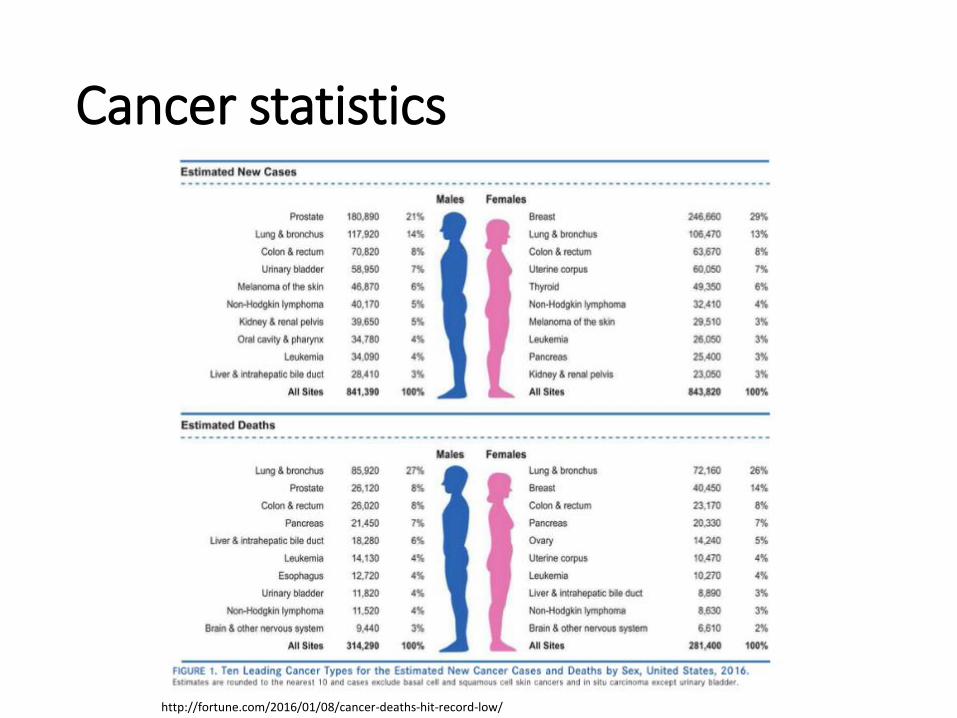
Cancer statistics
http://fortune.com/2016/01/08/cancer-deaths-hit-record-low/

Read the fine print
http://fortune.com/2016/01/08/cancer-deaths-hit-record-low/
cases exclude basal cell and squamous cell skin cacners and in situ carcinoma…
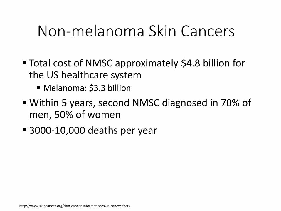
Non-melanoma Skin Cancers
Total cost of NMSC approximately $4.8 billion for the US healthcare system Melanoma: $3.3 billion
Within 5 years, second NMSC diagnosed in 70% of men, 50% of women
3000-10,000 deaths per year
http://www.skincancer.org/skin-cancer-information/skin-cancer-facts
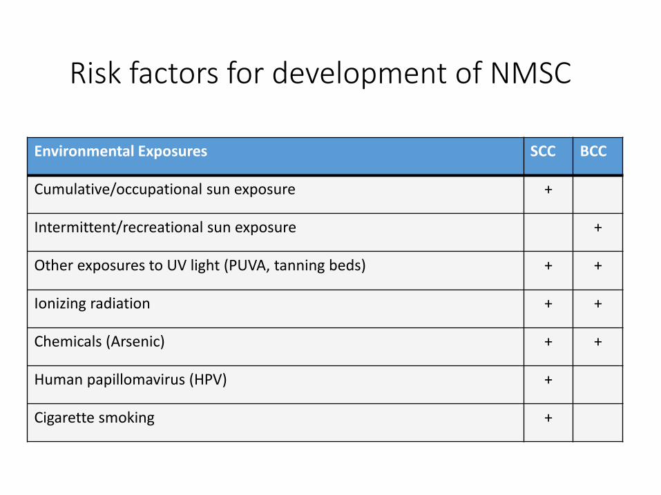
Risk factors for development of NMSC
Environmental Exposures SCC BCC
Cumulative/occupational sun exposure +
Intermittent/recreational sun exposure +
Other exposures to UV light (PUVA, tanning beds) + +
Ionizing radiation + +
Chemicals (Arsenic) + +
Human papillomavirus (HPV) +
Cigarette smoking +
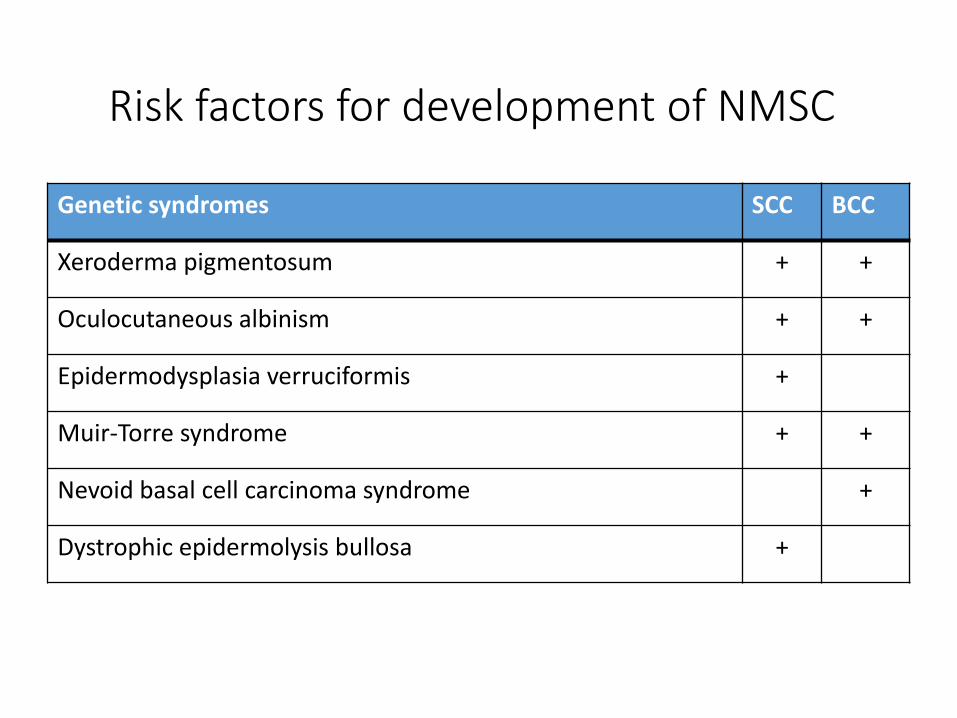
Risk factors for development of NMSC
Genetic syndromes SCC BCC
Xeroderma pigmentosum + +
Oculocutaneous albinism + +
Epidermodysplasia verruciformis +
Muir-Torre syndrome + +
Nevoid basal cell carcinoma syndrome +
Dystrophic epidermolysis bullosa +

Risk factors for development of NMSC
Predisposing clinical settings SCC BCC
Chronic non-healing wounds +
Long standing discoid lupus erythematosus +
Lichen planus (erosive) or lichen sclerosus +
Linear porokeratosis +
Risk factors for development of NMSC
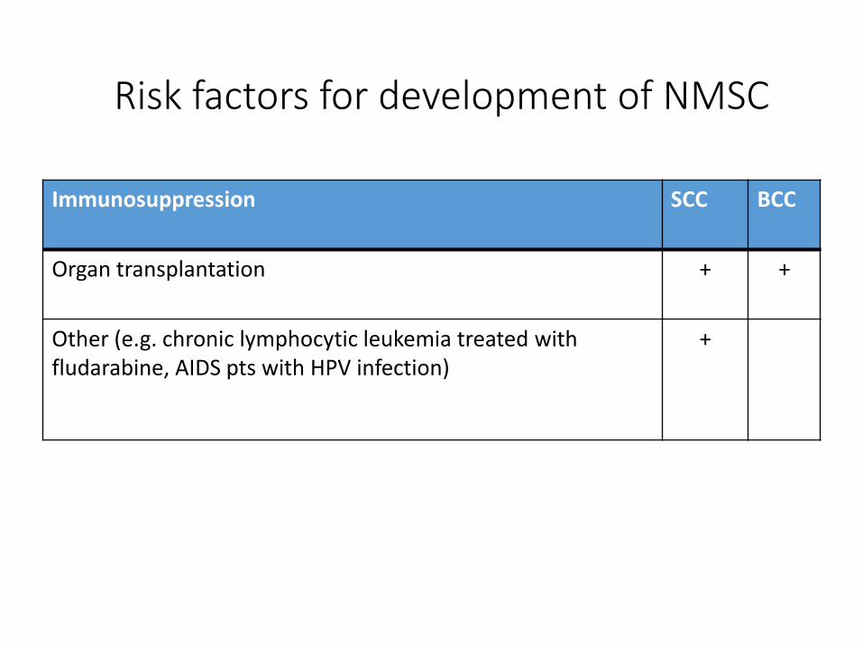
Immunosuppression SCC BCC
Organ transplantation + +
Other (e.g. chronic lymphocytic leukemia treated with fludarabine, AIDS pts with HPV infection)
+
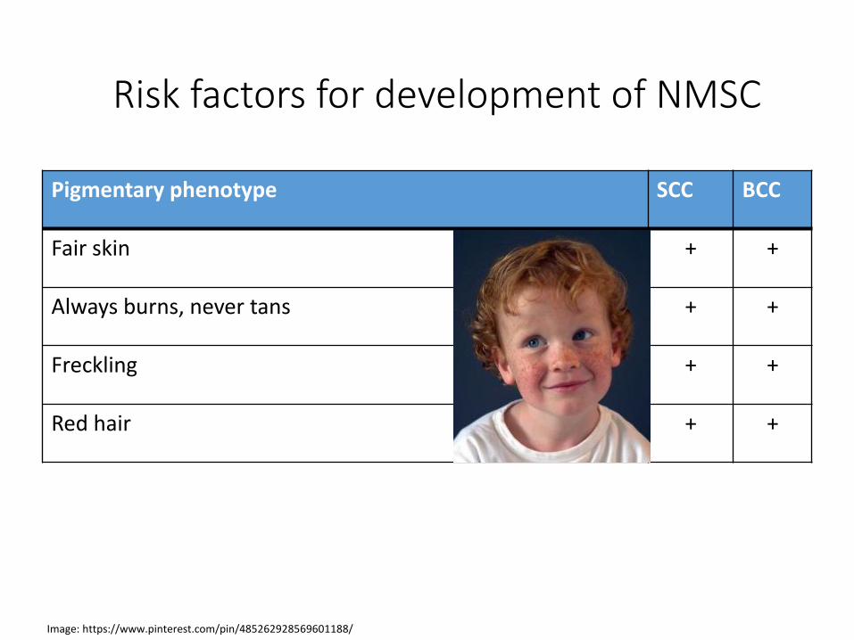
Risk factors for development of NMSC
Pigmentary phenotype SCC BCC
Fair skin + +
Always burns, never tans + +
Freckling + +
Red hair + +
Image: https://www.pinterest.com/pin/485262928569601188/
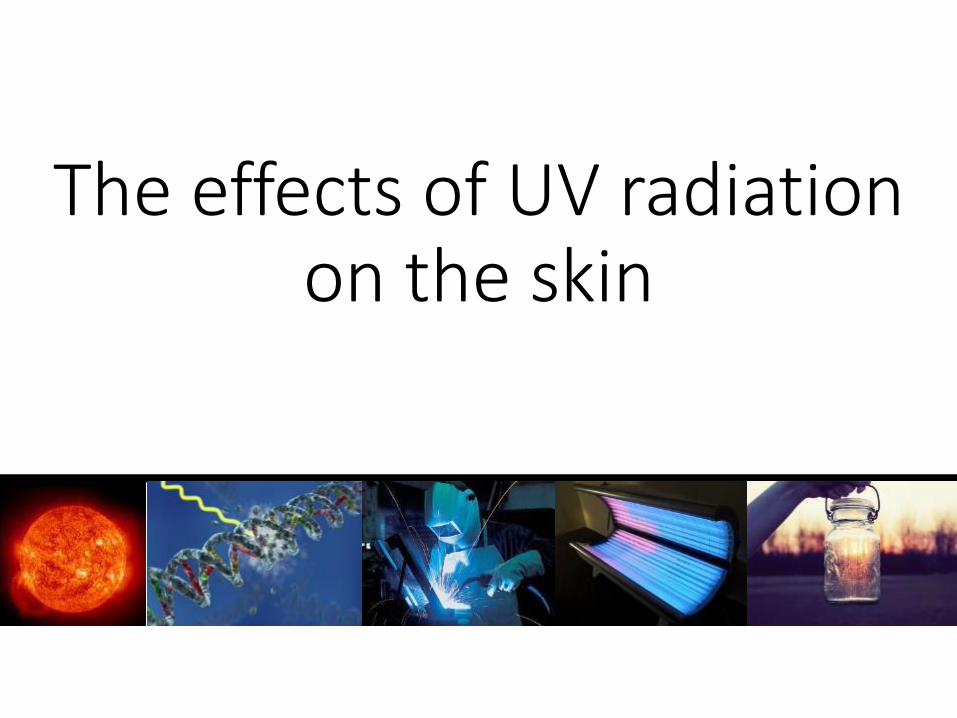
The effects of UV radiation on the skin
6/26/2013
Outline
• Introduction (definition, physical properties, UV sources, depth of penetration into the skin, action spectrums)
• Cellular effects of UVR (biomolecules effected by UV, DNA damage)
• UVR induced skin changes (sunburn, tanning, epidermal hypreplasia, vitamin D production, emotional effects, photoaging, photoimmunology, photocarcinogenesis)
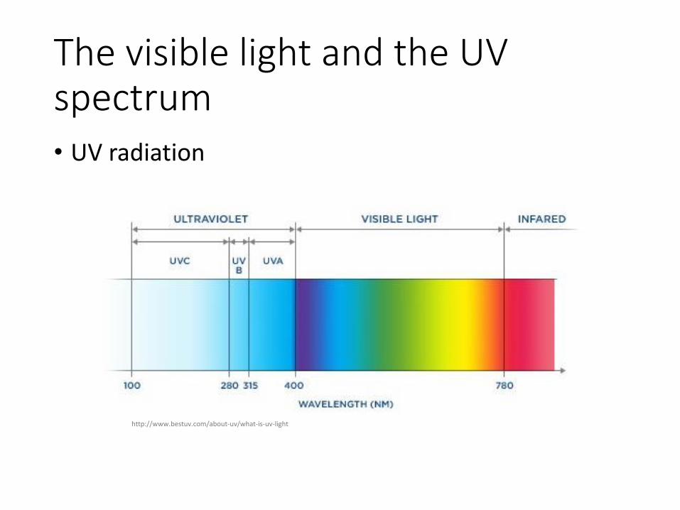
The visible light and the UV spectrum • UV radiation
http://www.bestuv.com/about-uv/what-is-uv-light
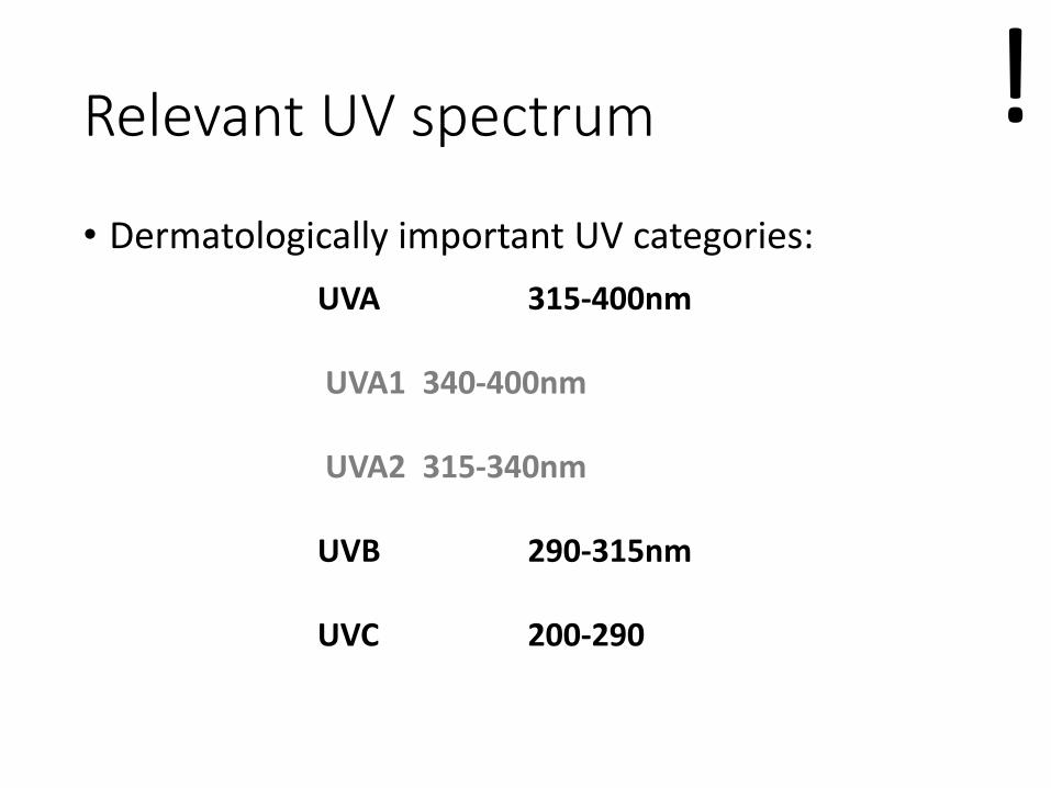
Relevant UV spectrum
• Dermatologically important UV categories:
UVA 315-400nm UVA1 340-400nm UVA2 315-340nm UVB 290-315nm UVC 200-290
!
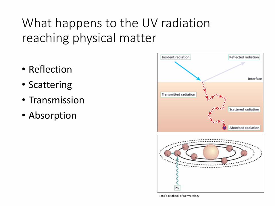
What happens to the UV radiation reaching physical matter
• Reflection
• Scattering
• Transmission
• Absorption
Rook’s Textbook of Dermatology
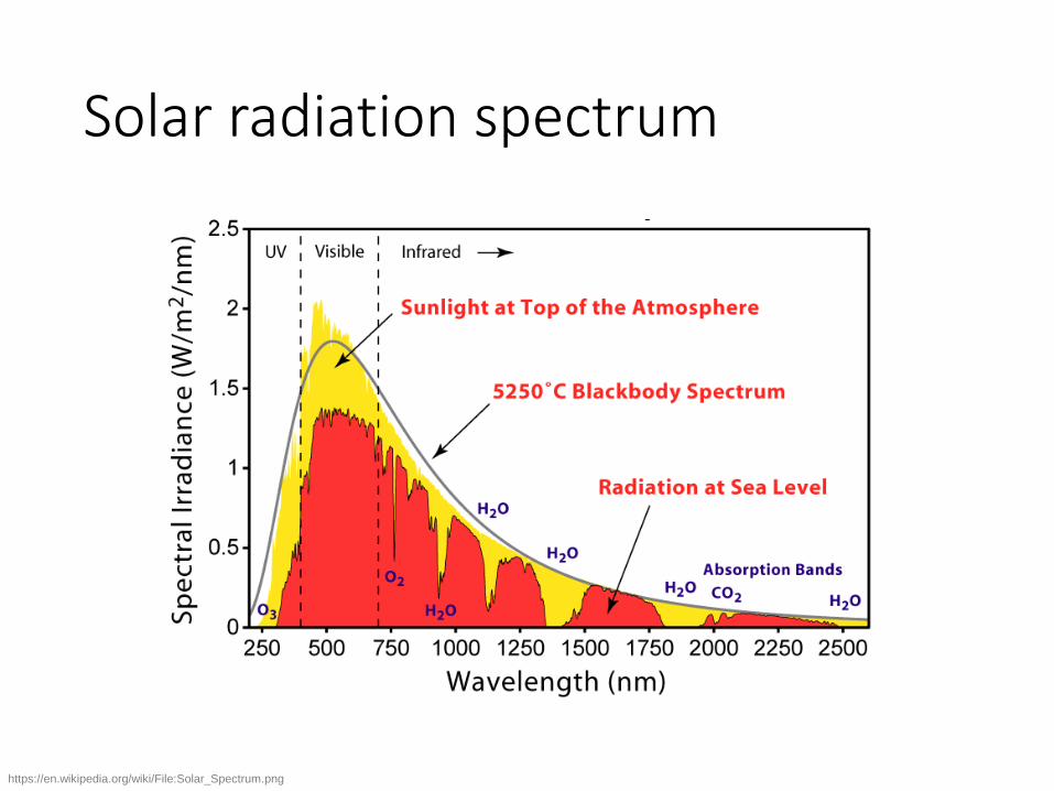
Solar radiation spectrum
https://en.wikipedia.org/wiki/File:Solar_Spectrum.png
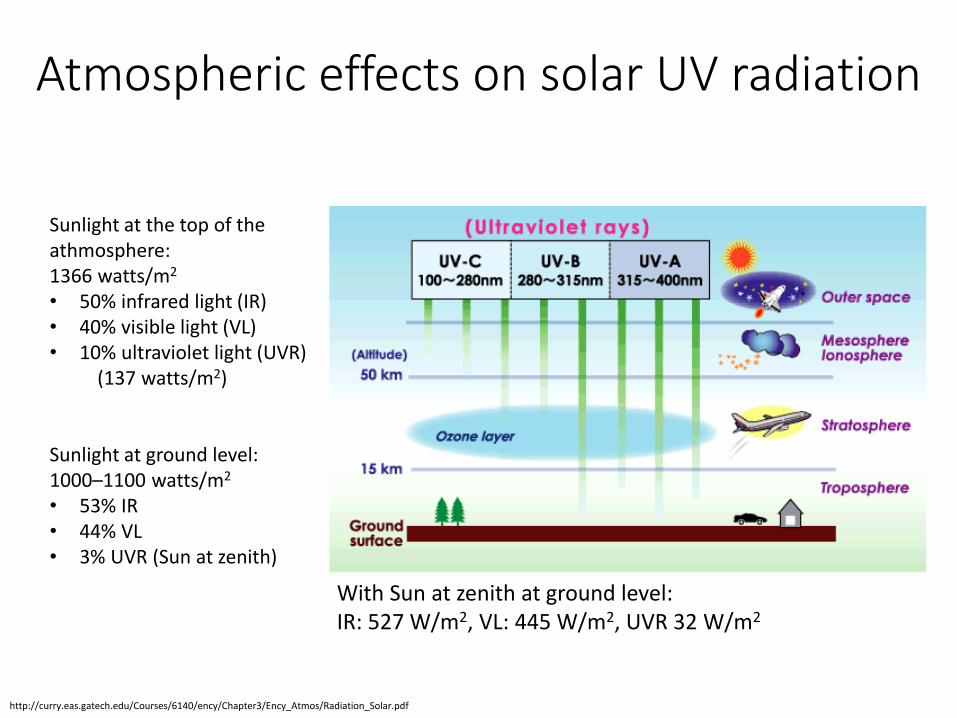
Atmospheric effects on solar UV radiation
Sunlight at the top of the athmosphere: 1366 watts/m2 • 50% infrared light (IR) • 40% visible light (VL) • 10% ultraviolet light (UVR)
(137 watts/m2)
Sunlight at ground level: 1000–1100 watts/m2 • 53% IR • 44% VL • 3% UVR (Sun at zenith)
With Sun at zenith at ground level: IR: 527 W/m2, VL: 445 W/m2, UVR 32 W/m2
http://curry.eas.gatech.edu/Courses/6140/ency/Chapter3/Ency_Atmos/Radiation_Solar.pdf
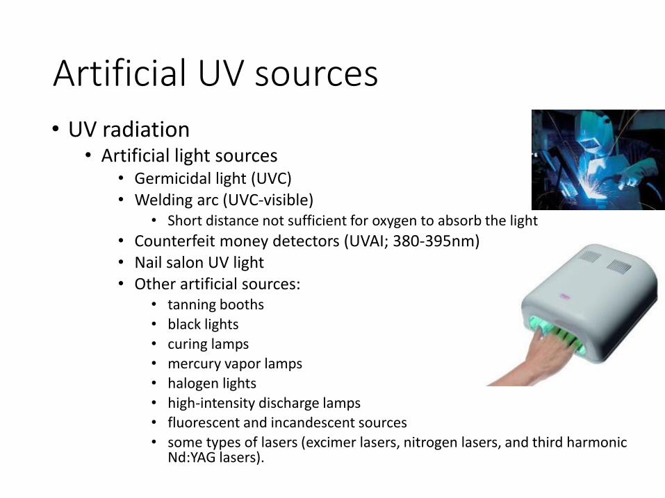
Artificial UV sources
• UV radiation • Artificial light sources
• Germicidal light (UVC) • Welding arc (UVC-visible)
• Short distance not sufficient for oxygen to absorb the light
• Counterfeit money detectors (UVAI; 380-395nm) • Nail salon UV light • Other artificial sources:
• tanning booths
• black lights
• curing lamps • mercury vapor lamps
• halogen lights
• high-intensity discharge lamps • fluorescent and incandescent sources
• some types of lasers (excimer lasers, nitrogen lasers, and third harmonic Nd:YAG lasers).
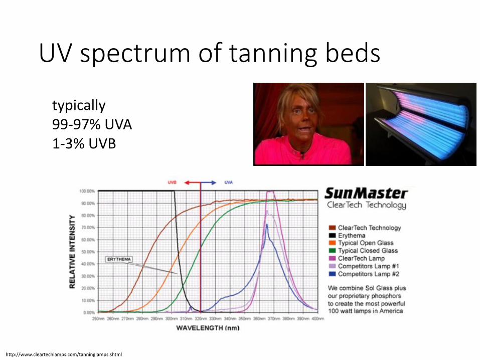
UV spectrum of tanning beds
http://www.cleartechlamps.com/tanninglamps.shtml
typically 99-97% UVA 1-3% UVB
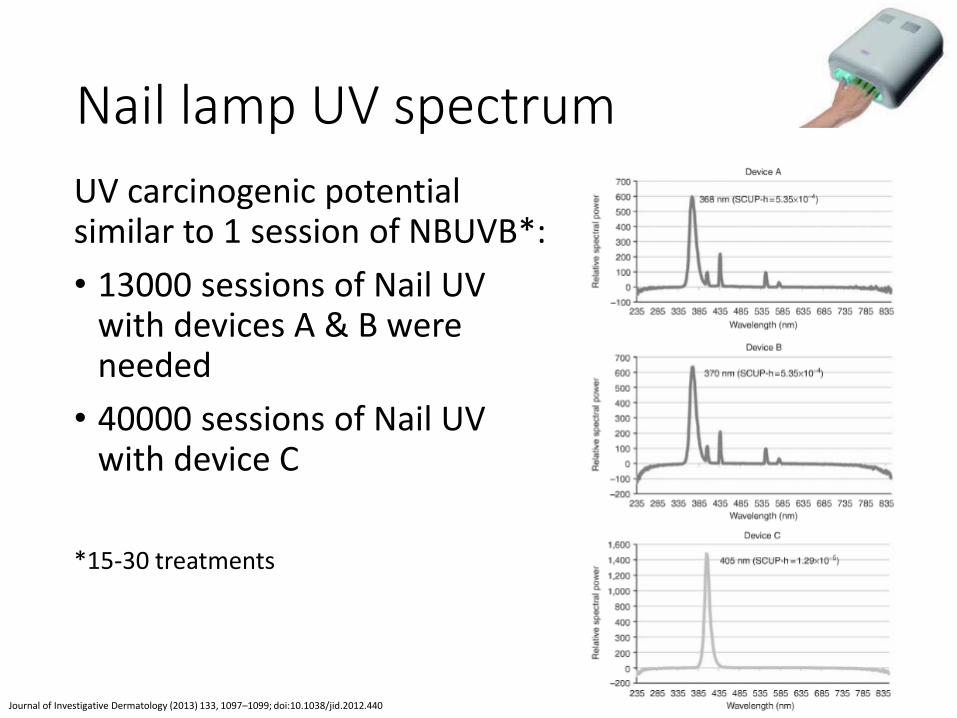
Nail lamp UV spectrum
UV carcinogenic potential similar to 1 session of NBUVB*:
• 13000 sessions of Nail UV with devices A & B were needed
• 40000 sessions of Nail UV with device C
*15-30 treatments
Journal of Investigative Dermatology (2013) 133, 1097–1099; doi:10.1038/jid.2012.440
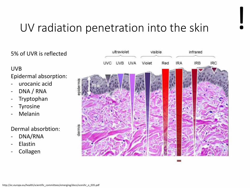
UV radiation penetration into the skin
http://ec.europa.eu/health/scientific_committees/emerging/docs/scenihr_o_035.pdf
5% of UVR is reflected UVB Epidermal absorption: - urocanic acid - DNA / RNA - Tryptophan - Tyrosine - Melanin
Dermal absorbtion: - DNA/RNA - Elastin - Collagen
!
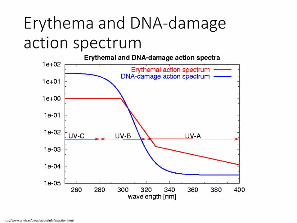
Erythema and DNA-damage action spectrum
http://www.temis.nl/uvradiation/info/uvaction.html
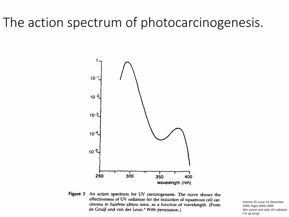
The action spectrum of photocarcinogenesis.
Volume 35, Issue 14, December 1999, Pages 2003–2009 Skin cancer and solar UV radiation F.R. de Gruijl
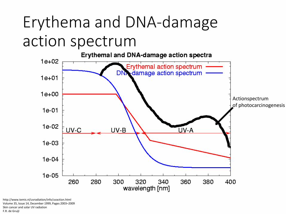
Erythema and DNA-damage action spectrum
http://www.temis.nl/uvradiation/info/uvaction.html Volume 35, Issue 14, December 1999, Pages 2003–2009 Skin cancer and solar UV radiation F.R. de Gruijl
Actionspectrum of photocarcinogenesis
Outline
• Introduction (definition, physical properties, UV sources, depth of penetration into the skin, action spectrums)
• Cellular effects of UVR (biomolecules effected by UV, DNA damage)
• UVR induced skin changes (sunburn, tanning, epidermal hypreplasia, vitamin D production, emotional effects, photoaging, photoimmunology, photocarcinogenesis)
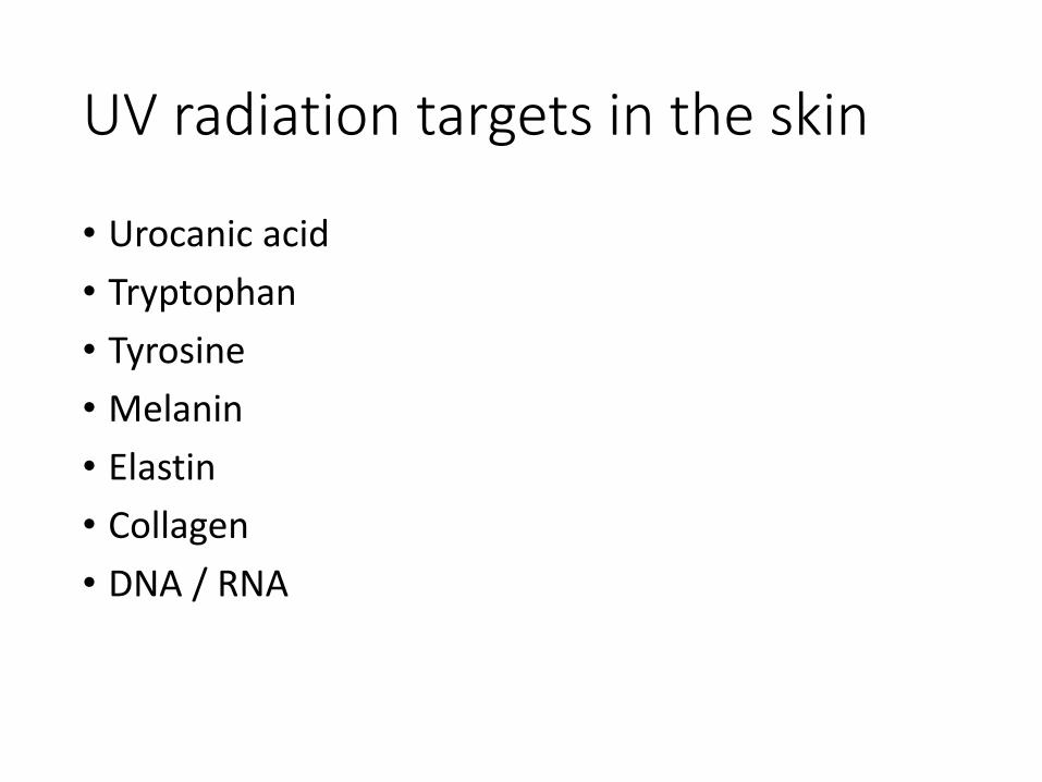
UV radiation targets in the skin
• Urocanic acid
• Tryptophan
• Tyrosine
• Melanin
• Elastin
• Collagen
• DNA / RNA
http://www.onepennysheet.com/2010/01/full-body-scanners-used-on-air-passengers-may-damage-human-dna/
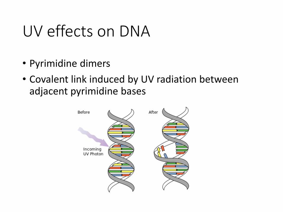
UV effects on DNA
• Pyrimidine dimers
• Covalent link induced by UV radiation between adjacent pyrimidine bases
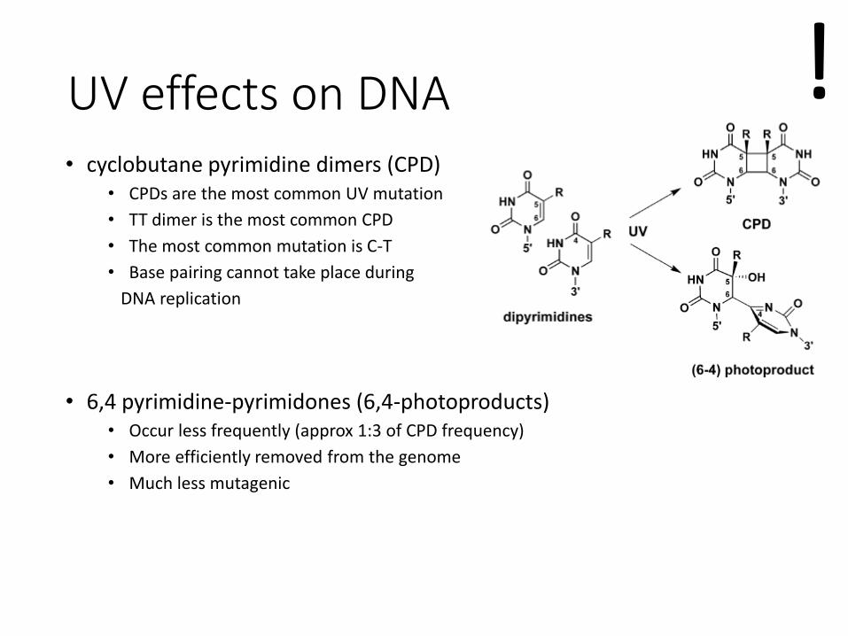
UV effects on DNA • cyclobutane pyrimidine dimers (CPD)
• CPDs are the most common UV mutation
• TT dimer is the most common CPD
• The most common mutation is C-T
• Base pairing cannot take place during
DNA replication
• 6,4 pyrimidine-pyrimidones (6,4-photoproducts) • Occur less frequently (approx 1:3 of CPD frequency)
• More efficiently removed from the genome
• Much less mutagenic
!
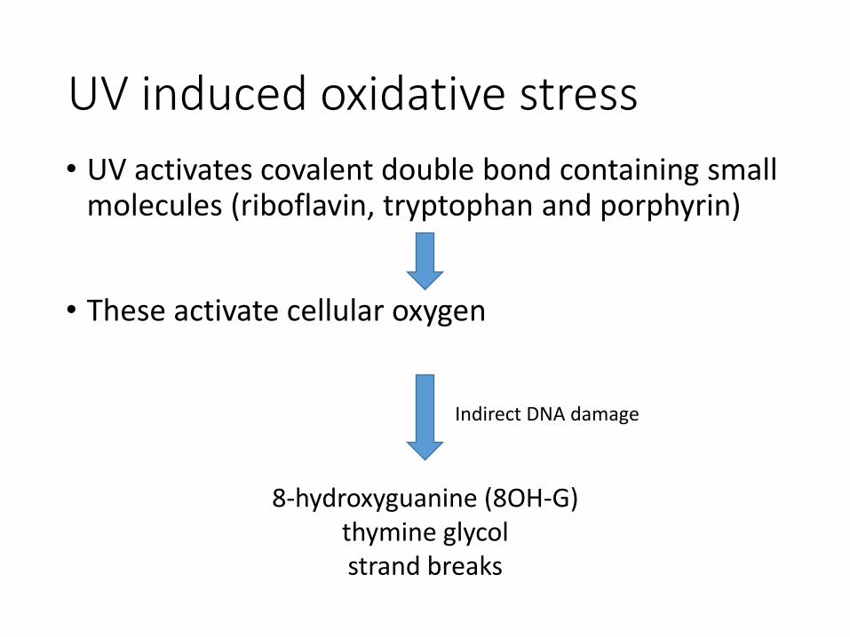
UV induced oxidative stress
• UV activates covalent double bond containing small molecules (riboflavin, tryptophan and porphyrin)
• These activate cellular oxygen
8-hydroxyguanine (8OH-G) thymine glycol strand breaks
Indirect DNA damage
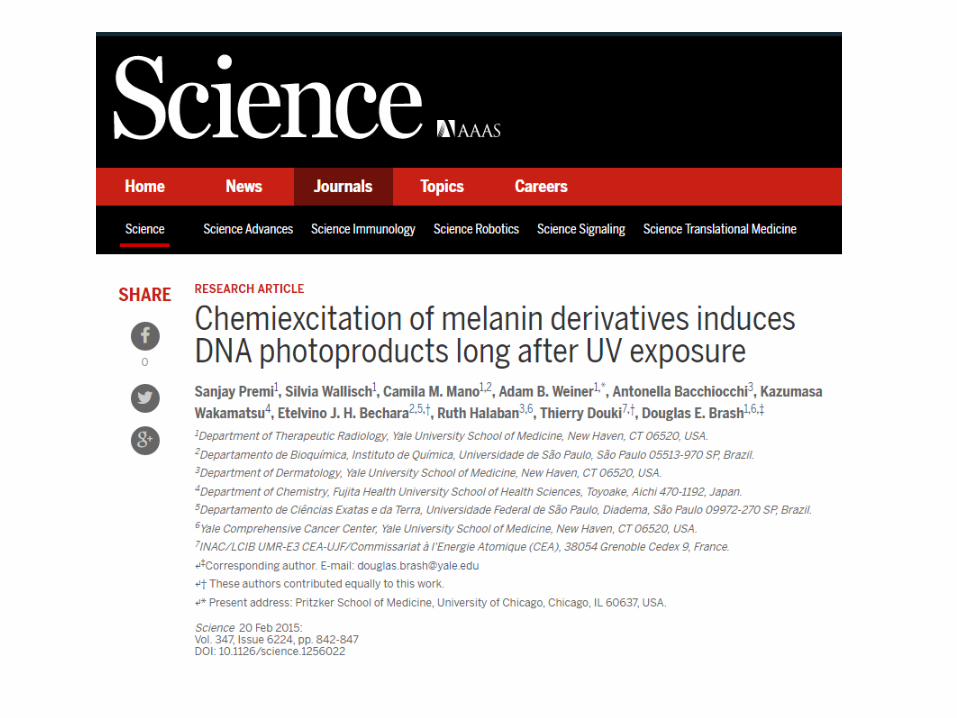
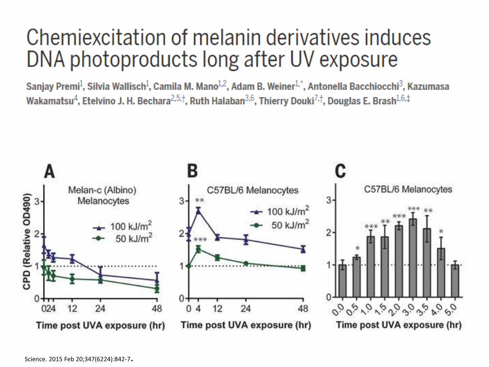
Science. 2015 Feb 20;347(6224):842-7.
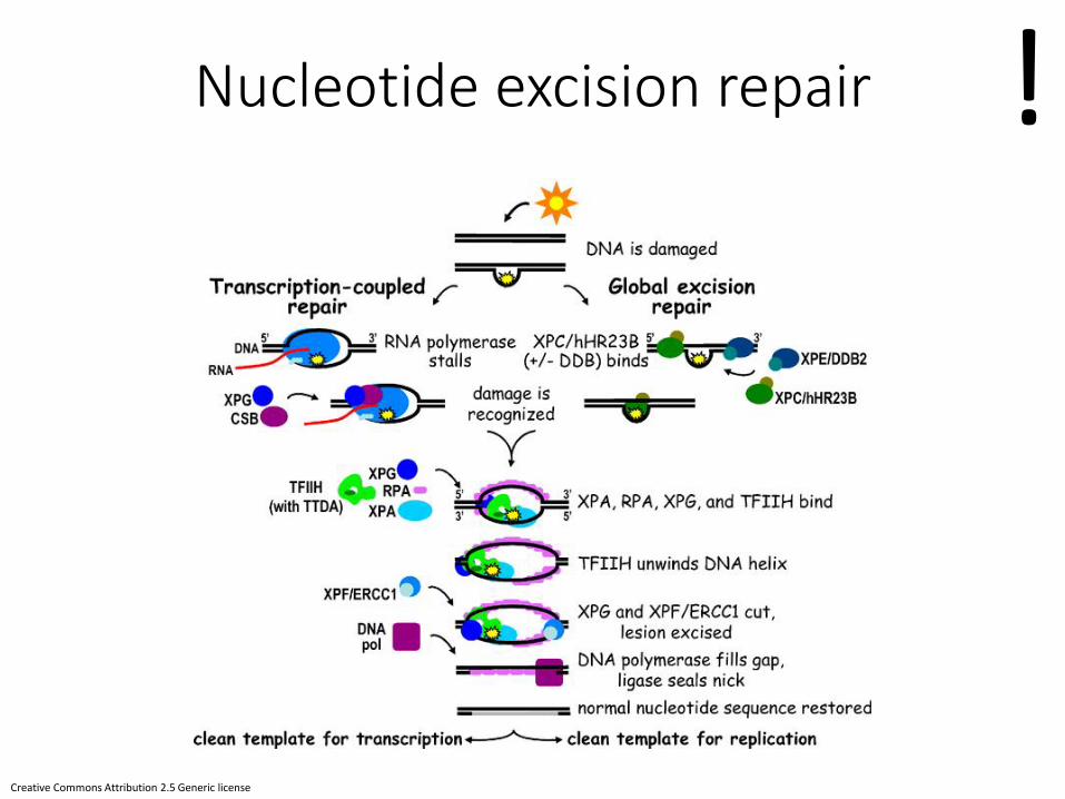
Nucleotide excision repair
Creative Commons Attribution 2.5 Generic license
!

Xeroderma pigmentosum
• Symptoms: Development of numerous lentigines at an early age
(DiGiovanna et al. 2012 )
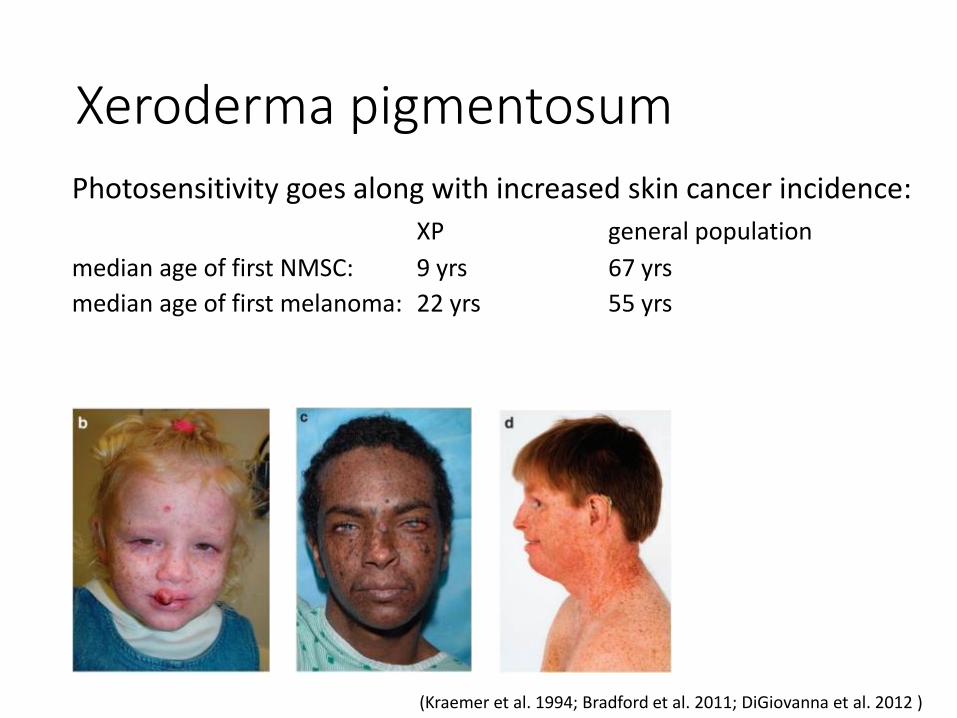
Xeroderma pigmentosum
Photosensitivity goes along with increased skin cancer incidence:
XP general population
median age of first NMSC: 9 yrs 67 yrs
median age of first melanoma: 22 yrs 55 yrs
(Kraemer et al. 1994; Bradford et al. 2011; DiGiovanna et al. 2012 )
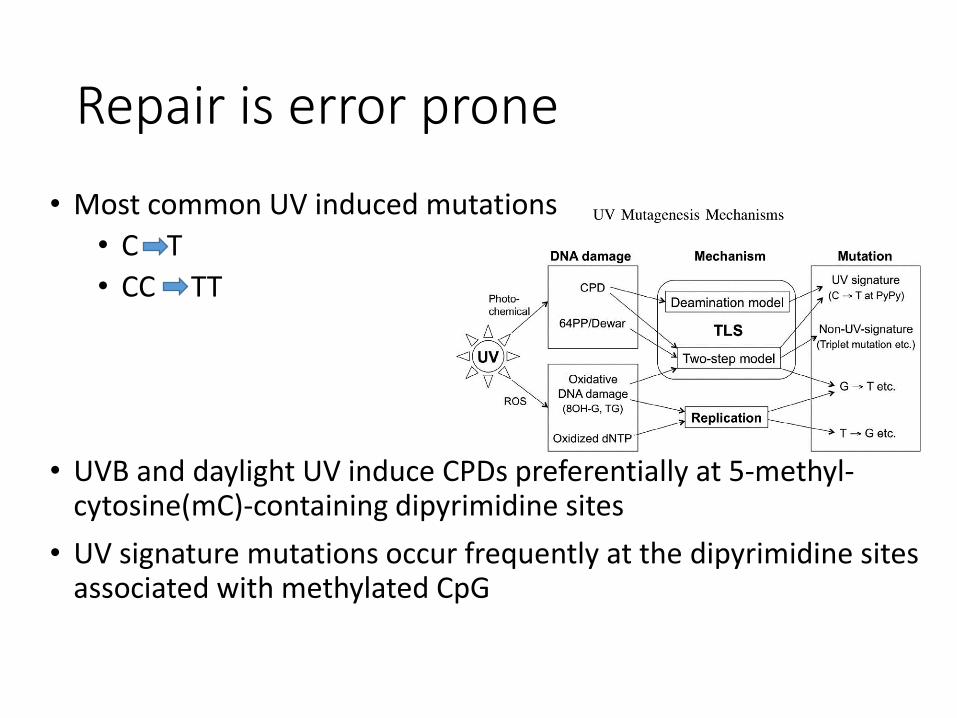
Repair is error prone
• Most common UV induced mutations
• C T
• CC TT
• UVB and daylight UV induce CPDs preferentially at 5-methyl-cytosine(mC)-containing dipyrimidine sites
• UV signature mutations occur frequently at the dipyrimidine sites associated with methylated CpG
Difference between UVA and UVB mutagenesis • UVB: mostly direct DNA damage with limited
secondary ROS effect
• UVA: 8-hydroxyguanine, CPDs (high amount compared to previously expected) no 6,4PPs or Dewar isomers
J. Radiat. Res., 52, 115–125 (2011) Hironobu IKEHATA*and Tetsuya ONO The Mechanisms of UV Mutagenesis

DNA damage signaling
Jackson SP and Bartek J. (2009) The DNA damage response in human biology and disease. Nature 461, 1071-1078
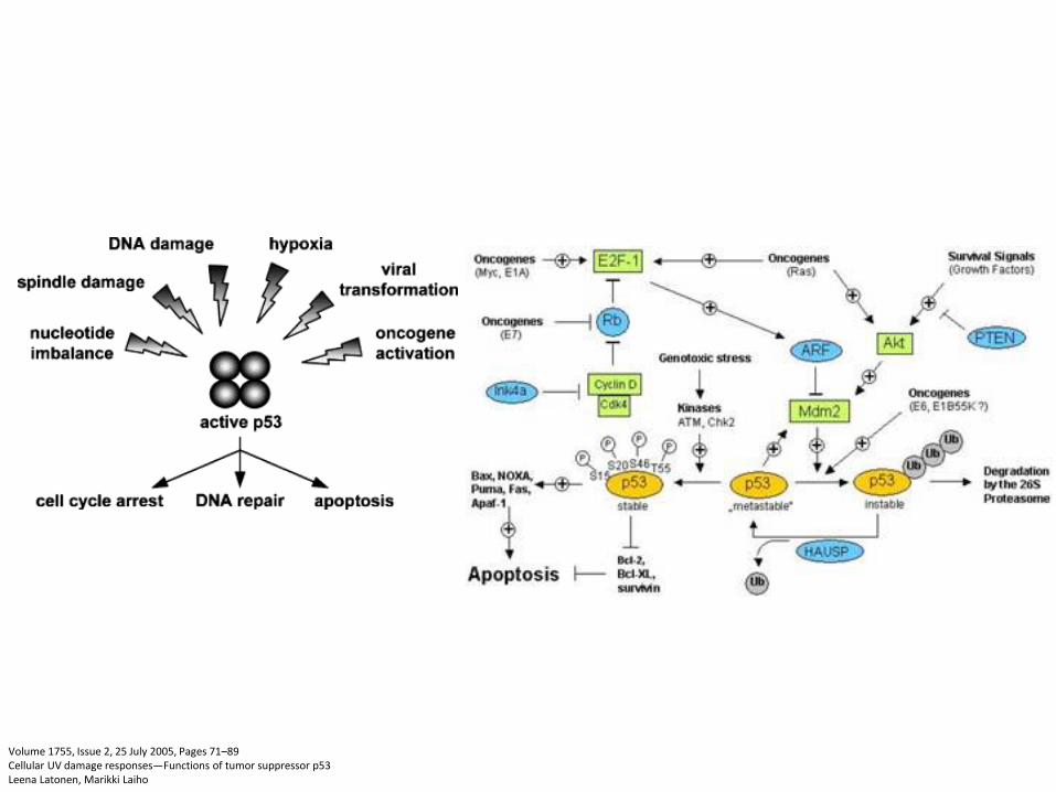
Volume 1755, Issue 2, 25 July 2005, Pages 71–89 Cellular UV damage responses—Functions of tumor suppressor p53 Leena Latonen, Marikki Laiho

Outline
• Introduction (definition, physical properties, UV sources, depth of penetration into the skin, action spectrums)
• Cellular effects of UVR (biomolecules effected by UV, DNA damage)
• UVR induced skin changes (sunburn, tanning, epidermal hypreplasia, vitamin D production, emotional effects, photoaging, photoimmunology, photocarcinogenesis)
UVR induced skin changes
sunburn
tanning
epidermal hypreplasia
vitamin D production
emotional effects
photoaging
photoimmunology
photocarcinogenesis
Photocarcinogenesis
8/28/2013
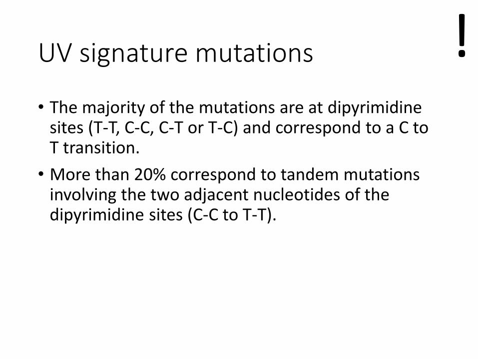
UV signature mutations
• The majority of the mutations are at dipyrimidine sites (T-T, C-C, C-T or T-C) and correspond to a C to T transition.
• More than 20% correspond to tandem mutations involving the two adjacent nucleotides of the dipyrimidine sites (C-C to T-T).
!
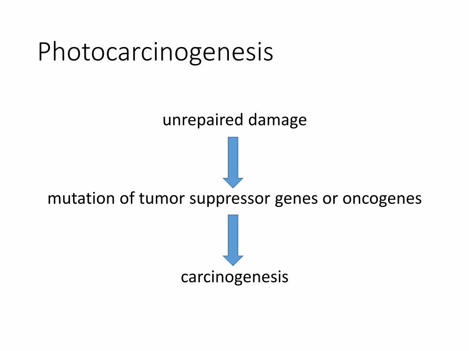
Photocarcinogenesis
unrepaired damage
mutation of tumor suppressor genes or oncogenes
carcinogenesis

Sunburn cells
• Pyknotic nucleus
• Eosinophilic cytoplasm
• Keratin 5 +
• Lack of late
differentiation markers
• Appear 6-24 hrs after
UV exposure
apoptotic keratinocytes
medscape.com
Photodermatol Photoimmunol Photomed. 1995 Aug;11(4):149-54
Sunburn cells
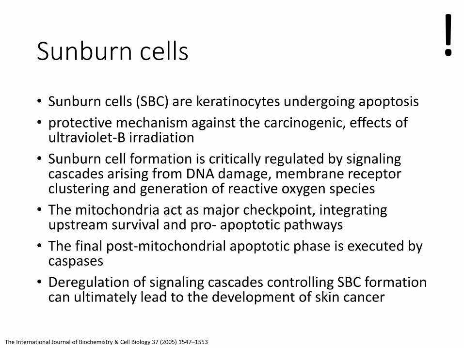
• Sunburn cells (SBC) are keratinocytes undergoing apoptosis
• protective mechanism against the carcinogenic, effects of ultraviolet-B irradiation
• Sunburn cell formation is critically regulated by signaling cascades arising from DNA damage, membrane receptor clustering and generation of reactive oxygen species
• The mitochondria act as major checkpoint, integrating upstream survival and pro- apoptotic pathways
• The final post-mitochondrial apoptotic phase is executed by caspases
• Deregulation of signaling cascades controlling SBC formation can ultimately lead to the development of skin cancer
The International Journal of Biochemistry & Cell Biology 37 (2005) 1547–1553
!
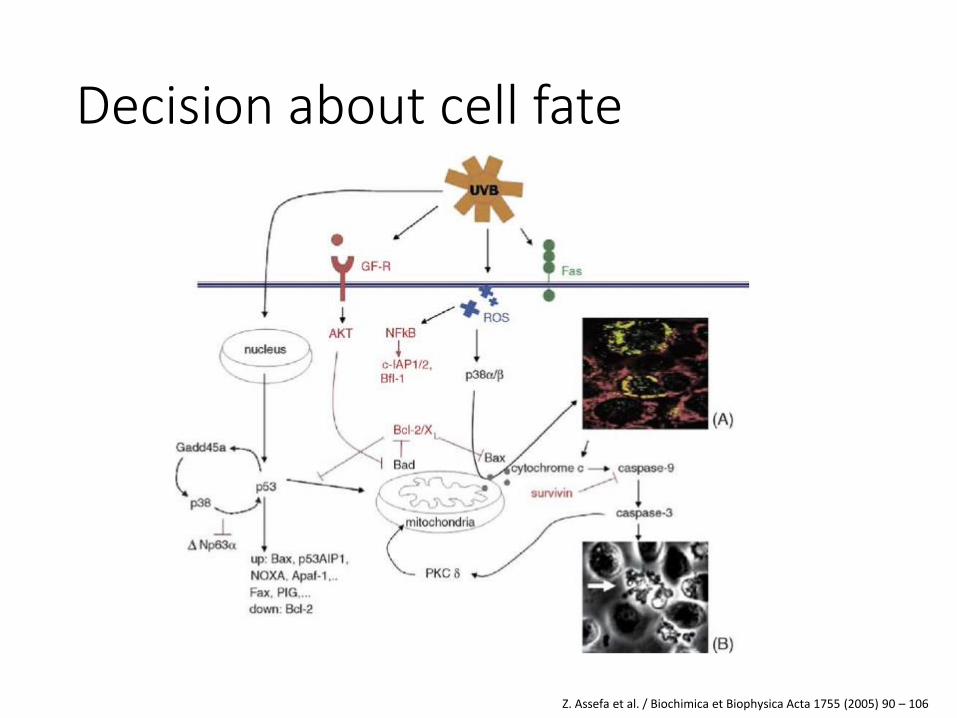
Decision about cell fate
Z. Assefa et al. / Biochimica et Biophysica Acta 1755 (2005) 90 – 106
Importance of sunburn cell formation
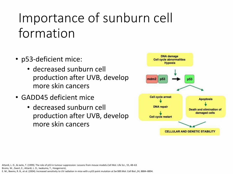
• p53-deficient mice:
• decreased sunburn cell production after UVB, develop more skin cancers
• GADD45 deficient mice
• decreased sunburn cell production after UVB, develop more skin cancers
Attardi, L. D., & Jacks, T. (1999). The role of p53 in tumour suppression: Lessons from mouse models.Cell Mol. Life Sci., 55, 48–63 Bruins, W., Zwart, E., Attardi, L. D., Iwakuma, T., Hoogervorst, E. M., Beems, R. B., et al. (2004). Increased sensitivity to UV radiation in mice with a p53 point mutation at Ser389.Mol. Cell Biol.,24, 8884–8894.
Sunburn cell formation and erythema
• Multiple studies show that erythema and inflammatory response and sunburn cell production correlate
• Suberythemogenic doses of UV radiation may still lead to small amount of sunburn cell formation
J Dermatol Sci. 2009 Jul;55(1):10-7. doi: 10.1016/j.jdermsci.2009.03.011. Epub 2009 May 2. J Drugs Dermatol. 2013 Apr;12(4):464-8. Journal of Investigative Dermatology (2004) 123, 781–787 Kaidbey KH: The photoprotective potential of the new superpotent sunscreens. J Amer Acad Dermatol 22(3):449-452, 1990. Ziegler A, Jonason AS, Leffell DJ, et al.: Sunburn and p53 in the onset of skin cancer. Nature 372(6508):773-776, 1994
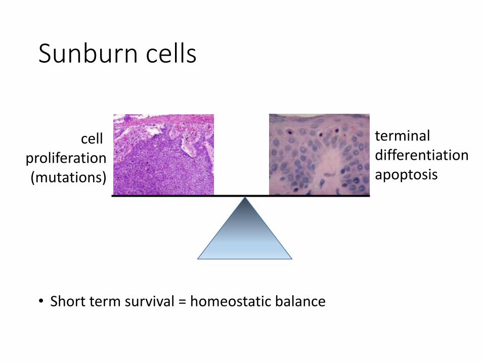
Sunburn cells
• Short term survival = homeostatic balance
terminal differentiation apoptosis
cell proliferation (mutations)
Early mutant cell clones

p53 mutations in UV carcinogenesis
• UV induced skin cancers show up to 100% p53 mutation rate (54-100%)
• p53 defective cells are less prone to apoptosis induction by UV light
• p53 mutations are present in sun-damaged skin and AKs
• Most loss of function p53 mutations result in increased p53 immunopositivity given accumulation of mutated / dysfunctional p53 protein
Cancer Res. 1993 Jul 1;53(13):2961-4.
Neoplasia. 1999 Nov;1(5):468-75.
Cancer Res.2007;67:4648-4656.
!
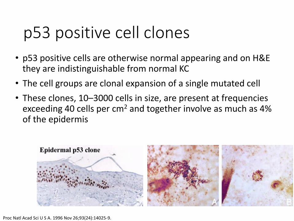
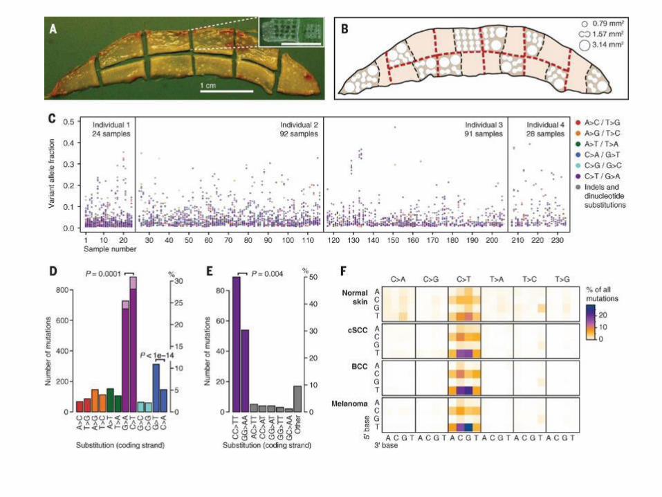
p53 positive cell clones
• p53 positive cells are otherwise normal appearing and on H&E they are indistinguishable from normal KC
• The cell groups are clonal expansion of a single mutated cell
• These clones, 10–3000 cells in size, are present at frequencies exceeding 40 cells per cm2 and together involve as much as 4% of the epidermis
Proc Natl Acad Sci U S A. 1996 Nov 26;93(24):14025-9.
• The mutation spectra found in epidermal p53 clones resemble that of non-melanoma skin cancer.
• Coexisting AK, CIS and SCC have been found to share similar mutations, further supporting the notion that p53 mutations appear early in the development of skin cancer
• The same mutations were not identified in the same geographic lactations.
• The p53 clones in normal skin surrounding SCC were significantly more frequent and larger in size than those in skin surrounding BCC or melanocytic nevus, indicating an association between p53 clones and SCC.
Exp Dermatol. 2004 Oct;13(10):643-50. Oncogene. 1996 Feb 15;12(4):765-73.
p53 positive cell clones are likely earliest detectable precursors of SCC
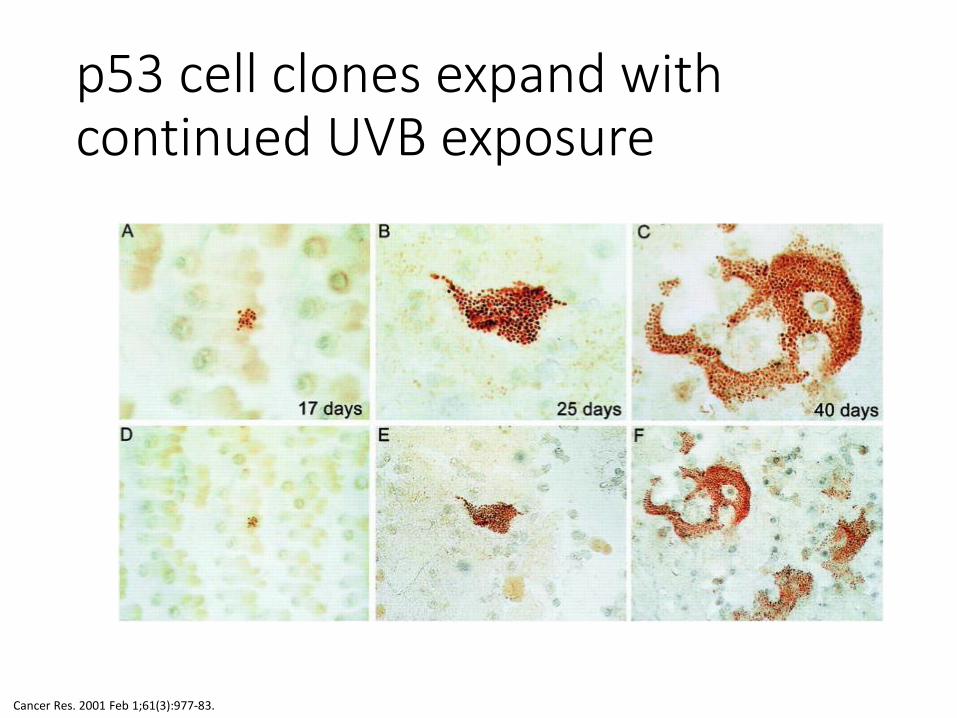
p53 cell clones expand with continued UVB exposure
Cancer Res. 2001 Feb 1;61(3):977-83.
p53 clone growth is promoted by UVB
• p53 clones contain p53 UV signature mutations
• In mouse models after p53 clone formation is stopped clones regress.
• Regression also takes place in mice defective in adaptive immunity.
What is the driving force behind UV induced p53 clone expasion?
Cancer Res. 2001 Feb 1;61(3):977-83. Semin Cancer Biol. 2005 Apr;15(2):97-102. Cancer Res.2007;67:4648-4656.
Apoptosis of surrounding normal keratinocytes drives p53 clone expansion
• Survivin is an apoptosis inhibitor
• Keratinocyte overexpression of survivin (decreased apoptosis) increases number of p53 clones but decreases their size and rate of growth.
• UV induced apoptosis of KCs surrounding p53 clones is one of the driving forces of clone expansion
• p53 clones regress with good sun protection
Cancer Res. 2001 Feb 1;61(3):977-83. Semin Cancer Biol. 2005 Apr;15(2):97-102. Cancer Res.2007;67:4648-4656.

Effect of marked genotoxic trauma on p53 clones
• UV-induced ablation of the epidermal basal layer including p53-mutant clones reduces UV induced keratinocyte carcinogenesis
Carcinogenesis (2012) 33 (3):714-720.
!
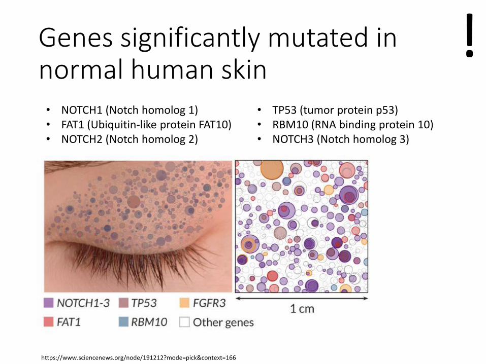
Genes significantly mutated in normal human skin
• TP53 (tumor protein p53) • RBM10 (RNA binding protein 10) • NOTCH3 (Notch homolog 3)
• NOTCH1 (Notch homolog 1) • FAT1 (Ubiquitin-like protein FAT10) • NOTCH2 (Notch homolog 2)
https://www.sciencenews.org/node/191212?mode=pick&context=166
!
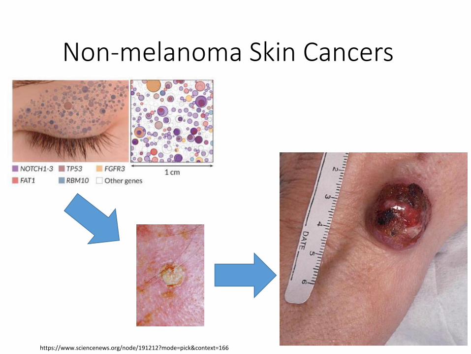
Non-melanoma Skin Cancers
https://www.sciencenews.org/node/191212?mode=pick&context=166
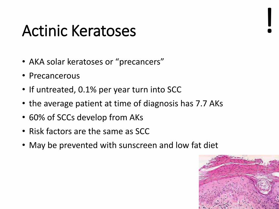
Actinic Keratoses
• AKA solar keratoses or “precancers”
• Precancerous
• If untreated, 0.1% per year turn into SCC
• the average patient at time of diagnosis has 7.7 AKs
• 60% of SCCs develop from AKs
• Risk factors are the same as SCC
• May be prevented with sunscreen and low fat diet
!
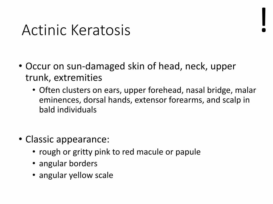
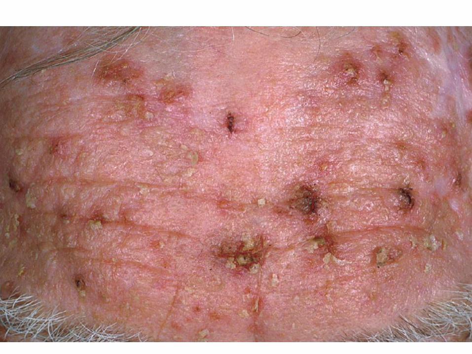
Actinic Keratosis
• Occur on sun-damaged skin of head, neck, upper trunk, extremities • Often clusters on ears, upper forehead, nasal bridge, malar
eminences, dorsal hands, extensor forearms, and scalp in bald individuals
• Classic appearance: • rough or gritty pink to red macule or papule
• angular borders
• angular yellow scale
!

Destructive treatment
• Cryotherapy

Field treatment
• 5-fluorouracil cream
• photodynamic therapy (PDT with ALA or MAL)
• imiquimod
• ingenol mebutate gel 0.05%
• diclofenac 3% gel
• chemical peels
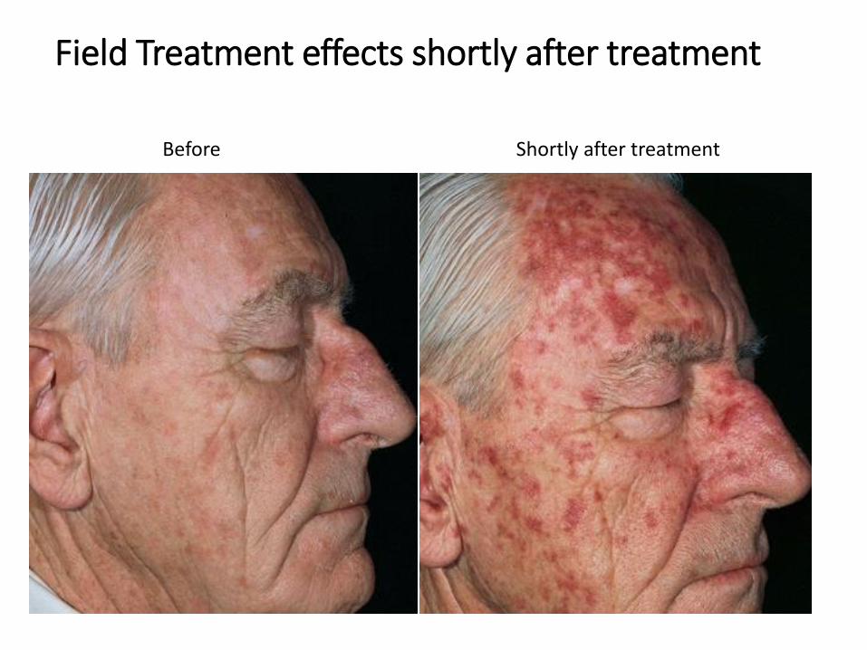
Field Treatment effects shortly after treatment
Before Shortly after treatment
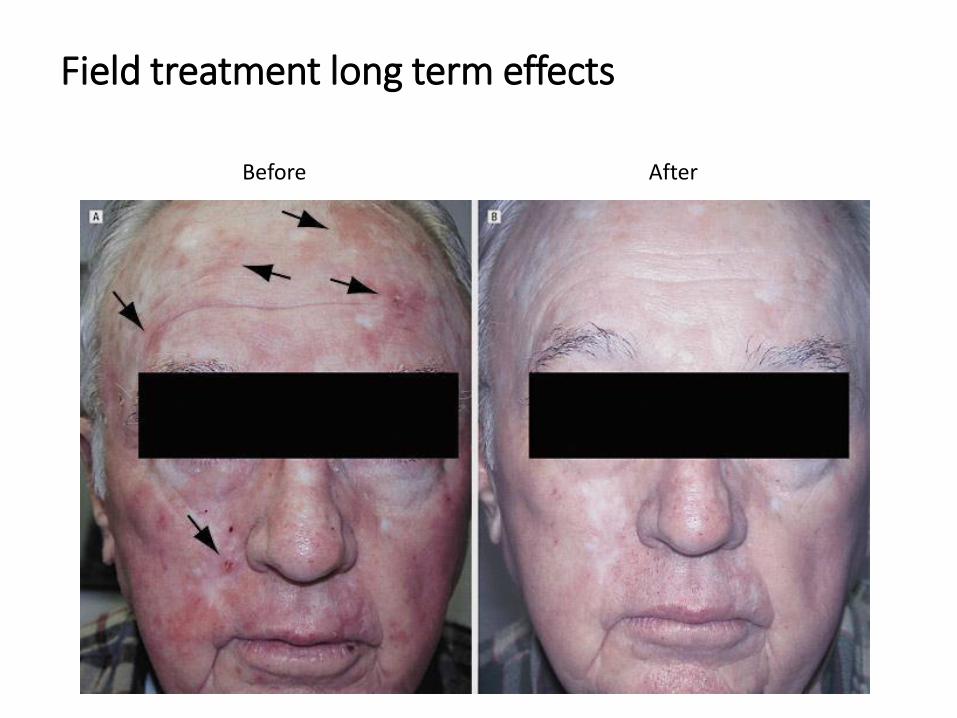
Before After
Field treatment long term effects
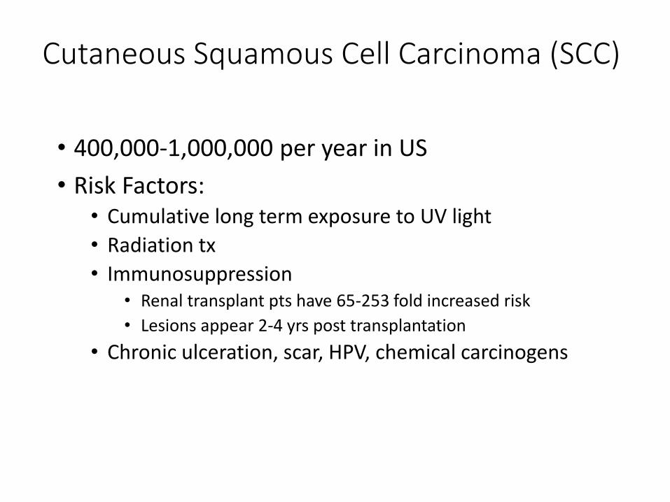
Cutaneous Squamous Cell Carcinoma (SCC)
• 400,000-1,000,000 per year in US
• Risk Factors: • Cumulative long term exposure to UV light
• Radiation tx
• Immunosuppression • Renal transplant pts have 65-253 fold increased risk
• Lesions appear 2-4 yrs post transplantation
• Chronic ulceration, scar, HPV, chemical carcinogens
Squamous Cell Carcinoma
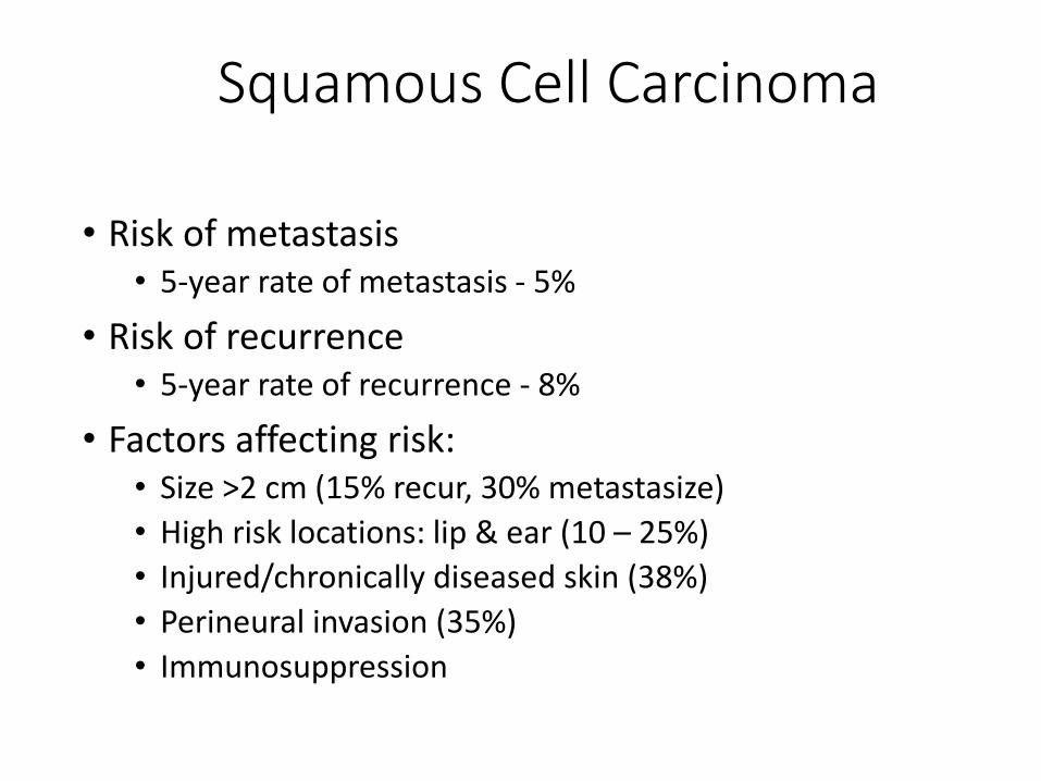
• Risk of metastasis • 5-year rate of metastasis - 5%
• Risk of recurrence • 5-year rate of recurrence - 8%
• Factors affecting risk: • Size >2 cm (15% recur, 30% metastasize)
• High risk locations: lip & ear (10 – 25%)
• Injured/chronically diseased skin (38%)
• Perineural invasion (35%)
• Immunosuppression
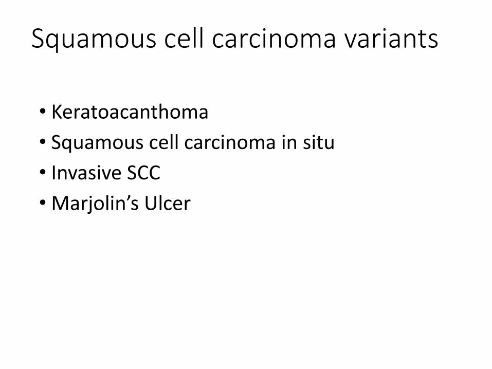
Squamous cell carcinoma variants
• Keratoacanthoma
• Squamous cell carcinoma in situ
• Invasive SCC
• Marjolin’s Ulcer
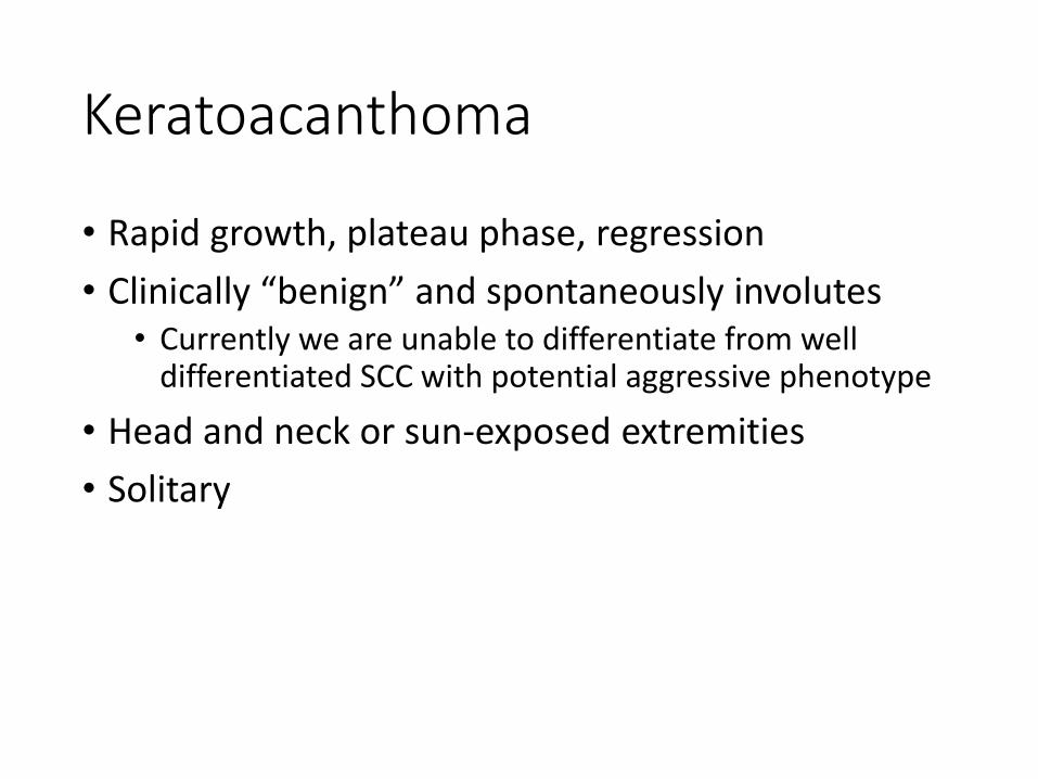
Keratoacanthoma
• Rapid growth, plateau phase, regression
• Clinically “benign” and spontaneously involutes • Currently we are unable to differentiate from well
differentiated SCC with potential aggressive phenotype
• Head and neck or sun-exposed extremities
• Solitary
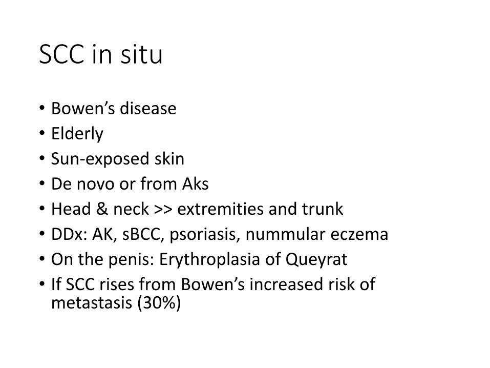
SCC in situ
• Bowen’s disease
• Elderly
• Sun-exposed skin
• De novo or from Aks
• Head & neck >> extremities and trunk
• DDx: AK, sBCC, psoriasis, nummular eczema
• On the penis: Erythroplasia of Queyrat
• If SCC rises from Bowen’s increased risk of metastasis (30%)
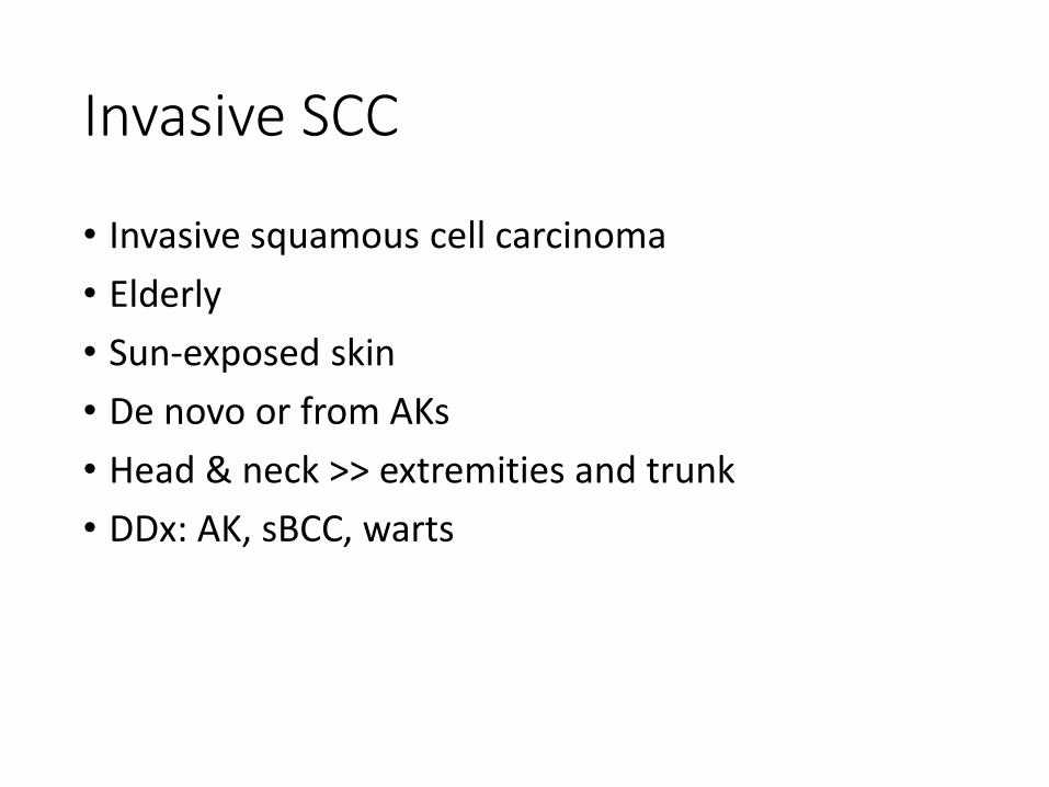
Invasive SCC
• Invasive squamous cell carcinoma
• Elderly
• Sun-exposed skin
• De novo or from AKs
• Head & neck >> extremities and trunk
• DDx: AK, sBCC, warts
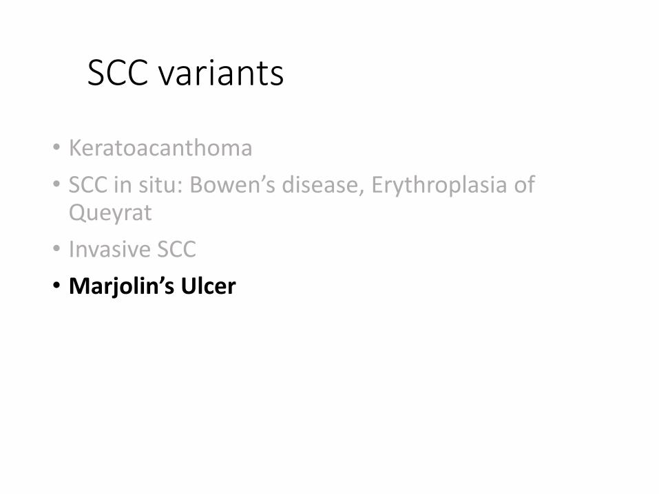
SCC variants
• Keratoacanthoma
• SCC in situ: Bowen’s disease, Erythroplasia of Queyrat
• Invasive SCC
• Marjolin’s Ulcer
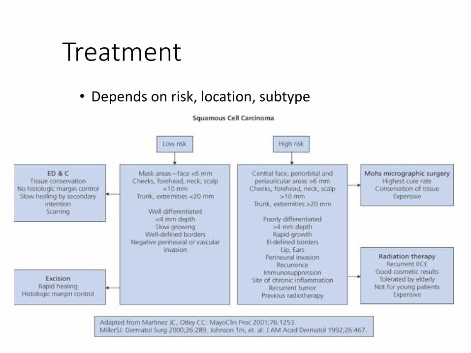
Treatment
• Depends on risk, location, subtype
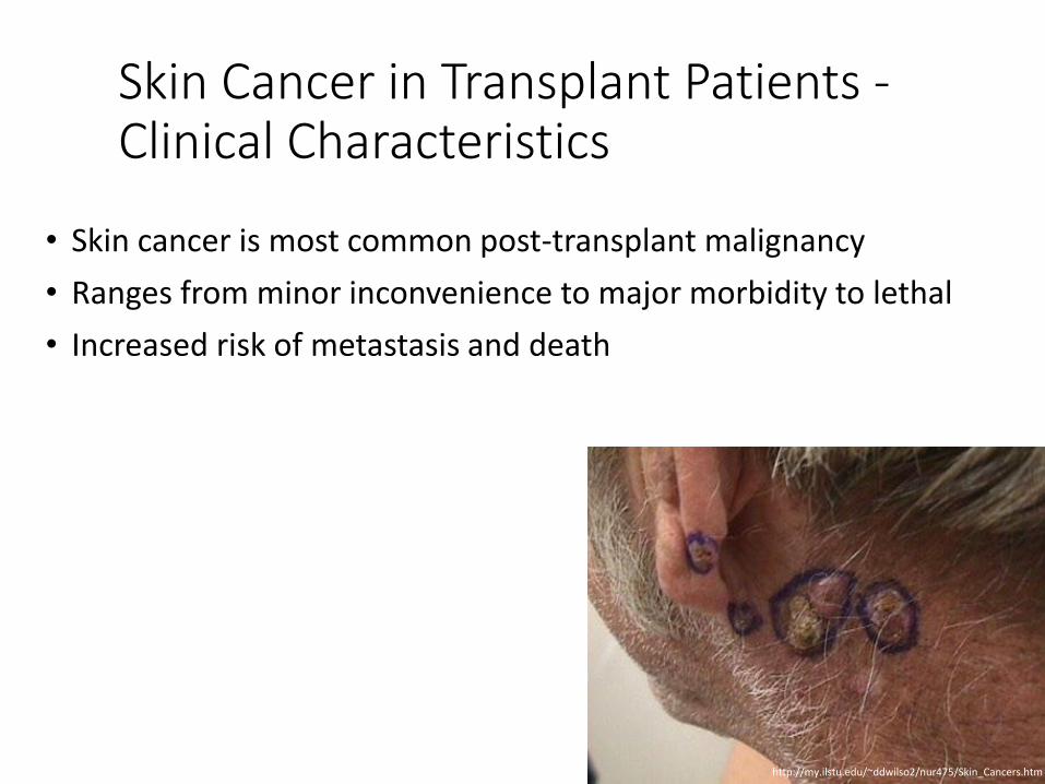
Skin Cancer in Transplant Patients - Clinical Characteristics
• Skin cancer is most common post-transplant malignancy
• Ranges from minor inconvenience to major morbidity to lethal
• Increased risk of metastasis and death
http://my.ilstu.edu/~ddwilso2/nur475/Skin_Cancers.htm
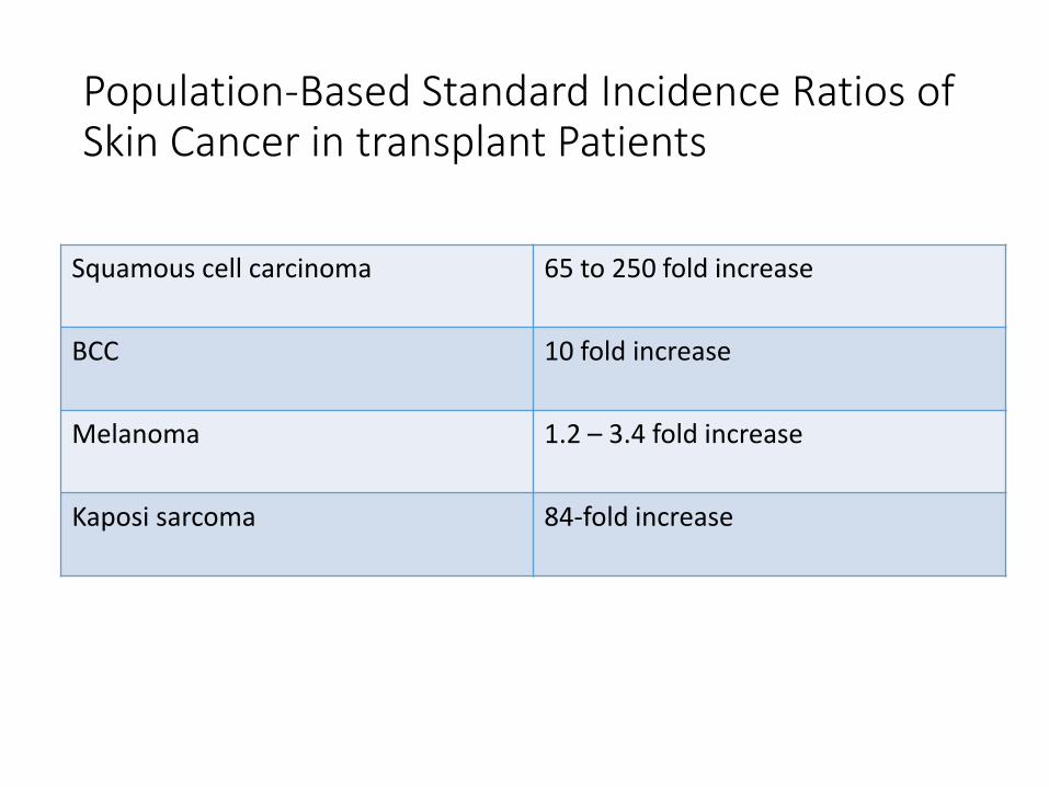
Population-Based Standard Incidence Ratios of Skin Cancer in transplant Patients
Squamous cell carcinoma 65 to 250 fold increase
BCC 10 fold increase
Melanoma 1.2 – 3.4 fold increase
Kaposi sarcoma 84-fold increase
Skin Cancer in Different Types of Transplants
• Cardiac transplants have a 2.9-fold higher risk of SCC compared to renal transplants • Cardiac transplant pts older
• Immunosuppression more intense
• Skin cancer is less common in liver transplants than renal or cardiac
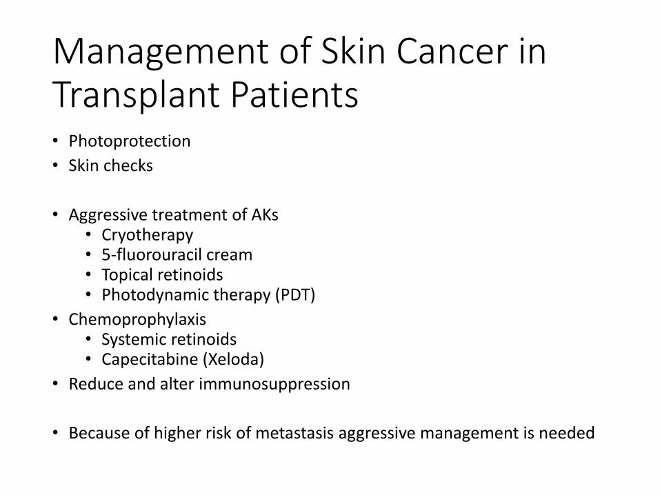
Management of Skin Cancer in Transplant Patients • Photoprotection
• Skin checks
• Aggressive treatment of AKs • Cryotherapy • 5-fluorouracil cream • Topical retinoids • Photodynamic therapy (PDT)
• Chemoprophylaxis • Systemic retinoids • Capecitabine (Xeloda)
• Reduce and alter immunosuppression
• Because of higher risk of metastasis aggressive management is needed
Basal Cell Carcinoma
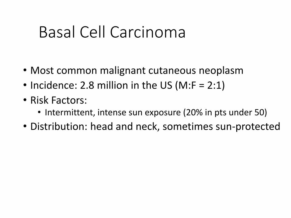
• Most common malignant cutaneous neoplasm
• Incidence: 2.8 million in the US (M:F = 2:1)
• Risk Factors: • Intermittent, intense sun exposure (20% in pts under 50)
• Distribution: head and neck, sometimes sun-protected
Basal Cell Carcinoma
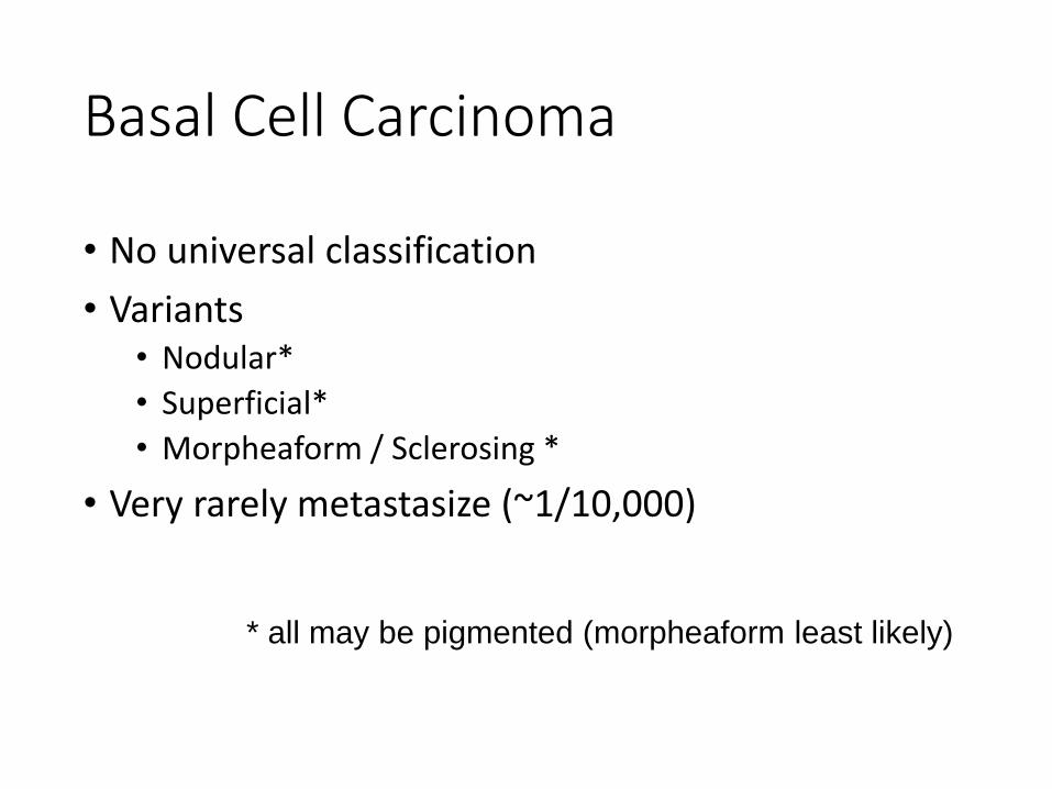
• No universal classification
• Variants • Nodular*
• Superficial*
• Morpheaform / Sclerosing *
• Very rarely metastasize (~1/10,000)
* all may be pigmented (morpheaform least likely)
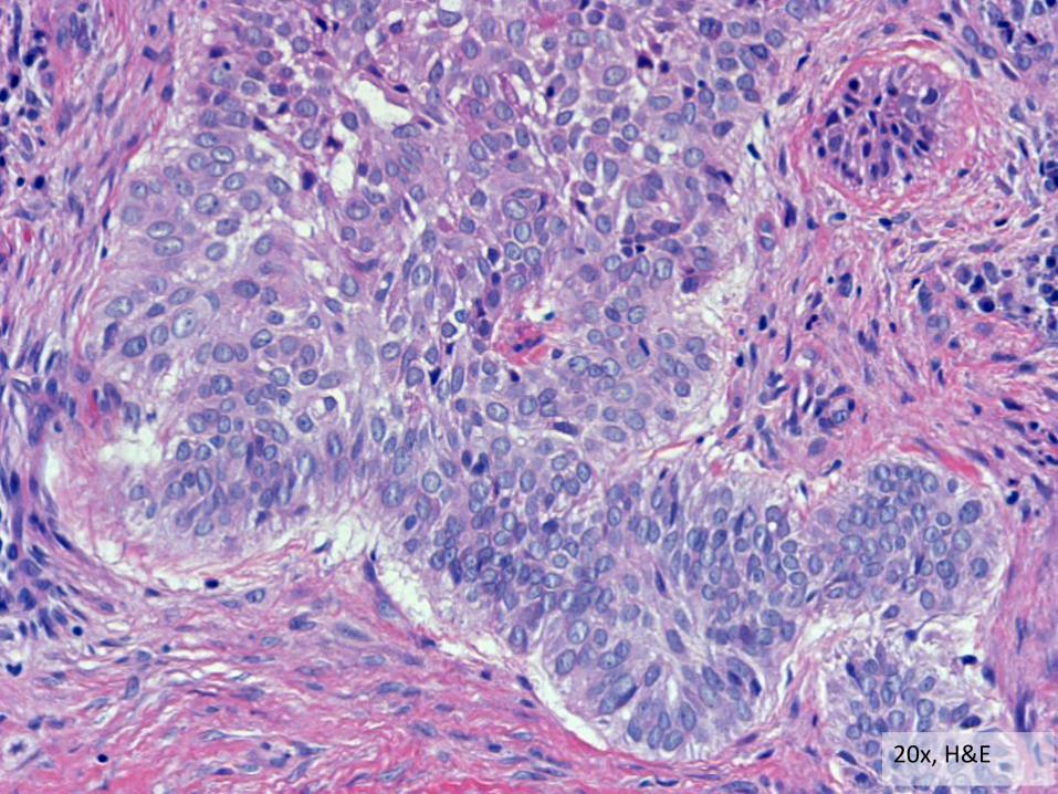
Basal Cell Carcinoma
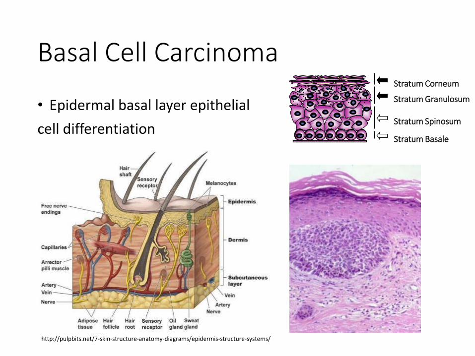
• Epidermal basal layer epithelial
cell differentiation
Stratum Granulosum
Stratum Spinosum
Stratum Basale
Stratum Corneum
http://pulpbits.net/7-skin-structure-anatomy-diagrams/epidermis-structure-systems/
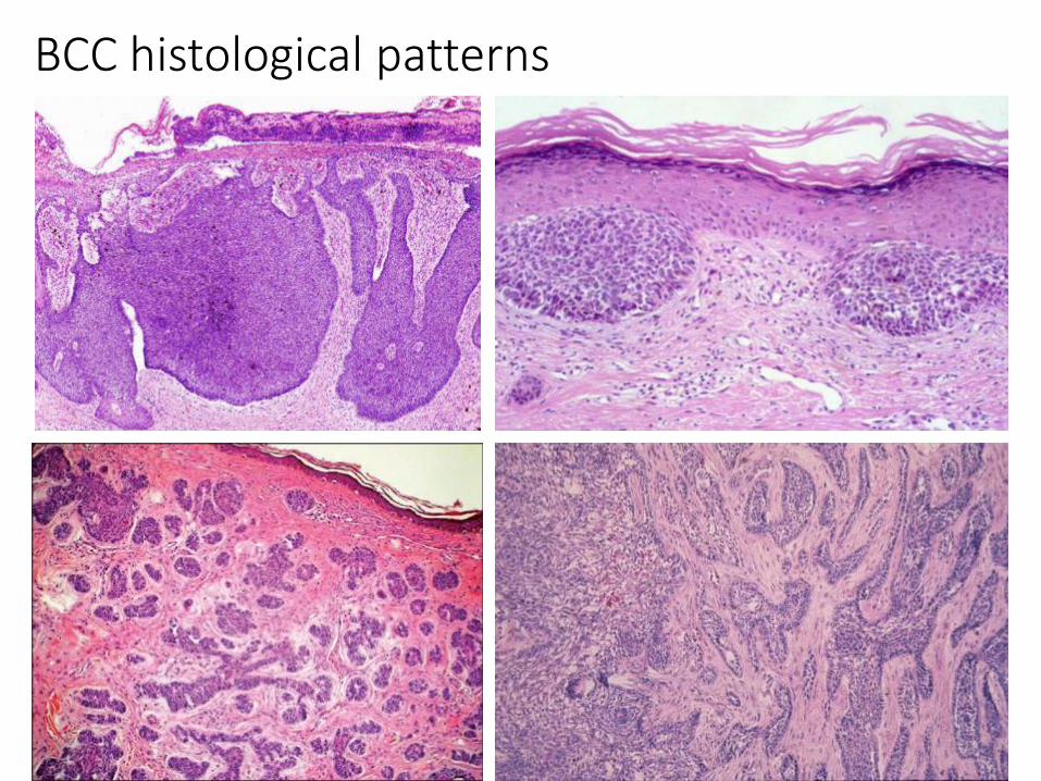
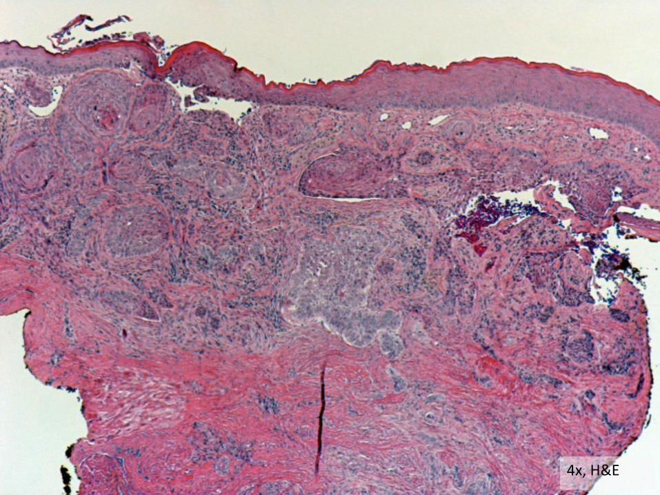
BCC histological patterns
Nodular BCCs
• Most common variant
• 60% of all BCCs
• Face
• Raised, glassy/pearly papule or nodule
• Overlying telangiectases
• Large, extend deeply
• Ulceration
• Pigmented
Superficial BCC
• Second most common
• 15% of BCC
• Favors the trunk and extremities
• Pink, erythematous macule/thin plaque
• Difficult to differentiate from benign inflammatory lesion/SCC/AK
Morpheaform BCC
• AKA sclerosing or infiltrating
• Locally aggressive subtype
• Flat, slightly atrophic or ill-defined plaque
• May appear scar-like
• Actual size often greater than clinically apparent
• Often a histologic determination
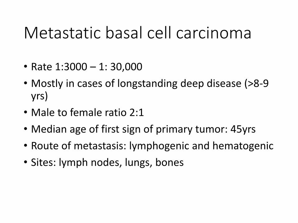
Metastatic basal cell carcinoma
• Rate 1:3000 – 1: 30,000
• Mostly in cases of longstanding deep disease (>8-9 yrs)
• Male to female ratio 2:1
• Median age of first sign of primary tumor: 45yrs
• Route of metastasis: lymphogenic and hematogenic
• Sites: lymph nodes, lungs, bones
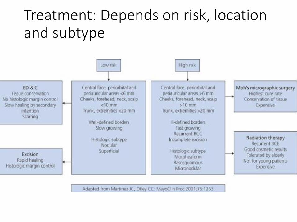
Treatment: Depends on risk, location and subtype
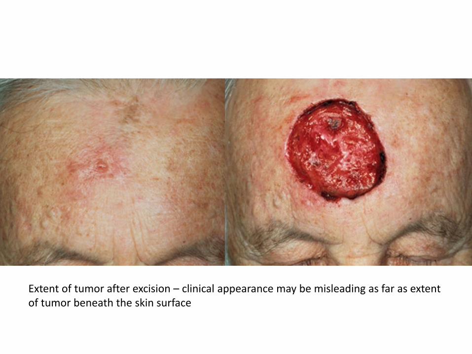
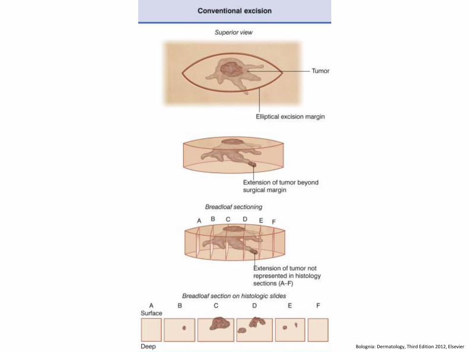
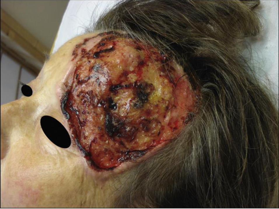
Extent of tumor after excision – clinical appearance may be misleading as far as extent of tumor beneath the skin surface

Mohs Surgery
• Developed in the 1938 by Dr Frederick Mohs, a Wisconsin General Surgeon
• Provides superior margin control, maximal tissue sparing and the highest cure rates
• Drawbacks: time and labor intensive, relatively few Mohs surgeons
Bolognia: Dermatology, Third Edition 2012, Elsevier
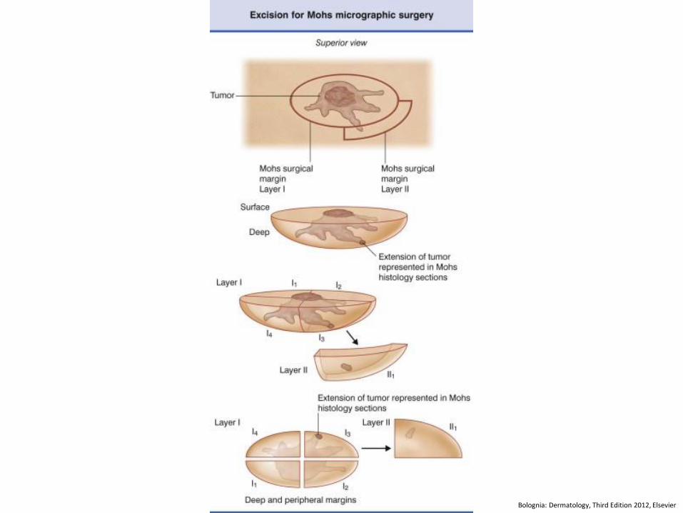
Bolognia: Dermatology, Third Edition 2012, Elsevier

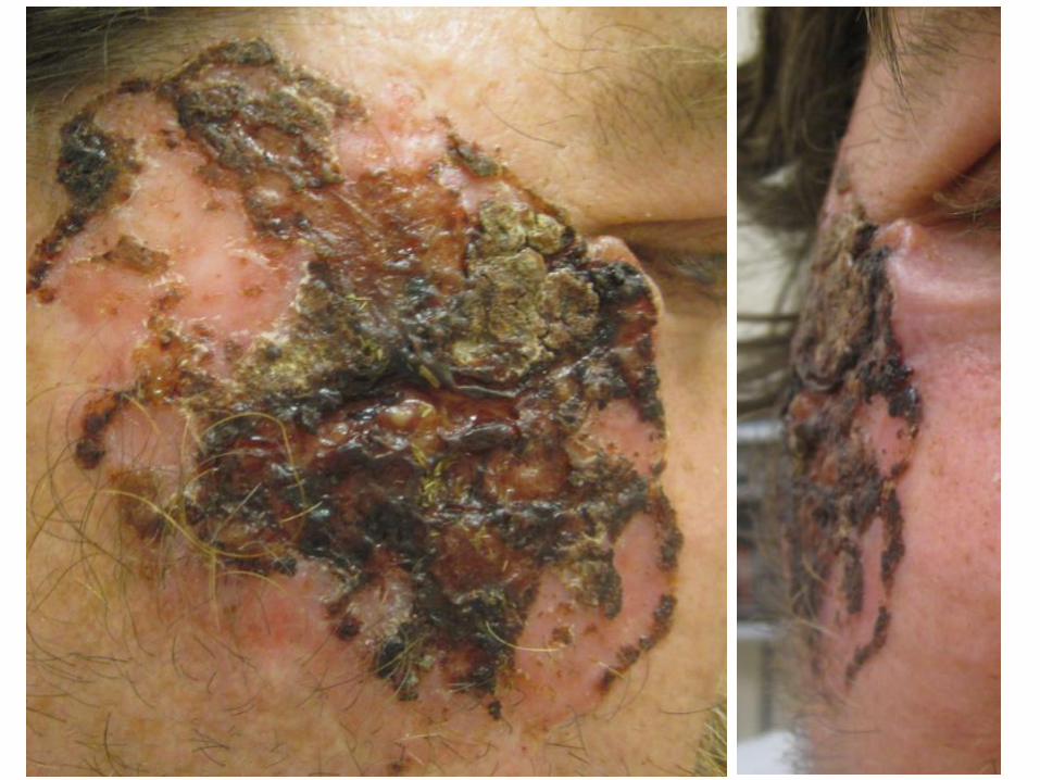
Other therapeutic options for advanced BCC
Radiation therapy
Systemic therapies:
alpha-interferon, capecitabine, retinoids
Small molecular inhibitors
Basal cell carcinoma: beyond surgery and irradiation
4x, H&E
20x, H&E
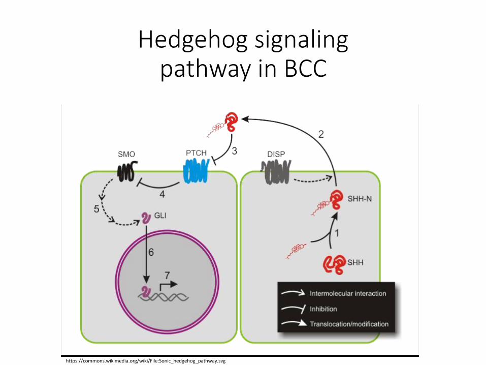
Hedgehog signaling pathway in BCC
https://commons.wikimedia.org/wiki/File:Sonic_hedgehog_pathway.svg
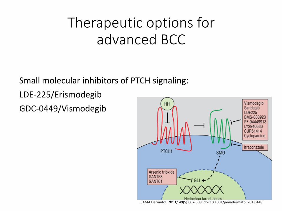
Therapeutic options for advanced BCC
Small molecular inhibitors of PTCH signaling:
LDE-225/Erismodegib
GDC-0449/Vismodegib
JAMA Dermatol. 2013;149(5):607-608. doi:10.1001/jamadermatol.2013.448
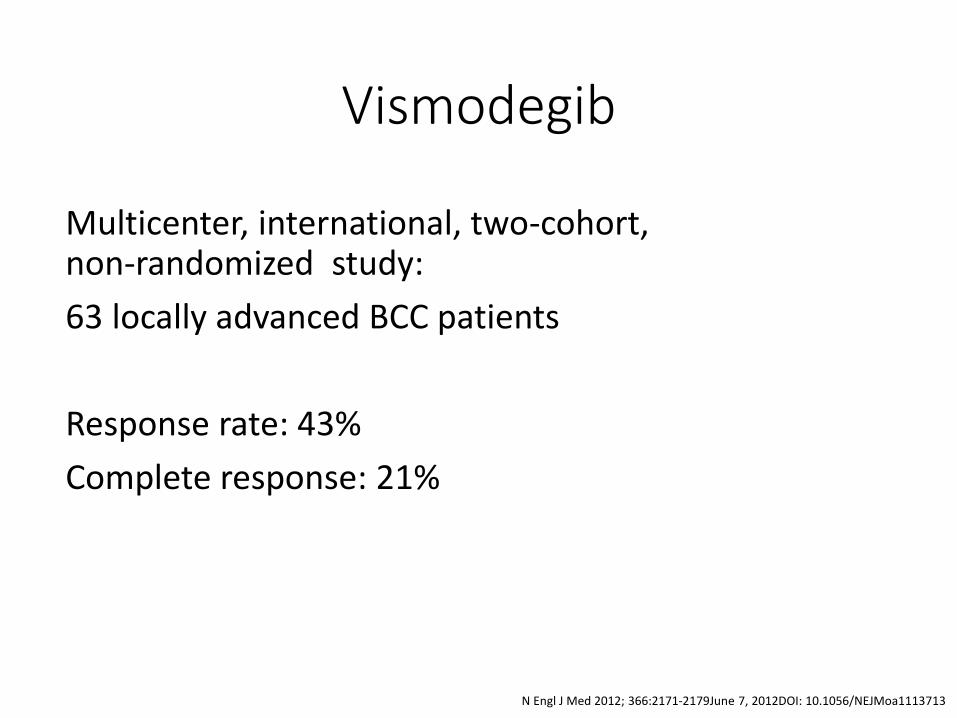
Vismodegib
Multicenter, international, two-cohort, non-randomized study:
63 locally advanced BCC patients
Response rate: 43%
Complete response: 21%
N Engl J Med 2012; 366:2171-2179June 7, 2012DOI: 10.1056/NEJMoa1113713
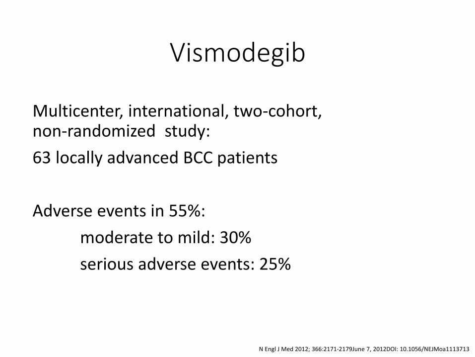
Vismodegib
Multicenter, international, two-cohort, non-randomized study:
63 locally advanced BCC patients
Adverse events in 55%:
moderate to mild: 30%
serious adverse events: 25%
N Engl J Med 2012; 366:2171-2179June 7, 2012DOI: 10.1056/NEJMoa1113713
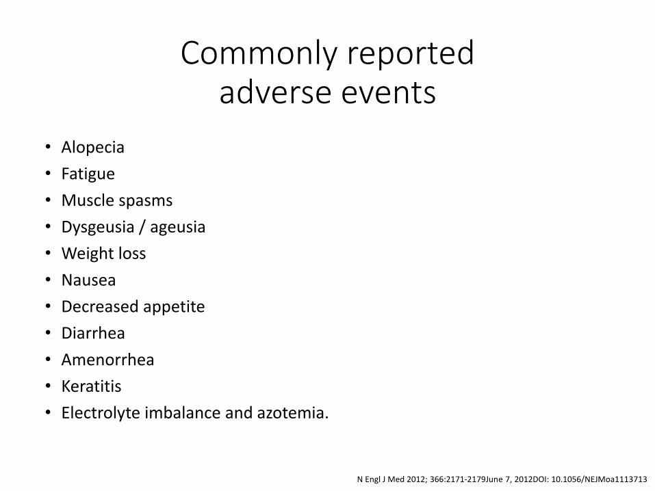
Commonly reported adverse events
• Alopecia
• Fatigue
• Muscle spasms
• Dysgeusia / ageusia
• Weight loss
• Nausea
• Decreased appetite
• Diarrhea
• Amenorrhea
• Keratitis
• Electrolyte imbalance and azotemia.
N Engl J Med 2012; 366:2171-2179June 7, 2012DOI: 10.1056/NEJMoa1113713
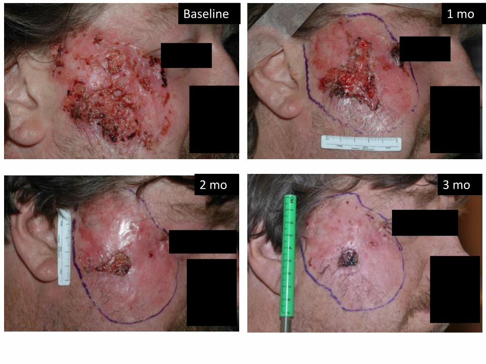
Baseline 1 mo
2 mo 3 mo
Potential drawbacks
• Unknown long term efficacy and side effects
• Potential development of resistance
• Only free for qualifying patients through Genentech Access Solutions, potential significant expenses
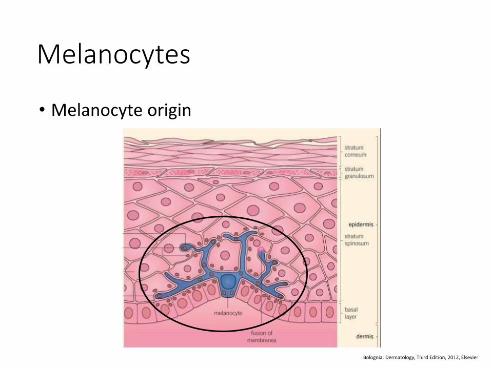
Melanocytes
• Melanocyte origin
Bolognia: Dermatology, Third Edition, 2012, Elsevier
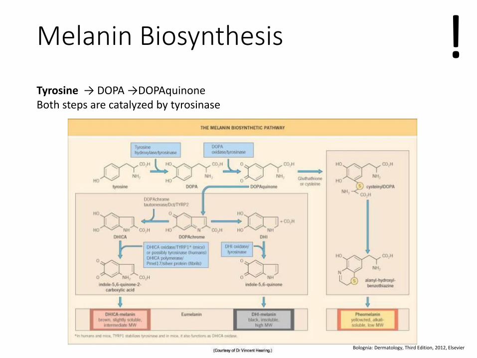
Melanin Biosynthesis
Bolognia: Dermatology, Third Edition, 2012, Elsevier
Tyrosine → DOPA →DOPAquinone Both steps are catalyzed by tyrosinase
!
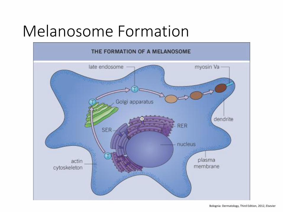
Melanosome Formation
Bolognia: Dermatology, Third Edition, 2012, Elsevier
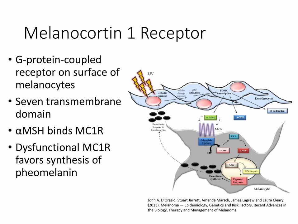
Melanocortin 1 Receptor
• G-protein-coupled receptor on surface of melanocytes
• Seven transmembrane domain
• αMSH binds MC1R
• Dysfunctional MC1R favors synthesis of pheomelanin
John A. D’Orazio, Stuart Jarrett, Amanda Marsch, James Lagrew and Laura Cleary (2013). Melanoma — Epidemiology, Genetics and Risk Factors, Recent Advances in the Biology, Therapy and Management of Melanoma

Melanocyte function in the epidermis
• Melanocytes load keratinocytes with melanosomes for protection against ultraviolet radiation
Bolognia: Dermatology, Third Edition, 2012, Elsevier
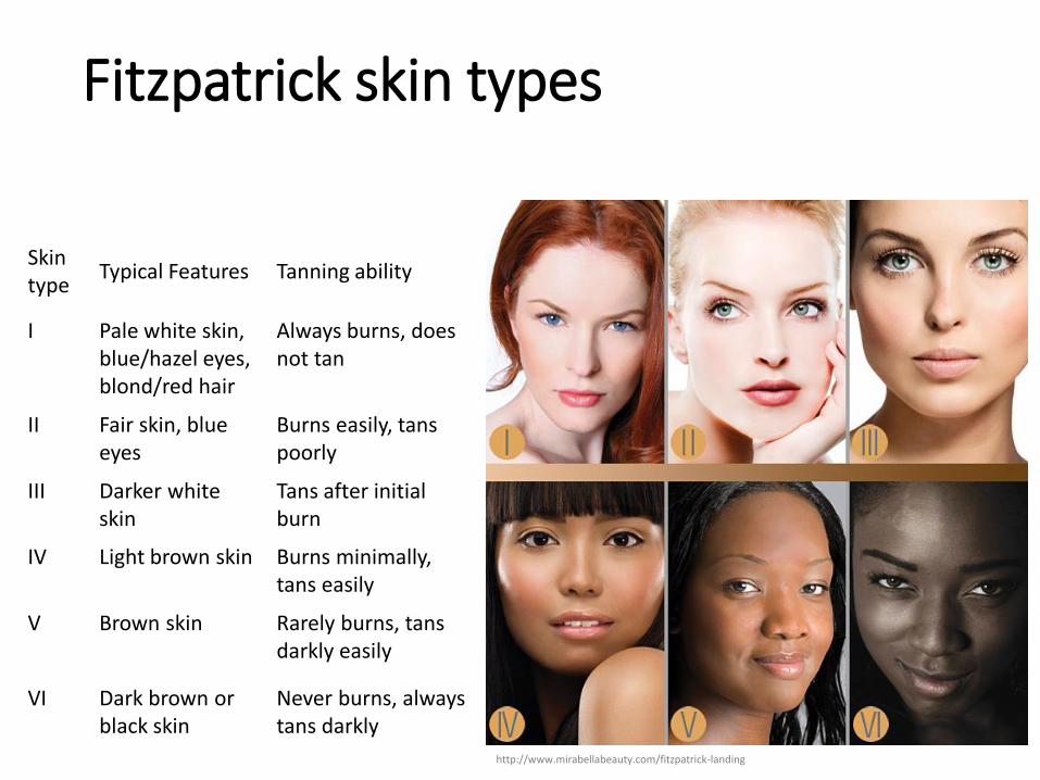
Fitzpatrick skin types
Skin type
Typical Features Tanning ability
I Pale white skin, blue/hazel eyes, blond/red hair
Always burns, does not tan
II Fair skin, blue eyes
Burns easily, tans poorly
III Darker white skin
Tans after initial burn
IV Light brown skin Burns minimally, tans easily
V Brown skin Rarely burns, tans darkly easily
VI Dark brown or black skin
Never burns, always tans darkly
http://www.mirabellabeauty.com/fitzpatrick-landing
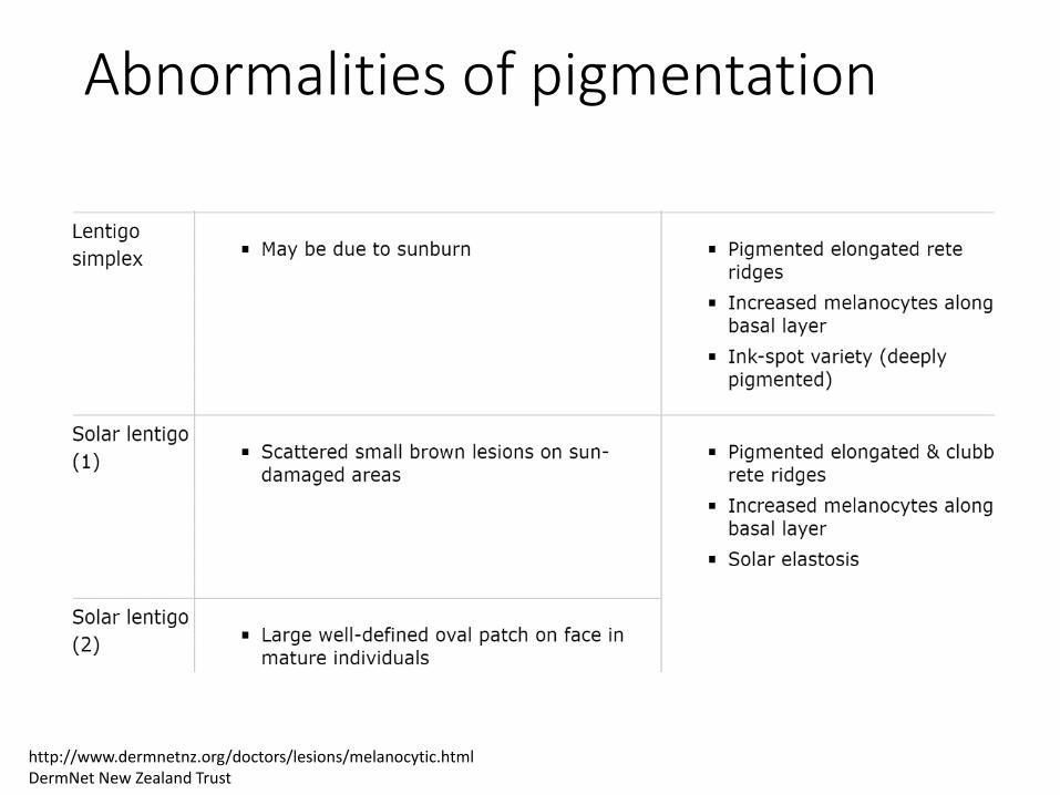
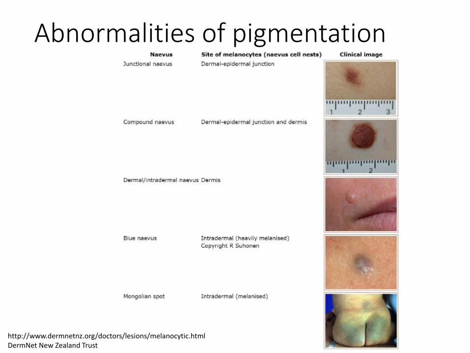
Abnormalities of pigmentation
http://www.dermnetnz.org/doctors/lesions/melanocytic.html DermNet New Zealand Trust
Abnormalities of pigmentation
http://www.dermnetnz.org/doctors/lesions/melanocytic.html DermNet New Zealand Trust
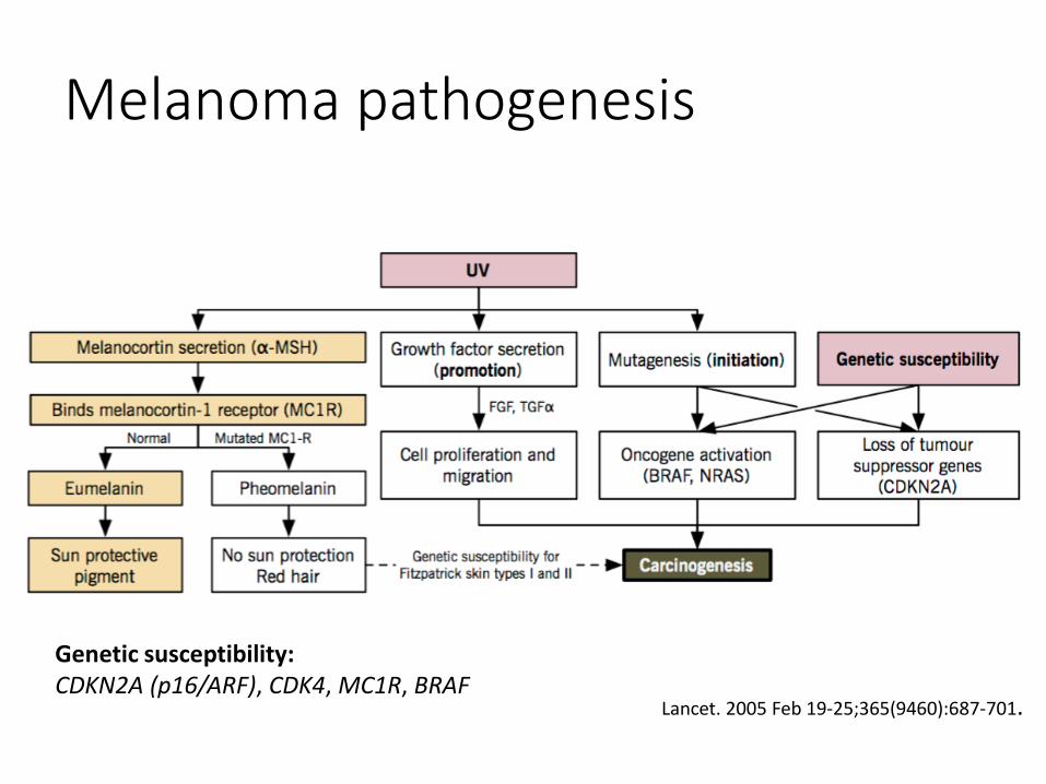
Melanoma pathogenesis
Lancet. 2005 Feb 19-25;365(9460):687-701.
Genetic susceptibility: CDKN2A (p16/ARF), CDK4, MC1R, BRAF
Lancet. 2005 Feb 19-25;365(9460):687-701.
Melanoma pathogenesis
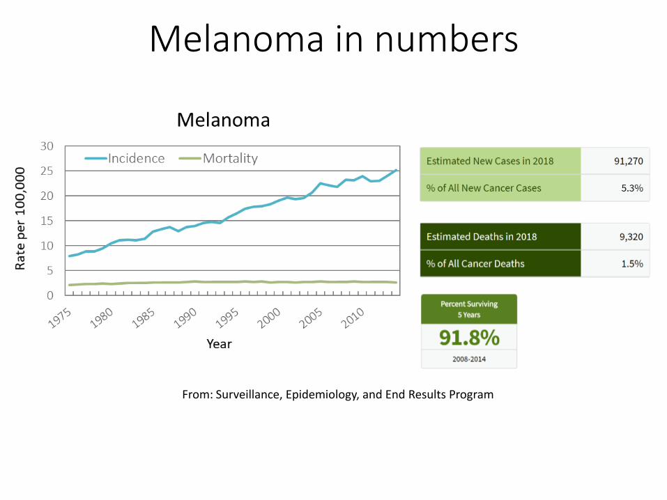
Melanoma in numbers
From: Surveillance, Epidemiology, and End Results Program
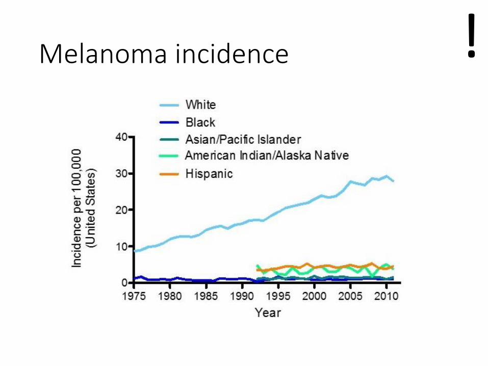
Melanoma incidence !
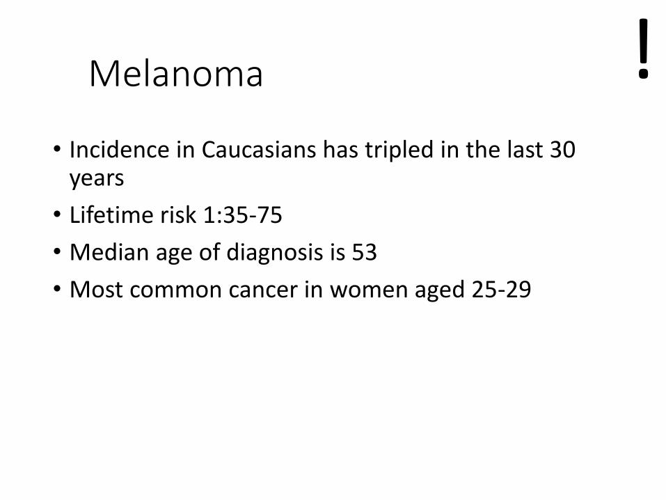
Melanoma
• Incidence in Caucasians has tripled in the last 30 years
• Lifetime risk 1:35-75
• Median age of diagnosis is 53
• Most common cancer in women aged 25-29
!
Melanoma Types
• Superficial Spreading Melanoma
• Nodular Melanoma
• Acral Lentiginous Melanoma
• Lentigo Maligna Melanoma
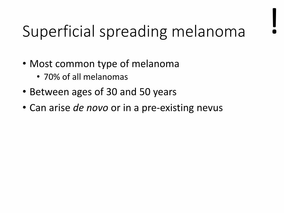
Superficial spreading melanoma
• Most common type of melanoma • 70% of all melanomas
• Between ages of 30 and 50 years
• Can arise de novo or in a pre-existing nevus
!
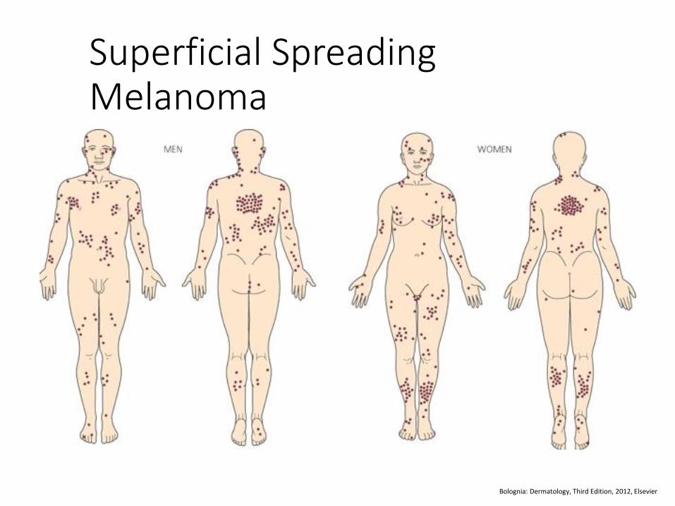
Superficial Spreading Melanoma
Bolognia: Dermatology, Third Edition, 2012, Elsevier
Nodular Melanoma
• Second most common • 15-30% of melanomas
• Commonly in 6th decade of life
• No radial growth phase Rapid growth
• Trunk, head and neck most common
• Male predominance
• Usually thicker and more advanced stage at diagnosis • Poorer prognosis
!
Acral Lentiginous Melanoma
• Uncommon • 5 – 10% of all melanomas
• Incidence similar across all racial groups
• 7th decade of life
• Represents disproportionate percentage of melanomas in African Americans (70%) and Asians (40%)
• Palms and soles or in and around the nail apparatus
!
Lentigo Maligna Melanoma
• Lentigo maligna analogous to the radial growth phase(5-20yrs)
• Lentigo maligna melanoma means invasive growth
• 4-15% of Melanomas
• Sun-damaged skin: face - nose and cheek
• Usually slow growth of large precursor lesion
!
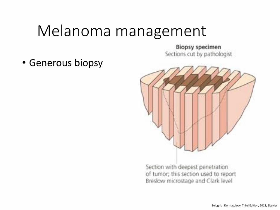
Melanoma management
• Generous biopsy
Bolognia: Dermatology, Third Edition, 2012, Elsevier
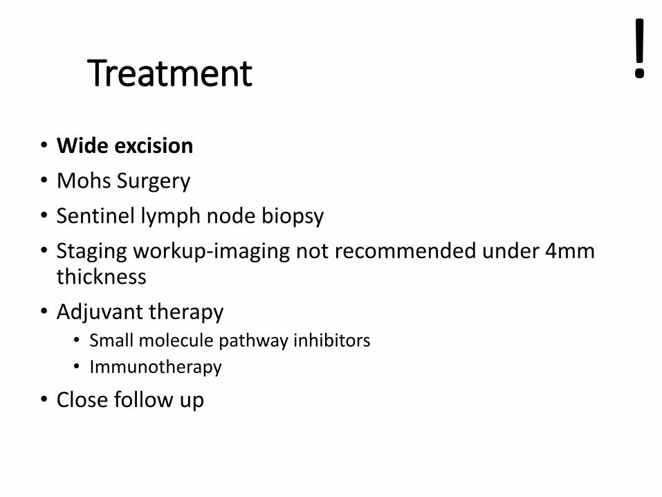
Treatment
• Wide excision
• Mohs Surgery
• Sentinel lymph node biopsy
• Staging workup-imaging not recommended under 4mm thickness
• Adjuvant therapy • Small molecule pathway inhibitors
• Immunotherapy
• Close follow up
!
http://www.riceland.com/all-about-rice/
http://www.riceland.com/all-about-rice/ GETTY IMAGES
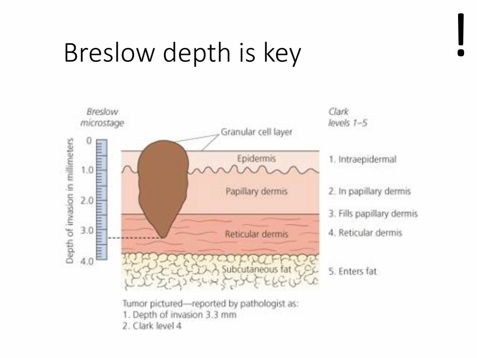
Breslow depth is key !
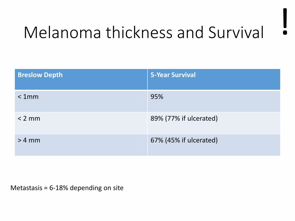
Melanoma thickness and Survival
Breslow Depth 5-Year Survival
< 1mm 95%
< 2 mm 89% (77% if ulcerated)
> 4 mm 67% (45% if ulcerated)
Metastasis = 6-18% depending on site
!
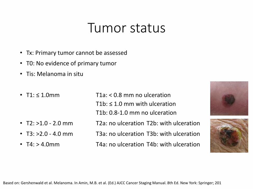
Tumor status
• Tx: Primary tumor cannot be assessed
• T0: No evidence of primary tumor
• Tis: Melanoma in situ
• T1: ≤ 1.0mm T1a: < 0.8 mm no ulceration
T1b: ≤ 1.0 mm with ulceration
T1b: 0.8-1.0 mm no ulceration
• T2: >1.0 - 2.0 mm T2a: no ulceration T2b: with ulceration
• T3: >2.0 - 4.0 mm T3a: no ulceration T3b: with ulceration
• T4: > 4.0mm T4a: no ulceration T4b: with ulceration
Based on: Gershenwald et al. Melanoma. In Amin, M.B. et al. (Ed.) AJCC Cancer Staging Manual. 8th Ed. New York: Springer; 201
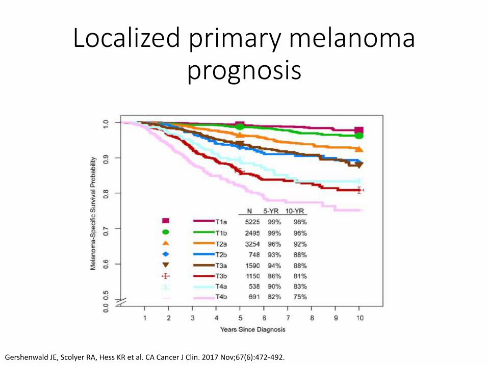
Localized primary melanoma prognosis
Gershenwald JE, Scolyer RA, Hess KR et al. CA Cancer J Clin. 2017 Nov;67(6):472-492.
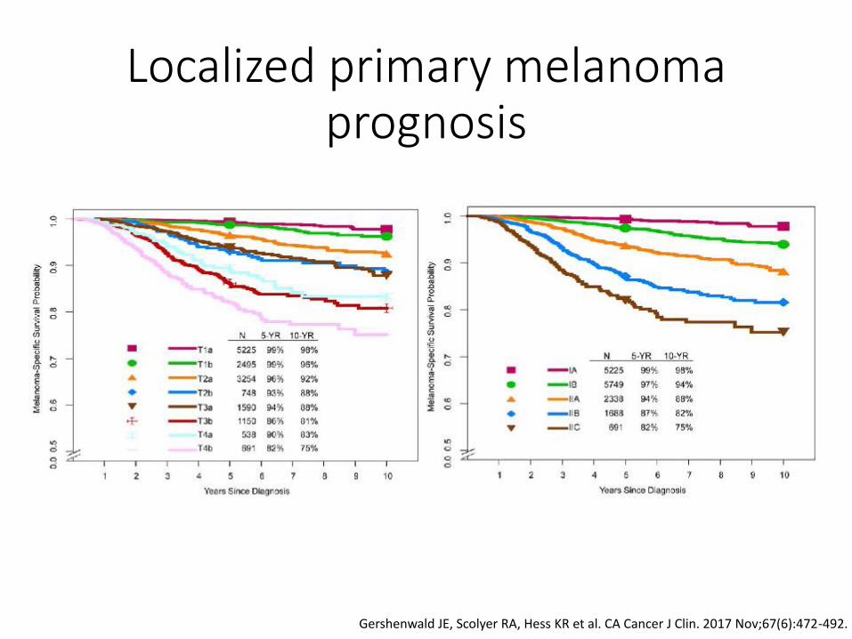
Localized primary melanoma prognosis
Gershenwald JE, Scolyer RA, Hess KR et al. CA Cancer J Clin. 2017 Nov;67(6):472-492.
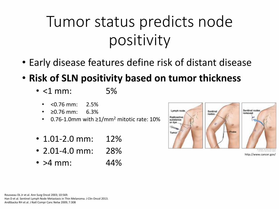
Tumor status predicts node positivity
• Early disease features define risk of distant disease
• Risk of SLN positivity based on tumor thickness • <1 mm: 5%
• 1.01-2.0 mm: 12%
• 2.01-4.0 mm: 28%
• >4 mm: 44%
Rousseau DL Jr et al. Ann Surg Oncol 2003; 10:569. Han D et al. Sentinel Lymph Node Metastasis in Thin Melanoma. J Clin Oncol 2013. Andtbacka RH et al. J Natl Compr Canc Netw 2009; 7:308
• <0.76 mm: 2.5% • ≥0.76 mm: 6.3% • 0.76-1.0mm with ≥1/mm2 mitotic rate: 10%
http://www.cancer.gov/
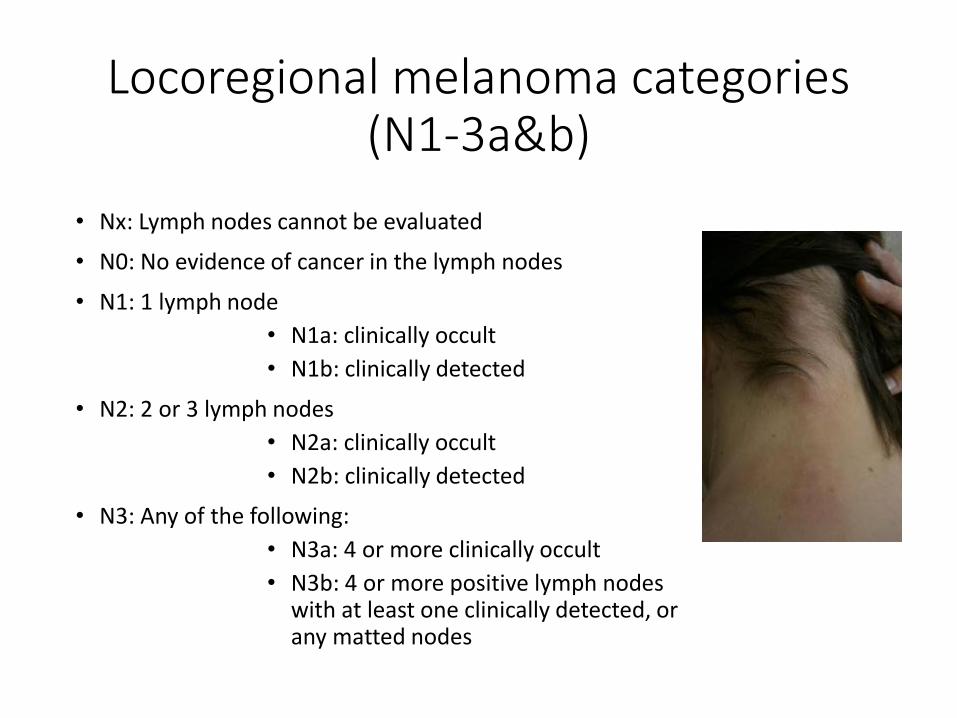
Locoregional melanoma categories (N1-3a&b)
• Nx: Lymph nodes cannot be evaluated
• N0: No evidence of cancer in the lymph nodes
• N1: 1 lymph node
• N1a: clinically occult
• N1b: clinically detected
• N2: 2 or 3 lymph nodes
• N2a: clinically occult
• N2b: clinically detected
• N3: Any of the following:
• N3a: 4 or more clinically occult
• N3b: 4 or more positive lymph nodes with at least one clinically detected, or any matted nodes
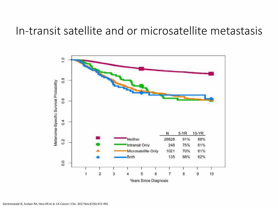
In-transit satellite and or microsatellite metastasis
Gershenwald JE, Scolyer RA, Hess KR et al. CA Cancer J Clin. 2017 Nov;67(6):472-492.
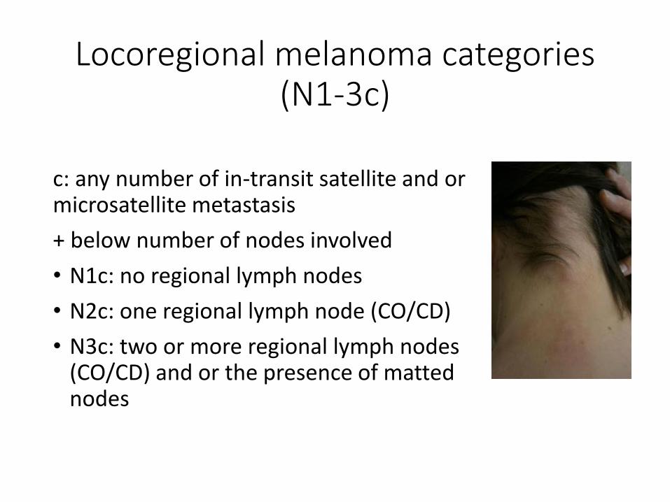
Locoregional melanoma categories (N1-3c)
c: any number of in-transit satellite and or microsatellite metastasis
+ below number of nodes involved
• N1c: no regional lymph nodes
• N2c: one regional lymph node (CO/CD)
• N3c: two or more regional lymph nodes (CO/CD) and or the presence of matted nodes
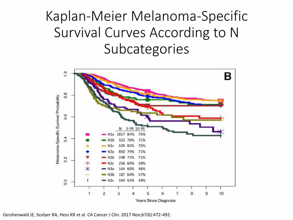
Kaplan-Meier Melanoma-Specific Survival Curves According to N
Subcategories
Gershenwald JE, Scolyer RA, Hess KR et al. CA Cancer J Clin. 2017 Nov;67(6):472-492.
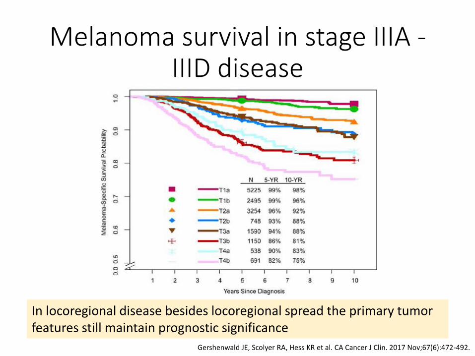
Melanoma survival in stage IIIA - IIID disease
Gershenwald JE, Scolyer RA, Hess KR et al. CA Cancer J Clin. 2017 Nov;67(6):472-492.
In locoregional disease besides locoregional spread the primary tumor features still maintain prognostic significance
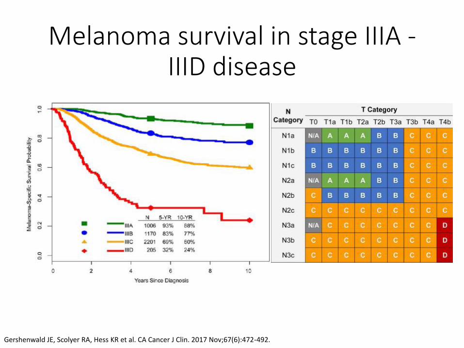
Melanoma survival in stage IIIA - IIID disease
Gershenwald JE, Scolyer RA, Hess KR et al. CA Cancer J Clin. 2017 Nov;67(6):472-492.
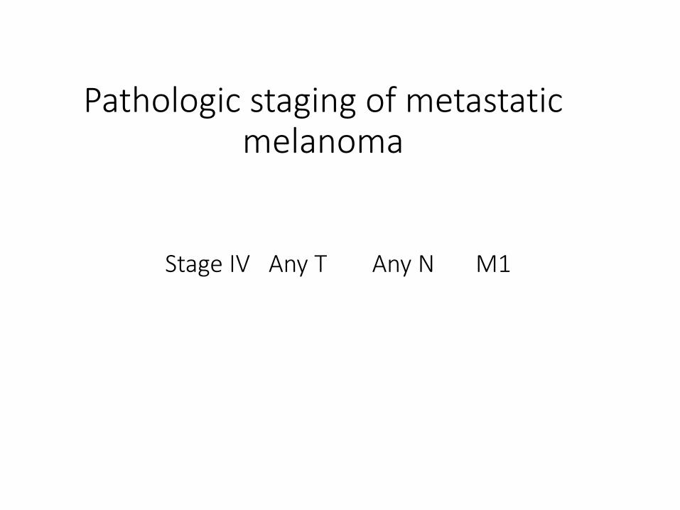
Pathologic staging of metastatic melanoma
Stage IV Any T Any N M1
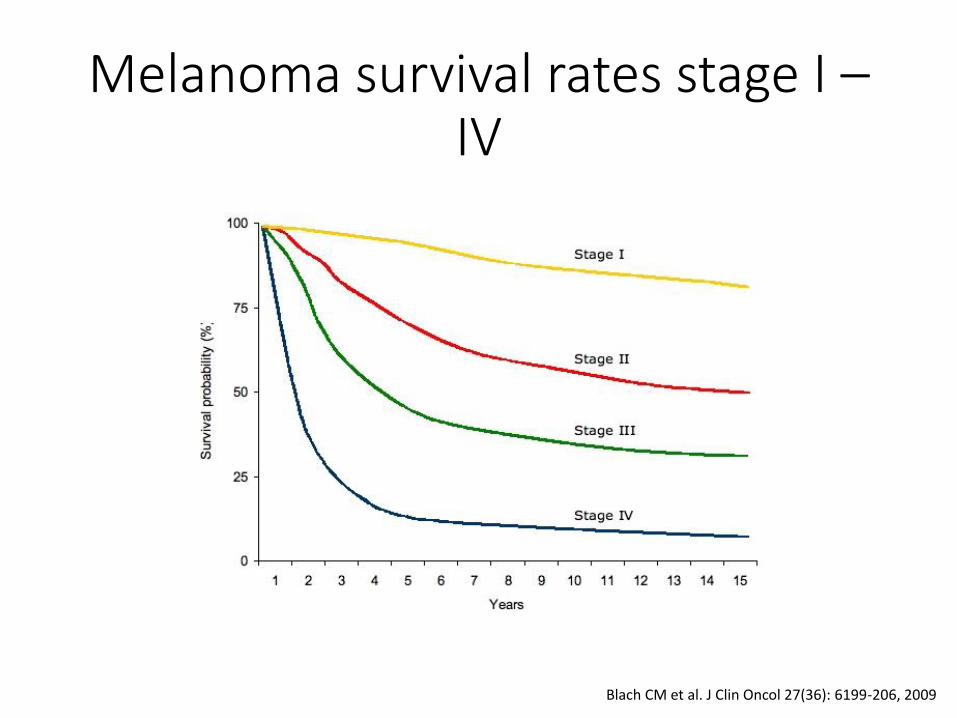
Melanoma survival rates stage I – IV
Blach CM et al. J Clin Oncol 27(36): 6199-206, 2009
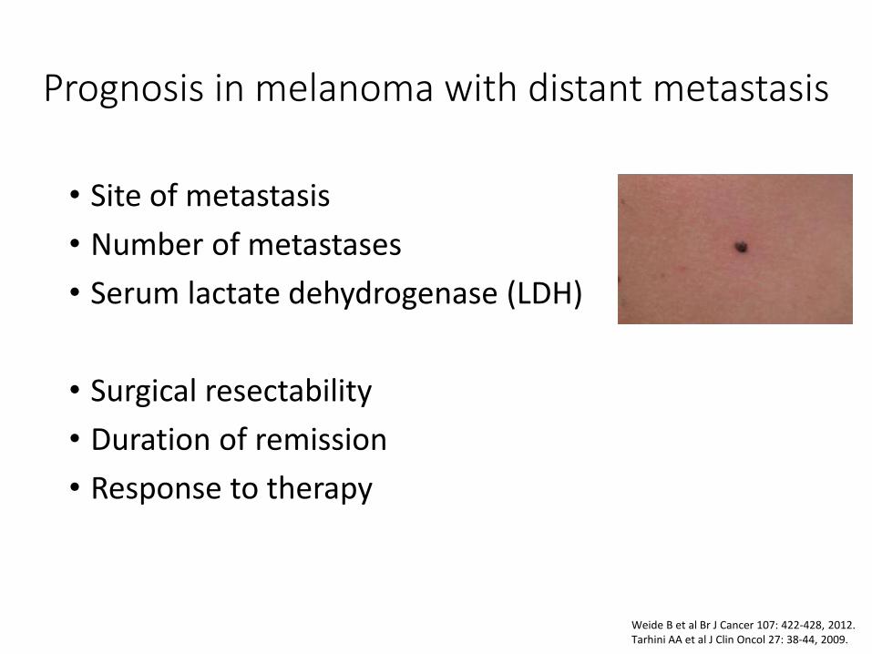
Prognosis in melanoma with distant metastasis
• Site of metastasis
• Number of metastases
• Serum lactate dehydrogenase (LDH)
• Surgical resectability
• Duration of remission
• Response to therapy
Weide B et al Br J Cancer 107: 422-428, 2012. Tarhini AA et al J Clin Oncol 27: 38-44, 2009.
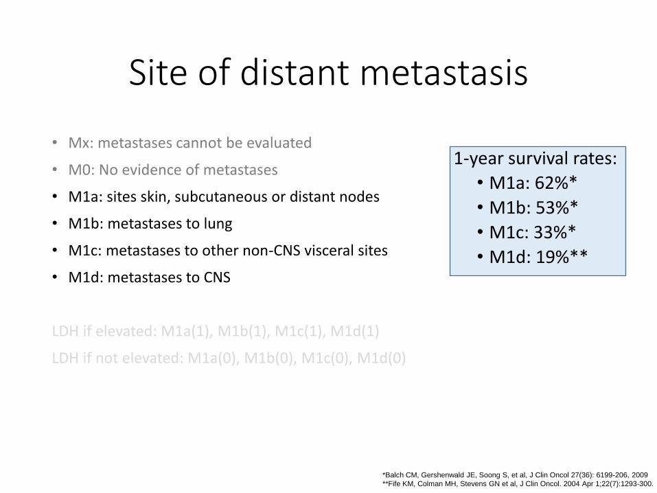
Site of distant metastasis
• Mx: metastases cannot be evaluated
• M0: No evidence of metastases
• M1a: sites skin, subcutaneous or distant nodes
• M1b: metastases to lung
• M1c: metastases to other non-CNS visceral sites
• M1d: metastases to CNS
LDH if elevated: M1a(1), M1b(1), M1c(1), M1d(1)
LDH if not elevated: M1a(0), M1b(0), M1c(0), M1d(0)
*Balch CM, Gershenwald JE, Soong S, et al, J Clin Oncol 27(36): 6199-206, 2009
**Fife KM, Colman MH, Stevens GN et al, J Clin Oncol. 2004 Apr 1;22(7):1293-300.
1-year survival rates:
• M1a: 62%*
• M1b: 53%*
• M1c: 33%*
• M1d: 19%**
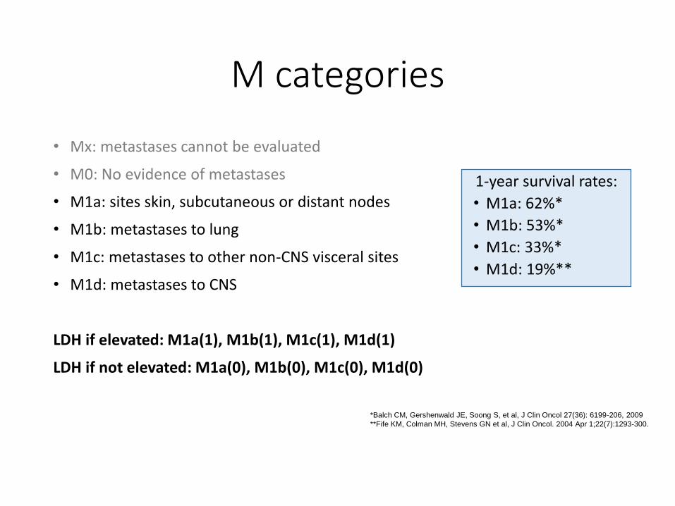
M categories
• Mx: metastases cannot be evaluated
• M0: No evidence of metastases
• M1a: sites skin, subcutaneous or distant nodes
• M1b: metastases to lung
• M1c: metastases to other non-CNS visceral sites
• M1d: metastases to CNS
LDH if elevated: M1a(1), M1b(1), M1c(1), M1d(1)
LDH if not elevated: M1a(0), M1b(0), M1c(0), M1d(0)
*Balch CM, Gershenwald JE, Soong S, et al, J Clin Oncol 27(36): 6199-206, 2009
**Fife KM, Colman MH, Stevens GN et al, J Clin Oncol. 2004 Apr 1;22(7):1293-300.
1-year survival rates:
• M1a: 62%*
• M1b: 53%*
• M1c: 33%*
• M1d: 19%**
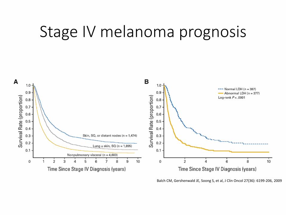
Stage IV melanoma prognosis
Balch CM, Gershenwald JE, Soong S, et al, J Clin Oncol 27(36): 6199-206, 2009
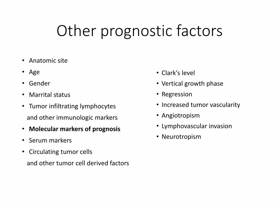
Other prognostic factors
• Anatomic site
• Age
• Gender
• Marrital status
• Tumor infiltrating lymphocytes
and other immunologic markers
• Molecular markers of prognosis
• Serum markers
• Circulating tumor cells
and other tumor cell derived factors
• Clark's level
• Vertical growth phase
• Regression
• Increased tumor vascularity
• Angiotropism
• Lymphovascular invasion
• Neurotropism
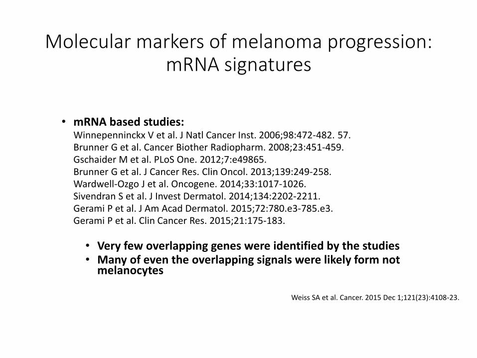
Molecular markers of melanoma progression: mRNA signatures
• mRNA based studies: Winnepenninckx V et al. J Natl Cancer Inst. 2006;98:472-482. 57. Brunner G et al. Cancer Biother Radiopharm. 2008;23:451-459. Gschaider M et al. PLoS One. 2012;7:e49865. Brunner G et al. J Cancer Res. Clin Oncol. 2013;139:249-258. Wardwell-Ozgo J et al. Oncogene. 2014;33:1017-1026. Sivendran S et al. J Invest Dermatol. 2014;134:2202-2211. Gerami P et al. J Am Acad Dermatol. 2015;72:780.e3-785.e3. Gerami P et al. Clin Cancer Res. 2015;21:175-183.
• Very few overlapping genes were identified by the studies • Many of even the overlapping signals were likely form not
melanocytes
Weiss SA et al. Cancer. 2015 Dec 1;121(23):4108-23.
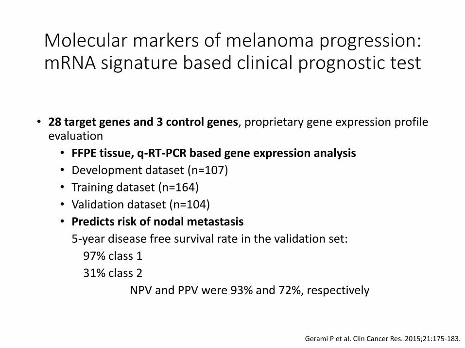
Molecular markers of melanoma progression: mRNA signature based clinical prognostic test
• 28 target genes and 3 control genes, proprietary gene expression profile evaluation
• FFPE tissue, q-RT-PCR based gene expression analysis
• Development dataset (n=107)
• Training dataset (n=164)
• Validation dataset (n=104)
• Predicts risk of nodal metastasis
5-year disease free survival rate in the validation set:
97% class 1
31% class 2
NPV and PPV were 93% and 72%, respectively
Gerami P et al. Clin Cancer Res. 2015;21:175-183.
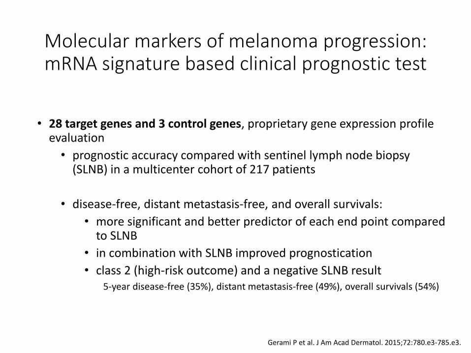
Molecular markers of melanoma progression: mRNA signature based clinical prognostic test
• 28 target genes and 3 control genes, proprietary gene expression profile evaluation
• prognostic accuracy compared with sentinel lymph node biopsy (SLNB) in a multicenter cohort of 217 patients
• disease-free, distant metastasis-free, and overall survivals:
• more significant and better predictor of each end point compared to SLNB
• in combination with SLNB improved prognostication
• class 2 (high-risk outcome) and a negative SLNB result 5-year disease-free (35%), distant metastasis-free (49%), overall survivals (54%)
Gerami P et al. J Am Acad Dermatol. 2015;72:780.e3-785.e3.

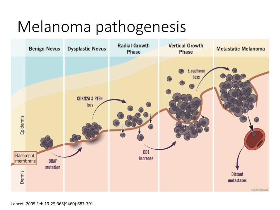
Lancet. 2005 Feb 19-25;365(9460):687-701.
Melanoma pathogenesis
This is not this simple
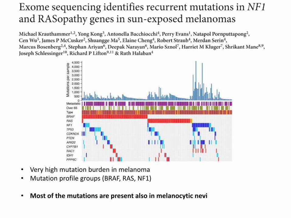
• Very high mutation burden in melanoma • Mutation profile groups (BRAF, RAS, NF1)
• Most of the mutations are present also in melanocytic nevi
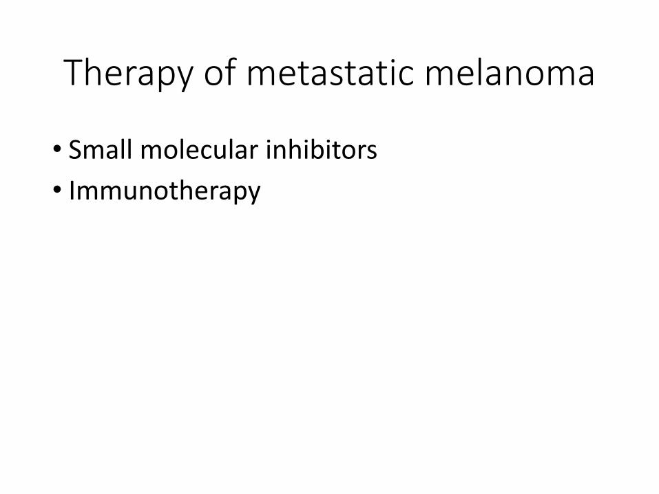
Therapy of metastatic melanoma
• Small molecular inhibitors
• Immunotherapy
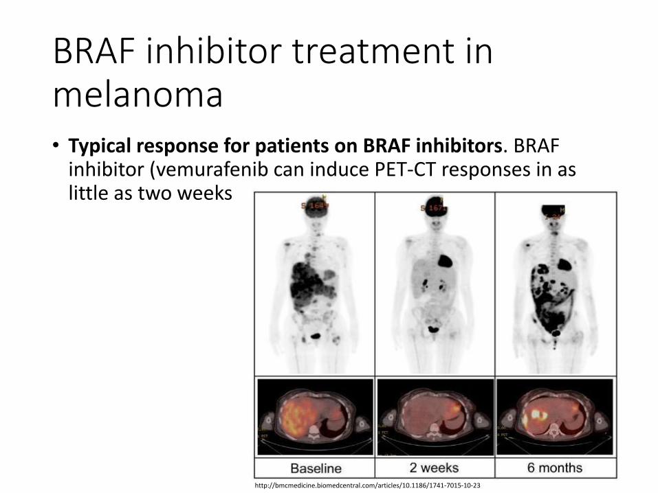
BRAF inhibitor treatment in melanoma • Typical response for patients on BRAF inhibitors. BRAF
inhibitor (vemurafenib can induce PET-CT responses in as little as two weeks
http://bmcmedicine.biomedcentral.com/articles/10.1186/1741-7015-10-23
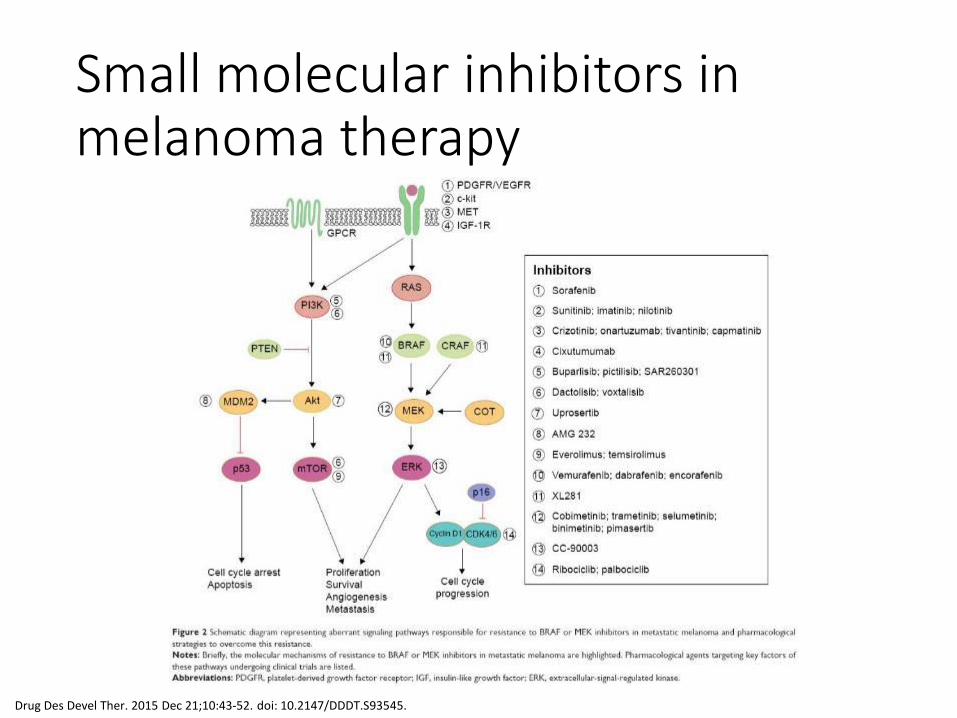
Small molecular inhibitors in melanoma therapy
Drug Des Devel Ther. 2015 Dec 21;10:43-52. doi: 10.2147/DDDT.S93545.
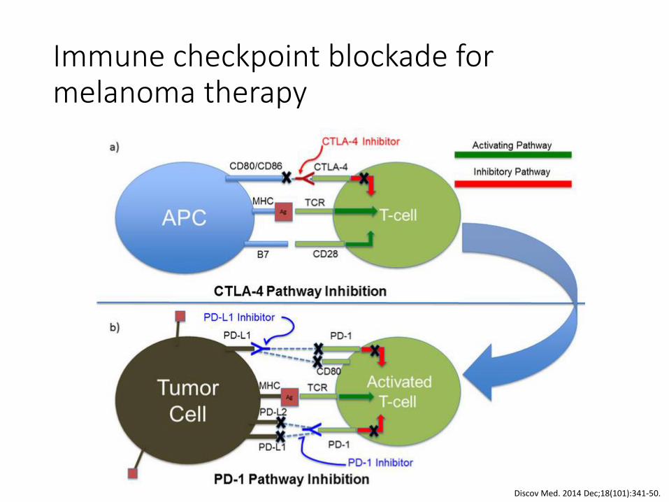
Immune checkpoint blockade for melanoma therapy
Discov Med. 2014 Dec;18(101):341-50.
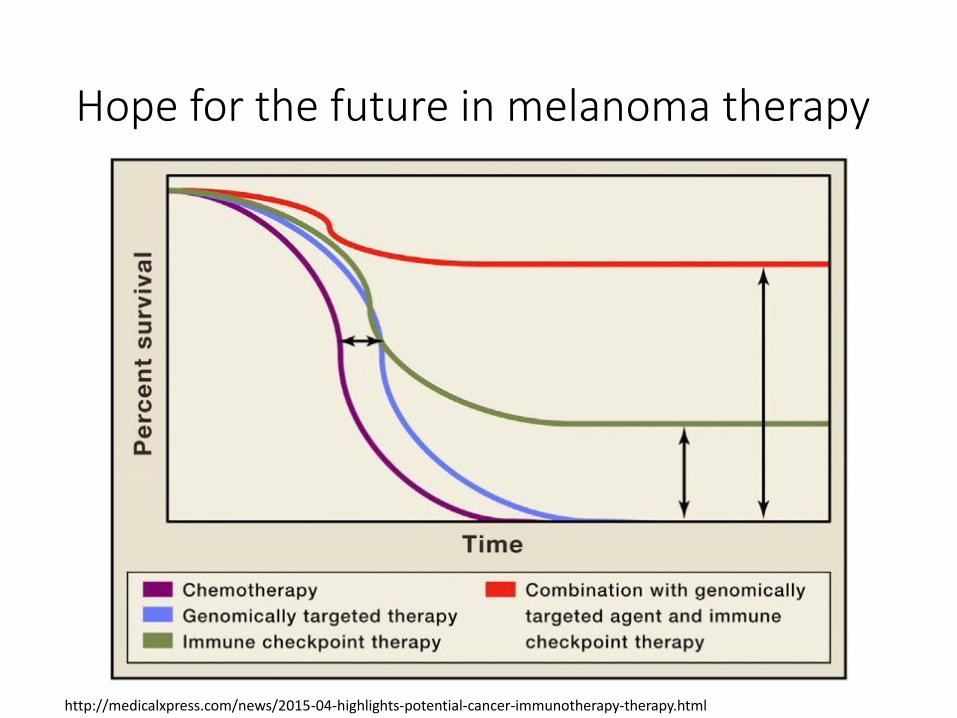
Hope for the future in melanoma therapy
http://medicalxpress.com/news/2015-04-highlights-potential-cancer-immunotherapy-therapy.html

Prevention of skin cancer
• Tanning of the skin is started by DNA damage
http://www.skincancer.org
TAN = damage
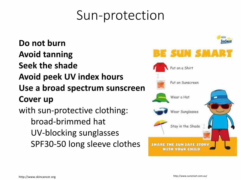
Sun-protection
http://www.skincancer.org
Do not burn Avoid tanning Seek the shade Avoid peek UV index hours Use a broad spectrum sunscreen Cover up with sun-protective clothing:
broad-brimmed hat UV-blocking sunglasses SPF30-50 long sleeve clothes
http://www.sunsmart.com.au/
Sun protection
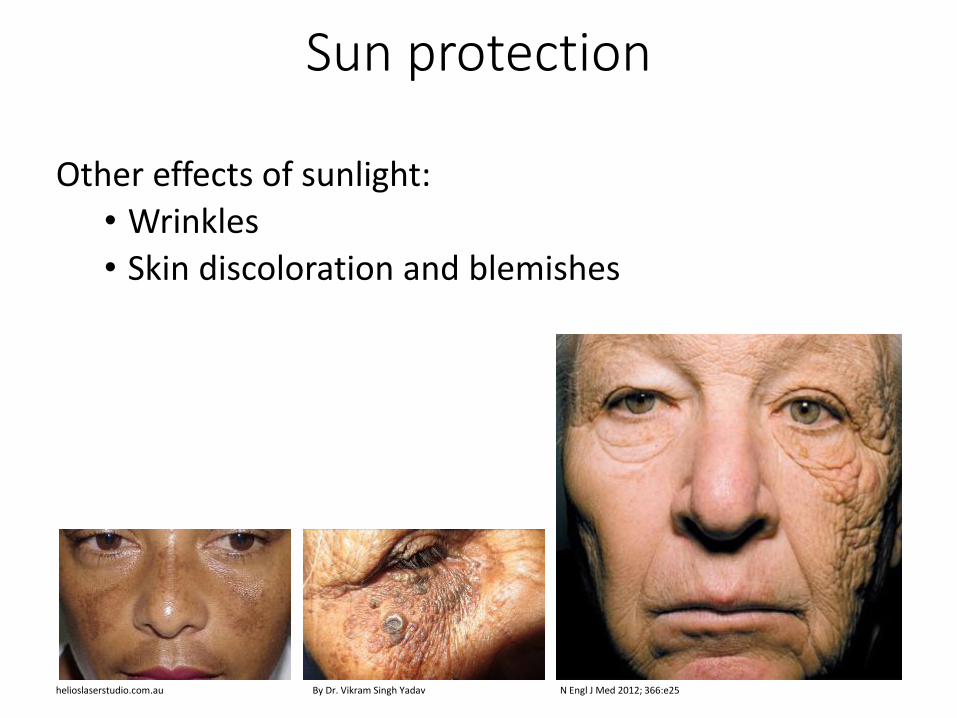
Other effects of sunlight:
• Wrinkles
• Skin discoloration and blemishes
By Dr. Vikram Singh Yadav helioslaserstudio.com.au N Engl J Med 2012; 366:e25
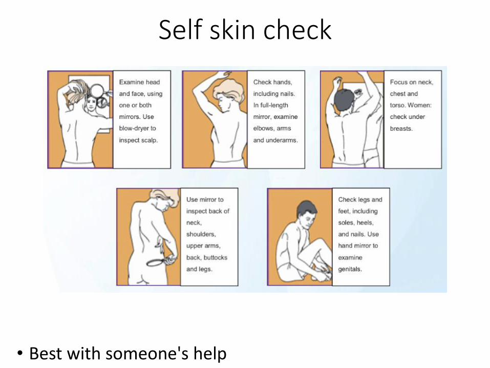
Self skin check
• Best with someone's help
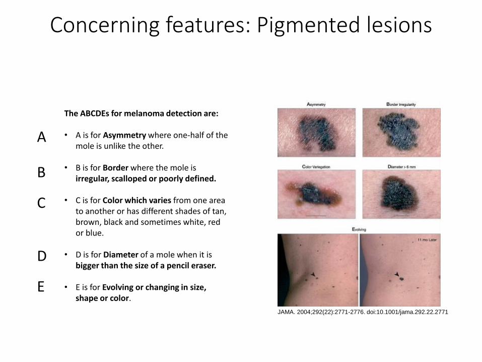
JAMA. 2004;292(22):2771-2776. doi:10.1001/jama.292.22.2771
The ABCDEs for melanoma detection are: • A is for Asymmetry where one-half of the
mole is unlike the other. • B is for Border where the mole is
irregular, scalloped or poorly defined. • C is for Color which varies from one area
to another or has different shades of tan, brown, black and sometimes white, red or blue.
• D is for Diameter of a mole when it is bigger than the size of a pencil eraser.
• E is for Evolving or changing in size, shape or color.
Concerning features: Pigmented lesions
A
B
C
D
E
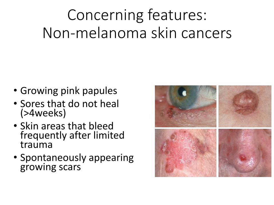
Concerning features: Non-melanoma skin cancers
• Growing pink papules • Sores that do not heal
(>4weeks) • Skin areas that bleed
frequently after limited trauma
• Spontaneously appearing growing scars
• Early diagnosis is key to cure
• Report concerning skin lesions to dermatology
Summary: Cutaneous malignancies
• Skin: barrier
• UV radiation: most important epidermal carcinogen
• Precancerous lesions: Actinic keratosis
• Most common skin cancers • Non melanoma skin cancers:
• Basal cell carcinoma
• Squamous cell carcinoma
• Melanoma