current status of antifibrinolytics in cardiopulmonary...
TRANSCRIPT

Anesthesiology Clin N Am
21 (2003) 527–551
Current status of antifibrinolytics in
cardiopulmonary bypass and elective deep
hypothermic circulatory arrest
Jeffrey A. Green, MD*, Bruce D. Spiess, MDDepartment of Anesthesiology, Virginia Commonwealth University,
Medical College of Virginia Campus, 1200 East Broad Street,
PO Box 980695, Richmond, VA 23209 USA
Bleeding after cardiopulmonary bypass
Cardiopulmonary bypass (CPB) alters the hemostatic balance and predisposes
cardiac surgery patients to an increased risk of microvascular bleeding. Bleeding
and the need for transfusion are among the most common complications of
cardiac surgery. In fact, until recently, blood transfusions seemed to be required
for about 50% of all cardiac surgery patients [1]. Currently, CPB accounts for
10% to 20% of the transfusions performed in the United States [2,3]. Transfusion,
however, exposes patients to added risks such as infectious disease transmission
[4], transfusion reactions [5], graft-versus-host disease [6], transfusion-induced
lung injury [7], and decreased resistance to postoperative infection [8,9].
Transfusion increases the risk of infection by 35% to 300% and increases the
risk of pneumonia in coronary artery bypass (CABG) patients by 5% per unit
transfused [10]. The primary purported benefit of transfusion, increased oxygen
carrying capacity, has not been definitively proven.
Excessive postoperative bleeding may necessitate surgical reexploration,
increasing morbidity, and mortality. Postoperatively, the risk of excessive
bleeding is 11% [11], and 5% to 7% of patients lose more than 2 L of blood
in the first 24 hours after CPB [12]. Reexploration for hemorrhage is required in
3.6% to 4.2% of patients [13], and mortality rates range from 10% to 22% [14].
Bleeding and reexploration consume hospital resources by increasing the
operative time and blood-product usage, and the need for mechanical ventilation,
intensive care, and longer hospitalization [15,16]. Reexploration also increases
0889-8537/03/$ – see front matter D 2003 Elsevier Inc. All rights reserved.
doi:10.1016/S0889-8537(03)00042-7
* Corresponding author.
E-mail address: [email protected] (J.A. Green).

J.A. Green, B.D. Spiess / Anesthesiology Clin N Am 21 (2003) 527–551528
the rates of renal failure, sepsis, and atrial arrhythmia [13]. Less than half of re-
explored patients have a surgical cause for bleeding [15]. It is these patients who
might have benefited from prophylactic antifibrinolytic drugs.
Because of the substantial cost, limited blood supply, multiple risks, and lack
of proven benefit, health care professionals are now avoiding transfusion by
accepting moderate anemia and using blood-saving techniques. In addition,
concern about allogeneic blood safety has led to methods intended to minimize
perioperative transfusion. Multiple nonpharmacologic blood-saving methods are
available for use during CPB, but they are beyond the scope of this article. In-
stead of using complex blood salvaging techniques, the most effective approach
is to avoid blood loss from the beginning [17]. Pharmacologic therapies
accomplish this goal. One of these techniques is prophylactic antifibrinolytic
therapy. Antifibrinolytic drugs prevent primary fibrinolysis and preserve platelet
function by preventing platelet activation. The efficacy of these drugs as
prophylactic hemostatic agents in cardiac surgery patients has been validated in
numerous studies [3,18–26]. The most widely administered antifibrinolytic drugs
are the synthetic lysine analogs (epsilon-aminocaproic acid [EACA] and trane-
xamic acid [TXA]) and the nonspecific serine protease inhibitor aprotinin [24].
This article briefly reviews the pathophysiology of bleeding after CPB. It
then reviews the current status, efficacy, and safety of antifibrinolytic drugs and
discusses the use of these agents in patients undergoing routine CPB and
elective DHCA.
Pathophysiology of excessive bleeding after cardiopulmonary bypass
The pathophysiology of bleeding after CPB is complex, involving hypother-
mia, hemodilution, activation of coagulation, endothelial cell and tissue injury [2],
foreign-surface contact [2], consumption of clotting factors [27], platelet activa-
tion [28], platelet dysfunction [29], and hyperfibrinolysis [30]. These factors are
all interrelated and variable from one individual to another; in combination, they
lead to a homeostatic imbalance between coagulation and fibrinolysis.
Activation of hemostasis
During CPB, the hemostatic system is activated by way of multiple pathways,
including the intrinsic and extrinsic coagulation cascades [31,32]. Contact with
the foreign surface of the CPB circuit results in contact-phase activation of factor
XII, prekallikrein, and high-molecular-weight kininogen, thus initiating the
intrinsic pathway [33]. Simultaneously, surgical trauma, exposure of raw tissue
surfaces to air, generation of factor VIIa (through endothelial cell activation) [34],
and reinfusion of pericardial blood result in tissue-factor activation of the
extrinsic pathway [35–37]. These pathways converge, leading to the generation
of thrombin [35]. Controversy exists about which system provides a more com-
mon, or a greater, insult. Recent studies suggest that the extrinsic system may

J.A. Green, B.D. Spiess / Anesthesiology Clin N Am 21 (2003) 527–551 529
lead to the greatest activation of the hemostatic system. Despite adequate
heparinization, thrombin is still generated before, during, and after CPB
[38,39]. Thrombin activates hemostasis, platelets, and endothelial cells, leading
to the release of vasoactive substances and other mediators of hemostasis,
fibrinolysis, and inflammation [33,34].
Activation of fibrinolysis
Fibrinolysis is activated by several mechanisms, including the production of
thrombin, release of tissue plasminogen activator (t-PA) by vascular endothelium,
and activation of the intrinsic coagulation pathway [40–42]. The release of t-PA
from endothelium can be caused by thrombin [43], epinephrine, bradykinin,
factor Xa, factor XII [34], hypothermia [44,45], or CPB alone [46]. The largest
surges in t-PA release occur on initiation of CPB and after infusion of protamine
[39,47]. Concentrations of plasminogen activator rise rapidly during CPB, while
levels of plasminogen activator inhibitor (PAI-1) slowly increase and peak during
the postoperative period [48]. Individual variability is high, for t-PA production
and PAI-1 release. The genetic characteristics of these proteins are complex, but
known polymorphisms exist and have been correlated with early myocardial
infarction (MI) [49,50]. Elevations or exaggerated surges of PAI-1 may be related
to elevated cytokine and inflammatory activity. This patient-to-patient variability
is just now being investigated.
The release of plasma-derived urokinase, as a direct consequence of contact-
phase activation by way of activated Hageman factor and kallikrein, also
contributes to fibrinolysis by causing further t-PA release and direct fibrinolysis
[51]. Serine proteases cleave plasminogen to form plasmin. Plasmin splits fibrin-
ogen and fibrin at specific sites to form fibrin degradation products and D-dimers
(Fig. 1).
Fibrin breakdown is a normal hematologic activity that remodels formed clots
and removes thrombus as the endothelium heals. The fibrin formation and lysis
that occur during CPB are usually mild and self-limited. Excessive fibrinolysis is
occasionally seen however, and it can contribute to excessive bleeding.
Role of platelets
The increased plasmin and fibrin degradation products produced during CPB
have deleterious effects on platelets [52]. Activation of the fibrinolytic system
contributes to platelet dysfunction. Fibrinolysis affects platelet membrane recep-
tors, causing platelet activation and granule release [53].
When hemostasis and fibrinolysis are both activated, a consumptive process
ensues that is mediated by thrombin and plasmin. Thrombin mediates the
conversion of fibrinogen to fibrin monomers, initiates fibrinolysis by mediating
the release of t-PA, and activates factors V, VIII, and XIII, and platelets.
Thrombin activates protein C in combination with thrombomodulin on the endo-
thelial surface, consuming previously generated, activated factors V and VIII.

Fig. 1. Overview of the fibrinolytic system. TPA, tissue plasminogen activator, PAI-1, plasminogen
activator inhibitor 1; AP antiplasmin (From Chandler W. The fibrinolytic response during
cardiopulmonary bypass: Pro or anticoagulant? In: The relationship between coagulation,
inflammation and endothelium. Baltimore: Lippincott Williams & Wilkins; 2000. p. 147–68;
with permission).
J.A. Green, B.D. Spiess / Anesthesiology Clin N Am 21 (2003) 527–551530
Thrombin down-regulates hemostasis by releasing tissue factor pathway inhib-
itor; this agent inhibits the extrinsic pathway and stimulates the release of t-PA,
which cleaves plasminogen to plasmin. Plasmin cleaves fibrinogen and fibrin
monomers to fibrin degradation products and cleaves cross-linked fibrin poly-
mers to D-dimers. Plasmin inactivates factors V and VIII and inhibits the
glycoprotein receptors of platelets. Therefore, it is important to attenuate the
exaggerated effects of both thrombin and plasmin [54].
Antifibrinolytic therapy can minimize the platelet defect mediated by plasmin
and fibrin degradation products [55,56]. Antifibrinolytic agents protect platelet
function by inhibiting the proteolytic degradation of glycoprotein platelet surface
receptors Ib and IIb/IIIa, allowing platelet function to be preserved after CPB
[57,58].
Use of antifibrinolytic agents during routine cardiopulmonary bypass
Discovery of the usefulness of antifibrinolytic drugs followed the development
of CPB during the 1950s and 1960s, because at the time CPB devices that were
not optimally biocompatible resulted in severe bleeding related to hyperfibrinol-
ysis. Recently, outcome-based evidence has revealed the effectiveness and safety
of these drugs, leading to their increased use during routine low-risk cardiac
operations [22].
J.A. Green, B.D. Spiess / Anesthesiology Clin N Am 21 (2003) 527–551 531
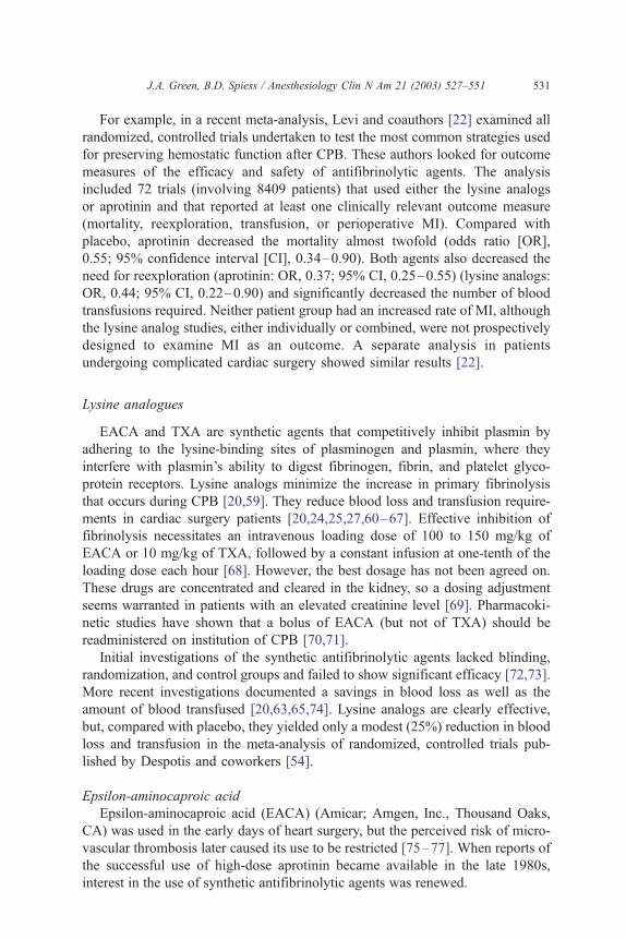
For example, in a recent meta-analysis, Levi and coauthors [22] examined all
randomized, controlled trials undertaken to test the most common strategies used
for preserving hemostatic function after CPB. These authors looked for outcome
measures of the efficacy and safety of antifibrinolytic agents. The analysis
included 72 trials (involving 8409 patients) that used either the lysine analogs
or aprotinin and that reported at least one clinically relevant outcome measure
(mortality, reexploration, transfusion, or perioperative MI). Compared with
placebo, aprotinin decreased the mortality almost twofold (odds ratio [OR],
0.55; 95% confidence interval [CI], 0.34–0.90). Both agents also decreased the
need for reexploration (aprotinin: OR, 0.37; 95% CI, 0.25–0.55) (lysine analogs:
OR, 0.44; 95% CI, 0.22–0.90) and significantly decreased the number of blood
transfusions required. Neither patient group had an increased rate of MI, although
the lysine analog studies, either individually or combined, were not prospectively
designed to examine MI as an outcome. A separate analysis in patients
undergoing complicated cardiac surgery showed similar results [22].
Lysine analogues
EACA and TXA are synthetic agents that competitively inhibit plasmin by
adhering to the lysine-binding sites of plasminogen and plasmin, where they
interfere with plasmin’s ability to digest fibrinogen, fibrin, and platelet glyco-
protein receptors. Lysine analogs minimize the increase in primary fibrinolysis
that occurs during CPB [20,59]. They reduce blood loss and transfusion require-
ments in cardiac surgery patients [20,24,25,27,60–67]. Effective inhibition of
fibrinolysis necessitates an intravenous loading dose of 100 to 150 mg/kg of
EACA or 10 mg/kg of TXA, followed by a constant infusion at one-tenth of the
loading dose each hour [68]. However, the best dosage has not been agreed on.
These drugs are concentrated and cleared in the kidney, so a dosing adjustment
seems warranted in patients with an elevated creatinine level [69]. Pharmacoki-
netic studies have shown that a bolus of EACA (but not of TXA) should be
readministered on institution of CPB [70,71].
Initial investigations of the synthetic antifibrinolytic agents lacked blinding,
randomization, and control groups and failed to show significant efficacy [72,73].
More recent investigations documented a savings in blood loss as well as the
amount of blood transfused [20,63,65,74]. Lysine analogs are clearly effective,
but, compared with placebo, they yielded only a modest (25%) reduction in blood
loss and transfusion in the meta-analysis of randomized, controlled trials pub-
lished by Despotis and coworkers [54].
Epsilon-aminocaproic acid
Epsilon-aminocaproic acid (EACA) (Amicar; Amgen, Inc., Thousand Oaks,
CA) was used in the early days of heart surgery, but the perceived risk of micro-
vascular thrombosis later caused its use to be restricted [75–77]. When reports of
the successful use of high-dose aprotinin became available in the late 1980s,
interest in the use of synthetic antifibrinolytic agents was renewed.

J.A. Green, B.D. Spiess / Anesthesiology Clin N Am 21 (2003) 527–551532
Many investigators have assessed the efficacy of EACA [62,64,78,79]. In
studying 40 patients undergoing primary coronary artery bypass grafting
(CABG), Daily and associates [62] demonstrated that, compared with a control
group, the patients who received a 30-g dose of EACA had decreased blood loss
and transfusion. In a retrospective analysis by Jordan and colleagues [64], EACA
usage reduced the percentage of patients who received red blood cells by 53%,
the number of units used per patient by 57%, the percentage of patients who
received platelets by 66%, and the number of platelet units administered per
patient by 68%. Also, the EACA patients had a reduced operative time after
being weaned from CPB, as well as fewer re-explorations.
In a meta-analysis using transfusion as the outcome variable, Laupacis and
Fergusson [21] found that EACA had no significant effect on the proportion of
patients transfused despite an odds ratio considerably less than 1.0 (OR, 0.20;
95% CI, 0.04–1.12). The authors attributed this lack of significance to the fact
that the analysis included only three randomized, controlled studies (including
118 patients) and to the use of different dosages.
Another recent meta-analysis, by Munoz and coworkers [24], who examined
EACA and aprotinin use in cardiac surgery, included nine trials of EACA, which
reduced blood loss by 35%. Compared with placebo recipients, the EACA group
had 61% fewer transfusions (mean reduction, 0.74 U) and was only 32% as
likely to need blood (OR, 0.32; 95% CI, 0.15–0.69; P = 0.004). The authors
concluded that, despite large differences in the dosage ranges among the studies,
EACA, which is substantially less expensive than aprotinin, is just as effective.
They found little evidence of thrombosis-related complications, including stroke
and MI, in either group. On the contrary, both groups tended to have a con-
siderable reduction in the risk of postoperative stroke, although this finding did
not reach significance.
In a randomized, double-blind, placebo-controlled study, Slaughter and
associates [27] demonstrated that EACA decreased fibrinolysis 3 hours after
CPB (50% reduction in D-dimer concentration; P < 0.005) and decreased
bleeding (35% reduction compared with placebo; P < 0.05), but there were no
intergroup differences in the perioperative generation of thrombin or fibrin. These
results suggest that there is at least a potential for a perioperative hypercoagulable
or prothrombotic state in EACA recipients who are genetically predisposed to
this condition. Although clinical trials have failed to show definitive evidence
that EACA therapy can lead to thrombosis, there are numerous reports of this
complication in the literature [80–82].
In randomized clinical trials comparing EACA to aprotinin, two studies found
no differences in postoperative transfusion rates despite reduced blood loss in the
aprotinin group [83,84]. Furthermore, in an efficacy and cost-effectiveness study
involving patients undergoing repeat cardiac operations, Bennett-Guerrero and
coworkers [19] compared EACA to aprotinin therapy and found no intergroup
difference in allogeneic blood use despite the fact that aprotinin was more
efficacious in reducing blood loss. The authors concluded that EACA is the more
cost-effective agent.

J.A. Green, B.D. Spiess / Anesthesiology Clin N Am 21 (2003) 527–551 533
Tranexamic acid
Tranexamic acid (TXA) (Cyklokapron; Pharmacia Corporation, Peapack, New
Jersey) is a synthetic lysine analog that is similar to EACA but ten times more
potent [68]. The mechanism of action of TXA is similar to that of EACA. The
usual dose for CPB patients is 1.5 to 10.0 g intravenously. Despite a variability in
dosage, there is little doubt that the 10-g dosage scheme is efficacious (Table 1)
In a meta-analysis of drugs used to minimize perioperative blood loss that
included 12 studies, Laupacis and Fergusson [21] found that TXA significantly
decreased the proportion of cardiac surgical patients transfused (OR, 0.50; 95%
CI, 0.34–0.76; P = 0.0009). It had no effect on perioperative MI or reexploration,
but these variables were not prospectively evaluated.
In repeat operations, TXA is of particular benefit. Shore-Lesserson and
coauthors [25] demonstrated that TXA decreased blood loss and reduced transfu-
sion by 33% in patients undergoing repeat cardiac surgery.
The timing of TXA therapy has been examined by several authors. In a study
by Horrow and colleagues [85], TXA given before the initiation of CPB de-
creased chest-tube drainage by 30% during the first 12 hours postoperatively and
decreased the transfusion rate from 41% to 22%. Large doses were not beneficial.
Administration of TXA after CPB or bleeding becomes established is a
controversial practice that could lead to prothrombotic complications [59]. In a
study by Casati and colleagues [92] involving both high- and low-risk patients in
whom TXA therapy was continued for 12 hours after CPB, this approach had no
advantage over placebo in reducing the incidence of bleeding. In a randomized,
placebo-controlled, double-blind study by Brown and coworkers [90], TXA
decreased bleeding and transfusion in patients- undergoing primary CABG when
this agent was administered before and during, but not after, CPB.
The literature contains multiple studies that compare the efficacy of TXA to that
of aprotinin. In a randomized, double-blind study of 150 patients undergoing
primary CABG, Mongan and associates [3] showed that TXA and aprotinin were
equally effective in reducing blood loss, the incidence of transfusion, and the
amount of blood products transfused. In two more recent, larger trials, Casati and
colleagues [60,61] demonstrated that TXAwas as effective as aprotinin in reducing
blood loss in low-risk patients.
In a randomized trial comparing TXA and aprotinin therapy in high-risk
patients, Nuttall and coauthors [91] found that TXA, used with the ‘‘platelet-
sparing’’ acute normovolemic hemodilution technique, was similar to aprotinin
with respect to reduced blood loss and transfusion requirements. In another study
involving high-risk, repeat, multiple valve, combined, or aortic arch procedures
[93], 80 patients were prospectively randomized to receive TXA or aprotinin.
There were no intergroup differences in complications (including stroke, MI, or
death), blood loss, or blood transfused. Aprotinin however, negated the usual
effect of CPB duration on chest-tube blood loss, but TXA did not.
TXA treatment results in very few side effects, but few safety data are available
from large, prospective, placebo-controlled, double-blind studies. However, there
is no evidence that TXA has a thrombogenic effect in noncardiac patients

Table 1
Summary of studies comparing treatment with TXA versus placebo or untreated controls
Author
Type of surgery
(TXA/control) Dose of TXA
% Receiving
donor blood
TXA
% Receiving
donor blood
control
Mean
transfusion
requirement
(units) TXA
Mean
transfusion
requirement
(units) control
Mean total
blood loss
TXA
Mean total
blood loss
control
Horrow, 1995 [85] CABG or valves
(n = 21/27)
10 mg/kg then
1mg/kg/h for 12h
29 29 NA NA 365 g for 12h 552 g for 12h
Horrow, 1991 [74] CABG or valves
(n = 37/34)
10 mg/kg then
1mg/kg/h for 12h
32 36 NA NA 328 g for 12h* 462 g for 12h
Horrow, 1990 [20] CABG or valves
(n = 18/20)
10 mg/kg then
1mg/kg/h for 10h
NA NA 1.1 0.9 496 mL for 12h* 750 ml for 12h
DePeppo, 1995 [84] CABG or valves
(n = 15/15)
10 mg/kg then
1mg/kg/h for 10h
7 20 0.07 0.4 534 mL for 24h 724ml for 24h
Speeken-brink,
1995 [86]
Primary CABG
(n = 15/15)
10 mg/kg then
1 mg/kg/h up to 1g
NA NA 2.9 3.1 352 mL for 6h* 674 mL for 6h
Blauhut, 1994 [87] Primary CABG
(n = 15/14)
10 mg/kg then
1 mg/kg/h for 10h
47 60 0.8 1.6 403 mL for 24h 453 mL for 24h
J.A.Green
,B.D.Spiess
/Anesth
esiologyClin
NAm
21(2003)527–551
534

Corbeau, 1995 [88] Primary CABG
(n = 41/20)
30 mg/kg in 2 doses 37 60 0.8 1.7 1015 mL for 48h* 1416ml for 48h
Katsaros, 1996 [89] CABG or valves
(n = 104/106)
10 g 11* 25 0.2* 0.5 474 ml for 24h* 906ml for 24h
Karski, 1995 [65] CABG or valves
(n = 49/48)
10 g 40 40 0.8 0.8 640 mL for 24h* 980ml for 24h
Rousou, 1995 [67] Primary CABG
(n = 206/209)
10 g NA NA 0.7* 1.3 804 mL for 24h* 1114ml for 24h
Shore-Lesserson,
1996 [25]
Repeat CABG or
valve, (n = 17/13)
20 mg/kg then
2 mg/kg/h until end
59* 92 — — 649ml for 24h* 923ml for 24h
Brown, 1997 [90] Primary CABG
(n = 30/30)
15 mg/kg then
1 mg/kg/h for 5h
27* 66 0.5* 5 710ml for 24h* 1202ml for 24 h
Nuttall, 2000 [91] Repeat CABG or
valve (n = 45/43)
10 mg/kg then
1 mg/kg/h until ICU
33 48 — — 700ml for 24h* 950ml for 24h
Mongan, 1998 [3] Primary CABG
(n = 75/30)
15 mg/kg then
2 mg/kg/h
for 6h + 1g
pump prime
25* 66 0* 4.5 700ml for 24h* 1200ml for 24h
CABG, coronary artery bypass grafting; TXA, tranexamic acid
* Results significant P < 0.05 compared to placebo or control (Data from Schumann F: Hemostatic Restoration during Cardiopulmonary Bypass. In: Pifarre R, editor.
New anticoagulants for the cardiovascular patient. Philadelphia: Hanley & Belfus, Inc.; 1997. p. 453–69.)
J.A.Green
,B.D.Spiess
/Anesth
esiologyClin
NAm
21(2003)527–551
535

J.A. Green, B.D. Spiess / Anesthesiology Clin N Am 21 (2003) 527–551536
predisposed to thrombosis [94].Most accounts of thrombosis come from individual
case reports [95].
Aprotinin
Aprotinin (Trasylol; Bayer Corporation, Pittsburgh, PA) is a protein serine
protease inhibitor derived from bovine lung. This substance is the most potent
antifibrinolytic agent known and is used in laboratory medicine to stop fibrinol-
ysis in vitro [34]. The dosage for adults is 2 million kallikrein inactivator units
(KIU) intravenously for both the patient and the bypass pump, followed by
500,000 KIU/h [96]. Aprotinin has a much longer elimination half-life (7 hours)
than either of the synthetic antifibrinolytics. It effectively decreases blood loss
during cardiac surgery and preserves platelet function after CPB [3,18,19,21–23,
60,61,96–102,].
Mechanism of action
The mechanism of action of aprotinin is complex but is becoming progres-
sively better understood. This agent has anti-inflammatory and anticoagulant
properties that preserve hemostasis by inhibiting the contact pathway and
possibly the tissue-factor pathway [103]. Aprotinin reversibly complexes with
the active serine site in various proteases, including trypsin, plasmin, and
kallikrein [104]. It inhibits factor XIIa, activation of complement, and kallikre-
in-mediated conversion of plasminogen to plasmin [54,104]. As a plasmin
inhibitor, aprotinin diminishes fibrinolysis and the increase in D-dimer levels
seen in CPB patients [48,98,99]. It decreases thrombin generation and fibrinolysis
during CPB [98,105].
The hemostatic mechanism of aprotinin may be better explained by its ability
to preserve platelet glycoprotein receptors Ib and IIb/IIIa or to block a plasmin-
mediated platelet defect [56–58,106,107]. Aprotinin preserves platelet function
and integrity by inhibiting proteolytic alterations in von Willebrand factor and on
platelet glycoprotein Ib and IIb/IIIa receptors [108,109]. It also attenuates the
inhibition of platelet function by heparin and decreases thrombin-mediated
platelet consumption by inhibiting the PAR1 (protease-activated) receptor
[33,102,110].
Aprotinin protects platelets from desensitization by thrombin generated during
CPB, increasing the number of platelets available to participate in wound he-
mostasis [111]. With respect to platelets, aprotinin is antithrombotic because it
selectively blocks the major thrombin receptor (PAR1) but not other receptors of
platelet activation, such as collagen, adenosine diphosphate (ADP), or epineph-
rine. The selective targeting of PAR1 protects platelets from unwanted activation
by thrombin generated during CPB while allowing platelets to participate in the
formation of hemostatic plugs at wound and suture sites where collagen, ADP,
and epinephrine are expressed [111,112]. Aprotinin inhibits proteolytic activa-
tion of platelets, but platelets can still be activated by nonproteolytic mecha-
nisms [113].

J.A. Green, B.D. Spiess / Anesthesiology Clin N Am 21 (2003) 527–551 537
Aprotinin also inhibits factor XIIa by 20% and factor IXa by more than 50%,
resulting in heparin-like activity. Some investigators have reported decreased rates
of thrombotic complications, presumably because of this antithrombotic tendency.
For example, aprotinin recipients undergoing total hip arthroplasty had a decreased
incidence of deep venous thrombosis and other thrombotic complications
[114,115]. Therefore, aprotinin is described as both antithrombotic and hemostatic.
Efficacy
The clinical efficacy of aprotinin has been extensively studied ever since the
drug was originally developed as a trypsin inhibitor for the treatment of acute
pancreatitis. Aprotinin costs significantly more than any other fibrinolytic agent
and some blood-salvage strategies [116].
Because aprotinin therapy is expensive, research has focused primarily on the
drug’s efficacy and side effects to justify its use, particularly in lower-risk patients.
Although some early reports and clinical studies in the 1970s and 1980s
showed that aprotinin reduced blood loss in heart surgery patients, it was not until
1987 that a new interest in the hemostatic properties of aprotinin arose. At
Hammersmith Hospital in London, Royston and Bidstrup [117,118], using a novel
high-dose scheme, evaluated the effect of aprotinin on complement activation in
patients undergoing open heart surgery. Although the drug did not affect the levels
of C3a and C4a, it reduced blood loss and transfusion by 50%.
In an attempt to achieve better results at less cost, many investigators undertook
original studies using only moderate amounts of aprotinin (half of the Hammer-
smith dose), but these studies showed no decreased blood loss. However, most
studies showed that ‘‘full’’ or high-dose aprotinin decreases blood loss and
transfusion more effectively [18,111,119,120]. High-dose aprotinin decreases
blood loss by 29% to 50% (Table 2) [56,98,117,118].
In a parallel, double-blind, placebo-controlled, multicenter study of primary
elective CABG, Lemmer and coworkers [101] demonstrated that full-dose, half-
dose, and ‘‘pump-prime-only’’ dosing regimens of aprotinin all reduced the
amount of transfused blood products by 50% compared with placebo. Further, in
a meta-analysis of all available studies up to 1997 [21], aprotinin was shown to
decrease transfusion by an average of 1.4 units per patient (OR, 0.31; 95% CI,
0.25–0.39; P < 0.0001). This effect was achieved regardless of type of surgery
(primary or reoperation), aspirin use, aprotinin dosage, or transfusion trigger. The
aprotinin recipients needed fewer reexplorations (OR, 0.44; 95% CI, 0.27–0.73;
P = 0.001), had a decreased incidence of stroke, and had a nonsignificantly
increased incidence of MI.
Despotis and colleagues [54] reviewed four well-controlled, double-blind,
randomized, US multicenter clinical trials that documented improved outcomes
in aprotinin users compared with placebo recipients. The authors showed an
overall 50% decrease in blood loss, a 50% to 90% decrease in donor exposures,
and a 40% to 60% decrease in the reexploration rate. In another study, by Lemmer
and associates [101], involving patients undergoing primary CABG, aprotinin
reduced the rate of early reoperation from 7.3% to 1.1%.
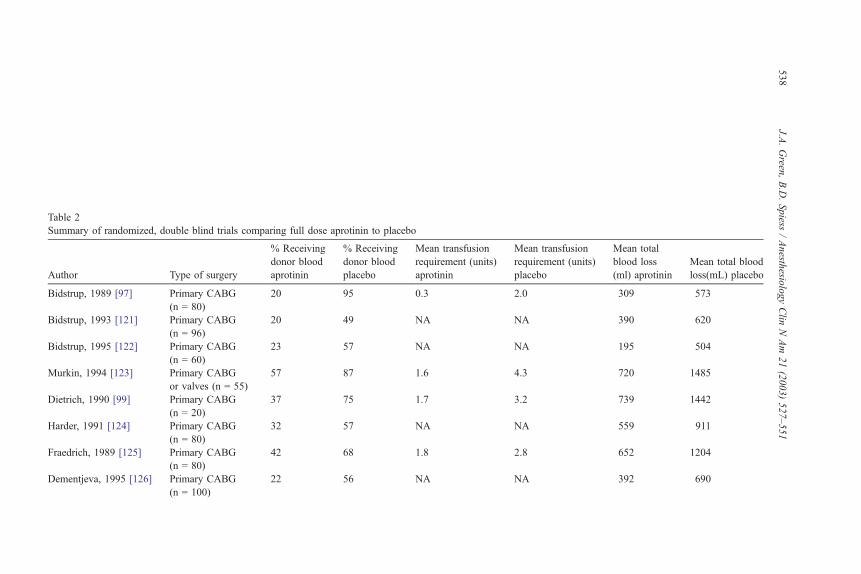
Table 2
Summary of randomized, double blind trials comparing full dose aprotinin to placebo
Author Type of surgery
% Receiving
donor blood
aprotinin
% Receiving
donor blood
placebo
Mean transfusion
requirement (units)
aprotinin
Mean transfusion
requirement (units)
placebo
Mean total
blood loss
(ml) aprotinin
Mean total blood
loss(mL) placebo
Bidstrup, 1989 [97] Primary CABG
(n = 80)
20 95 0.3 2.0 309 573
Bidstrup, 1993 [121] Primary CABG
(n = 96)
20 49 NA NA 390 620
Bidstrup, 1995 [122] Primary CABG
(n = 60)
23 57 NA NA 195 504
Murkin, 1994 [123] Primary CABG
or valves (n = 55)
57 87 1.6 4.3 720 1485
Dietrich, 1990 [99] Primary CABG
(n = 20)
37 75 1.7 3.2 739 1442
Harder, 1991 [124] Primary CABG
(n = 80)
32 57 NA NA 559 911
Fraedrich, 1989 [125] Primary CABG
(n = 80)
42 68 1.8 2.8 652 1204
Dementjeva, 1995 [126] Primary CABG
(n = 100)
22 56 NA NA 392 690
J.A.Green
,B.D.Spiess
/Anesth
esiologyClin
NAm
21(2003)527–551
538

Swart, 1994 [127] Primary CABG
or valves (n = 100)
68 86 1.8 2.8 506 783
Casas, 1995 [128] Primary CABG
or valves (n = 99)
26 56 0.7 1.8 195 489
Lemmer, 1994 [129] Primary CABG
(n = 151)
38 52 1.1 2.2 855 1503
Lemmer, 1994 [129] Repeat CABG
(n = 65)
30 72 0.4 3.3 1255 1979
Cosgrove, 1992 [130] Repeat CABG
(n = 115)
46 79 NA NA 720 1121
Minami, 1993 [131] Repeat CABG
(n = 49)
28 52 NA NA 441 772
Levy, 1995 [34] Repeat CABG
(n = 145)
54 75 1.6 3.4 900 1700
Alderman, 1998 [18] Primary CABG
(n = 401)
38 54 0.8 1.6 664 1168
CABG, coronary artery bypass grafting.
(Data from Schumann F. Hemostatic restoration during cardiopulmonary bypass. In: Pifarre R, editor. New anticoagulants for the cardiovascular patient. Philadelphia:
Hanley & Belfus, Inc.; 1997. p.453–69.)
J.A.Green
,B.D.Spiess
/Anesth
esiologyClin
NAm
21(2003)527–551
539

J.A. Green, B.D. Spiess / Anesthesiology Clin N Am 21 (2003) 527–551540
The efficacy of aprotinin has been especially well documented for high-risk
patients or those undergoing complex procedures. Royston and coauthors [117]
demonstrated that aprotinin recipients undergoing repeat operations had a
decrease in blood loss. In aspirin-treated patients, aprotinin reduces blood loss
and transfusion to the same degree as in non-aspirin patients undergoing primary
[123] or repeat CABG [23,130]. In a multicenter, double-blind, placebo-
controlled trial in repeat CABG patients [23], aprotinin decreased blood loss
and transfusion in both the high- and low-dose groups without increasing the
incidence of MI; the aprotinin recipients also had a lower incidence of reexplo-
ration and stroke. Overall, aprotinin therapy seemed to offer a consistent, sig-
nificant, and clinically relevant reduction in blood loss and transfusion
requirements [17].
Clinical safety/graft patency
Aprotinin is the only agent that has been approved by the US Food and Drug
Administration (FDA) to reduce bleeding in cardiac surgery patients [34]. The
clinical safety of aprotinin has been confirmed extensively throughout the
literature. The drug has very few side effects and is remarkably well tolerated,
even in the highest doses [68,132]. High-dose aprotinin seems to induce a
reversible microproteinuria related to a transient, isolated defect of tubular protein
absorption [133], but it has no effect on the serum creatinine level or the incidence
of adverse renal events [134]. Aprotinin can safely be administered to patients
with renal failure [135].
Very rarely (in < 1 per 1000 patients), aprotinin can trigger anaphylaxis
[98,136]. With repeat exposure, the incidence of anaphylaxis is as high as 5%
[137], but it decreases if the interval between the initial and subsequent dose is
greater than 3 months [138]. Prophylactic antihistamines reduce the severity and
incidence of these reactions [139]. Complications reported in the literature
include histamine-mediated cardiovascular collapse, heparin resistance, potenti-
ation of muscle relaxants, and renal toxicity [18,130]. Despite several case reports
of renal dysfunction, minimal renal effects have been observed in animal
experiments and controlled clinical studies [34].
Concerns have been raised regarding thrombotic complications with aprotinin.
There have been some case reports of clot formation on catheters [140], throm-
boembolic complications [141], and decreased graft patency [130,142] in CABG
patients. Investigators studying graft patency rates in aprotinin recipients,
however, have consistently been confounded by the aprotinin dosage, imaging
modality, time interval for patency assessment, heparin dosage, aspirin dosage,
distal vessel quality, saphenous vein graft (SVG) quality, and SVG preservation
techniques. For example, in a placebo-controlled study in redo CABG patients,
Cosgrove and coworkers [130] demonstrated that aprotinin decreased bleeding
and transfusion with no difference in mortality but with a (nonsignificant)
increase in the incidence of Q-wave MI. The authors concluded that aprotinin
may increase the risk of SVG thrombosis in redo CABG patients. The authors’
conclusion was based on post mortem examination of graft closures; although the
J.A. Green, B.D. Spiess / Anesthesiology Clin N Am 21 (2003) 527–551 541
incidence of MI was numerically higher in the group receiving aprotinin, no
denominator was disclosed, and no significance was shown.
High-dose aprotinin prolongs the celite-activated clotting time (ACT), leading
to decreased heparin doses [143]. Before this effect on the celite-ACT was
recognized, many reports and clinical studies cited an increased incidence
of thrombotic complications, graft closure, and MI [121,130]. In their study,
Cosgrove and associates [130] used celite-ACT values and gave less hepa-
rin, which probably influenced their results. Therefore, in the presence of aprotinin,
heparin activity must be monitored with kaolin-ACT, high-dose thrombin
time, blood heparin levels by protamine titration, or an empiric dosing scheme
[144–146]. Some clinicians recommend giving a total heparin dose of as much as
600 to 700 U/kg.
To perform a definitive prospective, randomized trial of vein graft patency,
Alderman and colleagues [18] participated in the International Multicenter
Aprotinin Graft Patency Experience (IMAGE) trial in 1995. Eight hundred
seventy patients undergoing primary CABG were randomized to receive high-
dose aprotinin or placebo treatment, followed by early coronary angiography to
evaluate SVG patency. In the aprotinin recipients, blood loss was reduced by
43%, transfusion by 49%, and reexploration by 47%. Occlusion of the SVG was
observed in 15.4% of the aprotinin-treated patients compared with 10.9% of the
placebo group (P = 0.03). However, in a subanalysis of the US patients only, the
occlusion rates were similar (9.4% aprotinin versus 9.5% placebo). The authors
attributed the dramatic difference in occlusion rates (aprotinin: non-US 23%
versus US 9.4%; placebo: non-US 12.4% versus US 9.5%) to differences in
gender prevalence, to vein harvesting and preservation techniques, to the
prevalence of aspirin therapy, and (most important) to small distal anastomotic
sites. Aprotinin had no effects on MI or mortality. In following up the IMAGE
trial patients, a survey showed no difference in survival or morbidity at a mean of
4 years postoperatively [147].
Subsequently, many studies showed no early SVG occlusion once the effect of
aprotinin on the celite-ACT was recognized and alternate methods of measuring
the heparin effect were used [100,129,148,149]. Further investigations reported
no increased thrombosis or graft closure in placebo-controlled, blinded, random-
ized trials when graft patency was determined by magnetic resonance imaging,
computed tomography, or angiography [34,111,150].
Use of antifibrinolytic agents in elective deep hypothermic circulatory arrest
The use of DHCA during cardiothoracic surgery is associated with an
increased risk of perioperative blood loss and renal dysfunction. The coagulop-
athy induced by hypothermia and circulatory stasis is complex and multifactorial
in origin. In brief, hypothermia induces kinetic slowing of coagulation, kinin and
kallikrein activation, platelet dysfunction, and fibrinolysis [151]. Furthermore,
blood stasis causes a thrombin-induced increase in activated protein C, which

J.A. Green, B.D. Spiess / Anesthesiology Clin N Am 21 (2003) 527–551542
results in further anticoagulation and fibrinolysis by way of endothelial release of
t-PA [152].
Use of antifibrinolytic agents to preserve hemostasis would seem to be of
greatest benefit in DHCA patients. However, concern over the possibility of
thrombotic complications in the setting of blood stasis has caused a controversy
to surround the use of antifibrinolytics, particularly aprotinin. Although all of the
antifibrinolytic agents have been used for DHCA, none has received as much
attention as aprotinin in the literature. Therefore, this section focuses on the use
of aprotinin in DHCA.
Early experience raised alarms about the hazards of aprotinin use in DHCA.
Excessive mortality, renal failure, and complications were reported in clinical
series in which the adequacy of heparinization was questionable. To date, there has
been very little evidence from prospective, randomized trials that aprotinin is
unusually hazardous. However, mostly because of the lack of well-controlled
studies, investigators have also failed to prove that its use offers a definitive benefit.
Some authors have speculated that unproven prothrombotic effects of apro-
tinin are uniquely expressed under the conditions of stasis and hypothermia that
distinguish DHCA from routine CPB alone [153,154]. Aprotinin may attenuate
the role of activated protein C in maintaining the fluidity of blood during periods
of stasis. This has led to the concern that aprotinin may potentiate thrombosis,
especially in the presence of inadequate heparinization [151]. As a serine protease
inhibitor, however, aprotinin would also decrease kinin/kallikrein activation
directly and would prevent platelet dysfunction by protecting glycoprotein
receptors and inhibiting fibrinolysis. These effects would make aprotinin attract-
ive in the setting of DHCA.
Clinical investigations comparing aprotinin recipients with control patients
have focused on neurologic outcome, renal dysfunction, and mortality. Most of
the thrombotic complications associated with the use of aprotinin during DHCA
were described in the early 1990s and may have been the consequence of
inadequate heparinization, when it was not clear how to adjust the dose of
heparin. The earliest studies were extremely alarming and led to warnings about
the use of aprotinin in the product brochure of aprotinin. In an uncontrolled series
studied by Sundt and coauthors [26], the aprotinin patients had 30% greater
mortality, 60% greater renal dysfunction, and a 25% greater need for dialysis than
the control patients. However, the aprotinin group received substantially less
heparin than the control group (27,850 versus 40,250 U). In a retrospective study,
Westaby [155] found increased coagulation abnormalities, increased reexplora-
tion for bleeding, and increased thrombosis-related mortality in aprotinin-treated
patients. Both of these studies were completed before 1995, when it was
recognized that aprotinin prolongs the celite-ACT. Thus, the complications of
these initial studies were attributed to inadequate heparinization.
Current recommendations include higher heparin dosing, keeping the celite-
ACT at > 750 seconds, or using kaolin-ACT measurements. Studies using these
criteria have shown no difference in mortality, neurologic defects, renal dysfunc-
tion, or renal failure in aprotinin versus placebo recipients [156,157]. The first

J.A. Green, B.D. Spiess / Anesthesiology Clin N Am 21 (2003) 527–551 543
positive results were reported by Goldstein and associates [158], who, in a
retrospective study, found that aprotinin-treated patients had a significantly lower
incidence of postoperative transfusion, although they had the same chest-tube
output as control patients. Okita and coworkers [159], who described a consec-
utive series of 112 partially randomized patients, reported decreased blood loss
and transfusion in recipients of low-dose aprotinin.
In a randomized, controlled study, Ehrlich and coauthors [156] compared low-
dose aprotinin to placebo in DHCA patients undergoing surgery of the thoracic
aorta. Compared with the control group, the aprotinin group had less blood loss
and transfusion, and no effect on renal function. In a more recent retrospective
review of a similar series of patients, Seigne and coworkers [160] found that low-
dose aprotinin decreased blood transfusion and had no deleterious renal or
myocardial effects. Similarly, in a recent retrospective study, Mora-Mangano
and colleagues [151] demonstrated that aprotinin did not increase renal dysfunc-
tion, although it also had no effect on blood loss or reexploration. Predictors of
postoperative renal dysfunction included transfusion of more than 5 units of
blood, low urine output in the operating room, lack of dopamine use, a
perioperative hematocrit of < 21%, and low urine output in the ICU [151].
In a prospective study concerning the effect of heparin dosage in patients
undergoing DHCA with aprotinin, Okita and associates [159] determined that
those who received additional heparin, regardless of their ACT, had better platelet
preservation and less severe activation of the coagulation system than did patients
who received heparin only when their ACT decreased to < 500 seconds. Overall,
recent studies seem to indicate that aprotinin is not unusually hazardous in DHCA
patients as long as adequate heparinization is used; however, the benefits of
aprotinin are unclear.
Before this controversy is resolved, further prospective, randomized, placebo-
controlled, double-blind studies must be performed on a much larger scale.
Based the most recent studies, however, there seems to be no convincing evidence
that aprotinin should be withheld from patients undergoing cardiac surgery
with DHCA.
Summary and future considerations
During cardiac surgery, CPB increases thrombin levels, which augments t-PA
and plasmin formation, leading to an increase in fibrinolytic activity. Future
therapies may be directed against increased hemostatic and inflammatory activa-
tion. The result should be less thrombin formation, better hemostasis, and less
blood loss. To this end, numerous strategies have been developed and are being
tested. For instance, the process of heparin dosing and protamine reversal are being
reexamined. Furthermore, administration of recombinant antithrombin III, which
enhances the antithrombotic properties of heparin, has decreased thrombin forma-
tion and fibrinolytic activity [161,162]. Also, development of other antithrombins
and heparin analogs—such as argatroban, bivalirudin, dermatin sulfate, pentasac-

J.A. Green, B.D. Spiess / Anesthesiology Clin N Am 21 (2003) 527–551544
charide, and recombinant hirudin—is ongoing. The short half-life and potent
antithrombotic activity of some of these agents could prove quite useful during
CPB. Lastly, other agents such as recombinant activated factor VIIa are being used
in cardiac surgery for patients with refractory bleeding [54,163].
The potential for severe bleeding after cardiac surgery and CPB remains a
major problem. Current pharmacologic approaches to attenuating the activation
of the hemostatic system and preventing fibrinolysis are limited. Antifibrinolytic
agents result in decreased consumption of clotting factors and better preservation
of platelet function. Prophylactic administration of agents with antifibrinolytic,
anticoagulant, and antiinflammatory properties could decrease operative time,
blood loss, transfusion, and reexploration, thereby improving patient outcomes
and potentially reducing overall health care costs.
The choice of which agent to use in specific patient groups remains
controversial, institutionally variable, and somewhat polarized. History shows
all three agents to be safe, yet the literature contains individual case reports of
thrombotic events. Further research into individual genetic variability among
patients may someday elucidate the cause of these rare events. Although
antifibrinolytic agents may increase patient safety during cardiac surgery, further
investigation into their use is essential.
References
[1] Magovern J, Sakert T, Benkhart D, et al. A model for predicting transfusion after coronary
artery bypass grafting. Ann Thorac Surg 1996;61:27–32.
[2] Boyle E, Verrier E, Spiess B. Endothelial cell injury in cardiovascular surgery: The procoagu-
lant response. Ann Thorac Surg 1996;62:1549–57.
[3] Mongan P, Brown R, Thwaites B. Tranexamic acid and aprotinin reduce postoperative bleeding
and transfusions during primary coronary revascularizations. Anesth Analg 1998;87:258–65.
[4] Dodd R. The risk of transfusion-transmitted infection. N Engl J Med 1992;327:419–20.
[5] Seyfried H, Walewska I. Immune hemolytic transfusion reactions. World J Surg 1987;11:25–9.
[6] Greenbaum B. Transfusion-associated graft-versus-host disease: historical perspectives, inci-
dence, and current use of irradiated blood products. J Clin Oncol 1991;9:1889–902.
[7] Goodnough L, Brecher M, Kanter M, et al. Transfusion Medicine. N Engl J Med 1999;340:
438–47.
[8] Klein H. Immunomodulatory aspects of transfusion: A once and future risk? Anesthesiology
1999;91:861–5.
[9] Murphy P, Connery C, Hicks G, et al. Homologous blood transfusions as a risk factor for
postoperative infection after coronary artery bypass operations. J Thorac Cardiovasc Surg
1992;104:1092–9.
[10] Spiess B. Blood transfusion: The silent epidemic. Ann Thorac Surg 2001;72:S1832–7.
[11] Nuttall G, Oliver W, Santrach P, et al. Efficacy of a simple intraoperative transfusion algorithm
for nonerythrocyte component utilization after cardiopulmonary bypass. Anesthesiology
2001;94:773–81.
[12] Despotis G, Filos K, Zoys T, et al. Factors associated with excessive postoperative blood loss
and hemostatic transfusion requirements: A multivariate analysis in cardiac surgical patients.
Anesth Analg 1996;82:13–21.
[13] Dacey L, Munoz J, Baribeau Y, et al. Reexploration for hemorrhage following coronary artery
bypass grafting: Incidence and risk factors. Arch Surg 1998;133:442–7.

J.A. Green, B.D. Spiess / Anesthesiology Clin N Am 21 (2003) 527–551 545
[14] Unsworth-White M, Herriot A, Valencia O, et al. Resternotomy for bleeding after cardiac
operation: a marker for increased morbidity and mortality. Ann Thorac Surg 1995;59:664–7.
[15] Moulton M, Creswell L, Mackey M, et al. Reexploration for bleeding is a risk factor for adverse
outcomes after cardiac operations. J Thorac Cardiovasc Surg 1996;111:1037–46.
[16] Woodman R, Harker L. Bleeding complications associated with cardiopulmonary bypass.
Blood 1990;76:1680–97.
[17] Schumann F. Hemostatic restoration during cardiopulmonary bypass. In: Pifarre R, editor. New
Anticoagulants for the Cardiovascular Patient. Philadelphia: Hanley & Belfus, Inc.; 1997.
p. 453–69.
[18] Alderman E, Levy J, Rich J, et al. Analyses of coronary graft patency after aprotinin use:
Results from the international multicenter aprotinin graft patency experience (IMAGE) trial.
J Thorac Cardiovasc Surg 1998;116:716–30.
[19] Bennett-Guerrero E, Sorohan J, Gurevich M, et al. Cost-benefit and efficacy of aprotinin
compared with epsilon-aminocaproic acid in patients having repeated cardiac operations: A
randomized, blinded clinical trial. Anesthesiology 1997;87:1373–80.
[20] Horrow J, Hlavacek J, Strong M, et al. Prophylactic tranexamic acid decreased bleeding after
cardiac operations. J Thorac Cardiovasc Surg 1990;99:70–4.
[21] Laupacis A, Fergusson D. Drugs to minimize perioperative blood loss in cardiac surgery:
Meta-analyses using perioperative blood transfusion as the outcome. Anesth Analg 1997;85:
1258–67.
[22] Levi M, Cromheecke M, de Jonge E, et al. Pharmacologic strategies to decrease excessive
blood loss in cardiac surgery: A meta-analysis of clinically relevant endpoints. Lancet 1999;
354:1940–7.
[23] Levy J, Pifarre R, Schaff H, et al. A multicenter, double-blind, placebo-controlled trial of
aprotinin for reducing blood loss and the requirement for donor-blood transfusion in patients
undergoing repeat coronary artery bypass grafting. Circulation 1995;92:2236–44.
[24] Munoz J, Birkmeyer N, Birkmeyer J, et al. Is epsilon-aminocaproic acid as effective as apro-
tinin in reducing bleeding with cardiac surgery? A meta-analysis. Circulation 1999;99:81–9.
[25] Shore-Lesserson L, Reich D, Vela-Cantos F, et al. Tranexamic acid reduces transfusions and
mediastinal drainage in repeat cardiac surgery. Anesth Analg 1996;83:18–26.
[26] Sundt T, Kouchoukos N, Saffitz J, et al. Renal dysfunction and intravascular coagulation with
aprotinin and hypothermic circulatory arrest. Ann Thorac Surg 1993;55:1418–24.
[27] Slaughter T, Faghih F, Greenberg C, et al. The effects of epsilon-aminocaproic acid on fibri-
nolysis and thrombin generation during cardiac surgery. Anesth Analg 1997;85:1221–6.
[28] Ray M, Marsh N, Hawson G. Relationship of fibrinolysis and platelet function to bleeding after
cardiopulmonary bypass. Blood Coagul Fibrinolysis 1994;5:679–85.
[29] Despotis G, Santoro S, Spitznagel E, et al. Prospective evaluation and clinical utility of on-site
monitoring of coagulation in patients undergoing cardiac operation. J Thorac Cardiovasc Surg
1994;107:271–9.
[30] Spiess B. The contribution of fibrinolysis to post bypass bleeding. J Cardiothorac Vasc Anes-
thesiology 1991;5:13–7.
[31] Davie E, Fujikawa K, Kisiel W. The coagulation cascade: Initiation, maintenance, and regu-
lation. Biochemistry 1991;30:10363–70.
[32] Furie B, Furie BC. Molecular and cellular biology of blood coagulation. N Engl J Med
1992;326:800–6.
[33] Dietrich W. Reducing thrombin formation during cardiopulmonary bypass: Is there a benefit of
the additional anticoagulant action of aprotinin? J Cardiovasc Pharm 1996;27:S50–7.
[34] Levy J. Pharmacologic preservation of the hemostatic system during cardiac surgery. Ann
Thorac Surg 2001;72:S1814–20.
[35] Boisclair M, Lane D, Philippou H, et al. Mechanisms of thrombin generation during surgery
and cardiopulmonary bypass. Blood 1993;82:3350–7.
[36] de Haan J, Boonstra P, Monnink S, et al. Retransfusion of suctioned blood during cardiopul-
monary bypass impairs hemostasis. Ann Thorac Surg 1995;59:901–7.

J.A. Green, B.D. Spiess / Anesthesiology Clin N Am 21 (2003) 527–551546
[37] Heimark R, Kurachi K, Fujikawa K, et al. Surface activation of blood coagulation, fibrinolysis
and kinin formation. Nature 1980;286:456–60.
[38] Gravlee G, Haddon W, Rothberger H, et al. Heparin dosing and monitoring for cardiopulmo-
nary bypass. J Thorac Cardiovasc Surg 1990;99:518–27.
[39] Tanaka K, Takao M, Yada I, et al. Alterations in coagulation and fibrinolysis associated with
cardiopulmonary bypass during open heart surgery. J Cardiothorac Anesth 1989;3:181.
[40] Levin E, Stern D, Nawroth P, et al. Specificity of the thrombin-induced release of tissue
plasminogen activator from cultured human endothelial cells. Thromb Haemost 1986;56:
115–9.
[41] Murkin J. Cardiopulmonary bypass and the inflammatory response: A role for serine protease
inhibitors? J Cardiothorac Vasc Anesth 1997;11:S19–23.
[42] Vang S, Stammers A, Mejak B, et al. Co-administration of aprotinin and epsilon-aminocaproic
acid during cardiopulmonary bypass in a swinemodel. J Extra-corporeal Tech 2000;32:196–206.
[43] Teufelsbauer H, Proidl S, Havel M, et al. Early activation of hemostasis during cardiopulmo-
nary bypass. Evidence for thrombin-mediated hyperfibrinolysis. Thromb Haemost 1992;68:
250–2.
[44] Stibbe J, Kluft C, Brommer E, et al. Enhanced fibrinolytic activity during cardiopulmonary
bypass in open-heart surgery in man is caused by extrinsic (tissue-type) plasminogen activator.
Eur J Clin Invest 1984;14:375–82.
[45] Yoshihara H, Yamamoto T, Mihara H. Changes in coagulation and fibrinolysis occurring in
dogs during hypothermia. Thromb Res 1985;37:503.
[46] Umlas J. Fibrinolysis and disseminated intravascular coagulation in open heart surgery. Trans-
fusion 1976;16:460.
[47] Hanss M, Collen D. Secretion of tissue-type plasminogen activator and plasminogen activator
inhibitor by cultured human endothelial cells: Modulation by thrombin, endotoxin, histamine.
J Lab Clin Med 1987;109:97–104.
[48] Lu H, du Buit C, Soria J, et al. Postoperative hemostasis and fibrinolysis in patients under-
going cardiopulmonary bypass with or without aprotinin therapy. Thromb Haemost 1994;72:
438–43.
[49] Benza RL, Paramo JA, Panizo C, et al. Gene polymorphisms for plasminogen activator inhib-
itor-I/tissue plasminogen activator and development of allograft coronary artery disease. Cir-
culation 1998;98:2248–54.
[50] Heinrich J, Funke H, Rust S, et al. Impact of polymorphisms in the alpha and beta fibrinogen
gene and plasma fibrinogen concentrations and risk of myocardial infarction. The ECTIM
Study. Arterial Thrombosis 1993;13:886–91.
[51] Spannagl M, Dooijewaard G, Dietrich W, et al. Protection of single-chain urokinase-type
plasminogen activator (scu- PA) in aprotinin treated cardiac surgical patients undergoing car-
diopulmonary bypass. Thromb Haemost 1995;73:825–8.
[52] Rinder C, Bohnert J, Rinder H, et al. Platelet activation and aggregation during cardiopulmo-
nary bypass. Anesthesiology 1991;75:388.
[53] Adelman B, Rizk A, Hanners E. Plasminogen interactions with platelets in plasma. Blood
1988;72:1530.
[54] Despotis G, Avidan M, Hogue C. Mechanisms and attenuation of hemostatic activation during
extracorporeal circulation. Ann Thorac Surg 2001;72:S1821–31.
[55] Soslau G, Horrow J, Brodsky I. Effect of Tranexamic acid on platelet ADP during extracorpo-
real circulation. Am J Hematol 1991;38:113–9.
[56] van Oeveren W, Harder M, Roozendaal K, et al. Aprotinin protects platelets against the initial
effect of cardiopulmonary bypass. J Thorac Cardiovasc Surg 1990;99:788–97.
[57] Lu H, Soria C, Li H, et al. Role of active center and lysine binding sites of plasmin in plasmin-
induced platelet activation and disaggregation. Thromb Haemost 1991;65:67–72.
[58] Lu H, Soria C, Soria J, et al. Reversible translocation of glycoprotein 1b in plasmin-treated
platelets: Consequences for platelet function. Eur J Clin Invest 1993;23:785–93.
[59] Vander Salm T, Ansell J, Okike O, et al. The role of epsilon-aminocaproic acid in reducing

J.A. Green, B.D. Spiess / Anesthesiology Clin N Am 21 (2003) 527–551 547
bleeding after cardiac operation: A double-blind randomized study. J Thorac Cardiovasc Surg
1988;95:538–40.
[60] Casati V, Guzzon D, Oppizi M, et al. Hemostatic effects of aprotinin, tranexamic acid and
epsilon-aminocaproic acid in primary cardiac surgery. Ann Thorac Surg 1999;68:2252–6.
[61] Casati V, Guzzon D, Oppizi M, et al. Tranexamic acid compared with high-dose aprotinin in
primary elective heart operations: Effects on perioperative bleeding and allogeneic transfusions.
J Thorac Cardiovasc Surg 2000;120:520–7.
[62] Daily P, Lamphere J, Dembitsky W. Effect of prophylactic epsilon-aminocaproic acid on blood
loss and transfusion requirements in patients undergoing first-time coronary artery bypass
grafting. J Thorac Cardiovasc Surg 1994;108:99–108.
[63] DelRossi A, Cernaianu A, Bostros S, et al. Prophylactic treatment of post perfusion bleeding
using EACA. Chest 1989;96:27–30.
[64] Jordan D, Delphin E, Rose E. Prophylactic epsilon-aminocaproic acid (EACA) administration
minimizes blood replacement therapy during cardiac surgery. Anesth Analg 1995;80:827–9.
[65] Karski J, Teasdale S, Norman P, et al. Prevention of post bypass bleeding with tranexamic acid
and epsilon-aminocaproic acid. J Cardiothorac Vasc Anesth 1993;7:431–5.
[66] Montesano R, Gustafson P, Palanzo D, et al. The effect of low-dose epsilon-aminocaproic acid
on patients following coronary artery bypass surgery. Perfusion 1996;11:53–6.
[67] Rousou J, Engelman R, Flack J, et al. Tranexamic acid significantly reduces blood loss asso-
ciated with coronary revascularization. Ann Thorac Surg 1995;59:671–5.
[68] Verstraete M. Clinical application of inhibitors of fibrinolysis. Drugs 1985;29:236.
[69] Andersson L, Eriksson O, Hedlund P, et al. Special considerations with regard to the dosage of
tranexamic acid in patients with chronic renal diseases. Urol Res 1978;6:83–8.
[70] Butterworth J, James R, Kennedy D, et al. Pharmacokinetics of epsilon-aminocaproic acid in
adult patients undergoing coronary artery surgery. Anesthesiology 1996;85:A151.
[71] Horrow J, DiGregorio G, Ruch E, et al. The dose-plasma concentration relationship of tranexa-
mic acid during surgery. Am J Ther 1994;1:206–9.
[72] McClure P, Izsak J. The use of epsilon-aminocaproic acid to reduce bleeding during cardiac
bypass in children with congenital heart disease. Anesthesiology 1974;40:604.
[73] Midell A, Hallman G, Bloodwell R, et al. Epsilon-aminocaproic acid for bleeding after car-
diopulmonary bypass. Ann Thorac Surg 1971;11:577.
[74] Horrow J, van Riper D, Strong M, et al. The hemostatic effects of tranexamic acid and
desmopressin during cardiac surgery. Circulation 1991;84:2063.
[75] Gans H, Krivit W. Problems in hemostasis during open heart surgery. III: Epsilon-aminocaproic
acid as an inhibitor of plasminogen activator activity. Ann Surg 1962;15:268–76.
[76] Gans H, Subramanian V, John S, et al. Theoretical and practical (clinical) considerations
concerning proteolytic enzymes and their inhibitors with particular reference to changes in
the plasminogen-plasmin system observed during assisted circulation in man. Ann N Y Acad
Sci 1968;146:721–36.
[77] Okamoto S, Oshiba S, Mihara H, et al. Synthetic inhibitors of fibrinolysis: In vitro and in vivo
mode of action. Ann N Y Acad Sci 1968;146:414–29.
[78] Fremes S, Wong B, Lee E, et al. Metaanalysis of prophylactic drug treatment in the prevention
of postoperative bleeding. Ann Thorac Surg 1994;58:1580–8.
[79] Hardy J, Belisle S. Natural and synthetic antifibrinolytics in adult cardiac surgery: Efficacy,
effectiveness and efficiency. Can J Anaesth 1994;41:1104–12.
[80] Bergin J. Complications of therapy with epsilon-aminocaproic acid. Med Clin North Am
1966;50:1669–78.
[81] Dentz M, Slaughter T, Mark J. Early thrombus formation on heparin-bonded pulmonary artery
catheters in patients receiving epsilon-aminocaproic acid. Anesthesiology 1995;82:583–6.
[82] Ratnoff O. Epsilon-aminocaproic acid: A dangerous weapon. N Engl J Med 1969;280:1124–5.
[83] Menichetti A, Tritapepe L, Ruvolo G, et al. Changes in coagulation patterns, blood loss and
blood use after cardiopulmonary bypass: Aprotinin vs. tranexamic acid vs. epsilon-aminoca-
proic acid. J Cardiovasc Surg 1996;37:401–7.

J.A. Green, B.D. Spiess / Anesthesiology Clin N Am 21 (2003) 527–551548
[84] Penta de Peppo A, Pierri M, Scafuri A, et al. Intraoperative antifibrinolysis and blood-saving
techniques in cardiac surgery: Prospective trial of 3 antifibrinolytic drugs. Tex Heart Inst J
1995;22:231–6.
[85] Horrow J, van Riper D, Strong M, et al. The dose-response relationship of tranexamic acid.
Anesthesiology 1995;82:383–92.
[86] Speekenbrink R, Vonk A, Wildevuur C, et al. Hemostatic efficacy of dipyridamole, tranexamic
acid, and aprotinin in coronary artery bypass grafting. Ann Thorac Surg 1995;59:438–42.
[87] Blauhut B, Harringer W, Bettelheim P, et al. Comparison of the effects of aprotinin and
tranexamic acid on blood loss and related variables after cardiopulmonary bypass. J Thorac
Cardiovasc Surg 1994;108:1083–91.
[88] Corbeau J, Monrigal J, Jacob J, et al. Comparative effects of aprotinin and tranexamic acid on
blood loss in cardiac surgery. Ann Fr Anesth Reanim 1995;14:154–61.
[89] Katsaros D, Petricevic M, Snow N, et al. Tranexamic acid reduces post bypass blood use: A
double-blind, prospective, randomized study of 210 patients. Ann Thorac Surg 1996;61:1131–5.
[90] Brown R, Thwaites B, Mongan P. Tranexamic acid is effective in decreasing postoperative
bleeding and transfusions in primary coronary artery bypass operations: A double-blind,
randomized, placebo-controlled trial. Anesth Analg 1997;85:963–70.
[91] Nuttall G, Oliver W, Ereth M, et al. Comparison of blood-conservation strategies in cardiac
surgery patients at high risk for bleeding. Anesthesiology 2000;92:674–82.
[92] Casati V, Bellotti F, Gerli C, et al. Tranexamic acid administration after cardiac surgery:
A prospective, randomized, double-blind, placebo-controlled study. Anesthesiology 2001;
94:8–14.
[93] Wong B, McLean R, Fremes S, et al. Aprotinin and tranexamic acid for high transfusion risk
cardiac surgery. Ann Thorac Surg 2000;69:808–16.
[94] Lindoff C, Rybo G, Astedt B. Treatment with tranexamic acid during pregnancy, and the risk of
thromboembolic complications. Thromb Haemost 1993;70:238–40.
[95] Ovrum E, Holen E, Abdelnoor M, et al. Tranexamic acid (cyclocapron) is not necessary to reduce
blood loss after coronary bypass operations. J Thorac Cardiovasc Surg 1993;105:78–83.
[96] Royston D. The serine antiprotease aprotinin (Trasylol): A novel approach to reducing post-
operative bleeding. Blood Coag Fibrin 1990;1:55.
[97] Bidstrup B, Royston D, Sapsford R, et al. Reduction in blood loss and blood use after car-
diopulmonary bypass with high-dose aprotinin (Trasylol). J Thorac Cardiovasc Surg 1989;97:
364–72.
[98] Blauhut B, Gross C, Necek S, et al. Effects of high-dose aprotinin on blood loss, platelet
function, fibrinolysis, complement, and renal function after cardiopulmonary bypass. J Thorac
Cardiovasc Surg 1991;101:958–67.
[99] Dietrich W, Spannagl M, Jochum M, et al. Influence of high-dose aprotinin treatment on blood
loss and coagulation patterns in patients undergoing myocardial revascularization. Anesthesi-
ology 1990;73:1119–26.
[100] Lass M, Welz A, Kochs M, et al. Aprotinin in elective primary bypass surgery. Graft patency
and clinical efficacy. Eur J Cardiothorac Surg 1995;9:206–10.
[101] Lemmer J, Dilling E, Morton J, et al. Aprotinin for primary coronary artery bypass grafting:
A multicenter trial of three dose regimens. Ann Thorac Surg 1996;62:1659–67.
[102] Poullis M, Manning R, Laffan M, et al. The antithrombotic effect of aprotinin: Actions medi-
ated via the protease-activated receptor 1. J Thorac Cardiovasc Surg 2000;120:370–8.
[103] Chabbat J, Porte P, Tellier M, et al. Aprotinin is a competitive inhibitor of the factor VIIa-tissue
factor complex. Thromb Res 1993;71:205–15.
[104] Horrow J. Transfusion medicine and coagulation disorders. In: Kaplan JA, editor. Cardiac
Anesthesia. Philadelphia: WB Saunders; 1987. p. 1111–54.
[105] Marx G, Pokar K, Reuter H, et al. The effects of aprotinin on hemostatic function during
cardiac surgery. J Cardiothorac Vasc Anesth 1991;5:467–74.
[106] Edmunds L, Niewiarowski S, Colman R. Aprotinin (letter). J Thorac Cardiovasc Surg 1991;
101:1103.

J.A. Green, B.D. Spiess / Anesthesiology Clin N Am 21 (2003) 527–551 549
[107] Wenger R, Lukasiewicz H, Mikuta B, et al. Loss of platelet fibrinogen receptors during clinical
cardiopulmonary bypass. J Thorac Cardiovasc Surg 1989;97:235–9.
[108] Huang H, Ding W, Su Z, et al. Mechanism of the preserving effect of aprotinin on platelet
function and its use in cardiac surgery. J Thorac Cardiovasc Surg 1993;106:11–8.
[109] Tabuchi N, De Haan J, Boonstra P, et al. Aprotinin effect on platelet function and clotting
during cardiopulmonary bypass. Eur J Cardiothorac Surg 1994;8:87–90.
[110] John L, Rees G, Kovacs I. Reduction of heparin binding to and inhibition of platelets by
aprotinin. Ann Thorac Surg 1993;55:1175–9.
[111] Landis R, Asimakopoulos G, Poullis M, et al. The antithrombotic and anti-inflammatory mech-
anisms of action of aprotinin. Ann Thorac Surg 2001;72:2169–75.
[112] Landis R, Haskard D, Taylor K. New anti-inflammatory and platelet-preserving effects of
aprotinin. Ann Thorac Surg 2001;72:S1808–13.
[113] Poullis M, Landis R, Taylor K. Aprotinin: Is it prothrombotic? Perfusion 2001;16:401–9.
[114] Clagett G, Anderson F, Levine M, et al. Prevention of venous thromboembolism. Chest
1992;102:3915–4075.
[115] Murkin J, Shannon N, Bourne R, et al. Aprotinin decreases blood loss in patients undergoing
revision or bilateral total hip arthroplasty. Anesth Analg 1995;80:343–8.
[116] Harmon D. Cost/benefit analysis of pharmacologic hemostasis. Ann Thorac Surg 1996;61:
S21–5.
[117] Royston D, Bidstrup B, Taylor K, et al. Effect of aprotinin on need for blood transfusion after
repeat open-heart surgery. Lancet 1987;ii:1289–91.
[118] van Oeveren W, Jansen N, Bidstrup B, et al. Effects of aprotinin on hemostatic mechanism
during cardiopulmonary bypass. Ann Thorac Surg 1987;44:640–5.
[119] Royston D. High-dose aprotinin therapy: A review of the first five years’ experience. J Car-
diothorac Vasc Anesth 1992;6:76–100.
[120] Tice D, Reed G, Clauss R, et al. Hemorrhage due to fibrinolysis occurring with open-heart
operations. J Thorac Cardiovasc Surg 1963;46:673–9.
[121] Bidstrup B, Underwood S, Sapsford R, et al. Effect of aprotinin (Trasylol) on aorto-coronary
bypass graft patency. J Thorac Cardiovasc Surg 1993;105:147–53.
[122] Bidstrup B. Pretreatment with aprotinin and aspirin. In: Pifarre R, editor. Blood Conservation
with Aprotinin. Philadelphia: Hanley & Belfus; 1995. p. 1–44.
[123] Murkin J, Lux J, Shannon N, et al. Aprotinin significantly decreases bleeding and transfusion
requirements in patients receiving aspirin and undergoing cardiac operations. J Thorac Cardi-
ovasc Surg 1994;107:554–61.
[124] Harder M, Eijsman L, Roozendaal K, et al. Aprotinin reduces intraoperative and postoper-
ative blood loss in membrane oxygenator cardiopulmonary bypass. Ann Thorac Surg 1991;
51:936–41.
[125] Fraedrich G, Engler H, Kanz L, et al. High dose aprotinin regimen in cardiac surgery – A
prospective randomised double-blind trial. Circulation 1989;80SII:158.
[126] Dementieva I, Dzemeshkevich S, Charnaya M, et al. The use of high doses of aprotinin to
decrease blood loss in aortocoronary bypass surgery. J Cardiovasc Surg 1994;35SI:185–6.
[127] Swart M, Gordon P, Hayse-Gregson P, et al. High-dose aprotinin in cardiac surgery – A
prospective randomized study. Anaesth Intens Care 1994;22:529–33.
[128] Casas J, Zuazu-Jausoro I, Mateo J, et al. Aprotinin versus desmopressin for patients undergoing
operations with cardiopulmonary bypass. J Thorac Cardiovasc Surg 1995;110:1107–17.
[129] Lemmer J, Stanford W, Bonney S, et al. Aprotinin for coronary bypass operations: Efficacy,
safety, and influence on early saphenous vein graft patency. J Thorac Cardiovasc Surg
1994;107:543–53.
[130] Cosgrove D, Heric B, Lytle B, et al. Aprotinin therapy for reoperative myocardial revascula-
rization: A placebo-controlled study. Ann Thorac Surg 1992;54:1031–8.
[131] Minami K, Notohamiprodjo G, Buschler H, et al. Alpha-2-plasmin inhibitor-plasmin complex
and postoperative blood loss: Double-blind study with aprotinin in reoperation for myocardial
revascularization. J Thorac Cardiovasc Surg 1993;106:934–6.

J.A. Green, B.D. Spiess / Anesthesiology Clin N Am 21 (2003) 527–551550
[132] Davis R, Whittington R. Aprotinin. A review of its pharmacology and therapeutic efficacy in
reducing blood loss associated with cardiac surgery. Drugs 1995;49:954–83.
[133] Fraedrich G, Neukamm K, Schneider T, et al. Safety and risk/benefit assessment of aprotinin in
primary CABG. In: Friedel N, Hetzer R, Royston D, editors. Blood use in cardiac surgery.
Darmstadt: Steinkopff Verlag; 1991. p. 221–31.
[134] Lemmer J, Stanford W, Bonney S, et al. Aprotinin for coronary artery bypass grafting: Effect on
postoperative renal function. Ann Thorac Surg 1995;59:132–6.
[135] Lemmer J, Metzdorf M, Krause A, et al. Aprotinin use in patients with dialysis-dependent renal
failure undergoing cardiac operations. J Thorac Cardiovasc Surg 1996;112:192–4.
[136] Bohrer H, Bach A, Fleischer F, et al. Adverse haemodynamic effects of high-dose aprotinin in a
paediatric cardiac surgical patient. Anaesthesia 1990;45:853.
[137] Goldstein D, Oz M, Smith C, et al. Safety of repeat aprotinin administration for LVAD recip-
ients undergoing cardiac transplantation. Ann Thorac Surg 1996;61:692–5.
[138] Dietrich W, Spath P, Ebell A, et al. Incidence of anaphylactic reactions to aprotinin – analysis
of 248 reexposures to aprotinin. Anesthesiology 1995;83S:A104.
[139] Lamparter-Schummert B, Mugaragu I, Hetzer R, et al. Aprotinin re-exposure in patients under-
going repeat cardiac surgery: Effect of prophylaxis with H1- and H2-receptor antagonists. Br J
Anaesth 1995;74:S3.
[140] Bohrer H, Fleischer F, Lang J, et al. Early formation of thrombi on pulmonary artery catheters
in cardiac surgical patients receiving high-dose aprotinin. J Cardiothorac Vasc Anesthesiology
1990;4:222–5.
[141] Umbrain V, Christiaens F, Camu F. Intraoperative coronary thrombosis: Can aprotinin and
protamine be incriminated? J Cardiothorac Vasc Anesth 1994;8:198–201.
[142] Van der Meer J, Hillege H, Ascoop C, et al. Aprotinin in aorto-coronary bypass surgery:
Increased risk of vein graft occlusion and myocardial infarction? Supportive evidence from a
retrospective study. Thromb Haemost 1996;75:1–3.
[143] Dietrich W, Dilthey G, Spannagl M, et al. Influence of high-dose aprotinin on anticoagulation,
heparin requirement, and celite- and kaolin-activated clotting time in heparin-pretreated patients
undergoing open heart surgery. A double-blind, placebo-controlled study. Anesthesiology
1995;83:679.
[144] Dietrich W, Jochum M. Effect of celite and kaolin on activated clotting time in the presence of
aprotinin: Activated clotting time is reduced by binding of aprotinin to kaolin. J Thorac
Cardiovasc Surg 1995;109:177–8.
[145] Huyzen R, Harder M, Huet R, et al. Alternative perioperative anticoagulation monitoring during
cardiopulmonary bypass in aprotinin-treated patients. J Cardiothorac VascAnesth 1994;8:153–6.
[146] Wang J, Lin C, Hung W, et al. Monitoring of heparin induced anticoagulation with kaolin-
activated clotting time in cardiac surgical patients treated with aprotinin. Anesthesiology
1992;77:1080–4.
[147] Niemcryk S, Pettersson G, Rasmussen C, et al. Follow-up analyses of the international multi-
center aprotinin graft patency experience (IMAGE). J Out Res 2001;5:1–10.
[148] Kalangos A, Tayyareci G, Pretre R, et al. Influence of aprotinin on early graft thrombosis in
patients undergoing myocardial revascularization. Eur J Cardiothorac Surg 1994;8:651–6.
[149] Royston D. Aprotinin vs. lysine analogues: The debate continues. Ann Thorac Surg 1998;65:
S9–19.
[150] Havel M, Grabenwoger F, Schneider J, et al. Aprotinin does not decrease early graft patency
after coronary artery bypass grafting despite reducing postoperative bleeding and use of do-
nated blood. J Thorac Cardiovasc Surg 1994;107:807–10.
[151] Mora Mangano C, Neville M, Ping H, et al. Aprotinin, blood loss, and renal dysfunction in
deep hypothermic circulatory arrest. Circulation 2001;104:I276–81.
[152] Espana F, Esrelles A, Griffin J, et al. Aprotinin is a competitive inhibitor of activated protein C.
Thromb Res 1989;56:751–6.
[153] Westaby S. Coagulation disturbances in profound hypothermia: The influence of antifibrino-
lytic therapy. Semin Thorac Cardiovasc Surg 1997;9:246–56.

J.A. Green, B.D. Spiess / Anesthesiology Clin N Am 21 (2003) 527–551 551
[154] Westaby S. Safety and efficacy of aprotinin in profoundly hypothermic circulatory arrest. J Card
Surg 1997;12S:207–13.
[155] Westaby S, Forni A, Dunning J, et al. Aprotinin and bleeding in profoundly hypothermic
perfusion. Eur J Cardiothorac Surg 1994;8:82–6.
[156] Ehrlich M, Grabenwoger M, Cartes-Zumelzu F, et al. Operations on the thoracic aorta and
hypothermic circulatory arrest: Is aprotinin safe? J Thorac Cardiovasc Surg 1998;115:220–5.
[157] Parolari A, Antona C, Alamanni F, et al. Aprotinin and deep hypothermic circulatory arrest:
There are no benefits even when appropriate amounts of heparin are given. Eur J Cardiothorac
Surg 1997;11:149–56.
[158] Goldstein D, DeRosa C, Mongero L, et al. Safety and efficacy of aprotinin under conditions of
deep hypothermia and circulatory arrest. J Thorac Cardiovasc Surg 1995;110:1615–22.
[159] Okita Y, Takamoto S, Ando M, et al. Is use of aprotinin safe with deep hypothermic circulatory
arrest in aortic surgery? Investigations on blood coagulation. Circulation 1996;94SII:177–81.
[160] Seigne P, Shorten G, Johnson R, et al. The effects of aprotinin on blood product transfusion
associated with thoracic aortic surgery requiring deep hypothermic circulatory arrest. J Cardi-
othorac Vasc Anesth 2000;14:676–81.
[161] Hashimoto K, Yamagishi M, Sasaki T, et al. Heparin and antithrombin III levels during car-
diopulmonary bypass: Correlation with sub clinical plasma coagulation. Ann Thorac Surg
1995;58:799–805.
[162] Levy J, Despotis G, Olson P, et al. Transgenically produced recombinant human ATIII enhances
the antithrombotic effects of heparin in patients undergoing cardiac surgery. Blood 1997;
90:298.
[163] Al Douri M, Shafi T, Al Khudairi D, et al. Effect of the administration of recombinant activated
factor VII (rFVIIa; NovoSeven) in the management of severe uncontrolled bleeding in patients
undergoing heart valve replacement surgery. Blood Coagul Fibrinol 2000;11:S121–7.