current approaches to flexible intramedullary nailing for ... · current approaches to flexible...
TRANSCRIPT
CURRENT CONCEPT REVIEW
Current approaches to flexible intramedullary nailing for bonelengthening in children
Dmitry Popkov1 • Pierre Lascombes2 • Pierre Journeau3 • Arnold Popkov1
Received: 19 October 2016 /Accepted: 19 October 2016 / Published online: 8 November 2016
� The Author(s) 2016. This article is published with open access at Springerlink.com
Abstract Limb-length discrepancies and extremity defor-
mities are among the most common non-traumatic ortho-
paedic conditions for which children are hospitalised.
There is a need to develop new treatment options for lower-
limb length discrepancy in order to ameliorate treatment
outcomes, avoid or reduce rates of complication and pro-
vide early rehabilitation. The authors report on the basic
principles, experimental and clinical data, advantages,
problems and complications of a combined technique
associating the Ilizarov method and flexible intramedullary
nailing (FIN) in limb lengthening and deformity correction
in children. They describe features of the use of hydrox-
yapatite-coated intramedullary nails in patients with certain
metabolic bone disorders and in cases where bone con-
solidation has been compromised. The advantages of bone
lengthening using a combined technique (circular fixator
plus FIN) are a lower healing index, quicker distraction-
consolidation, a reduced rate of septic and bone compli-
cations, the ability to correct deformities gradually and the
increased stability of bone fragments during the external
fixation period and after frame removal.
Keywords Lengthening � Ilizarov technique � Flexibleintramedullary nailing � Hydroxyapatite-coatedintramedullary nails
Introduction
Limb-length discrepancies and extremity deformities are
among the most common non-traumatic orthopaedic con-
ditions for which children are referred to hospital [1]. New
treatment options developed for lower-limb length dis-
crepancies include ‘‘guided growth’’, hexapod external
fixators, lengthening over an intramedullary nail and
lengthening then nailing [2–5]. These techniques have
expanded the indications for the surgical management of
limb-length discrepancies and decreased the incidence and
severity of complications in bone lengthening. Leg
lengthening using external fixators has now become an
accepted, well-established procedure based on the princi-
ples of the Ilizarov method, where distraction osteogenesis
encourages the spontaneous consolidation of lengthened
and regenerated bone [6, 7].
The incidence of fixator-related problems, however, can
prolong the duration of external fixation [8, 9]. Patients
must often wear fixators for long periods and the healing
index (HI) varies from 35 to 58 days/cm, depending on the
publication [5, 10, 11]. Longer periods of fixator attach-
ment are associated with numerous complications: local
and deep infections, joint stiffness, delayed consolidation,
axial deviation and fractures after frame removal, and a
significant psychological impact on the patient [12–15].
Lengthening over nail in adults or fully
implantable lengthening nails have been described as
techniques which permit the early removal of external
fixators or make them unnecessary, providing protection
& Dmitry Popkov
1 Russian Ilizarov Scientific Centre for Restorative
Traumatology and Orthopaedics, 6 M. Ulyanova Street,
Kurgan 640014, Russian Federation
2 Division of Paediatric Orthopaedics, Hopitaux Universitaires
de Geneve, rue Willy Donze 6, 1211 Geneva 14, Switzerland
3 CHU Brabois, Hopital d’Enfants, Chirurgie Infantile
Orthopedique, Rue du Morvan, 54500 Vandoeuvre, France
123
J Child Orthop (2016) 10:499–509
DOI 10.1007/s11832-016-0781-1
against refracture and earlier rehabilitation [3, 16]. How-
ever, in children, the use of rigid intramedullary nails alone
can cause physeal injuries and proximal femoral
osteonecrosis [17]. Major complications have been repor-
ted, including osteomyelitis, mechanical failure of the
intramedullary device, prolonged bone consolidation,
transfusion requirements, collapse of the lengthened seg-
ment with breakage of locking screws and the risk of the
nail becoming unreachable inside the canal [18–20]. Fur-
thermore, the presence of an open physis meant that
lengthening over nail or implantable nails are contraindi-
cated in children, and an implantable nail can only be used
after epiphyseal closure [21].
In paediatric limb-lengthening, approaches for the pro-
phylactic stabilisation of lengthened femurs in children
include Rush� pins, unreamed interlocking nails [22] or
flexible intramedullary nailing (FIN) [23]—‘‘lengthening
then rodding’’. However, when using the FIN technique,
manipulating the bone during frame removal or nail
implant can lead to fractures. In 101 femoral lengthening
operations, Schiedel et al. observed five fractures occurring
in connection with fixator removal and then seven more in
connection with in situ FIN [22]. Another disadvantage of
the technique is the risk of infection due to the one-stage
change from the external to the internal fixation.
The FIN technique involves bone fixation by means of
two bent elastic nails inserted so that they curve in two
opposite directions. It was initially described for the man-
agement of paediatric fractures, and its efficacy has been
proven [24, 25]. In order to reduce the external fixation
period in paediatric limb-lengthening and to avoid any risks
related to the one-stage change from the external to the
internal procedure, we have combined these limb-length-
ening methods: progressive distraction with a circular
external fixator and FIN applied during the same procedure.
The present article reports on the basic principles,
experimental and clinical data, advantages, problems and
complications of a combined technique associating the
Ilizarov method and FIN. The external fixator can be a
classic Ilizarov fixator or any kind of modern external
fixator.
Experimental data [26]
In an experimental study conducted in 28 dogs, the results
of tibia lengthening using the conventional Ilizarov tech-
nique (group I, 14 animals) were compared with the results
of tibia lengthening done using an Ilizarov device and FIN
(group II, 14 animals). In group II, the FIN was made up of
two 1.5 mm diameter stainless-steel nails with opposing
curves positioned in the same plane. The distraction period
was 28 days for both groups.
Radiological images of group II showed more intense
and extensive bone regeneration (Fig. 1), which forced an
increase in the distraction speed for some dogs. After
2 weeks, the regeneration was well structured and com-
pletely filled the interfragmentary gap. The regenerated
area was wider than the tibia. After about 15 days of fix-
ation, the regenerated area was completely consolidated in
all the animals. Neither fracture nor deformity occurred
after removal of the external fixator because the regener-
ated area was already solid, and also because the dog’s
bones were protected by the FIN.
Comparatively, bone consolidation in group I (conven-
tional Ilizarov technique) had only been achieved in half of
the animals in the 30–60 days of the fixation period.
Arteriographies were performed at the beginning of
lengthening, at the end of the fixation period and after
removing the frame and nails. The medullary artery was
visible in group I’s arteriographies but also in those of
group II’s dogs of group II (Fig. 2a).
By day 15 of the fixation period, histology (Fig. 2b)
showed the disappearance of the fibrous ‘‘growth zone’’
layer of the regenerated bone, and the cortical continuity in
all the group II dogs, but this was not the case in group I.
Indeed, complete bone union was observed 2 weeks after
lengthening had been stopped. Furthermore, extensive bone
trabeculae were observed along the length of the intrame-
dullary nails (Fig. 2c).
An experimental study suggested that an intramedullary
elastic-nail diameter of 20–25% of the medullary canal was
the most appropriate with which to avoid any wires and
half-pins blocking the external fixator and to allow the nail
to glide smoothly along the inner wall of medullary canal
during limb lengthening. Indeed, the diameter of the
combined structure (conglomerate) of the ‘‘nail plus sur-
rounding ossification’’ had expanded to 35–40% of
medullary canal by the end of fixation period (Fig. 2c). In
paediatric traumatology, however, it is well known that the
diameter of flexible intramedullary nails must be at least
40% of that of the medullary canal in order to obtain
stable fixation [27]. Finally, the optimal geometric
parameters for intramedullary nails were determined in a
biomechanical study modelling the stresses and strains
exerted on a bent intramedullary nail inserted into the
medullary canal [28].
Thus, experimental and biomechanical studies have
proven that the combined method, using a circular external
fixator and FIN, in no way contradicts the principles of the
Ilizarov method for bone lengthening: elasticity and sta-
bility of fixation; preserved intramedullary circulation
which stimulates endosteal and periosteal bone formation;
and the ability to maintain an optimal rate of gradual
lengthening or deformity correction.
500 J Child Orthop (2016) 10:499–509
123
Features of surgical technique
FIN for limb lengthening in hospitals should comply with
several basic rules. Depending on the degree of lengthening
selected, skin incisions and entry holes should be made
more or less close to the osteotomy site. Different
approaches have been described in the Nancy University
manual [24]. For bifocal humeral or femoral lengthening,
however, it is preferable to make two holes—one distal and
one proximal—into the cortex, with two nails in a mixed
antegrade and retrograde arrangement, and each nail
passing through both osteotomies.
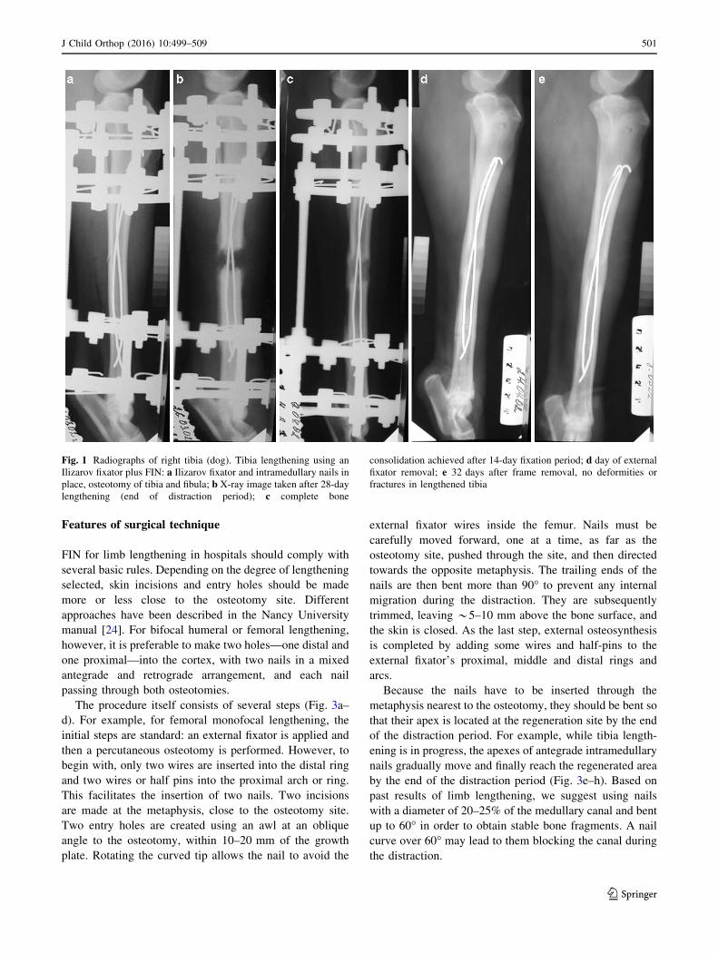
The procedure itself consists of several steps (Fig. 3a–
d). For example, for femoral monofocal lengthening, the
initial steps are standard: an external fixator is applied and
then a percutaneous osteotomy is performed. However, to
begin with, only two wires are inserted into the distal ring
and two wires or half pins into the proximal arch or ring.
This facilitates the insertion of two nails. Two incisions
are made at the metaphysis, close to the osteotomy site.
Two entry holes are created using an awl at an oblique
angle to the osteotomy, within 10–20 mm of the growth
plate. Rotating the curved tip allows the nail to avoid the
external fixator wires inside the femur. Nails must be
carefully moved forward, one at a time, as far as the
osteotomy site, pushed through the site, and then directed
towards the opposite metaphysis. The trailing ends of the
nails are then bent more than 90� to prevent any internal
migration during the distraction. They are subsequently
trimmed, leaving *5–10 mm above the bone surface, and
the skin is closed. As the last step, external osteosynthesis
is completed by adding some wires and half-pins to the
external fixator’s proximal, middle and distal rings and
arcs.
Because the nails have to be inserted through the
metaphysis nearest to the osteotomy, they should be bent so
that their apex is located at the regeneration site by the end
of the distraction period. For example, while tibia length-
ening is in progress, the apexes of antegrade intramedullary
nails gradually move and finally reach the regenerated area
by the end of the distraction period (Fig. 3e–h). Based on
past results of limb lengthening, we suggest using nails
with a diameter of 20–25% of the medullary canal and bent
up to 60� in order to obtain stable bone fragments. A nail
curve over 60� may lead to them blocking the canal during
the distraction.
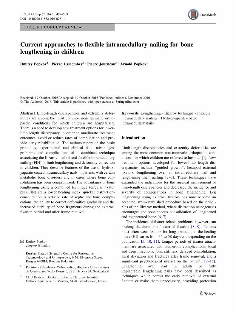
Fig. 1 Radiographs of right tibia (dog). Tibia lengthening using an
Ilizarov fixator plus FIN: a Ilizarov fixator and intramedullary nails in
place, osteotomy of tibia and fibula; b X-ray image taken after 28-day
lengthening (end of distraction period); c complete bone
consolidation achieved after 14-day fixation period; d day of external
fixator removal; e 32 days after frame removal, no deformities or
fractures in lengthened tibia
J Child Orthop (2016) 10:499–509 501
123
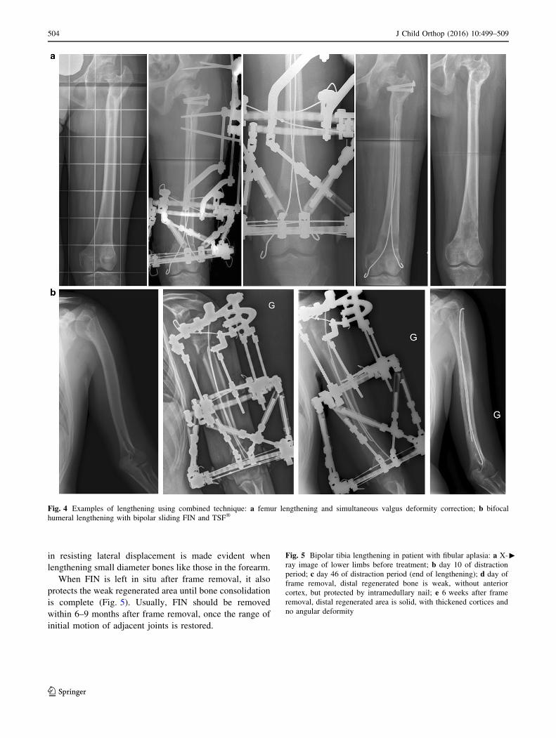
If limb lengthening is associated with a deformity cor-
rection, the surgeon can align the concave curves of both
nails toward the convexity of the deformity. In doing so,
the progressive realignment is effective and the stability of
the fragments is increased. The elasticity of both nails
allows for simultaneous lengthening and a gradual cor-
rection of angular deformity (Fig. 4a).
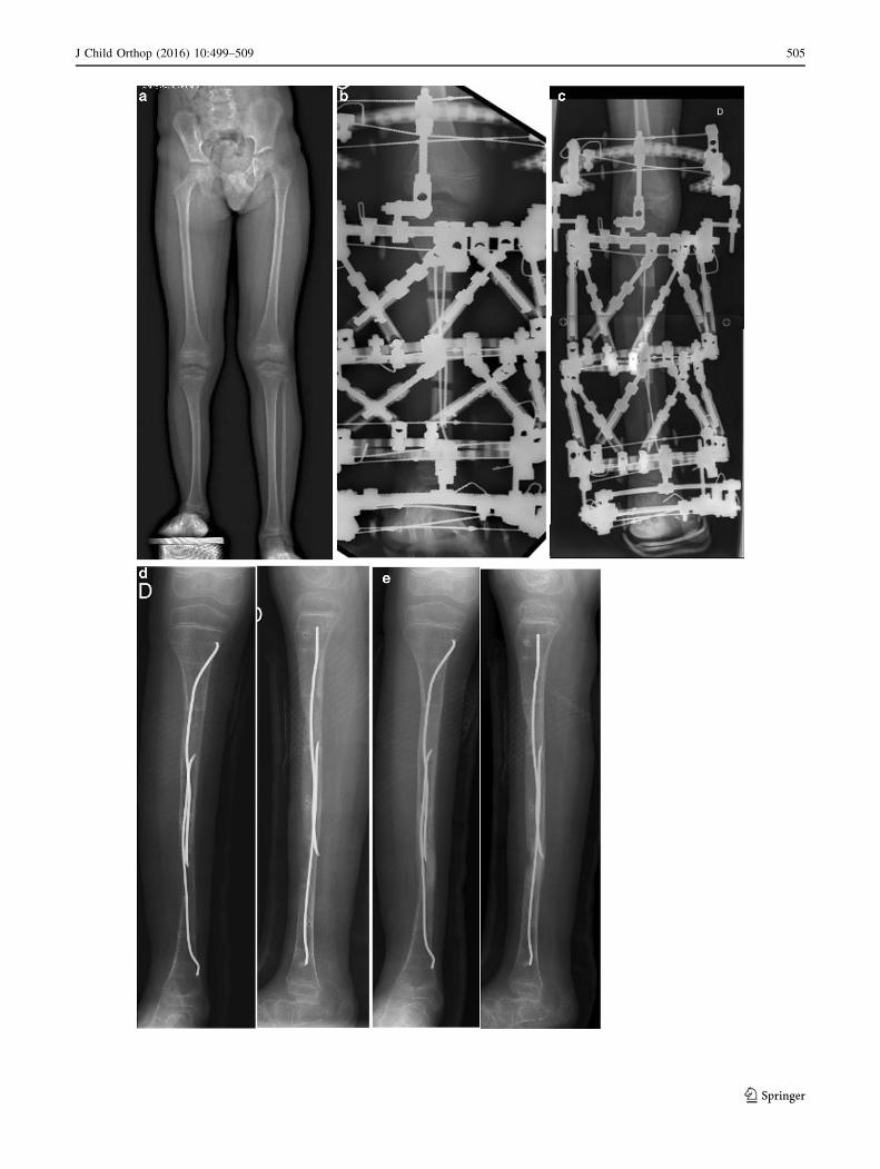
Bipolar, sliding FIN is more appropriate in cases
requiring bifocal lengthening. For instance, in the humerus
in Fig. 4b, an antegrade nail has been inserted through the
Fig. 2 Morphological study:
a medullary artery was still
visible at the end of the
distraction period;
b disappearance of fibrous
‘‘growth zone’’ layer of the
regenerated bone and presence
of a cortical continuity by day
15 of the fixation period,
extensive bone trabeculae
observed along alignment of the
intramedullary nails, and the
diameter of the conglomerate
‘‘nail plus surrounding
ossification’’ structure filled
40% of the medullary canal;
c intramedullary nail
surrounded by newly formed
bone tissue, thickened cortices
(above) in comparison to intact
healthy tibia (below)
502 J Child Orthop (2016) 10:499–509
123
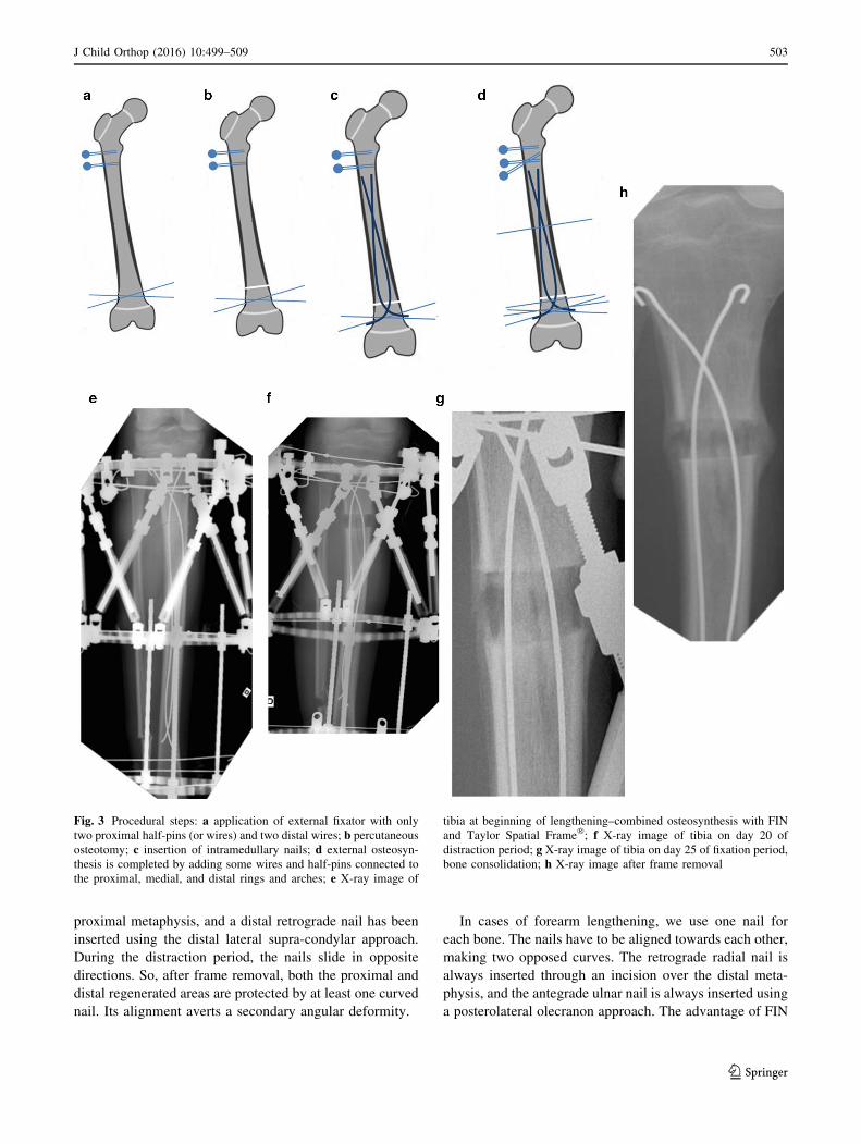
proximal metaphysis, and a distal retrograde nail has been
inserted using the distal lateral supra-condylar approach.
During the distraction period, the nails slide in opposite
directions. So, after frame removal, both the proximal and
distal regenerated areas are protected by at least one curved
nail. Its alignment averts a secondary angular deformity.
In cases of forearm lengthening, we use one nail for
each bone. The nails have to be aligned towards each other,
making two opposed curves. The retrograde radial nail is
always inserted through an incision over the distal meta-
physis, and the antegrade ulnar nail is always inserted using
a posterolateral olecranon approach. The advantage of FIN
Fig. 3 Procedural steps: a application of external fixator with only
two proximal half-pins (or wires) and two distal wires; b percutaneous
osteotomy; c insertion of intramedullary nails; d external osteosyn-
thesis is completed by adding some wires and half-pins connected to
the proximal, medial, and distal rings and arches; e X-ray image of
tibia at beginning of lengthening–combined osteosynthesis with FIN
and Taylor Spatial Frame�; f X-ray image of tibia on day 20 of
distraction period; g X-ray image of tibia on day 25 of fixation period,
bone consolidation; h X-ray image after frame removal
J Child Orthop (2016) 10:499–509 503
123
in resisting lateral displacement is made evident when
lengthening small diameter bones like those in the forearm.
When FIN is left in situ after frame removal, it also
protects the weak regenerated area until bone consolidation
is complete (Fig. 5). Usually, FIN should be removed
within 6–9 months after frame removal, once the range of
initial motion of adjacent joints is restored.
Fig. 4 Examples of lengthening using combined technique: a femur lengthening and simultaneous valgus deformity correction; b bifocal
humeral lengthening with bipolar sliding FIN and TSF�
cFig. 5 Bipolar tibia lengthening in patient with fibular aplasia: a X-
ray image of lower limbs before treatment; b day 10 of distraction
period; c day 46 of distraction period (end of lengthening); d day of
frame removal, distal regenerated bone is weak, without anterior
cortex, but protected by intramedullary nail; e 6 weeks after frame
removal, distal regenerated area is solid, with thickened cortices and
no angular deformity
504 J Child Orthop (2016) 10:499–509
123
J Child Orthop (2016) 10:499–509 505
123
Combined technique using FIN in limb lengthening
for congenital or acquired (growth arrest following
trauma or neonatal osteomyelitis) upper- and lower-
limb length discrepancy [29]
Using a prospective study, we compared the HI of two
groups of children who had undergone upper- and lower-
limb lengthening carried out using the Ilizarov external
fixator alone (194 cases) or a combination of the Ilizarov
fixator and FIN (92 cases). The HI was lower in the
combined technique group. Significant differences were
noted in:
• congenital pathologies: monofocal, monosegmental
lengthening of the femur and forearm, bifocal length-
ening of the tibia, and polysegmental lengthening;
• acquired discrepancies: monofocal tibia, bifocal
femoral, and forearm lengthening.
The HI difference between the two techniques varied
from 2 to 19.1 days/cm. This means that patients in the
combined technique group using FIN required 20–33%
fewer days in the external fixator than those in the con-
ventional Ilizarov technique group. The largest difference
in HI was noted in cases of bifocal acquired femoral dis-
crepancies (59.9%) and monofocal acquired forearm dis-
crepancies (51.3%). The association of the Ilizarov device
and FIN showed a mean HI reduction of 7 days/cm. Fur-
thermore, the Ilizarov plus FIN technique avoided and/or
considerably decreased the number of complications rela-
ted to longer-term external fixation, such as pin-tract
infection, osteomyelitis, and fractures and deformities after
frame removal. However, there were eight cases of skin
irritation where the bent FIN exited the bone; these
required one nail to be removed per bone—two during the
distraction period and six during the consolidation phase.
The removal of those nails had no influence on the final
outcomes.
The combined, simultaneous Ilizarov external fixator
and FIN technique (with two oppositely curving nails) has
been described positively by other authors. They have
shown the advantages of shorter durations of external fix-
ation, good protection of the lengthened bones against
refracture, earlier rehabilitation and its applicability to
children [14, 30–32]. Different published studies have used
monolateral [9, 14, 30] and circular [14, 31, 32] external
fixators. All these authors agreed that the key element of
the combined technique was FIN.
FIN in limb lengthening and deformity correction
in patients with Ollier disease [33]
Dyschondroplasia, or Ollier disease, is a rare, non-heredi-
tary skeletal disorder. This disease is responsible for
various troubles linked to the development of multiple
enchondromas secondary to growth disturbances: limb-
length discrepancy, complex deformities and pathological
fractures. These orthopaedic complications require more
specific management.
A retrospective study [33] assessed the efficiency of FIN
combined with a circular external fixator (such as the Ili-
zarov or Taylor Spatial Frame�) for lower-limb lengthen-
ing and associated deformity correction procedures, against
the results achieved with external fixation alone, i.e.
without FIN. The mean HI was significantly lower in
patients who underwent lengthening and deformity cor-
rection using the combined technique. The HI varied from
19 to 28.2 days/cm for monosegmental lengthening, and
from 10.9 to 12.3 days/cm for polysegmental lengthening.
The average duration of external fixation treatment was
thus reduced by about 8 days for each centimetre of
monosegmental lengthening.
In the group treated using the conventional Ilizarov
technique, three pathological fractures at the site of
enchondromas and three deformities at the lengthening site
were observed after the external fixator’s removal from 37
patients. On the other hand, among the seven patients
treated using the combined technique, no secondary frac-
tures were observed over the 25-month follow-up.
Bioactive FIN in lower-limb deformity correction
in children with X-linked hypophosphatemic rickets
(XHPR) [34]
Surgical procedures to correct multiplanar bone deformi-
ties may be indicated for the prevention of secondary
orthopaedic complications in children with XHPR, how-
ever, different problems related to these procedures have
been reported: increased rates of delayed union, recurrent
deformity, deep intramedullary infection, refracture and
pin-tract infection.
In a retrospective study, we compared the results of
corrections in children with XHPR who had undergone
treatment using either an Ilizarov device alone or a com-
bined technique (Ilizarov fixator plus bioactive hydroxya-
patite-coated FIN). The results of surgery were compared
in short-term (2–6 months) and long-term (over 5 years)
follow-up.
Applying the combined technique demonstrated a con-
siderably lower duration of external fixation (87.4 days)
than the external fixator technique alone (124.7 days), as
well as a decrease in the number of infectious complica-
tions, an absence of secondary fragment displacement
during correction, and no deformity at the osteotomy after
frame removal. In most cases, at long-term follow-up,
neither recurrence nor development of any deformity was
observed. In children treated using the combined
506 J Child Orthop (2016) 10:499–509
123
technique, no recurrent deformity appeared around the FIN
left in situ. Nevertheless, some new deformities appeared,
either in the distal femoral or proximal tibial metaphysis
during residual spontaneous growth. These areas were no
longer protected by the intramedullary nails. The bioactive
layers prevented the migration of the nails at total weight-
bearing and long-term follow-up.
We suggest the use of intramedullary nails coated with
hydroxyapatite or another bioactive layer in patients with
metabolic bone disorders and in cases where bone con-
solidation is compromised [33–35].
Disadvantages of the combined technique for limb
lengthening and deformity correction
[9, 14, 25, 29–36]
Here, we list some opinions and judgments about the
potential inconveniences of using the combined technique
rather than the Ilizarov procedure alone, as well as com-
plications which have been observed in different groups of
our patients:
1. The surgical procedure lasts 10–20 min longer.
2. In patients with congenital or acquired limb-length
discrepancies, nail removal should be adequately
planned. This usually requires a period of outpatient
hospitalisation.
3. Besides the general complications of surgery and
anaesthesia, there are complications specifically
related to the insertion or removal of FIN:
• A prominent nail end, leading to irritation or
perforation of the skin, due to insufficient trimming
or external migration of the nail;
• nerves or tendons rubbing the prominent end of a
nail;
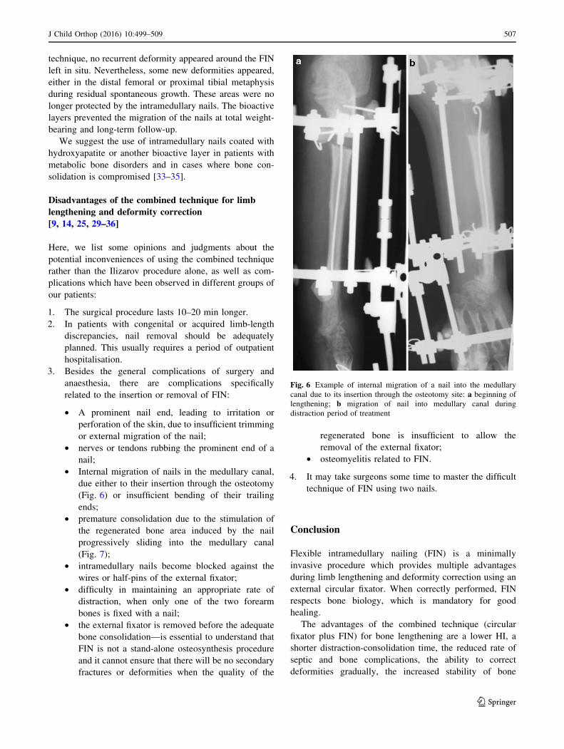
• Internal migration of nails in the medullary canal,
due either to their insertion through the osteotomy
(Fig. 6) or insufficient bending of their trailing
ends;
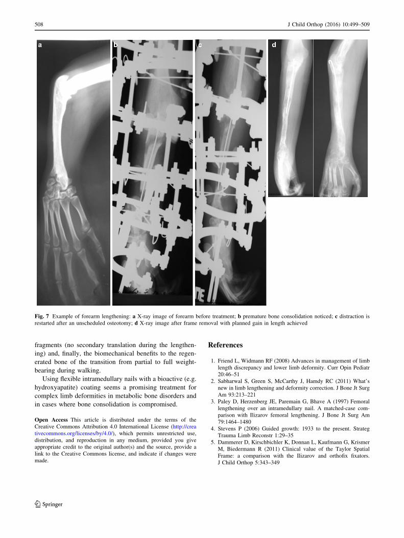
• premature consolidation due to the stimulation of
the regenerated bone area induced by the nail
progressively sliding into the medullary canal
(Fig. 7);
• intramedullary nails become blocked against the
wires or half-pins of the external fixator;
• difficulty in maintaining an appropriate rate of
distraction, when only one of the two forearm
bones is fixed with a nail;
• the external fixator is removed before the adequate
bone consolidation—is essential to understand that
FIN is not a stand-alone osteosynthesis procedure
and it cannot ensure that there will be no secondary
fractures or deformities when the quality of the
regenerated bone is insufficient to allow the
removal of the external fixator;
• osteomyelitis related to FIN.
4. It may take surgeons some time to master the difficult
technique of FIN using two nails.
Conclusion
Flexible intramedullary nailing (FIN) is a minimally
invasive procedure which provides multiple advantages
during limb lengthening and deformity correction using an
external circular fixator. When correctly performed, FIN
respects bone biology, which is mandatory for good
healing.
The advantages of the combined technique (circular
fixator plus FIN) for bone lengthening are a lower HI, a
shorter distraction-consolidation time, the reduced rate of
septic and bone complications, the ability to correct
deformities gradually, the increased stability of bone
Fig. 6 Example of internal migration of a nail into the medullary
canal due to its insertion through the osteotomy site: a beginning of
lengthening; b migration of nail into medullary canal during
distraction period of treatment
J Child Orthop (2016) 10:499–509 507
123
fragments (no secondary translation during the lengthen-
ing) and, finally, the biomechanical benefits to the regen-
erated bone of the transition from partial to full weight-
bearing during walking.
Using flexible intramedullary nails with a bioactive (e.g.
hydroxyapatite) coating seems a promising treatment for
complex limb deformities in metabolic bone disorders and
in cases where bone consolidation is compromised.
Open Access This article is distributed under the terms of the
Creative Commons Attribution 4.0 International License (http://crea
tivecommons.org/licenses/by/4.0/), which permits unrestricted use,
distribution, and reproduction in any medium, provided you give
appropriate credit to the original author(s) and the source, provide a
link to the Creative Commons license, and indicate if changes were
made.
References
1. Friend L, Widmann RF (2008) Advances in management of limb
length discrepancy and lower limb deformity. Curr Opin Pediatr
20:46–51
2. Sabharwal S, Green S, McCarthy J, Hamdy RC (2011) What’s
new in limb lengthening and deformity correction. J Bone Jt Surg
Am 93:213–221
3. Paley D, Herzenberg JE, Paremain G, Bhave A (1997) Femoral
lengthening over an intramedullary nail. A matched-case com-
parison with Ilizarov femoral lengthening. J Bone Jt Surg Am
79:1464–1480
4. Stevens P (2006) Guided growth: 1933 to the present. Strateg
Trauma Limb Reconstr 1:29–35
5. Dammerer D, Kirschbichler K, Donnan L, Kaufmann G, Krismer
M, Biedermann R (2011) Clinical value of the Taylor Spatial
Frame: a comparison with the Ilizarov and orthofix fixators.
J Child Orthop 5:343–349
Fig. 7 Example of forearm lengthening: a X-ray image of forearm before treatment; b premature bone consolidation noticed; c distraction is
restarted after an unscheduled osteotomy; d X-ray image after frame removal with planned gain in length achieved
508 J Child Orthop (2016) 10:499–509
123
6. Ilizarov GA (1990) Clinical application of the tension-stress
effect for limb lengthening. Clin Orthop 250:8–26
7. Gubin AV, Borzunov DY, Malkova TA (2013) The Ilizarov
paradigm: thirty years with the Ilizarov method, current concerns
and future research. Int Orthop 37:1533–1539
8. Paley D (1990) Problems, obstacles, and complications of limb
lengthening by the Ilizarov technique. Clin Orthop Relat Res
250:81–104
9. Saraph V, Roposch A, Zwick EB, Linhart WE (2004) Tibial
lengthening over nails in children using modified Ender nails:
preliminary results of a new treatment. J Pediatr Orthop B
13:383–388
10. Matsubara H, Tsuchiya H, Sakurakichi K, Watanabe K, Tomita K
(2006) Deformity correction and lengthening of lower legs with
an external fixator. Int Orthop 30:550–554
11. Launay F, Jouve JL, Guillaume JM, Viehweger E, Jacquemier M,
Bollini G (2001) Progressive forearm lengthening in children: 14
cases. Rev Chir Orthop Rep Appar Mot 87:786–795
12. Popkov AV (1991) Errors and complications of operative
lengtheningvof the lower extremities in adults by the Ilizarov
method. VestnvKhir I I Grek 1:113–116
13. Antoci V, Ono CM, Antoci V Jr, Raney EM (2006) Bone
lengthening in children: how to predict the complications rate and
complexity? J Pediatr Orthop 26:634–640
14. Launay F, Younsi R, Pithioux M, Chabrand P, Bollini G, Jouve
JL (2013) Fracture following lower limb lengthening in children:
a series of 58 patients. Orthop Traumatol Surg Res 99:72–79
15. Moraal JM, Elzinga-Plomp A, Jongmans MJ, Roermund PM,
Flikweert PE, Castelein RM, Sinnema G (2009) Long-term psy-
chosocial functioning after Ilizarov limb lengthening during
childhood. Acta Orthop 80:704–710
16. Black SR, Kwon MS, Cherkashin AM, Samchukov ML, Birch
JG, Jo CH (2015) Lengthening in congenital femoral deficiency:
a comparison of circular external fixation and a motorized
intramedullary nail. J Bone Jt Surg Am 97:1432–1440
17. Krieg AH, Speth BM, Foster BK (2008) Leg lengthening with a
motorized nail in adolescents: an alternative to external fixators?
Clin Orthop Relat Res 466:189–197
18. Song HR, Oh CW, Mattoo R, Park BC, Kim SJ, Park IH, Jeon IH,
Ihn JC (2005) Femoral lengthening over an intramedullary nail
using the external fixator: risk of infection and knee problems in
22 patients with a follow-up of 2 years or more. Acta Orthop
76:245–252
19. Gordon JE, Goldfarb CA, Luhmann SJ, Lyons D, Schoenecker
PL (2002) Femoral lengthening over a humeral intramedullary
nail in preadolescent children. J Bone Jt Surg Am 84:930–937
20. Kristiansen L, Steen H (1999) Lengthening of the tibia over an
intramedullary nail using the Ilizarov external fixator. Major
complications and slow consolidation in 9 lengthenings. Acta
Orthop Scand 70:271–274
21. Herzenberg JE, Paley D (1997) Tibial lengthening over nails.
Tech Orthop 12:250–259
22. Schiedel F, Elsner U, Gosheger G, Vogt B, Rodl R (2013) Pro-
phylactic titanium elastic nailing (TEN) following femoral
lengthening (Lengthening then rodding) with one or two nails
reduces the risk for secondary interventions after regenerate
fractures: a cohort study in monolateral vs bilateral lengthening
procedures. BMC Musculoskelet Disord 14:302
23. Grill F, Dungl P, Steinwender G, Hosny G (1999) Congenital
short femur. J Pediatr Orthop Part B 2:35–41
24. Lascombes P (2010) Flexible intramedullary nailing in children.
Springer, Berlin
25. Shevtsov VI, Popkov AV, Popkov DA, Yerofeev SA, Prevot J,
Lascombes P (2004) Embrochage centro-medullaire dans les
allongements osseux selon Ilizarov. Rev Chir Orthop 90:399–410
26. Popkov DA, Popkov AV, Kononovich NA, Barbier D, Ceroni D,
Journeau P, Lascombes P (2014) Experimental study of pro-
gressive tibial lengthening in dogs using the Ilizarov technique.
Comparison with and without associated intramedullary K-wires.
Orthop Traumatol Surg Res 100:809–814
27. Lascombes P, Huber H, Fay R, Popkov D, Haumont T, Journeau
P (2013) Flexible intramedullary nailing in children: nail to
medullary canal diameters optimal ratio. J Pediatr Orthop
33:403–408
28. Burlakov EV, Alatov DV, Popkov DA, Shutov RB (2008) Cal-
culation of the main parameters of spokes for intramedullary
reinforcement of tubular bones. Med Tekh 3:26–28
29. Popkov D, Popkov A, Haumont T, Journeau P, Lascombes P
(2010) Flexible intramedullary nail use in limb lengthening.
J Pediatr Orthop 30:910–918
30. Lampasi M, Launay F, Jouve JL, Bollini G (2009) Femoral
lengthening over elastic stable intramedullary nailing in children
using the monolateral external fixator. Chir Org Mov 93:57–64
31. Bukva B, Brdar R, Nikolic D, Petronic I, Ducic S, Abramovic D
(2013) Combined external fixation and intramedullary alignment
in correction of limb length discrepancies. Acta Orthop Belg
79:411–416
32. Bukva B, Vrgoc G, Rakovac I, Ducic S, Sindik J, Coklo M,
Marinovic M, Bakota B (2015) Complications in leg lengthening
using an Ilizarov external fixator and intramedullary alignment in
children: comparative study during a fourteen-year period. Injury
46(Suppl 6):S48–S51
33. Popkov D, Journeau P, Popkov A, Haumont T, Lascombes P
(2010) Ollier’s disease limb lenghtening: should intramedullary
nailing be combined with circular external fixation? Orthop
Traumatol Surg Res 96:348–353
34. Popkov A, Aranovich A, Popkov D (2015) Results of deformity
correction in children with X-linked hereditary hypophos-
phatemic rickets by external fixation or combined technique. Int
Orthop 39:2423–2431
35. Aranovich A, Popkov A, Barbier D, Popkov D (2014) Femoral
lengthening by combined technique in melorheostosis: a case
report. Eur Orthop Traumatol 5:175–179. doi:10.1007/s12570-
013-0220-4
36. Popkov D, Popkov A (2016) Progressive lengthening of short
congenital forearm stump in children for prosthetic fitting. Int
Orthop 40:547–554
J Child Orthop (2016) 10:499–509 509
123