cucurbitacins: elucidation of their interactions with the ... · [email protected], wink@uni...
TRANSCRIPT

Submitted 7 December 2016Accepted 26 April 2017Published 30 May 2017
Corresponding authorsXiaojuan Wang,[email protected] Wink,[email protected],[email protected]
Academic editorMaría Ángeles Esteban
Additional Information andDeclarations can be found onpage 14
DOI 10.7717/peerj.3357
Copyright2017 Wang et al.
Distributed underCreative Commons CC-BY 4.0
OPEN ACCESS
Cucurbitacins: elucidation of theirinteractions with the cytoskeletonXiaojuan Wang, Mine Tanaka, Herbenya Silva Peixoto and Michael WinkInstitute of Pharmacy and Molecular Biotechnology, Ruprecht-Karls-Universität Heidelberg, Heidelberg,Germany
ABSTRACTCucurbitacins, a class of toxic tetracyclic triterpenoids in Cucurbitaceae, modulatemany molecular targets. Here we investigated the interactions of cucurbitacin B, Eand I with cytoskeletal proteins such as microtubule and actin filaments. The effectsof cucurbitacin B, E and I on microtubules and actin filaments were studied inliving cells (Hela and U2OS) and in vitro using GFP markers, immunofluorescencestaining and in vitro tubulin polymerization assay. Cucurbitacin B, E and I apparentlyaffected microtubule structures in living cells and cucurbitacin E inhibited tubulinpolymerization in vitro with IC50 value of 566.91 ± 113.5 µM. Cucurbitacin E did notaffect the nucleation but inhibited the growth phase and steady state duringmicrotubuleassembly in vitro. In addition, cucurbitacin B, E and I all altered mitotic spindles andinduced the cell cycle arrest at G2/M phase. Moreover, they all showed potent effectson actin cytoskeleton by affecting actin filaments through the depolymerization andaggregation. The interactions of cucubitacin B, E and I with microtubules and actinfilaments present new insights into their modes of action.
Subjects Cell Biology, PharmacologyKeywords Cucurbitacins, Cucurbitaceae, Cytotoxicity, Actin filament, Microtubule
INTRODUCTIONCucurbitacins are a class of cucurbitane-type tetracyclic triterpenoids that are mainly pro-duced by plants of the family of Cucurbitaceae (Duncan et al., 1996; Kaushik, Aeri & Mir,2015;Wink & Van Wyk, 2008). Hundreds of cucurbitacins that occur in a diversity of plantsshare the same tetracyclic scaffold and can be divided into 12 main categories according totheir substituents (Chen et al., 2012; Lee, Iwanski & Thoennissen, 2010). Cucurbitacins Band E (Fig. 1) have been identified to be the primary cucurbitacin types by plant secondarymetabolism studies (Abbas et al., 2013; Gry, Søborg & Andersson, 2006; Kaushik, Aeri &Mir, 2015). Under certain environmental conditions, other cucurbitacin types could begenerated by enzymatic reactions. For instance, cucurbitacins A, C, D can be produced fromcucurbitacin B, while cucurbitacins I, J, K can be generated from cucurbitacin E (Ahmed& Halaweish, 2014; Chen et al., 2012).
Cucurbitacins exhibit a broad range of pharmacological properties such as anti-inflammatory, antioxidant, antiviral, antipyretic, analgesic and anti-malaria activities (Chenet al., 2005; Jayaprakasam, Seeram & Nair, 2003;Miro, 1995). Current studies have revealedseveral molecular targets of cucurbitacins such as JAK2/STAT3 pathway, cofilin, cyclins,cdc2, COX-2, TYR and EcR, among which actin cytoskeleton appears to be an early target
How to cite this article Wang et al. (2017), Cucurbitacins: elucidation of their interactions with the cytoskeleton. PeerJ 5:e3357; DOI10.7717/peerj.3357

Figure 1 The structure of cucurbitacins. (A) The skeleton of cucurbitacin A, B and D. (B) The skeletonof cucurbitacin E, I, J and K.
(Blaskovich et al., 2003; Chen et al., 2012). Additionally, cucurbitacin B has been reportedto disrupt microtubule polymerization in several studies (Yin et al., 2008; Duangmano etal., 2012). However, only few studies have explored the effects of cucurbitacins on themicrotubule-based cytoskeleton and the underlying mechanisms of action of cucurbitacinsremains elusive.
Actin filaments and microtubules, the two major networks of the eukaryotic cellcytoskeleton, become attractive targets for natural compounds in cancer research due totheir importance in a board range of processes such as vesicular and organelle transport, cellproliferation and migration (Jordan & Wilson, 2004; Petrasek & Schwarzerova, 2009; Wick-stead & Gull, 2011). In this study, we investigated the effects of cucurbitacin B, E and I onmicrotubules and actin filaments in living cells (cancer cell linesHela,MCF7, andU2OS) us-ingGFPmarkers and immunofluorescence staining. Their interactionswith tubulin dynam-ics were further determined in vitro using tubulin polymerization assay. Reference drugs
Wang et al. (2017), PeerJ, DOI 10.7717/peerj.3357 2/18

such as colchicine and vinblastine (microtubule-binding agents) and latrunculin B (actin-binding agent) were used as comparing controls. We can provide evidence for unidentifiedinteractions between cucurbitacins and the cytoskeleton in this study.
MATERIALS AND METHODSCell lines, chemicals and laboratory materialsThe human cervical cancer cell line Hela was purchased from ATCC (Wesel, Germany) andthe MCF-7 human breast cancer cell line was provided by Prof. Dr. Stefan Wölfl (Instituteof Pharmacy and Molecular Biotechnology, Heidelberg University, Heidelberg, Germany);U2OS human osteosarcoma cancer cells which were stably transfected with α-tubulin-GFPconstruct were supplied by Prof. Dr. Thomas Efferth (Institute of Pharmacy and Bio-chemistry, Johannes Gutenberg University, Mainz, Germany); cucurbitacin E and I (purity> 99% by HPLC) came from Phytoplan GmbH (Heidelberg, Germany) and cucurbitacinB (purity > 98% by HPLC) from Baoji Herbest Bio-Tech Co., Ltd. (Baoji, Shannxi, China);vinblastine (1 mg/mL) were purchased from Central Pharmacy of the University HospitalHeidelberg (Heidelberg, Germany); colchicine (purity > 95% by HPLC), latrunculin B(purity > 80% byHPLC), G418, Atto 390 phalloidin, paraformaldehyde, propidium iodide,ATP, BSA, Dimethyl sulfoxide (DMSO), EDTA, EGTA, FBS, GTP, MTT, piperazine-N, N′-bis(2-ethanesulfonic acid) (PIPES), RNaseA andCoomasie bluewere obtained fromSigma-Aldrich Chemie GmbH (Steinheim, Germany) and mowiol 4–88 from Carl Roth GmbH &Co.KG (Karlsruhe, Germany); DMEM, non-essential amino acids, penicillin-streptomycin,CellLight R© Actin-RFP BacMam 2.0 actin-RFP, trypsin-EDTA came from Life technologies(Paisley, United Kingdom) and triton X-100 from Merck KgaA (Darmstadt, Germany);mouse anti-α-tubulin monoclonal IgG and goat anti-mouse IgM-FITC were obtainedfrom Santa Cruz Biotechnology (Heidelberg, Germany); 96-well-plates, 24-well-plates and6-well-plates were purchased from Greiner (Frickenhausen, Germany) and circular glasscoverslips from Thermo Scientific (Braunschweig, Germany).
Cell cultureHela, MCF-7 and U2OS cancer cells were cultivated as previously described (Wang et al.,2016b).
MTT assayThe anti-proliferative effects of cucurbitacins were assessed using MTT assay, as previouslydescribed (Nurcahyanti & Wink, 2015). In brief, cells (1×10⁴) were seeded in 96-well platesand incubated with different concentrations of cucurbitacins for 48 h (Hela, U2OS) and72 h (MCF-7). MTT solution was then added and incubated for 2 h. The plates were readat 570 nm after the addition of DMSO using Tecan infinite M200 Pro (Tecan, Crailsheim,Germany).
Imaging of tubulin-GFP transfected U2OS cellsα-Tubulin-GFP U2OS cells (1×10⁵) were seeded in 24-well-plates and treated with 200 µldifferent concentrations (IC80, IC50 based onMTTdata) of cucurbitacins. Cells were imaged
Wang et al. (2017), PeerJ, DOI 10.7717/peerj.3357 3/18

using a Keyence BZ-9000 microscope (Keyence; Neu-Isenburg, Germany) after incubationfor 2 h, 4 h, 24 h and 48 h. The images were analyzed using BZ-II Analyzer software (version2.1, Keyence; Neu-Isenburg, Germany).
Immunofluorescence stainingThe immunofluorescence staining was carried out as established in our laboratory (Wanget al., 2016a).
Imaging of actin-RFP transfected hela cells2×104 Hela cells were seeded in 24-well-plates and mixed with CellLight R© Actin-RFP Bac-Mam2.0 which is a fusion construct of human actin and TagRFP, providing an accurate andspecific targeting to cellular actin filaments. After 16 h of incubation, 200 µl differentconcentrations of cucurbitacins (IC80, IC50 based on MTT data) were added and the cellswere analyzed as described above (‘Imaging of tubulin-GFP transfected U2OS cells’).
In vitro tubulin polymerization assayPorcine brain tubulin plusMAPs was prepared by two cycles of polymerization and depoly-merization according to a standard protocol (Gell et al., 2011). In-vitro tubulin polymeriza-tion assays were carried out in PEM buffer (100 mM PIPES, 2 mM EGTA, 0.1 mM EDTA,3 mM MgCl2, 1 mM ATP and 1 mM GTP, pH 6.85) by mixing 5.6 mg/ml tubulin-MAPswith different concentrations of cucurbitacins in 96-well plates at 37 ◦C for 40 min. Therate and extent of the polymerization reaction were monitored by light scattering at 360nm using Tecan infinite M200 Pro.
Cell cycle analysisCell cycle analysis was carried out as established in our laboratory (Su, Cheng & Wink,2015). Briefly, Hela cells (5×10⁵) were seeded in 6-well-plates and treated with differentconcentrations of cucurbitacins for 24 h. Cells were then collected, centrifuged and fixed in70% ice-cold ethanol for at least 8 h. After washing steps, cells were treated with 0.2 mg/mlRNase A for 30 min at 37 C and then stained with 0.1 mg/ml propidium iodide. Sampleswere analyzed using a FACScan flow cytometer (Becton Dickinson, Heidelberg, Germany).Data were analyzed using Cell QuestTM Pro software (Becton Dickinson) and Microsoftexcel (Microsoft Corporation, Washington, USA).
Statistical analysisThe data of reference drugs colchicine, vinblastine and latrunculin B have been publishedbefore by us Wang et al. (2016a). The IC50 and IC80 were determined as the amount ofthe substances needed to reduce 50% or 80% cell viability/tubulin polymerization andcalculated from concentration–response curves by Sigmaplot software (Systat SoftwareInc., San Jose, USA). All experiments were done in triplicate, repeated three times. Data arepresented as mean± standard deviation (SD). Statistical comparison between controls anddifferent treatments were performed by an unpaired student’s t test using Microsoft excel2013 (Microsoft Corporation, Washington, USA). Significance was considered at p< 0.05.
Wang et al. (2017), PeerJ, DOI 10.7717/peerj.3357 4/18

Table 1 Cytotoxic activities of cucurbitacins and reference drugs against Hela, MCF-7 and U2OS cells.
Compounds IC80 IC50
Hela MCF-7 U2OS Hela MCF-7 U2OS
Colchicinea 27.01± 7.48 nM 79.89± 40.85 nM 0.87 ± 1.46 µM 14.9± 3.94 nM 30.29± 8.02 nM 25.2± 19.58 nMVinblastinea 0.7 ± 0.34 µM 2.08 ± 0.92 µM 1.15 ± 0.51 µM 0.02± 0.01 nM 0.06± 0.05 nM 0.11± 0.07 nMLatrunculin Bb 63.94 ± 5.68 µM 140.1± 6.58 µM 37.17 ± 15.68 µM 11.19± 1.27 µM 38.5 ± 1.7 µM 5.67 ± 0.59 µMCucurbitacin B 22± 1.39 nM 43.71± 10.61 nM 28.05± 15.25 nM 12.2± 1.42 nM 22.93± 4.51 nM 17.07± 4.55 nMCucurbitacin E 15.09± 2.67 nM 0.92 ± 0.2 µM 26.27± 18.50 nM 6.43± 1.05 nM 54± 3.16 nM 15.07± 4.51 nMCucurbitacin I 55.49± 3.63 nM 0.29 ± 0.08 µM 34.03± 17.74 nM 44.77± 1.54 nM 64.67± 14.29 nM 23.47± 16.92 nM
Notes.aActive on tubulin/microtubules.bActive against actin filaments; data are presented as mean± SD.
RESULTSCytotoxicity of cucurbitacinsThe anti-proliferative activities of cucurbitacins against Hela, MCF-7 and U2OS cells wereassessed by MTT assay (Table 1). Among the reference drugs, the known actin-bindingagent latrunculin B inhibited the growth of three cell lines with IC50 values between 5.67µM and 38.5 µM. Compared to latrunculin B, cucurbitacin B, E and I exhibited strongercytotoxicity against all three cell lines with IC50 values between 6.43 nM and 64.67 nM.The known microtubule-binding agent colchicine and vinblastine also showed strongeranti-proliferative activity than latrunculin B with IC50 values between 0.02 nM and 30.29nM. Compared to colchicine and vinblastine, cucurbitacin B, E and I exhibited lower IC80
values (15.09 nM–0.92 µM) but greater IC50 values (6.43 nM–64.67 nM). Among thesecucurbitacins, cucurbitacin B and E caused a higher toxicity than cucurbitacin I, which isclose to the microtubule-binding agent colchicine.
Cucurbitacins interfered with microtubule structures in living cellsInfluence on microtubulesThe U2OS cells which express α-tubulin-GFP were treated with cucurbitacins to determinetheir effects on the cellular microtubule network by observing the changes in living cells(Fig. 2). In non-treated cells, microtubules extended continuously through the cytoplasmand formed an extensive intracellular network. Treatment with colchicine at both concen-trations (IC80, IC50) decreased the microtubule mass, which exhibited a reduced intensityat the cell periphery compared to non-treated cells. Vinblastine depolymerized micro-tubules in a way different from colchicine that tubulin paracrystals were formed and dis-persed through out the cytoplasm at the concentration of IC80. While at the concentrationof IC50, tubulin paracrystals disappeared and extensively depolymerized microtubules wereobserved. Latrunculin B immediately changed the cell morphology from stretching stateinto round state at both concentrations (IC80, IC50), which was returned to normalmorphology after 24 h incubation with the microtubule mass slightly decreased. The effectof cucurbitacins on microtubule network was concentration-dependent and different fromother reference drugs. Cucurbitacin B and E firstly changed themorphology of microtubulenetwork into half-stretching state after 2 h incubation then round state after 24 h incubation,
Wang et al. (2017), PeerJ, DOI 10.7717/peerj.3357 5/18

Figure 2 Cucurbitacins changed the morphology of microtubule network in U2OS cells. Panels showmicrographs of U2OS cells treated for 2 h, 4 h, 24 h and 48 h with all six compounds at concentrationsof IC80 (A) and IC50 (B). Known tubulin inhibitors colchicine and vinblastine induced microtubule de-polymerization and tubulin paracrystals, respectively. Actin-binding agent latrunculin B caused the rapidchange of cell morphology. Bar= 10 µm.
which exhibited their significant interference on microtubule network. Cucurbitacin I alsoinduced the similar but weaker effect on microtubule network after 4 h incubation.
Influence on spindle apparatusThe effects of cucurbitacins onmitotic microtubules were further evaluated by immunoflu-orescence staining in Hela cells (Fig. 3). The effects of colchicine, vinblastine, latrunculin Band cucurbitacins on Hela interphase microtubule network (Figs. 3A and 3B) were compa-rable with the findings in U2OS cells. In non-treated Hela metaphase cells (Fig. 3C), micro-tubules formed symmetric bipolar spindles with chromosomes aligning at the metaphaseplate. The completely depolymerized spindle with the compacted chromosomes wereobserved in colchicine-treated cells, while depolymerized bipolar spindles were found invinblastine-treated cells. Latrunculin B did not alter mitotic spindles and chromosomearrangements in Hela cells. Cucurbitacin B and E caused disordered distribution of spindlearray, while cucurbitacin I led to multipolar spindles and chromosomes mis-segregationon the metaphase plate.
Wang et al. (2017), PeerJ, DOI 10.7717/peerj.3357 6/18
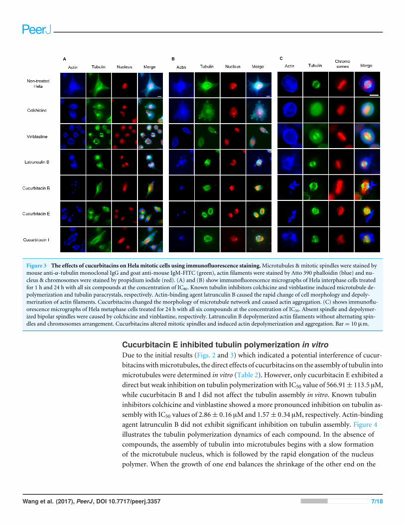
Figure 3 The effects of cucurbitacins on Hela mitotic cells using immunofluorescence staining.Microtubules & mitotic spindles were stained bymouse anti-α-tubulin monoclonal IgG and goat anti-mouse IgM-FITC (green), actin filaments were stained by Atto 390 phalloidin (blue) and nu-cleus & chromosomes were stained by propidium iodide (red). (A) and (B) show immunofluorescence micrographs of Hela interphase cells treatedfor 1 h and 24 h with all six compounds at the concentration of IC80. Known tubulin inhibitors colchicine and vinblastine induced microtubule de-polymerization and tubulin paracrystals, respectively. Actin-binding agent latrunculin B caused the rapid change of cell morphology and depoly-merization of actin filaments. Cucurbitacins changed the morphology of microtubule network and caused actin aggregation. (C) shows immunoflu-orescence micrographs of Hela metaphase cells treated for 24 h with all six compounds at the concentration of IC50. Absent spindle and depolymer-ized bipolar spindles were caused by colchicine and vinblastine, respectively. Latrunculin B depolymerized actin filaments without alternating spin-dles and chromosomes arrangement. Cucurbitacins altered mitotic spindles and induced actin depolymerization and aggregation. Bar= 10 µm.
Cucurbitacin E inhibited tubulin polymerization in vitroDue to the initial results (Figs. 2 and 3) which indicated a potential interference of cucur-bitacinswithmicrotubules, the direct effects of cucurbitacins on the assembly of tubulin intomicrotubules were determined in vitro (Table 2). However, only cucurbitacin E exhibited adirect but weak inhibition on tubulin polymerization with IC50 value of 566.91± 113.5 µM,while cucurbitacin B and I did not affect the tubulin assembly in vitro. Known tubulininhibitors colchicine and vinblastine showed a more pronounced inhibition on tubulin as-sembly with IC50 values of 2.86± 0.16 µMand 1.57± 0.34 µM, respectively. Actin-bindingagent latrunculin B did not exhibit significant inhibition on tubulin assembly. Figure 4illustrates the tubulin polymerization dynamics of each compound. In the absence ofcompounds, the assembly of tubulin into microtubules begins with a slow formationof the microtubule nucleus, which is followed by the rapid elongation of the nucleuspolymer. When the growth of one end balances the shrinkage of the other end on the
Wang et al. (2017), PeerJ, DOI 10.7717/peerj.3357 7/18

Table 2 Inhibition of tubulin polymerization in vitro.
Compounds IC50
Colchicinea 2.86 ± 0.16 µMVinblastinea 1.57 ± 0.34 µMLatrunculin Bb >1 mMCucurbitacin B >1 mMCucurbitacin E 566.91 ± 113.5 µMCucurbitacin I >1 mM
Notes.aActive on tubulin/microtubules.bActive against actin filaments; data are presented as mean± SD.
polymer, the polymerization dynamic reaches steady state (Grintsevich & Reisler, 2013;Margolis & Wilson, 1998). The effects of colchicine and vinblastine on tubulin assemblywere similar: As the concentration increased, the time needed for nucleation was longerand the growth phase of microtubule polymer was shorter, which led the system to theequilibrium phase sooner. However, the mode of action of cucurbitacin E was differentthat it did not affect the nucleation but inhibited the growth phase and steady state.Figure 4D showed that 1 mM cucurbitacin B and I did not affect the assembly, while1 mM latrunculin B weakly inhibited the polymerization around 30%.
Cucurbitacins exhibited dramatic effects on actin filamentsInfluence on mitotic actin filamentsThe effects of cucurbitacins on actin filamentswere firstly evaluated by immunofluorescencestaining in Hela mitotic cells (Fig. 3). The actin-binding agent Latrunculin B significantlyaltered the cell shape after 1 h incubation, which partially recovered after 24 h withthe actin cytoskeleton extensively disrupted. Latrunculin B also affected metaphase cellsby depolymerizing the actin filaments without alternating spindles and chromosomesarrangement. No apparent changes on actin filaments were found in colchicine-treatedcells. In vinblastine-treated cells, a slight reduction of actin filament mass was observedafter 24 h incubation at the concentration of IC80 (Fig. 3B). Cucurbitacins exhibitedremarkable effects on actin filaments both in Hela interphase and metaphase cells: after1 h treatment, the actin network started to depolymerize and the cell shape was slightlychanged (Fig. 3A): After 24 h incubation, the cell morphology was dramatically deformedand the aggregation of actin filaments into one piece was observed (Fig. 3B); in metaphasecells, actin depolymerization and aggregation were greatly accentuated (Fig. 3C).
Influence on cellular actin filamentsHela cellular actin filaments were further visualized by actin-RFP and treated withcucurbitacins to evaluate their effects on cellular actin filaments in living cells (Fig. 5). Theresults were in agreement with the findings shown in Fig. 3. No notable changes on actinfilaments were observed in colchicine/vinblastine-treated cells. The actin-binding agent La-trunculin B immediately altered the cell shape after 2 h incubation, which partially recoveredafter 24 hwith the actin cytoskeleton significantly disrupted. Cucurbitacins acted differentlyfrom latruculin B: At the concentration of IC80, the actin network was extensively disrupted
Wang et al. (2017), PeerJ, DOI 10.7717/peerj.3357 8/18

Figure 4 Cucurbitacin E inhibited tubulin polymerization in vitro. Polymerization of tubulin withMAPs in the assembly buffer was measured in the absence (�) and in the presence of different concen-trations of compounds. (A), (B) Colchicine and vinblastine inhibited the nucleation and growth phaseduring the assembly. (C) Cucurbitacin E did not affect the nucleation but inhibited the growth phase andsteady state. (D) Cucurbitacin B and I did not affect tubulin polymerization and latrunculin B showedweak inhibition on the dynamic.
within 24 h incubation and granulated aggregations of condensed actin were dispersedthrough out the cytoplasm; After 24 h incubation, the cell morphology started to change,while actin aggregation accentuated and tented to gather into one instead of distributingthrough out the whole cell (Fig. 5A). These effects were weakened as cucurbitacinsconcentration decreased (Fig. 5B), but slight aggregation of actin still can be observed atthe concentration of IC20 (Fig. 5C). These results suggest that cucurbitacins remarkablyrearrange actin cytoskeleton and their mechanism of action is different from that oflatrunculin B.
Wang et al. (2017), PeerJ, DOI 10.7717/peerj.3357 9/18

Figure 5 Cucurbitacins changed the cell morphology and reduced the mass of actin filaments after 24 h treatment. Panels show micrographs ofHela cells which were transduced with actin-RFP treated for 2 h, 4 h, 24 h and 48 h with all six compounds at concentrations of IC80 (A), IC50 (B)and IC20 (C). Actin-binding agent latruculin B induced the change of cell morphology and extensive depolymerization of actin network. Colchicinecaused few changes on actin network and vinblastine slightly reduced actin filament mass after 4 h incubation at high concentration of IC80. Bar=10 µm.
Cucurbitacins arrested cell cycle at G2/M phaseFigure 6 represents the effects of cucurbitacins on cell cycle. Colchicine, vinblastine, latrun-culin B and cucurbitacins all induced a dose-dependent G2/M cell cycle arrest. Colchicine,vinblastine and latrunculin B exhibited stronger effects on cell cycle than cucurbitacins.Colchicine and vinblastine promoted the G2/M population to 89.66 ± 2.04% (p< 0.001)and 78.04 ± 14.78% (p< 0.01) at the concentration of 0.1 µM and 10 nM, respectively.While cucurbitacin B, E and I promoted the G2/M population to 66.41 ± 3.73% (p< 0.01),59.35 ± 5.69% (p< 0.001) and 59.18 ± 7.2% (p< 0.01) at the concentration of 1.6 µM,0.3 µMand 0.6 µM, respectively. These results indicate the potential ability of cucurbitacinsto act as antimitotic agents.
DISCUSSIONIn this study, we provide evidence that cucurbitacin B, E and I interactedwith actin filamentsthrough the induction of aggregation and depolymerization in Hela and U2OS cells, whichallows a more comprehensive understanding of the changes of actin filaments in cancer
Wang et al. (2017), PeerJ, DOI 10.7717/peerj.3357 10/18

Figure 6 Cell cycle analysis in Hela cells. Cells were harvested after 24 h of drug treatment and subse-quently assayed for their DNA content by flow cytometry. Colchicine, vinblastine, latrunculin B and cu-curbitacins all blocked cell cycle at G2/M phase (A–F). Data are represented as mean± SD from three in-dependent experiments. ∗p< 0.05, ∗∗p< 0.01, ∗∗∗p< 0.001.
Wang et al. (2017), PeerJ, DOI 10.7717/peerj.3357 11/18

cells responding to cucurbitacins. In addition, they also interfered with microtubulestructure and altered mitotic spindles in living cells though their effects on tubulinpolymerization are weak.
The cytotoxicity of each cucurbitacin was similar on Hela, MCF-7 and U2OS cells(Table 1), indicating cucurbitacins do not have an apparent specific toxicity on cancer celllines. Cucurbitacins exhibited strong anti-proliferative activities against the three cell lines,among which cucurbitacin B and E showed the similar cytotoxicity as colchicine, suggestingtheir potential applications on cancer treatment. Cucurbitacins contain a Michael acceptor( ) at the side chain and a hydroxyl group at C3, which have been revealed to playimportant roles in their cytotoxicity via structure–activity relationship study (Chen et al.,2012; Duncan et al., 1996). Additionally, based on our data, cucurbitacin B and E caused ahigher toxicity than cucurbitacin I, suggesting the –OAc group at the side chain may alsocontribute to their cytotoxic properties.
The cytotoxicity of cucurbitacins is most likely correlated with their actions on actinfilaments. In this work, we found that cucurbitacins rearranged actin cytoskeleton even atlow concentration (IC20, Fig. 5C), indicating that cucurbitacins affect actin filaments withhigh affinity and may induce apoptosis mainly through the disruption on actin filaments.Themode of action of cucurbitacins on actin filamentswas different from that of latrunculinB in that the actin network was extensively depolymerized and the disrupted filaments werecondensed and aggregated (Fig. 5). Cucurbitacins have been reported to covalently bind tocofilin which is an actin-binding protein, resulting in the increase of actin depolymerization(Gabrielsen et al., 2013; Lappalainen & Drubin, 1997; McGough et al., 1997; Nakashima etal., 2010). Interestingly, Sari-Hassoun et al. (2016) recently found that cucurbitacin I doesnot bind to cofilin; instead, it is a direct inhibitor of LIMK1, a kinase that regulates thephosphorylation of cofilin. Although these hypotheses are controversial, one still couldspeculate that in cucurbitacin-treated cells, the pathways involved in cofilin activation arerelated to the actin depolymerization caused by cucurbitacins. However, the actin networkwas not only severed but also condensed in cucurbitacin-treated cells. Phalloidin and jas-plakinolide are actin-stabilizing agents that inhibit depolymerization and stabilize the struc-ture of actin filament (Cooper, 1987; Holzinger, 2009). Cucurbitacins have been reportedto substoichiometrically bind to actin and stabilize the polymerized actin without affectingits assembly (Momma et al., 2008; Sorensen et al., 2012). Cucurbitacins do not compete withphalloidin and jasplakinolide for the same binding site, which reveals that theirmechanismsof action are different from phalloidin and jasplakinolide (Sorensen et al., 2012). Zhang etal. (2014) suggest that the actin aggregation induced by cucurbitacin B is mediated via Gα13/RhoA/PKA/VASP pathway.While Sari-Hassoun et al. (2016) suggest the aggregation ofactin induced by cucurbitacin I most probably results from the stimulation of theRho/ROCK pathway. Another less probable proposal is that cucurbitacins may sever andstabilize the actin via the modifications of its cysteines, since theMichael acceptor of cucur-bitacins can react with -SH protein by forming a covalent bond (Gabrielsen et al., 2013; Ku-mar et al., 2016; Sorensen et al., 2012). The effects of cucurbitacins on the actin cytoskeletonhave been observed two decades ago; however, the precise mechanism is still not fully orcorrectly understood and more work is needed.
Wang et al. (2017), PeerJ, DOI 10.7717/peerj.3357 12/18

Furthermore, we discovered that cucurbitacins significantly interfered with microtubulestructure and altered mitotic spindles in cells (Figs. 2 and 3), which indicates the new rela-tionship between cucurbitacins and microtubules. However, the in vitro tubulin polymer-ization assay showed that except cucurbitacin B and I, only cucurbitacin E exhibited a directbut weak inhibition on the assembly (Table 2), indicating that the –OAc group at sidechain and the ring may be involved in the interaction between cucurbitacin E and tubulinassembly. Duangmano et al. (2012) also observed that cucurbitacin B did not affect tubulinpolymerization in vitro using the same assay. Taken together, it can be assumed that cucur-bitacins act on cellular microtubules not mainly by the direct interaction with tubulin, butby the indirect effects. Through to the above findings, the effects of cucurbitacins onmicrotubules and actin filaments may throw up the questions that did these effects correlatewith each other and was one effect the cause of the other? Microtubules and actin filamentscooperate functionally in a board range of processes, including vesicular andorganelle trans-port, cell and nuclear migration, spindle rotation and cleavage furrow placement via a seriesof accessory proteins such as kinesin, myosin, dynein, Anillin, RacGAP50C etc. (D’Avinoet al., 2008; Goode, Drubin & Barnes, 2000). Microtubule-binding agents such as colchicineand vinblastine which bound to tubulin subunit and depolymerized microtubules, did notaffect actin filaments (Figs. 2–5), indicating the direct alteration to microtubules does notdirectly affect actin filaments. Thus, it highly suggests that the interactionwithmicrotubuleswould not lead to the alteration on actin fialments. On the other hand, though cucurbitacinssignificantly affected actin filaments, their effects onmicrotubules were indirect and there isno relevant evidence to demonstrate the relationship between cucurbitacins and those acces-sory proteins. Thus, we hardly make the conclusion that the alteration on actin filamentsby cucurbitacins is the cause of their effects on microtubules. It can be suggested thatcucurbitacinsmay suppressmicrotubules by indirectly affecting themicrotubule-regulatingproteins that are involved in microtubule dynamics.
In cell cycle analysis, reference drugs colchicine and vinblastine were shown to induceG2/M arrest, which agrees with the literature that they depolymerize microtubules orprevent tubulin assembly by binding to colchicine domain and vinca domain, respectively(Kavallaris, 2010; Wink, 2007; Wink & Schimmer, 2010). Colchicine and vinblastine alterthe dynamic of mitotic spindles during mitosis, which triggers the cell cycle checkpointand thus arrests the cell cycle at G2/M phase (Jordan & Wilson, 2004; Wink, 2016). Theactin-binding agent latrunculin B induced G2/M arrest as well. Cdc25 has been reported tobe involved in cell size monitoring via a checkpoint mechanism during mitosis (Coleman& Dunphy, 1994; Rupes et al., 2001; Russell & Nurse, 1986). Latrunclin B can dramaticallyalter cell morphology, which activates the checkpoint that linked to Cdc25 and thus blockthe cell cycle. Cucurbitacin B, E and I also showed the potential ability to arrest cell cycleat G2/M phase during the study (Fig. 6). These results consist with the findings fromother studies (Deng et al., 2016; Duangmano et al., 2012; Yin et al., 2008), which furtherdemonstrates their role as anti-mitotic agents. The concentrations of cucurbitacin B, Eand I to arrest cell cycle were consistent with their cytotoxic concentration, suggestingthat cucurbitacin B, E and I induce apoptosis mainly via cell cycle arrest. Cucurbitacinshas been reported to induce G2/M arrest by decreasing cyclin A, cyclin B, cdc25C and
Wang et al. (2017), PeerJ, DOI 10.7717/peerj.3357 13/18

increasing p21WAF1 (Chen et al., 2012). According to our previous findings, the alterationof microtubule dynamics could be a new explanation for their modes of action.
CONCLUSIONSOur study systematically investigated the roles of cucurbitacins in biological processesrelated to cytoskeletal microtubules and actin filaments. Our data suggest that cucurbitacinB, E and I interact with the cytoskeleton by mainly affecting actin filaments throughdepolymerization and aggregation, which provides evidence that actin may be one ofthe key targets of cucurbitacins. In addition, cucurbitacins altered mitotic spindles andinduced G2/M arrest, indicating their potential role as anti-mitotic agents. These resultsallow a more comprehensive understanding of the changes of cancer cells responding tocucurbitacins. More studies at a molecular level are necessary to better understand theseresults and to use cucurbitacins in chemotherapy.
ACKNOWLEDGEMENTSWe thank Prof. Dr. StefanWölfl for supplying us the MCF-7 cell line and Prof. Dr. ThomasEfferth for the U2OS cell line.
ADDITIONAL INFORMATION AND DECLARATIONS
FundingXiaojuan Wang is supported by a scholarship of the Chinese Scholarship Council (CSC)(2011844277). Herbenya Silva Peixoto is supported by a scholarship of National Councilfor Scientific and Technological Development (CNPq) (242516/2012-9) The funders hadno role in study design, data collection and analysis, decision to publish, or preparation ofthe manuscript.
Grant DisclosuresThe following grant information was disclosed by the authors:Chinese Scholarship Council (CSC): 2011844277.National Council for Scientific and Technological Development (CNPq): 242516/2012-9.
Competing InterestsMichael Wink is an Academic Editor for PeerJ.
Author Contributions• Xiaojuan Wang conceived and designed the experiments, performed the experiments,analyzed the data, contributed reagents/materials/analysis tools, wrote the paper,prepared figures and/or tables.• Mine Tanaka and Herbenya Silva Peixoto analyzed the data, contributed reagents/ma-terials/analysis tools.• Michael Wink conceived and designed the experiments, reviewed drafts of the paper.
Wang et al. (2017), PeerJ, DOI 10.7717/peerj.3357 14/18

Data AvailabilityThe following information was supplied regarding data availability:
Wang, Xiaojuan; Tanaka, Mine; Peixoto, Herbenya Silva; Wink, Michael (2016):Supplemental Files.zip. figshare. https://doi.org/10.6084/m9.figshare.4284572.v1.
REFERENCESAbbas S, Vincourt JB, Habib L, Netter P, Greige-Gerges H, Magdalou J. 2013. The
cucurbitacins E, D and I: investigation of their cytotoxicity toward human chon-drosarcoma SW 1353 cell line and their biotransformation in man liver. ToxicologyLetters 216:189–199 DOI 10.1016/j.toxlet.2012.11.014.
AhmedMS, Halaweish FT. 2014. Cucurbitacins: potential candidates targeting mitogen-activated protein kinase pathway for treatment of melanoma. Journal of EnzymeInhibition and Medicinal Chemistry 29:162–167 DOI 10.3109/14756366.2012.762646.
BlaskovichMA, Sun J, Cantor A, Turkson J, Jove R, Sebti SM. 2003. Discovery of JSI-124 (cucurbitacin I), a selective Janus kinase/signal transducer and activator oftranscription 3 signaling pathway inhibitor with potent antitumor activity againsthuman and murine cancer cells in mice. Cancer Research 63:1270–1279.
Chen X, Bao J, Guo J, Ding Q, Lu J, HuangM,Wang Y. 2012. Biological activities andpotential molecular targets of cucurbitacins: a focus on cancer. Anti-Cancer Drugs23:777–787 DOI 10.1097/CAD.0b013e3283541384.
Chen JC, ChiuMH, Nie RL, Cordell GA, Qiu SX. 2005. Cucurbitacins and cucurbitaneglycosides: structures and biological activities. Natural Product Reports 22:386–399DOI 10.1039/b418841c.
Coleman TR, DunphyWG. 1994. Cdc2 regulatory factors. Current Opinion in CellBiology 6:877–882 DOI 10.1016/0955-0674(94)90060-4.
Cooper JA. 1987. Effects of cytochalasin and phalloidin on actin. Journal of Cell Biology105:1473–1478 DOI 10.1083/jcb.105.4.1473.
D’Avino PP, Takeda T, Capalbo L, ZhangW, Lilley KS, Laue ED, Glover DM. 2008.Interaction between Anillin and RacGAP50C connects the actomyosin contractilering with spindle microtubules at the cell division site. Journal of Cell Science121:1151–1158 DOI 10.1242/jcs.026716.
Deng C, Zhang B, Zhang S, Duan C, Cao Y, KangW, Yan H, Ding X, Zhou F,Wu L,Duan G, Shen S, Xu G, ZhangW, ChenM, Huang S, Zhang X, Lv Y, Ling T,WangL, Zou X. 2016. Low nanomolar concentrations of Cucurbitacin-I induces G2/Mphase arrest and apoptosis by perturbing redox homeostasis in gastric cancer cellsin vitro and in vivo. Cell Death & Disease 7:e2106 DOI 10.1038/cddis.2016.13.
Duangmano S, Sae-Lim P, Suksamrarn A, Domann FE, Patmasiriwat P. 2012. Cu-curbitacin B inhibits human breast cancer cell proliferation through disruption ofmicrotubule polymerization and nucleophosmin/B23 translocation. BMC Comple-mentary and Alternative Medicine 12:185 DOI 10.1186/1472-6882-12-S1-P185.
Wang et al. (2017), PeerJ, DOI 10.7717/peerj.3357 15/18

Duncan KLK, DuncanMD, ALley MC, Sausville EA. 1996. Cucurbitacin E-induceddisruption of the actin and vimentin cytoskeleton in prostate carcinoma cells.Biochemical Pharmacology 52:1553–1560 DOI 10.1016/S0006-2952(96)00557-6.
GabrielsenM, Schuldt M, Munro J, Borucka D, Cameron J, BaughM,Mleczak A, LillaS, Morrice N, OlsonMF. 2013. Cucurbitacin covalent bonding to cysteine thiols: thefilamentous-actin severing protein Cofilin1 as an exemplary target. Cell CommunSignal 11:58 DOI 10.1186/1478-811X-11-58.
Gell C, Friel CT, Borgonovo B, Drechsel DN, Hyman AA, Howard J. 2011. Purificationof tubulin from porcine brain. In: Straube A, ed.Microtubule dynamics: methods andprotocols. Totowa: Humana Press, 15–28.
Goode BL, Drubin DG, Barnes G. 2000. Functional cooperation between the mi-crotubule and actin cytoskeletons. Current Opinion in Cell Biology 12:63–71DOI 10.1016/S0955-0674(99)00058-7.
Grintsevich EE, Reisler E. 2013. Cytoskeleton dynamics and binding factors. In:Dermietzel R, ed. The cytoskeleton: imaging, isolation, and interaction. Totowa:Humana Press, 63–83.
Gry J, Søborg I, Andersson H. 2006. Cucucurbitacins in plant food. Denmark: NordicCouncil of Ministers.
Holzinger A. 2009. Jasplakinolide: an actin-specific reagent that promotes actin polymer-ization.Methods in Molecular Biology 586:71–87 DOI 10.1007/978-1-60761-376-3_4.
Jayaprakasam B, SeeramNP, Nair MG. 2003. Anticancer and antiinflammatoryactivities of cucurbitacins from Cucurbita andreana. Cancer Letters 189:11–16DOI 10.1016/S0304-3835(02)00497-4.
JordanMA,Wilson L. 2004.Microtubules as a target for anticancer drugs. NatureReviews: Cancer 4:253–265 DOI 10.1038/nrc1317.
Kaushik U, Aeri V, Mir SR. 2015. Cucurbitacins—an insight into medicinal leads fromnature. Pharmacognosy Reviews 9:12–18 DOI 10.4103/0973-7847.156314.
Kavallaris M. 2010.Microtubules and resistance to tubulin-binding agents. NatureReviews Cancer 10:194–204 DOI 10.1038/nrc2803.
Kumar RP, Roopa L, Nongthomba U, MohammedMM, Kulkarni N. 2016. Docking,molecular dynamics and QM/MM studies to delineate the mode of binding ofCucurbitacin E to F-actin. Journal of Molecular Graphics and Modelling 63:29–37DOI 10.1016/j.jmgm.2015.11.007.
Lappalainen P, Drubin DG. 1997. Cofilin promotes rapid actin filament turnover in vivo.Nature 388:78–82 DOI 10.1038/40418.
Lee DH, Iwanski GB, Thoennissen NH. 2010. Cucurbitacin: ancient compoundshedding new light on cancer treatment. Scientific World Journal 10:413–418DOI 10.1100/tsw.2010.44.
Margolis RL,Wilson L. 1998.Microtubule treadmilling: what goes around comesaround. Bioessays 20:830–836DOI 10.1002/(SICI)1521-1878(199810)20:10<830::AID-BIES8>3.0.CO;2-N.
Wang et al. (2017), PeerJ, DOI 10.7717/peerj.3357 16/18

McGough A, Pope B, ChiuW,Weeds A. 1997. Cofilin changes the twist of F-actin:implications for actin filament dynamics and cellular function. Journal of Cell Biology138:771–781 DOI 10.1083/jcb.138.4.771.
MiroM. 1995. Cucurbitacins and their pharmacological effects. Phytotherapy Research9:159–168 DOI 10.1002/ptr.2650090302.
Momma K, Masuzawa Y, Nakai N, ChujoM,Murakami A, Kioka N, Kiyama Y, Akita T,NagaoM. 2008. Direct interaction of cucurbitacin E isolated from alsomitra macro-carpa to actin filament. Cytotechnology 56:33–39 DOI 10.1007/s10616-007-9100-5.
Nakashima S, Matsuda H, Kurume A, Oda Y, Nakamura S, Yamashita M, YoshikawaM. 2010. Cucurbitacin E as a new inhibitor of cofilin phosphorylation in humanleukemia U937 cells. Bioorganic & Medicinal Chemistry Letters 20:2994–2997DOI 10.1016/j.bmcl.2010.02.062.
Nurcahyanti AD,WinkM. 2015. Cytotoxic potentiation of vinblastine and paclitaxelby L-canavanine in human cervical cancer and hepatocellular carcinoma cells.Phytomedicine 22:1232–1237 DOI 10.1016/j.phymed.2015.10.007.
Petrasek J, Schwarzerova K. 2009. Actin and microtubule cytoskeleton interactions.Current Opinion in Plant Biology 12:728–734 DOI 10.1016/j.pbi.2009.09.010.
Rupes I, Webb BA, Mak A, Young PG. 2001. G2/M arrest caused by actin disruption is amanifestation of the cell size checkpoint in fission yeast.Molecular Biology of the Cell12:3892–3903 DOI 10.1091/mbc.12.12.3892.
Russell P, Nurse P. 1986. Cdc25+ functions as an inducer in the mitotic control of fissionyeast. Cell 45:145–153 DOI 10.1016/0092-8674(86)90546-5.
Sari-HassounM, Clement MJ, Hamdi I, Bollot G, Bauvais C, Joshi V, Toma F, Burgo A,Cailleret M, Rosales-HernandezMC, Perez ME, Chabane-Sari D, Curmi PA. 2016.Cucurbitacin I elicits the formation of actin/phospho-myosin II co-aggregates bystimulation of the RhoA/ROCK pathway and inhibition of LIM-kinase. BiochemicalPharmacology 102:45–63 DOI 10.1016/j.bcp.2015.12.013.
Sorensen PM, Iacob RE, FritzscheM, Engen JR, BrieherWM, Charras G, EggertUS. 2012. The natural product cucurbitacin E inhibits depolymerization of actinfilaments. ACS Chemical Biology 7:1502–1508 DOI 10.1021/cb300254s.
Su S, Cheng X,WinkM. 2015. Cytotoxicity of arctigenin and matairesinol against theT-cell lymphoma cell line CCRF-CEM. Journal of Pharmacy and Pharmacology67:1316–1323 DOI 10.1111/jphp.12426.
Wang X, TanakaM, Krstin S, Peixoto HS, Moura CC,WinkM. 2016a. Cytoskeletalinterference—a new mode of action for the anticancer drugs camptothecin andtopotecan. European Journal of Pharmacology 789:265–274DOI 10.1016/j.ejphar.2016.07.044.
Wang X, TanakaM, Krstin S, Peixoto HS,WinkM. 2016b. The interference of selectedcytotoxic alkaloids with the cytoskeleton: an insight into their modes of action.Molecules 21:906 DOI 10.3390/molecules21070906.
Wickstead B, Gull K. 2011. The evolution of the cytoskeleton. Journal of Cell Biology194:513–525 DOI 10.1083/jcb.201102065.
Wang et al. (2017), PeerJ, DOI 10.7717/peerj.3357 17/18

WinkM. 2007.Molecular modes of action of cytotoxic alkaloids: from DNA intercala-tion, spindle poisoning, topoisomerase inhibition to apoptosis and multiple drug re-sistance. Alkaloids Chemistry and Biology 64:1–47DOI 10.1016/S1099-4831(07)64001-2.
WinkM. 2016. Alkaloids: toxicology and health effects. In: Caballero B, Finglas PM,Toldrá F, eds. Encyclopedia of food and health. Cambridge: Academic Press, 106–114.
WinkM, Schimmer O. 2010. Molecular modes of action of defensive secondarymetabolites. In: Wink M, ed. Annual plant reviews: functions and biotechnology ofplant secondary metabolites. 2nd edition. Oxford: Wiley-Blackwell, 21–161.
WinkM, VanWyk B-E. 2008.Mind-altering and poisonous plants of the world. Portland:Timber Press.
Yin D,Wakimoto N, Xing H, Lu D, Huynh T,Wang X, Black KL, Koeffler HP. 2008.Cucurbitacin B markedly inhibits growth and rapidly affects the cytoskeletonin glioblastoma multiforme. International Journal of Cancer 123:1364–1375DOI 10.1002/ijc.23648.
Zhang YT, Xu LH, Lu Q, Liu KP, Liu PY, Ji F, Liu XM, Ouyang DY, He XH. 2014.VASP activation via the Galpha13/RhoA/PKA pathway mediates cucurbitacin-B-induced actin aggregation and cofilin-actin rod formation. PLOS ONE 9:e93547DOI 10.1371/journal.pone.0093547.
Wang et al. (2017), PeerJ, DOI 10.7717/peerj.3357 18/18