ct - anatomy and pathology of uterus and ovaries migdalia ordonez ohsu summer 2012
TRANSCRIPT
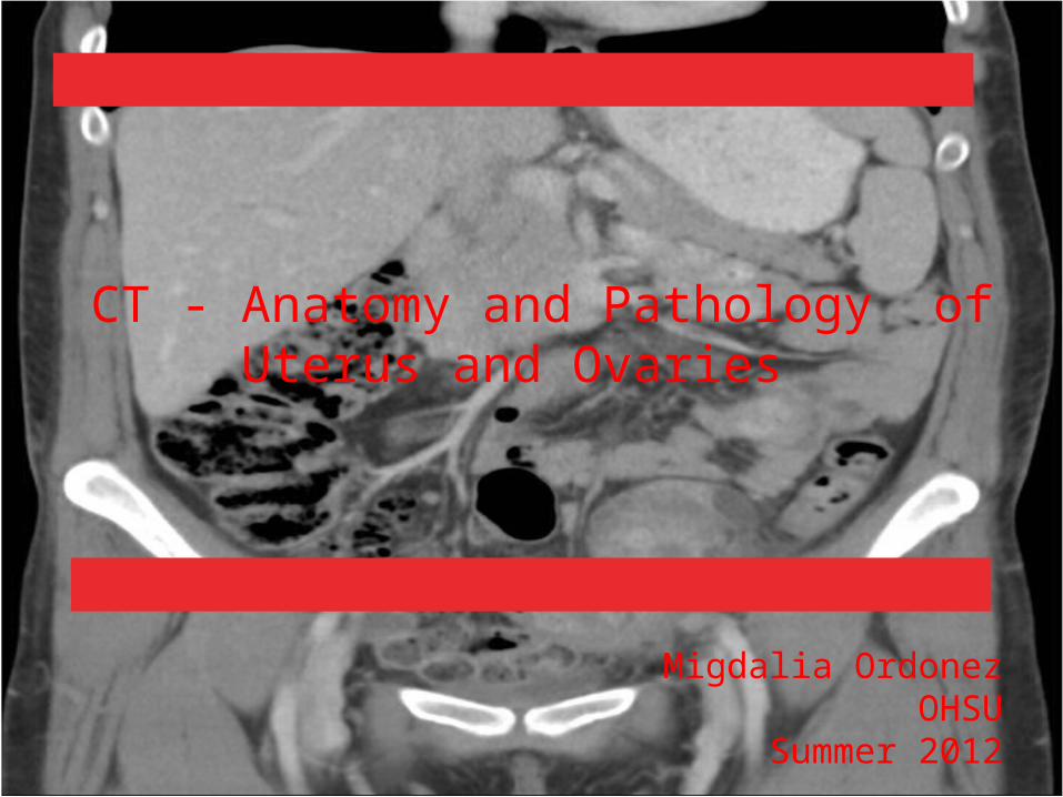
CT - Anatomy and Pathology ofUterus and Ovaries
Migdalia OrdonezOHSU
Summer 2012

Purpose of this Presentation
• Review Pelvic anatomy on CT.• Review common Pelvic pathologies on CT.
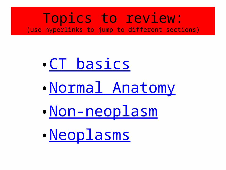
Topics to review:(use hyperlinks to jump to different sections)
•CT basics•Normal Anatomy•Non-neoplasm•Neoplasms
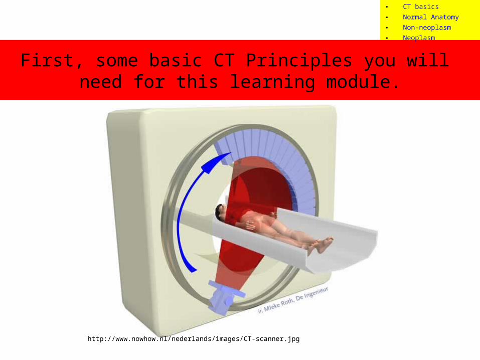
First, some basic CT Principles you will need for this learning module.
http://www.nowhow.nl/nederlands/images/CT-scanner.jpg
• CT basics• Normal Anatomy• Non-neoplasm• Neoplasm
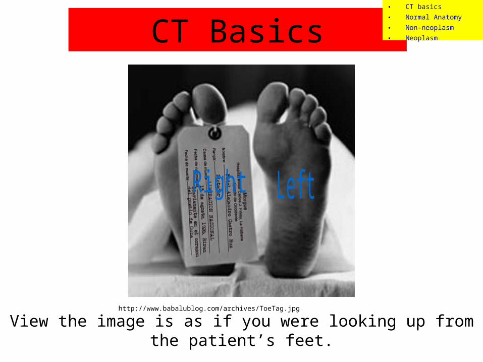
View the image is as if you were looking up from the patient’s feet.
http://www.babalublog.com/archives/ToeTag.jpg
CT Basics• CT basics• Normal Anatomy• Non-neoplasm• Neoplasm
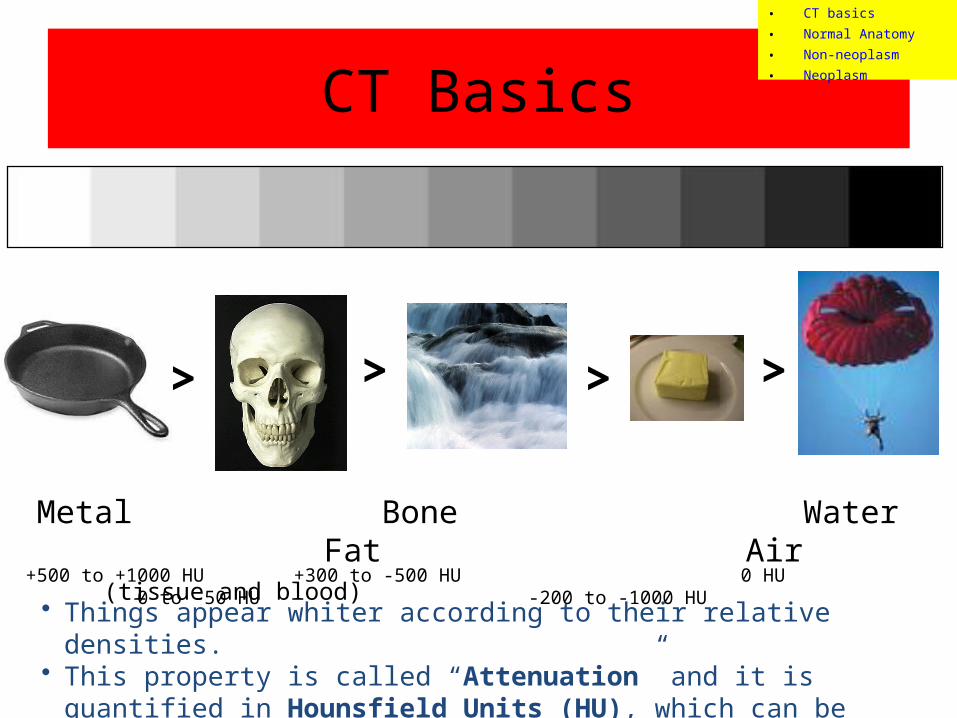
CT Basics
> > >
Metal Bone Water Fat Air (tissue and blood)
• Things appear whiter according to their relative densities. • This property is called “Attenuation” and it is quantified in Hounsfield Units
(HU), which can be measured on CT viewing software.
>
+500 to +1000 HU +300 to -500 HU 0 HU 0 to -50 HU -200 to -1000 HU
• CT basics• Normal Anatomy• Non-neoplasm• Neoplasm
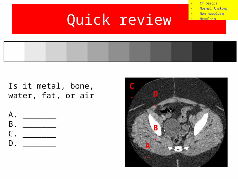
Quick review
Is it metal, bone, water, fat, or air
A. _______B. _______C. _______D. _______
A.
B.
C.D.
• CT basics• Normal Anatomy• Non-neoplasm• Neoplasm

Answers
Is it metal, bone, water, fat, or air
A. MuscleB. BoneC. AirD. Fat
A.
B.
C.D.
• CT basics• Normal Anatomy• Non-neoplasm• Neoplasm
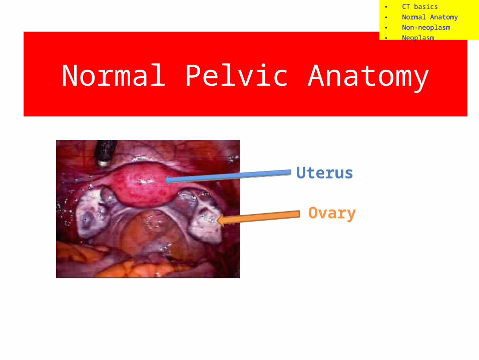
Normal Pelvic AnatomyNormal Pelvic Anatomy
Uterus
Ovary
• CT basics• Normal Anatomy• Non-neoplasm• Neoplasm
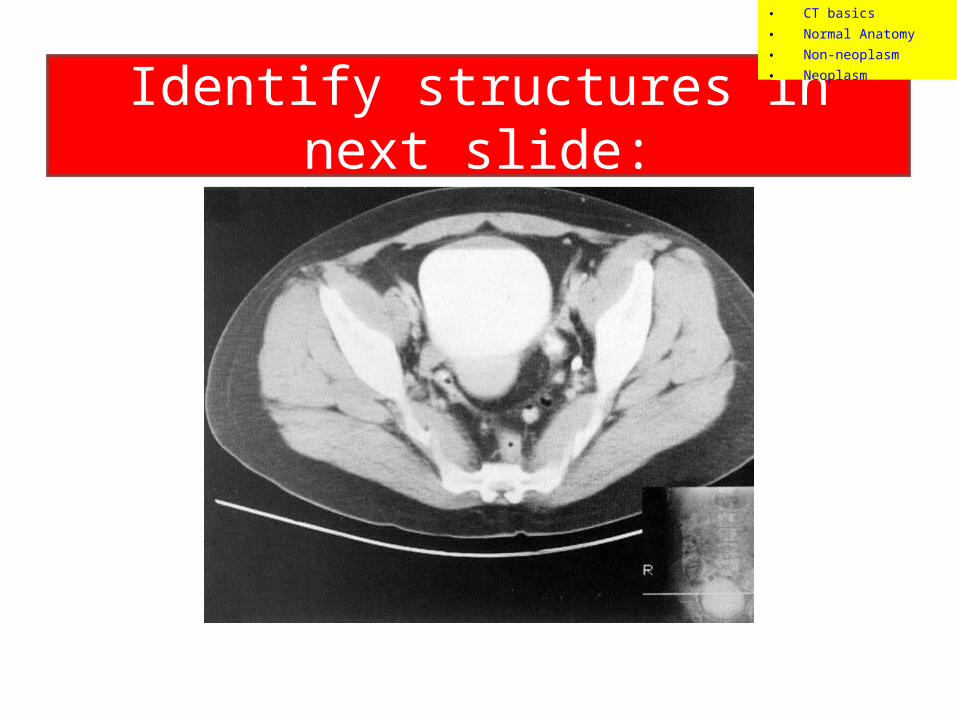
Identify structures in next slide:
• CT basics• Normal Anatomy• Non-neoplasm• Neoplasm
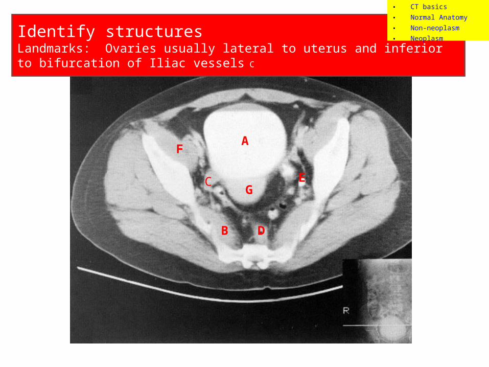
Identify structuresLandmarks: Ovaries usually lateral to uterus and inferior to bifurcation of Iliac vessels C
A
B
C
D
E
F
G
• CT basics• Normal Anatomy• Non-neoplasm• Neoplasm
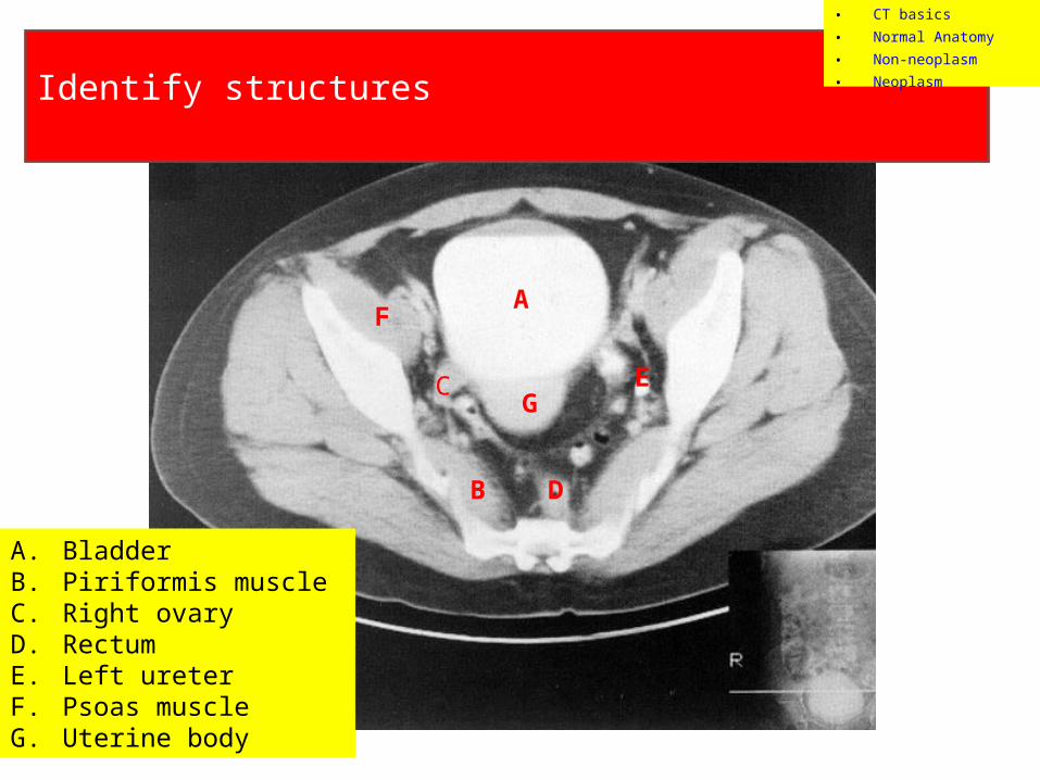
Identify structures
A
B
C
D
E
F
G
• CT basics• Normal Anatomy• Non-neoplasm• Neoplasm
A. BladderB. Piriformis muscleC. Right ovaryD. RectumE. Left ureterF. Psoas muscleG. Uterine body
Another look:
• CT basics• Normal Anatomy• Non-neoplasm• Neoplasm
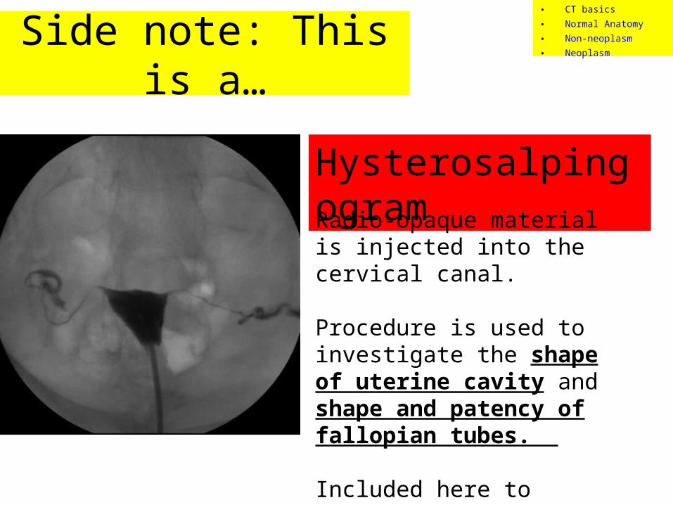
Side note: This is a…
Hysterosalpingogram
Radio-opaque material is injected into the cervical canal.
Procedure is used to investigate the shape of uterine cavity and shape and patency of fallopian tubes.
Included here to review anatomy
• CT basics• Normal Anatomy• Non-neoplasm• Neoplasm

Pathology
• CT basics• Normal Anatomy• Non-neoplasm• Neoplasm

Describe what you see:
• Attenuation (density)• Heterogeneous v. Homogenous• Well-circumscribed v. Indistinct Borders• Enhancing (lights up with contrast) v. non-enhancing• Location
Pathology – Non-neoplasm
• CT basics• Normal Anatomy• Non-neoplasm• Neoplasm
Case #1
• 28 year old female presents with fever, lower abdominal pain, new vaginal discharge and complaints of painful intercourse.
• Physical exam: Febrile and cervical motion tenderness.
• A computed tomography (CT) was done, see next slide.
• CT basics• Normal Anatomy• Non-neoplasm• Neoplasm
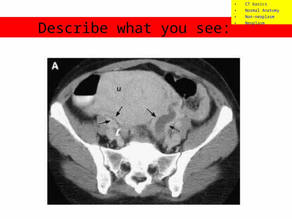
Describe what you see:
• CT basics• Normal Anatomy• Non-neoplasm• Neoplasm
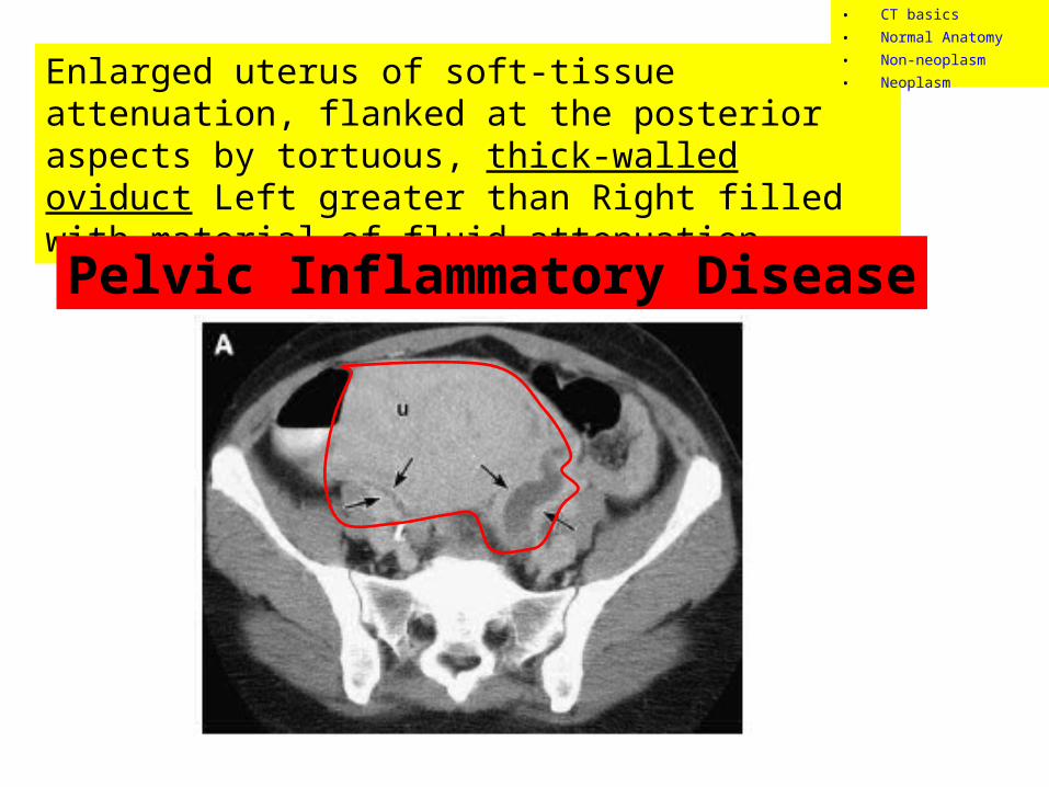
Enlarged uterus of soft-tissue attenuation, flanked at the posterior aspects by tortuous, thick-walled oviduct Left greater than Right filled with material of fluid-attenuation.
Pelvic Inflammatory Disease
• CT basics• Normal Anatomy• Non-neoplasm• Neoplasm
Case #2• 36 year old female presents with sudden onset
bilateral pelvic pain, left side worse than right. History of pelvic inflammatory disease a year ago treated with antibiotics.
• Physical exam: Tender to palpation in bilateral lower abdomen, L greater than R. Entire pelvis tender to palpation.
• A computed tomography (CT) was done, see next slide.
• CT basics• Normal Anatomy• Non-neoplasm• Neoplasm
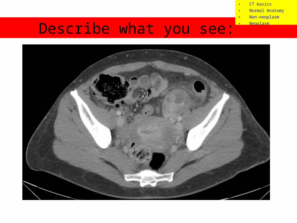
Describe what you see:
• CT basics• Normal Anatomy• Non-neoplasm• Neoplasm
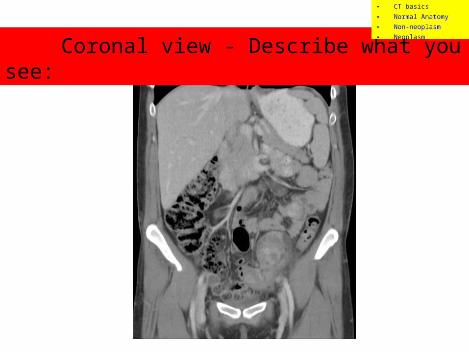
Coronal view - Describe what you see:
• CT basics• Normal Anatomy• Non-neoplasm• Neoplasm
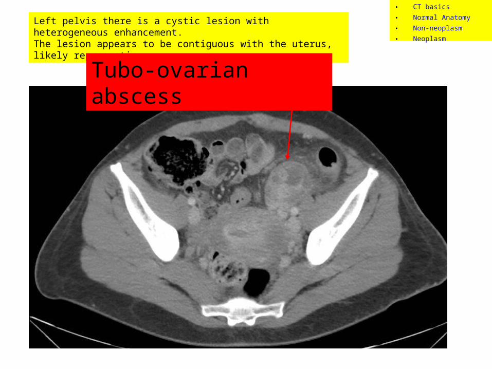
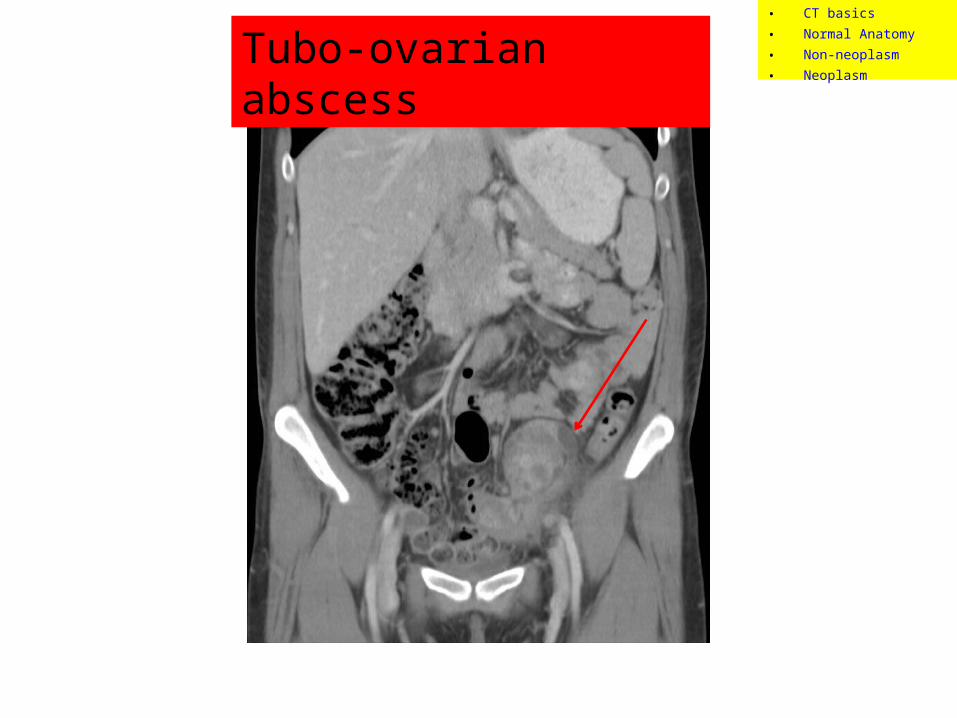
Left pelvis there is a cystic lesion with heterogeneous enhancement. The lesion appears to be contiguous with the uterus, likely representing…
Tubo-ovarian abscess
• CT basics• Normal Anatomy• Non-neoplasm• Neoplasm
Tubo-ovarian abscess• CT basics• Normal Anatomy• Non-neoplasm• Neoplasm
Case #3
• 23 year old female presents with fever, chills, lower abdominal pain, recent history of PID treated with antibiotics.
• Physical exam: Febrile and cervical motion tenderness.
• A computed tomography (CT) was done, see next slide.
• CT basics• Normal Anatomy• Non-neoplasm• Neoplasm
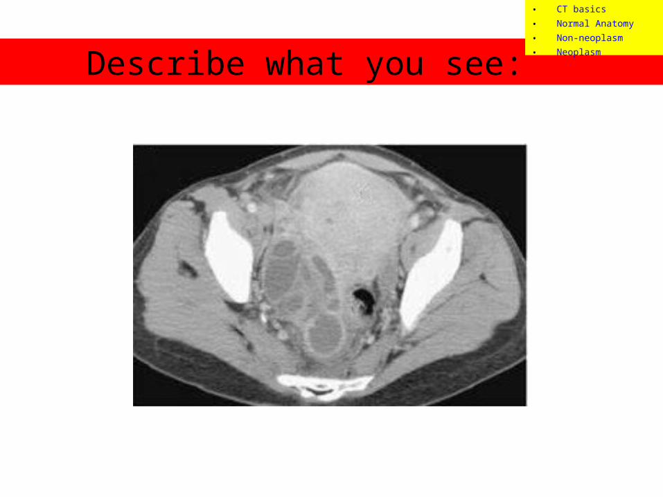
Describe what you see:
• CT basics• Normal Anatomy• Non-neoplasm• Neoplasm
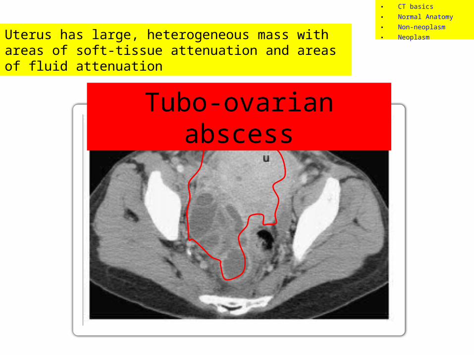
Uterus has large, heterogeneous mass with areas of soft-tissue attenuation and areas of fluid attenuation
Tubo-ovarian abscess
• CT basics• Normal Anatomy• Non-neoplasm• Neoplasm
Case #4
• 23 year old female presents to ED by ambulance due to motor vehicle accident. She is complaining of lower abdominal / pelvic pain.
• Physical exam: Pelvis tender to palpation.
• A computed tomography (CT) was done, see next slide.
• CT basics• Normal Anatomy• Non-neoplasm• Neoplasm

Describe what you see:
• CT basics• Normal Anatomy• Non-neoplasm• Neoplasm

Highly attenuated object in uterus, otherwise normal pelvic CT
IUD
• CT basics• Normal Anatomy• Non-neoplasm• Neoplasm
Case #5
• 21 year old female with 2 days of progressively worsening pelvic pain. She missed last period. She has been feeling nauseated for past 3 weeks.
• Physical exam: right pelvic tenderness, breast tenderness.
• A computed tomography (CT) was done, see next slide.
• CT basics• Normal Anatomy• Non-neoplasm• Neoplasm

Describe what you see:
• CT basics• Normal Anatomy• Non-neoplasm• Neoplasm
• CT basics• Normal Anatomy• Non-neoplasm• Neoplasm
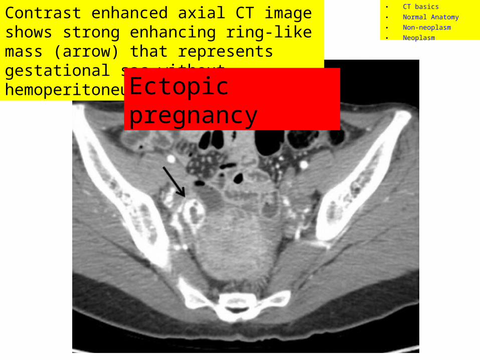
Contrast enhanced axial CT image shows strong enhancing ring-like mass (arrow) that represents gestational sac without hemoperitoneum
Ectopic pregnancy
Describe what you see:• Attenuation (density)• Heterogeneous v. Homogenous• Well-circumscribed v. Indistinct Borders• Enlarged v. atrophied• Enhancing (lights up with contrast) v. non-enhancing• Location
Pathology - Neoplasm
• CT basics• Normal Anatomy• Non-neoplasm• Neoplasm
Case #6
• 13 year old female presents with abdominal discomfort and feeling bloated. Stomach seems to be growing wider.
• Physical exam: Increased abdominal girth
• A computed tomography (CT) was done, see next slide.
• CT basics• Normal Anatomy• Non-neoplasm• Neoplasm
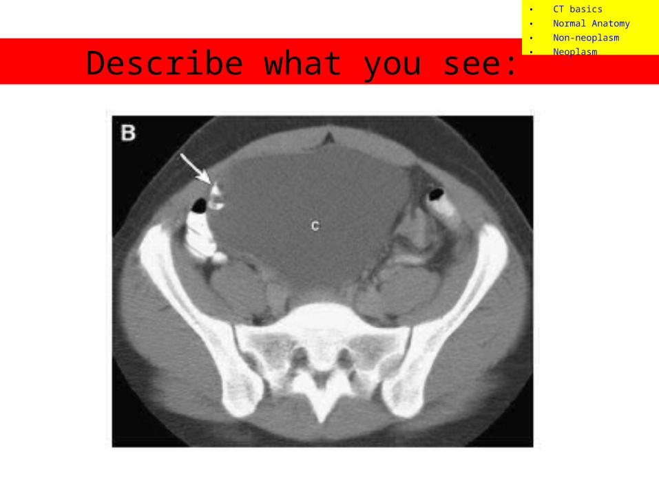
Describe what you see:
• CT basics• Normal Anatomy• Non-neoplasm• Neoplasm
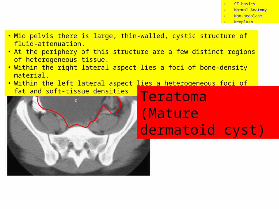
• Mid pelvis there is large, thin-walled, cystic structure of fluid-attenuation. • At the periphery of this structure are a few distinct regions of heterogeneous tissue. • Within the right lateral aspect lies a foci of bone-density material. • Within the left lateral aspect lies a heterogeneous foci of fat and soft-tissue densities
Teratoma (Mature dermatoid cyst)
• CT basics• Normal Anatomy• Non-neoplasm• Neoplasm
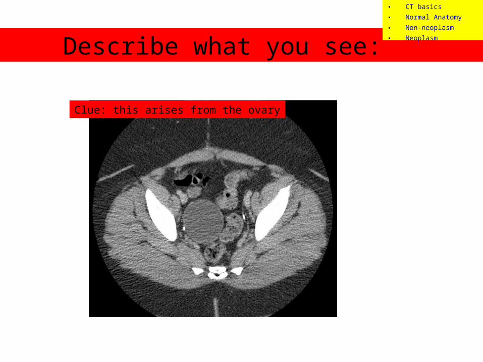
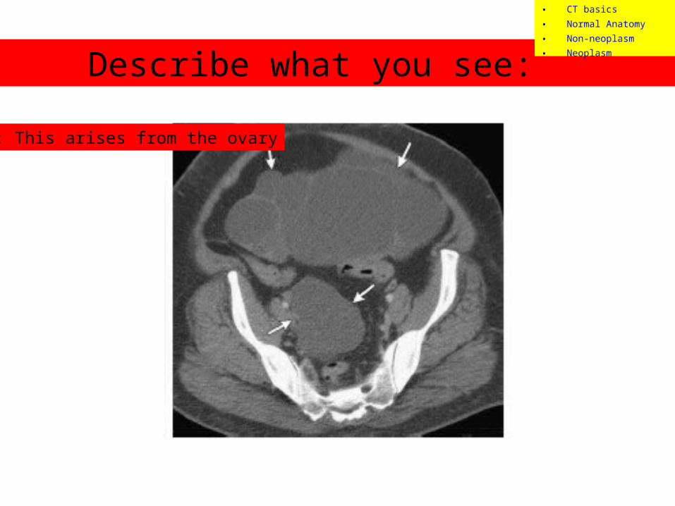
Case #7
• 48 year old female was involved in motor vehicle accident. She is shaken up from accident but otherwise feeling fine.
• Physical exam: Pt is in no acute distress. No signs or symptoms of pain. Patient insisted having a CT to rule out bleeds.
• A computed tomography (CT) was done, see next slide.
• CT basics• Normal Anatomy• Non-neoplasm• Neoplasm
Describe what you see:
Clue: this arises from the ovary
• CT basics• Normal Anatomy• Non-neoplasm• Neoplasm
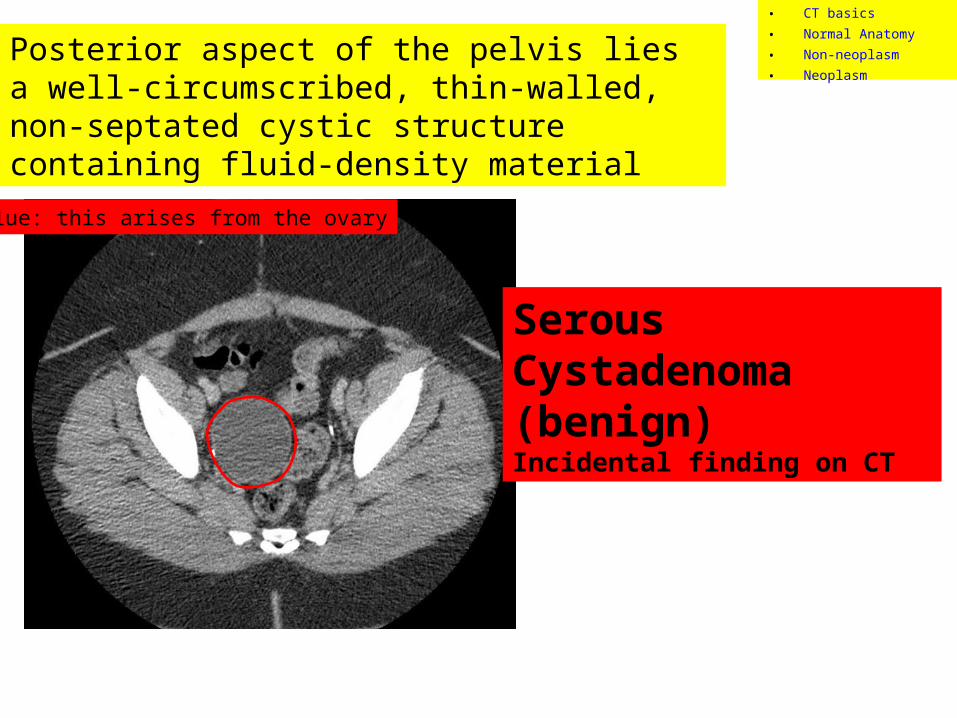
Clue: this arises from the ovary
Posterior aspect of the pelvis lies a well-circumscribed, thin-walled, non-septated cystic structure containing fluid-density material
Serous Cystadenoma(benign)Incidental finding on CT
• CT basics• Normal Anatomy• Non-neoplasm• Neoplasm

Case #8• 58 year old female presents to clinic with
bloating, back pain, urinary urgency, constipation, and tiredness for 6 months. Recently she developed pelvic pain, vaginal bleeding, and unintentional weight loss.
• Physical exam: Abdomen tender to palpation throughout. Pelvic tenderness.
• A computed tomography (CT) was done, see next slide.
• CT basics• Normal Anatomy• Non-neoplasm• Neoplasm
Describe what you see:
Clue: This arises from the ovary
• CT basics• Normal Anatomy• Non-neoplasm• Neoplasm
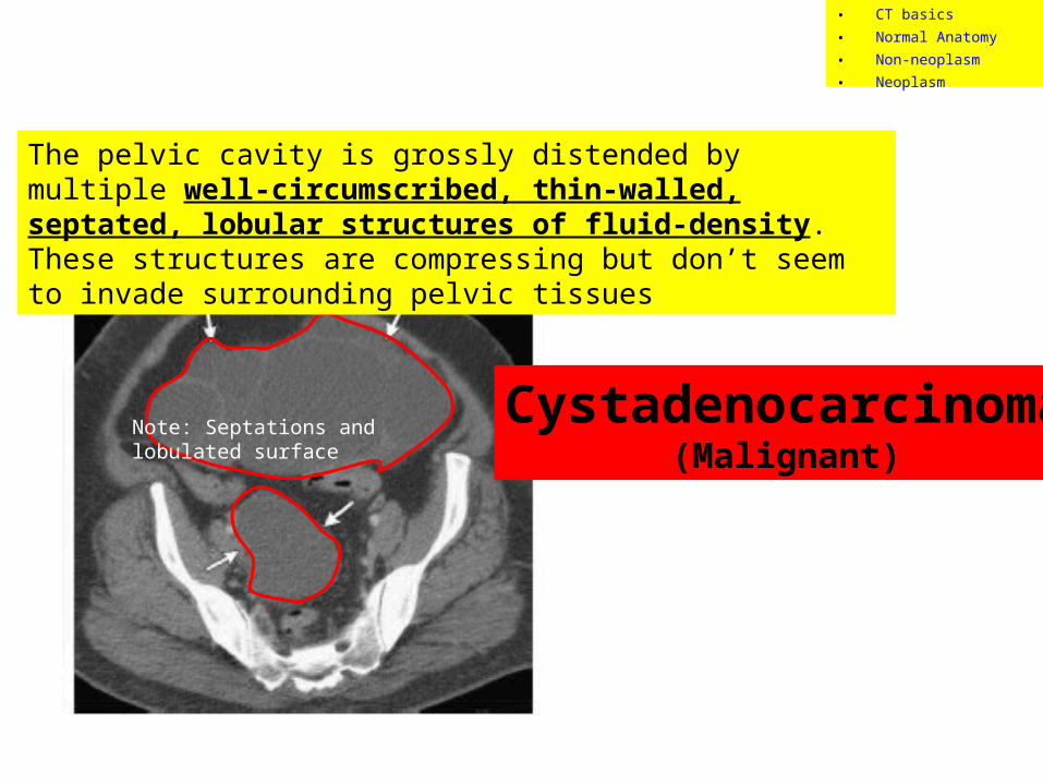
The pelvic cavity is grossly distended by multiple well-circumscribed, thin-walled, septated, lobular structures of fluid-density. These structures are compressing but don’t seem to invade surrounding pelvic tissues
Cystadenocarcinoma(Malignant)
Note: Septations and lobulated surface
• CT basics• Normal Anatomy• Non-neoplasm• Neoplasm
SourcesSiddall KA. Multidetector CT of the female pelvis. Radiol Clin North Am. 01-NOV-2005; 43(6): 1097-118
Casillas J, Joseph RC, Guerra JJ Jr. CT appearance of uterine leiomyomas. Radiographics. 1990 Nov;10(6):999-1007.
Foshager MC, Walsh JW. CT anatomy of the female pelvis: a second look. Radiographics. 1994 Jan;14(1):51-64;
Outwater EK, Siegelman ES, Hunt JL. Ovarian teratomas: tumor types and imaging characteristics. Radiographics. 2001 Mar-Apr;21(2):475-90.
Pannu, HK, et al. MD CT Evaluation of Cervical Cancer: Spectrum of Disease. Radiographics 2001; 21:1155–1168
Rha SE, et al. CT and MR imaging features of adnexal torsion. Radiographics. 2002 Mar-Apr;22(2):283-94.
Roberts JL, Dalen K, Bosanko CM, Jafir SZ. CT in abdominal and pelvic trauma. Radiographics. 1993 Jul;13(4):735-52.
Roobolamini, SA. Imaging of Pregnancy-related Complications. Radiographics 1993; 13:753-770.
Saksouk FA, Johnson SC. Recognition of the ovaries and ovarian origin of pelvic masses with CT. Radiographics. 2004 Oct;24 Suppl 1:S133-46.
Sam JW, Jacobs JE, Birnbaum BA. Spectrum of CT findings in acute pyogenic pelvic inflammatory disease. Radiographics. 2002 Nov-Dec;22(6):1327-34.
Yang DM. Retroperitoneal cystic masses: CT, clinical, and pathologic findings and literature review. Radiographics. 2004 Sep-Oct;24(5):1353-65.
Buy, J-N, et al. Cystic Teratoma of the Ovary: CT Detection. Radiology 1989; 171:697-701
As well as IMPAX, EPIC, and WIKIPEDIA
• CT basics• Normal Anatomy• Non-neoplasm• Neoplasm