cryptorchidism and associated problems in animals … · cryptorchidism and associated problems in...
TRANSCRIPT
Anim. Reprod., v.3, n.2, p.108-120, April/June. 2006
__________________________________________________________________
1Conference paper. International Symposium on Animal Biology of Reproduction, Nov. 15-18, 2006, Belo Horizonte, Brazil. 2Corresponding author: [email protected]
Phone: +1 (970)226-0682; Fax: +1(970)226-2340
Cryptorchidism and associated problems in animals1
R. P. Amann2 and D. N. R. Veeramachaneni
Animal Reproduction and Biotechnology Laboratory
Colorado State University, Fort Collins, CO 80523-1683 USA.
Abstract
Cryptorchidism is not a single disease; it
apparently results from failure of one of at least two
functions involving products of Leydig cells.
Cryptorchidism is symptomatic of underlying testis
dysgenesis. Precise terminology is important to
distinguish and understand three phases of testis
descent. Abdominal translocation involves holding a
testis near the internal inguinal ring plus slight
downward migration, via an enlarged gubernaculum
(Insl3 obligatory) as the abdominal cavity expands
away. Concurrently, a cylindrical evagination of
peritoneum invades the gubernaculum, cremaster
muscle(s) develop within the gubernaculum, and the
cephalic ligament regresses (testosterone not
obligatory). Transinguinal migration moves the testis
through the abdominal wall, via the inguinal canal
distended by the gubernaculum. Inguinoscrotal
migration involves subcutaneous movement of
gubernaculum, vaginal process, epididymis, and testis to
proper positions in the scrotum. Directional guidance is
provided by chemoattractant calcitonin gene-related
peptide, secreted by the genitofemoral nerve
(testosterone dependent). Probably incidence of
cryptorchidism is higher in companion animals or pigs
than in cattle or sheep. In dogs and horses, a retained
testis most commonly is abdominal. In horses, but not
other species, retention of testes within the inguinal
canal is common. In humans, subcutaneous testes
predominate. Overall, cryptorchidism usually is
unilateral; scrotal testes in unilateral cryptorchid males
often produce fewer than normal numbers of sperm,
with an increased percentage of abnormal sperm. Non-
cryptorchid siblings might manifest similar testicular
dysgenesis. Although risk of tumors is greater for
cryptorchid males, non-cryptorchid males also develop
testis tumors. Germ cell tumors are most common in dogs
and horses. Leydig or Sertoli cell tumors are not unusual in
dogs. Testis tumors rarely are reported for cattle, pigs,
sheep, and rabbits; in cattle Leydig cell tumors are twice as
common as Sertoli cell tumors. Producers and veterinarians
should recognize that inadvertent exposure of a
pregnant dam to estrogenic or anti-androgenic agents
could result in testicular dysgenesis.
Keywords: cryptorchidism, testis descent, testis
dysgenesis, testis tumors, terminology.
Introduction
Cryptorchidism is failure of one or both testes
to be positioned in the scrotum at the time normal for a
species. Typically, cryptorchidism is detected at birth or
shortly thereafter. Postpubertal abnormalities associated
with cryptorchidism (e.g., testis tumors, atypical
concentrations of reproductive hormones, altered
spermatogenesis in scrotal testis,) are not caused by
elevated temperature of an abdominal location
(Veeramachaneni, 2006), but as a delayed manifestation
of testis dysgenesis. Such underlying testis dysgenesis
of fetal origin might be evident in males who are not
cryptorchid. The concept of a generalized testicular
dysgenesis syndrome (TDS) is considered by Sharpe in
this volume (also Skakkebaek et al., 1998, 2001;
Rajpert-De Meyts, 2006; Sharpe, 2006).
Early reports on cryptorchidism (e.g., de Graaf,
1668) provided evidence of two or more diseases,
because undescended testes are not located at a common
non-scrotal site. Nevertheless, the general perception
had been that cryptorchidism is a single disease with
moderate heritability, incomplete penetrance, expressed
only in males (sex specific expression), and
concentrated by inbreeding or minimized by culling
affected males and all siblings. However, the notion of a
single-locus gene problem gave way to acceptance of a
polygenic recessive model, based on relatively small
studies with pigs (Sittmann and Woodhouse, 1977;
Rothschild et al, 1988) and dogs (Cox et al, 1978;
Nielen et al., 2001); also data for men (Czeizel et al.,
1981). It is evident that abnormalities in >20 genes are
associated with human cryptorchidism (Klonisch et al.,
2004) and, currently it is accepted that cryptorchidism
has many causes including genetic, epigenetic, and
environmental components.
Terminology
For reasons detailed elsewhere (Amann and
Veeramachaneni, 2007), certain traditional
nomenclature does not adequately describe where a
testis was found or when in the process of testis descent
the normal process went wrong. Hence, this review
defines and utilizes precise terms and nomenclature
equally applicable to companion, food-producing, or
wild animals; rodents or rabbits; and humans.
Testes are found in one of four general
locations: abdominal cavity, inguinal canal,
Amann and Veeramachaneni. Cryptorchidism and testis dysgenesis.
Anim. Reprod., v.3, n.2, p.108-120, April/June. 2006 109
subcutaneous, or scrotal. The distinction between
inguinal and subcutaneous locations is important.
An abdominal testis is within the abdominal
cavity, typically between the kidney and bladder or near
the internal inguinal ring. Distinction between proximal
vs near inguinal locations is important because it might
point to underlying cause.
An inguinal testis is within the space limited by
the internal and external inguinal rings. At least for
horses (inguinal canal might be 10 cm long), position
should be precisely defined (Beltran-Brown and
Villegas-Alvarez, 1988).
A subcutaneous testis usually is found in the
femoral triangle, but ectopia of the vaginal process
might place the testis at some distance or near a
malformed scrotum. Unfortunately, imprecision in
defining testis location typifies literature on mice or rats
administered an agent which might affect testis descent,
and the uninformative “ectopic testis” (i.e., abnormal
location of testis) often is used to describe location of a
testis not within a normal scrotum or within the
abdominal cavity.
A scrotal testis is found, and remains, in a
scrotum located at the site typical for that species.
However, in rodents and rabbits testes can move in and
out from the lower abdominal cavity throughout life, as
the inguinal canal never constricts. Occasionally, a
scrotal testis might later be retracted permanently into
the inguinal canal (retractile testis) or a testis initially
deemed to be inguinal or subcutaneous might later be
positioned permanently in the scrotum (late descent).
Such migration is more common in horses, humans, or
pigs than in cattle or sheep.
To stipulate discrete phases in the overall
process of testis descent, three terms are needed since
there are four general locations (see above). The
following terms are descriptive of the process and are
useful with any species.
Abdominal translocation of the testis reflects
what happens during the first phase. At the end of this
phase, the testis is positioned at the inner inguinal ring
(ready to enter), with the cauda epididymidis just within
the inguinal canal. The absolute distance between a
testis and inguinal area changes little during this phase.
Rather, the testes “stay put” as the fetus grows and the
distance between locations of testes and kidneys widens
by slight “downward” relocation to the developing inner
inguinal ring (Wensing, 1968; Shono et al., 1994a).
Hence, the widely used “abdominal descent”
overemphasizes what occurs.
Transinguinal migration of the testis pertains to
movement through the abdominal wall, from an abdominal
to a subcutaneous location. This is a rapid process.
Inguinoscrotal migration of the testis describes
the third phase. It covers the quest of the gubernaculum
for the scrotum, which can be rather distant from the
external inguinal ring, and consequent movement of the
attached cauda epididymidis and testis to proper
positions in the scrotum.
What went wrong during testis descent, in a
simple sense, can be deduced from observed testis
location and knowledge of the process. Three terms are
needed, but unfortunately and illogically some authors
have combined the latter two, without differentiation.
An abdominal testis reflects failure to initiate
and complete abdominal translocation of the testis, so
the testis is not poised near the internal inguinal ring,
but rather near the bladder or part way between the
inguinal area and kidney.
An inguinal testis reflects failure to initiate and
complete transinguinal migration of a testis.
A subcutaneous testis reflects failure to initiate
and complete inguinoscrotal migration of a cauda
epididymidis and testis, from outside the inguinal canal
to their final destination in the scrotum.
In horses, many retained testes are within the
inguinal canal per se and few are subcutaneous (Cox et
al., 1979; Rodgerson and Hansen, 1997), but testes
which actually are subcutaneous often are classified
inappropriately as inguinal (Genetzky, 1984). In
humans, however, most undescended testes are
subcutaneous in the groin, just outside the external
inguinal ring, or near the neck of the scrotum (Hutson et
al., 1992, 1997). It is imprecise to attribute both
inguinal and subcutaneous testes to failure of
“inguinoscrotal testis descent”; as discussed below,
different regulating mechanisms likely are involved.
Testis descent
To present an overview applicable to common
species ranging from mice and rabbits to bulls or horses,
this compilation reinterprets some information in older
publications. This is done with benefit of recent
information and without discounting underlying
observations in classic papers based on many
dissections. Literature was cited extensively and
concepts were discussed in Amann and Veeramachaneni
(2007), which should be consulted. Here the aim is ease
of comprehension.
Structures involved
The crucial starting point is differentiation of
an indifferent gonad to a testis (Rajpert-De Meyts,
2006; Sharpe, 2006; Amann and Veeramachaneni,
2007). In brief, primordial germ cells (PGCs) migrate
from the hind gut to the gonadal ridge. Then, in the
male, mesenchymal cells move into the developing
gonad, proliferate, surround the PGCs, and differentiate
into fetal Sertoli cells. Shortly afterwards other cells
from the mesonephros arrive (later become peritubular
myoid cells) to organize nests of fetal Sertoli cells plus
PGCs into seminiferous cords. Finally, other
mesenchymal cells migrate into spaces among the
seminiferous cords and differentiate into fetal Leydig
cells. The interval between entrance of PGCs into the
Amann and Veeramachaneni. Cryptorchidism and testis dysgenesis.
Anim. Reprod., v.3, n.2, p.108-120, April/June. 2006 110
indifferent gonad and differentiation of the gonad to a
functional testis is <14 days. This happens near
gestational day 14, 33, 35, 40, and 41 in the mouse, dog,
pig, horse and bull. Within 2-3 days after arrival, fetal
Leydig cells achieve maximum production of
testosterone (as a fetal structure, on a per gram basis),
and probably insulin-like peptide 3 (Insl3). Initially,
testosterone is produced constitutively, but later GnRH
and LH come into play to regulate the process.
As the testis is formed, a thin fold of
peritoneum covering the gonad evolves as the mesorchium
to suspend the gonad dorsally from the mesonephros. The
same fold continues cranially as the cephalic ligament
(cranial suspensory ligament) and caudally as the posterior
gonadal ligament (epididymal ligament; Fig. 1A). The
gubernaculum originates early in development from
mesenchymal cells among muscle fibers of the abdominal
wall, grows into the peritoneal fold forming the posterior
gonadal ligament, and soon dominates the fold caudal to
where it fuses to the mesonephric duct. The demarcation
between the gubernaculum and posterior gonadal ligament
is where the cauda epididymidis later transitions to the
deferent duct. Scrotal swellings become evident under the
skin, although they are not in the final location for the
scrotum.
Hunter (1762) coined the term gubernaculum
and described its structure. He slightly modified his
description and then (Hunter, 1786) wrote “... which at
present I shall call the ligament, or gubernaculum testis,
because it connects the testis with the scrotum, and
seems to direct its course through the rings of the
abdominal muscles. ... it is certainly vascular and
fibrous, and the fibers run in the direction of the
ligament itself which is covered by the fibers of the
cremaster or musculus testis, placed immediately behind
the peritoneum.” We now know that the gubernaculum
is rich in hyaluronic acid, glycosaminoglycans, and
collagen; it often is described as gelatinous although it
has collagen fibers and the cells proliferate during
expansion.
Clearly, Hunter stated that the cremaster
muscle covers the gubernaculum and, hence, considered
them as separate structures (but see van der Schoot,
1996; Amann and Veeramachaneni, 2007 for further
details). This distinction between the gubernaculum and
cremaster muscle(s) is unequivocal in reports pertaining
to non-rodent species, but in most reports on rodents or
rabbits the cremaster muscles are considered as part of
the gubernaculum. This inclusive usage hampers
ascribing observations to mechanistic problems (e.g.,
development of the gubernaculum per se is affected by
lack of Insl3, cremaster muscles are not affected by
Insl3, development of the gubernaculum per se is not
affected by anti-androgen, cremaster muscles are
affected by anti-androgen). Herein, the term
“gubernaculum” excludes the cremaster muscles or the
vaginal process. Formation of the gubernaculum and
cremaster muscles, and differences between rodents and
rabbits or other species, are detailed in Amann and
Veeramachaneni (2007).
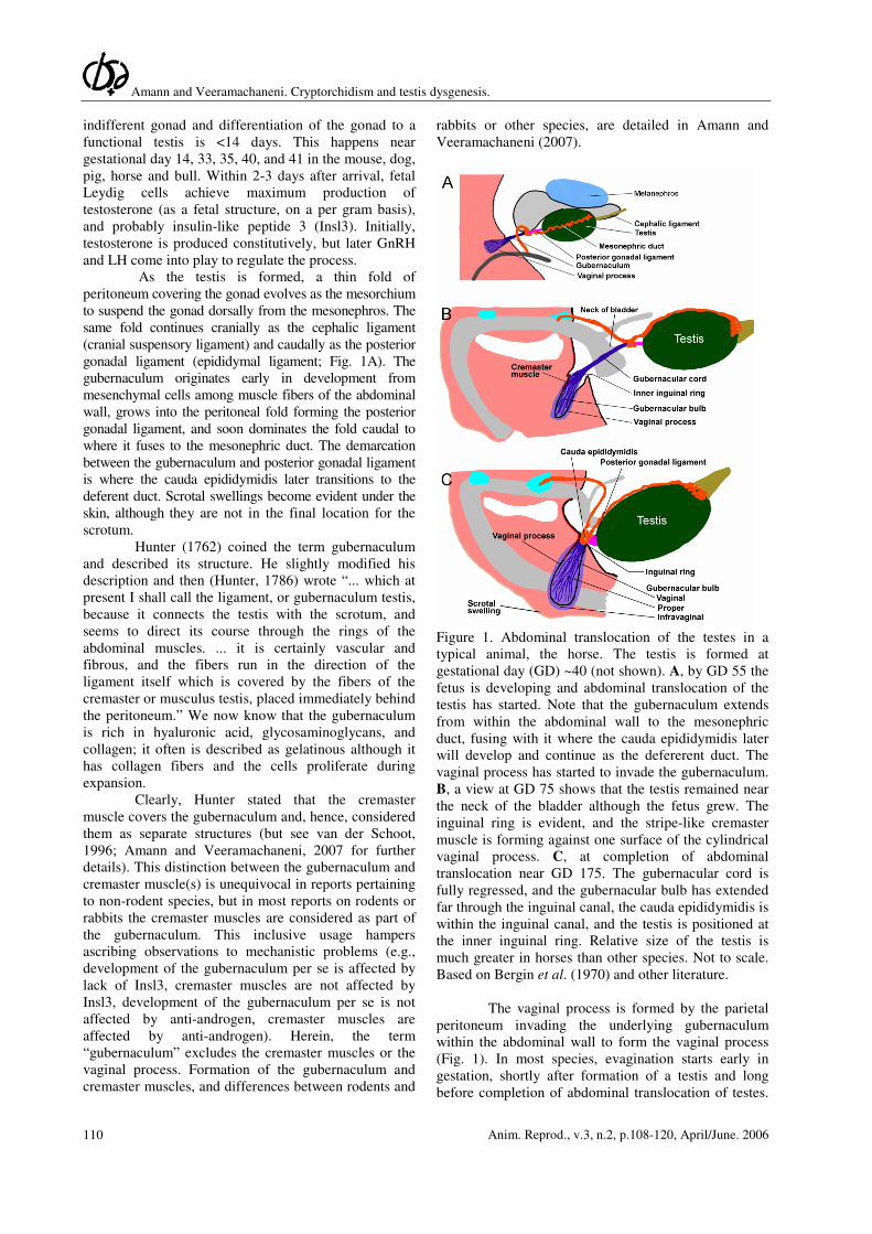
Figure 1. Abdominal translocation of the testes in a
typical animal, the horse. The testis is formed at
gestational day (GD) ~40 (not shown). A, by GD 55 the
fetus is developing and abdominal translocation of the
testis has started. Note that the gubernaculum extends
from within the abdominal wall to the mesonephric
duct, fusing with it where the cauda epididymidis later
will develop and continue as the defererent duct. The
vaginal process has started to invade the gubernaculum.
B, a view at GD 75 shows that the testis remained near
the neck of the bladder although the fetus grew. The
inguinal ring is evident, and the stripe-like cremaster
muscle is forming against one surface of the cylindrical
vaginal process. C, at completion of abdominal
translocation near GD 175. The gubernacular cord is
fully regressed, and the gubernacular bulb has extended
far through the inguinal canal, the cauda epididymidis is
within the inguinal canal, and the testis is positioned at
the inner inguinal ring. Relative size of the testis is
much greater in horses than other species. Not to scale.
Based on Bergin et al. (1970) and other literature.
The vaginal process is formed by the parietal
peritoneum invading the underlying gubernaculum
within the abdominal wall to form the vaginal process
(Fig. 1). In most species, evagination starts early in
gestation, shortly after formation of a testis and long
before completion of abdominal translocation of testes.
Amann and Veeramachaneni. Cryptorchidism and testis dysgenesis.
Anim. Reprod., v.3, n.2, p.108-120, April/June. 2006 111
The vaginal process divides the gubernacular bulb into
three areas (labeled in Fig. 1C): proper, central to the
cylindrical vaginal process and continuous with the
gubernacular cord; vaginal, concentric and outside the
vaginal process; and infravaginal, cup-shaped and between
the invading peritoneum and distal tip. In rodents and
rabbits, however, initial evagination of the vaginal process
occurs neonatally, just before completion of abdominal
testis translocation discussed below.
A striated cremaster muscle(s) is formed by
myoblasts, migrating from the muscles of the abdominal
wall and/or differentiating from mesenchymal cells in
the gubernaculum. The cremaster muscle is stripe-like
in companion and food-producing animals, or humans,
and invades the vaginal portion of the gubernaculum on
the lateral aspect of the developing vaginal process. In
rodents or rabbits, concentric cremaster muscles
develop and encompass the proper portion of the
gubernacular bulb. This results in formation of a
“gubernacular-cremaster complex”, one on each side
(Fig 2A). The gubernacular-cremaster complex includes
the intra-abdominal gubernaculum and both cremaster
muscles, but excludes the thin connection (gubernacular
cord) extending to the mesonephric duct and the extra-
abdominal gubernaculum. We recommend and use the
term gubernacular-cremaster complex, rather than
“gubernacular cones” commonly used in literature on
rodents and favored by van der Schoot (1993, 1996),
because this makes clear that the complex has two
elements and recognizes that they have different roles
during testis descent and later in adults. The sexually
dimorphic genitofemoral nerve (not shown) is carried
downward with the gubernaculum and innervates the
cremaster muscle.
Process of testis descent
There is limited information on regulation of
testis descent in common animals, although there are
good descriptions on changes in morphology. Hence,
this summary is based on what is known from model
animals, augmented with data for companion or food-
producing animals.
Abdominal translocation results in positioning
a testis near the developing internal inguinal ring. The
extra-abdominal portion of the gubernaculum becomes
longer and wider, but there is little change in the
distance between a testis and the inguinal area. In a bull
fetus, distance from a testis to the internal inguinal ring
decreases by 4 mm whereas the distance from testis to
kidney increases by 13 mm, during the time frame
required for abdominal translocation; distance from the
internal inguinal ring to kidney becomes >25 mm
(Edwards et al., 2003). In rats, distance from a testis to the
internal inguinal ring decreases 0.3 mm whereas testis to a
kidney distance increases 4.4 mm (Shono et al., 1994a).
The testis is suspended by the cephalic
ligament cranially and the posterior gonadal ligament
plus gubernaculum caudally (Fig. 1A). Initially, both the
posterior gonadal ligament and gubernaculum are short
and thin. Leydig cells in the rapidly evolving testis
secrete Insl3 and testosterone. Under stimulation of
Insl3, the extra-abdominal gubernaculum expands and
invades deep into the abdominal musculature (Fig. 1B),
to anchor the testis. The vaginal process invades the
gubernaculum (especially conspicuous in Fig. 1C), and
grows downward as the gubernaculum increases in size.
Testosterone might facilitate gradual dissolution of the
cephalic ligament, which elongates as the abdominal
cavity expands. Consequently, the testis is retained in
the inguinal region and the distance between the testis
and other structures in the abdomen (e.g., kidney)
widens. At final abdominal positioning (Fig. 1C), the
cauda epididymidis is just within the inguinal canal and
the testis is near the internal inguinal ring. At this time,
relative length of the posterior gonadal ligament is
variable, so a testis might not be hard against the
inguinal ring. Although the testis grows in all species, in
horses the testis becomes very large (primarily growth
of interstitial tissue) by the time it is positioned near the
inner inguinal ring.
In rodents and rabbits, as in other species,
formation of the vaginal process is a feature of
abdominal translocation of a testis. Hence, it should be
included in the first phase of testis descent. By late
gestation, the gubernacular-cremaster complex has
formed and assumed a conical or cylindrical shape
protruding into the abdominal cavity (Fig. 2A) in the
femoral triangle area. Then the base of the
gubernacular-cremaster complex “sinks” slightly below
the plane of the abdominal wall, accommodated by
slight cylindrical down-growth of the peritoneal lining
(Fig. 2B). This is the first evidence of the forming
inguinal canal. In other species, this phenomenon
happens much earlier. By this time, the gubernacular
cord has fully regressed, so that the cauda epididymidis
is positioned against the abdominal face of the future
vaginal process and, hence, in close proximity to the
intra-abdominal gubernaculum per se or the muscle of
the gubernacular-cremaster complex. In any case, the
testis is positioned near the entrance to the still forming
inguinal canal. Abdominal translocation of testes is
completed (Fig. 2B) around gestational day 28 in
rabbits, postnatal day 1-2 in mice, or postnatal day 4-5
in rats (Elder et al., 1982; Shono et al., 1994b, 1996;
Lam et al., 1998).
In large animals such as horses, at the end of
abdominal testis translocation the gubernaculum and
vaginal process extend far below the newly formed
inguinal canal and the testis is positioned against the
internal inguinal ring, with the cauda epididymidis
within the inguinal canal (Fig. 1C). In most species, this
position is maintained for some time, like a “pause”
between 2 separate processes. During the pause before
actual transinguinal migration, the gubernacular bulb
enlarges greatly (see Fig. 7 in Gier and Marion, 1970),
Amann and Veeramachaneni. Cryptorchidism and testis dysgenesis.
Anim. Reprod., v.3, n.2, p.108-120, April/June. 2006 112
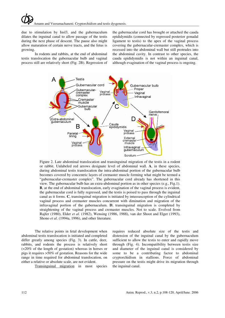
due to stimulation by Insl3, and the gubernaculum
dilates the inguinal canal to allow passage of the testis
during the next phase of descent. The pause also might
allow maturation of certain nerve tracts, and the fetus is
growing.
In rodents and rabbits, at the end of abdominal
testis translocation the gubernacular bulb and vaginal
process still are relatively short (Fig. 2B). Regression of
the gubernacular cord has brought or attached the cauda
epididymidis (connected by regressed posterior gonadal
ligament to testis) to the apex of the vaginal process
covering the gubernacular-cremaster complex, which is
recessed into the abdominal wall but still protrudes into
the abdominal cavity. In contrast to other species, the
cauda epididymidis is not within an inguinal canal,
although evagination of the vaginal process is ongoing.
Figure 2. Late abdominal translocation and transinguinal migration of the testis in a rodent
or rabbit. Unlabeled red arrows designate level of abdominal wall. A, in these species,
during abdominal testis translocation the intra-abdominal portion of the gubernacular bulb
becomes covered by concentric layers of cremaster muscle forming what might be termed a
“gubernacular-cremaster complex”. The gubernacular cord already has shortened in this
view. The gubernacular bulb has an extra-abdominal portion as in other species (e.g., Fig.1).
B, at the end of abdominal translocation, early evagination of the vaginal process is evident,
the gubernacular cord is fully regressed, and the testis is poised to pass through the inguinal
canal as it forms. C, transinguinal migration is initiated by intussusception of the cylindrical
vaginal process and cremaster muscles concurrent with diminution and migration of the
infravaginal portion of the gubernaculum. D, transinguinal migration is completed by
straightening of the vaginal process and cremaster muscles. Not to scale. Evolved from
Rajfer (1980), Elder et al. (1982), Wensing (1986, 1988), van der Shoot and Elger (1993),
Shono et al. (1994a, 1996), and other literature.
The relative points in fetal development when
abdominal testis translocation is initiated and completed
differ greatly among species (Fig. 3). In cattle, deer,
rabbits, and rodents the process is relatively short
(<20% of the length of gestation) whereas in horses or
pigs it requires >50% of gestation. Reasons for the wide
range in time required for abdominal translocation, on
either a relative or absolute scale, are not evident.
Transinguinal migration in most species
requires reduced absolute size of the testis and
distension of the inguinal canal by the gubernaculum
sufficient to allow the testis to enter and rapidly move
through (Fig. 4). Incompatibility between testis size
and diameter of the inguinal canal is considered by
some to be a contributing factor to abdominal
cryptorchidism in stallions. Force of abdominal
pressure on the testis might drive its migration through
the inguinal canal.
Amann and Veeramachaneni. Cryptorchidism and testis dysgenesis.
Anim. Reprod., v.3, n.2, p.108-120, April/June. 2006 113
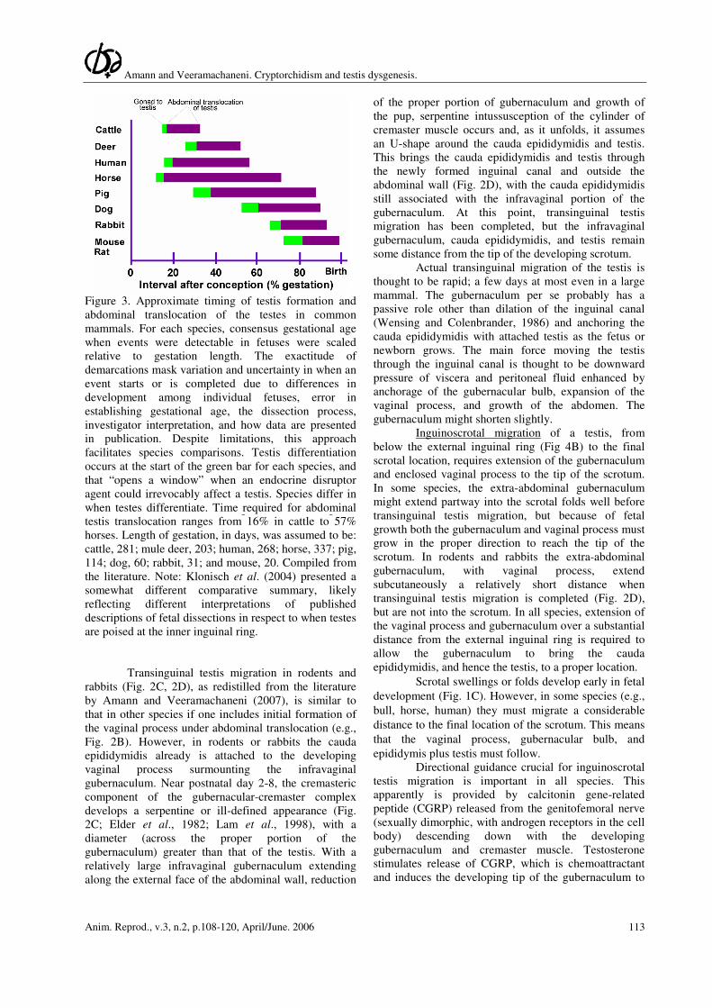
Figure 3. Approximate timing of testis formation and
abdominal translocation of the testes in common
mammals. For each species, consensus gestational age
when events were detectable in fetuses were scaled
relative to gestation length. The exactitude of
demarcations mask variation and uncertainty in when an
event starts or is completed due to differences in
development among individual fetuses, error in
establishing gestational age, the dissection process,
investigator interpretation, and how data are presented
in publication. Despite limitations, this approach
facilitates species comparisons. Testis differentiation
occurs at the start of the green bar for each species, and
that “opens a window” when an endocrine disruptor
agent could irrevocably affect a testis. Species differ in
when testes differentiate. Time required for abdominal
testis translocation ranges from 16% in cattle to 57%
horses. Length of gestation, in days, was assumed to be:
cattle, 281; mule deer, 203; human, 268; horse, 337; pig,
114; dog, 60; rabbit, 31; and mouse, 20. Compiled from
the literature. Note: Klonisch et al. (2004) presented a
somewhat different comparative summary, likely
reflecting different interpretations of published
descriptions of fetal dissections in respect to when testes
are poised at the inner inguinal ring.
Transinguinal testis migration in rodents and
rabbits (Fig. 2C, 2D), as redistilled from the literature
by Amann and Veeramachaneni (2007), is similar to
that in other species if one includes initial formation of
the vaginal process under abdominal translocation (e.g.,
Fig. 2B). However, in rodents or rabbits the cauda
epididymidis already is attached to the developing
vaginal process surmounting the infravaginal
gubernaculum. Near postnatal day 2-8, the cremasteric
component of the gubernacular-cremaster complex
develops a serpentine or ill-defined appearance (Fig.
2C; Elder et al., 1982; Lam et al., 1998), with a
diameter (across the proper portion of the
gubernaculum) greater than that of the testis. With a
relatively large infravaginal gubernaculum extending
along the external face of the abdominal wall, reduction
of the proper portion of gubernaculum and growth of
the pup, serpentine intussusception of the cylinder of
cremaster muscle occurs and, as it unfolds, it assumes
an U-shape around the cauda epididymidis and testis.
This brings the cauda epididymidis and testis through
the newly formed inguinal canal and outside the
abdominal wall (Fig. 2D), with the cauda epididymidis
still associated with the infravaginal portion of the
gubernaculum. At this point, transinguinal testis
migration has been completed, but the infravaginal
gubernaculum, cauda epididymidis, and testis remain
some distance from the tip of the developing scrotum.
Actual transinguinal migration of the testis is
thought to be rapid; a few days at most even in a large
mammal. The gubernaculum per se probably has a
passive role other than dilation of the inguinal canal
(Wensing and Colenbrander, 1986) and anchoring the
cauda epididymidis with attached testis as the fetus or
newborn grows. The main force moving the testis
through the inguinal canal is thought to be downward
pressure of viscera and peritoneal fluid enhanced by
anchorage of the gubernacular bulb, expansion of the
vaginal process, and growth of the abdomen. The
gubernaculum might shorten slightly.
Inguinoscrotal migration of a testis, from
below the external inguinal ring (Fig 4B) to the final
scrotal location, requires extension of the gubernaculum
and enclosed vaginal process to the tip of the scrotum.
In some species, the extra-abdominal gubernaculum
might extend partway into the scrotal folds well before
transinguinal testis migration, but because of fetal
growth both the gubernaculum and vaginal process must
grow in the proper direction to reach the tip of the
scrotum. In rodents and rabbits the extra-abdominal
gubernaculum, with vaginal process, extend
subcutaneously a relatively short distance when
transinguinal testis migration is completed (Fig. 2D),
but are not into the scrotum. In all species, extension of
the vaginal process and gubernaculum over a substantial
distance from the external inguinal ring is required to
allow the gubernaculum to bring the cauda
epididymidis, and hence the testis, to a proper location.
Scrotal swellings or folds develop early in fetal
development (Fig. 1C). However, in some species (e.g.,
bull, horse, human) they must migrate a considerable
distance to the final location of the scrotum. This means
that the vaginal process, gubernacular bulb, and
epididymis plus testis must follow.
Directional guidance crucial for inguinoscrotal
testis migration is important in all species. This
apparently is provided by calcitonin gene-related
peptide (CGRP) released from the genitofemoral nerve
(sexually dimorphic, with androgen receptors in the cell
body) descending down with the developing
gubernaculum and cremaster muscle. Testosterone
stimulates release of CGRP, which is chemoattractant
and induces the developing tip of the gubernaculum to

Amann and Veeramachaneni. Cryptorchidism and testis dysgenesis.
Anim. Reprod., v.3, n.2, p.108-120, April/June. 2006 114
grow towards the source of CGRP (Hutson et al., 1998;
Huston and Hasthorpe, 2005; Ng et al., 2005).
Assuming this occurs in all common mammals, factors
controlling outgrowth and direction of the genitofemoral
nerve would have a critical role in final positioning of
the testis. Also, lack of testosterone at this time could
result in malpositioned subcutaneous testes.
Figure 4. Transinguinal migration of the testes in a
typical animal, the horse. The transition from A to B
requires a few days and in horses typically occurs
between GD 290 and GD 300. A, the testis has become
smaller in both relative and absolute size (critical in the
horse) than in Fig. 1C, and the gubernaculum has
distended the inguinal canal and extended within
subcutaneous tissue towards the scrotal folds. B, the
testis is almost entirely through the inguinal canal. Not
to scale. Based on Bergin et al. (1970) and other
literature.
Sequential control of testis descent
There is a large number of genes and gene
products involved in regulation of testis descent
(Klonisch et al., 2004; Basrur and Basrur, 2004;
Huhtaniemi and Poutanen, 2004). It is obvious that, at a
minimum, products of Insl3, Great, androgen receptor,
and CGRP genes plus testosterone must be available
during critical points in development (Fig. 5; Amann
and Veeramachaneni, 2007), and various molecules plus
transcription factors probably are obligatory for a
normal differentiation of the testis and testicular
descent. Studies using estrogenic and anti-androgenic
molecules in cattle and pigs, as well as rabbits, rodents
and humans, establish that expression of Insl3 and
testosterone must occur in different critical time
windows. During abdominal testis translocation, Insl3
and testosterone probably are provided by Leydig cells
to nearby tissues in a paracrine fashion. By the time of
inguinoscrotal migration, GnRH and LH are involved in
regulation of testosterone production by Leydig cells;
the hypothalamic-pituitary-gonadal axis apparently is
operational.
Abdominal testis translocation is blocked by
elimination of Insl3 or Great genes in mice, or
inactivation of their products (Klonisch et al., 2004).
Expression of Insl3 gene can be blocked by estrogenic
molecules (Nef et al., 2000), because they bind to
estradiol receptor present in Leydig cells and suppresses
transcription of the Insl3 gene. The gubernaculum
remains under-developed, can not retain the testis near
the neck of the bladder, and the testis is moved
cranially. An anti-androgen with high affinity for
androgen receptor (e.g., flutamide) can compete with
testosterone for sites on androgen receptors in the
cephalic ligament. However, in both pigs and rats,
administration of flutamide at appropriate times during
gestation did not affect abdominal translocation of a
testis in most fetuses or litters (McMahon et al., 1995;
Mylchreest et al., 1999; Spencer et al., 1991), although
differentiation of mesonephric duct usually was
blocked.
In respect to transinguinal migration of testes,
blocking action of testosterone with flutamide usually
has no effect. In rat studies cited above, 82 and 87% of
testes were found outside the abdominal cavity in pups
from pregnant rats administered flutamide. Similarly in
the pig study, 95% of testes were outside the abdominal
cavity; 42% were subcutaneous and 53% were in the
scrotum. Clearly, testosterone is not obligatory for
transinguinal migration of a testis.
Inguinoscrotal migration of testes requires
availability of testosterone, but not Insl3. Inguinoscrotal
testis migration is blocked in null-mice lacking GnRH-
promoter, GnRH, LH, or LH-receptor genes; hence,
Leydig cells no longer have constitutive capacity to
secrete testosterone (Hutson et al., 1997; Klonisch et al.,
2004; Huhtaniemi and Poutanen, 2004). Similarly, in
boys displaying failure of inguinoscrotal migration
(testes below the external inguinal ring) the main
etiological factor was impairment of the hypothalamic-
pituitary-testis axis (Hadziselimovic et al., 1984). Prior
to or early during migration of a testis from the external
inguinal ring to deep in the scrotum, it is likely that
testosterone masculinizes the genitofemoral nerve and
induces it to secrete CRGP which bind to its receptors in
the tip of the gubernaculum to help provide migratory
direction. Concurrently, it is likely that testosterone
exerts negative actions to regress the gubernacular bulb
in terms of volume and molecular composition. Also,
Amann and Veeramachaneni. Cryptorchidism and testis dysgenesis.
Anim. Reprod., v.3, n.2, p.108-120, April/June. 2006 115
testosterone might induce closure of the inguinal canal
(except in rodents and rabbits) and final regression of
the cephalic ligament.
Studies with exogenous agents, or null-mice,
might lead one to conclude defective gene expression
was an important cause of cryptorchidism. Appropriate
studies apparently have not been undertaken with food
or companion animals. However, comprehensive
analyses of gene sequences in cryptorchid men revealed
that Insl3 or Great genes were aberrant in 3-5% of such
individuals (Ferlin et al., 2003; Klonisch et al., 2004;
Roh et al., 2003) and aberrant androgen receptor or
estrogen receptor genes in <16% of cryptorchid men
(Yoshida et al., 2005; Garolla et al., 2005)
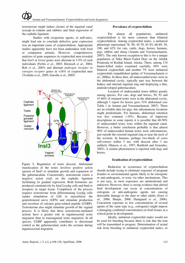
Figure 5. Regulation of testis descent. Abdominal
translocation of the testes involves positive action
(green) of Insl3 to stimulate growth and expansion of
the gubernaculum. Concurrently, testosterone exerts a
negative action (red) on the cephalic ligament
facilitating its gradual regression. Both hormones are
produced constitutively by fetal Leydig cells and bind to
receptors in target tissue. Completion of the process
requires testosterone from differentiating Leydig cells
(under stimulation of LH) to masculinize the
genitofemoral nerve (GFN) and stimulate production
and secretion of calcium gene-related peptide (CGRP).
Testosterone also might stimulate growth of the vaginal
process. It is likely that these testosterone-mediated
actions have a greater role in inguinoscrotal testis
migration than in transinguinal testis migration. In all
species, CGRP apparently contributes to directional
control as the gubernaculum seeks the scrotum during
inguinoscrotal migration.
Prevalence of cryptorchidism
For almost all populations, unilateral
cryptorchidism is far more common than bilateral
cryptorchidism. Among cryptorchid males, a unilateral
phenotype represented 78, 90, 45-70, 81-93, 66-89, 59,
100, and 62% for cats, cattle, dogs, horses, humans,
pigs, rabbits and sheep (Amann and Veeramachaneni,
2007). The only known exception to this is for a unique
population of Sitka Black-Tailed Deer on the Aliulik
Peninsula of Kodiak Island, Alaska. There, among 134
hunter-killed males examined carefully, 69% were
bilateral cryptorchids and another 7% were unilateral
cryptorchids (unpublished update of Veeramachaneni et
al., 2006a). In these deer, all undescended testes were in
the abdominal cavity, typically part way between the
kidney and internal inguinal ring and displaying a thin,
underdeveloped gubernaculum.
Location of undescended testes differs greatly
among species. For cats, dogs and horses, 50, 92 and
47-60% of retained testes were in the abdominal cavity,
although 1 report for horses gave 33% abdominal (see
Table 1 in Amann and Veeramachaneni, 2007). There
are no reliable data for pigs, but subcutaneous locations
might predominate. For humans, abdominal retention
was less common (~8%). Because of imprecise
descriptions in some reports it is possible that 40-50%
of undescended testes were within the inguinal canal.
However, a better conclusion probably is that almost
90% of undescended human testes were subcutaneous,
just outside the external inguinal ring or near the neck of
the scrotum. In humans, perhaps two-thirds of cases
self-correct within 3 mo, with descent after 3 mo
unlikely (Hutson et al., 1997; Barthold and Gonzalez,
2003). A similar phenomenon is reported with dogs and
horses.
Eradication of cryptorchidism
Reduction in occurrence of cryptorchidism
should include trying to eliminate exposure of pregnant
females to environmental agents likely to be estrogenic
or anti-androgenic, or toxic via other mechanisms. This
is not easy, as most exposures are unintentional and
unknown. However, there is strong evidence that altered
fetal development can occur at concentrations of
estrogenic or anti-androgenic agents not causing
detectable damage to the dam or other adults (Gore et
al., 2006; Sharpe, 2006; Damgaard et al., 2006).
Concurrent exposure to low concentrations of several
agents of the same type (e.g., estrogenic) might impose
a damaging combined concentration on fetal tissues at a
critical point in development.
Ideally, unilateral cryptorchid males would not
be used for breeding because there is risk that the trait
will be transmitted to progeny. Determination of actual
risk from breeding to unilateral cryptorchid males, or
Amann and Veeramachaneni. Cryptorchidism and testis dysgenesis.
Anim. Reprod., v.3, n.2, p.108-120, April/June. 2006 116
the sire or siblings of cryptorchids, would be difficult
and costly. However, special brother-sister matings of
dogs or pigs over several generations increases the
incidence of cryptorchidism (Cox et al., 1978; Mikami
and Fredeen, 1979; McPhee and Buckley, 1984). There
is anecdotal opinion that, for horses or pigs, there is
familial prevalence of cryptorchidism in some sire lines.
Breeders of race horses or dogs, simply remove
an undescended testis from a valuable unilateral
cryptorchid and then continue to use him for breeding.
They do not eliminate parents or siblings from breeding
stock. Most cryptorchid bulls are slaughtered and
unilateral cryptorchid bulls usually are not used for
breeding. Cryptorchid boars typically are killed
neonatally as they are deemed unsuitable for breeding
and rearing to market weight would result in a carcass
with greatly reduced value [problem is with “boar odor”
resulting from 5-androst-16ene-3one produced by
remaining testis tissue]. To cull non-cryptorchid male or
female littermates, much less the dam, would impose an
unacceptable economic penalty.
Other abnormalities associated with cryptorchidism
For reasons evident in Fig. 6, fetal testis
dysgenesis resulting in cryptorchidism also might be
manifested in a diversity of other reproductive
abnormalities later in life. Importantly, non-cryptorchid
males might display abnormalities in testis function or
behavior consequent to fetal testis dysgenesis. It is
known from breeding studies and pedigree analysis that
cryptorchidism is among many genetic conditions
displaying “incomplete penetrance”; not all animals
with abnormal DNA express the undesired phenotype.
Also, it is not evident that an aberrant genome accounts
for most reproductive abnormalities.
Environmental agents can produce a spectrum
of detected (and undetected) alterations in reproductive
development (Sharpe and Irvine, 2004; Sharpe, 2006).
A variety of animals including cattle (Veeramachaneni
et al., 1986), horses (Veeramachaneni and Sawyer,
1996, 1998), and deer (Veeramachaneni et al., 2006a)
“spontaneously” (i.e., in non-experimental situations)
manifest TDS. Rabbits (Higuchi et al., 2003;
Veeramachaneni et al., 2006b, 2007) and rats (Gray et
al., 2000, 2001) can be experimentally manipulated to
produce multiple lesions associated with TDS. Most
important to individuals working with companion or
food-producing animals might be reduced production of
sperm and/or increased proportion of abnormal sperm
(Veeramachaneni, 2000, 2006).
Testis tumors occur in non-cryptorchid males
as well as unilateral or bilateral cryptorchid males.
Abdominal location per se does not induce cell
transformations (Veeramachaneni, 2006). However, it is
likely that testis tumors occur 4-11 times more frequently
in cryptorchid than non-cryptorchid males.
Classification of testis tumors in animals (Kennedy et
al., 1998; Amann and Veeramachaneni, in preparation)
groups them as: germ cell tumors including carcinoma
in situ (CIS), gonocytic seminoma, and spermatocytic
seminoma; non-germ cell tumors including Leydig cell
tumors, Sertoli cell tumors, stromal cell tumors, and
adenoma of the rete testis; mixed tumors; and tumor-
like lesions including Leydig cell hyperplasia and
microlithiasis.
Based primarily on data for humans, it is
thought that CIS cells are formed, from primordial germ
cells, early in testis development and later give rise to
other types of germ cell tumors (Fig. 6; Almstrup et al.,
2004; Rajpert-De Meyts et al., 2004). CIS cells are
found in both abdominal and scrotal testes, without or
with overt evidence of other abnormalities (Giwercman,
1992, Hoei-Hansen et al., 2003; Veeramachaneni et
al., 2001, 2006a, 1996b, 2007). Failure of testicular
descent per se does not explain the transformation of
primordial germ cells into CIS cells (Veeramachaneni,
2006).
Tabulations on occurrence of testis tumors in
animals are inadequate to allow any meaningful
estimation of prevalence within a species, because of
under reporting and disparity in age when most males of
different species are castrated or killed/die. However,
there are important species differences in most
commonly reported testis tumors (Kennedy et al., 1998;
Amann and Veeramachaneni, in preparation). Germ cell
tumors are rare in cattle, but there are approximately
64% Leydig cell tumors and 30% Sertoli cell tumors.
Although testis tumors are not common in horses,
gonocytic seminoma is the most frequent testis tumor in
horses, followed by teratoma or Leydig cell tumors.
Spermatocytic seminoma accounts for 32-48% of testis
tumors in dogs (some of these might be gonocytic
seminomas), with 27-42 % Leydig cell tumors and 20-
40% Sertoli cell tumors. Gonocytic seminomas are more
likely to be invasive than spermatocytic seminomas
(Maiolino et al., 2004). In pigs, sheep, and rabbits, testis
tumors are rarely reported. However, in the first two
species gonocytic seminomas are most common
whereas in rabbits Leydig cell tumors apparently are
most common.
For rodents, incidence of spontaneous testis
tumors is dependent on strain, but not inordinate at 24
mo of age. It might be higher in older rats than older
mice (Clegg et al., 1997; Cook et al., 1999; Biegel,
2001). In both species Leydig cell tumors are by far the
most common. In rats, incidence of Leydig cell tumors
at 24 mo is 0.1-7.0%, except for Fisher 344 males or
cross-breed animals with 76-94% of males developing
tumors. In 24-mo old mice, incidence of Leydig cell
tumors at 0.4-2.5% and Sertoli cell tumors at 0.1% were
tabulated on the web site of one vendor.
Amann and Veeramachaneni. Cryptorchidism and testis dysgenesis.
Anim. Reprod., v.3, n.2, p.108-120, April/June. 2006 117
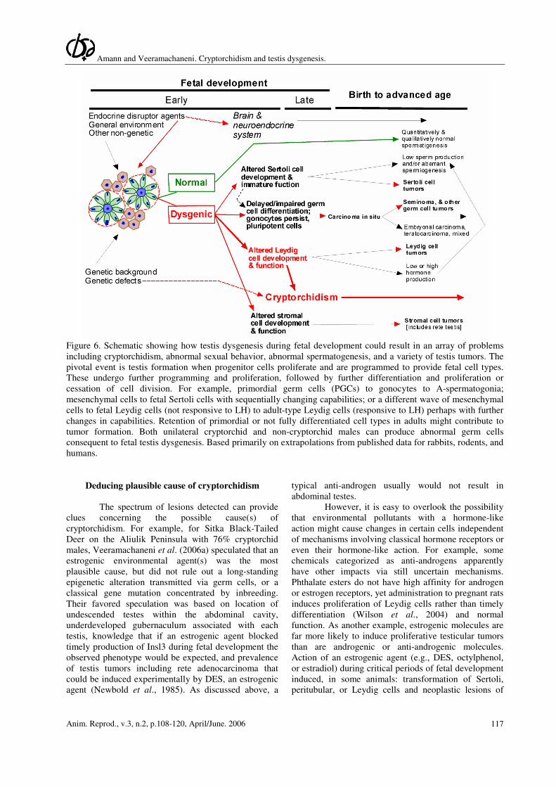
Figure 6. Schematic showing how testis dysgenesis during fetal development could result in an array of problems
including cryptorchidism, abnormal sexual behavior, abnormal spermatogenesis, and a variety of testis tumors. The
pivotal event is testis formation when progenitor cells proliferate and are programmed to provide fetal cell types.
These undergo further programming and proliferation, followed by further differentiation and proliferation or
cessation of cell division. For example, primordial germ cells (PGCs) to gonocytes to A-spermatogonia;
mesenchymal cells to fetal Sertoli cells with sequentially changing capabilities; or a different wave of mesenchymal
cells to fetal Leydig cells (not responsive to LH) to adult-type Leydig cells (responsive to LH) perhaps with further
changes in capabilities. Retention of primordial or not fully differentiated cell types in adults might contribute to
tumor formation. Both unilateral cryptorchid and non-cryptorchid males can produce abnormal germ cells
consequent to fetal testis dysgenesis. Based primarily on extrapolations from published data for rabbits, rodents, and
humans.
Deducing plausible cause of cryptorchidism
The spectrum of lesions detected can provide
clues concerning the possible cause(s) of
cryptorchidism. For example, for Sitka Black-Tailed
Deer on the Aliulik Peninsula with 76% cryptorchid
males, Veeramachaneni et al. (2006a) speculated that an
estrogenic environmental agent(s) was the most
plausible cause, but did not rule out a long-standing
epigenetic alteration transmitted via germ cells, or a
classical gene mutation concentrated by inbreeding.
Their favored speculation was based on location of
undescended testes within the abdominal cavity,
underdeveloped gubernaculum associated with each
testis, knowledge that if an estrogenic agent blocked
timely production of Insl3 during fetal development the
observed phenotype would be expected, and prevalence
of testis tumors including rete adenocarcinoma that
could be induced experimentally by DES, an estrogenic
agent (Newbold et al., 1985). As discussed above, a
typical anti-androgen usually would not result in
abdominal testes.
However, it is easy to overlook the possibility
that environmental pollutants with a hormone-like
action might cause changes in certain cells independent
of mechanisms involving classical hormone receptors or
even their hormone-like action. For example, some
chemicals categorized as anti-androgens apparently
have other impacts via still uncertain mechanisms.
Phthalate esters do not have high affinity for androgen
or estrogen receptors, yet administration to pregnant rats
induces proliferation of Leydig cells rather than timely
differentiation (Wilson et al., 2004) and normal
function. As another example, estrogenic molecules are
far more likely to induce proliferative testicular tumors
than are androgenic or anti-androgenic molecules.
Action of an estrogenic agent (e.g., DES, octylphenol,
or estradiol) during critical periods of fetal development
induced, in some animals: transformation of Sertoli,
peritubular, or Leydig cells and neoplastic lesions of
Amann and Veeramachaneni. Cryptorchidism and testis dysgenesis.
Anim. Reprod., v.3, n.2, p.108-120, April/June. 2006 118
rete testis and/or cystic lesions of excurrent ducts
(Newbold et al., 1985, 2000); and transformation of
primordial germ cells into CIS cells and/or
cryptorchidism (Veeramachaneni, 2000, 2006;
Veeramachaneni et al., 2007). Tumors in the rete testis
occurred transgenerationally without further exposure of
descendents (Newbold et al., 2000). Mechanism of
tumor-induction in fetal testes is uncertain.
Acknowledgment
Access to historic publications by John Hunter
was graciously provided by Simon Chaplin, Senior
Curator, Museums of the Royal College of Surgeons of
England, London, UK. Partial support provided by NIH
Grant 1R21-ES014607-01. The authors have nothing to
declare in respect to conflicting financial interests or
relationships with any commercial product or entity.
References
Almstrup K, Hoei-Hansen, Wirkner, Blake J, Leffers
H. 2004. Embryonic stem cell-like features of testicular
carcinoma in situ revealed by genome-wide gene
expression profiling. Cancer Res, 64:4736-4743.
Amann RP, Veeramachaneni DNR. 2007. Testicular
dysgenesis in animals: Cryptorchidism. Reproduction,
Submitted.
Barthold SJ, Gonzalez R. 2003. The epidemiology of
congenital cryptorchidism, testicular ascent and
orchiopexy. J Urol, 170:2396-2401.
Basrur PK, Basrur VR. 2004. Genes in genital
malformations and male reproductive health. Anim
Reprod, 1:64-85.
Beltran-Brown F, Villegas-Alverez F. 1988. Clinical
classification for undescended testes: experience in
1,000 orchidopexies. J Pediatr Surg, 23:444-447.
Bergin WC, Gier HT, Marion GB, Coffman JR.
1970. A developmental concept of equine
cryptorchidism. Biol Reprod, 3:82-92.
Biegel LB, Hurtt ME, Frame SR, O’Connor JC,
Cook JC. 2001. Mechanisms of extrahepatic tumor
indication by peroxisome proliferators in male CD rats.
Toxicol Sci, 60:44-55.
Clegg ED, Cook JC, Chapin RE, Foster PMD,
Daston GP. 1997. Leydig cell hyperplasia and adenoma
formation: mechanisms and relevance to humans.
Reprod Toxicol, 11:107-121.
Cook JC, Klinefelter GR, Hardisty JF, Sharpe RM, Foster PMD. 1999. Rodent Leydig cell tumorigenesis:
a review of the physiology, pathology, mechanisms, and
relevance to humans. Crit Rev Toxicol, 29:169-261.
Cox JE, Edwards GB, Neal PA. 1979. An analysis of
500 cases of equine cryptorchidism. Equine Vet J,
11:113-116.
Cox VS, Wallace LJ, Jessen CR. 1978. An anatomic
and genetic study of canine cryptorchidism. Teratology,
18:233-240.
Czeizel A, Erödi E, Toth J. 1981. Genetics of
undescended testes. J Urol, 126:528-529.
Damgaard IN, Skakkebaek NE, Toppari J, Virtanen
HE, Shen H, Schramm K-W, Petersen JH, Jensen TK, Main KM. 2006. Persistent pesticides in human
breast milk and cryptorchidism. Environ Health
Perspect, 114:1133-1138.
de Graaf R. 1668. A treatise concerning the generative
organs of men [in Latin]. Translation in: Jocelyn HD
and Setchell BP. 1972. Regnier de Graaf on the human
reproductive organs. J Reprod Fertil Suppl, 17:12.
Edwards MJ, Smith MSR, Freeman B. 2003.
Measurement of the linear dynamics of the descent of
the bovine fetal testis. J Anat, 203:133-142.
Elder JS, Isaacs JT, Walsh PC. 1982. Androgens
sensitivity of the gubernaculum testis: evidence for
hormonal/mechanical interactions in testicular descent.
J Urol, 127:170-176.
Ferlin A, Simonato M, Bartoloni L, Rizzo G, Bettella A,
Dottorini T, Dallapiccola B, Foresta C. 2003. The Insl3-
LGR8/GREAtligand receptor pair in human
cryptorchidism. J Clin Endocrinol Metab, 88:4273-4279.
Garolla A, Ferlin A, Vinanzi C, Roverato A, Sotti G,
Artiani W, Foresta C. 2005. Molecular analysis of the
androgen receptor gene in testicular cancer. Endo-
Related Cancer, 12:645-655.
Genetzky RM. 1984. Equine cryptorchidism:
pathogenesis, diagnosis, and treatment. Compend Cont
Educ, 6:S577-S582.
Gier HT, Marion GB. 1970. Development of the
mammalian testis. In: Johnson AD, Gomes WR,
VanDemark NL (Eds.). The Testis. New York, USA:
Academic Press. Vol.1, pp.1-45.
Giwercman A. 1992. Carcinoma-in-situ of the testis:
Screening and management. Scand J Urol Nephrol
Suppl, 148:1-47.
Gore AC, Heindel JJ, Zoeller RT. 2006. Endocrine
disruption for endocrinologists (and others).
Endocriniology, 147(Suppl): S1-S3.
Gray LE Jr, J Ostby, J Furr, M Price, DNR
Veeramachaneni, Parks L. 2000. Perinatal exposure to
the phthalates DEHP, BBP and DINP, but not DEP,
DMP or DOTP alters sexual differentiation of the male
rat. Toxicol Sci, 58:350-365.
Gray LE Jr, Ostby J, Furr CJ, Wolf CJ, Lambright
C, Parks L, Veeramachaneni DN, Wilson V, Price
M, Hotckiss A, Orlando E, and Guillette L. 2001.
Effects of environmental antiandrogens on reproductive
development in experimental animals. Human Reprod
Update, 7:248-264.
Hadziselimovic F, Herzog B, Girard J, Stalder G.
1984. Cryptorchidism: histology, fertility and treatment.
Prog Reprod Biol Med, 10:1-15.
Higuchi TT, Palmer JS, Gray LE Jr,
Veeramachaneni DN. 2003. Effects of dibutyl
phthalate in male rabbits following in utero, adolescent,
or postpubertal exposure. Toxicol Sci, 72:301-313.
Hoei-Hansen C, Hilm M, Rajpert-De Meyts,

Amann and Veeramachaneni. Cryptorchidism and testis dysgenesis.
Anim. Reprod., v.3, n.2, p.108-120, April/June. 2006 119
Skakkebaek NE. 2003. Histological evidence of
testicular dysgenesis in contralateral biopsies from 218
patients with testicular germ cell cancer. J Pathol,
200:370-374.
Huhtaniemi I, Poutanen M. 2004. Transgenic and
knockout mouse models for aberrant pituitary-testicular
function: relevance to the pathogenesis of
cryptorchidism. Turk J Pediat, 46(Suppl):28-34.
Hunter J. 1762. Observations on the state of the testes
in the foetus, and on the hernia congenita. In: Hunter W.
Medical commentaries, Part 1. London: A Hamilton for
A Millar. pp.107-120. Available at http://
surgicat.rcseng.ac.uk/media/pdfs/hunter-1762-p75-
89.pdf.
Hunter J. 1786. A description of the situation of the
testis in the foetus, with its descent into the scrotum. In:
Hunter J. Observations on certain parts of the animal
oeconomy. Read as reprinted in Palmer JF (Ed.).
Collected works of John Hunter, FRS, London:
Longman and Green. Vol.4, pp.1-26. Available
at.http://surgicat.rcseng.ac.uk/ media/pdfs/works-v4-p1-
19.pdf]
Hutson JM, Hasthorpe S. 2005. Testicular descent and
cryptorchidism: the state of the art in 2004. J Pediatr
Surg, 40:297-302.
Hutson JM, Hasthorpe S, Heyns CF. 1997.
Anatomical and functional aspects of testicular descent
and cryptorchidism. Endocr Rev, 18:259-280.
Hutson JM, Watts LM, Farmer PJ. 1998. Congenital
undescended testes in neonatal pigs and the effect of
exogenous calcitonin gene-related peptide. J Urol,
159:1025-1028
Hutson JM, Baker ML, Griffiths AL, Momosa Y,
Goh DW, Middlesworth W, Yum ZB, Cartwright E.
1992. Endocrine and morphological perspectives in
testicular descent. Reprod Med Rev, 1:165-177.
Kennedy PC, Cullen JM, Edwards JF, Goldsmidt
MH, Larsen S, Munson L, Nielsen S. 1998. Tumors of
the genital system of domestic animals; Series 2.
Washington, DC: American Registry of Pathology.
Vol.4.
Klonisch T, Fowler PA, Hombach-Klonisch S. 2004.
Molecular and genetic regulation of testis descent and
external genitalia development. Dev Biol, 270:1-18.
Lam SKL, Clarnette TD, Hutson JM. 1998. Does the
gubernaculum migrate during inguinoscrotal testicular
descent in the rat? Anat Rec, 250:159-163.
Maiolino P, Restucci B, Papparella S, Paciello O, de
Vico G. 2004. Correlation of nuclear morphometric
features with animal and human World Health
Organization international classifications of canine
spontaneous seminomas. Vet Pathol, 41:608-611.
McMahon DR, Kramer SA, Husmann DA. 1995.
Antiandrogen induced cryptorchidism in the pig is
associated with failed gubernacular regression and
epididymal malformations. J Urol, 154:553-557.
McPhee HC, Buckley SS. 1984. Inheritance of
cryptorchidism in swine. J Hered, 25:295-303.
Mikami H, Fredeen HT. 1979. A genetic study of
cryptorchidism and scrotal hernia in pigs. Can J Genet
Cytol, 21:9-19.
Mylchreest E, Sar M, Cattley RC, Foster PMD.
1999. Disruption of androgen-regulated male
reproductive development by di(-butyl)phthalate during
late gestation in rats is different from flutamide. Toxicol
Appl Pharm, 156:81-95.
Nef S, Shipman T, Parada LF. 2000. A molecular
basis for estrogen-induced cryptorchidism. Dev Biol,
224: 354-361.
Newbold RR, Bullock BC, McLachlan JA. 1985.
Lesions of the rete testis in mice exposed prenatally to
diethylstilbestrol. Cancer Res, 45:5145-5150.
Newbold RR, Hanson RB, Jefferson WN, Bullock BC, Haseman J, McLachlan JA. 2000. Proliferative
lesions and reproductive tract tumors in male
descendants of mice exposed developmentally to
diethylstilbestrol. Carcinogenesis, 21:1355-1363.
Ng SL, Bidkar SS, Sourial M, Farmer PJ, Donath S,
Hutson JM. 2005. Gubernacular cell division in
different rodent models of cryptorchidism supports
indirect androgenic action via the genitofemoral nerve. J
Pediatr Surg, 40:434-441.
Nielen ALJ, Janss LLG, Knol BW. 2001. Heritability
estimations for diseases, coat color, body weight, and
height in a birth cohort of Boxers. Am J Vet Res,
62:1198-1206.
Rajfer J. 1980. Morphological study of testicular
descent in the rabbit. Invest Urol, 18:293-295
Rajpert-De Meyts E. 2006. Developmental model for
the pathogenesis of testicular carcinoma in situ: genetic
and environmental aspects. Hum Reprod Update,
12:303-323.
Rajpert-De Meyts E, Hanstein R, Jørgensesn N,
Graem N, Vogt PH, Skakkebaek NE. 2004.
Developmental expression of POU5F1 (OCT-3/4) in
normal and dysgenetic human gonads. Human Reprod,
19:1338-1344.
Rodgerson DH, Hanson RR. 1997. Cryptorchidism in
horses. Part I. Anatomy, causes, and diagnosis.
Compend Cont. Educ Pract Vet, 19:1280-1288.
Roh J, Virtanen H, Kumagai J, Sudo S, Kaleva M,
Hsueh AJW. 2003. Lack of LGR8 gene mutation in
Finish patients with a family history of cryptorchidism.
Reprod Bio Med Online, 7:400-406.
Rothschild MF, Christian LL, Blanchard W. 1988.
Evidence for multigene control of cryptorchidism in
swine. J Hered, 79:313-314.
Sharpe RM. 2006. Pathways of endocrine disruption
during male sexual differentiation and masculinisation.
Best Pract Res Clin Endocr Metabol, 20:91-110.
Sharpe RM, Irvine DS. 2004. How strong is the
evidence of a link between environmental chemicals
and adverse effects on human health? Br Med J,
328:447-451.
Shono T, Ramm-Anderson S, Hutson JM. 1994a.
Transabdominal testicular descent is really ovarian

Amann and Veeramachaneni. Cryptorchidism and testis dysgenesis.
Anim. Reprod., v.3, n.2, p.108-120, April/June. 2006 120
ascent. J Urol, 152:781-784.
Shono T, Ramm-Anderson S, Goh DW, Hutson JM.
1994b. The effect of flutamide on testicular descent in
rats examined by scanning electron microscopy. J
Pediat Surg, 29:839-844.
Shono T, Hutson JM, Watts L, Goh DW, Momose Y,
Middlesworth B, Zhou B, Ramm-Anderson S. 1996.
Scanning electron microscopy shows inhibited
gubernacular development in relation to undescended
testes in oestrogen-treated mice. Int J Androl, 19:263-
270.
Sittmann K, Woodhouse B. 1977. Sex-limited and sex-
modified genetic defects in swine — cryptorchidism.
Can J Genet Cytol, 19:487-502.
Skakkebaek NE, Rajpert-De Meyts E, Main KM.
2001. Testicular dysgenesis syndrome: an increasingly
common developmental disorder with environmental
aspects. Hum Reprod, 16:972-978.
Skakkebaek NE, Rajpert-De Meyts E, Jørgensen N,
Carlsen E, Petersen P, Giwercman A, Andersen A-
G, Jensen TK, Andersen A-M, Müller J. 1998. Germ
cell cancer and disorders of spermatogenesis: an
environmental connection? APMIS, 106:3-12.
Spencer JR, Torrado T, Sanchez RS, Vaughn ED Jr,
Imperato-McGinley J. 1991. Effects of flutamide and
finasteride on rat testicular descent. Endocrinology,
129:741-748.
van der Schoot P. 1993. Foetal testes control the
prenatal growth and differentiation of the gubernacular
cones in rabbits – a tribute to the late Professor Alfred
Jost. Development, 118:1327-1334.
van der Schoot P. 1996. Towards a rational
terminology in the study of the gubernaculum testis:
arguments in support of the notion that the cremasteric
sac should be considered the gubernaculum in postnatal
rats and other mammals. J Anat, 189:97-108.
van der Schoot P, Elger W. 1993. Prenatal
development of gubernacular cones in rats and rabbits:
effect of exposure to anti-androgens. Anat Rec,
236:399-407.
Veeramachaneni DN. 2000. Deteriorating trends in
male reproduction: idiopathic or environmental? Anim
Reprod Sci, 60-61:121-130.
Veeramachaneni DN. 2006. Germ cell atypia in
undescended testes hinges on the aetiology of
cryptorchidism but not the abdominal location per se.
Int J Androl, 29:235-240.
Veeramachaneni DNR, Sawyer HR. 1996. Use of
semen as a biopsy material for assessment of health
status of the stallion reproductive tract. Vet Clin North
Am: Equine Pract, 12:101-110.
Veeramachaneni DNR, Sawyer HR. 1998. Carcinoma
in situ and seminoma in equine testis. Acta Pathol
Microbiol Immunol Scand, 106:183-185.
Veeramachaneni DNR, Amann RP, Jacobson JP.
2006a. Testis and antler dysgenesis in Sitka Black-
Tailed Deer on Kodiak Island, Alaska: sequela of
environmental endocrine disruption? Env Hlth Perspect,
114(Suppl 1):51-59.
Veeramachaneni DNR, Palmer JS, Amann RP, Pau
K-YF. 2007. Sequelae in male rabbits following
developmental exposure to p,p'-DDT or a mixture of
p,p'-DDT and vinclozolin: cryptorchidism, germ cell
atypia, and sexual dysfunction. Reprod Toxicol,
Submitted.
Veeramachaneni DNR, Ott RS, Heath EH, McEntee
K, Bolt DJ, Hixon JE. 1986. Pathophysiology of small
testes in beef bulls: Relationship between scrotal
circumference, histopathologic features of testes and
epididymides, seminal characteristics and endocrine
profiles. Am J Vet Res, 47:1988-1999.
Veeramachaneni DNR, Palmer JS, Amann RP, Kane
CM, Higuchi TT, Pau K-YF. 2006b. Disruption of
sexual function, FSH secretion, and spermiogenesis in
rabbits following developmental exposure to
vinclozolin, a fungicide. Reproduction,131:805-816.
Wensing CJG. 1968. Testicular descent in some
domestic mammals. I. Anatomical aspect of testicular
descent. Proc Kon Neder Akad Wet, Series C, 71:423-
434.
Wensing CJG. 1986. Testicular descent in the rat and a
comparison of this process in the rat with that in the pig.
Anat Rec, 214:154-160.
Wensing CJG. 1988. The embryology of testicular
descent. Horm Res, 30:144-152.
Wensing CJG, Colenbrander B. 1986. Normal and
abnormal testicular descent. In: Clarke JR (Ed.). Oxford
reviews reproductive biology. Oxford, UK: Claredon
Press. Vol.8, pp 125-130.
Wilson VS, Lambright C, Furr J, Ostby J, Wood C,
Held G, Gray LE Jr. 2004. Phthalate ester-induced
gubernacular lesions are associated with reduced Insl3
gene expression in the fetal rat testis. Toxicol Let,
146:207-215.
Yoshida R, Fukami M, Sasagawa I, Hasegawa T,
Kamatani N, Ogata T. 2005. Association of
cryptorchidism with a specific haplotype of the estrogen
receptor gene: implication for the susceptibility to
estrogenic environmental endocrine disruptors. J Clin
Endo Metabol, 90:4716-4721.