cryopreservation of mesenchymal stem...
TRANSCRIPT

CRYOPRESERVATION OF MESENCHYMAL STEM CELL AND
TISSUE ENGINEERED CONSTRUCTS USING NON-TOXIC
CRYOPROTECTIVE AGENTS
A THESIS SUBMITTED FOR THE AWARD OF THE DEGREE OF
Doctor of Philosophy
in
Biotechnology and Medical Engineering
By
AKALABYA BISSOYI
(Roll No – 509BM101)
Under the Guidance of
Prof. Krishna Pramanik & Prof. S. K. Mahapatra
Department of Biotechnology and Medical Engineering
National Institute of Technology, Rourkela
2013


Dedicated To
My Parents

1 | P a g e
ACKNOWLEDGEMENT
I avail this opportunity to express my indebtedness, deep gratitude and sincere thanks to my
supervisor Prof. (Mrs.) K. Pramanik ,Professor and Head of the Department of Biotechnology and
Medical Engineering, National institute of technology, Rourkela for her in depth supervision and
guidance, constant encouragement and co-operative attitude for bringing out this thesis work
successfully. I enjoyed the freedom she gave me throughout this work that was instrumental in
discovering a scientific “self” within me. I feel proud that I am one of her doctoral students and I
consider myself extremely lucky to get opportunity to work under the guidance of such a dynamic
personality.
My special thanks are due to Prof. S. K. Sarangi, Director, National Institute of Technology, Rourkela
for his encouragement, valuable suggestion and providing all the facilities to successfully complete
this dissertation work.
I would like to extend a special thanks to Prof. S. K. Mahapatra for guidance to successfully complete
this work. I am also very thankful to all the members of my doctoral scrutiny committee – Prof. M. K.
Gupta and Prof A. Biswas of the Department of Biotechnology and Medical Engineering, Prof. S.
Mishra of the Department of Chemical Engineering and Prof. S. Das of the Department of Life science
for their thoughtful advice, inspiration and encouragement throughout the research work. I take this
opportunity to thank the other faculty members and the supporting staff members of the Department of
Biotechnology and Medical Engineering for their timely co-operation and support at various phases
of experimental work. I would like to extend a special thanks to my dear friends Divyanshu Mahajan,
Nadeem Siddique, Biswadeep Chaudhuri, Niladri Nath panda, Varshini Vishwanath and Bhisma
Narayan singh for their valuable suggestions and encouragement.
And it goes without saying, that I am indebted to my parents Mr. Sarat Chandra Bissoyi and Mrs.
Shanti Bissoyi and brother Sabyasachi Bissoyi, whose patience, support and endurance made
completion of my thesis. Above all, I would like to thank the Almighty for his enormous blessings.
I greatly indebted to my wife, Sumana Bissoyi who had always been very supportive, caring and
everlasting support she has given me.

2 | P a g e
ABSTRACT
The thesis work deals with the development of cryopreservation strategy for long term storage
of MSCs and MSCs seeded tissue engineered constructs using non toxic cryoprotective agents
as freezing medium. In the first phase, different freezing medium consisting of a combination
of natural extracellular cryoprotectants namely trehalose, hydroxyl ethyl starch, polyvinyl
pyrolidine and intracellular CPAs like erythritol, taurine and ectoin were used for
cryopreservation of MNCs following the Taguchi Orthogonal Array method. Among the
various combinations, freezing medium consisting of trehalose (0.05mM), ectoin (0.10mM)
and catalase (100µg/ml) has shown maximum MNCs viability. These CPAs were further
investigated individually as well as in combination to see their effectiveness towards long
term preservation of MSCs. Among the freezing solutions, solution prepared using trehalose
(0.3mM), ectoin (0.3mM), and catalase (100µg/ml) was found to be the most effective in
preserving MSCs in long term basis. The viability of MSCs (73%) is found to be higher than
the viability achieved with 10% (v/v) Me2SO (61%) used as control. The apoptotic study has
indicated that the addition of general caspase and calpain inhibitors can reduce the apoptosis
rate upto 10-15% thereby achieving increased cell viability of 80%. The optimum condition
for the controlled rate freezing of MSCs was established as prenucleation cooling rate -
1oC/min, nucleation temperature -7.5
oC, cold spike -80
oC/min, post nucleation holding time
5min, post nucleation cooling rate -1oC/min, cell density (3×10
6/ml/cell) and storage
temperature (-150°C ) using the most effective freezing medium achieving cell viability of
85%. The developed freezing medium has also shown its ability to preserve MSCs seeded
tissue engineered construct. The maximum viability of 80% achieved at optimum controlled
rate freezing of TECs was established as cooling rate -1oC/min, nucleation temperature -7.5OC
and freezing medium consisting of trehalose (0.3mM), ectoin (0.3mM), catalase (100µg/ml)
in presence of caspase (50µg) and calpain (50µg) inhibitors. Overall, it is demonstrated that
the developed freezing medium may pave the way for long term preservation of MSCs and
also MSCs seeded scaffold.
Keywords: Cryopreservation, scaffold, caspase inhibitors, trehalose, ectoin, tissue engineered
constructs, control rate freezer

3 | P a g e
LIST OF CONTENTS
Certificate i
Acknowledgement ii
Abstract iii
Contents iv
List of Figures vii
List of tables viii
List of abbreviations ix
CHAPTER I: General Introduction 1-12
1.1. Background 1
1.2 Preservation techniques for cells & Tissues 2
1.2.1 Mechanical freezers 2
1.2.2 Liquid nitrogen storage vessels 2
1.2.3 Controlled rate freezers 3
1.2.4 Vitrification 3
1.2.5 Sterling engines 3
1.3 Cryopreservation of cells and tissue 4
1.3.1 Principle of Cryopreservation 4
1.3.2 History of cryopreservation 4
1.4 Important factors involved in cryopreservation 5
1.4.1 Ice formation in a cell suspension 5
1.4.2 Rate of cooling and warming 5
1.5 Cryoprotectants 6
1.5.1. Membrane-permeating CPA 7
1.5.2. Non-permeating cryoprotectants 8
1.5.1 Cryoprotectant and toxicity 8
1.6 Stem cells 9
1.6.1 Types of stem cells 9
1.6.2 Application of stem cells 10
1.6.3 Mesenchymal stem cells: For tissue engineering 10
1.6.4 Stem cell preservation 11
1.6.5 Cryopreservation of Tissue engineering constructs 11

4 | P a g e
1.7 Cryopreservation of Tissue Engineering Construct 11
Chapter II: Literature review 13-30
2.1 Preservation of MSCs 13
2.1.1 Alternative cryoprotectant for preserving MSCs 13
2.1.2 Use of antioxidants and inhibitors as additive in freezing medium 15
2.2 Cryopreservation of TECs 18
2.2.1 Controlled Rate Freezing of TECs 20
2.2.2 Vitrification of TECs 20
2.3 Cryopreservation induced cell death 22
2.3.1 Modes of Cell Death 23
2.3.2 Apoptosis 23
2.3.3 Extrinsic and Intrinsic pathway 24
2.3.4 Necrosis and autophagy 27
2.3.5 Motivation for Cryopreservation 29
Chapter III: Scope and objective 31-34
Chapter IV: Materials and Methods 35-49
4.1 Chemicals and culture media 36
4.1.1 Cryoprotective agents 36
4.1.2 Antioxidants and inhibitors 36
4.1.3 Cell culture media and antibodies 36
4.2 Collection and processing of umbilical cord blood 37
4.3 Isolation and culture of MNCs 37
4.4 Sorting and culturing of MSCs 37
4.5 Characterization of cells 38
4.5.1 Morphological Characterisation 38
4.5.2 Immunophenotypic characterisation 38
4.6 Cryopreservation of cells 38
4.6.1 Preparation of cryopreservation solution 38
4.6.2 Cryopreservation experiments 39
4.6.3 Thawing and post-thaw culturing 39
4.6.4 Cell viability assay 40
4.6.5 Cell proliferation assay 40
4.6.6 MTT assay 40
4.6.7 Apoptotic assay 40
4.6.8 Mitochondrial membrane potential assay 41

5 | P a g e
4.6.9 Differentiation potential 41
4.6.10 CFU-F assay 42
4.6.11 Cell cycle analysis 42
4.6.12 Western blotting 42
4.6.13 Optimization of cryopreservation parameters 43
4.7 Cryopreservation of TECs 43
4.7.1 Preparation of scaffolds 43
4.7.2 Formation of TECs 44
4.7.3 Preparation of cryopreservation solutions 44
4.7.4 Cryopreservation experiment 44
4.8.4 Characterization of TECs 45
4.8.4.1 Cell morphology by SEM 45
4.8.4.2 Cell viability 45
4.8.4.3 Cell metabolic activity 46
4.8.4.4 Cytoskeleton analysis by confocal microscopy 46
4.8.4.4 Osteo-induction assessment 46
4.8.4.4 Alkaline phosphatase activity 46
4.8.4.5 Histological study 47
4.8.4.6 Osteogenic-specific gene Expression 47
4.8.4.7 Mechanical strength 48
Chapter V: Results and Discussion 50-66
5.1 A preliminary study on cryopreservation of MNCs: selection of
non-toxic CPAs 52
5.1.1 Isolation and culture MNCs 52
5.1.2 Cell morphology study 52
5.1.3 Cryopreservation solution 56
5.1.4 Cell viability and apoptosis study 56
5.1.5 Statistical analysis 59
5.1.6 Analysis of Variance (ANOVA) 59
5.1.7 Cell morphology analysis of cryopreserved cells 61
5.1.8 MTT assay 62
5.1.9 Post-thaw cytoskeleton assessment 63
5.1.10 Morphological analysis of cryopreserved MNCs 65
5.1.11 Conclusion 66
5.2 Cryopreservation of MSCs using freezing medium prepared from
non-toxic CPAs 68

6 | P a g e
5.2.1 Sorting of MSCs 68
5.2.2 Cell morphology study 70
5.2.3 Immunophenotypic Characterization of MSCs 71
5.2.4 Effect of CPA composition on post-thaw MSCs viability 74
5.2.5 Effect of Me2SO on cryopreservation of MSCs 75
5.2.6 MTT Assay 75
5.2.7 Post-thaw phenotyping of cryopreserved MSCs 76
5.2.8 Change in growth curve (population doubling) in 77
cryopreserved MSCs 77
5.2.9 Change of mitochondrial membrane potential in
cryopreserved MSCs 78
5.2.10 Cytoskeleton analysis of post-thawed MSCs 80
5.2.11 Effect of storage duration on cell viability 82
5.2.12 Effect of cryopreservation on differentiation
potential of hMSCs 85
5.2.13 Effect of cryopreservation on clonogenic potential
of MSCs 87
5.2.14 Study on apoptosis pathways in cryopreserved
induced cell death of MSCs 88
5.2.14.1 Post thaw cell viability 89
5.2.14.2 Caspase-mediated apoptotic signaling pathways 89
5.2.14.3 Western blotting analysis of caspase proteins 92
5.2.14.4 Mitochondrial membrane potential 93
5.2.14.5 Calpain activity 94
5.2.14.6 Western blotting analysis for calpain protein 97
5.2.14.7 Effect of calpain inhibitor on mitochondrial
membrane potential and DNA Degradation 97
5.2.15 Optimization of Controlled rate freezing parameters
for cryopreservation of hMSCs using T30E30C100
freezing medium 98
5.2.15.1 Effect of cooling profile on MSCs viability 98
5.2.15.2 Effect of cell density 101
5.2.15.3 Effect of storage temperature during cryopreservation 102
5.2.15.4 Verification of cryopreservation protocol 103
5.2.15.4.1 The effect on the MSCs cytoskeleton and mitochondria distribution
after cryopreservation 103
5.2.15.4.2 Cell cycle analysis 105
5.3 Cryopreservation of hMSCs seeded silk nanofibers based tissue
engineered constructs 107

7 | P a g e
5.3.1 Structural integrity and cell morphology in TECs 108
5.3.2 MSCs viability 111
5.3.3 Proliferation assay 112
5.3.4 Cytoskeleton analysis 113
5.3.5 Alkaline Phosphatase activity 115
5.3.6 Effect of cryopreservation on Mechanical properties
of TECs 116
5.3.7 Calcium deposition 117
5.3.8 Expression of osteogenic-specific genes 118
5.3.9 Optimization of controlled rate freezing parameters for
preservation of TECs using T30E30C100
freezing medium 120
5.3.9.1 Effect of cooling rate on MSCs viability in TECs 120
5.3.9.2 The effects of seeding temperature on the cell viability 120
5.3.9.3 Cell Morphology 121
Chapter V: Summary and Conclusion 124-127
Bibliography 128-151
List of Publications 152-153

8 | P a g e
LIST OF FIGURES
Sl. no. Figure caption Page
no.
Figure 1 Graphical presentation of two distinctive apoptosis pathways namely
intrinsic pathways and extrinsic pathways.
26
Figure 2 Mediators of apoptosis during cryopreservation and the effective
apoptosis inhibitors
29
Figure 3 Control rate setup for cryopreservation of MSCs, (A) controlled rate
freezer and (B) vapor phase storage tank
39
Figure 4 Control rate setup for cryopreservation of MSCs seeded SF scaffold,
(A) controlled rate freezer and (B) vapor phase storage tank.
45
Figure 5 Collection of Cord blood: (A) clamped and cut the cord just after the
birth of baby, (B) and (C) needle is inserted in umbilical cord with
collection bag, (D) cord blood allowed to fill the collection bag by
gravity.
53
Figure 6 Schematic representation of isolation procedure of MNC from
umbilical derived cord blood
54
Figure 7 Cell morphology within 7 days after initial culturing 55
Figure 8 Images show dot plots of cryopreserved and non-cryopreserved MNCs 59
Figure 9 Illustrates S/N ratio averages for each parameter at three levels. 60
Figure 10 Conformity test for best obtained level with the respective factor 62
Figure 11 MTT assay of post-thawed MNCs cryopreserved in different
compositions of extracellular cryoprotectant, intracellular, antioxidant
and FBS.
63
Figure 12 Growth and attachment of cryopreserved MNCs after 48 hrs of
incubation
64
Figure 13 SEM images of UCB derived MNCs 65
Figure14 Schematic representation of isolation procedure of MNCs from
umbilical derived cord blood
69
Figure 15 Phase contrast microscopy images showing morphological
characteristics of cultured hMSCs
70
Figure 16 Flowcytometric analysis on the expression of MSC markers CD90,
CD105, CD44, and CD73 as well as hematopoietic markers CD34,
72

9 | P a g e
HLA-DR and CD45
Figure 17 Immnuofluorescence observation of passage 4 post thaw MSCs 73
Figure 18 Effect of CPA composition on cell viability 74
Figure 19 MTT assay of post-thawed MSCs cryopreserved in different
compositions of extracellular cryoprotectant, intracellular, antioxidant
with and without Me2SO
76
Figure 20 Culture growth curves of post thaw hMSCs with different composition
of Trehalose, ectoin, and catalase
77
Figure 21: Analysis of mitochondrial membrane potential (ΔΨ m) of post thaw
MSCs.
79-
80
Figure 22 F-actin morphology and distribution in cryopreserved hMSCs 81
Figure 23 Cytometric analysis of MSCs apoptosis performed using 7AAD and
annexin.
84
Figure 24 Representative phase contrast images of differentiated hMSCs. 86
Figure 25 Effect of cryopreservation on CFU-F ability of MSCs. 87
Figure 26 Effects of cryopreservation on cell viability in presence of caspase and
calpain inhibitors
89
Figure 27 Effect of caspase inhibitors on cryopreserved-induced apoptosis of
MSCs
91
Figure 28 Effect of cryopreservation on activation of caspase and Bid proteins. 93
Figure 29 Effect of caspase inhibition on cryopreserved-induced mitochondrial
membrane potential and DNA degradation in MSCs after 24 hrs of
post thaw
94
Figure 30 Effect of calpain inhibitors (PD 150606) on cryopreserved-induced
apoptosis.
95
Figure 31 Effect of cryopreservation on activation of µ-calpain 96
Figure 32 Effect of calpain inhibition on cryopreserved-induced mitochondria
membrane potential and DNA degradation in MSCs after 24 h of post
thaw
97
Figure 33 Percentage recovery of MSCs after cryopreservation in controlled rate
freezer under different cooling variables.
100
Figure 34 Temperature profile of cryochamber and sample with time 101

10 | P a g e
Figure 35 Effect of cell concentration during cryopreservation on cell viability. 102
Figure 36 Effect of different storage temperature of cryopreserved MSCs on cell
viability
103
Figure 37 Fluorescence image of F-actin and mitochondria distribution in fresh
and cryopreserved hMSCs.
104
Figure 38 Cell cycle analysis of (A) non-cryopreserved MSCs and (B)
Cryopreserved MSCs with T30/E30/C100
105
Figure 39 SEM images of (A) Precryopreserved scaffold and (B) cryopreserved
scaffold
109
Figure 40 SEM images of cryopreserved scaffold 110
Figure 41 Cell viability after 24h of post thaw TECs in different freezing
solution
111
Figure 42 MTT assay of the metabolic activities of post thaw MSCs in SF
scaffolds
112
Figure 43 Confocal images of cytoskeleton of MSCs in cryopreserved and non
cryopreserved TECs.
114
Figure 44 ALP activity of post thawed MSCs in TECs on 1, 7, 14 and 21days
culture.
127
Figure 45 Ultimate tensile strength of TECs under three different conditions. 128
Figure 46 Optical images of alizarin red staining for calcium deposition of
osteogenically induced MSCs in post thaw TECs
129
Figure 47 SEM and EDX analysis of post-thaw TECs cultured in osteogenic
differentiation medium for 21 days
130
Figure 48 Osteogenic gene expression levels of post thaw MSCs in TECs
cultured in osteogenic media
131
Figure 49 Effect of different cooling rate on MSCs seeded TECs 132
Figure 50 Effect of seeding temperature on MSCs seeded TECs 133
Figure 51 SEM images of silk-nanofiber TECs under different cooling rate 134
LIST OF TABLE

11 | P a g e
Sl. no. Table caption Pag
e no.
Table 1 Factors influencing cryopreservation of TEC 21
Table .2 Cryopreservation of tissue engineering constructs by vitrification
as reported
23
Table 3 The specific primers for RT-PCR 47
Table 4 Taguchi L9 array for nine cryoprotectant solutions with different
compositions
56
Table 5 S/N ratio and percentage of cells in Q3 quadrant (viable cell) 57
Table 6 ANOVA analysis of experiments consider in Taguchi orthogonal
array
61
Table 7 Effect of Me2SO as an additive in freezing solution on cell viability 75
Table 8 Immunophenotypic characterisation of MSCs after cryopreservation 77
Table 9 Population doubling time of MSCs 78
Table 10 Effect of storage time on MSCs viability 83
Table 11 Comparison of cell viability of cryopreserved MSCs in T30E30C100
solutions and Me2SO after 12months of storage
106

12 | P a g e
LIST OF ABBREVIATION
ADP Adenosine diphosphate
ASC AT derived Stromal Cells
AIF Apoptosis inducing factor
Apo3L Apo3 ligand
Apo2L Apo2 ligand
Apaf-1 Apoptotic protease activating factor 1
BAG BCL2 associated athanogene
BA Bongkrekic acid IAPs
BAD BCL2 antagonist of cell death
BAK BCL2 antagonist killer 1
BAX Bcl-2–associated X protein
Bcl-w BCL2 like 2 protein
Bcl-x BCL2 like 1
βME β-mercaptoethanol
BM-MSCs Bone Marrow derived MSCs
BIK BCL2 interacting killer
Bcl-XL BCL2 related protein, long isoform
Bcl-XS BCL2 related protein, short isoform
Bcl-2 B-cell lymphoma 2
BIM BCL2 interacting protein BIM
BNIP3 BCL2/adenovirus
BSA Bovine Serum Albumin
13 | P a g e
CAD Caspase-activated deoxyribonuclease
Caspase c Cysteinyl aspartic acid-protease
CD Cluster of Differentiation
c-FLIP Cellular form of FLICE-inhibitory protein
CFU-F Fibroblast Colony Forming Unit
CIDOCD Cryopreservation-induced, delayed-onset cell death
C100 Catalase 100µg
CPDA Citrate Phosphate Dextrose Adenine
CB Cord Blood
dATP Deoxyadenosine triphosphate
DD Death domain
D10 10% v/v Dimethyl sulfoxide
DR3 Death receptor 3
DR4 Death receptor 4
DR5 Death receptor 5
DISC Death-inducing signaling complex
DNA Deoxyribonucleic acid
DMEM-LG Dulbecco’s modified Eagle’s media-low glucose
EndoG Endonuclease G
EDTA Ethylenediaminetetraacetic acid
EGF Epidermal Growth Factor
E30 Ectoin (30mM)
ELISA Enzyme Linked Immuno-sorbent Assay
ERK Extracellular signal-regulated kinase
FACS Flow Activated Cell Sorter
FADD Fas-associated death domain
FasL Fatty acid synthetase ligand

14 | P a g e
FBS Fetal Bovine Serum
FSC Forward Scatter
FasR Fatty acid synthetase receptor
IAPs Inhibitors of apoptosis proteins
GAGs Glycosaminoglycans
IGH Ispat General Hospital
JNK c-Jun N-terminal kinases
hAdMSCs human omentum Adipose derived MSCs
HLA Human leukocyte antigen
HTS Hypothermosol
HtrA2/Omi High-temperature requirement
KV Kilo Volts
kDa Kilo Dalton
MAP kinase Mitogen activated protein kinase
Me2SO Dimethyl sulfoxide
MPT Mitochondrial permeability transition pores
MNCs Mononuclear Cells
Me2SO Dimethyl sulfoxide or DMSO
MTT (3-(4,5-Dimethylthiazol-2-yl)-2,5-diphenyltetrazolium
bromide)
N2 Nitrogen
p53 Tumor protein 53
PARP Poly (ADP-ribose) polymerase
PBS Phosphate Buffered Saline
ROS Reactive oxygen species
RIP Receptor interacting protein
RT-PCR Reverse Transcription Polymerase Chain
RPMI Rosewell Park Memorial Institute

15 | P a g e
SEM Scanning Electron Microscope
SSC Side Scatter
SVF Stromal Vascular Fraction
SF Silk fibroin
SMAC/DIABLO Second mitochondria-derived activator of Caspases/direct IAP binding
protein with low PI
SD-282 Indole-5-carboxamide (ATP-competitive inhibitor of p38 kinase)
S/N ratio Signal to noise ratio
tbid truncated Bid
TNF Tumor necrosis factor
TNF-a Tumor necrosis factor alpha
TNFR1 Tumor necrosis factor receptor 1
TRADD Tumor necrosis factor receptor type 1- associated DEATH domain
protein
TRAIL TNF-related apoptosis-inducing ligand
T30/E30C100/I Trehalose (30mM), Ectoin (30mM), Catalase
(30mM) & apoptosis inhibitor
T30 Trehalose (30mM)
UC Umbilical Cord
UCB Umbilical Cord Blood
ZVAD-FMK Carbobenzoxy-valyl-alanyl-aspartyl-[O-methyl]- fluoromethylketone

16 | P a g e
GENERAL INTRODUCTION
1.1 Background
Millions of people worldwide have been suffering from various tissue and blood related
diseases such as diabetes, parkinson diseases, osteoarthritis, bone, cartilage, tendon, muscle,
neuronal etc. due to the consequence of trauma, degeneration, birth defects, infection and
others. The current clinical treatment methods involving autograft, allograft and xenograft
suffer from several limitations including immune-rejection, donor site morbidity and disease
transmission [1]. As a result, none of these methods is able to fully cure the patients from such
diseases and ultimately suffering continues. In this context, cell and tissue based therapies
have emerged as promising techniques for the treatment of such diseases [2]. However, for
success of this technique, a wide variety of cells and cell-scaffold constructs (cell seeded on
artificial extra cellular matrix) are needed to be supplied to the health sector.
Mesenchymal stromal cells or mesenchymal stem cells (MSCs) are multipotent cells present
in all tissues and have the ability to differentiate into several cell types including osteoblast,
adipocytes, chondrocytes, skeletal muscle cell, myoblast and the like. Recently, MSCs and
MSCs derived tissue engineered constructs (TECs) have been considered as the most
promising clinical products that can be used for a variety of therapeutic and tissue engineering
applications. This has lead to many researchers worldwide to produce MSCs and MSCs
seeded tissue engineered constructs (TECs) for their therapeutic applications in repairing and
replacing defect and/or diseased tissues and organs. However, the long-term storage of these
cells and constructs is the most critical aspect to ensure the off-the-shelf availability of such
products for clinical applications at the time of injury or diseases. In this context,
cryopreservation using freezing medium consisting of a suitable cryoprotective agent has been
considered as an effective preservation method. Freezing solution containing Me2SO as
cryoprotective agents is normally used for preservation of various cells and other biological
species. However, freezing protocol using Me2SO as cryoprotective agent in freezing solution
is reported to have several detrimental effects on the cryopreserved cells that are harmful to
patients. Therefore, an urgent research program calls for the development of an appropriate

17 | P a g e
cryopreservation strategy using freezing medium using a cryoprotective agent alternative to
toxic Me2SO to meet the growing demand of MSCs and TECs for clinical applications.
1.3 Cryopreservation of cells and tissues
1.3.1 Principle of cryopreservation
Cryobiology is a new modern science studies effect of low temperature on biological system.
Cryobiology is the branch of science, which deals with the effects of reduced temperatures on
living organisms, their constituent parts, and their products. [3] Cryobiology is the study of
the effects of extremely low temperatures on biological systems, such as cells or organisms
diversity of cells and other biological materials. The main aim of cryobiology is to protect and
preserve viable cells and tissues at very low temperature for a long duration of time without
alteration of their characteristics.
1.3.2 History of cryopreservation
In 1949, Polge and colleagues discovered the effectiveness of glycerol as a cryoprotective
agent for fowl spermatozoa [8]. These early studies resulted in the development of the
discipline that is now known as cryobiology. Within a few short years, human red blood cells
were successfully cryopreserved. In the 1960s, Peter Mazur conducted extensive experiments
to model the responses of microorganisms when subjected to low temperatures and freezing
[6]. Through the 1960's other call lines were successfully cryopreserved along with unstable,
complex bio-organic molecules especially labile proteins, In 1972 the methodologies
necessary for the maintenance of "frozen" embryos were developed [7] and new concepts in-
vitro fertilization were adopted. To date, list of cell lines and tissue types that are routinely
cryopreserved is extensive. Both profit and non-profit banking system exist worldwide.
1.4 Important factors involved in cryopreservation
1.4.1 Ice formation in a cell suspension
An aqueous solution usually supercools at few degrees below its freezing point before
freezing is initiated by heterogeneous nucleation that is by a foreign particle (nucleating
agent) that mimics the organization of water molecules in ice and catalyzes ice crystallization.

18 | P a g e
Once nucleation occurred ice crystal growth starts, the amount of water that separates from
the solution to form ice increases progressively with decreasing temperature [8].Evidence to
suggest that although intracellular freezing is initiated by heterogeneous nucleation,
intracellular nucleators are very inefficient [9]. Consequently, when a cell suspension is
cooled, the cells super cool to a greater extent than the bathing medium, and freezing occurs
almost exclusively in the extracellular solution. The resulting increase in extracellular solute
concentration lowers the chemical potential of water outside the cells, which are, therefore, no
longer in osmotic equilibrium be with the external solution. Under these conditions, the
chemical potential of water inside the cells could be lowered and osmotic equilibrium is
restored in three ways: by an efflux of water, by an influx of solute, or by intracellular
freezing. As a consequence of the semi permeable properties of the plasma membrane, many
types of cell respond as osmometers to changes in the extracellular concentration of
nonpermeating solutes. Osmotic equilibrium is restored, therefore, by the movement of water,
rather than solute, across the plasma membrane; water moves from the region of higher to the
region of lower chemical potential until the chemical potential of water is once more the same
on both sides of the plasma membrane. Consequently, cell volume is a linear function of the
reciprocal of external osmolality [10].
1.4.2 Rates of cooling and warming
Cooling rate is one of the major factors affecting cell survival after freezing and thawing.
Each cell type has its own characteristic optimum cooling rate, which is, to a large extent,
determined by the water permeability of the cell but also influenced by the presence of
cryoprotectants. The terms rapid and slow cooling for a particular cell type are defined in
relation to the optimum cooling rate. The decline in survival at rates higher than the optimum
is due to the likelihood that cells contain ice [11]. Although the formation of intracellular ice
during cooling is not necessarily harmful, the consequences of the presence of intracellular ice
during warming are usually lethal [12-18].
Several mechanisms have been proposed for slow-cooling injury: namely, direct damage to the
membrane caused by high electrolyte concentrations [19-20]; excessive cellular shrinkage, which
results in damaging stresses in the plasma membrane [21-25]; thermal shock and dilution shock
[26]; and changes in the volume of the liquid phase in the ice lattice [27].

19 | P a g e
Some types of cell, however, survive rapid cooling when they are cooled by the so-called two-
step method [28-35]. Cells are initially cooled to a relatively high subzero temperature (e.g., -
25°C), held at that temperature for a few minutes, and then cooled rapidly by immersion in liquid
nitrogen. The period spent at the intermediate temperature allows the cells time to dehydrate
sufficiently to avoid intracellular ice formation during subsequent rapid cooling in liquid
nitrogen.
The survival of cryopreserved cells is also dependent on the rate of warming [36-38]. When
cells are cooled at rates higher than the optimum, they are likely to contain intracellular ice; the
survival of these cells is higher when warming is rapid than when it is slow. Furthermore, cells
cooled at suboptimal rates survive better when; for example, the first successful recovery of
mouse embryos was achieved only when they were both cooled and warmed slowly [39].
1.5 Cryoprotectants
Cryoprotectants are chemical compounds that are able to protect cells against the stresses of
freezing and thawing. Glycerol and Me2S0 are the most widely used cryoprotectants, but there
is a large range of chemically diverse additives that possess cryoprotective properties [40, 41].
The only common features that these compounds seem to possess are high solubility in water and
low toxicity to cells. Cryoprotectants are divided broadly on the basis of whether they permeate
cells; for example, cells are permeable to glycerol and Me2S0 but are impermeable to sucrose
and dextran. Although direct interactions between cryoprotectants and cells cannot be
excluded, the main effect of additives, such as glycerol, is to reduce both the amount of ice
formed and the rise in electrolyte concentration at any given temperature [11]. The high molecular
weight of polymers, such as polyvinylpyrrolidone, precludes a similar colligative effect.
However, their extremely non ideal behavior at high concentrations can reduce the amount of
water that crystallizes [42, 43]. Cryoprotectants do not mitigate the effects of intracellular
freezing; instead, the presence of a cryoprotectant can reduce the cooling rate at which intracellular
nucleation first occurs [44]. Cryoprotectants are therefore only effective at protecting cells
against slow cooling injury, and as the initial concentration of cryoprotectant is increased, the
optimum cooling rate is reduced and survival improves [45]. In two-step cooling procedures the
main action of a cryoprotectant is thought to permit survival of the cells at the intermediate

20 | P a g e
holding temperature, rather than during subsequent rapid cooling. The optimum holding
temperature is itself dependent on cryoprotectant concentration [46, 47]. Cryoprotectants are
divided into two types: membrane-permeating (e.g., glycerol, ethylene glycol (EG), dimethyl
sulfoxide (Me2S0), propanediol) and membrane non-permeating (e.g., sucrose, glucose, Ficol,
proteins, lipoproteins).
1.5.3 Cryoprotectant and toxicity
Cryoprotectants are reported to have adverse effects on cells due to chemical toxicity, or they
could be a result of osmotic stress during the addition of the cryoprotectant before freezing and
during dilution of the cryoprotectant after thawing. If a cell is more permeable to water than to a
solute, then a sharp increase in the concentration of that solute in the bathing medium results in
cellular shrinkage as water moves out of the cell to restore osmotic equilibrium.. It is accompanied
by water, and the cell returns asymptotically to its normal volume. Conversely, if the external
concentration of the solute is reduced abruptly, the cell will swell as water enters to restore
osmotic equilibrium. The cell then returns to its normal volume as solute and water leave the cell
simultaneously. Reducing the rate of change of solute concentration reduces the associated
osmotic stress. Stepwise addition and dilution protocols to minimize osmotic stress can
be devised in a rational manner, instead of empirically, providing that values for certain
parameters are available for the cell: namely, the limits to which the cell can shrink and
swell without damage and the membrane permeability coefficients.
1.6 Stem cells
Stem cells are exceptional cells with unique ability to differentiate into diverse cell types in
the body, right from the time of nativity to death of an organism [3]. They are found in
internal repair systems where they divide without limit, to either repair or replace damaged
and diseased cells throughout a person’s life. A stem cell has the capability of forming a new
cell that either has the potential of a stem cell or becomes a more dedicated type of cell with
definite function. Stem cells can be defined as undifferentiated cells with high proliferative
capacity, self renewal ability and are found in all multi-cellular organisms. Stem cells are
distinguished from other cell types by two important characteristics: i) they are
undifferentiated cells, capable of self-renewing for continued periods of time. ii) They can be

21 | P a g e
differentiated into a more dedicated lineage with specialized functions under certain specific
physiological environmental conditions.
1.6.1 Types of stem cells
Stem cells are of three types based on their proliferation potential namely totipotent stem
cells, pluripotent and multipotent stem cells. totipotent stem cells are obtained after initial cell
division from a fertilized ovum and have the capacity to become all cell types including
placenta. After blastocyst stage, cells become pluripotent, which develop into cells from the 3
primary germ layers. Multipotent cells can develop into cells that are closely related to where
they reside and self renew for a lifetime. Some sources of stem cells have a potential to
differentiate to lineages of endoderm, ectoderm and mesoderm which form the entire human
body whereas others are more specialized to differentiate into a few lineages only. There are
two types of stem cells namely embryonic stem cells and somatic or adult stem cells.
Embryonic stem cells (ESCs) are derived from the inner cell mass of 3 to 5 day old embryo.
ESCs can give rise to the entire body of the organism, including all of the many specialized
cell types and organs. An adult stem cell is an undifferentiated cell, found among
differentiated cells in a tissue or organ [5]. The primary role of adult stem cells in a living
organism is to maintain and repair the tissue in which it is found [3-7]. However, in recent
past experiments have shown that they can differentiate to cells that are not closely related to
them by the phenomenon called transdifferentiation [5, 8]. Adult stem cells are
immunocompatible, immunomodulators and immunosuppressant, and there are no ethical
concerns with their use.
1.6.3 Mesenchymal stem cells
The most promising attractive stem cell sources for the regeneration or repair of damaged
tissues. MSCs have the ability to proliferate in vitro and differentiate into a series of
mesoderm-type lineages, including osteoblasts, chondrocytes, adipocytes, myocytes, vascular
cells and the like [9-14]. The advantages of MSCs as potential source for tissue engineering
are: ease of availability and large quantity of starting samples for isolating cells [12], high
proliferation ability, no sign of senescence for >16 passages [10], immuno-compatible and
immunosuppressant, useful in autologous and allologous stem cell transplantation, no
formation of teratomas or tumor as in case of ESCs [15-16], immuno-modulator property,

22 | P a g e
useful in GHVD and inflammation [17-18], no ethical concerns. MSCs were first identified by
Friedenstein and Petrakova (1968) [19] from rat marrow as plastic adherent bone-forming
progenitor cells. MSCs in culture have a fibroblastic morphology and adhere to the tissue
culture substrate [20]. MSCs isolated from bone marrow are obtained by invasive procedure
and it has been found that the passaging capacity, maximum culture lifetime as well as
differention potential [21-24]. The potential sources are bone marrow, umbilical cord (UC),
umbilical cord blood (CB), placental tissue (PT), adipose tissue (AT) , amniotic fluid (AF)
and the like. The MSCs s derived from all these sources may but differ in proliferation and
differentiation capability [25-26].
However the advantage of using birth associated tissues like UC, CB, AF and PT are ease of
their availability in large quantity, non invasive procedure to harvest, usually discarded as
waste and ability to proliferate and maintain self renewal capacity for longer period of time in
culture, unlike BM-MSCs [6]. AT is also less invasive source available in large quantity and
AT derived stromal cells (ASC) has shown similar properties as bone marrow derived
mesenchymal stem cells (BM-MSCs) [12, 27]. Phenotypically, the minimal criteria to
consider human MSCs are adherence to plastic surface, >95% expression of CD105, CD73,
CD90; < 2% of CD45, CD34, CD14 or CD11b, CD79α or CD19, HLA-DR and
differentiation to osteoblasts, adipocytes, chondrocytes [28].
1.7 Cryopreservation of Tissue Engineering Construct
Tissue defects are one of the most frequent, devastating, and costly problems in human health
care. The need for substitutes to repair restores, or replaces tissues due to disease, trauma, or
congenital problems is over whelming. In recent years, tissue-engineering constructs (TECs)
that can be developed by seeding of cells on the scaffold (artificial ECM) through tissue
engineering technique. TECs have the potentiality to revolutionize current methods of health
care treatment and significantly improve the quality of life of patients when compared with
the three major types of clinical treatment methods are currently utilized: autograft, allograft
and xenograft. However, living TECs are not easily available due to time and space
constraints. One of the major obstacles in the industrialization and large- scale clinical
applications of tissue engineering is the preservation of TECs. TECs stored at ambient
temperature require expensive human involvement to satisfy metabolic demands and can be

23 | P a g e
potentially infected and biologically altered. Safe and long-term preservation methods for
these engineered tissues are needed [29-31].
Organisation of thesis
The work embodied in this thesis has been presented in the following six chapters
Chapter I: Presents a general introduction emphasizing on the background of the study,
preservation techniques for cells and tissue, principle of cryopreservation, history of
cryopreservation and important factors involved in cryopreservation. The problem associated
with cryopreservation of stem cells and cryopreserved TECs have been highlighted.
Chapter II: An extensive literature survey on the preservation of MSCs, cryoprotectants
alternative to DMSO for preserving MSCs, cryopreservation of TECs and cryopreserved
induced cell death has been presented.
Chapter III: Includes the aims and scope of the present work.
Chapter IV: Describes the materials and detailed experimental procedure to carry out various
stages of present research work including i) collection and isolation of MNCs ii) sorting and
culturing of MSCs iii) Characterizations before and after cryopreservation of MSCs iv)
Cryopreservation study on MNCs and MSCs iv) Cryopreservation study of MSCs seeded
TECs vi) Characterizations of TECs before and after cryopreservation.
Chapter V: Deals with the results and discussion on the experimental work. The results and
discussions section is divided into three phases. The first phase includes the cryopreservation
of MNCs for the selection of most efficient natural CPAs included in this phase. The second
phase involves the Cryopreservation of MSCs using most efficient CPA with the aim to
improve long term cryopreservation efficacy MSCs. In the last phase of this chapter
Cryopreservation of TECs using most efficient freezing solution has been presented.
Chapter VI: Includes a brief summary and conclusion of the whole investigation.

24 | P a g e
CHAPTER II
Literature Review

25 | P a g e
LITERATURE REVIEW
2.1 Preservation of MSCs
Cryopreservation plays a vital role in obtaining off the-shelf availability for a variety of tissues
and cells[4]. Recently MSCs are considered to be the main cell source for tissue engineering and
stem cell therapy. The availability of stem cell and amount makes cryopreservation important
[32]. If stem cells can be cryopreserved for long duration with retention of high level of viability
and potentiality to differentiate into tissue-specific cells, their clinical applications are greatly
simplified. However, an important prerequisite for prospering applications of MSCs is to
cryopreserved these cells under well defined conditions to satisfy needs in clinical applications.
Currently, there are available approaches for cryopreservation of cells such as (i) typical slow
cooling and (ii) vitrification using Me2SO as CPA.
Slow freezing has the advantage of using low concentrations of CPAs, which are reported to be
associated with chemical toxicity and osmotic shock [33-34]. More significantly slow freezing
can handle large quantity of cells, which makes it more clinically relevant. After the invention of
glycerol as cryopreservation of sperm in 1949 [5], it has become a common practice to add one or
several CPAs to the freezing media [6]. Afterwards, the discovery of Me2SO as cryoprotectant
increases the growth of cryopreservation applications. Recent studies have confirmed that 10%
Me2SO and slow cooling/rapid warming does not affect the viability or differentiation potential of
adipose-derived MSCs [7]. Adult MSCs derived from human dental pulp also showed high post-
thaw recovery and trilineage differentiation potential after slow cooling in 1–1.5mol/l Me2SO [8].
Me2SO is known to have an effect on the epigenetic profile of, and induce differentiation in,
murine stem cells [9]. Cryopreserved cells treated with Me2SO may cause other adverse effect

26 | P a g e
including gene mutation. Therefore, it is important to reduce the levels of Me2SO in freezing
solutions even if complete elimination is not possible.
2.1.1 Alternative cryoprotective agents for preserving MSCs
The use of polyvinylpyrrolidone (PVP), an extracellular cryoprotectant, has been investigated as
an alternative to Me2SO and fetal calf serum (FCS) [14-13] for cryopreservation of MSCs.
Recovery of MSCs cryopreserved in 10% PVP with human serum was slightly lower than cells
cryopreserved in Me2SO with animal serum as reported[10]. A similar study utilizing
methylcellulose either individually or in conjunction with reduced levels of Me2SO indicated that
human serum could replace in standard Me2SO mixtures without affecting the recovery of cells
[11] and that 1% methylcellulose produced comparable results with Me2SO concentrations as low
as 2% when an annexin V apoptosis assay was used to analyze cells after 24 h post-thaw [12]. In
an another study, MSCs exposed to trehalose loading medium for 24 h prior to addition of
cryoprotectant mixture (10% Me2SO and serum)[13] showed significant increase in post-thaw
viability of more than 50% . They showed a beneficial effect of trehalose in the elution solution,
Polyampholytes (polyelectrolytes bearing both cationic and anionic repeat group) such as poly-L-
lysine have been used successfully to cryopreserve MSCs isolated from rat [14] and may offer an
alternative to Me2SO. Sericin (a protein derived from the silkworm) has been shown to improve
the attachment of cryopreserved hepatocytes and mammalian cells [15-17].
2.1.2 Use of antioxidants and inhibitors as additive in freezing medium
Antioxidants in the form of β-mercaptoethanol are routinely present in culture medium of human
embryonic stem cells (hESCs). But the overproduction of reactive oxygen species (ROS) during
cryopreservation has led to the addition of antioxidants to the freezing solution in an attempt to
reduce damage [35- 36]. Addition of glutathione to the cryoprotectant and post-thaw recovery
solution has shown an improvement in the survival of embryonic stem cells following
cryopreservation [18-20]. Rho-associated protein kinase (ROCK) inhibitors have been reported to
improve post-thaw survival in a number of studies [38-42]. It has been demonstrated that Y-
27632 added to the post-thaw culture medium increased hES survival rate 10-fold, with colony
growth rates similar to unfrozen controls [21]. Furthermore, the effect of ROCK inhibitor Y-
27632 has shown to have similar effects on the recovery from cryopreservation of both adult stem

27 | P a g e
cells and bone marrow-derived MSCs[22] as well as human iPS cells in both feeder-associated
and feeder-free conditions [43].
It is reported that Rho GTPase enzyme target rho-associated kinase I (ROCK-I) is cleaved by
caspase inhibitor during apoptosis and activated ROCK-I brings about bleb formation in apoptotic
cells [27] which is one of the essential characteristics of apoptosis. A potent ROCK matter Y-
27632 inhibits apoptosis and is proved to be effective in increasing post thaw recovery and
survival of many cryopreserved cells like human embryonic cells , induced pluripotent stem cells
(iPS cells), human mesenchymal cells, cord blood-derived CD34+ hematopoietic the cells and
frozen human embryos. Targeting and inhibiting Furthermore, various molecules including
proteases, signaling molecules and other proteins bring about apoptosis and necrosis down the
cell death cascades is the prime goal of the researchers in this field.
Various caspase inhibitors are reported to provide protection against apoptosis [44]. The most
widely studied is the synthetic broad-spectrum irreversible inhibitor of caspase enzymes and N-
Benzyloxycarbonyl-Val-Ala-Asp (O-Me) fluoromethyl ketone (zVAD-fmk) [45]. However,
incorporation of only zVAD-fmk in the freezing medium, does not much improve post thaw cell
viability. But an increase in cell viability is observed when it is added to post thaw culture
medium and incorporated in both freezing and post thaw culture medium [38]. In another study,
zVAD-fmk not only inhibited caspase activity in cryopreserved cells but also reduced
mitochondrial injury by caspase inhibition by increasing mitochondrial membrane potential in
cryopreserved porcine hepatocytes [39]. As mentioned earlier, cryopreservation induced stress
activates mitochondrial death pathway resulting in an increase in mitochondrial membrane
permeability and reduction in mitochondrial membrane potential. Besides zVAD-fmk, there are
many other potential caspase inhibitors has been reported. Recently Xia Xu et. Al.1999 studied
caspase-9 inhibitor and zVAD-fmk [46]. They reported that though these inhibitors significantly
inhibited caspase-8 and caspase-9 activity, but they did not enhance post thaw cell viability [23].
This implies that inhibition of caspase activity does not protect the cells [24]. Hence it is stated
that caspase 8 and caspase 9 acts as downstream effectors in cryopreservation induced cell death
and their inhibition does not improve cell recovery.
Ferrusola et. al. (2010), reported that the addition of bongkrekic acid (BA), a specific inhibitor of
mitochondrial permeability transition pore (PT–pore), during cryopreservation of sperm stallion,

28 | P a g e
significantly reduced caspase activity after thawing, reduced the increase in membrane
permeability, and increased the mitochondrial membrane potential of thawed sperm cells [47].
The addition of broad spectrum protease inhibitors to the cryopreservation medium significantly
reduces the cleavage of apoptosis proteins like caspase 3 and 8 as reported [48]. The p38 MAPK
is one of the mitogen-activated protein kinase (MAPK) signal transduction pathways which can
be activated in response to various environmental stimuli and induce apoptosis. It is shown to be
activated during freezing-thawing and cryopreservation processes [49-51]. Inclusion of p38
MAPK inhibitor (SD-282) inhibits the phosphorylation of p38 MAPK substrate, heat shock
protein (HSP27), which in turn reduces cell damage/apoptosis, thereby results in the improvement
in viability of cryopreserved cells [52].
The F-actin cytoskeleton plays a vital role in various cellular functions like cell motility, shape
and regulating complex signaling systems [53]. It has been observed that alteration of the
equilibrium between G-actin and F-actin in response to environmental stimuli can induce
apoptosis in mammalian cells [54]. Cytoskeleton may be affected by hypertonic conditions and
may bring about remodeling of actin [55]. It is well known fact that during cryopreservation, cells
are exposed to anisotonic conditions, which result in elevation of F-actin levels inside cellular
cytoplasm [56]. This elevation in F-actin levels itself may be the reason for low recovery after
cryopreservation as alteration in cellular F-actin levels causes apoptosis [57]. Rho GTPases is
reported to regulate cytoskeleton dynamics. In particular, ROCK triggers a signalling pathway
that leads to actin-myosin coupling to the plasma membrane leading to the contraction of actin-
myosin and blebbing of plasma membrane [58-62]. It has been reported that ROCK pathway can
be activated by a variety of external stimuli including osmotic stress [63]. Pizzolla D. et al. has
reported that ROCK leads to phosphorylation of ezrin, resulting in clustering of FAS and
ultimately apoptosis via extrinsic pathway [64]. A potent ROCK inhibitor Y-27632 inhibits
apoptosis and is proved to be effective in improving the post thaw recovery and survival of many
cryopreserved cells like human embryonic cells hESCs), induced pluripotent stem cells (iPS
cells), human mesenchymal cells and frozen human embryos[65-75] except for cord blood-
derived CD34+ hematopoietic progenitor cells [76]. It has been reported that the inhibition of
ROCK by Y-27632 following cryopreservation reduces caspase 8 activity [64]. Thus ROCK is
involved in the regulation of activation of caspase 8 during cryopreservation which may be
through ezrin phosphorylation and induces apoptosis via extrinsic pathway. However, it is still not

29 | P a g e
clear whether caspase 8 induces apoptosis directly or via mitochondrial pathway. Thus the
mechanism of cryopreservation induced ROCK pathway and relation between F-actin and ROCK
needs to be further studied. Recently, Barbaric et. al. found an alternative to the potent ROCK
inhibitor Y-27632. They found an FDA approved drug, pinacidil through a high-throughput
screening assay with similar properties to that of Y-27632. Like Y-27632, the compound is
reported to promote attachment of post thawed hESCs, reduce apoptosis and enhance their
survival when added to the post thaw culture medium [76]. The advantage of pinacidil over Y-
27632 is that it has been used over many years as a vasodilator drug for treating hypertension, its
availability and much cheaper than Y-27632, thus, it is expected to develop cryopreservation and
post thaw culture medium economical.
As mentioned earlier, in response to various stimuli, p53 plays an important role in regulation of
apoptosis [52]. At low levels of reactive oxygen species (ROS), p53 acts as a survival factor but at
higher concentrations, p53 induces the expression of pro-oxidant genes [76]. Xu et al. have
reported that Bax-peptide inhibitor promotes cell recovery to some extent and also demonstrated
and correlated the high level of intracellular ROS and p53 activation caused by the procedure of
freezing in cryopreservation. They have also reported that in presence of p53 inhibitor, pifithrin µ,
there was a reduction in caspase 9 activity but not caspase 8 activity [77]. This suggests that
apoptosis following cryopreservation may be induced by activation and accumulation of p53 in
the cytosol through the intrinsic pathway. Furthermore, study has shown that there is an increased
level of LC3-II (phagophore protein to lysosomal degradation) during storage of stallion
spermatozoa [78]. Autophagy is reported to be detected after cold storage of fatty livers [79].
However, further study is essential to understand the exact relationship between autophagy and
cell survival due to cryopreservation.
2.2 Cryopreservation of TECs
Biopreservation techniques that maintain ex-vivo TECs viability and function represents the
foundation of modern tissue engineering. The ability to preserve the integrity of TECs outside the
native environment for extended periods has not only separated donors and recipient’s time, but
also has made it possible for bio-banks to provide safe, high quality TECs products in an efficient
and effective manner. It is fact that successful cryopreservation of ready to use TECs would
support the implementation of routine cryopreservation practices during preparation of engineered

30 | P a g e
cells and tissues for clinical applications and benefit high throughput cell based assays by
reducing batch to batch variation and eliminating the time consuming, labor intensive process of
inoculation and expansion from a frozen vial of cells, thus reducing time between cell storage and
use in experimental settings. For this reason, cryopreservation of not only cells, but also tissue
engineered constructs (TECs) is an important part.
Various natural and synthetic materials have been used to construct extracellular matrices (ECMs)
for in vitro cell culture and in vivo tissue regeneration. The biomaterials used as scaffolds are
natural or synthetic polymers such as polysaccharides, hydrogels or thermoplastic elastomers.
However, there is currently no scaffolding material that simultaneously offers superior
biocompatibility, biofunctionality, effective mechanical properties and tractability.
There are several challenges of cryopreservation of TECs such as preservation of integrity of
TECs (Table 1) adapted from Sarangi et al [25]. Maintenance of cell-cell interaction, cell
substrate contact, cell viability, cell functions, cell proliferation and differentiation ability, It was
hence the objective of this review to discuss how the surface properties of biomaterial affect
cryopreservation outcome of adherent hMSCs.
Table1: Factors influencing cryopreservation of TEC
Irregular physical dimensions of TECs Uneven distributions of cryoprotectant occur,
some regions are not sufficiently protected due to
low concentration of cryoprotectant and some are
damaged by over exposure to the cryoprotectant.
Non uniform rates of temperature change Optimal cooling and warming rates are difficult to
achieve in large sections of the tissue constructs.
Thermal gradients difference Uneven expansion or contraction in the TECs
Vascularization of tissue Vascularisation results in extracellular ice
formation inside the intravascular space,
ultimately resulting in over distention and rupture
of blood vessels and sinusoids
Stem cell transplantation has been in clinical use for decades to treat diseases. The principal
sources for those therapeutic strategies are stem cells derived from umbilical cord blood, non
hematopoietic stem cells, such as embryonic and induced pluripotent stem cells. The therapeutic
strategies utilizing such cellular products depend heavily on the effective preservation of those

31 | P a g e
cell products for clinical use. The need for ready availability of such products calls for appropriate
storage procedures with favorable graft survival rates and a tolerable toxicity profile. Broad
research on stem cells open an area of promising therapeutic ability of stem cells has shed light on
shortage of tissue grafts. Stem cells derived from bone marrow, cord blood, cord, adipose tissue,
dental pulp are considered as an important source for tissue regeneration. These stem cells have
shown good proliferation and differentiation ability into various functional cell types of
mesenchymal tissues, including bone, cartilage, tendon, fat and have been considered appropriate
candidates for various tissue regeneration. MSCs derived from various source can be successfully
cryopreserved in suspension, such as embryonic stem cells, with high recovery of viability and
functions, but this technology has not been successfully extended to cells on substrates/scaffolds.
Only limited success has been achieved in cryopreservation of encapsulated hepatocytes. The
majority of cell scaffold construct have been shown not to accommodate freezing protocols due to
various factors such as various location in the tissue changes the concentration of cryoprotectant
and temperature which leads to non uniform mass and heat transfer [26-27].
2.2.1 Cryopreservation of TECs by slow freezing
Polge et al. in 1949 was first reported the successful slow cooling protocol using glycerol as
cryoprotectant[28]. Slow cooling consists of steps of various steps like CPA addition, seeding,
cooling, thawing and CPA removal. The traditional methods used for cryopreservation of TECs
are based on the slow cooling approach. For this, TECs are usually cooled in presence of one or
more cryoprotectant, at a selected cooling rate in a programmable freezing device. The slow
cooling rate allows TECs to partial dehydration by maintaining equilibrium with the frozen extra-
cellular environment. For successful cryopreservation, cooling rate is most important part as it
determines the rate of water efflux from cell to extracellular component. However, slow freezing
method is not cost-effective as it requires a controlled rate freezer. More significantly, it is
extremely tricky to preserve the intactness of biomaterials and the integrity of neo-tissue during
the liquid-ice phase transition[4, 29]. Uncontrolled freezing i.e. at -20ºc and -80ºc is reported to
more injurious for fragile TECs seeded with stem cells. Table 2 shows the some of the important
cryopreservation study on tissue engineering constructs.
2.2.2 Cryopreservation of TECs by vitrification

32 | P a g e
Vitrification refers to the physical phenomenon relating the solidification of any solutions into a
glass state. Vitrification occurs due to rise in viscosity during sudden cooling which eliminate ice
formation. Vitrification of cells depends on various factors such as (a) cooling and warming rates
(b) concentration of cryoprotectant (c) substitution of an amino group for the hydroxyl (OH−)
group of an alcohol (d) hydrostatic pressure of the solution (e) reduction of Cv (f) sample size and
carrier systems [80]. Table 2 shows some of the important cryopreservation study on tissue
engineering constructs.
Table 2: Cryopreservation of tissue engineering constructs by vitrification as reported
Year Author Cryoprotectant Application Morphology/viability
2004 Pegg, D Me2S0 Cartilage Inadequate and variable
2006 Han, D 10%Me2SO & 20% FBS Osteoblasts 50%
2006 Wang 10% Me2SO PGA seeded dermal
cells
Regain its natural shape & No
fracture was observed
2013 Chen 10% Me2SO Alginate encapsulation
stem cells
Gave 74% (mESC) and
80%(hMSC) survival rates
2010 Malpique CryoStor Neurospheres 60%
2009 Malpique CryoStor & 10% Me2SO N2a, Caco-2 91 ± 15% and 68 ± 15%
2011 Khalil 12% Me2SO/Celsior+/- Liver Spheroids Improve multi-cellular constructs
2009 Miyoshi 10% Me2SO Bioartificial livers 65% cell attached to scaffold
2006 Lehle 10% Me2SO Vascular tissue Between 60% and 80%
2009 Sui 10% Me2SO 3Dgelatin constructs 78.7 ± 3.94%
2011 Sambu 20 % (v/v) Me2SO ESCs alginate beads 60 %
2013 Massie 15% Me2SO HepG2 monolayer
cell & Alginate
encapsulation
65% to 80%
2011 Umemura 10% ethylene glycol
1.0M sucrose & 0.00075
M (PVP)
3D gelatin 49% to 96%
2012 Costa Me2SO Starch & PCL blend Off-The-Shelf Engineered Tissue Substitutes
2012 Xu 10% Me2SO glass,gelatin, matrigel
and a matrigel sandwich
cell viability of adherent hMSCs
is significantly lower (up to 35%)

33 | P a g e
2011 Chen Me2SO & trehalose Epithelial sheets Mechanical property decreased
2.3 Cryopreservation induced cell death
Cryopreservation is defined as the promising technique of storing biological living cells and
tissues at sub freezing temperatures below -80°C and particularly at a cryogenic temperature
below -140°C. In long term storage of cells, tissues and other biological materials, during
cryopreservation, cells face various physical and chemical stresses such as cold stress, formation
of intracellular and extracellular ice crystals, osmotic stress during cryoprotectant addition and
removal, solution effects and chemical toxicity of solutes added as potential cryoprotectants [30].
Despite all the marked developments in cryopreservation protocols, post thaw cell death is still
significant, especially within the first 24 h of post thaw culturing. However, the majority of
cryoinjuries occur during the freezing and thawing processes [81].
During the development of preservation protocol and preservation medium, scientists have always
focused on overcoming cryopreservation induced cell death that mainly occurs due to intracellular
ice formation and chemo-osmotic stress leading to morphological and functional damage of cells
and subsequent necrosis. To overcome this difficulty, many investigators have used
cryoprotective agents like Me2SO and glycerol to preserve biologics in an extracellular-like
carrier solution i.e. standard cell culture medium [1-5]. However, low survival rate of
cryopreserved cells is still observed in spite of using most effective cryoprotectants like 10%
Me2SO [6]. The involvement of molecular-based mode of cell death following cell preservation is
first reported by Nagle et. al. (1990) under non freezing condition. This was further studied by
Baust et. al. (2009) under freezing condition [82-85]. Later on many scientists have reported
cryopreservation induced cell death and given efforts to modify cryopreservation solution
accordingly. Baust et al. (2000) used Hypothermosol® (HTS) as an intracellular-like hypothermic
maintenance solution and modified it by adding 5% Me2SO and caspase I inhibitor V to HTS to
control cryopreservation induced cell death and traumatic necrosis [85]. The modified HTS is
known as CryoStor® CS 5N. In their study, the addition of a caspase inhibitor showed a
remarkable improvement in cryopreservation outcome as compared to the parent solution without
adding inhibitor.

34 | P a g e
2.3.1 Modes of cell death
Multicellular organisms eliminate unwanted, damaged or dying cells by two main mechanisms:
necrosis and apoptosis. Necrosis or pathological cell death is an energy independent process that
is characterized by cellular and organelle swelling, formation of blebs and vacuoles,
disaggregation and detachment of ribosomes, loss of organelle and cell membrane integrity and
rupture, random DNA degradation by endonucleases resulting in cell lysis and activation of
active immune and inflammatory response due to release of cytokines [86-92]. Necrosis is
triggered by multiple stresses such as osmotic shock, hypothermia and cryopreservation,
ischemia, ionic disregulation and exposure to toxic agents. On the other hand, apoptosis or
programmed cell death is an energy dependent process involved in homeostatic maintenance of
cell number and tissue size in complex organisms. It is characterized by the activation of
intracellular proteases from their inactive zymogens (procaspases to caspases), cellular shrinking,
chromatin condensation, non random cleavage of DNA into 180 KDa fragments by exonucleases,
maintenance of intact cell membrane, externalization of phosphotidylserine, and formation of
membrane blebs (apoptotic bodies). There is no recruitment of immune and inflammatory
response. Apoptosis may be initiated by various stresses such as anoxia, nutrient depletion,
withdrawal of essential growth factor, radiation, cytotoxic agents and exposure to extreme
temperature [92-100].
It is believed in many cases that when a cell commits to cell death, first apoptosis is activated and
then continues via the classical apoptotic pathway till stresses experienced by the cell exceed
normal limits or the energy reserves become very low for apoptosis to continue. It is at this point
the cells switch from apoptotic to necrotic pathway of cell death called secondary necrosis [101,
102].
2.3.2 Apoptosis
Apoptosis may be defined as a physiological cell death that occur in response to environmental
and developmental signals during which dying cells silently vanish out, without any traces being
left behind. In mammals, apoptosis can be triggered by mainly two different pathways (Figure 1)
extrinsic pathway (Death receptor pathway) and intrinsic pathway (Mitochondrial pathway).
There is an additional pathway termed as the granzyme pathway that involves T cell mediated
cytotoxicity and perforin granzyme dependent killing of cell via either granzyme A or granzyme

35 | P a g e
B. The extrinsic, intrinsic and granzyme B pathways finally converge to a common execution
path of apoptosis that involves proteolytic activation of caspases -3 and /or -7 from their inactive
procaspases [103-109]. The granzyme-A pathway is a caspase independent cell death pathway
that is characterized by single stranded DNA damage [110].
Caspases play a major role in the final stage of “execution” in apoptosis. These are highly
conserved family of cysteine proteases with specificity for aspartic acid in their substrates. The
cleaveage of these substrates induces the cell death. Caspases are constitutively present in the
cytoplasm of most of the cells in the form of single chain proenzyme (Procaspase). These proteins
undergo two proteolytic cleavages, wherein first divides the chain into large and small domains
and second one cleaves the N-terminal domain (prodomain) to give its fully activated and
functional caspase. Caspases have been broadly categorized into initiators (caspase-2,-8,-9,-10),
effectors or executioners (caspase-3,-6,-7) and inflammatory caspases (caspase-1,-4,-5) [29, 30].
Caspase cascade activation and DNA fragmentation are reported to the main features of apoptosis
[30]. Apoptotic cells also exhibit phosphatidyl serine externalisation [110].
2.3.3 Extrinsic and intrinsic pathway
The receptors for extrinsic pathways located in the plasma membrane are activated by ligands
present in extracellular membrane. Some of the well characterized ligands and corresponding
death receptors include FasL/FasR, TNF-α/TNFR1, Apo3L/DR3, Apo2L/DR4 and Apo2L/DR5
[111]. These receptors contain a death domain on their cytosolic side. The binding of FasL to
FasR results in the recruitment of adaptor protein FADD (Fas-Associated protein with Death
Domain). The attachment of TNF ligand to TNF receptor results in the binding of TRADD (tumor
necrosis factor receptor type 1-associated DEATH domain protein) and recruitment of FADD and
RIP (receptor interacting protein)[39, 40]. FADD then binds to procaspae-8 via dimerization of
their death domain (DD) to the DD on the cytoplasmic side of the receptors [111] resulting in the
formation of death inducing signalling complex (DISC) [112-117]. Formation of DISC results in
the autocatalytic activation of initiator procaspases [31] which activates downstream effector
caspases (caspases-3 or -7) that further carry out the degradation of cellular targets. On the other
hand, activated caspase 8 can also cleave BH3 only protein Bid resulting in the formation of
truncated Bid (tBid). tBid then translocates to the mitochondria, changes the permeability of inner
mitochondrial membrane and induces release of cytochrome C, leading to the activation of

36 | P a g e
caspase 9 and caspase 3 [118]. Extrinsic pathway can be blocked by the expression of c-FLIP
(cellular form of FLICE-inhibitory protein), which is physiologically present as a negative
caspase-8 that leads to the formation of a signalling non-functional DISC [119-124].
The intrinsic or mitochondrial pathway is triggered by a variety of extracellular and intracellular
stresses such as high temperature, oxidative stress (excess of reactive oxygen species), irradiation
etc. All these stimuli result in opening of the mitochondrial permeability transition pore (MPT).
This results in loss of mitochondrial membrane potential that leads to the release of two
specialised types of proteins from the intermembrane space into the cytosol [124]. The first group
of proteins consists of cytochrome c, serine protease HtrA2/Omi and Smac/DIABLO [125-128].
These proteins further transduce the apoptotic signal via the caspase-dependent pathway.
Following release into cytosol, cytochrome c binds to Apaf-1 in conjunction with dATP, leading
to the recruitment of pro-caspase-9 to this complex, termed as apoptosome [129-132] which
contains several units of above molecules. Active caspase-9 further activates caspase-3 and
initiates proteolytic cascade causing cell death [56]. Smac/DIABLO and Omi/HtrA2 activate
caspases by neutralising the inhibitory effects of inhibitors of apoptosis proteins (IAPs) [133,134].
The second group of proteins released from the intermembrane space of mitochondria include
AIF (apoptosis inducing factor) [135], endonuclease G [136] and CAD (caspase-activated
deoxyribonuclease) [137]. These proteins are released generally at later stages of apoptosis.
Apoptosis-inducing factor (AIF) and endonuclease G bring about DNA damage and condensation
[61-63] in a caspase independent manner. CAD also results in DNA fragmentation and brings
about a more advanced level of condensation [139]. Apoptosis also may occur due to p53 protein
by its signalling pathway. It has been reported that signals from external stress can cause DNA
damage, which can activate p53 protein. High level of p53 switches on the apoptosis genes and
switches off the survival genes. The intrinsic apoptotic pathway is regulated by many p53
regulated genes. These genes contribute to the activation of caspase 9 and ultimately cause
apoptosis [117]. p53 protein is normally present in the cytosol at low concentration that prevents
it from entering into the nucleus. Various cellular stress activate p53, leading to apoptosis in
either transcription dependent manner or transcription independent manner [140]. In transcription
independent manner, p53 directly acts on mitochondria and induces apoptosis [141-144]. p53
also enters the nuclei and activates various pro-apoptotic genes in response to generation of
Reactive oxygen species (ROS).

37 | P a g e
Figure 1: Graphical representation of two distinctive apoptosis pathways namely death receptor
pathway (extrinsic pathway) and mitochondrial pathway (intrinsic pathway). In the extrinsic pathway
death receptors present on cell membrane ligate due to the binding of their ligands, resulting in the
recruitment of adaptor protein Fas-associated protein with death domain (FADD) and procaspase 8.
The complex is an “apoptosome” in which the aggregated procaspase transactivates. The active
caspase 8 then acts to cleave and activate the downstream caspases. Caspase 8 also activates Bid which
further represents the main link between extrinsic and intrinsic pathways. Truncated Bid (tBid)
favours aggregation and permeabilization of Bax and Bak to the outer membrane of mitochondria. In
the intrinsic pathway, various forms of cellular stress cause mitochondrial membrane
permeabilisation which triggers mitochondrial release of cytochrome c, which binds to Apaf1, which
in turn self-associates and binds procaspase-9, resulting in an apoptosome. Transactivation of the
complex procaspase-9 to active caspase-9 cleaves and activates downstream caspases. Mitochondria
also initiates apoptosis in a caspase independent manner by releasing pro-apoptotic proteins like
Smac/Diablo, Omi/HtrA2 (caspase dependent), AIF and Endo G.

38 | P a g e
Mitochondrial pathway is basically regulated by two types of proteins namely pro-apoptotic and
anti-apoptotic proteins belonging to the Bcl (B-cell lymphoma 2) family of proteins [144] which
are controlled and regulated by the tumor suppressor protein p53 [145]. The pro-apoptotic
proteins like Bcl-10, Bax, Bak, Bid, Bad, Bim and Bik insert into the mitochondrial membrane,
resulting in release of cytochrome c from the intermembranal space of mitochondria into the
cytosol [146]. In contrast, anti-apoptotic Bcl-2 family members, such as Bcl-2, Bcl-x, Bcl-XL,
Bcl-XS, Bcl-w and BAG, prevent cytochrome release by binding and inhibiting pro-apoptotic
proteins.
2.3.4 Necrosis and autophagy
Necrosis also known as pathological cell death is a term used to define “cellular murder” [65].
Morphologically, apoptosis and necrosis are very different. However, with the development of the
techniques which differentiate apoptosis and necrosis, it is revealed that there are many examples
when biochemical and morphological characteristics of both apoptosis and necrosis are found in
the same cell. This indicates that apoptosis and necrosis are just two extremes of a series of cell
death program that occur in a dying cell. Furthermore, apoptosis is considered as a process of cell
elimination without disruption of the plasma membrane, while necrosis is the result of rapid
efflux of cell constituents into the extracellular space. However, it is reported that in vitro
apoptosis finally leads to the disruption of plasma membrane due to absence of phagocytosis.
Members of the TNF (tumor necrosis factor) receptor family Fas, and TNF-related apoptosis-
inducing ligand (TRAIL) not only initiate apoptosis but also necrosis [147,148]. RIP 1 is a death-
domain-containing kinase associated with the death receptors that is required for activating
necrosis by the death receptor ligands (FasL or TNFα) [147]. Another important sensor is massive
DNA damage brought about due to excessive ROS production. Excess of DNA damage activates
Poly (ADP-ribose) polymerase (PARP), thus depleting ATP and activating necrosis due to energy
depletion [149,150]. PARP is a nuclear enzyme activated in response to excessive DNA breaks.
Upon activation it binds to oligomers of ADP-ribose, thus utilizing ATP for synthesis of PARP
substrate NAD+, Ca
2+ ions plays an important role as an inducer and secondary messenger
involved in receptor mediated necrosis [151]. Mitochondria play a crucial role in determining cell
fate under stress conditions. It is a source of death promoting factors as it can initiate and amplify
caspase dependent death pathway by effluxing cytochrome C, or initiate cell death in a caspase

39 | P a g e
independent manner by secreting AIF or EndoG. It also produces ROS that may trigger necrosis
[152-153]. Being the factory for ATP production, mitochondria chooses between ATP dependent
and independent processes depending upon its availability for bringing about cell death under
stress conditions. The anti-apoptotic members of Bcl-2 family (Bcl-2, Bcl-XL) can inhibit both
apoptosis and necrosis [154-158]. A protein belonging to Bcl-2 family named BNIP3
(BCL2/adenovirus E1B 19 kDa protein-interacting protein 3) has been discovered possessing
pronecrotic activity [158]. Stress kinases like JNK (c-Jun N-terminal kinases) and p38 activated
by ligation of death receptors, oxidative stress and DNA damage have the ability to initiate both
apoptotic and necrotic programs. Inhibition and suppression of these stress kinases decreased
necrotic death in many cells [159, 160]. On the other hand, stress induced apoptosis protected by
AKT kinase and MAP kinase ERK can also provide protection against necrotic cell death [161-
164]. This indicates that proapoptotic (JNK and p38) and antiapoptotic (AKT and ERK) function
in similar manner in necrosis (Figure 2).
Finally, the terminal step of necrosis is the activation of proteases. Caspases may or may not be
involved in the execution stage of necrosis as it has been observed in many cases that inhibition of
caspases during stress conditions may trigger necrosis. Calpains, cathepsins and serine proteases
are the main proteases involved in necrosis [165].
Besides apoptosis and necrosis, autophagy has also an important role in cell death machinery.
Autophagy is a degradative process where cyoplasmic organelles are hydrolysed to provide
metabolic precursor to fulfill the energy demand of the cells which are under stress condition such
as starvation. Though, initially it is believed that autophagy plays an important role in cell
survival by elimination of damaged organelles, but recently it has been reported that Autophagy
also kills the cells [166]. This autophagy cell death mechanism is also called as type II cell death
mechanism. It works as secondary death mechanism when apoptosis death pathways are not
available. Though apoptosis and autophagy have distinct mechanism of action, but both are
regulated by each other [167].
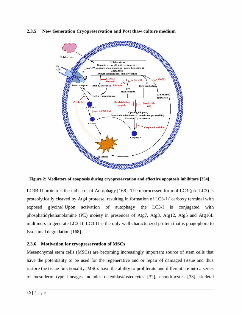
40 | P a g e
2.3.5 New Generation Cryopreservation and Post thaw culture medium
Figure 2: Mediators of apoptosis during cryopreservation and effective apoptosis inhibitors [254]
LC3B-II protein is the indicator of Autophagy [168]. The unprocessed form of LC3 (pro LC3) is
proteolytically cleaved by Atg4 protease, resulting in formation of LC3-I ( carboxy terminal with
exposed glycine).Upon activation of autophagy the LC3-I is conjugated with
phosphatidylethanolamine (PE) moiety in presences of Atg7, Atg3, Atg12, Atg5 and Atg16L
multimers to generate LC3-II. LC3-II is the only well characterized protein that is phagophore to
lysosomal degradation [168].
2.3.6 Motivation for cryopreservation of MSCs
Mesenchymal stem cells (MSCs) are becoming increasingly important source of stem cells that
have the potentiality to be used for the regenerative and or repair of damaged tissue and thus
restore the tissue functionality. MSCs have the ability to proliferate and differentiate into a series
of mesoderm type lineages includes osteoblast/osteocytes [32], chondrocytes [33], skeletal

41 | P a g e
muscles[34], adipocyte and myocytes, vascular cells and the like [35]. With the advancement of
MSCs isolation and expansion, the development of technology and or strategy for storing and
preserving of MSCs for long time without change in its properties is of paramount importance so
that these MSCs can be used when patient needs. The current technique using Me2SO as CPA has
potentiality for preservation of various cells and tissue. However, the use of Me2SO has
detrimental effects causing cell death. Me2SO is considered to possess an impact on the genetic
profile and induce differentiation in murine stem cells[36]. Use of nontraditional cryoprotectant
in preservation of stem cells a crucial facet because the entire long term storages are done with
customary Me2SO and glycerol. Several studies have already been done on the cryopreservation
of mesenchymal stem cell with trehalose and few with ectoin also [13, 37-47]. However, very
little work has been done on long term cryopreservation of hMSC using natural osmolytes and
impact of these cryoprotectants on long time storage of MSCs. The aim of our present study is,
therefore, to investigate the effect of trehalose and ectoin as major cryoprotectant using a reduced
amount of Me2SO for long term cryopreservation of MSCs.
Furthermore, many factors affect the effectiveness of MSCs cryopreservation, including
incubation temperature, the freezing medium, cooling rate, storage temperature, and cell
concentration during freezing and duration of storage, thawing temperature, and thawing medium.
And also the cooling profile which is important for the cell viability is not taken into
consideration [125]. Long time biopreservation technique that maintains ex-vivo TECs viability
and functioning is one of the most important research aspects of modern tissue engineering and
regenerative medicine. The ability to preserve the integrity of TECs outside the native
environment for an extended period shall not only save time of the donors and the recipients, but
it may also be possible to establish cell bio-banks worldwide for the safe preservation of high
quality TECs products for use when need arises. Such a ready to use TECs would support the
implementation of routine cryopreservation practices during preparation of even engineered cells
and tissues for the clinical applications when needed.

42 | P a g e
Chapter III
Scope and Objective
Mesenchymal stem cell is an important and promising cell source for various therapeutic
applications and development of various tissue constructs/tissue grafts through tissue engineering
approach. Thus there is a worldwide demand of mesenchymal stem cells (MSCs) and MSCs
seeded scaffold (TECs) in clinical sector for the treatment of variety of blood and tissue related
diseases. With the advancement of isolation and expansion of MSCs and development of tissue

43 | P a g e
engineering products utilizing MSCs as an important cell source, the development of technology
and/or strategy for storing and preserving of such clinical products for long time without change
in their properties is of paramount importance keeping in view of their actual use when patient
needs. Cryopreservation has been found to be the most effective method of preserving cells using
Me2SO as CPA. However, the use of Me2SO in cryopreservation medium cause cell death and
also harmful to the patient.
In this context, one of the key challenges is the development of a cryopreservation strategy using
an alternative freezing medium that can preserve MSCs and MSCs seeded scaffold for a longer
period of time with high recovery of cells, differentiation ability and structural integrity of the
scaffold used in TECs formation.
Therefore, the main objective of the present research is the development of cryopreservation
strategy using natural osmolytes for long term cryopreservation of MSCs and MSCs seeded
scaffold.
The specific objectives of the present research work are as follows:
1. To select potential non-toxic (natural) cryoprotective agents
2. To evaluate the potentiality of natural osmolytes in preserving MSCs
3. To study the role of apoptosis pathways in cryopreserved induced cell death of MSCs
4. To optimize the controlled rate freezing condition for cryopreservation of MSCs using natural
CPAs
5. To evaluate the potentiality of natural CPAs in preserving MSCs seeded constructs
6. To optimize the controlled rate freezing MSCs seeded constructs using natural CPAs
Scope of work
Cryopreservation is proven to be an efficient method for preserving varies of cells and other
biological species. This method uses Me2SO is as a potential CPA which is reported to have
several detrimental effect causing cell death. To overcome the limitation and adverse effect of

44 | P a g e
Me2SO, the recent research is directed towards the search of natural CPAs which seem to be
promising without having harmful effect like Me2SO. However there is very limited research has
been done on the study of these natural osmolytes for its actual use in long term preservation of
MSCs and other clinical product using MSCs.
Therefore the present investigation aimed to formulate efficient cryopreservation medium from
potential natural CPAs for preserving MSCs and develop a cryopreservation system that will be
helpful to preserve MSCs and other tissue products on long term basis.
The specific scope of work under this research includes the following
1. Selection of potential CPAs alternative to Me2SO
In this part, efforts would be given to select a single or a combination of potential natural
cryoprotective agents by a preliminary study on cryopreservation of umbilical cord blood (UCB)
derived MNCs in a controlled rate freezer. Different batches of cryopreservation solutions
prepared using extracellular cryoprotectants like trehalose, hydroxyl ethyl starch, polyvinyl
pyrrolidine and intracellular CPAs like erythritol, taurine and ectoin will be investigated and the
most effective CPAs will be selected for further study on cryopreservation of MSCs.
The formation of reactive oxygen is another important factor which causes cellular damage during
low temperature storage. In this context, the addition of selective antioxidants in cryopreservation
medium is reported to be beneficial to overcome this oxidative effect and thus improve post-thaw
cell viability. Keeping this in view, cryopreservation experiments will be performed to investigate
the performance of the best obtained freezing solution in presence of potential antioxidants such
as catalase, co-enzyme Q10 and N-acetyl cystine and thus an optimal freezing solution is
expected to be obtained giving maximum cell viability after cryopreservation.
2. Development of an effective freezing solution for cryopreservation MSCs
This phase of research will focus on the development of a freezing solution from the selected
CPAs and antioxidant for long term preservation of MSCs. Different batches of freezing solutions
will be prepared using individual as well as combination of selected CPAs. The solutions will be
evaluated by cryopreservation experiment to see their ability towards preserving MSCs and the
most effective freezing solution will be selected.

45 | P a g e
3. Study on apoptosis signaling pathways
Apoptosis that involves program cell death is an important phenomenon that occurs in
cryopreserved cells. The inhibition of cryopreserved induced apoptosis is important to improve
the post thaw viability of cryopreserved cells. Therefore, an effort will be given to study the
influence of important caspase and calpain inhibitors on the cryopreservation of MSCs and the
potential inhibitors will be selected to improve further the effectiveness of the developed freezing
solutions.
4. Optimization of cryopreservation condition for MSCs
The controlled rate freezing condition will be optimized further by studying the effect of key
parameters such prenucleation cooling rate, nucleation temperature, cold spike temperature, post
nucleation holding temperature, post nucleation cooling rate, cell concentration and storage
temperature using the best obtained freezing solution to maximize the MSCs recovery
5. Evaluation of developed freezing solution for preserving MSCs seeded scaffold
In this phase, research work will further be extended to evaluate the potentiality of the freezing
solution developed for MSCs cryopreservation towards the preservation of TECs formed from
MSCs seeded on scaffold.
6. Optimization of cryopreservation condition for TECs
The controlled rate freezing condition will be further optimized by studying the effect of key
parameters such as prenucleation cooling rate and nucleation temperature using the best obtained
freezing solution to maximize the MSCs recovery in TECs

46 | P a g e
Chapter IV
Materials & Methods
Materials and Methods
4.1 Chemicals and culture media
4.1.1 Cryoprotective agents

47 | P a g e
The various cryoprotective agents such as trehalose, Me2SO, hydroxy ethyl starch (HES), ectoin,
erythritol and poly vinyl chloride (PVC) used in this thesis work were purchased from Sigma
Chemical Co. (St. Louis, MO, USA).
4.1.2 Antioxidants and inhibitors
The chemicals such as co-enzyme Q-10, ascorbic acid, catalase and N-acetyl cystine were
purchased from Sigma Chemical Co. (St. Louis, MO, USA) and used as antioxidants. Caspase
inhibitors (z-LEHD-FMK, z-IETD-FMK, z-DEVD-FMK, z-VAD-FMK) and calpain inhibitors
(3-(4-iodophenyl)-2-mercapto-(Z)-2-propenoic acid) were purchased from BD pharminogen
(Becton Dickenson, San Jose, CA, USA).
4.1.3 Cell culture media and antibodies
Dulbecco’s modified eagle medium (DMEM), Penicillin-Streptomycin solution, fetal bovine
serum (FBS), Dulbecco’s phosphate buffered saline (DPBS), chondrogenic basal medium and
supplement, Collagenase type I and 0.25% Trypsin /ethylene diamine tetra acetic acid (EDTA)
solution were procured from Gibco (BRL, USA). Phalloidin-Alexa Fluor 488 conjugate, calcein
green AM and JC1 were used of Invitrogen, USA. Ficol Hypaque solution (Hi sep LSM 1073),
adipogenic differentiation media, Giemsa 10x stain, Triton X-100, formaldehyde, reagent for
MTT assay and Commassie blue G-250 are of HiMedia labs, India. Growth factors EGF and
bFGF, BD FACS lysis buffer, propidium iodide (PI), CD34-PE, CD44-FITC, CD45-PerCP/cy5.5,
CD73-PE/APC, CD90-APC, CD105-PE and HLA-DR-PerCP/cy5.5, Annexin-V conjugated with
FITC were from BD pharminogen (Becton Dickenson, San Jose, CA). Dexamethasone,
staurosporine, α-MEM, L- glutamine, L-ascorbate, β-glycerophosphate, bovine serum albumin
(BSA), Oil red O, Alizarin red S, Safranin O were from Sigma-Aldrich (St Louis, MO, USA). All
the tissue culture plastic wares and RNase were from BD falcon and MP Biomedicals, USA,
respectively.
4.2 Collection and processing of umbilical cord blood
Umbilical cord blood (UCB) samples were collected from Ispat General Hospital, Rourkela, India
with prior consent of the full-term delivery patients. A blood collection bag containing 17 ml of

48 | P a g e
citrate phosphate dextrose adenine (CPDA) anticoagulant was used for aseptic transfer of the cord
blood sample each time and brought to our culture laboratory for processing. The Institutional
ethical board approved the collection procedure.
4.3 Isolation and culture of MNCs
The MNCs were isolated from umbilical cord blood by standard Ficol hypaque density gradient
method [33]. 30 ml of cord blood sample was diluted with RPMI 1640 in the ratio of 4:1 (v/v) and
added to 20 ml of ficol hypaque solution. The solution is then centrifuged (swing bucket rotor) at
430×g for 30 min at 4°C. From the three layers formed, the middle layer (buffy coat) containing
MNCs was separated, followed by the addition of 1x Lysis buffer (1:9 dilution of 10x stock buffer
in distilled water) and incubated for 10min at room temperature. The cell suspension was
centrifuged at 200×g for 10 min at 4°C and supernatant was discarded. The pellet was washed to
remove lysis buffer and other impurities by centrifugation twice with D-PBS at 200×g for 10 min
each time. Finally the pellet obtained was resuspended in expansion medium consisting of α-
MEM, 10% FBS, 2mM glutamine and 1% antibiotic-antimycotic solution and plated at density of
1×10⁶
cells/cm3
. The non adherent cells were discarded after incubating culture plates at 37°C
with 5% humidified CO2 for 24 h. The adherent cells were washed thoroughly with D-PBS/EDTA
and then supplemented with freshly prepared expansion medium. The cells were cultured with
media change weekly twice.
4.4 Sorting and culturing of MSCs
Mesenchymal stem cells (MSCs) were isolated from the cultured human mononuclear cells
(hMNCs) by florescence activated cell sorting (FACS ARIA III; BD Biosciences) using
antibodies like CD90-APC, CD73-APC, CD105-APC, CD44-FITC, CD45 PerCP Cy5-5, HLA-
DR PerCP Cy5-5, CD34 PE. UCB derived MSCs was isolated from MNC populations by positive
selection using CD44, CD90, CD73vand CD105 positive antibodies (BD Biosciences, USA).
After sorting, the cells were counted by haemocytometer with Trypan Blue dye used for staining
dead cells. MSCs were suspended in an expansion medium consisting of Dulbecco's modified
eagle medium (DMEM), 10% FBS and 1% antibiotic-antimycotic solution and plated initially at
cell density of 1x10⁶cells/cm
3 in T50 culture flasks (Beckon Dickinson, San José, CA, USA). Non
adherent cells were discarded after incubating culture plates at 37°C with 5% humidified CO2 for
24 h. The adherent cells were washed thoroughly with D-PBS/EDTA two or three times and then

49 | P a g e
supplemented with freshly prepared culture media. Thereafter the media was changed twice
weekly.
4.5 Characterization of cells
4.5.1 Morphological characterisation
MSCs were harvested and morphology was observed from the culture flask under phase-contrast
inverted microscope. Images of MSCs were captured using a Carl Zeiss Axiovert 40 microscope.
No specific staining was carried out.
4.5.2 Immunophenotypic characterisation
The specific surface antigens expressed by MSCs were characterized by flow cytometry analysis.
The trypsinized post-thaw cells (5 × 10⁵ cells per sample) were stained with human monoclonal
antibodies against CD44, CD45, CD73, CD90, CD105, CD34 and HLA-DR. The CD expressions
were analyzed using a flow cytometer (Becton Dickinson and Co, San Jose, CA, USA).
4.6. Cryopreservation of cells
4.6.1 Preparation of cryopreservation solution
1×10⁶
cells/ml MNCs were transferred into standard 1-ml cryovials (Nalge Nunc, IL, USA)
containing each batch of freezing solutions prepared from 10mM extracellular cryoprotectant,
5mM intracellular cryoprotectant and 100μg of antioxidant in FBS at 6ºC. The concentrations of
CPAs were selected based on the published literature [169-173]. Each time, MNCs were treated
with intracellular cryoprotective agents for 20mins followed by the treatment with extracellular
cryoprotectants for 10 mins. Similarly, MNCs were resuspended in 10% Me2SO culture medium
solution without FBS at 4°C as a positive control. For the culture of MSCs, medium was replaced
with DMEM containing ectoin and trehalose at 0.3 M concentration each and cultured at 37 °C in
CO2 incubator for overnight [174]. All freezing vials contained 1ml of final freezing medium after
the addition of cells. Different molar concentrations of osmolytes were added to the cryovials. In
all cases, 100μg/ml catalase was added to the cryovials. Cryopreservation solution used for this
study was prepared by adding different concentration of molar ration of trehalose, ectoin and
100 μg/ml catalase.
4.6.2 Cryopreservation experiments

50 | P a g e
The sealed cryovials containing each cryoprotectant solution and cells were placed in a controlled
rate freezer (Kryo360-17, Planer, Inc.., UK) as shown in Figure 3. The cryopreservation
experiments were carried out following the programmable cooling protocol [175] as- ramp 1, for
4ºC for 10min; ramp 2, -3ºC/min to -10ºC/min; ramp 3, -15ºC/min to -30ºC/min; ramp 4, -
3ºC/min to -60ºC; Ramp 5, -10ºC/min to -180ºC. After achieving -180°C, the cell suspension was
taken out of the freezer and stored in vapor phase storage tank (-180 ºC) for further use. Each
batch of experiments was repeated three times.
Figure 3: Controlled rate freezer for cryopreservation of MSCs
4.6.3 Thawing and post-thaw culturing
The cryopreserved cells were thawed by immersing cryovials rapidly in water bath at 37°C and
then subjected to gradual dilution of cryoprotectant solution with prewarmed thawing medium
(DMEM + 10% FBS). The cells were then centrifuged at 200×g for 5 mins and resuspended in
DMEM supplemented with 10% FBS, 100U/ml penicillin G, 100 mg/ml streptomycin and
10ng/ml epidermal growth factor (EGF). The cells were then cultured in 6-well tissue-culture
plates at 37ºC in a humidified atmosphere of 5% CO2 [177, 178].
4.6.4 Cell viability assay

51 | P a g e
After 24h of incubation, medium containing the dead cells were separated and the attached cells
were trypsinized. All the cells were stained with propidium iodide (PI) and analysed by flow
cytometry for cell viability. Flow cytometry analysis was done within 10min trypsinization [176].
4.6.5 Cell proliferation assay
1×10⁴/ well of cell were seeded into 96-well plates with 100µl of growth medium that consists of
DMEM supplemented with 10% FBS, and 10ng/ml EGF and incubated at 37ºC and 5% CO2.
These cells were detached with trypsin/EDTA mixture and the cell number counted from day 1 to
7 day. The number of viable cells was counted using automated cell counter with cells stained
with 0.2% trypan blue [177].
4.6.6 MTT assay
The cell viability of cryopreserved cells was further verified by MTT (5-dimethylthiazol-2-y1]-2,
5-diphenyl tetrazolium bromide) assay using standard micro plate absorbance reader [178]. Post
thaw cryopreserved MNCs was treated with 10% Me2SO (control) to determine baseline cell
viability. For viability tests of different batches of CPA solutions, the post thaw cells at
concentration of 1×10⁵ cells/ml were plated (in triplicate) by adding 10µl MTT solution in each
well and incubated at 37ºC for 4h in 5% CO2. The optical density of the solution is then measured
using ELISA plate Reader (Perkin Elmer) at 570 nm.
4.6.7 Apoptotic assay
Post-thawed MNCs were labeled with AV-FITC (Annexin V conjugated with FITC) as per
manufacturer’s instruction (BD Pharmingen, San Diego, CA) [179] and 1×10⁶
cells/ml were
suspended in PBS (pH 7.4) supplemented with 2mM CaCl2 solution. AV-FITC binding was
analyzed with respect to fresh cells as positive control. The cells were incubated with AV-FITC
for 15 min in dark at room temperature and then subjected to flow cytometry analysis using
FACS Aria III flow cytometer (Becton Dickinson, San Jose, CA). For each sample, 10,000 events
were acquired from the region defined for MNCs and analyzed by BD FACSDiva Software 6.0
(Becton Dickinson, San Jose, CA). Compensation was required for the annexin V assay. Firstly,
this was done by analyzing an autofluorescent control to determine the level of autofluorescence.
Secondly, annexin V-FITC labeled cell population was analysed to establish optimal
compensation for annexin V-FITC. Finally, only PI-labeled cells were analyzed on these settings

52 | P a g e
and the cell populations was recompensated by subtracting the overlapping of PI and FITC. Cells
cryopreserved with 10% Me2SO (v/v) were used as negative control.
4.6.8 Mitochondrial membrane potential assay
Mitochondrial membrane potential of cryopreserved MSCs were evaluated by flowcytometry
using JC-1 as per method previously described [180]. Post thaw cryopreserved MSCs for 12
month were exposed to JC-1 for 15min at 10μg/ml concentration in culture medium and incubated
in 5% Co2 at 37°C. For the assessment of JC-1 staining, a total of 10,000 gated events were
analyzed per sample. In addition, the entire samples were viewed by fluorescence microscopy to
confirm JC-1 labeling.
4.6.9 Differentiation potential
After 12 months of storage in vapor phase liquid nitrogen, the cryopreserved MSCs were thawed
by usual procedure. Both cryopreserved and fresh (non-cryopreserved) MSCs were then seeded at
a concentration of 3×10³ cells/cm
2 into a 6 well-plates coated with type I collagen and cultured in
DMEM medium supplemented with 10% FBS for 24h.
Osteogenic differentiation
The osteogenic differentiation ability of cryopreserved MSCs was assessed by following the
method described in literature [181]. The differentiation of hMSCs were induced by culturing
with osteogenic differentiation medium consisting of DMEM supplemented with 100nM
dexamethasone, 0.05µM ascorbate-2-phosphate, 10mM β-glycerophosphate, 1%
antibiotic/antimycotic and 10% FBS for 14 and 21days. The culture medium was replaced twice a
week. After a specific period of culture, cells were washed twice with PBS and fixed with 2%
methanol for 10 min at room temperature. The cells were then stained with 1% alizarin Red S for
15min and used for assessment of mineral deposition.
Adipogenic differentiation
For the assessment of adipogenic differentiation potential, cryopreserved and unfrozen hMSCs
were cultured in adipogenic differentiation medium. The medium contains DMEM supplemented
with 10% FBS, 0.5 Mm isobutyl-methyl xanthine, 1µM dexamethasone, 10 µg/ml insulin, 200
µM indomethacin, and 1% antibiotic/antimycotic. The culturing was done for 7 and 14 days with

53 | P a g e
the change of media twice a week. The cells were washed with PBS and then fixed with 2%
methanol. The cells were stained with oil red O to visualize the intracellular lipid droplets.
Chondrogenic differentiation
The chondrogenic differentiation of post thaw cultured cells was performed using pellet culture
method as previously described [181]. Briefly, 2.5×10⁵ cryopreserved and non-cryopreserved
hMSCs were centrifuged at 800rpm for 15min. The pellets were resuspended in chondrogenic
differention medium, the composition of which is DMEM supplemented with 10% FBS, 1%
penicillin, 1% insulin-transferrin-selenium, 10-7
M dexamethasone, 150µM ascorbic-2-phosphate,
20 µM linoic acid and 0.1ng/ml TGF-β. The cells were incubated at 37ºC and 5%CO2
environment for 21days. The chondrogenic culture medium was replaced twice a week. The cells
were washed in PBS and fixed with methanol before staining with Saffranin O.
4.6.10 CFU-F assay
Colony forming assays were done following the procedure described in publish literature [182].
Post thawed as well as non cryopreserved MSCs were seeded in 75 cm2 tissue culture flasks at a
concentration of 8 cells/cm2 in cell culture medium. The medium was changed every 2 to 3 days.
After 12 days, cells were washed with PBS and fixed with methanol for 15 min. CFU-F colonies
were stained with 0.4%w/v Giemsa (diluted with deionized water for 30 min) and the culture
plate was observed under optical microscope (Leica DM750 equipped with digital camera).
Colonies were considered as clusters of more than 50 cells.
4.6.11 Cell cycle analysis
Post thaw MSCs were fixed with the addition of 70% ethanol and stored at 4°C. The ethanol-
suspended cells were centrifuged at 300 g for 5 min and the pellet was suspended in 2 ml of D-
PBS. This pellet was centrifuged again to remove any ethanol and finally the pallet was
suspended in 1ml D-PBS/EDTA containing 20 μg/ml PI and 100 μg/ml RNase A for 30 min.
Cells were then analyzed by flow cytometry by gating on PE-area versus PE-width discriminating
all the doublet population [183].
4.6.12 Western blotting
Cryopreserved MSCs were suspended in buffer containing 20 mM TRIS, 2 mM EDTA, 150 mM
NaCl, 0.5%Triton X-100 and protease inhibitors (Sigma). The samples were sonicated,

54 | P a g e
centrifuged and the pellets were discarded. The cryopreserved MSCs were assayed for protein
concentration following standard Bradford assay. Protein extracts of 50μg/sample, control or
post-thaw cryopreserved MSCs were separated on 10% SDS PAGE and transferred to PVDF
membrane. The membranes were incubated with primary antibodies such as caspase-3 (1:1000,
BD Biosciences), caspase-8 (1:250, BD Pharmingen), caspase-9 (1:250, BD Pharmingen),
cytochrome C (1:250, BD Pharmingen), PARP (1:500, BD Pharmingen), Bid (1:250, BD
Pharmingen) and calpain (Cell Signaling Technology, Inc., Beverly, USA) for overnight [185].
The membranes were then incubated with HRP Goat anti-mouse Ig, following the manufacturers
recommended protocol and visualized on film with the ECL substrate (Abcam Inc., Cambridge,
MA, USA).
4.6.13 Optimization of cryopreservation process
The cryopreservation of MSCs was optimized by investigating the effects of five aspects of
cryopreservation cooling profile, storage temperature and cell concentration on cell viability. The
cryopreservation experiments were carried out using (Kryo Cooler 1.6 Planner Biomed, U.K).
The various controlled rate freezing parameters investigated are: prenucleation cooling rate
(1ºC/min, 2.5ºC/min, 5ºC/min, and 10ºC/min), holding temperature (0ºC, -2.5ºC, -5ºC, -7.5ºC and
-10ºC), nucleation temperature (-2.5ºC,-5ºC, -7.5ºC and -10ºC), cold spike temperature (-20 ºC, -
40ºC, -60ºC, -80ºC) and post nucleation cooling temperature ( 1ºC/min, 2.5ºC/min, 5ºC/min,
10ºC/min). Various MSCs concentrations were compared at 0.5, 1, 3, 6×10⁶ /ml. To select
optimal storage temperature, final storage temperature was maintained at -80°C, -150°C, or -
196°C. For all experiments, in addition to comparing their own conditions, the medium
composition used as 0.3M trehalose, 0.3M ectoin, 100μg/ml catalase and 50µm/ml of calpain
inhibitor (PD 150606) and general caspase inhibitor (z-VAD-fmk) each. The cooling rate of
1°C/min, storage temperature of −180°C and cell concentration of 2×10⁶ /ml. were maintained.
4.7 Cryopreservation of TECs
4.7.1 Preparation of scaffolds
3-D nanofiberous mats developed using semi automated electrospinning machine (ESPINNANO
M/s. Physics Instruments Company, India) was used as scaffold for TECs formation. The detail
description of the method is reported in our patent filed (Patent number: 713/KOL/2012). In brief,
Silk fibroin (SF) was extracted from eri and tasar silk cocoon by degumming method using aq.

55 | P a g e
Na2CO3. SF blend were prepared and made electrospinnable by dissolving silk fibroin of eri and
tasar silk in achloroform and formic acid mixed in a ratio of 60:40v/v. The blend was electrospinned
at 15kV to generate nanofibrous scaffold of thickness 300µm and the average fiber diameter of
nanofiber is measured as 350nm (range 200-500nm). The contact angle and porosity were
determined as 54.7º and 80-82%.
4.7.2 Formation of TECs
The nanofibrous sheets of dimension 6mm diameter and 400µm thickness were used in this study.
The scaffolds were autoclaved and then sterilized with 70% EtOH. The sample was further exposed
to ultraviolet irradiation for 40min to avoid any morphological change. The sterilized scaffolds were
washed thoroughly with PBS and neutralized with growth medium at 37°C and 5% CO2. 5.0×10⁵
[48-49] cells/ml (MSCs) were seeded on scaffold by static method and cultured in DMEM
containing 10% MSC qualified fetal bovine serum and 1% antibiotics for a period of three weeks.
4.7.3 Preparation of cryopreservation solutions
Cryopreservation solutions were prepared with varying compositions as follows: (1)10% (v/v)
Me2SO with 50% (v/v) fetal bovine serum (D10) and T30/E30/C100. All solutions were prepared with
DMEM media.
4.7.4 Cryopreservation experiment
The precultured TECs were exposed to cryoprotectant solutions for 10 min at 4°C and then
transferred to a high-density polypropylene sterile pouch. The cryopreservation study was performed
in the computer-controlled programmable freezer following two steps cooling (shown in Figure 4).
TECs samples were cooled at the rate of 1°C/min down to -150°C and then immersed into liquid
nitrogen bath [186,187]. After 7 days of cryopreservation, the samples were removed from liquid
nitrogen and were thawed by shaking at 37°C. The TECs were transferred from pouch into a 35-mm
culture dish containing DMEM solution and washed thoroughly to remove cryoprotectants from
TECs. Precultured TECs without cryopreservation was used as control.

56 | P a g e
Figure 4: Controlled rate freezer for cryopreservation of TECs
4.7.5 Characterization of TECs
4.7.5.1 Cell morphology by SEM
Scanning electron microscopy was performed before and after cryopreservation at a specific interval
of time. Cryopreserved TECs were fixed in 2.5% glutaraldehyde at 4ºC overnight. TECs were
washed three times with deionized water and serially dehydrated with increasing gradient of ethanol.
Then samples were subjected to SEM study at 15kV accelerating voltage[180-181]. Elements,
especially calcium (Ca), phosphorous (P) and oxygen (O) on the surface of post-thawed TECs after
culture for 28days in osteogenic media were analyzed by energy-dispersive X-ray spectroscopy
(EDX, Philip Model XP 30 CP).
4.7.5.2 Cell viability
After cryopreservation study, TECs were removed from DMEM medium and washed twice with
PBS buffer. MSCs were removed from TECs by treating with 0.25% trypsin and 1mM EDTA. The
cells were then centrifuged at 200×g for 5 mins and resuspended in PBS. The percentage viability of
cells was assessed by flow cytometric analysis using propidium iodide [190] staining.

57 | P a g e
4.7.5.3 Cell metabolic activity
The metabolic activity of MSCs in scaffolds were quantitatively determined by MTT (3-(4, 5-
dimethylthiazol-2-yl)-diphenyltetrazolium bromide formazan) assay at an interval of 3, 5 and 7 days
of culture period. Culture medium was aspirated and washed with 600µl prewarmed DPBS. 400µl
prewarmed culture medium supplemented with 40µl MTT solution [2.5mg/ml MTT dissolved in
Dulbecco's Phosphate Buffered Saline (DPBS)] was added to each sample. After 3hr incubation at
37°C, the medium was removed from each well and scaffolds were transferred to 24-well petri dish.
250 µl Me2SO was added to each well to dissolve the formazan crystals[191-192]. The resulting
solution was removed and centrifuged at 500g. The optical density of the supernatant was measured
spectrophotometrically at 595 nm, using an ELISA plate Reader (Perkin Elmer).
4.7.5.4 Cytoskeleton analysis by confocal microscopy
Post-thaw TECs were fixed with 2% paraformaldehyde in PBS at room temperature for 5min. This
was followed by 40-min treatment with 2% Bovine serum albumin (BSA) and 1% glycine in PBS to
block any non-specific staining. Samples were then permeabilized with 0.5% Triton X-100 in PBS
for 15 min. For actin staining, samples were incubated with Alexa-Fluor 488 conjugated phalloidin
at 370 C for 40min. in dark condition. Samples were mounted on glass slides and confocal images of
the samples were acquired using Leica TCS SP5 X Super continuum Confocal Microscope [193-
194].
4.7.5.5 Osteo-induction assessment
The osteogenic differentiation potential of cryopreserved TECs was assessed and compared with
control. Cryopreserved and non cryopreserved TECs were cultured in osteogenic medium consisting
of high glucose–DMEM (Gibco, Invitrogen) supplemented with L-ascorbic acid-2-phosphate
(50mg/ml), β-glycerophosphate (10 mM), and dexamethasone (100nM) (Sigma) 24 hr after
cryopreservation [196-197]. Culture media was allowed to change every 3 days.
4.7.5.6 Alkaline phosphatase activity
The alkaline phosphatase (ALP) activity of post thawed TECs was determined quantitatively by 1, 7,
14, and 21days [195] of TECs culture. The cell lysate from cryopreserved TECs was prepared by
treating TECs with 1% Triton X-100 for 50 min and sonication for 10 min. The cell lysate is then
added to ALP substrate (para-Nitrophenylphosphate) with a ratio of 1:1. After 40 min of incubation,
1 M sodium hydroxide (Sigma) was added to block the enzymatic reaction. The OD of the solution

58 | P a g e
mixture was measured at 405nm in a microplate spectrophotometer. Each experiment was performed
in triplicate.
4.7.5.7 Histological study
The mineralization of hMSCs on the scaffolds was analyzed and quantified by alizarin red-assay.
The cells grown on the scaffolds were washed with PBS and fixed with paraformaldehyde for
15min. The cell-seeded constructs (TECs) were thoroughly washed with distilled water prior to the
addition of 500ml of 40mM alizarin red stain (pH 4.1). The plates were incubated at room
temperature for 20 min with gentle shaking. After aspiration of un-incorporated dye, the wells were
washed thoroughly with distilled water with stirring for 5 min. The plates were left for 2 min to
facilitate removal of excess water.
4.7.5.8 Osteogenic-specific gene expression
RNA was extracted using Trizol (Invitrogen, Carlsbad, CA, USA) according to the instructions of
manufacturer. After the treatment of cell extract with DNase (Invitrogen), 1 μg of total RNA was
used for cDNA synthesis (Superscript First-strand synthesis system, GibcoBRL, Life Technologies)
following the protocol of manufacturer. RT-PCR was performed to determine the expression of
genes using primers (Table-3) for the osteogenic differentiation of osteopontin [OPN], ALP and
core-binding factor alpha 1[Cbfa 1]. Glyceraldehydes phosphate dehydrogenase [GAPDH] was used
as a house keeping gene for internal control. The polymerase chain reaction (PCR) was carried out
using BIO-RAD Mycylcer and analyzed by software Bio-Rad. Detail description of the procedure
described in published literature [195].
Table 3: The specific primers used for RT-PCR
Gene Abbr. Forward primer Reverse primer
Collagenase 2 COL2 GTGGGCTTCCTGGTGA CAGCACCTGTCTCACCA
Osteocalcin OCN CCCAGGCGCTACCTG TATCAA GGTCAGCCAACTCGTCACAGTC
Osteopontin OPN TGTGAGCTCAATCCGGACTGT CCGATAGGCCTCCTGAAGC
Glyceraldehydes-3-
phosphate dehydrogenase
GAPDH GCACCGTCAAGGCTG AGAAC ATGGTGGTGAAGACGCCAGT

59 | P a g e
4.7.5.9 Mechanical strength
Scaffolds were subjected to tensile tests before and after cryopreservation for determining their
elastic modulus, using a Universal tensile testing machine (Instron 4505 Universal Machine)
following the procedure described in the published literature [197]. The tests were performed under
compression loading of 1KN with a crosshead speed of 2 mm/min till 60% strain was reached.
4.7.6 Optimization of MSCs cryopreservation parameters
The precultured TECs were exposed to cryoprotectant solutions for 10min at 4°C and then
transferred to a high-density polypropylene sterile pouch. The cryopreservation study was
performed in a computer-controlled programmable freezer (Kryo cooler, UK) following two steps
cooling. TECs samples were cooled at different cooling rate such as 0.5°C /min, 1°C/min and
2°C/min from 5°C to -150°C. Finally the scaffold was immersed into liquid LN2 bath. After 7
days of cryopreservation in vapors phase storages tank, the samples were removed from liquid
nitrogen and were thawed by shaking at 37°C. The TECs were transferred into a 35-mm culture
dish containing DMEM solution and washed thoroughly to remove cryoprotectants from TECs.
Precultured TECs without cryopreservation was used as control. Seeding was performed
manually at temperature -5° C,-7° C and -10° C to analyze the effect of seeding temperature.
4.7.7 Statistical analysis
Statistical differences were evaluated by the Tukey-Kramer test after one-way analysis of variance. p
< 0.05 was considered significant using online graphpad quickcalc software.

60 | P a g e
CHAPTER IV
Results and Discussion

61 | P a g e
PART I
A preliminary study on cryopreservation of MNCs:
Selection of natural cryoprotective agents & antioxidant

62 | P a g e
In recent years stem cell therapy using mesenchymal stem cells has been considered as a
promising treatment option for patients suffering from various therapeutic and tissue related
diseases [1-13]. Therefore, systematic efforts have been given by the scientists worldwide for the
isolation and expansion of MSCs keeping in view of their commercial applications. Umbilical
cord blood (UCB) is considered as a potential source of MSCs because of its easy availability as a
waste product, no risk to donor, juvenile source and better proliferation potential compared to
other sources. However, development of a long term preservation strategy is a prerequisite for
successful application of MSCs when need arises. Cryopreservation using Me2SO as CPA is a
proven method for long term storage of human cells with high cell viability and maintenance of
desired stem cell characteristics [197-200]. However, cellular damage occurs due to extracellular
and intracellular ice formation and toxicity effect.of using Me2SO. The search for novel
cryoprotective agent (CPA) as alternative to Me2SO is under active research consideration as
CPA has a critical role in reducing many of these factors encountered under extreme low
temperature treatment of cells.
In this context, treahalose, polyvinyl pyrrolidine (PVP) and hydroxy ethyl starch (HES) seem to
be attractive extracellular [200-212] and ectoin, taurine and erythritol as intracellular CPAs for the
preservation of cells [213-219]. These CPAs has important role in protecting cell membrane
integrity during low temperature preservation by preventing extracellular and intracellular ice
formation that lead to the damage of cell membrane and denaturation of cell. Furthermore, the use
of these CPAs in combination seems to be more efficient in preserving cells at low temperature
freezing conditions [22, 54]. However, not much investigation has been done so far to explore the
potentiality of these natural CPAs particularly their use in combination for preserving
mesenchymal stem cells. Another important aspect is that the choices of many CPAs are cell-
specific and their potentiality as CPA cannot be adjudged as a priori. So, in the present study three
extracellular (trehalose, PVP, HES) and three intracellular (ectoin, taurine and erythritol) CPAs
were chosen to assess their ability for cryopreservation of MNCs and select the most effective
CPA which can be further used for the study on MSCs preservation. The MNCs were chosen for
preliminary trial with intention to avoid the use of MSCs which takes long period of time for their
harvesting and the process is tedious, whereas MNCs that contains MSCs is easy to isolate and
handle [220].

63 | P a g e
The formation of reactive oxygen is another important factor which is reported to cause cellular
damage during low temperature storage. The addition of selective antioxidant in cryopreservation
medium is reported to improve post thaw cell viability by overcoming this oxidative effect [222].
The widely used antioxidants reported are catalase, co-enzyme Q10 and N-acetyl cystine [221-
223]. So the effect of these antioxidants was also investigated to enhance the cryopreservation
efficiency of the freezing solution.
5.1.1 Isolation and culture MNCs
A blood bag containing CPDA solution was used for the CB collections. The blood was collected
by gravity as shown in Figure 5. The cells isolated by Ficol- hypaqe technique were first cultured
on plates in DMEM media with supplement of 10%FBS and 1% antibiotic. After 14 days of
culture, MNCs were harvested using trypsin/EDTA solution (Figure 6).
5.1.2 Cell morphology study
The morphological variation in the cultured cells was studied by phase contrast microscopic
images as shown in Figure 7(A-F). After initial culture, the cells are observed to be round in
shape (Figure 7 A) and MNCs are shown to be adherent to culture plate after 48h (Figure 7B).
Cells became elongated as spindle fibroblastic shape after 4 days of culture (Figure 7C) and
confluence is shown to be reached after 14 days of culture (Figure 7F).

64 | P a g e
Figure 5: Collection of Cord blood: (A) Clamped and cut the cord just after the birth of baby, (B)
and (C) needle is inserted in umbilical cord with collection bag, (D) cord blood allowed to fill the
collection bag by gravity

65 | P a g e
Figure 6: Schematic representation of isolation procedure of MNC from umbilical derived cord
blood

66 | P a g e
Figure 7: Phase contrast microscopic images depicting the morphological changes during culture
period. After initial culture, the cells are observed to be round in shape (A) and MNCs are shown to
be adherent to culture plate after 48h (B). Cells became elongated as spindle fibroblastic shape after
4 days of culture (C) and confluence is shown to be reached after 14 days of culture ( F)
Day 1 Day 2
Day 3 Day 4
Day 7 Day 14
1414

67 | P a g e
5.1.3 Cryopreservation solution
Different freezing solutions for cryopreservation of MNCs prepared by taguchi orthogonal L9
array are presented in Table 4.
Table- 4: Taguchi L9 array for nine cryoprotectant solutions with different compositions
5.1.4 Cell viability and apoptosis study
An important phenomenon that occurs in cryopreserved cells is the programmed cell death, called
apoptosis. The measurement of this apoptosis is important because it permits the detection of cells
that are undergoing a programmed cell death without disrupting their membrane integrity and thus
it helps in selection of cryoprotective agents [50-51]. In this study, the cell viability and apoptosis
of post thaw cultured MNCs were studied by flowcytometry analysis using Annexin–V FITC/PI
staining. Four quadrants of dot plots consisting of Q1 -dead and necrotic cells, Q2-late apoptotic,
Q3-live and Q4-late apoptotic are defined as
Q1-Annexin V-FITC negative & PI positive –dead and necrotic cells,
Q2-Annexin V-FITC positive & PI positive –late apoptotic cells,
Q3-Annexin V-FITC negative & PI negative -live cells,
Cryopreservation solution 1
A
2
B
3
C
4
D
Solution-1 HES Erythritol Co-enzyme Q 10 10% FBS
Solution-2 HES Taurine Catalase 5% FBS
Solution-3 HES Ectoin N-acetyl cystine 0% FBS
Solution-4 PVP Erythritol Catalase 0% FBS
Solution-5 PVP Taurine N-acetyl cystine 10% FBS
Solution-6 PVP Ectoin Co-enzyme Q 10 5% FBS
Solution-7 Trehalose Erythritol N-acetyl cystine 5% FBS
Solution-8 Trehalose Taurine Co-enzyme Q 10 0% FBS
Solution-9 Trehalose Ectoin Catalase 10% FBS

68 | P a g e
Q4- Annexin V-FITC positive & PI negative-early apoptotic.
The dot plots of flowcytometry analysis found with post-thaw cultured samples are shown in
Figure 8 and the cell viability data generated from dot plots is tabulated in Table 5. The
experimental results indicate that the combination of hydroxy ethyl starch, ectoin, catalase and
10% FBS (solution-9) has shown maximum cell viability of 93.7%, 5.5% early apoptotic cells,
0.7% late apoptotic cells and 0.1% necrotic cells. But the combinations of polyvinlypyrrolidine,
erythritol and co-enzyme Q10 in absence of FBS have shown the least cell viability of 36.0%.
The cell viability is also compared with the cell viability obtained with the cells cryopreserved in
10% (v/v) Me2SO as control. The cell viability was observed to be significantly reduced when
cell cryopreserved in Me2SO. The cell viability achieved in later case is 60%. Percentage of cells
in Q3 quadrant was further used for Taguchi statistical analysis. Signal-to-Noise ratio (S/N), a
logarithmic functions of desired output, that serves as objective functions for optimization, helps
in data analysis and thus predicts the optimum results. There are three way of calculating Signal-
to-Noise ratios (1) smaller-the-better (2) larger-the-better and (3) nominal-the-best. In the present
study S/N ratio was calculated based on the principle of “smaller the better
Table 5: S/N ratio and percentage of cells in Q3 quadrant (viable cell)
Sample Repetition -1 Repetition-2 Repetition-3 Average viability S/N Ratio
Solution 1 83.70 84.4 84.2 84.1 47.5817
Solution 2 66.90 66.3 66.5 66.6 46.9271
Solution 3 69.90 70.8 67.8 69.6 41.0037
Solution 4 36.09 35.9 36.3 36.0 51.5668
Solution 5 68.80 72.0 70.0 70.5 32.8675
Solution 6 83.50 80.3 81.5 81.6 34.1221
Solution 7 80.50 79.6 78.0 79.4 42.6865
Solution 8 63.00 67.0 64.0 65.0 30.2377
Solution 9 91.60 93.8 95.5 93.7 37.4882

69 | P a g e
A B
C
D E F
G H I

70 | P a g e
Figure 8:Dot plots of cryopreserved and non-cryopreserved MNCs: control(A), cryopreserved
MSCs in Me2SO (B), HES, erythritol, Co-enzyme Q10 and 10%FBS(C), HES, taurine, catalase and
5%FBS(D), HES, ectoin, n-acetyl cystine and 0%FBS(E), PVP, erythritol, catalase and 0%
FBS(F), PVP, taurine, n-acetyl cystine and 10%FBS(G), PVP, ectoin, co-enzyme Q10 and
5%FBS(H), trehalose, erythritol, n-acetyl cystine and 5%FBS(I), trehalose, taurine, co-enzyme
Q10 and 0% FBS (J), trehalose, ectoin, catalase and 10%FBS(K)
5.1.5 Statistical analysis
The S/N ratio data as shown in Figure 9, was calculated based on Taguchi’s smaller the better
approach (224-225). A large variation in S/N ratio is observed among the extracellular
cryoprotectants, intracellular cryoprotectants and antioxidants as evident from Figure 2(C1), (C2)
and (C3). Furthermore, there was no significant variation in S/N ratio observed with the sample
containing FBS which indicates that variation in concentration of FBS does not have any
remarkable effect on cell viability. However, though a slightly increase in cell viability is
achieved with freezing medium containing 5%FBS. Among various cryoprotective agents
studied, trehalose and ectoin are found to be the most effective extracellular and intracellular
cryoprotective agents showing maximum cell viability in terms of S/N ratio. Similarly catalase
was found to be the most favorable antioxidant.
5.1.6 Analysis of Variance (ANOVA)
ANOVA helps in testing the significance of all main factors and their interactions by comparing
the mean square against an estimate of the experimental errors at specific confidence levels
[225]. The results are also compared with statistical data obtained by ANOVA analysis based on
analysis of variance as shown in Table 6. Data confirms that the extracellular (p value=0.0003)
J K

71 | P a g e
and intracellular CPAs (p value=0.009) have greater influence on cell viability compared to other
factors such as FBS concentration (p value= 0.05) and types of antioxidants (p value= 0.037).
Figure9: Illustrates S/N ratio averages for each parameter at three levels. As it is observed
extracellular cryoprotectants (C1) intracellular cryoprotectants (C2) and antioxidants
concentrations (C3) exhibit larger variations. However, the largest variation is found in case of
antioxidants
Since the Taguchi Orthogonal array adopted in this study is a factorial experiment, it is necessary
to conduct verification experiment to ascertain that the entire factor included in this analysis are
significant [224].Therefore, various experiments for verification under best obtained level with
the respective factor were performed following the methodology published earlier [225]. The
average viability is calculated based on flowcytometry as 73% which is shown to be within the
confidence level, where quadrant analysis shows the distribution of viable MNCs (7AAD -ve,
Annexin V-ve), apoptotic MNCs (7AAD -ve, Annexin V +ve), and necrotic MNCs (7AAD +ve,
Annexin V +ve).

72 | P a g e
Table 6: ANOVA analysis of experiments consider in Taguchi orthogonal array
Factor Degree of freedom Sum of squares Mean square P-value
Extracellular cryoprotectants 2 129.4 64.7 0.0003
Natural osmolytes 2 133.5 66.7 0.009
Antioxidants 2 94.8 47.4 0.037
Concentration of FBS 2 13.1 6.5 0.050
Total 8 370.8
5.1.7 Cell morphology analysis of cryopreserved cells
The cellular morphology of cryopreserved MNCs was studied by SEM and fluorescence
microscopic images (Figure 10). The cryopreserved cells are observed to retain their normal cell
morphology.

73 | P a g e
Figure 10: Conformity test for best obtained level with the respective factor. (A) Dot plot of non-
cryopreserved sample (negative control), (B) Post-thaw MNCs viability, (C) SEM image of after
24h post-thaw MNCs ,(D) fluorescence image after 24h post thaw of MNCs at 5x magnification. (B),
(C) and (D) show the result observed with combination of trehalose, ectoin, catalase and 5% FBS
5.1.8 MTT assay
The cell viability of cryopreserved MNCs was also measured quantitatively by MTT assay and
the experimental results are shown in Figure 11. The assay result indicates that the cells
preserved in solution 1, 2, 3, 7 and 9 are more viable after 24 h of post thaw compared to the cell
viability observed with other CPA solutions. Furthermore, the combination consisting of
A B
C D

74 | P a g e
trehalose, ectoin and catalases have shown better cell viability as compared to the other
antioxidant present in cryopreserved solution. However, a statistically significant increase in cell
viability is observed using cryoprotectant solution 9 (***p<0.0005) followed by 3 (**p<0.05) as
compared to the post thaw MNCs cryopreserved in 10% Me2SO. The result has shown similar
trend as the results obtained by using flow cytometer i.e apoptosis study. However, cell viability
with traditional Me2SO treated samples are found to be less as compared to any combination
except solution 4 (**p<0.05). Furthermore, no significant variation in cell viability is observed
with solution 5, 6 and 8 from the cell viability achieved with control.
Figure 11: MTT assay of post-thawed MNCs cryopreserved in different compositions of
extracellular cryoprotectant, intracellular, antioxidant and FBS. MNCs cryopreserved in 10%
Me2SO taken as control. All the data are compared with control using student t-test, Bars represent
data from three separate experiments with standard deviation (*p< 0.05, **p< 0.005, ***<0.0005)
5.1.9 Post-thaw cytoskeleton assessment
F-actin, a main constituent of cytoskeleton has an important role in maintaining cellular function
[226]. It is further reported that at extremely low temperature, the ultra structure of F-actin
becomes distorted that results in cell damage [227]. Therefore, the effect of cryopreservation was
investigated using non-toxic CPA solution on the F-actin which would be beneficial to design

75 | P a g e
cryopreservation strategy. The change in F-actin of 48h post thaw cultured samples was observed
under the florescence microscope (Figure10). Non-cryopreserved cell shows prominent
cytoplasmaic extension with maximum cell attached to the flask as depicted in Figure 10A and
MNCs cryopreserved with solution-9 shows good cell attachment. However, the cell attachment
is found to be lesser than the normal cultured cell (Figure 12B) and higher than the sample
cryopreserved with 10% Me2SO (Figure 12 C)
Figure 12: Growth and attachment of cryopreserved MNCs after 48 h of incubation. (A) non-
cryopreserved MNCs, (B) MNCs cryopreserved in solution-9 and (C) MNCs cryopreserved in 10%
Me2SO (Scale bar 100 μm).
B A
C
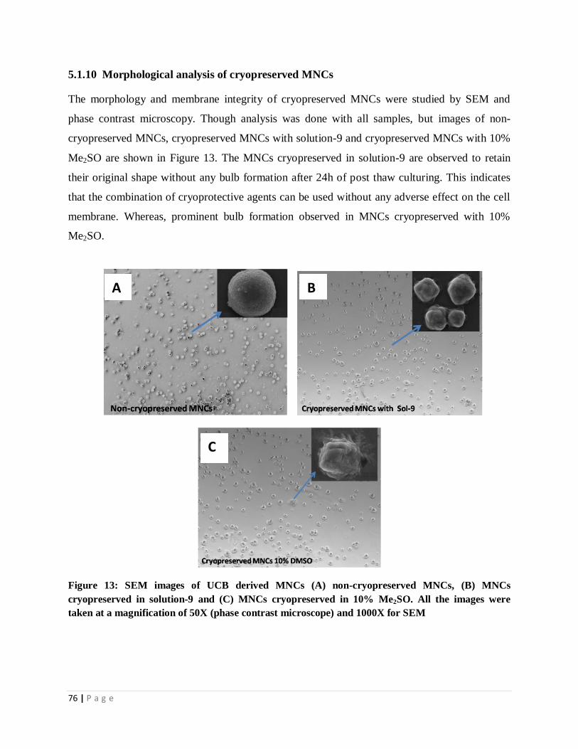
76 | P a g e
5.1.10 Morphological analysis of cryopreserved MNCs
The morphology and membrane integrity of cryopreserved MNCs were studied by SEM and
phase contrast microscopy. Though analysis was done with all samples, but images of non-
cryopreserved MNCs, cryopreserved MNCs with solution-9 and cryopreserved MNCs with 10%
Me2SO are shown in Figure 13. The MNCs cryopreserved in solution-9 are observed to retain
their original shape without any bulb formation after 24h of post thaw culturing. This indicates
that the combination of cryoprotective agents can be used without any adverse effect on the cell
membrane. Whereas, prominent bulb formation observed in MNCs cryopreserved with 10%
Me2SO.
Figure 13: SEM images of UCB derived MNCs (A) non-cryopreserved MNCs, (B) MNCs
cryopreserved in solution-9 and (C) MNCs cryopreserved in 10% Me2SO. All the images were
taken at a magnification of 50X (phase contrast microscope) and 1000X for SEM
B A
C

77 | P a g e
5.1.11 Conclusion
In this study, a number of potential natural osmolytes and antioxidants were investigated to see
their effects on post-thaw cell viability and early apoptosis of the cryopreserved MNCs isolated
from UCB. Among the CPAs and antioxidants used under study, trehalose and ectoin as
extracellular and intracellular CPAs and catalase as antioxidant are found to be the most effective.
Furthermore, the combination of these CPAs and antioxidant has shown encouraging results
towards the maintenance and survival of cell during and after freezing achieving maximum post
thaw viability of 76%, which is more than 10% higher compared to the cell viability obtained
using conventional Me2SO. Thus it has been demonstrated that trehalose, ectoin and catalase can
be the major constituents in developing a freezing solution for cryopreservation of MNCs and may
be useful to develop a cryopreservation strategy for MSCs.

78 | P a g e
PART II
Cryopreservation of UCB derived MSCs using
natural cryoprotective agents

79 | P a g e
As it has been already mentioned, transplantation of MSCs derived from umbilical cord blood is
a promising treatment option for patients suffering from various blood and tissue related diseases
because of their immunosuppressive properties and ability to differentiate into mesoderm type
lineages including osteocytes [32], chondrocytes [33], skeletal muscles [34], adipocyte and
myocytes, vascular cells and the like [35]. This has led to the advancement of isolation and
expansion of MSCs, for which the development of a successful preservation strategy is of prime
requirement to advance their clinical applications [59].
In previous chapter, trehalose and ectoin were found to be the most effective extracellular and
intracellular natural CPAs and catalase as antioxidant for the preservation of MNCs. In this
phase of thesis work, the research work has been extended further to evaluate the ability of these
CPAs and catalase towards preservation of MSCs and establish an optimized controlled rate
freezing protocol for its long term preservation.
5.2.1 Sorting and culturing of MSCs
Fluorescence activated cell sorting allows physical separation of sub-populations of cells of
interest from a heterogeneous population and thereby a high degree of purity (95-100%) of the
sorted population is obtained [60]. The cultured MNCs were sorted by this technique using
CD44, CD73, CD90, CD105, CD34, CD45 and HLA-DR markers. MSCs that are positive to
CD90/CD105/CD73/CD44 were sorted by cell sorter. As shown in Figure 14(A) primary gate
applied on FSC-A vs SSC-A to discriminate Mononuclear cells from other population, followed
by a gate to discriminate singlet from doublet with SSC-H vs SSC-W and FSC-H vs FSC-W
Figure 14 (B) a gate in FSC-H vs. FSC-W was applied to divide singlet and doublets from higher
level aggregates, and was refined by similar gate in SSC-H vs. SSC-W (Figure 14C). Singlet
population was further analyzed for the pattern of expression of CD45/HLA DR vs. CD 34
(Figure 14D). Negative expressed cells are again gated for CD44 positive cells (Figure 12E).
Stringent gating was done to remove any false CD44 positive cells based on CD34 vs
CD45/HLA-DR (Figure 14E). Cells were again gated for true positive CD90/CD73/CD105 cells
(Figure 14F).

80 | P a g e
Figure24: Image shows the sorting strategy used for isolation of MSCs. As shown in Figure 14A primary gate applied on FSC-A vs SSC-
A to discriminate Mononuclear cells from other population, followed by a gate to discriminate singlet from doublet with SSC-H vs SSC-W
and FSC-H vs FSC-W Figure 14 B, a gate in FSC-H vs. FSC-W was applied to divide singlet and doublets from higher level aggregates,
and was refined by similar gate in SSC-H vs. SSC-W (Figure 14C). Singlet population was further analyzed for the pattern of expression
of CD45/HLA DR vs. CD 34(Figure 14D). Negative expressed cells are again gated for CD44 positive cells (Figure 12E). Stringent gating
was done to remove any false CD44 positive cells based on CD34 vs CD45/HLA-DR (Figure 14E). Cells were again gated for true positive
CD90/CD73/CD105 cells (Figure 14F)

81 | P a g e
5.2.2 Cell morphology study
After initial culture, cells were observed to be round in shape as depicted in Figure 15 A. After
48 h of culture cells were shown to be adherent to culture plate (Figure 15B).
Figure 15: Phase contrast microscopy images showing morphological characteristics of cultured
hMSCs A) Initially the cells were round in shape B) Adherent cells after 48 h of culture C) Cells
became elongated as spindle fibroblastic shape after 4 days of culture D) hMSCs became confluent
in 14 days E) hMSCs after passage 1 and F) after passage 2. (Scale bar 100 μm)
Day 1 Day 2
Day 3 Day 4
Day 7 Day 14
1414
C D
A B
E F

82 | P a g e
Further, cells became elongated as spindle fibroblastic shape after 2 days of culture (Figure 15C)
and cultured cells reached confluence after 4 days (Figure 15D). Cells attains the fibroblastic
morphology after 1st passage (Figure 15 E & F)
5.2.3 Immunophenotypic characterization of MSCs
Isolated MSCs were cultured and colonies were found to appear within 5 to 7 days after initial
culturing. MSCs were harvested using 0.25% Trypsin/EDTA solution and cells were expanded
upto passage 4. The cells were then subjected to immunophenotypic characterization by flow
cytometry analysis, the data (dot plot) of which is shown in Figure 16. The immunophenotypic
characterizations are shown to be positive for CD90 (99.01%), CD73 (99.5%), CD105 (98%)
and CD44 (98.5%) expression and negative for CD34 (1.0%), CD45 (0.5%), and HLA-DR
(1.2%) indicating the cells are mesenchymal stem cells (Figure 16). Immnuofluorescence also
found to express CD90, and CD105, but not the hematopoietic origin marker CD34 as shown in
Figure 17.

83 | P a g e
Figure 16: Flowcytometric analysis on the expression of MSC markers CD90, CD105, CD44, and CD73 as well as hematopoietic markers
CD34, HLA-DR and CD45

84 | P a g e
Figure 17: Immnuofluorescence observation of passage 4 post thaw MSCs. (A, D) Nuclear staining with 4, 6-diamidino-2-phenylindole
(DAPI). (B, C) Cell-surface marker CD90 and (E, F) Cell-surface marker CD105. (Scale bar 100 μm)

85 | P a g e
5.2.4 Effect of CPAs on post-thaw MSCs viability
A series of cryopreservation experiments were conducted in controlled rate freezing equipment
for the evaluation of the selected CPAs in single as well as in combination for their ability to
preserve MSCs. The cell viability with post thawed MSCs cryopreserved in various freezing
solutions prepared from trehalose, ectoin and catalase were measured and the experimental
results are shown in Figure 18. The result shows that the higher cell viability is observed when
CPAs are used in combination than the single one. Furthermore, the highest cell viability of 82%
is obtained with freezing solution that consists of 30mM trehalose, 30mM ectoin and 100µM
catalase. This freezing solution is designated as T30/E30/C100. No further improvement in cell
viability is observed with increase in either trehalose or ectoin in the freezing solution. It is also
demonstrated that catalase has a positive impact on the freezing solution facilitating improved
cell viability Therefore, it is established that T30/E30/C100 is the most favorable freezing solution
achieving maximum MSCs viability.
Figure 18: Effect of CPAs on cell viability of cryopreserved MSCs

86 | P a g e
5.2.5 Effect of addition of Me2SO in T30/E30/C100 freezing solution
The effect of T30/E30/C100 0 freezing solution with and without Me2SO was also studied to
investigate any influence of Me2SO on cryopreservation of MSCs. The experimental data is
shown in table 7. It is observed that the cell viability of T30/E30/C100 is higher than the cell
viability obtained with conventional 10% v/v Me2SO. The highest cell viability of 84% is shown
by T30E30C100 with addition of 2.5%v/v Me2SO. The cell viability (82%) is comparable to the
cell viability obtained with T30E30C100 without adding Me2SO. However, keeping in view the
harmful effect of Me2SO, T30E30C100 is established as the most favorable freezing medium.
Table 7: Effect of Me2SO as an additive in freezing solution on cell viability
Solution Me2SO
(%)
Trehalose
(mMol/l)
Ectoin
(mMol/l)
Catalase
(µg/ml)
Viability
(%)
1 0 30 30 100 82
2 2.5 30 30 100 84
3 5 30 30 100 75
4 10 30 30 100 70
5 10 0 0 0 61
6 2.5 0 0 0 24
5.2.6. MTT Assay
The cell viability of cryopreserved MSCs was also evaluated by MTT assay. The assay result is
shown in Figure 19. It is indicated from Figure that solution containing T30/E30/C100 have
achieved better cell viability as compared to other freezing solutions tested after 24 h of post
thaw, though a variation in cell viability is observed among the freezing solutions. The maximum
cell viability as indicated by slightly higher OD value is achieved with cryopreserved solution
consisting of a combination of T30/E30/C100 with 2.5% v/v of Me2SO. A comparable cell viability
is also achieved with T30/E30/C100 freezing solution. Keeping in view the detrimental effect of
Me2SO, T30/E30/C100 is established as the most favorable freezing medium which is also well

87 | P a g e
supported by the cell viability result obtained by flowcytometry analysis as described in previous
section.
Figure 19: MTT assay of post-thawed MSCs cryopreserved in different compositions of
extracellular cryoprotectant, intracellular, antioxidant with and without Me2SO. MSCs
cryopreserved in 10% Me2SO taken as control. All the data are compared with control using
student t-test
5.2.7 Post-thaw phenotyping characterisation of cryopreserved MSCs
The phenotyping characterisation of MSCs cryopreserved in T30/E30/C100 with and without
Me2SO has been done by flow cytometry analysis. CD90, CD44, CD45, CD105, CD73 and
CD34 expressions of cryopreserved MSCs were measured and compared with control. The
results are tabulated in table 8. As expected, both cryopreserved and noncryopreserved MSCs
have shown low expression of hematopoietic stem cell markers CD34, HLA-DR and CD45 and
high expression of mesenchymal-associated marker CD90, CD73, CD105 and CD44. The
corresponding positive CD expressions were in the range of 98-99%. Furthermore, there is no
significant variation in phenotype characteristics are observed with MSCs crypreserved in
traditional Me2SO.

88 | P a g e
Table-8: Immunophenotypic characterisation of MSCs after cryopreservation
Cryoprotectant CD45 CD34 HLA-DR CD90 CD73 CD105
T30E30C100 0.60±0.41 0.33±0.26 1.73±37 98.77±31 97.66±78 98.94±37
T30E30 M10C100 1.36±0.08 0.92±0.48 1.55±33 97.22±71 98.79±24 99.77±66
M10 0.92±0.22 0.22±0.04 1.49±71 98.88±59 99.06±97 98.45±46
T30E30M2.5C100 0.60±0.41 0.33±0.26 1.73±37 98.77±31 97.66±78 98.94±37
T30E30M5C100 1.36±0.08 0.92±0.48 1.55±33 97.22±71 98.79±24 99.77±66
5.2.8 Cell proliferation assessment in cryopreserved MSCs
As it is expected, non-cryopreserved MSCs are found to proliferate rapidly than
cryopreserved MSCs [69]. The growth curve of post-thaw MSCs in all freezing solutions
used in this as well as non-cryopreserved MSCs have shown similar trend i.e. In first 2 days
after inoculation, no variation in cell number is observed that may be attributed to the
adherence of cell to the tissue culture plates, and on day 3, the cells entered the logarithmic
growth stage representing an exponential increase in cell number. The doubling time of
MSCs in freezing solutions are in the range of 29.0 to 31.9 h (Figure 20 and Table 9). Thus it
has been demonstrated that MSCs cryopreserved in the newly formulated freezing solution
T30/E30/C100 has retained their cell proliferation ability.
Figure 20: Growth curves of post thaw hMSCs cryopreserved with T30/E30/C100 and Me2SO
(10%v/v)

89 | P a g e
Table 9: Population doubling time of MSCs
Cryoprotectant Population doubling time (h)
T30E30C100 29.7
T30E30 M10C100 31
M10 30
T30E30M2.5C100 31
T30E30M5C100 31.6
Fresh 29.0
5.2.9 Change of mitochondrial membrane potential in cryopreserved MSCs
Recent studies have reported a rise in reactive oxygen species (ROS) like superoxide anion
and hydrogen peroxide during cryopreservation procedure [23]. Mitochondria are considered
as the main source of this ROS. This superoxide anion induces the release of cytochrome C
from the intermembrane of mitochondria into the cytoplasm that eventually leads to
apoptosis. Therefore, ROS suppression using strong antioxidants can be an effective agent
for improving cell recovery from cryopreserved cells. Furthermore it is reported that the
generation of ROS and mitochondrial potential are directly correlated [61]. Hence, it is
important to understand the changes in mitochondria during long term cryopreservation of
hMSCs. Therefore, the mitochondrial membrane potential (MMP) of MSCs was studied in
presence or absence of antioxidant using a mitochondrial membrane potential-sensitive dye,
JC-1. After thawing, cryopreserved cells with and without antioxidant (catalase 100µg) were
divided into two groups for analysis. The MMP rate was measured as 14±1.2% and 26±2.0%
for the combination of trehalose and ectoin with or without catalase which is stastically
significant as shown in Figure 21. Similarly the conventional 10%v/v Me2SO as sole
cryoprotectant has shown significant variation in MPP with (49±4%) or without (71±10%)
supplement of catalase. From this study, it has been established that the combination of
trehalose, ectoin, and antioxidant has very low level of depolarization and 10% v/v Me2SO
has shown comparatively higher depolarization rate.

90 | P a g e

91 | P a g e
Figure 21: Analysis of mitochondrial membrane potential (MMP) of post thaw MSCs. At indicated
time points, cell samples were stained with MMP sensitive dye JC-1. Change in red (polarized) to
green (depolarized) fluorescence of MSCs was analyzed by FACS, and the data present as dot plots
to depict percentage of MMP. Staurosporin was added to fresh MSCs as a positive control to induce
maximum MMP
5.2.10 Cytoskeleton analysis of post-thaw MSCs
As it has already been mentioned that F-actin a main constituent of cytoskeleton has a key role in
the maintenance of cellular function. However, F-actin depolymerises and accumulates during
low temperature freezing that lead to cell damage [225]. Therefore, it has been investigated the
effect of cryopreservation on the organization of F-actin. Fluorescence image analysis for change
in cytoskeleton was done with MSCs treated with different cryoprotectant solutions. However,
the result obtained with MSCs cryopreserved in most effective freezing solution consisting of
trehalose, ectoin, and catalase (T30/E30/C100) and traditional 10% v/v Me2SO are shown in Figure
22. It clearly shows that MSCs frozen in T30/E30/C100 solution retain their original shape without
undergoing any deformation or damage in F-actin ultra structure after 48 h of post thaw culture
(Figure 22 C) and similar type of morphology is also shown by control (Figure 22A). Moreover,
cells have shown normal cell behavior i.e. fibroblast like morphology during post thaw culturing
which is evident from Figure 22F. In contrast, the distortion in F-actin is observed when MSCs
are cryopreserved in 10%v/v Me2SO as it is indicated from Figure 22B. A similar type of
morphology is observed, after 72h of post thaw cryopreserved cells in both 10% v/v Me2SO and
T30E30C100 as shown in Figure 22D and E. It is further noticed (Figure 22F) that during the
progress of culture, MSCs frozen in T30/E30/C100 became long shuttle-shaped and on 7 days, the
occurrence of confluence indicates the start of passaging. Furthermore, cells have shown parallel
or whirlpool-shaped arrangement on prolonged culturing upto 2, 4 and 8 passages.

92 | P a g e
Figure 22: F-actin morphology and distribution in cryopreserved hMSCs. (A) Fresh 4th
passaged MSCs (B) hMSCs cryopreserved
in 10%Me2SO after 48h post thaw (C) hMSCs cryopreserved in T30/E30/C100 after 48h post thaw, (D) hMSCs cryopreserved in
10%Me2SO after 72h post thaw (E) hMSCs cryopreserved in T30/E30/C100 after 72h post thaw, (F) hMSCs cryopreserved in T30/E30/C1005
after 7 days of post thaw. All the cells were labeled with FITC-phalloidin and visualized by fluorescence microscopy (Scale bar 50 μm

93 | P a g e
5.2.11 Effect of storage duration on cell viability
It is evident from literature that the cell viability immediately after thawing cannot be reliable
criteria for the evaluation of cryopreservation efficacy. It is reported that the decrease in cell
viability observed with post thaw sample is related to apoptotic and necrotic processes which
occur within first 24 hr after thawing process [216]. Therefore the apoptosis assay was done with
24 hr after post thaw MSCs at an interval of 1, 6, and 12 months of freezing and was compared
with fresh MSCs using Annexin/7ADD kit. Freshly isolated MSCs from cord blood were used as
an experimental control. Results (table 7 and Figure 23) indicate that after 1 month, the freezing
medium T30E30C100 has shown maximum cell viability of 84.90%, 4.17% early apoptotic cells
(annexin-V+), 5.69% late apoptotic cells (annexin-V
+ and 7ADD+) and 5.24% necrotic cells
(annexin-V+
and 7ADD+)). The corresponding results obtained after six months of
cryopreservation are 75.74% cell viability, 20.87% early apoptotic, 0.50% late apoptotic and
2.89% necrotic cells. Comparable result is also obtained with cryopreserved MSCs after 12
months of storage. The cell viability achieved in later case is 73%. Apoptosis results with
T30E30C100 are also shown to be encouraging as compared to the result achieved using
conventional 10%(v/v) Me2SO as freezing medium (Table 10) and a representive data is shown
in Figure 23.

94 | P a g e
Table 10: Effect of storage time on MSCs viability
Freezing medium Storage duration %Viable cells %Necrotic
cells
%Early apoptosis
cells
%Late apoptosis cells
Freshly isolated MSCs 98.04±1.1 0.02±0.005 1.70±0.8 0.24±0.06
1 month 84.90±2.5 5.69±0.9 4.17±0.5 5.24±1.7
T30,E30, C100 6 months 75.74±1.1 2.89±0.12 20.87±0.21 0.50±0.1
12 months 73.9±2.3 3.93±1.4 17.07±2.2 3.93±0.3
1 month 61.57±3.2 4.54±1.3 24.23±2.1 9.57±1.1
10% Me2SO 6 months 51.74±1.7 1.42±0.5 35.86±3.2 10.98±2.5
12 months 43.76±2.3 17.37±1.7 20.14±2.6 18.73±1.9

95 | P a g e
Figure 23: Flowcytometric analysis of MSCs apoptosis performed using 7AAD and annexin. The image shows the level
of viability of MSCs cryopreserved in T30/E30/C100 after (B) 1 month (C) 6 months and (D) 12 months. Similarly, the
image shows the level of viability of MSCs cryopreserved in 10%Me2SO after (E) 1 month, (F) 6 months and (G) 12
months. Viability of all the samples are compared with freshly isolated MSCs (A)

96 | P a g e
5.2.12 Effect of cryopreservation on differentiation potential of hMSCs
hMSCs cryopreserved in T30/E30/C100 freezing medium were induced into adipogenic, osteogenic
and chondrogenic differentiation to examine whether the thawed hMSCs retained their ability to
differentiate into three specific lineages. The experimental observations are described here-
Osteogenic potential
The histological results of the osteogenic differentiation of hMSCs are shown in Figure 24(D, E).
hMSCs cryopreserved in T30/E30/C100 for 12 months is observed to be sufficiently differentiated
into osteoblasts as shown by the deposition of calcium on 14 days of culture (Figure 24D). The
differentiation is stronger on 21 days as it is evident from the deposition of more calcium when
major portion of the MSCs was mineralized (Figure 24E).
Chondrogenic potential
The chondrogenic differentiation of hMSCs cryopreserved in T30/E30/C100 and non cryopreserved
hMSCs is depicted in Figure 24 (A, B). The cells were stained positive with safranin O indicates
the formation of cartilage matrix on 14days of culture. The entire well plate is observed to be
covered with differentiated extracellular cartilage matrix on 21 days (Figure 24B).
Adipogenic potential
The histological results of adipogenic differentiation of cryopreserved hMSCs are shown in
Figure 24 (G, H). When the post thaw MSCs were exposed to the adipogenic medium, the cell
morphology underwent transition from elongated fibroblast shape into a round or polygonal
shape. After 14 days of induction, a consistent cell vacuolation is evident in the induced cells
(Figure 24G). Most of the differentiated cells showed red lipid droplets throughout the cytoplasm
when stained with 0.36% Oil red O stain as depicted in Figure 24H [64],[36].
Thus it has been demonstrated that the MSCs cryopreserved in T30/E30/C100 freezing solution has
retained its adipogenic, osteogenic and chondrogenic differentiation ability even after 12 months
of cryopreservation.

97 | P a g e
Figure 24: Representative phase contrast images of differentiated and undifferentiated hMSCs.
Images in first and second column are of differentiated post thaw MSCs cryopreserved in freezing
medium containing trehalose, ectoin & catalase (T30E30C100). (A, B) shows differentiation of MSCs to
osteocytes, (stained with alizarin Red S stain (D, E) differentiation of MSCs to chondrocytes (stained
with Saffranin O and (G, H) differentiation of MSCs to adipocytes (stained with oil red O) (Scale
bar 100 μm), Images in third column are for control. The images show the adipogenic, osteogenic
and chondriogenic differentiation ability of cryopreserved MSCs in new freezing medium.)

98 | P a g e
4.2.13 Effect of cryopreservation on clonogenic potential of MSCs:
Cryopreservation may affect colony forming capacity of stem cells that results in the reduction of
cell differentiation. In a study on controlled rate cryopreservation of UCB, it is observed that
cryopreserved CD34+ cells contain a significant population of non-clonogenic early apoptotic
cell which cannot be differentiated[65]. Therefore, colony-forming unit-fibroblast (CFU-F) assay
was performed to investigate the effect of cryopreservation solution T30E30C100 on the clonogenic
capacity of post thawed hMSCs [36]. As shown in Figure 25, non-cryopreserved MSCs
displayed a similar CFU-F potential as observed with post thaw MSCs cryopreserved in
T30E30C100 freezing media. The corresponding CFU-F values are measured as 11± 0.7 and 10 ±
0.5 (SD) respectively. Furthermore, hMSCs cryopreserved in conventional 10% v/v Me2SO is
shown to have lower colony forming capacity in comparison to hMSCs cryopreserved in
T30E30C100 indicating the superiority of new freezing solution than the conventional one.
Figure 25: Effect of cryopreservation on CFU-F ability of MSCs. Data represents the mean ± SD
per 1×10³ cells of three separate experiments

99 | P a g e
5.2.14 Study on apoptosis pathways in cryopreserved induced cell death
As already mentioned that, cryopreservation plays an important role in long term storage or
banking of MSCs. MSCs derived from various sources are cryopreserved by slow freezing
protocols utilizing Me2SO as cryoprotectant in freezing medium. However, it is reported that
Me2SO fails to protect hMSCs from cryopreserved induced cell death (CICD) due to external
stress developed during cryopreservation process. It is, therefore, essential to understand the
mechanism of cryopreserved induced cell death using cryoprotective agents, which may be
helpful in developing an effective cryopreservation protocol. It is further reported that the event
of CICD is largely due to apoptosis [228]. Cryopreservation induced apoptosis is significant in a
wide variety of cell systems including fibroblasts, hepatocytes [232], cord blood [231-232],
peripheral blood mononuclear cells [229] renal cells [235], spermatozoa [236], oocytes and
ovarian tissue [234]. These studies have thrown light on the molecular based cell death following
cryopreservation and thus affecting cryopreservation outcome. It is also reported that
manifestation of cryopreservation induced cell death may take few h to days. However, the
maximum apoptotic and necrotic activity occur at 24 h post thaw, following a decline in cell
survival upto 48 h post thaw. This study of the timing of cell death following cryopreservation is
termed as cryopreservation-induced Delayed-Onset Cell Death (CIDOCD). Previously, it has
been observed [235] that cryoinjury induced cell death is initiated by the intrinsic or
mitochondrial apoptotic pathway. However, according to recent report [236], both extrinsic and
intrinsic pathways are activated after post thaw. Initiation of apoptosis after-cryopreservation has
been shown via membrane-mediated Fas-receptor initiation by caspase-8 and caspase-9
activation (237, 238). Studies have also shown that apoptotic pathways are activated after
thawing that in turn activates caspases 3, 8 and 9 [239].
All these studies suggest that programmed cell death following cryopreservation is important to
characterize and study. The strategy to control both membrane and mitochondrial mediated
apoptosis pathway is essential to improve the outcome of cryopreservation procedure. The
increase in calpain-like activity during cryopreservation is another important factor that
influences post thaw cell survival [238-239] and the addition of calpain inhibitor results in higher
viability rate. However, the specific role of calpain in cryopreservation induced apoptosis and
interrelation between calpain and caspase mediated apoptosis so far remains unclear.

100 | P a g e
In this part of research, an attempt has been made to study the apoptosis pathways and
mechanism for cryopreserved induced cell death. The study also investigates the extrinsic and
intrinsic pathways as well as the involvement of calpain in the MSCs cryopreservation process.
5.2.14.1 Post thaw cell viability
To identify and characterize the cryopreserved induced apoptosis in MSCs, cells were
cryopreserved with inhibitors and analyzed using flowcytometer immediately after post thaw.
Result shows (Figure 26) that there is no significant difference in cell viability observed
immediately after post-thaw. It is further indicated that 10% cells died during cryopreservation
process which is independent of apoptosis. This cell death may be due to physical stress
produced during cryopreservation process [237]. Initial cell viability result shows that all
cryopreserved MSCs with or without pre-treatment of inhibitor have the same cell viability range
of 85±5%. Thus the result suggests that initial cell death cannot attribute to the activity of
caspase 3, 8, 9 or calpain.
Figure 26: Effect of cryopreservation on cell viability in presence of caspase and calpain
inhibitors. Bars represent the data from at least three experiments – standard error of the mean.
‘Cryo’ means cryopreserved sample without inhibitor. The result shows that all the cryopreserved
cells with or without inhibitor have same viability range of 85%±5.
5.2.14.2 Caspase-mediated apoptotic signaling pathways
One of the important characteristics of apoptosis is DNA degradation, which is thought to be a
caspase dependent process. To compare the effect of caspase on cryopreservation induced

101 | P a g e
apoptosis, MSCs were incubated with various caspase inhibitors. In this study, MSCs
cryopreserved in freezing solution shows a large population of cells with sub G0 phase (degraded
DNA). Pretreatment of MSCs with general caspase inhibitor (z-VAD-fmk) has shown its ability
to block the degradation of DNA, whereas pretreatment with z-IETD-fmk, z-LEHD-fmk, or z-
DEVD-fmk are not able to block DNA degradation (Figure-27 B) significantly. Furthermore to
investigate the kinetics of cell death, cells were subjected to Annexin V/7-AAD staining to detect
early apoptotic (Annexin V), late apoptotic/necrotic (Annexin V/7-AAD), and necrotic (7-AAD)
cells (Figure 27). In comparison to non-cryopreserved MSCs, a significant percentage of early
apoptotic and late apoptotic/necrotic cells are observed when MSCs were cryopreserved in 10%
Me2SO. However, within 24 h of post thaw, there is a significant amount (approximately 35%)
of cell death occurs even with caspase inhibition by z-VAD-fmk.
The kinetics of early, late apoptotic and necrotic cells in presence of different inhibitor treatment
after 24h of post thaw are summarized in Figure 27. Although the pretreatment of MSCs with z-
VAD-fmk before cryopreservation is not observed to be completely protective after 24 h of post
thaw, it inhibits the loss of cell viability significantly as compared to cryopreserved MSCs
without inhibitor pretreatment. In marked contrast, pretreatment of these cells with selective
caspase inhibitors such as z-IETD-fmk, z-LEHD-fmk, and z-DEVD-fmk did not have any effect
on the loss of cell viability induced by cryopreservation.

102 | P a g e
Figure 27: Effect of caspase inhibitors on cryopreservation-induced apoptosis of MSCs. (A) Untreated MSCs; (B) Cryopreserved
sample treated with 50 mM z-LEHD-FMK; (C) Cryopreserved sample treated with 50 mM z-VAD-FMK; (D) Cryopreserved sample
treated with 50 mM z-IETD-FMK; (E) Cryopreserved sample treated with 50 mM z-DEVD-FMK; (F) The % of viable, apoptotic, and
necrotic cells. Bars represent the data from at least three experiments ± standard error of the mean [* indicates difference from control
(i.e., cryopreserved MSCs without pretreatment with caspase inhibitor)]. Cryo = cryopreserved sample without inhibitor pretreatment

103 | P a g e
5.2.14.3 Western blotting analysis of caspase proteins
To dissect the potential role of caspases in cryopreservation-mediated MSCs apoptosis, MSCs
were pretreated with various caspase inhibitors before cryopreservation. The aim of investigation
is whether these caspase proteins are cleaved during cryopreservation induced apoptosis of
MSCs, and whether this process is selectively inhibited by z-VAD-fmk, z-IETD-fmk, z-LEHD-
fmk, or z-DEVD-fmk. MSCs with or without pretreatment with caspase inhibitors were
cryopreserved and 24h post thaw cells were harvested for Western blot analysis using antibodies
specific to caspases 8, 9, and 3.
The Western blot analysis result is shown in Figure 23. The Figure shows the appearance of
typical cleavage fragments of caspases 8, 9, and 3 proteins in cryopreserved MSCs with and
without treatment with caspase inhibitors except z-VAD-fmk. To determine the potential
involvement of the death receptor pathway in cryopreserved-induced apoptosis, we also
examined the activity of Bid protein. It has been reported that Bid relays the apoptotic signal
from the cell surface to the mitochondria. Furthermore, after cleavage due to apoptosis, truncated
Bid translocates into mitochondria and disrupts the mitochondrial membrane potential, thereby
triggers cytochrome c release [255]. Cryopreservation also induces activation of Bid as evident
from Figure 28. It is observed that 32 kDa-band represents the precursor of caspase-3 while two
smaller bands of 17 kDa and 12 kDa indicate the active subunits. A significant decrease in
amount of caspase-3 precursor (32 kDa) in cryopreserved MSCs is observed in presence of z-
DEV-fmk caspase inhibitor. Whereas a significant increase in caspase-8 (18 kDa) activity is
observed in MSCs cryopreserved in presence of other inhibitors. Moreover, the general caspase
inhibitor (z-VAD-fmk) tends to decrease the expression of caspase proteins to a non-detectable
level. Result also shows that z-VAD-fmk is able to significantly decrease the level of caspase-9
precursor. Other than z-VAD-fmk, there is no significant alteration of caspase-9 precursor in
MSCs cryopreserved with other apoptotic inhibitors observed. Cryopreservation induces the
activation of the initiator caspases 8, the effector caspase 3 and the pro-apoptotic protein Bid.
Caspase 9 is activated by release of cytochrome c from the mitochondria, which causes the
formation of the apaf-1/caspase 9 apoptosome. Caspase 8 is cleaved and activated during the
formation of the DISC at the plasma membrane. Caspase 3 is normally in the cell as an inactive
zymogen, where it is cleaved by initiator caspases such as 8 and 9 [26].

104 | P a g e
However, the result shows that neither caspase 8/caspase 3 nor caspase/tBid/mitochondria
pathway is solely responsible for cryopreserved induced MSCs cell death as both proteins from
both the pathways are activated.
Figure 28: Effect of cryopreservation on activation of caspase and Bid proteins. Actin has been used
as housekeeping marker protein. Bars represent data from at least three experiments – standard
error of the mean. C = cryopreserved sample without inhibitor pretreatment; D= cryopreserved
sample treated with 50 mM z-DEVD-FMK inhibitor; I = cryopreserved sample treated with 50 mM
z-IETD-FMK; L = cryopreserved sample treated with 50 mM z-LEHD-FMK; V= cryopreserved
sample treated with 50 mM z-VAD-FMK.
5.2.14.4 Mitochondrial membrane potential
The mitochondrial or intrinsic pathway is considered to be one of the two predominant signaling
cascades leading to apoptosis and many studies have described the depolarization of the
mitochondrial membrane as an important characteristic of this pathway [245, 246]. Further,
mitochondrial membrane depolarization is associated with release of cytochrome c and
subsequent activation of caspases 9 and 3 [247]. To examine whether cryopreservation induced

105 | P a g e
apoptosis of MSCs involves any loss of mitochondrial membrane potential, a dual-fluorescent
dye JC-1 was used. When the mitochondrial membrane depolarizes during cell death the dye
dissociates into its monomeric form, which fluoresces at 530nm. In this study, the monomer vs.
aggregate fluorescence plots (Figure 29 (A)) indicates a loss in aggregate fluorescence and an
increase in monomer fluorescence representing a decrease in mitochondrial membrane potential.
Interestingly, z-VAD-fmk, z-IETD- fmk, z-LEHD- fmk, z-DEVD-fmk caspase inhibitors has
shown to significantly inhibit the loss of mitochondrial membrane potential caused by
cryopreservation.
Figure 29: Effect of caspase inhibition on cryopreserved-induced mitochondrial membrane
potential and DNA degradation in MSCs after 24 h of post thaw (A) Bar graphs showing the
percentage of depolarized MSCs for cryopreserved MSCs with and without caspase inhibitor and
(B) the corresponding percentage of MSCs with degraded DNA. “Cryo” means cryopreserved
sample without pre-treatment with any inhibitor. Bars represent data from three separate
experiments ± standard error of the mean
5.2.14.5 Calpain activity
Calpain is a calcium-dependent neutral protease and is reported in initiating or progressing the
events associated with apoptosis causing cell death during cryopreservation [251]. So in this
study, the effect of calpain with and without calpain specific inhibitor (PD150606)) were
investigated in apoptosis induced cell death during cryopreservation of MSCs. The choice of
PD150606) as an effective calpain inhibitor is based on the published literature [252].

106 | P a g e
To study the effect of calpain inhibitor on cell cycle, MSCs were pretreated with calpain
inhibitor before cryopreservation. Result shows the significant inhibition of DNA degradation by
the addition of calpain inhibitor (Figure 30 B). To examine the effect of calpain inhibitor on post
thaw, MSCs were assessed by Annexin V assay for determining viable cells, apoptotic cells and
necrotic cells. From the results it is indicated that, the presence of calpain inhibitor does not
show any significant improvement in cell viability. However the addition of general caspase
inhibitor (z-VAD-fmk) and calpain inhibitor (PD150606) in combination improve the cell
viability upto 80±2%. Thus the result suggests that cryopreserved induced apoptosis is attributed
by both caspase dependent and caspase independent.
Figure 30: Effect of caspase inhibitors on cryopreservation-induced apoptosis of MSCs. (A)
Untreated MSCs; (B) Cryopreserved MSCs treated with 50 mM z-LEHD-FMK; (C) Cryopreserved
MSCs treated with 50 mM z-VAD-FMK; (D) Cryopreserved MSCs treated with 50 mM z-IETD-
FMK; (E) Cryopreserved MSCs treated with 50 mM z-DEVD-FMK; (F) denotes % viable,
apoptotic, and necrotic cells. Bars represent the data from at least three experiments – standard
error of the mean, [* indicates difference from control (i.e., cryopreserved MSCs without
pretreatment with caspase inhibitor)]. ‘Cryo’ means cryopreserved sample without inhibitor.The
flowcytometric analysis shows that cryopreserved induced apoptosis is attributed bt both caspase
dependant and caspase independent.

107 | P a g e
5.2.14.6 Western blotting analysis for calpain protein
The Western blot analysis has revealed that (Figure 31) the expression of calpain increases in
cryopreserved MSCs and addition of calpain inhibitor decreases the cleavage of calpain protein.
It is also observed that the addition of pan caspase inhibitor does not have any effect on
expression of calpain protein. Interestingly, the addition of both calpain and general caspase
inhibitor (z-VAD-fmk) has shown synergistic effect in lowering the expression of calpain. This
result suggests that the general caspase inhibitor is not able to inhibit calpain dependant pathway.
Also caspase and calpain mediated cell death pathways are interconnected.
Figure 31: Effect of calpain inhibitor PD150606 on activation of m-calpain proteins. Cryopreserved
MSCs were investigated after pretreatment with PD150606 with or without z-VAD-FMK. Bars
represent data from three separate experiments– standard error of the mean. ‘Cryo’ means
cryopreserved sample without inhibitor. PD150606 =MSCs pretreated with 50 mM PD 150606; z-
VAD-FMK=MSCs pretreated with 50 mM z-VAD-FMK; PD150606 and z-VADFMK=MSCs
pretreated with a combination of 50 mM PD150606 and 50 mM z-VAD-FMK.The result shows that
the general caspase inhibitor is not able to inhibit calpain dependent pathway

108 | P a g e
5.2.14.7 Effect of calpain inhibitor on mitochondrial membrane potential and DNA
degradation
The experimental result of the influence of calpain inhibitor is shown in Figure 32A. Calpain
inhibitor (PD150606) did not show any significant effect in mitochondrial membrane potential
(MMP) during cryopreservation. However, the combination of PD150606 and z-VAD-fmk
significantly maintained the mitochondrial membrane potential (MMP) and decreased the
amount of DNA degradation in post thaw MSCs. These data suggest that the apoptotic signaling
to the mitochondria has both caspase dependent and independent components. And the calpain
dependent pathway also works through mitochondrial pathway.
Figure 32: Comparative confocal images (F-actin) of cryopreserved and non-cryopreserved silk
nanofiber TECs. (A) (i) Shows noncryopreserved TECs (control), (ii and iii) shows TECs
immediately after cryopreservation with scale bar 5µm, (B) 24 h after cryopreservation with scale
bar 5 µm and (C) close-up images of actin cytoskeleton 48 h after cryopreservation (ii) with scale
bar 40 µm. In both Me2SO and T30/E30/A100/I cryopreserved groups, the cytoskeleton in the
frozen/thawed regions is damaged, with shortened extensions and less number of cells compare to
noncryopreserved cells. In Me2SO frozen group, the cells have condensed, distorted after freezing,
and less damage is observed in T30/E30/A100/I groups.

109 | P a g e
5.2.15Optimization of Controlled rate freezing parameters for
cryopreservation of hMSCs using T30E30C100 freezing medium
It is fact that depending on the type of cells and freezing media, there is need to maintain an
optimal condition that offers maximum cell recovery following cryopreservation. In this context,
incubation temperature, freezing medium, cooling rate, storage temperature, cell concentration
during freezing, and duration of storage, are some of the key factors that has great influence on
the cryopreservation outcome. while a few studies have examined the influence of freezing
conditions on post thaw viability of MSCs using Me2SO as cryoprotectant [41, 44], no such
study has been reported so far for optimization of cryopreservation conditions of MSCs using
combination of cryoprotectants. Keeping this in view, efforts has been given in this section to
investigate the effect of key controlled rate freezing parameters on cryopreservation of MSCs
and establish the optimum cryopreservation condition using the most effective freezing medium
T30E30C100 in presence of general caspase and calpain inhibitors.
5.2.15.1 Effect of cooling profile on MSCs viability
The cryopreservation experiment was carried out at different prenucleation cooling rate such as-
1ºC/min, 2.5ºC/min, 5ºC/min and 10ºC/min to examine the effect of prenucleation cooling rate
on cell viability. The others parameters such as: nucleation temperature (-7.5°C), cold spike
temperature (-40°C), hold time (10min), and post nucleation cooling rate (1°C) were maintained
constant. The parameters were chosen based on published literature [143]. As shown in Figure
33a, the effect of prenucleation temperature is shown to have a significant influence on cell
viability. The cell viability increases with decrease in cooling rate. The maximum cell viability
of 82% is obtained at prenucleation cooling rate of -1.0°C/min. Comparable cell viability is also
achieved at -2.5°C/min.
Similarly, the effect of nucleation temperature (-2.5°C, -5°C, -7.5°C and -10°C) on cell viability
was studied and the experimental result is shown in Figure 33b. The highest cell viability of 78%
is achieved at nucleation temperature of –7.5°C and a decline in cell viability is observed with
either higher or lower than –7.5°C nucleation temperature. The others parameters such as:

110 | P a g e
prenucleation cooling rate 1°C/min, cold spike temperature (-40°C), hold time (10min), and post
nucleation cooling rate (1°C) were maintained constant.
The effect of shock cooling or so called cold-spike temperature on cell viability was investigated
by performing cryopreservation experiment at varying shock cooling temperature in the range of
-20°C, -40°C, -60°C and -80°C. Figure 28c shows the effect of shock cooling temperature on
MSCs recovery. The optimal cell viability was recorded as 85% at -80°C. The others parameters
such as: prenucleation cooling rate 1°C/min, nucleation temperature (-7.5°C), hold time (10min),
and post nucleation cooling rate (1°C) were maintained constant.
Figure 33d shows the effect of different holding times at post-nucleation temperature of -30°C.
An upward trend of cell viability is observed till holding time 10min is reached and there is
decrease in cell viability observed beyond this holding time. The optimal cell recovery of more
than 80% is obtained with 10min holding time.
Finally, Figure 33e shows the effect of post nucleation cooling rate on post thaw cell viability. A
decrease in cell viability is observed over the range of cooling rate tested and the post nucleation
cooling rate of 1°C/min offers the maximum cell viability (84%) results. The others parameters
such as: prenucleation cooling rate 1°C/min, nucleation temperature (-7.5°C), and holding time
(10min) were maintained constant.
Our results demonstrate the sensitivity of cell viability to the cooling profile used during
cryopreservation in controlled rate freezing. However, the optimized cooling profile is
establishes as prenucleation cooling rate 1oC/min, nucleation temperature -7.5
oC, cold spike of
80oC/min, post nucleation holding time of 10min and post nucleation cooling rate of 1
oC/min.
The resultant optimum cooling profile is shown in Figure 34. The results from the controlled
rate freezer (CRF) temperature control study, illustrating the process temperature profiles for
sample in the CRF chamber with cold-spike nucleation control over the period of a
cryopreservation study. A blue line indicates the temperature profile of the chamber in response
to a programmed profile for freezing hMSCs. The typical cooling profile included five steps: (1)
equilibrate temperature, (2) induce ice formation with a cold spike, (3) warming rate, (4) hold at
–35 °C for 10 minutes, and (5) cool to –40 °C at a cooling rate of 1.0 °C/min. The uniformity of
nucleation is apparent right after the cold gas was plunged to –80 °C, as shown in the figure

111 | P a g e
temperature profiles of sample and chamber were tightly aligned together through the whole
freezing process. Optimization of cooling profile has been carried out based on the sample
temperature.
Figure 33: Percentage recovery of MSCs after cryopreservation in controlled rate freezer under
different cooling variables (a) pre-nucleation cooling rate in °C/min (b) prenucleation temperature
in °C; (c) cold-spike temperature in °C/min; (d) holding time in min(e) post nucleation cooling rate
in °C/min

112 | P a g e
Figure 34: Temperature profile of cryochamber and sample with time; blue line represents the
chamber temprature, green line represents the sample temperature. (a) equilibrium temperature;
(b) a pre-nucleation cooling step; (c) a pre-nucleation temperature step; (d) a cold spike step; (e) a
post-nucleation temperature hold step and (f) a final cooling step.
5.2.15.2 Effect of cell density
The cryopreservation experiment was designed to test whether cell concentration has any effect
on cell viability following cryopreservation. Study has shown that increase in cell concentration
decreases cell viability and metabolic activity in hepatocytes [70]. During slow cooling, ice
formation mainly occurs on extracellular spaces but as the cell concentration increases
extracellular spaces are decreased by cell packing due to which intracellular ice formation occurs
more readily. In this study the varying concentration of MSCs such as 0.5, 1, 2, 3, 4 and
5×10⁶/ml were investigated to establish optimal concentration to maximize cell viability. The
cell viability results of post thawed MSCs are shown in Figure 35. Each set of experiment was
performed in triplicate. An unpaired t-test was conducted taking 3×10⁶/ml MSCs as control. The

113 | P a g e
result indicates that the maximum cell recovery is achieved with 3×10⁶/ml followed by
2×10⁶/ml. Furthermore, a decrease in cell viability is observed with increase in cell concentration
beyond 3×10⁶/ml.
Figure 35: Effect of cell concentration on cell viability
5.2.15.3 Effect of storage temperature
Recent study suggests that storage temperature plays an important role in cell viability [91-102].
So, the effect of different storage temperature such as −80°C, −150°C and −180°C has been
studied on the cell viability. The different storage temperatures were chosen based on the
published literature [66-69]. The experimental data of MSCs viability after cryopreservation is
shown in Figure 36. Result shows that cell viability significantly varies with storage temperature
from -80°C to -150°C. However the maximum cell viability is achieved at -150°C and then a
decline in cell viability. is observed

114 | P a g e
Figure 36: Effect of different storage temperature on cryopreserved MSCs viability
5.2.15.4 Verification of cryopreservation protocol
5.2.15.4.1 Effect of cryopreservation on cytoskeleton and mitochondria distribution
As it is mentioned earlier, F-actin has a role in controlling cell morphology and surface
movement [120] as well as cellular functions [121-124]. It is reported that the critical factor that
changes F-actin morphology and distribution is not associated with addition or removal
cryoprotectant but freezing profile [123]. Result shows (Figure 37) that at the optimum
controlled rate of freezing (CRF) condition F-actin shows a better maintenance recovery in
morphology and distribution of cytoskeleton in cryopreserved MSCs than that of the cells
cryopreserved using uncontrolled rate of freezing using mechanical freezer. The result also
confirms the organization of cytoskeletons of cryopreserved MSCs is similar to the non-
cryopreserved MSCs. The quantitative and morphological alterations in the mitochondrial
potential were detected using JC-1, a fluorescent probe [125]. Result shows that there is no
significant changes in mitochondria fluorescence in cryopreserved MSCs at optimum condition
compared to non cryopreserved MSCs (as negative control) shown in Figure 37.

115 | P a g e
Figure 37: Fluorescence images of F-actin and mitochondria distribution in fresh and cryopreserved hMSCs. Scale bar: 100µm

116 | P a g e
5.2.15.4.2 Cell cycle and viability analysis
Cell cycle analysis was performed to determine the cell phase after cryopreservation (Figure 38).
The results indicate that both non cryopreserved and cryopreserved MSCs at optimum controlled
rate freezing condition show similar trend with highest percentage of cells in G0/G1 as compared
to, S and G2/M which signify that most of the cells are active and proliferating (Figure 38).
Analysis of cell cycle indicates that the cryopreserved MSCs are in 78% G0/G1; 4.67%G2/M; and
6.43% S phases (Figure 38B). The average viability is calculated based on flowcytometry is
85%.
Figure 38: Cell cycle analysis of (A) non-cryopreserved MSCs and (B) Cryopreserved MSCs
Comparison of the freezing solution
A comparison of the efficiency of freezing solutions for the cryopreservation of MSCs has been
shown in Table 11. It is indicated that after 12 months of cryopreservation 10% v/v Me2SO
shows the lowest viability. Further an increased viability of T30/E30/C100/I was obtained with
addition of apoptotic inhibitors .and the highest MSCs viability is shown by T30/E30/C100/I at
optimum controlled rate freezing condition
A B
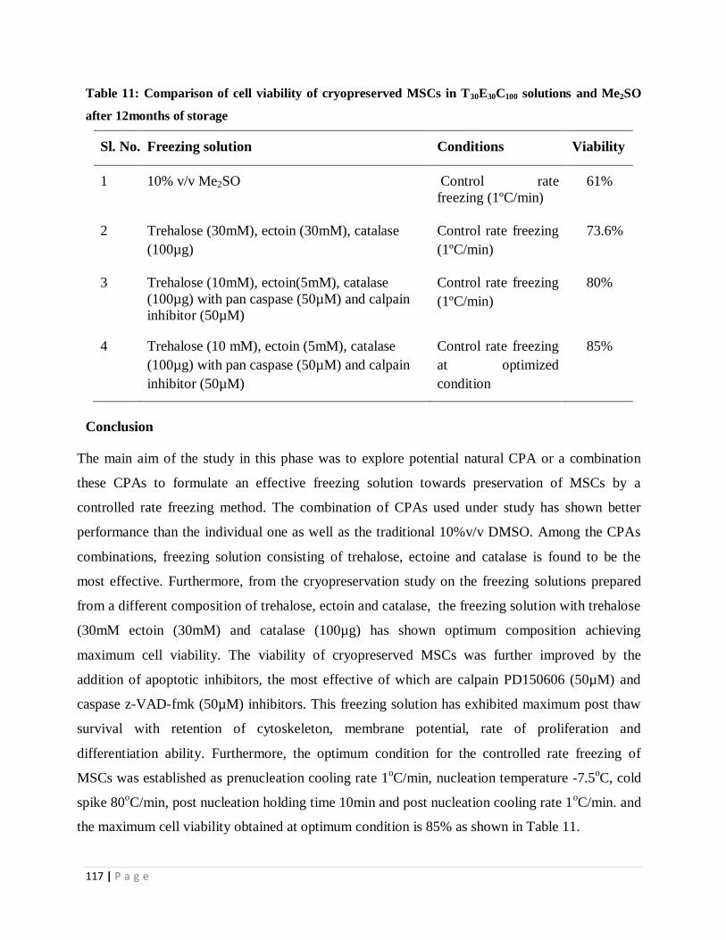
117 | P a g e
Table 11: Comparison of cell viability of cryopreserved MSCs in T30E30C100 solutions and Me2SO
after 12months of storage
Sl. No. Freezing solution Conditions Viability
1 10% v/v Me2SO Control rate
freezing (1ºC/min)
61%
2 Trehalose (30mM), ectoin (30mM), catalase
(100µg)
Control rate freezing
(1ºC/min)
73.6%
3 Trehalose (10mM), ectoin(5mM), catalase
(100µg) with pan caspase (50µM) and calpain
inhibitor (50µM)
Control rate freezing
(1ºC/min)
80%
4 Trehalose (10 mM), ectoin (5mM), catalase
(100µg) with pan caspase (50µM) and calpain
inhibitor (50µM)
Control rate freezing
at optimized
condition
85%
Conclusion
The main aim of the study in this phase was to explore potential natural CPA or a combination
these CPAs to formulate an effective freezing solution towards preservation of MSCs by a
controlled rate freezing method. The combination of CPAs used under study has shown better
performance than the individual one as well as the traditional 10%v/v DMSO. Among the CPAs
combinations, freezing solution consisting of trehalose, ectoine and catalase is found to be the
most effective. Furthermore, from the cryopreservation study on the freezing solutions prepared
from a different composition of trehalose, ectoin and catalase, the freezing solution with trehalose
(30mM ectoin (30mM) and catalase (100µg) has shown optimum composition achieving
maximum cell viability. The viability of cryopreserved MSCs was further improved by the
addition of apoptotic inhibitors, the most effective of which are calpain PD150606 (50µM) and
caspase z-VAD-fmk (50µM) inhibitors. This freezing solution has exhibited maximum post thaw
survival with retention of cytoskeleton, membrane potential, rate of proliferation and
differentiation ability. Furthermore, the optimum condition for the controlled rate freezing of
MSCs was established as prenucleation cooling rate 1oC/min, nucleation temperature -7.5
oC, cold
spike 80oC/min, post nucleation holding time 10min and post nucleation cooling rate 1
oC/min. and
the maximum cell viability obtained at optimum condition is 85% as shown in Table 11.

118 | P a g e
PART II
Cryopreservation of hMSCs seeded silk nanofibers
based tissue engineered constructs

119 | P a g e
As it is described in previous chapter that because of the possibility of autologous and non-
tumorogenic characteristics, MSCs has been used as an attractive cell source in recent research
focusing on the development of various tissue engineered constructs (TECs) to repair defect
and/or diseased tissues and organs. In view of the emerging demand of these tissue engineered
products, the development of an effective preservation technique is of paramount importance for
the potential future applications of these tissue constructs clinically.
Besides cell source, the development of an artificial extra cellular matrix so called scaffold from
biodegradable and biocompatible biopolymers is another important aspect in tissue engineering.
Among the various biomaterials, silk fibroin (SF) extracted from silkworm cocoons possesses
suitable surface, remarkable mechanical and desired biological properties that can mimic the
human bone and other tissue and thus is considered as a potential candidate for the development
of TECs using MSCs [71] .
Therefore, in this phase of dissertation work, the performance of the best freezing medium
consisting of the combination of trehalose, ectoin and catalase (T30/E30/C100/I) was evaluated
towards the preservation of MSCs seeded SF scaffold (TECs) and the optimum control rate
freezing condition was established.
5.3.1 Structural integrity and cell morphology in TECs
The crypreservation strategy has to be designed in such a way that it must be able to preserve the
constructs on long term basis without affecting any structural integrity of the scaffold and maintain
the membrane integrity of the cells in TEC. It is reported that the maintenance of structural integrity
of the scaffold is a prerequisite in cryopreservation of cell–biomaterial constructs. Any deformation
in the scaffold during cryopreservation may affect cell functionality. It was, therefore, investigated
whether the cryopreservation had any adverse effect on the scaffold structure and morphology of
MSCs in TECs by SEM study. SEM images (Figure 39, 40) of scaffolds and TECs before and after
cryopreservation were generated on 1 and 14 days of culture. As indicated in Figure 37, no visible
structural deformity on the surface of the nanofibrous scaffolds with (Figure 39) and without seeded
MSCs (Figure 39) as well as before and after cryopreservation are observed. Furthermore, while
comparing the type of freezing medium used for cryopreservation such as Me2SO and combination
of natural osmolytes (T30/E30/C100/I), TECs cryopreserved in T30/E30/C100/I freezing solution have
shown better MSCs attachment and spreading than Me2SO as evident from Figure 40. This enhanced

120 | P a g e
cell attachment may be due to less mass transfer impedance, uniform heat distribution and desired
pore size of scaffold that facilitated the penetration of cryoprotectant as reported earlier [256].
Furthermore, no significant morphological difference of MSCs was observed between non-
cryopreserved and cryopreserved TECs. This signifies that the microcellular environment of the
MSCs in nanofiber scaffold used in this study remain unaltered in post thaw TECs. Thus it has been
established that T30/E30/C100/I is an effective freezing medium for TECs preservation in terms of
maintenance of structural integrity of scaffold and retention of cell morphology during
cryopreservation process.
Figure 39:SEM images of (A) noncryopreserved and (B) cryopreserved scaffold. Scale bar: 5µm

121 | P a g e
Figure 40: SEM images of cryopreserved scaffold (A) precryopreserved constructs after 1 day, (D) precryopreserved constructs after
14th days , (B) Cryopreserved constructs using freezing solution T30/E30/C100/I after 1 day, (E) Cryopreserved constructs using freezing
solution T30/E30/C100/I after 14days, (C) Cryopreserved constructs using freezing solution Me2SO after 1 day and (F) Cryopreserved
constructs using freezing solution Me2SO after 14 days. Scale bar: 10µm

122 | P a g e
5.3.2 MSCs viability
The MSCs viability in cryopreserved TECs was assessed using flowcytometry analysis by
determining the percentage of post thaw necrotic cells. As shown in Figure 41, the cell viability
in TECs is obtained as 54.37±2.1% using 10% Me2SO, 72.5± 1.8% with T30/E30/A100/I freezing
solutions and 95±2% with non-cryopreserved MSCs. Among the various freezing medium, the
maximum cell viability was achieved with T30/E30/C100/I freezing solution. Furthermore, result
shows that CPAs containing trehalose/ ectoin/catalase with or without apoptotic inhibitors have
better cell viability than the standard 10% Me2SO solution which is statistically significant
(p=0.0003 and 0.0021). Furthermore, the cell viability of non-cryopreserved and cryopreserved
TECs groups showed statistically significant differences, the higher MSCs viability is obtained
with the former, which is obvious. The cell viability results were further confirmed by MTT
assay.
Figure 41: Cell viability after 24h of post thaw TECs in different freezing solution and
noncryopreserved MSCS. Bar represents standard deviation (n = 3)

123 | P a g e
5.3.3 Proliferation assay
The metabolic activity of MSCs seeded on SF scaffolds after post thaw was evaluated by MTT
assay and the result is shown in Figure 42. It is observed that the metabolic activity of the cells
on the scaffold cryopreserved in T30/E30/A100/I is significantly higher than the TECs
cryopreserved in 10% Me2SO (*p value > 0.05and **p value > 0.005). The result has shown
similar trend as the cell viability results obtained by flowcytometry analysis as presented in
previous section.
Figure 42: MTT assay of the metabolic activities of post thaw MSCs seeded on SF scaffolds.
Result demonstrates statistical differences between l0%v/v Me2SO (control) and T30/E30/C100/I
freezing solution after 3, 5 and 7 days (n = 3; ∗p < 0.05; ∗∗p < 0.01)

124 | P a g e
5.3.4 Cytoskeleton analysis
Organization of F-actin, largely determines the cellular morphology and surface movement [256,
257]. Because of their close proximity, any mechanical damage to the membrane might also
disrupt the F-actin. During cryopreservation, extracellular ice, mechanical stress and differential
thermal contraction between cells and substrates occur that may cause depolymerization and
accumulation of F-actin. Cytoskeleton integrity plays a major role in cell viability, cell
proliferation and differentiation. Loss of function of cell membranes interferes with transport
systems such as pH regulatory systems on the cell membrane. Disruption of organelle
membranes affects transport systems such as mitochondrial transport systems that is essential for
oxidative phosphorylation, the major energy-generating pathway [258-259] Therefore,
cytoskeleton integrity of TECs was assessed by confocal microscopy and confocal images are
shown in Figure 43. The investigation on the effect of cryopreservation on the organization of F-
actin has shown no remarkable impact on F-actin of MSCs in TECs by cryopreservation using
T30/E30/A100/I. Whereas significant distortion of F-actin, ruptured membrane, low intensity of
stress fiber and unclear boundary are observed with cryopreserved TECs using Me2SO.
It is further observed that the noncryopreserved TECs has retained its cell morphology as
represented by the existence of intact cell membrane. Immunofloroscence has shown better cell
attachment in TECs cryopreserved in T30/E30/A100/I compared to the cell attachment using
Me2SO. However, cell attachment is lower than that observed with unfrozen TECs in all the
cases.

125 | P a g e
Figure 43: Comparative confocal images (F-actin) of cryopreserved and non cryopreserved silk nanofiber TECs. (A) (i) Shows
noncryopreserved, (ii and iii) shows the TECs immediately after cryopreservation with scale bar 5 lm, (B) 24 h after cryopreservation
with scale bar 5 mm and (C) close-up images of actin cytoskeleton 48 h after cryopreservation (ii) with scale bar 40 µm. In both the
Me2SO and T30/E30/A100/I groups, the cytoskeleton in the frozen/thawed regions is damaged, with shortened extensions and less number of
cells compare to noncryopreserved cells. In the Me2SO frozen group, the cells have condensed, distorted after freezing, and less damage is
observed in T30/E30/A100/I groups.

Results & Discussion
5.3.5 Alkaline Phosphatase activity
The ALP activity of MSCs on the nanofibrous scaffolds up to 21 days culture in osteogenic
differentiation media is shown in Figure 44. The study shows the increase in ALP production
progressively on cryopreserved and non cryopreserved TECs indicating the osteoblastic
differentiation ability of MSCs on scaffold. Additionally, a highly significant difference (p <
0.05) in ALP production is observed between Me2SO and T30/E30/C100/I. Overall, an increase in
ALP activity from 8±0.9µg to 4000±67µg and 9±0.3µg to 3848±400 with cryopreserved TECs
in Me2SO and T30E30C100/I are achieved during 1 to 21 days of culture period.
Figure 44: ALP activity was studied on day 1, 7 and 21 of post thaw MSCs cultured on silk
nanofiber scaffolds in freezing solution of 10% Me2SO and T30/E30/A100/I as evaluated by NBT
substrate in osteogenic induction medium. Results shows significant difference (p > 0.05) in ALP
activity after 7 and 14 days post thaw culture.
116 | P a g e
5.3.6 Effect of cryopreservation on Mechanical properties of TECs
In tissue engineering, scaffold must have desired mechanical properties which become critical
when it is used for bone tissue regeneration in particular. During cryopreservation, the
mechanical strength of scaffold may be altered due to the occurrence of thermal stress[75-76].
Therefore, mechanical test was performed on cryopreserved and non cryopreserved TECs to
investigate the effect of cryopreservation on tensile strength of scaffold. Statistical analysis
indicates that, tensile strength of the non-cryopreserved, TECs cryopreserved in Me2SO and
T30/E30/C100/I were 1.82 ±0.31Mpa, 1.35±0.19Mpa and 1.79 ±0.21Mpa respectively (Figure 45).
Furthermore, no significant difference was observed between non-cryopreserved and
cryopreserved TECs in T30/E30/C100/I. It has been assessed that the combined CPA T30/E30/C100/I
did not show any remarkable effect on the mechanical property of the scaffold. However, the
tensile strength of the scaffold decreases significantly with Me2SO preserved TECs. Similar
result is also reported in another study [15]. Thus it is established the superiority of T30E30C100/I
over Me2SO as freezing solution.
Figure 45: Ultimate tensile strength of TECs cryopreserved in T30/E30/C100/I, Me2SO and
noncryopreserved TECs (Fresh)

117 | P a g e
5.3.7 Calcium deposition
The effect of cryopreservation on the osteogenic differentiation capacity of MSCs in TECs was
investigated qualitatively by histological staining of calcium deposition on the scaffold. As
shown in Figure 46 and 47, MSCs are found to attach and spread throughout the scaffolds. A
network of cells and ECM with a relatively small amount of granular deposition of calcium is
covered on the scaffold surface when TECs is cryopreserved in Me2SO. Whereas, a higher
deposition of calcium is observed with noncryopreserved and cryopreserved T30/E30/C100/I TECs
indicating the higher degree of osteogenic activity of MSCs. SEM results indicated the MSCs
remained adhered to the scaffold and mineralization structures intact (Figure 47). EDXA
detected the presence of phosphorus and calcium confirming the presence of mineralized
structures (Figure 47 B and D).
Figure 46: Optical images of alizarin red staining for calcium deposition of osteogenically induced
MSCs in post thaw TECs (A) TECs without osteogenic media (B) TECs with osteogenic media
without cryopreservation, (C) TECs cryopreserved in Me2SO (D) TECs cryopreserved in
T30/E30/C100/I. Scale bars represent 50 mm

118 | P a g e
Figure 47: SEM and EDX analysis of post-thaw TECs cultured in osteogenic differentiation
medium for 21 days -(A) TECs cryopreserved in T30/E30/C100/I, (C) TECs cryopreserved in 10%
Me2SO, (B) and (D) depicts the EDX analysis of the elemental components of calcium phosphate
salts, consisting of P, Ca and O elements in both groups scaffolds
5.3.8 Expression of osteogenic-specific genes
The quantitative osteogenic differentiation level of seeded MSCs on TECs was assessed by
quantitative PCR of bone-specific genes. From the analysis of Figure 48, the osteogenic
differentiation genes such as collagenase-1[Col 1], core binding protein [Cbfa 1], osteocalcin
[OCN] and alkaline phosphate [APL] are observed to be increasingly expressed on T30/E30/C100/I
cryopreserved group during 14 day of culture in comparison to Me2SO cryopreserved groups.

119 | P a g e
Figure 48: Osteogenic gene expression levels of post thaw MSCs in TECs cultured in osteogenic
media

120 | P a g e
5.3.9 Optimization of controlled rate freezing parameters for preservation of
TECs using T30/E30/C100 freezing medium
In this section, it was further investigated the effect of key controlled rate freezing parameters
on cryopreservation of TECs and establish the optimum cryopreservation condition using the
most effective freezing medium T30/E30/C100/I in presence of apoptotic inhibitors.
5.3.9.1 Effect of cooling rate on MSCs viability in TECs
The effect of cooling rate (-0.5°C/min, -1.0°C/min, -2.5°C/min, -5°C/min and -10°C/min) on
MSCs viability was studied and the experimental result is shown in Figure 49. As it is indicated
the highest cell viability of 74% is achieved at cooling rate of -1.0°C/min and a decline in cell
viability is observed with either higher or lower than -1.0°C/min cooling rate at constant seeding
temperature of -7.5 °C
Figure 49: Effect of different cooling rate on MSCs seeded TECs
5.3.9.2 The effect of seeding temperature on cell viability
The effect of seeding temperature on cell viability was investigated by performing
cryopreservation experiments at varying seeding temperature at -5 °C, -7.5 °C and -10°C. The
optimal cell viability was recorded as 80 % at -7.5 °C seeding temperature. The rate of cooling
temperature is maintained constant at1°C /min.

121 | P a g e
Figure 50: Effect of seeding temperature on MSCs seeded TECs
5.3.9.3 Cell Morphology
After cryopreservation of MSCs seeded scaffold, the cell population was observed to be
decreased in comparison to fresh unfrozen MSCs, representing that cryopreserved induced stress
resulted in cell detachment as shown in Figure 51. Furthermore, more number of cells is
detached in 5°C/min and 10°C/min rate cryopreserved cells in comparison to the cells
cryopreserved at 1°C/min. SEM result reveals that before cryopreservation, the adherent hMSCs
were in spindle shape. But after cryopreservation at 5 and 10°C/min, the cells did not maintain
their original shape, either in flatten shape or round shape. Moreover, the cell number becomes
less after cryopreservation at the cooling rates of 5 and 10 C/min than the fresh MSCs.
.

Results & Discussion
Figure 51: SEM images of TECs under different cooling rate and seeding temperature

Results & Discussion
Conclusion
From this study, it is established that T30/E30/C100/I is found to be an effective freezing medium
in preserving TECs developed from hMSCs and nanofiberous SF scaffold as confirmed by the
cell viability, proliferation and differention ability of hMSCs in TECs after and before
cryopreservation. Furthermore, an enhanced cell vitality of of MSCs is obtained by optimizing
the controlled rate freezing parameters. The optimum cryopreservation condition was established
s cooling rate of 1oC / min and nucleation temperature of -7.5
o C giving maximum cell viability
of 80%.

125 | P a g e
CHAPTER V
Summary & Conclusion

Bibliography
SUMMARY & CONCLUSION
In recent years, mesenchymal stem cells (MSCs) and MSCs derived tissue engineered constructs
(TECs) have been considered as important biological products that can be used for a variety of
therapeutic and tissue engineering applications including bone, cartilage, tendon, muscles, blood
vessel and other tissue regeneration. Therefore, recent research is directed towards the production
of MSCs and MSCs seeded tissue engineered constructs (TECs) to repair defect and/or diseased
tissues and organs. However, the successful clinical applications of these cells and tissue
engineering products rely on the development of an appropriate cryopreservation technology that
offers long time storage of such products with desired cell viability, metabolic activity and cellular
differential ability. Me2SO a traditional freezing solution is currently used for preservation of
various cells and other biological species. However, freezing protocol using Me2SO as
cryoprotecting solution is reported to have several detrimental effects on the cryopreserved cells
and hence harmful to patients.
In this context, a combination of a variety of natural osmolytes seems to be effective
cryoprotective agents in preserving MSCs and other cells. Furthermore, it has been reported that
the controlled rate freezing technology is superior to the uncontrolled freezing methods of
preserving cells. However, no systematic research effort has so far been given to develop an
efficient cryopreservation strategy for long term preservation of MSCs and TECs using natural
osmolytes. Therefore, the present dissertation work has been undertaken to develop a
cryopreservation strategy using a freezing medium consisting of natural cryoprotective agents that
can be useful for the long term preservation of MSCs and MSCs seeded tissue engineered
constructs.
The major outcome achieved through this research work is summarized as follows-
I. In the first phase of the dissertation work, a variety of natural cryoprotective agents in
combination were investigated to see their ability towards cryopreservation with the aim of
developing an effective freezing medium that can be useful for preserving MNCs and other cells
like MSCs. MNCs were isolated from umbilical cord blood by Ficol hypaque technique and
cultured. The extracellular cryoprotectants (10mM) namely trehalose, hydroxyl ethyl starch,

125 | P a g e
polyvinyl pyrrolidine and intracellular CPAs (5mM) like erythritol, taurine and ectoine were used
to prepare different combinations of freezing medium following L9 (34) Taguchi orthogonal array.
Catalase, coenzyme Q10 and n-acetyl cystine (100μg/m) were added as antioxidants. The
cryopreservation experiments were conducted in a controlled rate freezing condition. The
combination of extracellular, intracellular CPA and antioxidant has shown encouraging results
towards the maintenance and survival of cell during and after freezing. Among the CPAs and
antioxidants used under study, trehalose, ectoin and catalase have shown the best cryopreservation
performance achieving high cell viability. The post thaw viability is also observed to be 10%
higher than the cell viability obtained using conventional Me2SO as CPA. Furthermore, SEM and
phase contrast microscopy confirmed the normal cell morphology of the post-thaw cultured cells
with retaining their membrane integrity.
Thus it has been demonstrated that the cryopreservation medium consisting of trehalose, ectoin
and catalase is the most effective freezing solution that may be of further interest of research to
design a standard cryopreservation protocol for MSCs
II. In this part of the thesis work, the most effective natural osmolytes such as trehalose and ectoin
in presence of catalase antioxidant as obtained in the previous section were further evaluated for
their effectiveness towards the long term preservation of much demanding MSCs for therapeutic
and tissue engineering applications. To accomplish this task, MSCs were sorted from cultured
MNCs by fluorescence activated cell sorter (FACS), cultured and characterised for their
morphology, and immune-phenotype. MSCs were then subjected to controlled rate freezing using
various freezing solutions using trehalose and ectoin as CPAs individually and in combination
with varying ratios of trehalose and ectoin. The freezing solution containing the combination of
CPAs has shown better post thaw cell viability than the individual CPAs as well as control
(Me2SO). Furthermore, among various composition of CPAs tested under study, the freezing
medium having combination of 30mM trehalose, 30mM ectoin and 100µg catalase (T30/E30/C100/I)
exhibited maximum post thaw survival with retention of cytoskeleton, membrane potential and
rate of proliferation. The cryopreserved MSCs have also shown the desired differentiation ability
even after 12months of storage. It is thus concluded that T30/E30/C100 freezing solution developed

126 | P a g e
in this study is effective in preserving UCB derived MSCs in long term basis and the maximum
cell viability archived was 73%.
III. In this phase of research work, an attempt has been made to determine the cryopreservation
induced apoptosis signalling pathways and evaluate the role of caspase-dependent and
independent apoptotic pathways in MSCs. to accomplish the task MSCs were cryopreserved in a
control rate freezer, using T30/E30/C100/I as cryoprotective agent either with or without pre-
treatment with general caspase inhibitor z-VAD-fmk, or with the more selective caspase inhibitors
such as z-IETD-fmk, z-LEHD-fmk, and z-DEVD-fmk. Furthermore, to evaluate the effect of
calcium mediated pathway, the cryopreserved MSCs samples were tested with and without calpain
inhibitor. FACS was used to measure the cell viability, mitochondrial membrane potential and cell
cycle analysis. Processing of procaspases 3, 8, 9, µ calpain and Bid was determined by Western
blotting. Cryopreservation of MSCs resulted in characteristic apoptosis within 24 h after post
thaw. It has been established that the addition of calpain inhibitor and pan caspase inhibitor are
used in CPA solution increases the cell viability to 80 %.
IV. In this phase of work, the cryopreservation condition was further optimised to increase the
efficiency of the best obtained freezing solution (T30/E30/C100/I) in presence of caspase and calpain
inhibitors. The various parameters were investigated are prenucleation cooling rate, nucleation
temperature, cold spike temperature, post nucleation holding temperature, post nucleation cooling
rate, cell concentration and storage temperature. The result indicated that the cooling profile has a
significant impact on cell viability. The optimum condition for the controlled rate freezing of
MSCs was established as (i) Prenucleation cooling rate -1oC / min (ii) Nucleation temperature -
7.5oC (iii) Cold spike -80
oC/min (iv) Post nucleation holding time of 5
min (v) Post nucleation
cooling rate -1oC/min. (vi) 3×10
6/ml cell concentration. The maximum cell viability obtained at
optimum condition is 85%.
V. In this part of the thesis, research work was further extended to evaluate the potentiality of the
most effective freezing solution obtained in the previous section towards the preservation of TECs

127 | P a g e
derived by seeding of MSCs on nanofibrous silk fibroin scaffold (TECs) by conducting
cryopreservation experiments in a controlled rate freezer. The SF scaffolds used for TECs
formation is made from eri and tasar SF blend. To the best of our knowledge, the present study is
the first report on the cryopreservation of MSCs-seeded nanofibrous scaffold fabricated from silk
fibroin which is considered as a promising biomaterial for tissue engineering applications. It has
been observed from the experimental results that the viability and functionality of hMSCs on SF
scaffolds were well maintained and cultured after cryopreservation. These are well supported by
the assessment of cell viability (flowcytometry analysis & MTT assay), structural integrity (SEM
and confocal images) and functionality in terms of osteogenic differentiation ability
(Immunohistochemistry, RT-PCR analysis & ALP assay) of TECs. The newly formulated freezing
solution of combined CPAs (T30/E30/C100/I) further did not show any adverse impact on the
mechanical property of the scaffold in cryopreserved TECs. Thus it is established the effectiveness
of T30/E30/C100/I as freezing medium in preserving TECs developed from hMSCs and SF scaffold.
VI. In this phase of research work, an optimization study was carried out to maximise the recovery
of MSCs on TECs cryopreserved in T30/E30/C100/I freezing medium. The effect of key controlled
rate freezing parameters was investigated and optimum freezing condition was established as
cooling rate of -1oC/min and seeding temperature of -7.5
oC/min. The maximum MSCs viability of
80% was achieved at optimum condition.
Overall, in this dissertation work, a Me2SO free freezing solution consisting of a combination of
natural osmolytes such as trehalose (extracellular CPA) and ectoin (intracellular CPA) with
catalase as antioxidant has been established as an effective cryopreservation medium for long
term preservation of MSCs and MSCs seeded on tissue engineered constructs. The efficiency of
the freezing medium achieving high MSCs recovery can further be improved by the addition of
apoptotic inhibitors. It is finally concluded that the development of a controlled rate freezing
system using the freezing medium (T30/E30/C100/I)established in this study may pave the way for
long term preservation of MSCs and MSCs seeded tissue engineered products for clinical
applications

128 | P a g e
Bibliography

Bibliography
129 | P a g e
1. Meyer, U., U. Joos, and H. Wiesmann, Biological and biophysical principles in
extracorporal bone tissue engineering: Part III. International journal of oral and
maxillofacial surgery, 2004. 33(7): p. 635-641
2. Tuan, R.S., G. Boland, and R. Tuli, Adult mesenchymal stem cells and cell-based tissue
engineering. Arthritis Research and Therapy, 2003. 5(1): p. 32-45.
3. Nussbaum, Krista, Cryobiology, Low temperature studies of biological systems (2000).
4. Odorico, J.S., D.S. Kaufman, and J.A. Thomson, Multilineage differentiation from
human embryonic stem cell lines. Stem Cells, 2001. 19(3): p. 193-204
5. Fuller, B. and S. Paynter, Fundamentals of cryobiology in reproductive medicine.
Reproductive BioMedicine Online, 2004. 9(6): p. 680-691.
6. Fuller, B.J., Cryoprotectants: the essential antifreezes to protect life in the frozen state.
CryoLetters, 2004. 25(6): p. 375-388.
7. Woelders, H. and A. Chaveiro, Theoretical prediction of ‘optimal’freezing
programmes. Cryobiology, 2004. 49(3): p. 258-271.
8. Kuleshova, L., S. Gouk, and D. Hutmacher, Vitrification as a prospect for
cryopreservation of tissue-engineered constructs. Biomaterials, 2007. 28(9): p. 1585-
1596.
9. Wharton, David A., Gordon Goodall, and Craig J. Marshall. Freezing survival and
cryoprotective dehydration as cold tolerance mechanisms in the Antarctic nematode
Panagrolaimus davidi. Journal of experimental biology 206, no. 2 (2003): 215-221.
10. Sahagian, Michael E., and H. Douglas Goff. Fundamental aspects of the freezing
process. Food Science And Technology-New York-Marcel Dekker- (1996): 1-50.
11. Mazur, Peter, and Ulrich Schneider. Osmotic responses of preimplantation mouse and
bovine embryos and their cryobiological implications. Cell biophysics 8, no. 4 (1986):
259-285.
12. McGrath, J. J., E. G. Cravalho, and C. E. Huggins. An experimental comparison of
intracellular ice formation and freeze-thaw survival of HeLa S-3 cells. Cryobiology 12,
no. 6 (1975): 540-550.

Bibliography
130 | P a g e
13. Miller, Robert H., and Peter Mazur. Survival of frozen-thawed human red cells as a
function of cooling and warming velocities. Cryobiology 13, no. 4 (1976): 404-414.
14. Karlsson, Jens OM, and Mehmet Toner. Long-term storage of tissues by
cryopreservation: critical issues." Biomaterials 17, no. 3 (1996): 243-256.
15. Massip, A., P. Mermillod, and A. Dinnyes, Morphology and biochemistry of in-vitro
produced bovine embryos: implications for their cryopreservation. Human
Reproduction, 1995. 10(11): p. 3004-3011.
16. Gadea, J., et al., Reduced glutathione content in human sperm is decreased after
cryopreservation: effect of the addition of reduced glutathione to the freezing and
thawing extenders. Cryobiology, 2011. 62(1): p. 40-46.
17. Bueno, C., R. Montes, and P. Menendez, The ROCK inhibitor Y-27632 negatively
affects the expansion/survival of both fresh and cryopreserved cord blood-derived
CD34+ hematopoietic progenitor cells. Stem Cell Reviews and Reports, 2010. 6(2): p.
215-223.
18. Xu, X., et al., The roles of apoptotic pathways in the low recovery rate after
cryopreservation of dissociated human embryonic stem cells. Biotechnology progress,
2010. 26(3): p. 827-837.
19. Rozman-Pungerčar, J., et al., Inhibition of papain-like cysteine proteases and legumain
by caspase-specific inhibitors: when reaction mechanism is more important than
specificity. Cell Death & Differentiation, 2003. 10(8): p. 881-888.
20. Ferrusola, C.O., et al., Inhibition of the mitochondrial permeability transition pore
reduces “apoptosis like” changes during cryopreservation of stallion
spermatozoa. Theriogenology, 2010. 74(3): p. 458-465.
21. Men, H., et al., Degeneration of cryopreserved bovine oocytes via apoptosis during
subsequent culture. Cryobiology, 2003. 47(1): p. 73-81.
22. Seo, J.M., et al., Cryopreservation of amniotic fluid-derived stem cells using natural
cryoprotectants and low concentrations of dimethylsulfoxide. Cryobiology, 2011.
62(3): p. 167-173.

Bibliography
131 | P a g e
23. Sarangi, S. and K. Pramanik. Cryopreservation in Tissue Engineering: Challenges &
Prospects. in Advanced Technologies for Enhancing Quality of Life (AT-EQUAL),
2010. 2010: IEEE.
24. Niu, M., Engineering Liver Tissue Devices for Enhanced Transport, Improved
Function, and Storage. 2008: ProQuest.
25. Hunt, C.J., Cryopreservation of human stem cells for clinical application: a review.
Transfusion Medicine and Hemotherapy, 2011. 38(2): p. 107-123.
26. Balachandran, H., Optimization Of Methods For In-vitro Expansion And
Cryopreservation Of Mammalian Stem Cells. 2007.
27. Bernard, A. and B. Fuller, Cryopreservation of human oocytes: a review of current
problems and perspectives. Human Reproduction Update, 1996. 2(3): p. 193-207.
28. Benson, E.E., Cryopreservation theory. Plant Cryopreservation: A Practical Guide,
2008: p. 15-32.
29. Sakai, A. and F. Engelmann, Vitrification, encapsulation-vitrification and droplet-
vitrification: a review. CryoLetters, 2007. 28(3): p. 151-172.
30. Meryman, H., R. Williams, and M.S.J. Douglas, Freezing injury from “solution effects”
and its prevention by natural or artificial cryoprotection. Cryobiology, 1977. 14(3): p.
287-302.
31. Gao, D. and J. Critser, Mechanisms of cryoinjury in living cells. Ilar Journal, 2000.
41(4): p. 187-196.
32. Liu, Yang, Xia Xu, Xuehu Ma, Enca Martin‐Rendon, Suzanne Watt, and Zhanfeng Cui.
"Cryopreservation of human bone marrow‐derived mesenchymal stem cells with
reduced dimethylsulfoxide and well‐defined freezing solutions."Biotechnology
progress 26, no. 6 (2010): 1635-1643.

Bibliography
132 | P a g e
33. Picton, Helen M., Roger G. gosden, and Stanley P. Leibo. Cryopreservation of oocytes
and ovarian tissue." Current Practices and Controversies in Assisted
Reproduction (2002): 142.
34. Gao, Doyong, and J. K. Critser. Mechanisms of cryoinjury in living cells. ILAR
journal 41, no. 4 (2000): 187-196.
35. Hunt, Charles J. Cryopreservation of human stem cells for clinical application: a
review." Transfusion medicine and hemotherapy 38, no. 2 (2011): 107-123.
36. Picton, H. M., S. S. Kim, and R. G. Gosden. Cryopreservation of gonadal tissue and
cells. British medical bulletin 56, no. 3 (2000): 603-615.
37. Heng, Boon Chin. Effect of Rho-associated kinase (ROCK) inhibitor Y-27632 on the
post-thaw viability of cryopreserved human bone marrow-derived mesenchymal stem
cells. Tissue and Cell 41, no. 5 (2009): 376-380.
38. Li, Xiangyun, Guoliang Meng, Roman Krawetz, Shiying Liu, and Derrick E. Rancourt,
The ROCK inhibitor Y-27632 enhances the survival rate of human embryonic stem
cells following cryopreservation. Stem cells and development17, no. 6 (2008): 1079-
1086.
39. Martin-Ibanez, R., C. Unger, A. Strömberg, D. Baker, J. M. Canals, and O. Hovatta.
Novel cryopreservation method for dissociated human embryonic stem cells in the
presence of a ROCK inhibitor. Human Reproduction 23, no. 12 (2008): 2744-2754.
40. Gauthaman, Kalamegam, Chui-Yee Fong, Arjunan Subramanian, Arijit Biswas, and
Ariff Bongso. ROCK inhibitor Y-27632 increases thaw-survival rates and preserves
stemness and differentiation potential of human Wharton’s jelly stem cells after
cryopreservation. Stem Cell Reviews and Reports 6, no. 4 (2010): 665-676.
41. Nemati, Shiva, Maryam Hatami, Sahar Kiani, Katayoun Hemmesi, Hamid Gourabi,
Najmehsadat Masoudi, Sara Alaei, and Hossein Baharvand. Long-Term Self-
Renewable Feeder-Free Human Induced Pluripotent Stem Cell–Derived Neural
Progenitors. Stem cells and development 20, no. 3 (2010): 503-514.

Bibliography
133 | P a g e
42. Cortes, J. L., L. Sanchez, G. Ligero, I. Gutierrez-Aranda, P. Catalina, C. Elosua, P. E.
Leone et al. Mesenchymal stem cells facilitate the derivation of human embryonic stem
cells from cryopreserved poor-quality embryos. Human reproduction 24, no. 8 (2009):
1844-1851.
43. Martín-Ibáñez, Raquel, Outi Hovatta, and Josep M. Canals. Cryopreservation of human
pluripotent stem cells: are we going in the right direction."Cryopreservation/Book 1
(ISBN 979-953-307-300-1) (2012).
44. Hotchkiss, Richard S., and Donald W. Nicholson. Apoptosis and caspases regulate
death and inflammation in sepsis. Nature Reviews Immunology 6, no. 11 (2006): 813-
822.
45. Qu, Gui-Qin, Xiang Liu, Ya-Li Zhang, Dan Yao, Qiu-Min Ma, Ming-Yu Yang, Wen-
Hua Zhu, Shi Yu, and Yun-Bo Luo. Evidence for programmed cell death and activation
of specific caspase-like enzymes in the tomato fruit heat stress response. Planta 229, no.
6 (2009): 1269-1279.
46. Talanian, Robert V., Christopher Quinlan, Simone Trautz, Maria C. Hackett, John A.
Mankovich, David Banach, Tariq Ghayur, Kenneth D. Brady, and Winnie W. Wong.
Substrate specificities of caspase family proteases. Journal of Biological
Chemistry 272, no. 15 (1997): 9677-9682.
47. Ortega Ferrusola C, Gonzalez Fernandez L, Salazar Sandoval C, et al. Inhibition of the
mitochondrial permeability transition pore reduces "apoptosis like" changes during
cryopreservation of stallion spermatozoa. Theriogenology 2010;74:458–465.
48. Paasch U, Grunewald S, Agarwal A, Glandera HJ. Activation pattern of caspases in
human spermatozoa. Fertil Steril 2004;81:802–809.
49. Seo JM, Sohn MY, Suh JS, et al. Cryopreservation of amniotic fluid-derived stem cells
using natural cryoprotectants and low concentrations of dimethylsulfoxide.
Cryobiology 2011;62:167–173.
50. Heng BC, Clement MV, Cao T. Caspase inhibitor Z-VADFMK enhances the freeze-
thaw survival rate of human embryonic stem cells. Biosci Rep 2007;27:257–264.

Bibliography
134 | P a g e
51. Yagi T, Hardin JA, Valenzuela YM, et al. Caspase inhibition reduces apoptotic death of
cryopreserved porcine hepatocytes. Hepatology 2001;33:1432–1440.
52. Omori K, Valiente L, Orr C, et al. Improvement of human islet cryopreservation by a
p38 MAPK inhibitor. Am J Transplant 2007;7:1224–1232.
53. Franklin-Tong VE, Gourlay CW. A role for actin in regulating apoptosis/programmed
cell death: Evidence spanning yeast, plants and animals. Biochem J 2008;413:389–404.
54. Bishop AL, Hall A. Rho GTPases and their effector proteins. Biochemical J
2000;348:241–255.
55. Rizoli SB, Rotstein OD, Parodo J, Phillips MJ, Kapus A. Hypertonic inhibition of
exocytosis in neutrophils: Central role for osmotic actin skeleton remodeling. Am J
Physiol Cell Physiol 2000;279:C619–633.
56. Reinehr R, Haussinger D. Hyperosmotic activation of the CD95 death receptor system.
Acta Physiol (Oxf) 2006;187: 199–203.
57. Aznar S, Lacal JC. Rho signals to cell growth and apoptosis. Cancer Lett 2001;165:1–
10.
58. Coleman ML, Olson MF. Rho GTPase signalling pathways in the morphological
changes associated with apoptosis. Cell Death Different 2002;9:493–504.
59. Leverrier Y, Ridley AJ. Apoptosis: Caspases orchestrate the ROCK ’n’ bleb. Nature
Cell Biol 2001;3:E91–93.
60. Amano M, Chihara K, Kimura K, et al. Formation of actin stress fibers and focal
adhesions enhanced by Rho-kinase. Science 1997;275:1308–1311.
61. Leung T, Chen XQ, Manser E, Lim L. The p160 RhoAbinding kinase ROK alpha is a
member of a kinase family and is involved in the reorganization of the cytoskeleton.
Mol Cell Biol 1996;16:5313–5327.

Bibliography
135 | P a g e
62. Maekawa M, Ishizaki T, Boku S, et al. Signaling from Rho to the actin cytoskeleton
through protein kinases ROCK and LIM-kinase. Science 1999;285:895–898.
63. Di Ciano-Oliveira C, Thirone AC, Szaszi K, Kapus A. Osmotic stress and the
cytoskeleton: The R(h)ole of Rho GTPases. Acta Physiol (Oxf) 2006;187:257–272.
64. Piazzolla D, Meissl K, Kucerova L, Rubiolo C, Baccarini M. Raf-1 sets the threshold of
Fas sensitivity by modulating Rok-alpha signaling. J Cell Biol 2005;171:1013–1022.
65. Gauthaman K, Fong CY, Bongso A. Effect of ROCK inhibitor Y-27632 on normal and
variant human embryonic stem cells (hESCs) in vitro: Its benefits in hESC expansion.
Stem Cell Rev 2010;6:86–95.
66. Krawetz RJ, Li X, Rancourt DE. Human embryonic stem cells: Caught between a
ROCK inhibitor and a hard place. BioEssays 2009;31:336–343.
67. Li X, Krawetz R, Liu S, Meng G, Rancourt DE. ROCK inhibitor improves survival of
cryopreserved
68. Li X, Meng G, Krawetz R, Liu S, Rancourt DE. The ROCK inhibitor Y-27632
enhances the survival rate of human embryonic stem cells following cryopreservation.
Stem Cells Devel 2008;17:1079–1085.
69. Martin-Ibanez R, Unger C, Stromberg A, et al. Novel cryopreservation method for
dissociated human embryonic stem cells in the presence of a ROCK inhibitor. Hum
Reprod 2008;23:2744–2754.
70. Pakzad M, Totonchi M, Taei A, et al. Presence of a ROCK inhibitor in extracellular
matrix supports more undifferentiated growth of feeder-free human embryonic and
induced pluripotent stem cells upon passaging. Stem Cell Rev 2010;6:96–107.
71. Watanabe K, Ueno M, Kamiya D, et al. A ROCK inhibitor permits survival of
dissociated human embryonic stem cells. Nature Biotechnol 2007;25:681–686.
Bibliography
136 | P a g e
72. Claassen DA, Desler MM, Rizzino A. ROCK inhibition enhances the recovery and
growth of cryopreserved human embryonic stem cells and human induced pluripotent
stem cells. Mol Reprod Devel 2009;76:722–732.
73. Heng BC. Effect of Rho-associated kinase (ROCK) inhibitor Y-27632 on the post-thaw
viability of cryopreserved human bone marrow-derived mesenchymal stem cells.
Tissue Cell 2009;41:376–380.
74. Cortes JL, Sanchez L, Ligero G, et al. Mesenchymal stem cells facilitate the derivation
of human embryonic stem cells from cryopreserved poor-quality embryos. Hum Reprod
2009;24:1844–1851.
75. Bueno C, Montes R, Menendez P. The ROCK inhibitor Y- 27632 negatively affects the
expansion/survival of both fresh and cryopreserved cord blood-derived CD34 +
hematopoietic progenitor cells: Y-27632 negatively affects the expansion/survival of
CD34 + HSPCs. Stem Cell Rev 2010;6:215–223.
76. Barbaric I, Jones M, Buchner K, Baker D, et al. Pinacidil enhances survival of
cryopreserved human embryonic stem cells. Cryobiology 2011;63:298–305. Sablina
AA, Budanov AV, Ilyinskaya GV, et al. The antioxidant function of the p53 tumor
suppressor. Nature Med 2005;11:1306–1313.
77. Gallardo BJ, Miro´ MA´ , Balao SC, et al. Autophagy and apoptosis have a role in the
survival or death of stallion spermatozoa during conservation in refrigeration. PloS One
2012;7:e30688.
78. Minor T, Stegemann J, Hirner A, Koetting M. Impaired autophagic clearance after cold
preservation of fatty livers correlates with tissue necrosis upon reperfusion and is
reversed by hypothermic reconditioning. Liver Transplant 2009;15:798–805.
79. Dumont, Frédéric, Pierre-André Marechal, and Patrick Gervais. Cell size and water
permeability as determining factors for cell viability after freezing at different
cooling rates. Applied and environmental microbiology 70, no. 1 (2004): 268-272.
80. Ortega‐Ferrusola, C., Y. Sotillo‐Galán, E. Varela‐Fernández, J. M. Gallardo‐Bolaños,
A. Muriel, L. González‐Fernández, J. A. Tapia, and F. J. Pena. Detection of
“Apoptosis‐Like” Changes During the Cryopreservation Process in Equine
Sperm. Journal of andrology 29, no. 2 (2008): 213-221.

Bibliography
137 | P a g e
81. Nagle, William A., Bernard L. Soloff, A. J. Moss Jr, and Kurt J. Henle. "Cultured
Chinese hamster cells undergo apoptosis after exposure to cold but nonfreezing
temperatures." Cryobiology 27, no. 4 (1990): 439-451.
82. Baust JG, Dayong Gao, Baust JM. Cryopreservation: An emerging paradigm change.
Organogenesis 2009;3:90–96.
83. Baust JM. Three properties of cells and tissues influencing preservation outcome:
Molecular basis of preservationinduced cell death. Adv Biopreserv 2010;63.
84. Baust JM, Snyder KK, VanBuskirk RG, Baust JG. Changing paradigms in
biopreservation. Biopreserv Biobanking 2009;7:3–12.
85. Searle J, Kerr JF, Bishop CJ. Necrosis and apoptosis: Distinct modes of cell death with
fundamentally different significance. Pathol Ann 1982;17:229–259.
86. Walker NI, Harmon BV, Gobe GC, Kerr JF. Patterns of cell death. Methods
Achievements Exp Pathol 1988;13: 18–54.
87. Kerr JF. Shrinkage necrosis of adrenal cortical cells. J Pathol 1972;107:217–219.
88. Columbano A. Cell death: Current difficulties in discriminating apoptosis from necrosis
in the context of pathological processes in vivo. J Cell Biochem 1995;58:181–190.
89. Kerr JF, Wyllie AH, Currie AR. Apoptosis: A basic biological phenomenon with wide-
ranging implications in tissue kinetics. Br J Cancer 1972;26:239–257.
90. Majno G, Joris I. Apoptosis, oncosis, and necrosis. An overview of cell death. Am J
Pathol 1995;146:3–15.
91. Trump B.F, Berezesky IK, Chang SH, Phelps PC. The pathways of cell death: Oncosis,
apoptosis, and necrosis. Toxicol Pathol 1997;25:82–88.

Bibliography
138 | P a g e
92. Baust JM, Vogel MJ, Van Buskirk R, Baust JG. A molecular basis of cryopreservation
failure and its modulation to improve cell survival. Cell Transplant 2001;10: 561–571.
93. Peter ME, Heufelder AE, Hengartner MO. Advances in apoptosis research. Proc Natl
Acad Sci USA 1997;94: 12736–12737.
94. Gewirtz DA. Growth arrest and cell death in the breast tumor cell in response to
ionizing radiation and chemotherapeutic agents which induce DNA damage. Breast
Cancer Res Treatment 2000;62:223–235
95. Fink KB, Andrews LJ, Butler WE, et al. Reduction of post-traumatic brain injury and
free radical production by inhibition of the caspase-1 cascade. Neuroscience 1999;94:
1213–1218
96. Emery E, Aldana P, Bunge MB, et al. Apoptosis after traumatic human spinal cord
injury. J Neurosurg 1998;89: 911–920.
97. Elsasser A, Suzuki K, Schaper J. Unresolved issues regarding the role of apoptosis in
the pathogenesis of ischemic injury and heart failure. J Mol Cell Cardiol 2000;32:711–
724
98. Brune B, von Knethen A, Sandau KB. Nitric oxide and its role in apoptosis. Eur J
Pharmacol 1998;351:261–272.
99. Saikumar P, Dong Z, Weinberg JM, Venkatachalam MA. Mechanisms of cell death in
hypoxia/reoxygenation injury. Oncogene 1998;17:3341–3349.
100. Liu CY, Liu YH, Lin SM, et al. Apoptotic neutrophils undergoing secondary necrosis
induce human lung epithelial cell detachment. J Biomed Sci 2003;10: 746–756.
101. Jaeschke H, Lemasters JJ. Apoptosis versus oncotic necrosis in hepatic
ischemia/reperfusion injury. Gastroenterology 2003;125:1246–1257.
102. Salvesen GS, Dixit VM. Caspases: Intracellular signaling by proteolysis. Cell
1997;91:443–446.

Bibliography
139 | P a g e
103. Thornberry NA, Lazebnik Y. Caspases: Enemies within. Science 1998;281:1312–
1316.
104. Martinvalet D, Zhu P, Lieberman J. Granzyme A induces caspase-independent
mitochondrial damage, a required first step for apoptosis. Immunity 2005;22:355–370.
105. Rai NK, Tripathi K, Sharma D, Shukla VK. Apoptosis: A basic physiologic process in
wound healing. Intl J Lower Extremity Wounds 2005;4:138–144.
106. Adams JM. Ways of dying: Multiple pathways to apoptosis. Genes Devel
2003;17:2481–2495.
107. Bratton DL, Fadok VA, Richter DA, et al. Appearance of phosphatidylserine on
apoptotic cells requires calciummediated nonspecific flip-flop and is enhanced by loss
of the aminophospholipid translocase. J Biol Chem 1997; 272:26159–26165.
108. Martin SJ, Reutelingsperger CP, McGahon AJ, et al. Early redistribution of plasma
membrane phosphatidylserine is a general feature of apoptosis regardless of the
initiating stimulus: Inhibition by overexpression of Bcl-2 and Abl. J Exp Med
1995;182:1545
109. Vermes I, Haanen C, Reutelingsperger C. Flow cytometry of apoptotic cell death. J
Immunol Methods 2000;243: 167–190.
110. Chicheportiche Y, Bourdon PR, Xu H, et al. TWEAK, a new secreted ligand in the
tumor necrosis factor family that weakly induces apoptosis. J Biol Chem 1997;272:
32401– 32410.
111. Ashkenazi A, Dixit VM. Death receptors: Signaling and modulation. Science
1998;281:1305–1308.
112. Peter ME, Krammer PH. Mechanisms of CD95 (APO-1/ Fas)-mediated apoptosis.
Curr Opin Immunol 1998;10: 545–551.

Bibliography
140 | P a g e
113. Suliman A, Lam A, Datta R, Srivastava RK. Intracellular mechanisms of TRAIL:
Apoptosis through mitochondrialdependent and -independent pathways. Oncogene
2001; 20:2122– 2133.
114. Rubio-Moscardo F, Blesa D, Mestre C, et al. Characterization of 8p21.3 chromosomal
deletions in B-cell lymphoma: TRAIL-R1 and TRAIL-R2 as candidate dosage-
dependent tumor suppressor genes. Blood 2005;106:3214–3222.
115. Hsu H, Xiong J, Goeddel DV. The TNF receptor 1-associated protein TRADD signals
cell death and NF-kappa B activation. Cell 1995;81:495–504.
116. Wajant H. The Fas signaling pathway: More than a paradigm. Science 2002;296:1635
117. Boldin MP, Varfolomeev EE, Pancer Z, et al. A novel protein that interacts with the
death domain of Fas/APO1 contains a sequence motif related to the death domain. J
Biol Chem1995;270:7795–7798.
118. Kischkel FC, Hellbardt S, Behrmann I, et al. Cytotoxicitydependent APO-1
(Fas/CD95)-associated proteins form a death-inducing signaling complex (DISC) with
the receptor. EMBO J 1995;14:5579–5588.
119. Bodmer JL, Holler N, Reynard S, et al. TRAIL receptor-2 signals apoptosis through
FADD and caspase-8. Nature Cell Biol 2000;2:241–243.
120. Salvesen GS. Caspases and apoptosis. Essays Biochem 2002;38:9–19.
121. Li J, Yuan J. Caspases in apoptosis and beyond. Oncogene 2008;27:6194–6206.
122. Kataoka T, Schroter M, Hahne M, et al. FLIP prevents apoptosis induced by death
receptors but not by perforin/ granzyme B, chemotherapeutic drugs, and gamma
irradiation. J Immunol 1998;161:3936–3942.
123. Scaffidi C, Schmitz I, Krammer PH, Peter ME. The role of c-FLIP in modulation of
CD95-induced apoptosis. J Biol Chem 1999;274:1541–1548.

Bibliography
141 | P a g e
124. Saelens X, Festjens N, Vande Walle L, et al. Toxic proteins released from
mitochondria in cell death. Oncogene 2004;23:2861–2874.
125. Du C, Fang M, Li Y, Li L, Wang X. Smac, a mitochondrial protein that promotes
cytochrome c-dependent caspase activation by eliminating IAP inhibition. Cell 2000;
102:33–42.
126. Garrido C, Galluzzi L, Brunet M, et al. Mechanisms of cytochrome c release from
mitochondria. Cell Death Different 2006;13:1423–1433.
127. Li W, Srinivasula SM, Chai J, et al. Structural insights into the pro-apoptotic function
of mitochondrial serine protease HtrA2/Omi. Nature Structural Biology 2002;9: 436–
441.
128. Ghobrial IM, Witzig TE, Adjei AA. Targeting apoptosis pathways in cancer therapy.
CA Cancer J Clin 2005;55: 178–194.
129. Ly JD, Grubb DR, Lawen A. The mitochondrial membrane potential (deltapsi(m)) in
apoptosis; An update. Apoptosis 2003;8:115–128.
130. Chinnaiyan AM. The apoptosome: Heart and soul of the cell death machine. Neoplasia
1999;1:5–15.
131. Hill MM, Adrain C, Duriez PJ, Creagh EM, Martin SJ. Analysis of the composition,
assembly kinetics and activity of native Apaf-1 apoptosomes. EMBO J 2004;23:
2134–2145.
132. Li P, Nijhawan D, Budihardjo I, et al. Cytochrome c and dATP-dependent formation
of Apaf-1/caspase-9 complex initiates an apoptotic protease cascade. Cell
1997;91:479–489.
133. van Loo G, Saelens X, van Gurp M, et al. The role of mitochondrial factors in
apoptosis: A Russian roulette with more than one bullet. Cell Death Different
2002;9:1031–1042.

Bibliography
142 | P a g e
134. Schimmer AD. Inhibitor of apoptosis proteins: Translating basic knowledge into
clinical practice. Cancer Res 2004; 64:7183–7190.
135. HK, Susin SA, Penninger J, Kroemer G. Apoptosis inducing factor (AIF): A
phylogenetically old, caspaseindependent effector of cell death. Cell Death Different
1999;6:516–524.
136. Li LY, Luo X, Wang X. Endonuclease G is an apoptotic DNase when released from
mitochondria. Nature 2001; 412:95–99.
137. Enari M, Sakahira H, Yokoyama H, et al. A caspaseactivated DNase that degrades
DNA during apoptosis, and its inhibitor ICAD. Nature 1998;391:43–50.
138. Joza N, Susin SA, Daugas E, et al. Essential role of the mitochondrial apoptosis-
inducing factor in programmed cell death. Nature 2001;410:549–554.
139. Debatin KM. Apoptosis pathways in cancer and cancer therapy. Cancer Immunol
Immunother CII 2004;53: 153–159.
140. Pluquet O, Hainaut P. Genotoxic and non-genotoxic pathways of p53 induction.
Cancer Lett 2001;174:1–15.
141. Nakano K, Vousden KH. PUMA, a novel proapoptotic gene, is induced by p53. Mol
Cell 2001;7:683–694.
142. Leu JI, Dumont P, Hafey M, Murphy ME, George DL. Mitochondrial p53 activates
Bak and causes disruption of a Bak-Mcl1 complex. Nature Cell Biology 2004;6: 443–
450.
143. Mihara M, Erster S, Zaika A, et al. p53 has a direct apoptogenic role at the
mitochondria. Mol Cell 2003;11: 577–590.
144. Vaux DL, Cory S, Adams JM. Bcl-2 gene promotes haemopoietic cell survival and
cooperates with c-myc to immortalize pre-B cells. Nature 1988;335:440–442.

Bibliography
143 | P a g e
145. Schuler M, Green DR. Mechanisms of p53-dependent apoptosis. Biochem Soc
Transact 2001;29:684–688.
146. Kim R. Recent advances in understanding the cell death pathways activated by
anticancer therapy. Cancer 2005; 103;1551–1560.
147. Holler N, Zaru R, Micheau O, et al. Fas triggers an alternative, caspase-8-independent
cell death pathway using the kinase RIP as effector molecule. Nature Immunol
2000;1:489–495.
148. Boone E, Vanden Berghe T, Van Loo G, et al. Structure/ Function analysis of p55
tumor necrosis factor receptor and fas-associated death domain. Effect on necrosis in
L929sA cells. J Biol Chem 2000;275:37596–37603.
149. Eliasson MJ, Sampei K, Mandir AS, et al. Poly (ADPribose) polymerase gene
disruption renders mice resistant to cerebral ischemia. Nature Med 1997;3:1089–1095.
150. Ha HC, Snyder SH. Poly(ADP-ribose) polymerase is a mediator of necrotic cell death
by ATP depletion. Proc Natl Acad Sci USA 1999;96:13978–13982.
151. Yoshioka A, Yamaya Y, Saiki S, et al. Non-N-methyl-Daspartate glutamate receptors
mediate oxygen–glucose deprivation-induced oligodendroglial injury. Brain Res
2000;854:207–215.
152. Goossens V, De Vos K, Vercammen D, et al. Redox regulation of TNF signaling.
Biofactors 1999;10:145–156.
153. Fiers W, Beyaert R, Declercq W, Vandenabeele P. More than one way to die:
Apoptosis, necrosis and reactive oxygen damage. Oncogene 1999;18:7719–7730.
154. Shimizu S, Eguchi Y, Kamiike W, et al. Retardation of chemical hypoxia-induced
necrotic cell death by Bcl-2 and ICE inhibitors: Possible involvement of common
mediators in apoptotic and necrotic signal transductions. Oncogene 1996;12:2045–
2050.

Bibliography
144 | P a g e
155. Brocheriou V, Hagege AA, Oubenaissa A, et al. Cardiac functional improvement by a
human Bcl-2 transgene in a mouse model of ischemia/reperfusion injury. J Gene Med
2000;2:326–333.
156. Tan J, Town T, Placzek A, et al. Bcl-X(L) inhibits apoptosis and necrosis produced by
Alzheimer’s betaamyloid1- 40 peptide in PC12 cells. Neurosc Lett 1999; 272:5–8.
157. Single B, Leist M, Nicotera P. Differential effects of bcl-2 on cell death triggered
under ATP-depleting conditions. Exp Cell Res 2001;262:8–16.
158. Vande Velde C, Cizeau J, Dubik D, et al. BNIP3 and genetic control of necrosis-like
cell death through the mitochondrial permeability transition pore. Mol Cell Biol
2000;20:5454–5468.
159. Gabai VL, Meriin AB, Yaglom JA, et al. Suppression of stress kinase JNK is involved
in HSP72-mediated protection of myogenic cells from transient energy deprivation.
HSP72 alleviates the stewss-induced inhibition of JNK dephosphorylation. J Biol Chem
2000;275:38088–38094.
160. Ma XL, Kumar S, Gao F, et al. Inhibition of p38 mitogen- activated protein kinase
decreases cardiomyocyte apoptosis and improves cardiac function after myocardial
ischemia and reperfusion. Circulation 1999;99: 1685–1691.
161. Mochizuki T, Asai A, Saito N, et al. Akt protein kinase inhibits non-apoptotic
programmed cell death induced by ceramide. J Biol Chem 2002;277:2790–2797.
162. Matsui T, Tao J, del Monte F, et al. Akt activation preserves cardiac function and
prevents injury after transient cardiac ischemia in vivo. Circulation 2001; 104:330–335.
163. Punn A, Mockridge JW, Farooqui S, Marber MS, Heads RJ. Sustained activation of
p42/p44 mitogen-activated protein kinase during recovery from simulated ischaemia
mediates adaptive cytoprotection in cardiomyocytes. Biochem J 2000;350:891–899.
164. Yue TL, Wang C, Gu JL, et al. Inhibition of extracellular signal-regulated kinase
enhances ischemia/reoxygenationinduced apoptosis in cultured cardiac myocytes and
exaggerates reperfusion injury in isolated perfused heart. Circulation Res 2000;86:692–
699.

Bibliography
145 | P a g e
165. Festjens N, Tom VB, Peter V. Necrosis, a well-orchestrated form of cell demise:
Signalling cascades, important mediators and concomitant immune response. Biochim
Biophys Acta Bioenergetics 2006;1757:1371–1387.
166. Wang Y, Singh R, Massey AC, et al. Loss of macroautophagy promotes or prevents
fibroblast apoptosis depending on the death stimulus. J Biol Chem 2008;283: 4766–
4777.
167. Thorburn A. Apoptosis and autophagy: Regulatory connections between two
supposedly different processes. Apoptosis 2008;13:1–9.
168. Kabeya Y, Mizushima N, Ueno T, et al. LC3, a mammalian homologue of yeast
Apg8p, is localized in autophagosome membranes after processing. EMBO J 2000;19:
5720–5728.
169. Rodrigues, J. P., F. H. Paraguassu-Braga, L. Carvalho, E. Abdelhay, L. F. Bouzas, and
L. C. Porto. "Evaluation of trehalose and sucrose as cryoprotectants for hematopoietic
stem cells of umbilical cord blood."Cryobiology 56, no. 2 (2008): 144-151.
170. Buchanan, Sandhya S., Sherilyn A. Gross, Jason P. Acker, Mehmet Toner, John F.
Carpenter, and David W. Pyatt. Cryopreservation of stem cells using trehalose:
evaluation of the method using a human hematopoietic cell line.Stem cells and
development 13, no. 3 (2004): 295-305.
171. Sasnoor, Lalita M., Vaijayanti P. Kale, and Lalita S. Limaye. Supplementation of
conventional freezing medium with a combination of catalase and trehalose results in
better protection of surface molecules and functionality of hematopoietic cells. Journal
of hematotherapy & stem cell research 12, no. 5 (2003): 553-564.
172. Limaye, L. S., and V. P. Kale. Cryopreservation of human hematopoietic cells with
membrane stabilizers and bioantioxidants as additives in the conventional freezing
medium. Journal of hematotherapy & stem cell research 10, no. 5 (2001): 709-718.
173. Xiao, Hong-Hai, Tse-Chao Hua, Jun Li, Xue-Lian Gu, Xin Wang, Zhi-Jiang Wu,
Lvrong Meng, Qi-Rong Gao, Jian Chen, and Zhong-Ping Gong. Freeze-drying of

Bibliography
146 | P a g e
mononuclear cells and whole blood of human cord blood. Cryoletters 25, no. 2 (2004):
111-120.
174. Lyu, Shaw-Ruey, Wei Te Wu, Chien Chih Hou, and Wen-Hsin Hsieh. "Study of
cryopreservation of articular chondrocytes using the Taguchi method."Cryobiology 60,
no. 2 (2010): 165-176.
175. Day, John G., and Glyn Stacey, eds. Cryopreservation and freeze-drying protocols.
Vol. 368. Springer, 2007.
176. Shimizu S, Eguchi Y, Kamiike W, et al. Retardation of chemical hypoxia-induced
necrotic cell death by Bcl-2 and ICE inhibitors: Possible involvement of common
mediators in apoptotic and necrotic signal transductions. Oncogene 1996;12:2045–
2050.
177. Louis, Kristine S., and Andre C. Siegel. Cell viability analysis using trypan blue:
manual and automated methods. In Mammalian Cell Viability, pp. 7-12. Humana
Press, 2011.
178. Bissoyi, Akalabya, and K. Pramanik. Effects of Non-Toxic Cryoprotective Agents on
the Viability of Cord Blood Derived MNCs.CryoLetters 34, no. 5 (2013): 453-465.
179. Derby, Eric, Vasavi Reddy, William Kopp, Edward Nelson, Michael Baseler, Thomas
Sayers, and Anatoli Malyguine. Three-color flow cytometric assay for the study of the
mechanisms of cell-mediated cytotoxicity. Immunology letters78, no. 1 (2001): 35-39.
180. Pena, F. J., A. Johannisson, M. Wallgren, and H. Rodriguez Martinez. Antioxidant
supplementation in vitro improves boar sperm motility and mitochondrial
membrane potential after cryopreservation of different fractions of the
ejaculate. Animal reproduction science 78, no. 1 (2003): 85-98.
181. Pal, J. Kundu, S. Talukdar, T. Thomas, S.C. Kundu, An emerging functional natural
silk biomaterial from the only domesticated non-mulberry silkworm samia ricini,
Macromol. Biosci. 13 (8) (2013) 1020–1035.
182. Sekiya, Ichiro, Benjamin L. Larson, Jason R. Smith, Radhika Pochampally, Jian‐Guo
Cui, and Darwin J. Prockop. "Expansion of human adult stem cells from bone marrow

Bibliography
147 | P a g e
stroma: conditions that maximize the yields of early progenitors and evaluate their
quality." Stem cells 20, no. 6 (2002): 530-541
183. Crissman, H. A., and J. A. Steinkamp. "Rapid, one step staining procedures for
analysis of cellular DNA and protein by single and dual laser flow
cytometry."Cytometry 3, no. (1982): 84-90.
184. Pozarowski P, Darzynkiewicz Z. Analysis of cell cycle by flow cytometry. In:
Checkpoint Controls and Cancer 2004. Humana Press. pp. 301–311
185. Rao, Rammohan V., Susana Castro-Obregon, Harald Frankowski, Martin Schuler,
Veronica Stoka, Gabriel del Rio, Dale E. Bredesen, and H. Michael Ellerby. Coupling
endoplasmic reticulum stress to the cell death program An Apaf-1-independent intrinsic
pathway. Journal of Biological Chemistry 277, no. 24 (2002): 21836-21842.
186. F. Chen, W. Zhang, W. Wu, Y. Jin, L. Cen, J.D. Kretlow, W. Gao, Z. Dai, J. Wang, G.
Zhou, Cryopreservation of tissue-engineered epithelial sheets in trehalose, Biomaterials
32 (2011) 8426–8435
187. E. Umemura, Y. Yamada, S. Nakamura, K. Ito, K. Hara, M. Ueda, Viable
cryopreserving tissue-engineered cell-biomaterial for cell banking therapy in an
effective cryoprotectant, Tissue Eng. C Methods 17 (2011) 799–807.
188. F.K. Skinner, Y. Rotenberg, A.W. Neumann, Contact angle measurements from the
contact diameter of sessile drops by means of a modified axisymmetric drop shape
analysis, J. Colloid Interface Sci. 130 (1) (1989) 25–34.
189. L. Smith, P. Ma, Nano-fibrous scaffolds for tissue engineering, Colloids Surf. B
Biointerfaces 39 (3) (2004) 125–131.
190. C. Stoll, J.L. Holovati, J.P. Acker, W.F. Wolkers, Synergistic effects of liposomes,
trehalose, and hydroxyethyl starch for cryopreservation of human erythrocytes,
Biotechnol. Prog. 28 (2) (2012) 364–371.
191. T.K. Teh, S.-L. Toh, J.C. Goh, Aligned hybrid silk scaffold for enhanced
differentiation of mesenchymal stem cells into ligament fibroblasts, Tissue Eng. C
Methods 17 (2011) 687–703.

Bibliography
148 | P a g e
192. K.Y. Teo, T.O. DeHoyos, J.C. Dutton, F. Grinnell, B. Han, Effects of freezinginduced
cell–fluid–matrix interactions on the cells and extracellular matrix of engineered
tissues, Biomaterials 32 (2011) 5380–5390.
193. S. Thirumala, J.M. Gimble, R.V. Devireddy, Evaluation of methylcellulose and
dimethyl sulfoxide as the cryoprotectants in a serum-free freezing media for
cryopreservation of adipose-derived adult stem cells, Stem Cells Dev. 19 (2010) 513–
522.
194. P.B. Tuncer, U. Tasdemir, S. Büyükleblebici, T. Özgürtas, E. Coskun, H. Erol, I.S.
Gürcan, Effects of different doses of trehalose supplementation in egg yolk extender in
frozen–thawed Angora buck semen, Small Ruminant Res. (2013).
195. E. Umemura, Y. Yamada, S. Nakamura, K. Ito, K. Hara, M. Ueda, Viable
cryopreserving tissue-engineered cell-biomaterial for cell banking therapy in an
effective cryoprotectant, Tissue Eng. C Methods 17 (2011) 799–807.
196. F. Wen, R. Magalhães, S.S. Gouk, B. Gajadhar, K.H. Lee, D.W. Hutmacher, L.L.
Kuleshova, Vitreous cryopreservation of nanofibrous tissue-engineered constructs
generated using mesenchymal stromal cells, Tissue Eng. C Methods 15 (2009) 105–
114.
197. D.A. Lauffenburger, A.F. Horwitz, Cell migration: review a physically integrated
molecular process, Cell 84 (1996) 359–369.
198. Regidor, Carmen, Manuel Posada, Dolores Monteagudo, Carlos Garaulet, Nieves
Somolinos, Rafael Forés, Montserrat Briz, and Manuel-Nicolás Fernández. Umbilical
cord blood banking for unrelated transplantation: evaluation of cell separation and
storage methods. Experimental hematology27, no. 2 (1999): 380-385.
199. Hunt, Charles J., David E. Pegg, and Susan E. Armitage. Optimising cryopreservation
protocols for haematopoietic progenitor cells: a methodological approach for umbilical
cord blood. Cryoletters 27, no. 2 (2006): 73-85.
200. Hunt, Charles J., Susan E. Armitage, and David E. Pegg. Cryopreservation of
umbilical cord blood: 2. Tolerance of CD34< sup>+</sup> cells to multimolar

Bibliography
149 | P a g e
dimethyl sulphoxide and the effect of cooling rate on recovery after freezing and
thawing. Cryobiology 46, no. 1 (2003): 76-87.
201. Zhang, Xiao Bing, Karen Li, Kwai Heung Yau, Kam Sze Tsang, Tai Fai Fok, Chi
Kong Li, Shuk Man Lee, and Patrick Man Pan Yuen. Trehalose ameliorates the
cryopreservation of cord blood in a preclinical system and increases the recovery of
CFUs, long‐term culture‐initiating cells, and nonobese diabetic‐SCID repopulating
cells. Transfusion 43, no. 2 (2003): 265-272.
202. Rodrigues, J. P., F. H. Paraguassu-Braga, L. Carvalho, E. Abdelhay, L. F. Bouzas, and
L. C. Porto. "Evaluation of trehalose and sucrose as cryoprotectants for hematopoietic
stem cells of umbilical cord blood."Cryobiology 56, no. 2 (2008): 144-151.
203. Buchanan, Sandhya S., Sherilyn A. Gross, Jason P. Acker, Mehmet Toner, John F.
Carpenter, and David W. Pyatt. "Cryopreservation of stem cells using trehalose:
evaluation of the method using a human hematopoietic cell line."Stem cells and
development 13, no. 3 (2004): 295-305.
204. Sasnoor, Lalita M., Vaijayanti P. Kale, and Lalita S. Limaye. Supplementation of
conventional freezing medium with a combination of catalase and trehalose results in
better protection of surface molecules and functionality of hematopoietic cells. Journal
of hematotherapy & stem cell research 12, no. 5 (2003): 553-564.
205. Limaye, L. S., and V. P. Kale. "Cryopreservation of human hematopoietic cells with
membrane stabilizers and bioantioxidants as additives in the conventional freezing
medium." Journal of hematotherapy & stem cell research 10, no. 5 (2001): 709-718
206. Xiao, Hong-Hai, Tse-Chao Hua, Jun Li, Xue-Lian Gu, Xin Wang, Zhi-Jiang Wu,
Lvrong Meng, Qi-Rong Gao, Jian Chen, and Zhong-Ping Gong. "Freeze-drying of
mononuclear cells and whole blood of human cord blood." Cryoletters 25, no. 2 (2004):
111-120.
207. Hubel, Allison. "Parameters of cell freezing: implications for the cryopreservation of
stem cells." Transfusion medicine reviews 11, no. 3 (1997): 224-233.
208. Coda, Barbara A., Barbara O'Sullivan, Gary Donaldson, Sharol Bohl, C. Richard
Chapman, and Danny D. Shen. "Comparative efficacy of patient-controlled

Bibliography
150 | P a g e
administration of morphine, hydromorphone, or sufentanil for the treatment of oral
mucositis pain following bone marrow transplantation." Pain 72, no. 3 (1997): 333-346.
209. Fuller, Barry J. "Cryoprotectants: the essential antifreezes to protect life in the frozen
state." CryoLetters 25, no. 6 (2004): 375-388.
210. Liu, Yang, Xia Xu, Xuehu Ma, Jing Liu, and Zhanfeng Cui. "Effect of various
freezing solutions on cryopreservation of mesenchymal stem cells from different
animal species." Cryoletters 32, no. 5 (2011): 425-435.
211. Lynch, Andrew L., and Nigel KH Slater. "Influence of intracellular trehalose
concentration and pre-freeze cell volume on the cryosurvival of rapidly frozen human
erythrocytes." Cryobiology 63, no. 1 (2011): 26-31.
212. Wu, Chun Fang, Hsiao Chien Tsung, Wen Jie Zhang, Yan Wang, Jun Hong Lu, Zheng
Ya Tang, Yan Pin Kuang et al. "Improved cryopreservation of human embryonic stem
cells with trehalose." Reproductive biomedicine online 11, no. 6 (2005): 733-739.
213. Freimark, Denise, Constanze Sehl, Christian Weber, Klaus Hudel, Peter Czermak,
Nicola Hofmann, Ralf Spindler, and Birgit Glasmacher. "Systematic parameter
optimization of a Me< sub> 2</sub> SO-and serum-free cryopreservation protocol for
human mesenchymal stem cells." Cryobiology 63, no. 2 (2011): 67-75.
214. Seo, Ji Min, Mi Yeung Sohn, Jang Soo Suh, Anthony Atala, James J. Yoo, and Yun-
Hee Shon. "Cryopreservation of amniotic fluid-derived stem cells using natural
cryoprotectants and low concentrations of dimethylsulfoxide."Cryobiology 62, no. 3
(2011): 167-173.
215. Grein, Tanja A., Denise Freimark, Christian Weber, Klaus Hudel, Christine Wallrapp,
and Peter Czermak. "Alternatives to dimethylsulfoxide for serum-free cryopreservation
of human mesenchymal stem cells." The International journal of artificial organs 33,
no. 6 (2010): 370-380.
216. Limaye, L. S., and V. P. Kale. "Cryopreservation of human hematopoietic cells with
membrane stabilizers and bioantioxidants as additives in the conventional freezing
medium." Journal of hematotherapy & stem cell research 10, no. 5 (2001): 709-718.

Bibliography
151 | P a g e
217. Hanna, Jacob, and Allison Hubel. "Preservation of stem cells." Organogenesis5, no. 3
(2009): 134-137.
218. Campbell, Lia H., and Kelvin GM Brockbank. "Serum-free solutions for
cryopreservation of cells." In Vitro Cellular & Developmental Biology-Animal 43, no.
8-9 (2007): 269-275.
219. Lyu, Shaw-Ruey, Wei Te Wu, Chien Chih Hou, and Wen-Hsin Hsieh. "Study of
cryopreservation of articular chondrocytes using the Taguchi method."Cryobiology 60,
no. 2 (2010): 165-176.
220. Nadarajan, J., H. J. Staines, E. E. Benson, M. Marzalina, B. Krishnapillay, and K.
Harding. "Optimization of cryopreservation for sterculia cordata zygotic embryos using
vitrification techniques."Journal of Tropical Forest Science 19, no. 2 (2007).
221. Lin, Tsai-Ming, Jin-Lian Tsai, Sin-Daw Lin, Chung-Sheng Lai, and Chia-Cheng
Chang. "Accelerated growth and prolonged lifespan of adipose tissue-derived human
mesenchymal stem cells in a medium using reduced calcium and antioxidants." Stem
cells and development 14, no. 1 (2005): 92-102.
222. Limaye, Lalita S. "Bone marrow cryopreservation: improved recovery due to
bioantioxidant additives in the freezing solution." Stem Cells 15, no. 5 (1997): 353-
358.
223. Berz, David, Elise M. McCormack, Eric S. Winer, Gerald A. Colvin, and Peter J.
Quesenberry. "Cryopreservation of hematopoietic stem cells." American journal of
hematology 82, no. 6 (2007): 463-472.
224. Roy, Ranjit K. Design of experiments using the Taguchi approach: 16 steps to product
and process improvement. John Wiley & Sons, 2001.
225. Chen, Y. H., S. C. Tam, W. L. Chen, and H. Y. Zheng. "Application of the Taguchi
method in the optimization of laser micro-engraving of photomasks."International
journal of materials and product technology 11, no. 3 (1996): 333.
226. Rodrigues, J. P., F. H. Paraguassu-Braga, L. Carvalho, E. Abdelhay, L. F. Bouzas, and
L. C. Porto. "Evaluation of trehalose and sucrose as cryoprotectants for

Bibliography
152 | P a g e
hematopoietic stem cells of umbilical cord blood."Cryobiology 56, no. 2 (2008):
144-151.
227. Buchanan, Sandhya S., Sherilyn A. Gross, Jason P. Acker, Mehmet Toner, John F.
Carpenter, and David W. Pyatt. "Cryopreservation of stem cells using trehalose:
evaluation of the method using a human hematopoietic cell line."Stem cells and
development 13, no. 3 (2004): 295-305.
228. Baust JM, Van Buskirk R, Baust JG. Cell viability improves following inhibition of
cryopreservation-induced apoptosis. In Vitro Cell Devel Biol Anim 2000;36:262–270.
229. Schuurhuis GJ, Muijen MM, Oberink JW, et al. Large populations of non-clonogenic
early apoptotic CD34-positive cells are present in frozen-thawed peripheral blood
stem cell transplants. Bone Marrow Transplant 2001;27:487–498.
230. Xiao M, Dooley DC. Assessment of cell viability and apoptosis in human umbilical
cord blood following storage. J Hematother Stem Cell Res 2003;12:115–122.
231. de Boer F, Drager AM, Pinedo HM, et al. Extensive early apoptosis in frozen-thawed
CD34-positive stem cells decreases threshold doses for haematological recovery after
autologous peripheral blood progenitor cell transplantation. Bone Marrow Transplant
2002;29:249–255.
232. Fu T, Guo D, Huang X, et al. Apoptosis occurs in isolated and banked primary mouse
hepatocytes. Cell Transplant 2001;10:59–66.
233. Paasch U, Grunewald S, Agarwal A, Glandera HJ. Activation pattern of caspases in
human spermatozoa. Fertil Steril 2004;81:802–809.
234. Men H, Monson RL, Parrish JJ, Rutledge JJ. Degeneration of cryopreserved bovine
oocytes via apoptosis during subsequent culture. Cryobiology 2003;47:73–81.
235. Woo D. Apoptosis and loss of renal tissue in polycystic kidney diseases. New Engl J
Med 1995;333:18–25.

Bibliography
153 | P a g e
236. Baust JM. Properties of cells and tissues influencing preservation outcome: Molecular
basis of preservation-induced cell death. Adv Biopreserv 2010;63–83.
237. Baust JM. Molecular mechanisms of cellular demise associated with cryopreservation
failure.Cell Preserv Technol 2002;17–31.
238. Mukherjee IN. Thesis. A rational design approach for the cryopreservation of natural
and engineered tissues. Department of Chemical and Biomolecular Engineering,
Georgia Institute of Technology; Atlanta, GA: 2008.
239. Xu X, Cowley S, Flaim CJ, James W, Seymour L, Cui Z. The roles of apoptotic
pathways in the low recovery rate after cryopreservation of dissociated human
embryonic stem cells. Biotechnol Prog 2010;26:827–837.
240. Sindram D, Kohli V, Madden JF, Clavien PA. Calpain inhibition prevents sinusoidal
endothelial cell apoptosis in the cold ischemic rat liver 1, 2. Transplant 1999;68:136–
140.
241. Robilotto AT, Baust JM, Van Buskirk R, Baust JG. Calpain activation influences
cryopreservation outcome. Cell Preserv Technol 2006;41:17–30.
242. Cui XD, Gao DY, Fink BF, Vasconez HC, Pu LLQ. Cryopreservation of human
adipose tissues. Cryobiology 2007;55:269–278.
243. Pozarowski P, Darzynkiewicz Z. Analysis of cell cycle by flow cytometry. In:
Checkpoint Controls and Cancer 2004. Humana Press. pp. 301–311.
244. NeumarRW,XuYA, Gada H, Guttmann RP, Siman R. Crosstalk between calpain and
caspase proteolytic systems during neuronal apoptosis. J Biol Chem 2003;278:14162–
14167.
245. Reed JC. Mechanisms of apoptosis. Am J Pathol 2000; 157:1415–1430
246. Robilotto AT, Baust JM, Buskirk RV, Baust JG. Involvement of the cysteine protease
calpain family in cell death after cryopreservation. Cell Preserv Technol 2006;17–30.

Bibliography
154 | P a g e
247. Jones A, Van Blerkom J, Davis P, Toledo AA. Cryopreservation of metaphase II
human oocytes effects mitochondrial membrane potential: Implications for
developmental competence. Human Reprod 2004;19:1861–1866.
248. Kim T, Jung U, Cho DY, Chung AS. Semethylselenocysteine induces apoptosis
through caspase activation in HL- 60 cells. Carcinogenesis 2001;559–565.
249. Li Z, Lin Q, Liu R, Xiao W, Liu W. Protective effects of ascorbate and catalase on
human spermatozoa during cryopreservation. J Androl 2010;31:437–444.
250. Buchanan SS, Gross SA, Acker JP, Toner M, Carpenter JF, Pyatt DW.
Cryopreservation of stem cells using trehalose: Evaluation of the method using a
human hematopoietic cell line. Stem Cells Devel 2004;13:295–305.
251. Crocker SJ , Smith PD, Jackson-Lewis V, Lamba WR, Hayley SP, Grimm E, Park DS.
Inhibition of calpains prevents neuronal and behavioral deficits in an MPTP mouse
model of Parkinson’s disease. J Neurosci 2003;23:4081–4091.
252. Upadhya GA, Topp SA, Hotchkiss RS, Anagli J, Strasberg SM. Effect of cold
preservation on intracellular calcium concentration and calpain activity in rat
sinusoidal endothelial cells. Hepatology 2003;37:313–323.
253. StrohC,Cassens U, SamrajAK, Sibrowski W, Schulze-Osthoff K, Los M. The role of
caspases in cryoinjury: Caspase inhibition strongly improves the recovery of
cryopreserved hematopoietic and other cells. FASEB J 2002;16:1651–1653.
254. Bissoyi A, Nayak B, Pramanik K, Sarangi SK. Targeting cryopreservation-induced
cell death: A review. Biopreserv Biobanking 2014;12:23–34.
255. Corwin LW, Baust JM, Baust JG, Van BGR. Characterization and modulation of
human mesenchymal stem cell stress pathway response following hypothermic
storage. Cryobiology 2014;68:215–226.
256. S.Y. Park, C.S. Ki, Y.H. Park, H.M. Jung, K.M. Woo, H.J. Kim, Electrospun silk
fibroin scaffolds with macropores for bone regeneration: an in vitro and in vivo study,
Tissue Eng. A 16 (2010) 1271–1279.

Bibliography
155 | P a g e
257. T. Jiang, Y. Khan, L.S. Nair, W.I. Abdel-Fattah, C.T. Laurencin, Functionalization of
chitosan/poly (lactic acid–glycolic acid) sintered microsphere scaffolds via surface
heparinization for bone tissue engineering, J. Biomed. Mater. Res. A 93 (3) (2010)
1193– 1208.
258. X. Xu, Y. Liu, Z. Cui, Y. Wei, L. Zhang, Effects of osmotic and cold shock on
adherent human mesenchymal stem cells during cryopreservation, J. Biotechnol.
(2012).
259. X. Xu, Y. Liu, Z.F. Cui, Effects of cryopreservation on human mesenchymal stem
cells attached to different substrates, J. Tissue Eng. Regen. Med. (2012).

Bibliography
156 | P a g e
List of Publications
On PhD work
Patent
1. Patent applied: Niladri N Panda, Krishna pramanik, Amit Biswas, Akalabya Bissoyi.
Electrospun silk fibroin bland nanofibrous scaffold for tissue engineering (TR-5
C.B.R NO: 4716) Date-27/3/2012 17:07:01
Journals
Bissoyi, A., Nayak, B., Pramanik, K., & Sarangi, S. K. (2014). Targeting
Cryopreservation-Induced Cell Death:A Review. Biopreservation and
Biobanking, 12(1), 23-34.
Bissoyi, A., Pramanik, K., Panda, N. N., & Sarangi, S. K. (2014). Cryopreservation of
hMSCs seeded silk nanofibers based tissue engineered
constructs. Cryobiology.(68)332–342.
Bissoyi, A., & Pramanik, K. (2013). Effects of Non-Toxic Cryoprotective Agents on
the Viability of Cord Blood Derived MNCs. Cryoletters, 34(5), 453-465.
Bissoyi, A., Pramanik, K., (2014). Role of apoptosis pathway in cryopreserved
induced cell death in mesenchymal stem cells derived from umbilical Cord blood,
Biopreservation and Biobanking, 12(4), 21-32.
Bissoyi, A., Pramanik, K. Isolation, characterization and preservation of MSCs
derived from umbilical cord blood. Journal of Tissue Engineering and Regenerative
Medicine Vol 6, Issue Supplement s1, Article first published online: 3 SEP 2012
Manuscript under preparation
To be communicated: Akalabya Bissoyi, K. Pramanik, Sribatsha Mohapatra. “Long
term Cryopreservation of Umbilical Cord blood derived Mesenchymal Stem cell using
natural osmolytes and antioxidant” Biopreservation and Biobanking.
To be communicated: Akalabya Bissoyi, K. Pramanik. Optimization of
cryopreservation protocol for hMSCs using osmolytes in combination with antioxidant
and apoptotic inhibitor.

Bibliography
157 | P a g e
Conference
1. A. Bissoyi, K. Pramanik‘ A Study of the apoptosis on cord blood derived MNC
as novel method for selection of cryoprotectant solution’-2011 National
conference on Tissue engineering: prospects & challenges & post conference
workshop on Flowcytometer in tissue engineering (tepc-2011) JAN 22
2. A. Bissoyi, K. Pramanik ‘Molecular dynamic study of caspase-8 protein in
cryopreserved condition’-2011 National conference on Tissue engineering:
prospects & challenges & post conference workshop on Flowcytometer in tissue
engineering (tepc-2011) JAN-22
3. A. Bissoyi, K. Pramanik ‘Effect of osmolytes on apoptotic protein in
cryopreservation temperature by molecular dynamic simulation study’ . 2011
International Conference on Tissue Engineering & Regenerative Medicine

Bibliography
158 | P a g e
AKALABYA BISSOYI
Educational Qualification
2013: Ph.D (Thesis Submitted),
National Institute of Technology, Rourkela, India
2009: M. Tech. (Biotechnology),
National Institute of Technology, Rourkela, India
2007: B. Pharm (Pharmacy),
Indira Gandhi Institute of Pharmaceutical Sciences, Bhubaneswar
Research Interest
Tissue Engineering, Stem Cells, Cryobiology
Personal Details
Permanent Address Q. No.- 7, Type-4/Old A.G. Colony
Unit-4, Bhubaneshwar- 751001, Odisha
Date of Birth 7th
June, 1983
Nationality Indian