crusting and ulceration in a crossbred dog author: david granteditor: david lloyd © european...
TRANSCRIPT
Crusting and ulceration in a crossbred dog
Author: David GrantAuthor: David Grant Editor: David LloydEditor: David Lloyd
© European Society of Veterinary Dermatology © European Society of Veterinary Dermatology

History | Signs | Differentials | Tests | Therapy | NotesHistory | Signs | Differentials | Tests | Therapy | Notes
Click to reveal the text on this screen
Click the forward arrow to jump to the next screen
History – 1
HistoryHistory
• 2-year-old entire male crossbred dog
• Weight 10 kg
• Initial ‘sores’ on lip, nose, scrotum developed over ‘a week or two’
• Dog otherwise healthy. No pruritus.

History | Signs | Differentials | Tests | Therapy | NotesHistory | Signs | Differentials | Tests | Therapy | Notes
History - 2
HistoryHistory
• Treated with antibiotics and steroids
• 2 weeks later no response
• Dog now shows malaise, anorexia
History | Signs | Differentials | Tests | Therapy | NotesHistory | Signs | Differentials | Tests | Therapy | Notes
History - 3
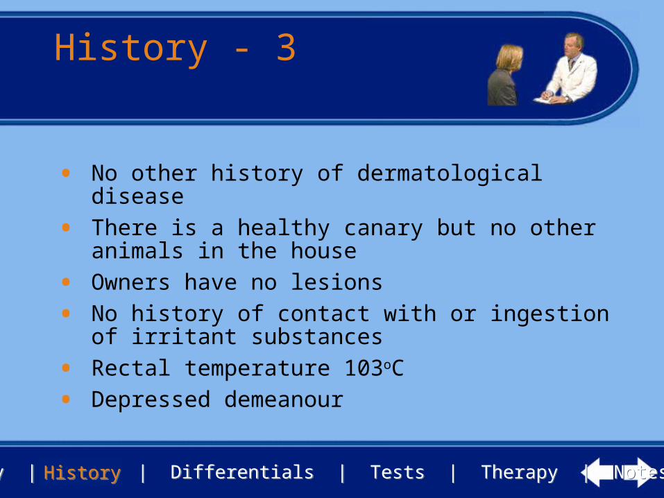
• No other history of dermatological disease
• There is a healthy canary but no other animals in the house
• Owners have no lesions
• No history of contact with or ingestion of irritant substances
• Rectal temperature 103oC
• Depressed demeanour
HistoryHistory

History | Signs | Differentials | Tests | Therapy | NotesHistory | Signs | Differentials | Tests | Therapy | Notes
Clinical signs - 1
SignsSigns
Crusting, mucopurulent discharges and ulceration are apparent at various sites
Ulceration and discharge from the lids of the left eye
Ulceration of the lip margins

History | Signs | Differentials | Tests | Therapy | NotesHistory | Signs | Differentials | Tests | Therapy | Notes
Clinical signs - 2
SignsSigns
Crusting, exudation and pustular lesions affecting the footpads

History | Signs | Differentials | Tests | Therapy | NotesHistory | Signs | Differentials | Tests | Therapy | Notes
How would youapproach this case?
• What are the next steps you would take?
• Make a list of your principle differential diagnoses
• List any samples you would collect
• List any tests you would perform to assist in making a definitive diagnosis
SignsSigns

History | Signs | Differentials | Tests | Therapy | NotesHistory | Signs | Differentials | Tests | Therapy | Notes
Differential diagnoses
DifferentialsDifferentials
• Bullous pemphigoid, drug eruption, SLE, mococutaneous candidiasis
• Also ulcerative stomatitis, neoplasia, dermatophytosis, secondary pyoderma

History | Signs | Differentials | Tests | Therapy | NotesHistory | Signs | Differentials | Tests | Therapy | Notes
Tests - 1
TestsTests
• Skin scrapings, Nikolsky test
• Blood tests: routine haematology and biochemical screens
• Fungal culture of crusts and exudate
• Multiple biopsy samples from intact vesicles/pustules and edges of ulcers

History | Signs | Differentials | Tests | Therapy | NotesHistory | Signs | Differentials | Tests | Therapy | Notes
Tests - 2
• Scrapings from crusted areas did not reveal ectoparasites or fungal structures
• The Nikolsky sign was not elicited
• Smears of exudate stained with Giemsa showed coccoid and rod- shaped bacteria in moderate numbers, neutrophils, and some acanthocytes
• No satisfactory smears were obtained from intact pustules or vesicles
TestsTests

History | Signs | Differentials | Tests | Therapy | NotesHistory | Signs | Differentials | Tests | Therapy | Notes
What now?
TestsTests
• What treatment should you now institute, if any, whilst waiting for the fungal cultures and biopsy results?
• What are now your principle differential diagnoses?
• Are there any other samples you would collect

History | Signs | Differentials | Tests | Therapy | NotesHistory | Signs | Differentials | Tests | Therapy | Notes
Tests - 3
TestsTests
• Blood screens showed a slight neutrophilia but were otherwise unremarkable
• Fungal cultures were negative for dermatophytes or yeasts
• Histopathological examination of biopsy samples revealed an intra- and sub-epidermal vesicular dermatitis

History | Signs | Differentials | Tests | Therapy | NotesHistory | Signs | Differentials | Tests | Therapy | Notes
Tests - 4
TestsTests
Acanthosis with suprabasilar and some subepidermal clefts. A lichenoid band of inflammatory cells and some pigmentary incontinence in upper dermis and around follicles
Histopathology

History | Signs | Differentials | Tests | Therapy | NotesHistory | Signs | Differentials | Tests | Therapy | Notes
What is yourdiagnosis?
• Do the investigations permit a definitive diagnosis?
• Are there any additional investigations which you think may need to be done?
TestsTests

History | Signs | Differentials | Tests | Therapy | NotesHistory | Signs | Differentials | Tests | Therapy | Notes
• Pemphigus vulgaris• Lesion type, location and histopathology are consistent• No history of previous drug therapy and histopathology
not consistent with EM and TEN• Vesicles or bullae are subepidermal in bullous
pemphigoid• Fungal culture was negative
Diagnosis
TestsTests

History | Signs | Differentials | Tests | Therapy | NotesHistory | Signs | Differentials | Tests | Therapy | Notes
How would you deal with this case?
• What is your prognosis?
• How will you advise the owner?
• What treatment would you consider?
TestsTests

History | Signs | Differentials | Tests | Therapy | NotesHistory | Signs | Differentials | Tests | Therapy | Notes
Prognosis
• Prognosis is guarded• Disease can be fatal if not successfully treated• Dogs may not tolerate steroids and other
immunomodulatory drugs• Lifelong therapy is necessary
TestsTests

History | Signs | Differentials | Tests | Therapy | NotesHistory | Signs | Differentials | Tests | Therapy | Notes
Therapy
TherapyTherapy
• Induction therapy - first 3 weeks• Methylprednisolone orally, 5 mg/kg daily• Azathioprine orally, 2.2 mg/kg every other day

History | Signs | Differentials | Tests | Therapy | NotesHistory | Signs | Differentials | Tests | Therapy | Notes
Response to therapy
• After 3 weeks the lesions were in remission
• Therapy continued as• Methylprednisolone, 2 mg/kg every other day• Azathioprine, 2.2 mg/kg on the alternate days
• At 6 months the dog was still in remission
NotesNotes

History | Signs | Differentials | Tests | Therapy | NotesHistory | Signs | Differentials | Tests | Therapy | Notes
Review
NotesNotes
• If you would like to review this case, please use the navigation buttons below