crossm - home | msphere · cell line. the viability profile of a549 cells exposed to ricin...
TRANSCRIPT

TRAIL (CD253) Sensitizes Human Airway Epithelial Cells toToxin-Induced Cell Death
Yinghui Rong,a Jennifer Westfall,a Dylan Ehrbar,a Timothy LaRocca,b Nicholas J. Mantisa
aDivision of Infectious Disease, Wadsworth Center, New York State Department of Health, Albany, New York,USA
bDepartment of Basic and Clinical Sciences, Albany College of Pharmacy and Health Sciences, Albany, NewYork, USA
ABSTRACT Inhalation of ricin toxin is associated with the onset of acute respiratorydistress syndrome (ARDS), characterized by hemorrhage, inflammatory exudates, andtissue edema, as well as the nearly complete destruction of the lung epithelium.Here we report that the Calu-3 human airway epithelial cell line is relatively impervi-ous to the effects of ricin, with little evidence of cell death even upon exposure tomicrogram amounts of toxin. However, the addition of exogenous soluble tumor ne-crosis factor (TNF)-related apoptosis-inducing ligand (TRAIL; CD253) dramatically sen-sitized Calu-3 cells to ricin-induced apoptosis. Calu-3 cell killing in response to ricinand TRAIL exposure was partially inhibited by caspase-8 and caspase-3/7 inhibitors,consistent with involvement of extrinsic apoptotic pathways in cell death. We em-ployed nCounter Technology to define the transcriptional response of Calu-3 cells toricin, TRAIL, and the combination of ricin plus TRAIL. An array of genes associatedwith inflammation and cell death were significantly upregulated upon treatmentwith ricin toxin and were further amplified upon addition of TRAIL. Of particularnote was interleukin-6 (IL-6), whose expression in Calu-3 cells increased 300-foldupon ricin treatment and more than 750-fold upon ricin and TRAIL treatment. IL-6secretion by Calu-3 cells was confirmed by cytometric bead array analysis. On thebasis of these finding, we speculate that the severe airway epithelial cell damageobserved in animal models following ricin exposure is a result of a positive-feedbackloop driven by proinflammatory cytokines such as TRAIL and IL-6.
IMPORTANCE Ricin toxin is a biothreat agent that is particularly damaging to lungtissue following inhalation. A hallmark of ricin exposure is widespread inflammationand concomitant destruction of the airway epithelium. In this study, we investigatedthe possible interaction between ricin and known proinflammatory cytokines associ-ated with lung tissue. Using an established human airway epithelial cell line, wedemonstrate that epithelial cell killing by ricin is significantly enhanced in the pres-ence of the proinflammatory cytokine known as TRAIL (CD253). Moreover, epithelialcells that are simultaneously exposed to ricin and TRAIL produced large amounts ofsecondary proinflammatory signals, including IL-6, which in the context of the lungwould be expected to exacerbate toxin-induced tissue damage. Our results suggestthat therapies designed to neutralize proinflammatory cytokines such as TRAIL andIL-6 may limit the bystander damage associated with ricin exposure.
KEYWORDS bioterrorism, cytokines, epithelial cells, inflammation, lung defense,toxins
NATO’s Biomedical Advisory Council recently concluded that ricin ranks at the topof the list of potential biothreat agents, due in large part to the toxin’s extreme
potency against numerous different cell types as well as to its capacity to be dissem-
Received 6 August 2018 Accepted 29 August2018 Published 26 September 2018
Citation Rong Y, Westfall J, Ehrbar D, LaRoccaT, Mantis NJ. 2018. TRAIL (CD253) sensitizeshuman airway epithelial cells to Toxin-inducedcell death. mSphere 3:e00399-18. https://doi.org/10.1128/mSphere.00399-18.
Editor Sarah E. F. D'Orazio, University ofKentucky
Copyright © 2018 Rong et al. This is an open-access article distributed under the terms ofthe Creative Commons Attribution 4.0International license.
Address correspondence to Timothy LaRocca,[email protected], or Nicholas J.Mantis, [email protected].
RESEARCH ARTICLETherapeutics and Prevention
crossm
September/October 2018 Volume 3 Issue 5 e00399-18 msphere.asm.org 1
on May 18, 2020 by guest
http://msphere.asm
.org/D
ownloaded from

inated via aerosol (1). In rodents, swine, and nonhuman primates (NHPs), inhalationalricin exposure evokes what is clinically equivalent to acute respiratory distress syn-drome (ARDS) (2–4). In rodents and NHPs, the 50% lethal dose (LD50) of ricin by aerosolis �4 �g/kg of body weight (5–7). The hallmarks of ricin-induced lung damage includeearly (h 6 to h 12) onset of alveolar macrophage apoptosis followed hours later byintra-alveolar edema, accumulation of inflammatory cytokines in bronchoalveolar la-vage (BAL) fluid, neutrophilic infiltration, and fibrinous exudate (5, 7–10). Airwayepithelial cells are also a primary target of ricin intoxication and may play a role inamplifying toxin-induced pathology through secretion of proinflammatory cytokinesand chemokines (5, 7, 11, 12).
Ricin itself is a potent inducer of apoptosis (13). The toxin is derived from castorbeans (Ricinus communis), where it accumulates in storage vesicles as a mature, 65-kDaglycosylated protein (14–16). Ricin’s two subunits, RTA and RTB, are joined by a singledisulfide bond. RTB is a galactose- and N-acetylgalactosamine (Gal/GalNAc)-specificlectin that promotes ricin attachment to cell surface glycoproteins and glycolipidsand facilitates ricin’s retrograde transport to the endoplasmic reticulum (ER). RTA is anRNA N-glycosidase (EC 3.2.2.22) that catalyzes the hydrolysis of a conserved adenineresidue within the sarcin/ricin loop (SRL) of 28S rRNA (13, 17, 18). In the ER, RTA isliberated from RTB and is retrotranslocated via the Sec61 complex into the cytoplasm,where it inactivates ribosomes with great efficiency (17–19). Programmed cell death inalveolar macrophages and primary human bronchial epithelial cells occurs via caspase-3-dependent mechanisms, although the exact upstream signals (e.g., ribosome inacti-vation, ribotoxic stress, and mitogen-activated protein kinase [MAPK] and NF�b signal-ing) are yet to be elucidated.
The goal of the current study was to better define the response of airway epithelialcells to the effects of ricin, especially in light of recent quantitative analyses ofribosomal depurination status that indicated that the pulmonary epithelial cells aredisproportionately affected by ricin following intranasal challenge (11). Specifically, wereasoned that cells already compromised by ricin would be hypersensitive to secondaryinsults such as those represented by proinflammatory cytokines which are known toaccumulate in the bronchoalveolar lavage (BAL) fluid of animals following a ricininhalation, especially the early response cytokines interleukin-1 (IL-1) and tumor ne-crosis alpha (TNF-�) (2, 8, 12, 20–24). TNF-� and related cytokines such as TRAIL (tumornecrosis factor-related apoptosis-inducing ligand; CD253) are suspected to have a rolein driving lung epithelial cell death considering their established capacities to triggerextrinsic apoptotic cell death in different cell types experiencing intracellular stressfrom another insult (25).
RESULTSTRAIL sensitizes Calu-3 cells to ricin toxin-induced death. Ricin is a promiscuous
toxin capable of killing virtually all mammalian cell types. In most cases, �50% cell deathoccurs within 12 to 48 h of exposure of cells to nanogram amounts of ricin (26). We choseCalu-3 cells as a model to understand the response of human airway epithelial cells to ricintoxin. Calu-3 cells are a lung adenocarcinoma line widely accepted as a model to study drugand nanomaterial interactions with pulmonary epithelium (27–31). Calu-3 cells have alsobeen used as a model to assess transcytosis of botulinum toxin (32) and mucosal IgA (33),as well as infection by respiratory pathogens (34).
To assess the sensitivity of Calu-3 cells to ricin toxin, nearly confluent Calu-3 cellsgrown in microtiter plates were treated with a range of ricin toxin doses (0.08 to10 �g/ml) and then assessed for viability 24 to 72 h later. We found that Calu-3 cellviability was largely unaffected by ricin, as determined using an ATP-based cell viabilityassay (Fig. 1A; see also Fig. S1 in the supplemental material). However, under theseconditions, protein synthesis was inhibited to a level comparable to that achieved withcycloheximide treatment, indicating that the cells were indeed affected by exposure toricin but that cell viability remained high (Fig. 2). Calu-3 cell monolayers grown onTranswell filters were similarly insensitive to killing by ricin, although transepithelial
Rong et al.
September/October 2018 Volume 3 Issue 5 e00399-18 msphere.asm.org 2
on May 18, 2020 by guest
http://msphere.asm
.org/D
ownloaded from

resistance (TER) declined significantly following ricin treatment, indicating that thetoxin impacted epithelial integrity (Fig. S2). To determine whether lung epithelial celllines are generally more resistant to ricin-induced cell death than other cell types, weperformed parallel studies with A549 cells, another established human lung epithelialcell line. The viability profile of A549 cells exposed to ricin resembled that of Calu-3 cells(Fig. S3) (35). The issue was not related to the potency of ricin, as exactly the same lotof toxin was used in numerous Vero cell studies and mouse lethal challenge studies, asreported in separate manuscripts (36, 37). Thus, we conclude that two well-establishedhuman lung epithelial cell lines are hyposensitive to ricin-induced cell death.
Lung epithelial cell death in vivo in response to infectious agents such as influenzavirus is typically driven by local inflammatory responses. Therefore, we reasoned thatproinflammatory cytokines such as TNF-�, which is known to be released by alveolarmacrophages in response to ricin, might sensitize Calu-3 cells to toxin-induced cell
FIG 1 The sensitizing effect of TRAIL on ricin-induced cell death in Calu-3 cells. (A) TNF-ɑ or TRAIL(starting at 1 �g/ml) in a 10-fold serial dilution was mixed with ricin (1 �g/ml) and then administered tothe cells seeded in 96-well plates for 24 h. The cells were then washed, and cell viability was measured72 h later, as described in Materials and Methods. (B) In the dose experiment, cell viability was assessedat 72 h after exposure of the cells to the indicated concentrations of ricin and TRAIL. (C) In the time courseexperiments, cell viability was assessed 24 h, 48 h, or 72 h after the cells were exposed to ricin (0.25�g/ml) and TRAIL (0.1 �g/ml). All treatments were performed in triplicate and repeated 3 times. Viabilityof 100% was defined as the average value obtained from wells in which cells had been treated withmedium only.
Ricin-Induced Killing of Airway Epithelial Cells
September/October 2018 Volume 3 Issue 5 e00399-18 msphere.asm.org 3
on May 18, 2020 by guest
http://msphere.asm
.org/D
ownloaded from
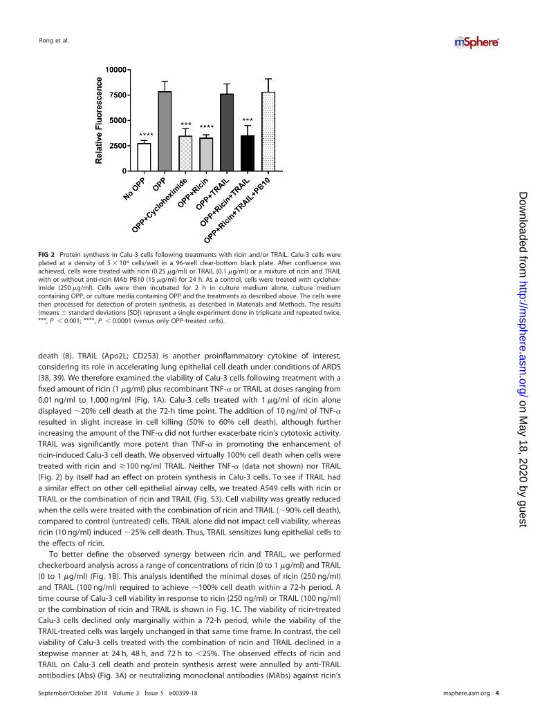
death (8). TRAIL (Apo2L; CD253) is another proinflammatory cytokine of interest,considering its role in accelerating lung epithelial cell death under conditions of ARDS(38, 39). We therefore examined the viability of Calu-3 cells following treatment with afixed amount of ricin (1 �g/ml) plus recombinant TNF-� or TRAIL at doses ranging from0.01 ng/ml to 1,000 ng/ml (Fig. 1A). Calu-3 cells treated with 1 �g/ml of ricin alonedisplayed �20% cell death at the 72-h time point. The addition of 10 ng/ml of TNF-�resulted in slight increase in cell killing (50% to 60% cell death), although furtherincreasing the amount of the TNF-� did not further exacerbate ricin’s cytotoxic activity.TRAIL was significantly more potent than TNF-� in promoting the enhancement ofricin-induced Calu-3 cell death. We observed virtually 100% cell death when cells weretreated with ricin and �100 ng/ml TRAIL. Neither TNF-� (data not shown) nor TRAIL(Fig. 2) by itself had an effect on protein synthesis in Calu-3 cells. To see if TRAIL hada similar effect on other cell epithelial airway cells, we treated A549 cells with ricin orTRAIL or the combination of ricin and TRAIL (Fig. S3). Cell viability was greatly reducedwhen the cells were treated with the combination of ricin and TRAIL (�90% cell death),compared to control (untreated) cells. TRAIL alone did not impact cell viability, whereasricin (10 ng/ml) induced �25% cell death. Thus, TRAIL sensitizes lung epithelial cells tothe effects of ricin.
To better define the observed synergy between ricin and TRAIL, we performedcheckerboard analysis across a range of concentrations of ricin (0 to 1 �g/ml) and TRAIL(0 to 1 �g/ml) (Fig. 1B). This analysis identified the minimal doses of ricin (250 ng/ml)and TRAIL (100 ng/ml) required to achieve �100% cell death within a 72-h period. Atime course of Calu-3 cell viability in response to ricin (250 ng/ml) or TRAIL (100 ng/ml)or the combination of ricin and TRAIL is shown in Fig. 1C. The viability of ricin-treatedCalu-3 cells declined only marginally within a 72-h period, while the viability of theTRAIL-treated cells was largely unchanged in that same time frame. In contrast, the cellviability of Calu-3 cells treated with the combination of ricin and TRAIL declined in astepwise manner at 24 h, 48 h, and 72 h to �25%. The observed effects of ricin andTRAIL on Calu-3 cell death and protein synthesis arrest were annulled by anti-TRAILantibodies (Abs) (Fig. 3A) or neutralizing monoclonal antibodies (MAbs) against ricin’s
FIG 2 Protein synthesis in Calu-3 cells following treatments with ricin and/or TRAIL. Calu-3 cells wereplated at a density of 5 � 104 cells/well in a 96-well clear-bottom black plate. After confluence wasachieved, cells were treated with ricin (0.25 �g/ml) or TRAIL (0.1 �g/ml) or a mixture of ricin and TRAILwith or without anti-ricin MAb PB10 (15 �g/ml) for 24 h. As a control, cells were treated with cyclohex-imide (250 �g/ml). Cells were then incubated for 2 h in culture medium alone, culture mediumcontaining OPP, or culture media containing OPP and the treatments as described above. The cells werethen processed for detection of protein synthesis, as described in Materials and Methods. The results(means � standard deviations [SD]) represent a single experiment done in triplicate and repeated twice.***, P � 0.001; ****, P � 0.0001 (versus only OPP-treated cells).
Rong et al.
September/October 2018 Volume 3 Issue 5 e00399-18 msphere.asm.org 4
on May 18, 2020 by guest
http://msphere.asm
.org/D
ownloaded from

A subunit (Fig. 3B) or B subunit (Fig. S4). The 50% inhibitory concentrations (IC50s) foreach of the RTA and RTB neutralizing MAbs in conjunction with the Calu-3 cells weresimilar to what has been reported for other cell types such as Vero cells and THP-1 cells(36, 40). For example, MH3’s IC50 for Calu-3 cells was � 1 �g/ml, which is similar to whatwe reported for MH3 on Vero cells (1.6 �g/ml) (36).
In macrophage and epithelial cells, ricin triggers the intrinsic apoptotic pathwaythrough a process dependent on caspase-3/7 activation (13). We therefore examinedcaspase-3/7 activity in Calu-3 cells following treatment with ricin (250 ng/ml) or TRAIL(100 ng/ml) or the combination of ricin and TRAIL. At the concentrations employed,neither ricin nor TRAIL alone was sufficient to induce caspase-3/7 activity in Calu-3 cells(Fig. 4A). However, the combination of ricin and TRAIL resulted in a significant (�4-fold)increase in caspase-3/7 activity, which was inhibited by Z-DVEVD (Fig. 4). In a Calu-3 cellviability assay, ZVAD (pan-caspase inhibitor), ZIETD (caspase-8 inhibitor), and ZDEVD(caspase-3/7 inhibitor) were each able to partially suppress the cytotoxic effects of ricinand TRAIL, but only at early time points (Fig. 5). Blocking the initiator caspase-9 with theinhibitor LEHD had no effect on Calu-3 cell viability following ricin and TRAIL treatment(Fig. S5), nor did treatment with the necrosis inhibitors necrosulfonamide (NSA),glycogen synthase kinase (GSK), and Nec-1 (Fig. S5). Collectively, these results areconsistent with ricin and TRAIL treatment activating apoptosis through multiplecaspase-8- and caspase-3/7-dependent pathways.
Transcriptional profiling of Calu-3 cells following ricin and TRAIL treatment. Tobetter understand the interaction between ricin and TRAIL, we subjected Calu-3 cells totranscriptional profiling using a human immunology nCounter array encompassing
FIG 3 Specificity of ricin and TRAIL in inducing Calu-3 cell death. (A) Anti-TRAIL Abs (starting at 1 �g/ml)or (B) anti-ricin MAbs (starting at 15 �g/ml) in a 2-fold serial dilution were mixed with ricin (0.25 �g/ml)and TRAIL (0.1 �g/ml) and then administered to the cells seeded in 96-well plates for 24 h. The cells werethen washed and cell viability was measured 72 h later, as described in Materials and Methods. Theresults (means � SD) represent a single experiment done in triplicate and repeated at least three times.
Ricin-Induced Killing of Airway Epithelial Cells
September/October 2018 Volume 3 Issue 5 e00399-18 msphere.asm.org 5
on May 18, 2020 by guest
http://msphere.asm
.org/D
ownloaded from

�600 target genes. RNA was isolated from Calu-3 cells treated with ricin (250 ng/ml) orTRAIL (100 ng/ml) or the combination of ricin and TRAIL for 3 h, 6 h and 18 h. At the 3-hand 6-h time points, there were no significant changes in RNA levels seen among thetarget genes represented on the human immunology array in comparisons of sampleswith ricin or TRAIL or ricin-plus-TRAIL treatments to medium control samples (data notshown). By 18 h, the picture was markedly different. Analysis of the RNA from Calu-3cells treated with the combination of ricin and TRAIL indicated that there were �80genes whose expression was elevated �2-fold over the levels seen with the untreatedcontrols, which corresponds to roughly 12% of all the genes on the human immunol-ogy nCounter array (Fig. 6; see also Table S1 and Fig. S6 in the supplemental material).Most notable was an increase in the level of IL-6 (�750-fold), followed by otherproinflammatory cytokines such as beta interferon (IFN-�) (�120-fold), TNF-� (�120-fold), IL-8 (88-fold), IL-1� (60-fold), and CCL20 (90-fold). Virtually the same transcrip-tional profile was observed when Calu-3 cells were treated with just ricin, although themagnitude of the response was dampened compared to that seen with cells treatedwith ricin plus TRAIL (Fig. 6; see also Table S1 and Fig. S6). TRAIL treatment alone didnot significantly alter Calu-3 gene expression. These results are consistent with TRAILenhancing the proinflammatory and apoptotic responses of Calu-3 to ricin rather thaninducing parallel or convergent proinflammatory and apoptotic pathways.
FIG 4 Increased caspase-3/7 activity in ricin- and TRAIL-treated Calu-3 cells. For the quantification ofcaspase-3/7 activity, Calu-3 cells were treated with ricin (0.25 �g/ml) or TRAIL (0.1 �g/ml) or ricin andTRAIL with or without caspase 3 inhibitor Z-DEVD-FMK(62.5 nM) for 24 h or with medium only (negativecontrol). The caspase-3/7 activity levels were determined by flow cytometry as described in Materials andMethods. Caspase-3/7 activity was expressed as a percentage of total cells. The results are presented asmeans � SD of three independent experiments. ***, P � 0.001 (versus control cells).
Rong et al.
September/October 2018 Volume 3 Issue 5 e00399-18 msphere.asm.org 6
on May 18, 2020 by guest
http://msphere.asm
.org/D
ownloaded from

To validate the transcriptional profiling studies, Calu-3 cells were treated for 24 hwith ricin or TRAIL or the ricin-plus-TRAIL combination, after which culture supernatantswere assayed for levels of proinflammatory cytokines IL-6, IL-8, IL-1, IL-10, TNF-�, andIL-12 by cytometric bead array (CBA). We found that the IL-8, IL-1, IL-10, TNF-�, andIL-12 levels were unchanged, irrespective of whether Calu-3 cells were treated with ricinor TRAIL or the ricin-plus-TRAIL combination (Fig. 7). IL-6 levels, in contrast, wereelevated �10-fold in supernatants from Calu-3 cells treated with ricin plus TRAIL,compared to controls (Fig. 7). Treatment of cells with ricin alone enhanced IL-6 levels,although not to a statistically significant degree. Thus, treatment of Calu-3 cells with thecombination of ricin plus TRAIL results in the preferential secretion of IL-6.
We postulated that the absence of TNF-� in the Calu-3 cell supernatants followingricin and TRAIL treatments might have been due to autocrine signaling such thatsoluble cytokine was rapidly captured by the TNF-� receptor, which in turn might havecontributed to ricin-induced cell death. To examine this possibility, Calu-3 cells weretreated with ricin plus TRAIL in the presence of neutralizing anti-TNF-� antibody. Wefound that anti-TNF-� antibody treatment did not prevent or even reduce Calu-3 cellkilling in response to ricin plus TRAIL, whereas treatment with an anti-TRAIL neutral-izing antibody did rescue the cells (Fig. 8).
DISCUSSION
Widespread damage to the airway epithelium is a hallmark of inhalational ricinexposure, although the exact molecular events that culminate in epithelial cell destruc-tion have not been fully elucidated (2, 3, 5, 7, 24, 41). For this study, we utilized thewell-characterized Calu-3 cell line as a prototype to better define the response ofhuman airway epithelial cells to ricin (27–31). We found that Calu-3 cells, when grownto confluence on solid or permeable substrates, were largely impervious to the effectsof ricin-induced cell death (as determined using an ATP-based cell viability assay), eventhough protein synthesis was effectively shut down (as determined using anO-propargyl-puromycin [OPP]-based assay) and epithelial integrity compromised (asmanifested by a reduction in TER). Coadministration of soluble TRAIL (and, to a lesserdegree, TNF-�) rendered Calu-3 cells �1,000-fold more sensitive to toxin-inducedapoptosis. Soluble TRAIL also magnified the proinflammatory transcriptional responseof Calu-3 cells to ricin and the preferential secretion of IL-6. While the current investi-gation was limited to in vitro studies, the results are consistent with a model in whichproinflammatory cytokines such as TRAIL amplify epithelial stress-induced signal trans-duction that promotes the recruitment of polymorphonuclear leukocytes (PMNs) to thelung and simultaneously lowers the threshold level of ricin required to induce epithelialapoptosis. We also present evidence to support the notion that ricin alters bronchial
FIG 5 Protective effect of caspase inhibitors on cell viability in ricin- and TRAIL-treated Calu-3 cells.Pan-caspase inhibitor Z-VAD-FMK, caspase-3 inhibitor Z-DEVD-FMK, or caspase-8 inhibitor Z-IETD-FMK at62.5 nM was mixed with ricin (0.25 �g/ml) and TRAIL (0.1 �g/ml) and administered to the cells for 24 h.The cells were then washed, and cell viability was measured 48 h or 72 h later. The results (means � SD)represent a single experiment done in triplicate and repeated at least three times.
Ricin-Induced Killing of Airway Epithelial Cells
September/October 2018 Volume 3 Issue 5 e00399-18 msphere.asm.org 7
on May 18, 2020 by guest
http://msphere.asm
.org/D
ownloaded from

epithelial integrity and barrier function, thereby contributing to enhanced transudationinto the alveolar space (42).
TRAIL has previously been implicated in driving respiratory pathology and airwayepithelial cell death in mice and humans in response to pathogenic agents, notably,
FIG 6 NanoString analysis of genes differentially expressed in ricin- and TRAIL-treated Calu-3 cells. Calu-3 cellswere treated with ricin (0.25 �g/ml) or TRAIL (0.1 �g/ml) or a mixture of ricin and TRAIL or medium only (negativecontrol) for 18 h. RNA was extracted and subjected to nCounter analysis using a human immunology array panel.(A) Volcano plot representation of gene expression changes in ricin-plus-TRAIL-treated cells, compared with controlcells. Red circles represent transcripts upregulated �32-fold (5 log2-fold). The vertical dashed red line marks the 5log2-fold change threshold. (B) Heat map showing the relative fold changes in expression of selected genes in eachtreatment group compared to the control group. Genes were selected based on a minimum of 4-fold (2log2-fold)-higher expression in comparisons between control and ricin-plus-TRAIL-treated cells. The color scale bardenotes maximum counts in blue and minimal counts in white. The measured fold change values (relative tocontrol cells) are listed in Table S1.
Rong et al.
September/October 2018 Volume 3 Issue 5 e00399-18 msphere.asm.org 8
on May 18, 2020 by guest
http://msphere.asm
.org/D
ownloaded from

influenza virus, respiratory syncytial virus (RSV), and chlamydia (38, 39, 43–45). In thecase of influenza virus infection, alveolar macrophages are the primary source of TRAIL(38, 39). Bronchial epithelial cells express a TRAIL receptor(s), which ultimately modu-lates TRAIL-dependent apoptosis (46). In rodents and nonhuman primates, alveolarmacrophages are a primary target of ricin following intranasal and inhalational chal-lenge, so it is plausible that these cells also serve as a source of TRAIL following toxinexposure (3, 5, 9, 47). It remains unclear whether this scenario applies to ricin. In
FIG 7 Cytokine secretion by Calu-3 cells following ricin and TRAIL treatment. Calu-3 cells were treatedwith ricin (0.25 �g/ml) or TRAIL (0.1 �g/ml) or a mixture of ricin and TRAIL or medium only (negativecontrol) for 24 h. Cell supernatants were collected from treated cells. The levels of cytokines IL-6, IL-8,IL-1�, IL-10, TNF-ɑ, and IL-12p70 (panels A to F, respectively) were measured by CBA. The results arepresented as the means � SD of three independent experiments. **, P � 0.01 (versus untreated cells[negative control]).
Ricin-Induced Killing of Airway Epithelial Cells
September/October 2018 Volume 3 Issue 5 e00399-18 msphere.asm.org 9
on May 18, 2020 by guest
http://msphere.asm
.org/D
ownloaded from

preliminary studies, we have not yet observed a measurable uptick in TRAIL levels in theBAL fluids from mice or nonhuman primates that had been challenged with ricin (Y.Rong and N. Mantis, unpublished results). However, we have observed that superna-tants from toxin-treated monocyte/macrophage cell lines do sensitize human lungepithelial cells to the effects of ricin, and we are actively examining whether this factoris TRAIL or a member of the TNF-� family of cytokines (C. Kempen, Y. Rong, N. Mantis,and T. LaRocca, unpublished results).
The issue of how TRAIL sensitizes Calu-3 cells to ricin-induced cell death is ofparticular interest, considering that TRAIL activates cell death through an extrinsicapoptotic pathway, whereas ricin triggers intrinsic pathways induced as a result of theribotoxic stress response (RSR), unfolded protein response (UPR), and/or increasedlevels of intracellular calcium (13, 25). We postulate that caspase-3 serves as the centralnode through which ricin and TRAIL intersect. In human cells, activation of TRAILreceptor 1 (TRAIL-R1) and/or TRAIL-R2 stimulates caspase-8 activation, which in turntriggers caspase-3 activation (46). Ricin-induced programmed cell death is also depen-dent on caspase-3 activation, although which specific upstream signaling pathway(s)(e.g., RSR, UPR, ER stress) is most relevant in airway epithelial cell killing has not beencompletely elucidated (2, 12). As demonstrated in this study, Calu-3 cell death followingwith ricin and TRAIL treatment coincided with an increase in caspase-3/7 activity andwas partially inhibited by the addition of Z-DEVD-fmk but was not impacted by LEHD,an inhibitor of caspase-9. Calu-3 cell death may also be exacerbated by a concomitantdecline in the levels of endogenous inhibitors of apoptosis such as cFLIP, due to ricin’scapacity to arrest protein synthesis, as reported previously in a study of cells treatedwith Shiga toxin (48). In fact, the protein synthesis inhibitor cycloheximide is commonlyused as a tool to sensitize cells to TRAIL-induced apoptosis (25, 49). While the issue issomewhat a question of semantics, we would argue that TRAIL sensitizes Calu-3 cellsto ricin-induced apoptosis rather than sensitizing ricin-sensitizing Calu-3 cells to TRAIL-induced cell death. This claim is best supported by the results of the transcriptionalprofiling that we performed, which demonstrated that TRAIL amplifies the effects ofricin on the treatment of Calu-3 cells across the board. Treatment with TRAIL alone hadno effect on Calu-3 gene expression, nor did TRAIL (by itself) negatively influence Calu-3cell viability.
The relationship between protein synthesis arrest and apoptosis in response to ricintoxin is another interesting aspect of this study. Using the puromycin analogue OPPcoupled with click chemistry to measure levels of protein synthesis (50), we found thatricin treatment induced protein synthesis arrest (i.e., a measurable decrease in ATPlevels) in Calu-3 cells without triggering cell death. Only upon the addition of TRAIL didthe cells undergo apoptosis. Thus, ricin-treated Calu-3 cells are primed to undergoprogrammed cell death upon exposure to a secondary insult, whether that insult is
FIG 8 Effects of TNF-�- and TRAIL-neutralizing Abs on the viability of Calu-3 cells following ricin andTRAIL treatment. Calu-3 cells were treated with ricin or ricin plus TRAIL in the presence of neutralizinganti-TNF-� Ab or anti-TRAIL Ab or the combination of the two Abs. Cell viability was measured 72 h later.The results (means � SD) represent a single experiment done in triplicate and repeated at least threetimes. **, P � 0.01; *** P � 0.001 (versus untreated cells [negative control]).
Rong et al.
September/October 2018 Volume 3 Issue 5 e00399-18 msphere.asm.org 10
on May 18, 2020 by guest
http://msphere.asm
.org/D
ownloaded from

represented by TRAIL or possibly by other signals in vivo (25). As shown in Fig. 2 and3 (see also Fig. S4 in the supplemental material), neutralizing antibodies against ricin orTRAIL protected Calu-3 cells from simultaneous ricin and TRAIL challenge. The relativekinetics of neutralizing activity were not investigated here. In other words, mightanti-TRAIL antibodies have activity when administered after treatment of Calu-3 cellswith ricin? Might the Calu-3 cell response to TRAIL be blunted with TRAIL receptor (e.g.,DR2) antibodies? These are pertinent questions that are currently being addressed in amouse model, even though the mouse differs considerably from humans with respectto TRAIL receptors (46).
We identified IL-6 as being markedly upregulated in Calu-3 cells at the transcrip-tional and protein levels following ricin and ricin-plus-TRAIL treatments. This findingmay have important implications for understanding the pathology associated withpulmonary ricin exposure, especially in nonhuman primates, where elevated levels ofIL-6 in bronchoalveolar lavage (BAL) fluids are associated with negative outcomesfollowing toxin exposure (C. Roy, Y. Rong, D. Ehrbar, and N. Mantis, unpublishedresults). IL-6 also accumulates in BAL fluids and serum of mice following intranasal ricinchallenge (Rong and Mantis, unpublished) (9, 22, 47, 51). DaSilva and colleagues alsonoted a significant increase (albeit modest) in IL-6 mRNA levels when total lung tissuesfrom ricin-challenged mice were analyzed by DNA microarray (20). Whether IL-6 is morethan just a biomarker of ricin intoxication remains to be determined, but there isconsiderable evidence to implicate this cytokine in driving local and systemic pathol-ogies (52, 53). Surprisingly, Calu-3 cells did not secrete detectable levels of other“initiator” cytokines such as TNF-� and IL-1 or the chemokine IL-8, even though each oftheir respective mRNA transcripts was significantly unregulated by ricin or ricin plusTRAIL. Wong et al. noted that TNF-� secretion actually declined when primary humanbronchial epithelial cells were exposed to ricin, presumably due to a global arrest inprotein synthesis (12). The continued (and possibly preferential) synthesis of IL-6 inricin-intoxicated cells may have to do with differential rates of mRNA stability, especiallywhen mitogen-activated protein kinase (MAPK) signaling pathways are activated (54).It is worth noting that Wong and colleagues reported that TNF-� and IL-1� expressionin primary human bronchial epithelial cells in response to ricin is dependent on NF�B,while IL-6 expression is not (12). In summary, our report advances our understandingof the factors that may drive the severe pathology associated with aerosolized ricinexposure and offers support for therapeutic approaches aimed at neutralizing ricin plusimmunomodulatory factors such as TRAIL (2, 24).
MATERIALS AND METHODSChemicals and biological reagents. Ricin toxin (Ricinus communis agglutinin II) was purchased from
Vector Laboratories (Burlingame, CA). Ricin was dialyzed against phosphate-buffered saline (PBS) at 4°Cin 10,000-molecular-weight (MW)-cutoff Slide-A-Lyzer dialysis cassettes (Pierce, Rockford, IL), prior to usein cytotoxicity studies. Fetal calf serum was purchased from Gibco-Invitrogen (Carlsbad, CA). Cells weremaintained in a humidified incubator at 37°C with 5% CO2. Recombinant human tumor necrosis factoralpha (TNF-�), recombinant human soluble TRAIL (sTRAIL)/Apo2L/CD253, anti-human sTRAIL-(s)-Apo2L,and rabbit polyclonal anti-TRAIL antibody were purchased from Peprotech (Rocky Hill, NJ). Human TNF-�neutralizing rabbit Ab was purchased from Cell Signaling Technology (Danvers, MA). Z-LEHD-FMK,Z-VAD-FMK, Z-DEVD-FMK, and Z-IETD-FMK were purchased from ApexbBio (Taiwan). Necrostatin-1(Nec-1), GSK=872, and necrosulfonamide (NSA) were purchased from EMD Millipore (Burlington, MA).Unless noted otherwise, all other chemicals were obtained from MilliporeSigma (St. Louis, MO). MurineMAbs against ricin toxin’s A subunit (PB10, SyH7, GD12, and IB2) and B subunit (24B11, SylH3, MH3, 8A1,8B3, LF1, and LC5) were purified by protein A chromatography at the Dana Farber Cancer Institute (DFCI)Monoclonal Antibody Core facility (Boston, MA), as described previously (36, 55).
Cell culture. The Calu-3 lung adenocarcinoma cell line was obtained from the American Type CultureCollection (ATCC; Manassas, VA) and cultured in Eagle’s minimum essential medium (EMEM) supple-mented with 10% fetal bovine serum, provided by the Wadsworth Center Media Services facility. Cellswere grown in a humidified incubator containing 5% CO2 and 95% air at 37°C. The cells were plated in75-cm2 cell culture flasks and subcultured at 70% to 90% confluence using a 0.25% trypsin–EDTA solution(Corning Life Sciences, Corning, NY). The culture medium was changed every 3 days. The cells were split1:5 during each passage. The numbers of passages used for the following experiments were under 10.The A459 human lung epithelial cell line (ATCC CCL-185) was obtained from ATCC and maintained asrecommended by ATCC.
Ricin-Induced Killing of Airway Epithelial Cells
September/October 2018 Volume 3 Issue 5 e00399-18 msphere.asm.org 11
on May 18, 2020 by guest
http://msphere.asm
.org/D
ownloaded from

Ricin cytotoxicity assay. Calu-3 and A549 cells were trypsinized, adjusted to 5 � 105 cells per ml,seeded (100 �l/well) into 96-well plates (Corning Life Sciences, Corning, NY), and incubated for 3 to4 days until confluence. Calu-3 cells were then treated with ricin, TRAIL, a mixture of ricin and TRAIL, ormedium alone (negative control) for 24 h. The cells were washed to remove noninternalized toxin orTRAIL and were then incubated for 24 to 72 h. Cell viability was assessed using CellTiter-GLO reagent(Promega, Madison, WI) and a SpectraMax L microplate reader (Molecular Devices, Sunnyvale, CA). Alltreatments were performed in triplicate and repeated at least 3 times. Viability of 100% was defined asthe average value obtained from wells in which cells were treated with medium only.
On the basis of the cytotoxicity results from the ricin and TRAIL treatments described above, ricin(0.25 �g/ml) and TRAIL (0.1 �g/ml) were used in all the subsequent experiments. To measure theneutralizing activity of ricin-specific MAbs and neutralizing anti-TRAIL Ab, the anti-RTA MAbs (starting at15 �g/ml), anti-RTB MAbs (starting at 30 �g/ml), or anti-TRAIL Abs (starting at 1 �g/ml) in a 2-fold serialdilution were mixed with ricin and TRAIL and then administered to the cells seeded in 96-well plates for24 h. After washing and then incubating for 3 days, cell viability was assessed using CellTiter-GLOreagent. Cell viability was normalized to cells treated with medium only. The protective effect of caspaseinhibitors (Z-VAD-FMK, Z-LEHD-FMK, Z-DEVD-FMK, and Z-IETD-FMK), RIPK1 inhibitor (Nec-1), RIPK3inhibitor (GSK=872), and MLKL inhibitor (NSA) was also evaluated using combinations with ricin and TRAILand the concentrations and incubation times indicated for each experiment. Dimethyl sulfoxide (DMSO)was used as a control vehicle for all experiments.
Protein synthesis assay. Calu-3 cells were plated at a density of 5 � 104 cells/well in a 96-wellclear-bottom black plate (Corning Life Science, Corning, NY) and incubated for 3 to 4 days untilconfluence. Cayman’s protein synthesis assay kit (Cayman Chemical, Ann Arbor, MI) was used to measurethe protein synthesis. Cells were treated with ricin (0.25 �g/ml), TRAIL (0.1 �g/ml), the mixture of ricinand TRAIL combined with PB10 (15 �g/ml), or cycloheximide (250 �g/ml; positive control) for 24 h. Cellswere then incubated for 2 h in culture medium alone, culture medium containing O-propargyl-puromycin (OPP), or culture medium containing OPP plus the treatments described above. After fixingand washing, cells were incubated with 5 FAM-azide staining solution in the dark for 30 min forsubsequent detection of OPP-labeled proteins. The cells were then examined immediately using afluorescent plate reader (Becton, Dickinson, Franklin Lakes, NJ) set to excitation/emission at 485/535 nm.
Caspase-3/7 activity assay. For the quantification of caspase-3/7 activities after treatment, Calu-3cells were labeled with 500 nM Cell Event caspase-3/7 green detection reagent (Invitrogen, Carlsbad, CA)for 30 min at 37°C in the dark. A total of 10,000 stained cells per sample were acquired and analyzed ina FACSCalibur flow cytometer by using CellQuest Pro software (Becton, Dickinson, Franklin Lakes, NJ).Data were expressed as a percentage of total cells.
Multiplex gene expression analysis using NanoString. Calu-3 cells were treated with ricin, TRAIL,the combination of ricin and TRAIL, or medium alone (negative control) for 24 h. RNA was extracted fromtreated or nontreated cells using an RNeasy Plus minikit with additional on-column DNase digestion withan RNase-Free DNase set (Qiagen Hilden, Germany). Protocols were followed according to the manu-facturer’s instructions. Extracted RNA samples were stored at �80°C until use. Upon assay, RNA integritywas verified by agarose gel electrophoresis. RNA quality and concentrations were measured using anAgilent 2100 Bioanalyzer (Life Technologies, Carlsbad, CA). RNA (100 ng) was hybridized with a prede-signed nCounter human immunology panel that included 594 target genes and 15 internal referencegenes. The geNorm algorithm was used to select the most stable of the reference genes (GAPDH[glyceraldehyde-3-phosphate dehydrogenase], PPIA, G6PD, EEF1G, GUSB, HPRT1, SDHA, and RPL19) usedfor normalization (56). The experimental procedures were carried out on a NanoString preparationstation and digital analyzer according to the manufacturer’s instructions. Two biological replicates wereselected from each of the four groups for analysis.
Cell supernatant cytokine quantification by cytometric bead array (CBA). Cell supernatantswere collected from treated Calu-3 cells. The BD CBA Human Inflammatory Cytokines kit (Becton,Dickinson, Franklin Lakes, NJ) was used to quantitatively measure levels of the following specific setsof cytokines: interleukin-8 (IL-8), interleukin-1� (IL-1�), interleukin-6 (IL-6), interleukin-10 (IL-10),TNF-�, and interleukin-12p70 (IL-12p70). Dilution series of human cytokine standards, included inthe kit and prepared according to the manufacturer’s instructions, were employed in each assay runto enable quantification. Assays were performed according to the manufacturer’s instructions, and50 �l of assay beads, 50 �l of the studied sample or standard, and 50 �l of phycoerythrin (PE)-labeled antibodies (Detection Reagent) were added consecutively to each sample tube and incu-bated at room temperature in the dark for 3 h. Next, the samples were washed and centrifuged, afterwhich the pellet was resuspended in wash buffer and analyzed on the same day in a flow cytometer.Flow cytometry was performed using a four-laser BD FACSCalibur system utilizing BD CellQuestsoftware for acquisition.
Statistical analyses. Statistical analyses were carried out using GraphPad Prism 7 (GraphPadSoftware, San Diego, CA), as well as the nCounter Advanced analysis module (v 1.1.4) of the nSolverAnalysis software (v3). Differential expression of the genes examined was determined by multivariatelinear regression, with group membership chosen as the predictor variable and binding density as aconfounder. All P values derived from NanoString analysis were adjusted with Benjamini-Yekutielicorrection for control of the false-discovery rate. Low-count genes were omitted using the defaultsettings in the nCounter Advanced analysis software for all analyses except the linear regression of geneexpression values.
Rong et al.
September/October 2018 Volume 3 Issue 5 e00399-18 msphere.asm.org 12
on May 18, 2020 by guest
http://msphere.asm
.org/D
ownloaded from

SUPPLEMENTAL MATERIALSupplemental material for this article may be found at https://doi.org/10.1128/
mSphere.00399-18.FIG S1, JPG file, 0.02 MB.FIG S2, JPG file, 0.03 MB.FIG S3, JPG file, 0.01 MB.FIG S4, JPG file, 0.03 MB.FIG S5, JPG file, 0.04 MB.FIG S6, JPG file, 0.1 MB.TABLE S1, DOCX file, 0.03 MB.
ACKNOWLEDGMENTSWe gratefully acknowledge Renjie Song of the Wadsworth Center’s Biochemistry
and Immunology Core facility for assisting and for the use of the flow cytometer. Wethank Navjot Singh and Susan L. McHale of the Wadsworth Center’s Applied GenomicsTechnologies Core for assistance with the nCounter instrumentation. We also thankAmanda Poon (University at Albany) for helpful discussions.
This work was supported by contract no. HHSN272201400021C from the NationalInstitute of Allergy and Infectious Diseases, National Institutes of Health. The content issolely our responsibility and does not necessarily represent the official views of theNational Institutes of Health. The funders had no role in study design, data collectionand analysis, decision to publish, or preparation of the manuscript. The nCounterexperiments were funded by the Director’s Office of at the Wadsworth Center insupport of collaborations between the Wadsworth Center and ACPHS.
REFERENCES1. Cieslak TJ, Kortepeter MG, Wojtyk RJ, Jansen HJ, Reyes RA, Smith JO;
NATO Biological Medical Advisory Panel. 2018. Beyond the dirty dozen:a proposed methodology for assessing future bioweapon threats. MilMed 183:e59 – e65. https://doi.org/10.1093/milmed/usx004.
2. Gal Y, Mazor O, Falach R, Sapoznikov A, Kronman C, Sabo T. 2017.Treatments for pulmonary ricin intoxication: current aspects and futureprospects. Toxins 9:311. https://doi.org/10.3390/toxins9100311.
3. Pincus SH, Bhaskaran M, Brey RN, III, Didier PJ, Doyle-Meyers LA, Roy CJ.2015. Clinical and pathological findings associated with aerosol expo-sure of macaques to ricin toxin. Toxins 7:2121–2133. https://doi.org/10.3390/toxins7062121.
4. Thompson BT, Chambers RC, Liu KD. 2017. Acute respiratory distresssyndrome. N Engl J Med 377:562–572. https://doi.org/10.1056/NEJMra1608077.
5. Brown RF, White DE. 1997. Ultrastructure of rat lung following inhalationof ricin aerosol. Int J Exp Pathol 78:267–276.
6. Roy CJ, Song K, Sivasubramani SK, Gardner DJ, Pincus SH. 29 September2011. Animal models of ricin toxicosis. Curr Top Microbiol Immunolhttps://doi.org/10.1007/82_2011_173.
7. Smallshaw JE, Richardson JA, Vitetta ES. 2007. RiVax, a recombinant ricinsubunit vaccine, protects mice against ricin delivered by gavage oraerosol. Vaccine 25:7459–7469. https://doi.org/10.1016/j.vaccine.2007.08.018.
8. Korcheva V, Wong J, Lindauer M, Jacoby DB, Iordanov MS, Magun B.2007. Role of apoptotic signaling pathways in regulation of inflamma-tory responses to ricin in primary murine macrophages. Mol Immunol44:2761–2771. https://doi.org/10.1016/j.molimm.2006.10.025.
9. Lindauer ML, Wong J, Iwakura Y, Magun BE. 2009. Pulmonary inflamma-tion triggered by ricin toxin requires macrophages and IL-1 signaling. JImmunol 183:1419 –1426. https://doi.org/10.4049/jimmunol.0901119.
10. Roy CJ, Brey RN, Mantis NJ, Mapes K, Pop IV, Pop LM, Ruback S, KilleenSZ, Doyle-Meyers L, Vinet-Oliphant HS, Didier PJ, Vitetta ES. 2015. Ther-mostable ricin vaccine protects rhesus macaques against aerosolizedricin: epitope-specific neutralizing antibodies correlate with protection.Proc Natl Acad Sci U S A 112:3782–3787. https://doi.org/10.1073/pnas.1502585112.
11. Falach R, Sapoznikov A, Gal Y, Israeli O, Leitner M, Seliger N, Ehrlich S,Kronman C, Sabo T. 2016. Quantitative profiling of the in vivo enzymatic
activity of ricin reveals disparate depurination of different pulmonarycell types. Toxicol Lett 258:11–19. https://doi.org/10.1016/j.toxlet.2016.06.003.
12. Wong J, Korcheva V, Jacoby DB, Magun B. 2007. Intrapulmonary deliveryof ricin at high dosage triggers a systemic inflammatory response andglomerular damage. Am J Pathol 170:1497–1510. https://doi.org/10.2353/ajpath.2007.060703.
13. Tesh VL. 2012. The induction of apoptosis by Shiga toxins and ricin. CurrTop Microbiol Immunol 357:137–178. https://doi.org/10.1007/82_2011_155.
14. Lamb FI, Roberts LM, Lord JM. 1985. Nucleotide sequence of clonedcDNA coding for preproricin. Eur J Biochem 148:265–270. https://doi.org/10.1111/j.1432-1033.1985.tb08834.x.
15. Lord JM. 1985. Precursors of ricin and Ricinus communis agglutinin.Glycosylation and processing during synthesis and intracellular trans-port. Eur J Biochem 146:411– 416. https://doi.org/10.1111/j.1432-1033.1985.tb08667.x.
16. Jolliffe NA, Craddock CP, Frigerio L. 2005. Pathways for protein transportto seed storage vacuoles. Biochem Soc Trans 33:1016 –1018. https://doi.org/10.1042/BST0331016.
17. Endo Y, Mitsui K, Motizuki M, Tsurugi K. 1987. The mechanism of actionof ricin and related toxic lectins on eukaryotic ribosomes. The site andthe characteristics of the modification in 28 S ribosomal RNA caused bythe toxins. J Biol Chem 262:5908 –5912.
18. Endo Y, Tsurugi K. 1987. RNA N-glycosidase activity of ricin A-chain.Mechanism of action of the toxic lectin ricin on eukaryotic ribosomes. JBiol Chem 262:8128 – 8130.
19. Spooner RA, Lord JM. 2015. Ricin trafficking in cells. Toxins (Basel)7:49 – 65. https://doi.org/10.3390/toxins7010049.
20. DaSilva L, Cote D, Roy C, Martinez M, Duniho S, Pitt ML, Downey T,Dertzbaugh M. 2003. Pulmonary gene expression profiling of inhaled ricin.Toxicon 41:813–822. https://doi.org/10.1016/S0041-0101(03)00035-7.
21. David J, Wilkinson LJ, Griffiths GD. 2009. Inflammatory gene expressionin response to sub-lethal ricin exposure in Balb/c mice. Toxicology264:119 –130. https://doi.org/10.1016/j.tox.2009.08.003.
22. Gal Y, Mazor O, Alcalay R, Seliger N, Aftalion M, Sapoznikov A, Falach R,Kronman C, Sabo T. 2014. Antibody/doxycycline combined therapy forpulmonary ricinosis: attenuation of inflammation improves survival of
Ricin-Induced Killing of Airway Epithelial Cells
September/October 2018 Volume 3 Issue 5 e00399-18 msphere.asm.org 13
on May 18, 2020 by guest
http://msphere.asm
.org/D
ownloaded from

ricin-intoxicated mice. Toxicol Rep 1:496 –504. https://doi.org/10.1016/j.toxrep.2014.07.013.
23. Lindauer M, Wong J, Magun B. 2010. Ricin toxin activates the NALP3inflammasome. Toxins (Basel) 2:1500–1514. https://doi.org/10.3390/toxins2061500.
24. Wong J, Magun BE, Wood LJ. 2016. Lung inflammation caused byinhaled toxicants: a review. Int J Chron Obstruct Pulmon Dis 11:1391–1401. https://doi.org/10.2147/COPD.S106009.
25. Spencer SL, Sorger PK. 2011. Measuring and modeling apoptosis insingle cells. Cell 144:926 –939. https://doi.org/10.1016/j.cell.2011.03.002.
26. Sandvig K, Olsnes S, Pihl A. 1976. Kinetics of binding of the toxic lectinsabrin and ricin to surface receptors of human cells. J Biol Chem 251:3977–3984.
27. Forbes B, Ehrhardt C. 2005. Human respiratory epithelial cell culture fordrug delivery applications. Eur J Pharm Biopharm 60:193–205. https://doi.org/10.1016/j.ejpb.2005.02.010.
28. Grainger CI, Greenwell LL, Lockley DJ, Martin GP, Forbes B. 2006. Cultureof Calu-3 cells at the air interface provides a representative model of theairway epithelial barrier. Pharm Res 23:1482–1490. https://doi.org/10.1007/s11095-006-0255-0.
29. Hittinger M, Juntke J, Kletting S, Schneider-Daum N, de Souza CarvalhoC, Lehr CM. 2015. Preclinical safety and efficacy models for pulmonarydrug delivery of antimicrobials with focus on in vitro models. Adv DrugDeliv Rev 85:44 –56. https://doi.org/10.1016/j.addr.2014.10.011.
30. Ong HX, Traini D, Young PM. 2013. Pharmaceutical applications of theCalu-3 lung epithelia cell line. Expert Opin Drug Deliv 10:1287–1302.https://doi.org/10.1517/17425247.2013.805743.
31. Shan J, Huang J, Liao J, Robert R, Hanrahan JW. 2011. Anion secretion bya model epithelium: more lessons from Calu-3. Acta Physiol (Oxf) 202:523–531. https://doi.org/10.1111/j.1748-1716.2011.02253.x.
32. Park JB, Simpson LL. 2003. Inhalational poisoning by botulinum toxinand inhalation vaccination with its heavy-chain component. Infect Im-mun 71:1147–1154. https://doi.org/10.1128/IAI.71.3.1147-1154.2003.
33. Loman S, Radl J, Jansen HM, Out TA, Lutter R. 1997. Vectorial transcytosisof dimeric IgA by the Calu-3 human lung epithelial cell line: upregulationby IFN-gamma. Am J Physiol 272:L951–L958. https://doi.org/10.1152/ajplung.1997.272.5.L951.
34. Tormakangas L, Markkula E, Lounatmaa K, Puolakkainen M. 2010. Chla-mydia pneumoniae infection in polarized epithelial cell lines. InfectImmun 78:2714 –2722. https://doi.org/10.1128/IAI.01456-09.
35. Konowalchuk J, Speirs JI, Stavric S. 1977. Vero response to a cytotoxin ofEscherichia coli. Infect Immun 18:775–779.
36. Rong Y, Van Slyke G, Vance DJ, Westfall J, Ehrbar D, Mantis NJ. 2017.Spatial location of neutralizing and non-neutralizing B cell epitopes ondomain 1 of ricin toxin’s binding subunit. PLoS One 12:e0180999.https://doi.org/10.1371/journal.pone.0180999.
37. Vance DJ, Tremblay JM, Rong Y, Angalakurthi SK, Volkin DB, MiddaughCR, Weis DD, Shoemaker CB, Mantis NJ. 5 December 2017. High-resolution epitope positioning of a large collection of neutralizing andnonneutralizing single-domain antibodies on the enzymatic and bind-ing subunits of ricin toxin. Clin Vaccine Immunol https://doi.org/10.1128/CVI.00236-17.
38. Herold S, Steinmueller M, von Wulffen W, Cakarova L, Pinto R, PleschkaS, Mack M, Kuziel WA, Corazza N, Brunner T, Seeger W, Lohmeyer J. 2008.Lung epithelial apoptosis in influenza virus pneumonia: the role ofmacrophage-expressed TNF-related apoptosis-inducing ligand. J ExpMed 205:3065–3077. https://doi.org/10.1084/jem.20080201.
39. Peteranderl C, Morales-Nebreda L, Selvakumar B, Lecuona E, Vadasz I,Morty RE, Schmoldt C, Bespalowa J, Wolff T, Pleschka S, Mayer K,Gattenloehner S, Fink L, Lohmeyer J, Seeger W, Sznajder JI, Mutlu GM,Budinger GR, Herold S. 2016. Macrophage-epithelial paracrine crosstalkinhibits lung edema clearance during influenza infection. J Clin Invest126:1566 –1580. https://doi.org/10.1172/JCI83931.
40. O’Hara JM, Kasten-Jolly JC, Reynolds CE, Mantis NJ. 2014. Localization ofnon-linear neutralizing B cell epitopes on ricin toxin’s enzymatic sub-unit (RTA). Immunol Lett 158:7–13. https://doi.org/10.1016/j.imlet.2013.11.009.
41. Wong J, Korcheva V, Jacoby DB, Magun BE. 2007. Proinflammatoryresponses of human airway cells to ricin involve stress-activated proteinkinases and NF-kappaB. Am J Physiol Lung Cell Mol Physiol 293:L1385–L1394. https://doi.org/10.1152/ajplung.00207.2007.
42. Rushing SR, Saylor ML, Hale ML. 2009. Translocation of ricin acrosspolarized human bronchial epithelial cells. Toxicon 54:184 –191. https://doi.org/10.1016/j.toxicon.2009.04.003.
43. Bem RA, Bos AP, Wosten-van Asperen RM, Bruijn M, Lutter R, Sprick MR,van Woensel JB. 2010. Potential role of soluble TRAIL in epithelial injuryin children with severe RSV infection. Am J Respir Cell Mol Biol 42:697–705. https://doi.org/10.1165/rcmb.2009-0100OC.
44. Kotelkin A, Prikhod’ko EA, Cohen JI, Collins PL, Bukreyev A. 2003. Respi-ratory syncytial virus infection sensitizes cells to apoptosis mediated bytumor necrosis factor-related apoptosis-inducing ligand. J Virol 77:9156 –9172. https://doi.org/10.1128/JVI.77.17.9156-9172.2003.
45. Starkey MR, Nguyen DH, Essilfie AT, Kim RY, Hatchwell LM, Collison AM,Yagita H, Foster PS, Horvat JC, Mattes J, Hansbro PM. 2014. Tumornecrosis factor-related apoptosis-inducing ligand translates neonatalrespiratory infection into chronic lung disease. Mucosal Immunol7:478 – 488. https://doi.org/10.1038/mi.2013.65.
46. von Karstedt S, Montinaro A, Walczak H. 2017. Exploring the TRAILs lesstravelled: TRAIL in cancer biology and therapy. Nat Rev Cancer 17:352–366. https://doi.org/10.1038/nrc.2017.28.
47. Sabo T, Gal Y, Elhanany E, Sapoznikov A, Falach R, Mazor O, Kronman C.2015. Antibody treatment against pulmonary exposure to abrin conferssignificantly higher levels of protection than treatment against ricin intox-ication. Toxicol Lett 237:72–78. https://doi.org/10.1016/j.toxlet.2015.06.003.
48. Hattori T, Watanabe-Takahashi M, Ohoka N, Hamabata T, Furukawa K,Nishikawa K, Naito M. 2015. Proteasome inhibitors prevent cell deathand prolong survival of mice challenged by Shiga toxin. FEBS Open Bio5:605– 614. https://doi.org/10.1016/j.fob.2015.06.005.
49. Wang L, Du F, Wang X. 2008. TNF-alpha induces two distinct caspase-8activation pathways. Cell 133:693–703. https://doi.org/10.1016/j.cell.2008.03.036.
50. Horisawa K. 2014. Specific and quantitative labeling of biomoleculesusing click chemistry. Front Physiol 5:457. https://doi.org/10.3389/fphys.2014.00457.
51. Korcheva V, Wong J, Corless C, Iordanov M, Magun B. 2005. Administra-tion of ricin induces a severe inflammatory response via nonredundantstimulation of ERK, JNK, and P38 MAPK and provides a mouse model ofhemolytic uremic syndrome. Am J Pathol 166:323–339. https://doi.org/10.1016/S0002-9440(10)62256-0.
52. Kishimoto T. 2010. IL-6: from its discovery to clinical applications. IntImmunol 22:347–352. https://doi.org/10.1093/intimm/dxq030.
53. Rincon M, Irvin CG. 2012. Role of IL-6 in asthma and other inflammatorypulmonary diseases. Int J Biol Sci 8:1281–1290. https://doi.org/10.7150/ijbs.4874.
54. Schoenberg DR, Maquat LE. 2012. Regulation of cytoplasmic mRNAdecay. Nat Rev Genet 13:246 –259. https://doi.org/10.1038/nrg3160.
55. Toth RT, IV, Angalakurthi SK, Van Slyke G, Vance DJ, Hickey JM, Joshi SB,Middaugh CR, Volkin DB, Weis DD, Mantis NJ. 5 December 2017. High-definition mapping of four spatially distinct neutralizing epitope clusterson RiVax, a candidate ricin toxin subunit vaccine. Clin Vaccine Immunolhttps://doi.org/10.1128/CVI.00237-17.
56. Vandesompele J, De Preter K, Pattyn F, Poppe B, Van Roy N, De Paepe A,Speleman F. 2002. Accurate normalization of real-time quantitative RT-PCR data by geometric averaging of multiple internal control genes.Genome Biol 3:RESEARCH0034.
Rong et al.
September/October 2018 Volume 3 Issue 5 e00399-18 msphere.asm.org 14
on May 18, 2020 by guest
http://msphere.asm
.org/D
ownloaded from