crosslinking density and resorption of dimethyl suberimidate-treated collagen
TRANSCRIPT

Crosslinking density and resorption of dimethylsuberimidate-treated collagen
V. Charulatha, A. RajaramDepartment of Biophysics, Central Leather Research Institute, Adyar, Madras-600 020, India
Received 8 March 1996; accepted 3 September 1996
Abstract: Collagen was purified from bovine Achilles ten- DMS crosslinked collagen membranes were more susceptibledon and crosslinked with dimethyl suberimidate (DMS) and to degradation by enzymes in vitro, while GTA-treated colla-glutaraldehyde (GTA). Under optimal conditions, the shrink- gen was highly resistant to degradation. The biocompatibilityage temperature (TS) was raised to 748C for collagen cross- of the collagen membranes was studied by subcutaneouslinked with DMS and to 808C for those crosslinked with implantation in rats. Uncrosslinked collagen membranes de-GTA. Crosslinking density measurements were done on the graded within 14 days with the formation of granulationhydrothermally denatured collagen by the method based on tissue. DMS crosslinked membranes degraded within 21 daysthe Flory–Rehner equation. GTA treatment was found to and the area was replaced by numerous fibroblasts andintroduce more number of crosslinks than DMS. The maxi- newly formed collagen. No calcification was observed. Formum tension attained during heating (after shrinkage has GTA-treated membranes, necrosis was observed after 7 daysoccurred) was greater for GTA-treated collagen than for DMS implantation and by 14 days the membrane had started toand control. The control collagen membranes broke during calcify. 1997 John Wiley & Sons, Inc. J Biomed Mater Res,heating (at 738C), while for the crosslinked membranes the 36, 478–486, 1997.tension kept on increasing up to 1008C. The crosslinkingdensity correlated well with the data determined from the Key words: collagen; crosslinking density; glutaraldehyde;in vitro and in vivo degradation studies. Uncrosslinked and dimethyl suberimidate; biocompatibility
INTRODUCTION
Collagenous materials have been widely used as bio-medical implants for a variety of applications duringthe past 30 years.1 Medical products based on collagenhave included sutures, hemostatic agents, burn wounddressings, and injectable collagen for soft-tissue aug-mentation.1,2 These biomaterials are generally cross-linked to reduce their immunogenicity and controltheir biodegradation on implantation.1 Various cross-linking agents have been used; glutaraldehyde (GTA)is the most widely used one.1,2 However, cytotoxicityof GTA is a major problem.1 Alternatives to GTA ascrosslinking agents are being examined.3,4
One such reagent is dimethyl suberimidate (DMS),which is used as a fixative for collagen matrices andhas been used to determine the number of subunitsof oligomeric proteins.5,6 DMS reacts with the aminogroups of collagen in the following way:
Coll NH2
+NH2
+
+
+
+
+NH2
C C
OMe[DMS]
[mono-amidine derivative (I)]
OMe
(CH2)6
Coll NH
+NH2
+NH2 +NH2
+NH2
C C
OMe
(CH2)6 CH3OH
CH3OHColl NH C NH CollC(CH2)6
CollI NH2
(diamidine derivative)
(Coll = collagen)
Rat fibrous dermal collagen has been crosslinkedCorrespondence to: V. Charulathawith DMS for use as dermal implant and was foundto be less cytotoxic than GTA.7 DMS has also beenevaluated as a crosslinking agent for human dermis
1997 John Wiley & Sons, Inc. CCC 0021-9304/97/040478–09 and porcine aortas for use as bioprostheses.8 Although
DIMETHYL SUBERIMIDATE-TREATED COLLAGEN 479
DMS has been reported to show less cytotoxicity, con- Collagen membranes (12 cm 3 3 mm) were dena-tured by placing them in water at 968C–1008C forditions for crosslinking with collagen have not been
optimized. The present study was undertaken to deter- 30 s for control and 120 s for crosslinked samples. Afterthe appropriate period, the membrane was rapidlymine the physicochemical properties of DMS cross-
linked collagen to assess it as a crosslinking agent for transferred into cold water. The membrane was thenmounted between two clamps which were immersedcollagenous matrices. Crosslinking conditions for DMS
treatment are optimized and the crosslinking density in water. The upper clamp was connected to the loadcell and the lower to the movable crosshead of anis determined. In vitro enzymatic degradation and bio-
compatibility of the DMS crosslinked matrix have been Instron tensile tester. After equilibrating for 2 h in thewater bath at the temperature of testing, the lowerstudied in relation to the crosslinking density.clamp was adjusted downward until the sample wasjust in tension and the unstressed length was noted.The specimen was then extended by 15% of the un-MATERIALS AND METHODS
stressed length in six equal steps at 3-min intervalsand was returned to the point of no stress in a similarAll analytical-grade chemicals, pepsin, and collagen-
ase were from Sigma chemicals. Trypsin was obtained manner. At each stage, the corresponding tension wasnoted. Then the sample was removed from the clampsfrom HI Media. Electron microscopy–grade GTA
(50%) aqueous solution (EM Sciences) and DMS was and blotted with a filter paper, and the density wasdetermined by the specific gravity bottle method. Frompurchased from Sigma Chemicals.
Collagen was extracted from bovine Achilles tendon the volume and unstressed length, the cross-sectionalarea was calculated and each equilibrium tension wasby limited proteolysis with pepsin and purified by
repeated acid-salt precipitation.1 A 0.9% w/v suspen- expressed as ( f ), the force per unit cross-sectional area.Corresponding values of (a 2 a22) were calculatedsion of collagen in acetic acid solution (0.5M) at 48C
was homogenized and degassed in a vacuum chamber from the unstressed length and known extensions andwere plotted against ( f ). The dry weight was obtainedto remove air bubbles. The resulting solution was
poured into a tray and the acid solution was neutral- by acetone dehydration of the sample followed by dry-ing at 608C for 1 h. From the dry weight and the wetized. The gel which formed was washed and dried at
ambient temperature to form a film. The thickness of weight, the volume fractions were calculated. A graphof ( f ) against (a 2 a22) would be a straight line withthe film varied from 60 to 80 mm.the slope giving RTrV2
1/3/Mc, where f 5 the force perunit area of the swollen unstretched sample; R 5 gas
Crosslinking of collagen constant; T 5 absolute temperature; V2 5 volume frac-tion; a 5 extension ratio; r 5 density of collagen; andCollagen films were treated with different concentra-Mc 5 average molecular weight of the chains betweentions of DMS in 0.2M Tris buffer, pH 9.0, at room crosslinks. The number of crosslinks per unit masstemperature for different periods.5 Crosslinking of would be given by (2Mc)21.films with GTA was done in 0.2M phosphate buffer,
pH 7.4. It was ascertained that the ratio of 280 : 235-Measurement of thermomechanical propertiesnm peak in ultraviolet (UV) absorbance spectrum of
the GTA used is 1.8–2.0. This confirmed that the poly-The shrinkage characteristics were measured usingmers of GTA were present in lesser amounts, as sug-
an adaptation of the Instron tensile tester. The samplegested by McPherson et al.2 At selected time points,was kept immersed in a saline bath at room tempera-treated samples were taken out and washed with lysineture. The upper end of the sample was connected to the(0.01M) to stop further crosslinking. The films wereload cell and the lower end to the movable crosshead. Astored in phosphate buffer (0.02M, pH 7.4) at 48C andload of about 5 g was given to the sample and the loadused for further studies.was allowed to relax with time. During the relaxationprocess, the solution (and sample) were heated at a
Measurement of crosslinking density uniform heating rate of 38C/min. At the shrinkagetemperature, when the sample contracted the load be-
The crosslinking density was estimated by the gan to increase and reached a maximum. The maxi-method based on the theory of rubber elasticity.9 Colla- mum load attained was a measure of the intermolecu-gen, on hydrothermal denaturation, behaves as a rub- lar crosslinking.10
berlike material. From the stress–strain behavior of theswollen denatured collagen, average molecular Estimation of lysine and hydroxyprolineweights between points of crosslinking can be deter-mined.9 From this quantity the number of crosslinks Lysine content was determined by the method of
Habeeb.11 Hydroxyproline analysis was done by theper unit mass can be calculated.

480 CHARULATHA AND RAJARAM
RESULTSmethod of Woesner.12 The collagen samples were hy-drolyzed with 6N HCl for 24 h at 1108C and the hydro-lysate were analyzed for hydroxyproline.
At physiologic pH of 7.4 and at low concentrations,DMS was ineffective in crosslinking collagen. An ap-
Enzymatic digestion preciable rise in the Ts was observed only at pH val-ues .9.0.
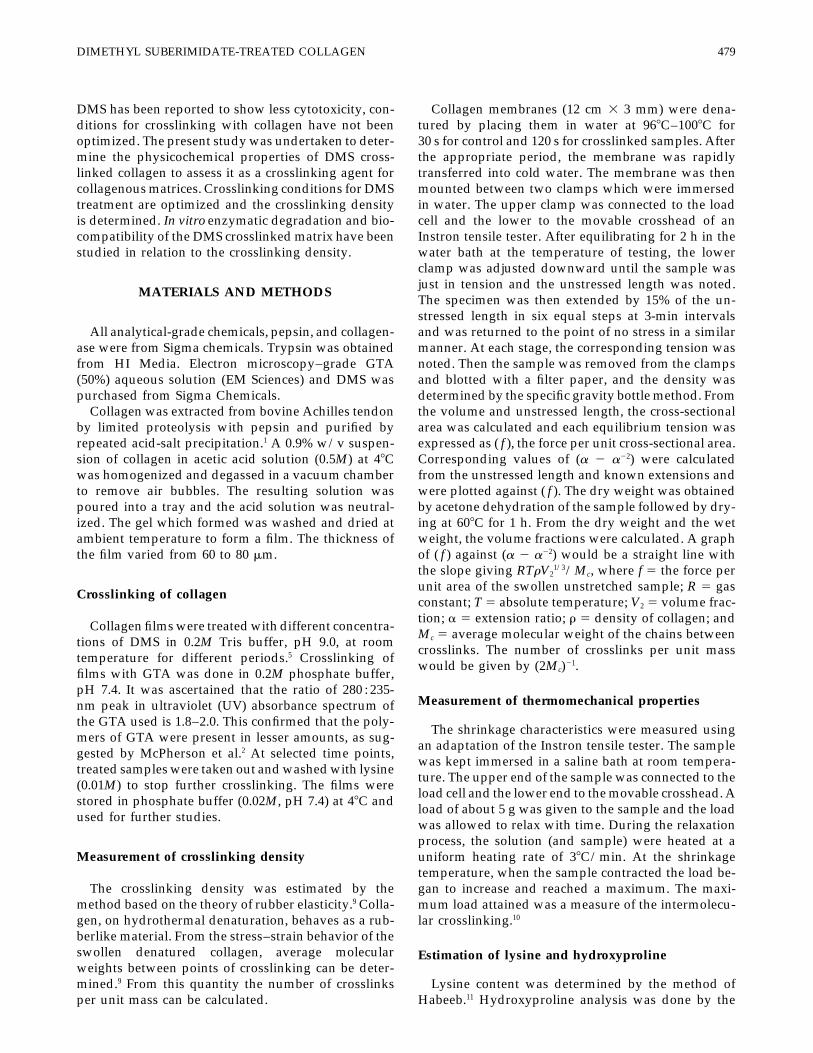
Figures 1 and 2 show the increase in shrinkage tem-Pepsin digestionperature values obtained for various concentrations ofGTA- and DMS-treated membranes, respectively, atA total of 5 mg of collagen was added to 10 mLdifferent time periods. It is seen that shrinkage temper-of 0.5M acetic acid containing 0.5 mg/mL pepsin.13
ature increased with time of crosslinking treatment.Samples were shaken in a water bath at 208C for 24 h.Our results show that treatment with 1% DMS forAfter the incubation period, the samples were centri-2 h led to the highest thermal stability of the collagenfuged and an aliquot of the supernatant was hy-membrane. There was a maximum increase of 138C fordrolyzed and hydroxyproline was estimated.these membranes. Treatment with higher concentra-tions of DMS or increasing the time of crosslinkingTrypsin digestiondid not further increase the thermal stability of themembrane. Similarly crosslinking conditions for GTAThis was done by the method of Diamond et al.13
were optimized. Crosslinking with GTA was carriedBriefly, a sample of collagen suspension (5 mg/mL)out at pH 7.4 as maximum crosslinking occurs at thisin glycerol/acetic acid, pH 3.5 was added to Tris-HClpH, and was found that treatment with 0.2% GTA for(0.1M, pH 7.8) and trypsin (1 mg/mL) in the same2 h brought about the maximum increase in shrinkagebuffer, resulting in a final pH of 7.5. The samples weretemperature. Hence, collagen membranes crosslinkedthen incubated at 378C for 2 h and centrifuged. Thewith 1% DMS (2 h) and 0.2% GTA (2 h) were takenamount of solubilized collagen was calculated fromup for further comparisons.the hydroxyproline content of the supernatant follow-
The crosslinking density of DMS crosslinked mem-ing hydrolysis.brane is given in Table I. The maximum number ofcrosslinks introduced by 1% DMS treatment was 7.Collagenase digestion
Collagen film (1 mg/mL) was taken in 0.05M Trisbuffer containing 10 mM CaCl2 at pH 7.4. The sampleswere then mixed with 10 mL of collagenase (1.36/103
U/mL) and incubated at 378C for 24 h.1 The materialsolubilized by collagenase was separated from the resi-due by centrifugation. The amount of collagen solubi-lized was determined by estimating the hydroxypro-line content in the supernatant.
Implantation studies
Collagen membranes were sterilized by immersionin 70% ethanol overnight and rinsed in sterile salineimmediately before implantation. Male albino rats ofWistar strain approximately 2 months of age were cho-sen for the study. They were ether-anesthetized andtwo midline incisions were made on the back. Collagenmembranes (1 3 1 cm) were implantated subcutane-ously to the right and left of these two incisions. Im-plants with the surrounding tissue were carefully ex-cised from the subcutaneous region at 3, 7, 14, 21,and 30 days. Implants were immediately fixed in 10%neutral-buffered formalin and processed for routinewax histology (hematoxylin and eosin for general mor- Figure 1. Increase in shrinkage temperature (TS) with dura-phology, van Gieson for collagen and von Kossa for tion of crosslinking for collagen membranes treated with
GTA.calcium).

DIMETHYL SUBERIMIDATE-TREATED COLLAGEN 481
TABLE IICrosslinks in Membranes Crosslinked with GTA
Duration of Temperature of No. ofCrosslinking Measurement Crosslinks/
(h) pH (8C) MC mol. wt 105
0 7.4 65 200,000 0.250.5 7.4 78 7200 7.01.0 7.4 80 5150 10.01.5 7.4 82 4300 12.02.0 7.4 85 3800 13.0
24 7.4 85 3900 13.0
broke) at 738C for the uncrosslinked samples, whereasthe tension was maintained to a considerable extentfor the crosslinked samples.
The results obtained from the enzymatic degrada-tion studies are shown in Table III. For DMS-treatedmembranes, 63% of collagen was solubilized by pepsincompared to 86% for control. Incubation with collagen-ase completely solubilized the uncrosslinked collagenmembranes, while the crosslinked membranes wereresistant to degradation.
Figure 2. Increase in shrinkage temperature (TS) with dura- An estimation of lysine content in the crosslinkedtion of crosslinking for collagen membranes treated withmembranes showed that 70% of these residues wereDMS.modified for membranes crosslinked with GTA (0.2%for 2 h) compared to 48.5% for DMS (1% for 2 h).
Higher concentrations of DMS do not increase thecrosslinking density. For GTA, the maximum cross-linking density obtained was 13 (Table II), which isconcomitant with the reported data.9
Isometric tension is the maximum tension attainedafter shrinkage of collagen has occurred.10 When themembrane is heated over a range of 308C–1008C, ten-sion starts to develop when the sample contracts at theshrinkage temperature (for samples held at constantextension) and increases rapidly up to a maximum.The change in tension between 408C and 1008C forDMS- and GTA-crosslinked membranes is given inFigures 3 and 4, respectively. Maximum tension oc-curred around 738C for the control samples and above908C for all samples crosslinked with DMS and GTA.Crosslinking with DMS and GTA resulted in an in-crease in isometric tension compared to the control.There was a rapid decrease in tension (the material
TABLE ICrosslinks in Membranes Crosslinked with DMS
Duration of Temperature of No. ofCrosslinking Measurement Crosslinks/
(h) pH (8C) Mc mol. wt 105
0 9.0 65 200,000 0.250.5 9.0 73 17,250 3.01.0 9.0 75 10,800 5.01.5 9.0 75 8100 6.02.0 9.0 78 7050 7.0
Figure 3. Change in isometric tension between 408C and24 9.0 78 7000 7.01008C for control and DMS-crosslinked membranes.

482 CHARULATHA AND RAJARAM
densities evaluated,9 no reports are available on theestimation of crosslinking density and its impact onthe properties of reconstituted collagen membranesand their compatibility. In the present study, the cross-linking density of reconstituted type I collagen treatedwith DMS and GTA was determined. An attempt wasmade to correlate the crosslinking density with in vitroand in vivo biodegradation and biocompatibility of thecollagenous matrices. As mentioned earlier, the cross-linking density of the collagenous matrices can be com-puted by the method based on the theory of rubberelasticity.9,15–17 From Tables I and II, it is obvious thatGTA is a more effective crosslinking reagent than DMS.The ability of GTA to form various types of crosslinksof different lengths may be the reason for the highvalue of crosslinking density obtained. On the otherhand, DMS does not polymerize and forms crosslinksonly with lysyl (hydroxylysyl) residues which are sepa-rated by 1.1 nm. This may restrict the number of cross-links being introduced in the collagen matrix. Degreeof crosslinking (as reflected in shrinkage temperature)also determines the extent of denaturation.3 Eventhough a direct correlation cannot be made betweenthe increase in crosslinking density and shrinkage tem-perature, it is clear from our shrinkage temperaturemeasurements (Figs. 1 and 2) that GTA-treated mem-branes are thermally more stable. The ability of GTA
Figure 4. Change in isometric tension between 408C and and DMS to form intermolecular crosslinks may be1008C for control and GTA-crosslinked membranes. confirmed from the data obtained from isometric ten-
sion measurements. The maximum isometric tensionobtained after shrinkage has occurred is dependent onThe results obtained from the histopathology studiesthe type and extent of intermolecular crosslinking.18
are shown in Figures 5(a–e).Consequently, an increase in the number of crosslinksand also the nature of crosslinks prevents the cross-linked matrices from breaking after denaturation at
DISCUSSION the shrinkage temperature. Further, the crosslinkingdensity compared well with the data obtained fromthe thermomechanical studies.The type of crosslinking agent and degree of cross-
Aldehydes can react with several groups in proteins.linking determine the biocompatibility of an implant.Amino, guanidino, phenol, and imidazole have beenPrevious studies have used different concentrations ofconsidered as possible modification centers. Treatmentcrosslinking agents treated for different time periodsof pericardial tissues with 5.0% GTA for 24 h led to aon different thicknesses of collagen materials, makingdecrease in the content of both the tyrosine and histi-it difficult to compare the efficacy of these crosslinkingdine residues by 13%, while the e-amino groups haveagents.1,3,7,8,13 Even though collagen fibers were cross-been modified up to 77%.19 Imidoesters such as DMSlinked by various aldehydes and their crosslinkingreact specifically with e-amino groups in proteins.5
GTA brings about a more effective modification ofTABLE III lysine residues, which explains the higher value of
Solubilization Levels* of Chemically Modified Collagen crosslinking density compared to DMS.on Enzymatic Hydrolysis For a long time, collagen has been investigated as
Pepsin Trypsin Collagenase a medical implant.1 A good correlation between thecrosslinking density and in vitro and in vivo biodegra-a 63.61 1 5.2 3.11 1 0.3 52.60 1 2.3dation was revealed in our studies. While the resistanceb 28.08 1 1.3 3.30 1 0.3 6.86 1 1.4
c 86.35 1 1.0 5.14 1 0.1 99.40 1 0.3 of GTA crosslinked collagen to proteolytic degradationhas been very well documented,1,2,13,20 this has not been*Mean values are expressed as a percentage of total collagendone for pure collagen with DMS. From our resultscontent. a 5 Crosslinked with 1% DMS; b 5 crosslinked with
0.2% GTA; c 5 control. (Table III), the resistance of crosslinked samples to en-

DIMETHYL SUBERIMIDATE-TREATED COLLAGEN 483
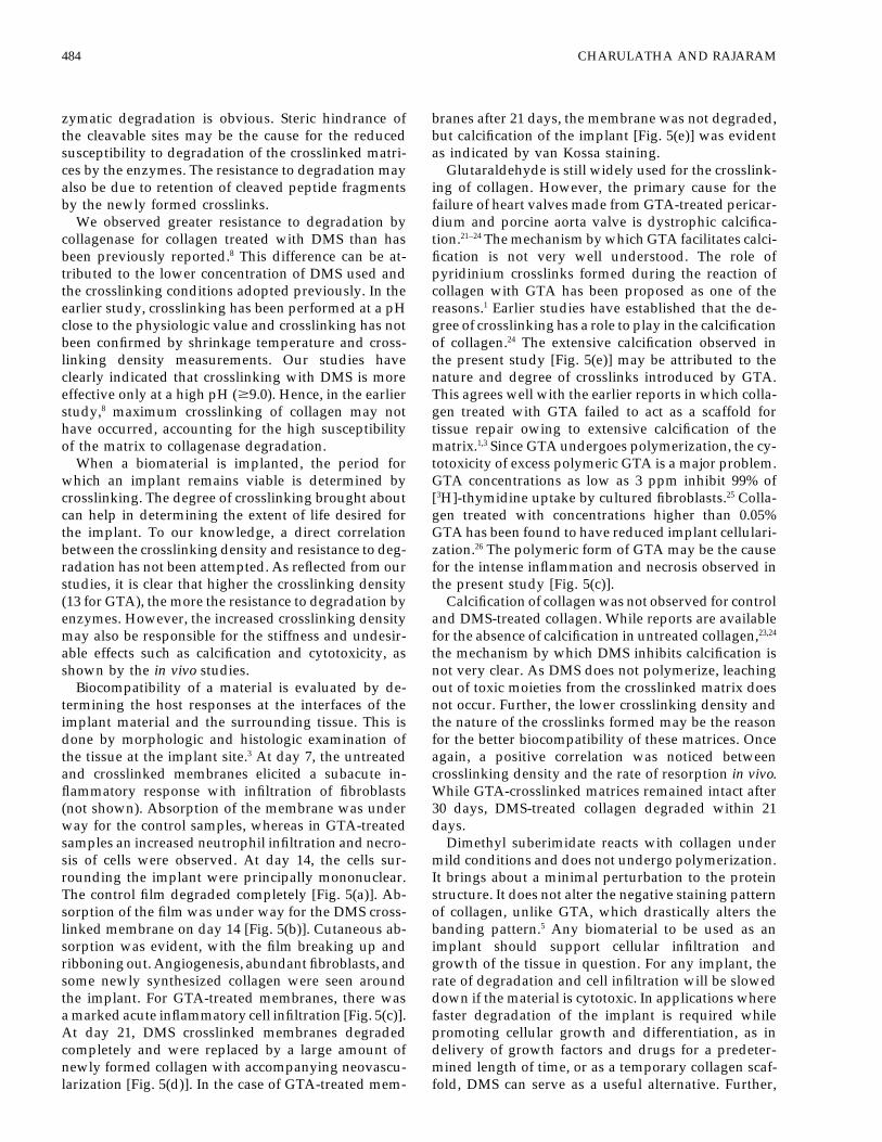
Figure 5. (a) Control collagen (uncrosslinked) membranes 14 days after implantation. The membrane has completelydegraded and the region is densely infiltrated by mainly mononuclear cells with early formation of granulation tissue (H &E, 375). (b) Collagen membrane crosslinked with DMS 14 days after implantation. The membrane is in the process ofdegradation and the cells around the implant are mainly mononuclear. Abundant fibroblasts, neovascularization, and newlyformed collagen are seen (H & E, 3175). (c) Collagen membranes crosslinked with GTA 14 days after implantation. Thereis an acute inflammatory response around the implant (H & E, 3125). (d) Collagen membrane crosslinked with DMS 21days after implantation. The implant has completely degraded and the area is filled with abundant fibroblasts. Neovasculariza-tion and newly formed collagen are also seen (van Gieson, 3150). (e) Collagen membrane crosslinked with GTA 21 daysafter implantation. Calcium deposits are seen on collagen (von Kossa, 3325).
484 CHARULATHA AND RAJARAM
zymatic degradation is obvious. Steric hindrance of branes after 21 days, the membrane was not degraded,but calcification of the implant [Fig. 5(e)] was evidentthe cleavable sites may be the cause for the reduced
susceptibility to degradation of the crosslinked matri- as indicated by van Kossa staining.Glutaraldehyde is still widely used for the crosslink-ces by the enzymes. The resistance to degradation may
also be due to retention of cleaved peptide fragments ing of collagen. However, the primary cause for thefailure of heart valves made from GTA-treated pericar-by the newly formed crosslinks.
We observed greater resistance to degradation by dium and porcine aorta valve is dystrophic calcifica-tion.21–24 The mechanism by which GTA facilitates calci-collagenase for collagen treated with DMS than has
been previously reported.8 This difference can be at- fication is not very well understood. The role ofpyridinium crosslinks formed during the reaction oftributed to the lower concentration of DMS used and
the crosslinking conditions adopted previously. In the collagen with GTA has been proposed as one of thereasons.1 Earlier studies have established that the de-earlier study, crosslinking has been performed at a pH
close to the physiologic value and crosslinking has not gree of crosslinking has a role to play in the calcificationof collagen.24 The extensive calcification observed inbeen confirmed by shrinkage temperature and cross-
linking density measurements. Our studies have the present study [Fig. 5(e)] may be attributed to thenature and degree of crosslinks introduced by GTA.clearly indicated that crosslinking with DMS is more
effective only at a high pH ($9.0). Hence, in the earlier This agrees well with the earlier reports in which colla-gen treated with GTA failed to act as a scaffold forstudy,8 maximum crosslinking of collagen may not
have occurred, accounting for the high susceptibility tissue repair owing to extensive calcification of thematrix.1,3 Since GTA undergoes polymerization, the cy-of the matrix to collagenase degradation.
When a biomaterial is implanted, the period for totoxicity of excess polymeric GTA is a major problem.GTA concentrations as low as 3 ppm inhibit 99% ofwhich an implant remains viable is determined by
crosslinking. The degree of crosslinking brought about [3H]-thymidine uptake by cultured fibroblasts.25 Colla-gen treated with concentrations higher than 0.05%can help in determining the extent of life desired for
the implant. To our knowledge, a direct correlation GTA has been found to have reduced implant cellulari-zation.26 The polymeric form of GTA may be the causebetween the crosslinking density and resistance to deg-
radation has not been attempted. As reflected from our for the intense inflammation and necrosis observed inthe present study [Fig. 5(c)].studies, it is clear that higher the crosslinking density
(13 for GTA), the more the resistance to degradation by Calcification of collagen was not observed for controland DMS-treated collagen. While reports are availableenzymes. However, the increased crosslinking density
may also be responsible for the stiffness and undesir- for the absence of calcification in untreated collagen,23,24
the mechanism by which DMS inhibits calcification isable effects such as calcification and cytotoxicity, asshown by the in vivo studies. not very clear. As DMS does not polymerize, leaching
out of toxic moieties from the crosslinked matrix doesBiocompatibility of a material is evaluated by de-termining the host responses at the interfaces of the not occur. Further, the lower crosslinking density and
the nature of the crosslinks formed may be the reasonimplant material and the surrounding tissue. This isdone by morphologic and histologic examination of for the better biocompatibility of these matrices. Once
again, a positive correlation was noticed betweenthe tissue at the implant site.3 At day 7, the untreatedand crosslinked membranes elicited a subacute in- crosslinking density and the rate of resorption in vivo.
While GTA-crosslinked matrices remained intact afterflammatory response with infiltration of fibroblasts(not shown). Absorption of the membrane was under 30 days, DMS-treated collagen degraded within 21
days.way for the control samples, whereas in GTA-treatedsamples an increased neutrophil infiltration and necro- Dimethyl suberimidate reacts with collagen under
mild conditions and does not undergo polymerization.sis of cells were observed. At day 14, the cells sur-rounding the implant were principally mononuclear. It brings about a minimal perturbation to the protein
structure. It does not alter the negative staining patternThe control film degraded completely [Fig. 5(a)]. Ab-sorption of the film was under way for the DMS cross- of collagen, unlike GTA, which drastically alters the
banding pattern.5 Any biomaterial to be used as anlinked membrane on day 14 [Fig. 5(b)]. Cutaneous ab-sorption was evident, with the film breaking up and implant should support cellular infiltration and
growth of the tissue in question. For any implant, theribboning out. Angiogenesis, abundant fibroblasts, andsome newly synthesized collagen were seen around rate of degradation and cell infiltration will be slowed
down if the material is cytotoxic. In applications wherethe implant. For GTA-treated membranes, there wasa marked acute inflammatory cell infiltration [Fig. 5(c)]. faster degradation of the implant is required while
promoting cellular growth and differentiation, as inAt day 21, DMS crosslinked membranes degradedcompletely and were replaced by a large amount of delivery of growth factors and drugs for a predeter-
mined length of time, or as a temporary collagen scaf-newly formed collagen with accompanying neovascu-larization [Fig. 5(d)]. In the case of GTA-treated mem- fold, DMS can serve as a useful alternative. Further,

DIMETHYL SUBERIMIDATE-TREATED COLLAGEN 485
5. J. A. Chapman, M. Tzaphlidou, K. Meek, and K. E.our work also shows the importance of estimating theKadler, ‘‘The collagen fibril—a model system for study-crosslinking density, as a quantitative aid in determin-ing the staining and fixation of a protein,’’ Electron.ing the implant stability and compatibility. Micro. Rev., 3, 143–182 (1990).
6. G. E. Davies and G. R. Stark, ‘‘Use of dimethyl suberimi-date in studying the subunit structure of oligomericproteins,’’ Proc. Natl. Acad. Sci. USA, 66, 651–656(1970).CONCLUSIONS
7. K. B. Hey, M. C. Lachs, M. J. Raxworthy, and E. J. Wood,‘‘Crosslinked fibrous collagen for use as a dermal im-plant: Control of cytotoxic effects of glutaraldehyde andThe crosslinking density of the DMS- and GTA- dimethyl suberimidate,’’ Biotech. Appl. Biochem., 12,
treated collagen membranes was determined. It is 85–93 (1990).shown quantitatively that GTA treatment introduces 8. D. M. Simmons and J. N. Kearny, ‘‘Evaluation of
collagen crosslinking techniques for the stabilisationa larger number of crosslinks than DMS. Treatmentof tissue matrices,’’ Biotech. Appl. Biochem., 17, 23–29with DMS and GTA increases the thermal stability of(1993).the collagen membranes, as indicated by the increase
9. C. W. Cater, ‘‘The evaluation of aldehydes and otherin TS. Crosslinking with both GTA and DMS increases difunctional compounds as crosslinking agents forthe intermolecular crosslinking, as shown by isometric collagen,’’ J. Soc. Leather Trades Chem., 47, 269–271
(1963).tension measurements. Crosslinking with DMS and10. J. C. Allain, M. Le Lous, S. Bazin, A. J. Bailey, and A.GTA is also confirmed by their increased resistance to
Delaunay, ‘‘Isometric tension developed during healingin vitro degradation by enzymes (collagenase, pepsin,of collagenous tissues. Relationship with collagen cross-
and trypsin). DMS-crosslinked membranes seem to be linking,’’ Biochem. Biophy. Acta, 533, 147–155 (1978).more biocompatible than GTA-treated membranes, as 11. A. F. S. A. Habeeb, ‘‘Determination of free amino
groups in proteins by trinitrobenzenesulfonic acid,’’revealed by subcutaneous implantation studies. UnlikeAnal. Biochem., 14, 328–336 (1966).GTA, which induced necrosis and calcification, DMS-
12. J. F. Woesner, Jr., ‘‘The determination of hydroxyprolinecrosslinked membranes promoted fibroblast migrationin tissue and protein samples containing small propor-and proliferation and acted as a scaffold for tissue tions of this imino acid,’’ Arch. Biochem. Biophys., 93,
regeneration. Finally crosslinking density and isomet- 440–447 (1961).ric tension measurements can serve as useful tech- 13. A. M. Diamond, S. D. Gorham, D. J. Etherington, J. G.
Robertson, and N. D. Light, ‘‘The effect of modificationniques for assessing the efficacy of a crosslinking agenton the susceptibility of collagen to proteolysis: 1. Chemi-for collagen matrices.cal modification of amino acid side chains,’’ Matrix, 11,321–329 (1991).
The authors are thankful to the Director, Central Leather 14. S. Srivastava, S. D. Gorham, D. A. French, A. A. Shivas,and J. M. Courtney, ‘‘In vivo evaluation and comparisonResearch Institute, for the facilities provided to carry out thisof collagen, acetylated collagen and collagen/glycos-work. The financial assistance (Fellowship grant) to one of theaminoglycan composite films and sponges as candidateauthors by the University Grants Commission, New Delhi, isbiomaterials,’’ Biomaterials, 11, 155–161 (1990).gratefully acknowledged. The authors are also grateful to
15. P. J. Flory and J. Rehner, ‘‘Statistical mechanics of cross-Dr. Shanti, Department of Pathology, Post Graduate Institutelinked polymer networks,’’ J. Chem. Phys., 11, 512–526of Basic Medical Sciences, for help with the histopathology. (1943).
16. N. M. Weiderhorn and G. V. Reardon, ‘‘Studies con-cerned with the structure of collagen. II. Stress-strain
References behaviour of thermally contracted collagen,’’ J. Polym.Sci., 9, 315–325 (1952).
17. I. V. Yannas, ‘‘Biologically active analogues of the extra-1. M. E. Nimni, D. T. Cheung, B. Strates, K. Kodama, andcellular matrix: Artificial skin and nerves,’’ Angew.K. Sheik, ‘‘Bioprosthesis derived from crosslinking andChem. Int. Ed. Engl., 29, 20–35 (1990).chemically modified collagenous tissues,’’ in Collagen:
18. A. J. Bailey and A. D. Lister, ‘‘Thermally labile crosslinksBiotechnology, Vol. III, M. E. Nimni (ed.), CRC Press,in native collagen,’’ Nature, 220, 280–281 (1968).Boca Raton, FL, 1988, pp. 1–38.
19. J. G. Gavilanes, G. Gonzalez De Buitrago, M. A. Li-2. J. M. McPherson, P. W. Ledger, S. Sawamura, A. Conti,zarbe, A. M. Municio, and N. Olmo, ‘‘Stabilisation ofS. Wade, H. Reihanian, and D. G. Wallace, ‘‘The prepa-pericadial tissue by glutaraldehyde,’’ Conn. Tissue Res.,ration and physicochemical characterisation of an in-13, 37–44 (1984).jectable form of reconstituted, GTA crosslinked, bovine
20. H. Petite, I. Rault, A. Huc, P. Menasche, and D. Herbage,corium collagen,’’ J. Biomed. Mater. Res., 20, 79–82 (1986).‘‘Use of acyl azide method for crosslinking collagen rich3. P. B. Van Wachem, M. J. A. Van Luyn, L. H. H. Oldetissue such as pericardium,’’ J. Biomed. Mater. Res., 24,Damink, P. J. Dijkstra, J. Feijen, and P. Nieuwenhuis,179–187 (1961).‘‘Biocompatibility and tissue regenerating capacity of
21. F. J. Schoen, H. Harasaki, K. M. Kim, H. C. Anderson,crosslinked dermal sheep collagen,’’ J. Biomed. Mater.and R. J. Levy, ‘‘Biomaterial associated calcification: Pa-Res., 28, 353–363 (1994).thology, mechanisms and strategy for prevention,’’ J.4. J. N. Gade, J. H. Fellman, and J. P. Bentley, ‘‘The sta-Biomed. Mater. Res., 22A1, 11–36 (1988).bilisation of fibrillar collagen matrices with 3,4-dihy-
droxy phenylalanine,’’ J. Biomed. Mater. Res., 25, 799– 22. D. Hirsch, F. J. Schoen, and R. J. Levy, ‘‘Effect of metallicions and diphosphonates on inhibition of pericardial811 (1991).

486 CHARULATHA AND RAJARAM
bioprosthetic tissue calcification and associated alkaline used in cardiac valve bioprostheses,’’ Am. J. Pathol., 127,122–130 (1987).phosphatase activity,’’ Biomaterials, 14, 371–377 (1993).
23. R. J. Levy, F. J. Schoen, F. S. Sherman, J. Nichols, M. A. 25. D. P. Speer, M. Chvapil, C. D. Eskelson, and U. Judith,‘‘Biological effects of residual GTA in GTA-tanned colla-Hawley, and S. A. Lund, ‘‘Calcification of subcutane-
ously implanted type I collagen sponges. Effect of for- gen biomaterials,’’ J. Biomed. Mater. Res., 14, 753–764(1980).maldehyde and glutaraldehyde pretreatments,’’ Am. J.
Pathol., 122, 71–82 (1986). 26. R. F. Oliver, R. A. Grant, R. W. Cox, and A. Cooke, ‘‘Ef-fect of aldehyde crosslinking on human dermal collagen24. G. Golomb, F. J. Schoen, M. S. Smith, J. Linden, M.
Dixon, and R. J. Levy, ‘‘The role of glutaraldehyde-in- implants in the rat,’’ Br. J. Exp. Pathol., 16, 544–549(1980).duced cross-links in calcification of bovine pericardium