critical care clotting catastrophies - university of …williams/delougheryreview.pdf · critical...
TRANSCRIPT

Crit Care Clin 21 (2005) 531–562
Critical Care Clotting Catastrophies
Thomas G. DeLoughery, MD
Oregon Health & Science University, Hematology L586, 3181 SW Sam Jackson Park Road,
Portland, OR 97201-3098, USA
Most patients in ICU will develop coagulation defects [1–4]. The immediate
priorities are to establish the severity of the coagulation defects, evaluate for life
threatening processes, and initiate therapy.
Initial evaluation
When an ICU patient is found to have a bleeding problem, the initial
assessment should focus on how serious the bleeding is and on the underlying
disorders that led to the ICU admission, on current medications, and on the past
medical history.
Clinical examination should seek first to determine whether the patient is
suffering from a ‘‘structural’’ cause of localized bleeding (ie, bleeding from a
gastric ulcer) or from more generalized bleeding suggesting a systemic coagu-
lation defect. Presence of the latter may be suggested by inspection of instrumen-
tation sites (eg, IV sites, chest tube drainage, or mucosa for bleeding). The digits
should be examined for evidence of emboli or ischemia, which, if present, again
suggest a systemic problem.
Exposure to medicines is a common cause of thrombocytopenia and can aug-
ment coagulation defects [5,6]. All the medicines the patient has received should
be noted on the medication sheets and the family should be quizzed about medi-
cation [7–9] the patient is taking (Table 1 and Box 1).
0749-0704/05/$ – see front matter D 2005 Elsevier Inc. All rights reserved.
doi:10.1016/j.ccc.2005.05.003 criticalcare.theclinics.com
E-mail address: [email protected]
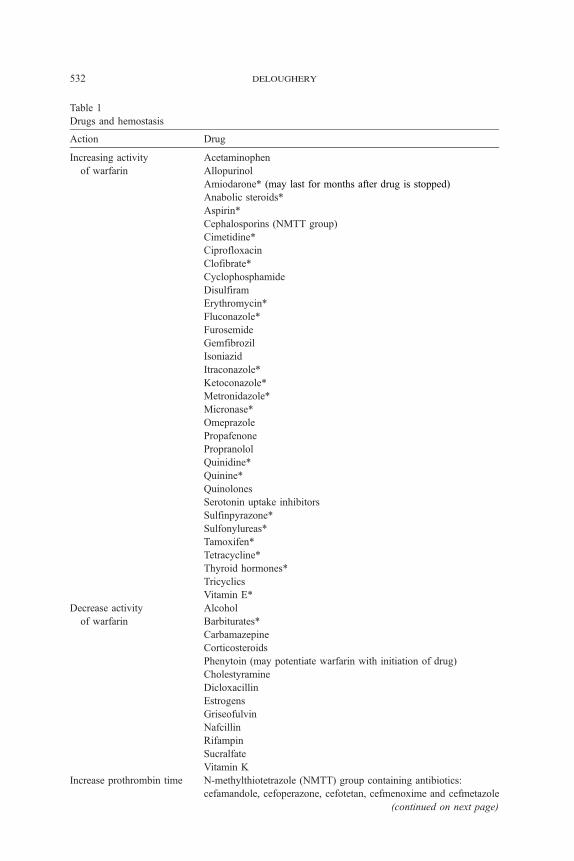
Table 1
Drugs and hemostasis
Action Drug
Increasing activity
of warfarin
Acetaminophen
Allopurinol
Amiodarone* (may last for months after drug is stopped)
Anabolic steroids*
Aspirin*
Cephalosporins (NMTT group)
Cimetidine*
Ciprofloxacin
Clofibrate*
Cyclophosphamide
Disulfiram
Erythromycin*
Fluconazole*
Furosemide
Gemfibrozil
Isoniazid
Itraconazole*
Ketoconazole*
Metronidazole*
Micronase*
Omeprazole
Propafenone
Propranolol
Quinidine*
Quinine*
Quinolones
Serotonin uptake inhibitors
Sulfinpyrazone*
Sulfonylureas*
Tamoxifen*
Tetracycline*
Thyroid hormones*
Tricyclics
Vitamin E*
Decrease activity
of warfarin
Alcohol
Barbiturates*
Carbamazepine
Corticosteroids
Phenytoin (may potentiate warfarin with initiation of drug)
Cholestyramine
Dicloxacillin
Estrogens
Griseofulvin
Nafcillin
Rifampin
Sucralfate
Vitamin K
Increase prothrombin time N-methylthiotetrazole (NMTT) group containing antibiotics:
cefamandole, cefoperazone, cefotetan, cefmenoxime and cefmetazole
(continued on next page)
deloughery532

TTP/HUS Mitomycin C, cyclosporine, FK 506, carbo- or cis-platinum,
ticlopidine, clopidogrel
Hemolysis/DIC syndrome Quinine, 2nd and 3rd generation cephalosporins
Thrombocytopenia See Table 8
* Major effect; bold, Strongest evidence for effect.
Data from: Tiede DJ, Nishimura RA, Gastineau DA, et al. Modern management of prosthetic valve
anticoagulation. Mayo Clin Proc 1998;73:665–80. Hirsh J, Dalen JE, Anderson DR, et al. Oral an-
ticoagulants: mechanisn of action, clinical effectivenss, and optimal theraueutic range. Chest 2001;
119:8S–21S. DeLoughery TG. Anticoagulant therapy in special circumstances. Curr Cardiol Rep
2000;2:74–9.
Table 1 (continued)
Action Drug
clotting catastrophies 533
Laboratories
The first step in laboratory evaluation of the bleeding patient is to obtain a
basic set of coagulation tests consisting of a prothrombin time international
normalized ratio (PT-INR), activated partial thromboplastin time (aPTT), platelet
count, and fibrinogen [10]. Three patterns of defects can be seen in the PT-INR
and aPTT (Box 2). Isolated elevations of the PT-INR are indicative of an isolated
factor VII deficiency. In sick patients, low factor VII levels are common because
of third-spacing and increased consumption [11]. A marked elevation of the
PT-INR out of proportion to the aPTT suggests vitamin K deficiency. Isolated
elevation of the aPTT has many causes. Mixing studies can provide information
to narrow the list of possible diagnoses. Prolongation of both the PT-INR and
aPTT suggest multiple defects or deficiency of factors II, V, or X. As discussed
later, marked prolongation of the PT-INR and aPTT can also be seen with low
levels of fibrinogen. Additional coagulation tests can be ordered based on the
PT-INR and aPTT to better define the defect if the reason for the coagulation
deficiency is not apparent by the history.
If the platelet count is low, examination of the blood smear is essential to make
sure that pseudothrombocytopenia [12] is not present. Examination of the blood
smear is also essential to diagnose microangiopathic processes. Although many
processes can cause a moderately low platelet count, the differential diagnosis
for isolated profound thrombocytopenia (b10,000/uL) is usually limited to im-
mune thrombocytopenia, drug-induced thrombocytopenia, or post-transfusion
purpura (Box 3).
Excessive bleeding has been reported with plasma fibrinogen levels under
50 mg/dL [13]. Fibrinogen is also essential for the proper function of coagu-
lation tests. Low fibrinogen levels reflect either severe liver disease, consump-
tive coagulopathy, or dilution by infusion of massive amounts of resuscitative
fluids. There are some bleeding defects that cannot be detected by routine
laboratory tests. These defects include platelet function defects or increases
in fibrinolysis.

Box 1. Herbal medicines and hemostasis
Possible increase risk of bleeding
Angelica rootHorse chestnutArnica flowerLicorice rootAniseLoavage rootAsafoetidaMeadowseetBogbeanOnionBorage seed oilParsleyBromelainPassionflower herbGinkgoCeleryQuassiaChamomileRed cloverCloveRueRenugreekSweet cloverFeverfewTurmericGarlicWillow barkGingerCapsicumPoplar
Possible increase in warfarin effect
DanshenDong quaiDevil’s clawPapain
deloughery534
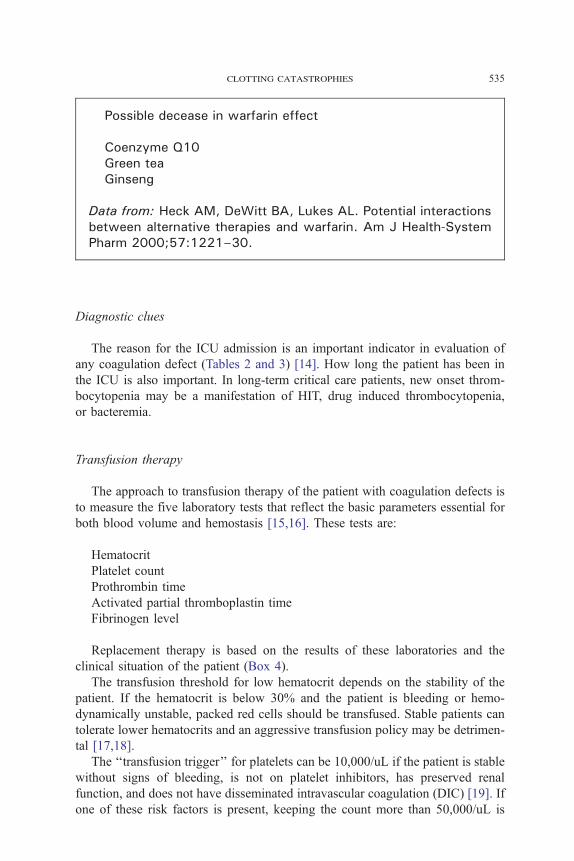
Possible decease in warfarin effect
Coenzyme Q10Green teaGinseng
Data from: Heck AM, DeWitt BA, Lukes AL. Potential interactionsbetween alternative therapies and warfarin. Am J Health-SystemPharm 2000;57:1221–30.
clotting catastrophies 535
Diagnostic clues
The reason for the ICU admission is an important indicator in evaluation of
any coagulation defect (Tables 2 and 3) [14]. How long the patient has been in
the ICU is also important. In long-term critical care patients, new onset throm-
bocytopenia may be a manifestation of HIT, drug induced thrombocytopenia,
or bacteremia.
Transfusion therapy
The approach to transfusion therapy of the patient with coagulation defects is
to measure the five laboratory tests that reflect the basic parameters essential for
both blood volume and hemostasis [15,16]. These tests are:
Hematocrit
Platelet count
Prothrombin time
Activated partial thromboplastin time
Fibrinogen level
Replacement therapy is based on the results of these laboratories and the
clinical situation of the patient (Box 4).
The transfusion threshold for low hematocrit depends on the stability of the
patient. If the hematocrit is below 30% and the patient is bleeding or hemo-
dynamically unstable, packed red cells should be transfused. Stable patients can
tolerate lower hematocrits and an aggressive transfusion policy may be detrimen-
tal [17,18].
The ‘‘transfusion trigger’’ for platelets can be 10,000/uL if the patient is stable
without signs of bleeding, is not on platelet inhibitors, has preserved renal
function, and does not have disseminated intravascular coagulation (DIC) [19]. If
one of these risk factors is present, keeping the count more than 50,000/uL is

Box 2. Interpretations of coagulation tests
Elevated prothrombin time, normal aPTT
Factor VII deficiencyVitamin K deficiencyWarfarinSepsisDIC (occasionally)
Normal prothrombin time, elevated aPTT
Isolated factor deficiency (VIII, IX, XI, XII, contact path-way proteins)
Specific factor inhibitorHeparinLupus inhibitor
Elevated prothrombin time, elevated aPTT
Multiple coagulation factor deficienciesDilutional effectLiver diseaseDisseminated intravascular coagulationIsolated factor X, V or II deficiencyFactor V inhibitorsHigh hematocrits (NNNNNNNNNNN60% - spurious)High heparin levelsSevere vitamin K deficiencyLow fibrinogen (bbbbbbbbbbbbb50 mg/dL)Dysfibrinogemia
deloughery536
reasonable [15,20]. The dose of platelets to be transfused should be 6–8 platelet
concentrates or one plateletpheresis unit.
For a fibrinogen level b100–125 mg/dL, transfusions of 10 units of cryo-
precipitate should increase the plasma fibrinogen level by 100 mg/dl.
In patients with an INR N1.6–2.0 and an abnormal aPTT, fresh frozen plasma
(FFP) is given with the dose, dependent on the aPTT. For an aPTT N1.5 times
normal, 2–4 units of plasma should be given. Elevation of the aPTT N1.8 times
normal is associated with bleeding in trauma patients [13]. Patients with marked
abnormalities such as an aPTT N2 times normal may require aggressive therapy
of at least 15–30ml/kg (4–8 units for an average adult) of plasma [21].

Box 3. Typical platelet counts in various disease states
Moderate thrombocytopenia (50–100,000/uL)
Thrombotic thrombocytopenic purpura (TTP)Heparin induced thrombocytopenia (HIT)Disseminated intravascular coagulation (DIC)Hemophagocytic syndromeLiver disease/ hypersplenism
Severe thrombocytopenia (bbbbbbbbbbbbb20,000/uL)
Drug induced thrombocytopeniaPost-transfusion purpuraImmune thrombocytopenia (ITP)Heparin induced thrombocytopenia (unusual)Thrombotic thrombocytopenic purpura (less common)
clotting catastrophies 537
The basic five laboratory tests should be repeated after administering the blood
products. This insures adequate replacement therapy was given for the coagu-
lation defects. Frequent checks of the coagulation tests also allow rapid iden-
tification and therapy of new coagulation defects in a timely fashion. A flow chart
of the test and the blood products administered should also be maintained.
Table 2
Diagnostic clues to thrombocytopenia
Clinical setting Differential diagnoses
Cardiac surgery Cardiopulmonary bypass, HIT, dilutional thrombocytopenia,
heart valve hemolysis
Interventional cardiac procedure Glycoprotein IIb/IIIa blockers, HIT
Sepsis syndrome DIC, ehrlichiosis, Sepsis hemophagocytosis syndrome,
drug-induced, misdiagnosed TTP, mechanical ventilation,
pulmonary artery catheters
Pulmonary failure DIC, hantavirus pulmonary syndrome, mechanical ventilation,
pulmonary artery catheters
Mental status changes/seizures TTP, ehrlichiosis
Renal failure TTP, dengue, HIT, DIC
Cardiac failure HIT, drug induced, pulmonary artery catheter, heart valve
hemolysis
Post-surgery Dilutional, drug-induced, HIT
Pregnancy HELLP syndrome, fatty liver of pregnancy, TTP/HUS
Acute liver failure Splenic sequestration, HIT, drug induced, DIC
Abbreviations: DIC, disseminated intravascular coagulation; HELLP, hemolysis, elevated liver func-
tion tests, and low platelets; HIT, heparin induced thrombocytopenia; TTP, thrombotic thrombocy-
topenic purpura.

Table 3
Diagnostic clues to coagulation defects
Clinical setting Differential diagnoses
Cardiac surgery Factor V inhibitor, heparin excess or rebound, protamine excess,
fibrinolysis
Sepsis syndrome Isolated factor VII deficiency, DIC, vitamin K deficiency,
Recent use of quinine, 2nd or
3rd generation cephalosporin
Drug induced hemolysis/DIC syndrome
Post-surgery Dilutional, DIC, thrombin inhibitors
Pregnancy HELLP syndrome, fatty liver of pregnancy, vitamin K deficiency
Acute liver failure Consumption, DIC, fibrinolysis, vitamin K deficiency
(biliary obstruction)
Abbreviations: DIC, disseminated intravascular coagulation; HELLP, hemolysis, elevated liver func-
tion tests, and low platelets.
deloughery538
Massive transfusions
The massively transfused patient is defined as one who receives greater
transfused blood than one blood volume in 24 hours or less [22]. A practical
definition is receiving one blood volume in 2 hours or less. The most common
settings for massive transfusion are trauma or gastrointestinal bleeding [23].
Management of blood products is outlined above. The use of a laboratory guided
transfusion protocol has helped to reduce the mortality in patients requiring
massive transfusions [24,25].
Box 4. Massive transfusions
The five basic tests of hemostasis
HematocritPlatelet countProthrombin time (PT-INR)Activated partial thromboplastin time (aPTT)Fibrinogen level
Management guidelines
Platelets bbbbbbbbbbbbb50–75,000/uL: give 1–2 units of apheresis (‘‘singledonor’’) platelets or 6–8 units of whole blood derived(‘‘random donor’’) platelets
Fibrinogen bbbbbbbbbbbbb100–125 mg/dL: give 10 units of cryoprecipitateHematocrit bbbbbbbbbbbbb30%: give red cellsPT-INR NNNNNNNNNNN1.6–2.0 and aPTT abnormal: give 2–4 units of FFP

clotting catastrophies 539
Coagulation defects are common in the massively transfused patients [26].
These can be caused by dilution of the plasma by massive fluid resuscitation
or by red cell transfusions. Packed red cell units contain little plasma (about
25–50 mL/unit), and massive replacement of blood volume with packed red
blood cells can lead to a dilutional coagulopathy. Patients may also develop a
coagulopathy caused by their underlying medical or surgical conditions. Pro-
longed hypotension may be associated with severe ongoing coagulopathy even
after normotension is restored.
It is not possible to predict the degree of coagulopathy from the amount of
blood transfused, and formulaic replacement of factors—give so many units of
plasma for so many units of red cells transfused—should be avoided [27]. Some
patients may receive 20 units of packed red cells and still have good hemostatic
functions; others may have florid coagulopathies caused by injuries before the
first unit of blood is given. Therefore, monitoring the patient’s coagulation status
during massive transfusions is crucial.
Correcting coagulation defects before procedures
A common question is, at what platelet count is it safe to perform invasive
procedures such as central venous line placement? Procedures such as central
venous line placement are frequently done successfully on patients with anti-
coagulation [28–31]. One study found the risk was not related to the degree
of hemostatic defects [32]. In this study, the risk of hemorrhage was higher
when inexperienced operators attempted line placement. For urgent line place-
ment, experience of the operator is more important than to waiting for transfu-
sion therapy [32]. In a non-urgent situation, increasing the platelet count to
30–50,000/uL may be a reasonable goal, a necessary procedure should not be
delayed by trying to achieve an arbitrary platelet count target.
Coagulation defects
Disseminated Intravascular Coagulation
DIC is the clinical manifestation of inappropriate thrombin activation [33–36].
The activation of thrombin leads to (1) fibrinogen conversion to fibrin, (2) plate-
let activation and consumption, (3) activation of factors V and VIII, (4) protein C
activation (and degradation of factors Va and VIIIa), (5) endothelial cell activa-
tion, and (6) fibrinolysis.
Patients with DIC can present in one of four patterns [33,35].
Asymptomatic. Patients can present with laboratory evidence of DIC but no
bleeding or thrombosis. This is often seen in patients with sepsis or cancer.
However, with further progression of the underlying disease, these patients
can rapidly become symptomatic.

deloughery540
Bleeding. The bleeding is caused by combinations of factor depletion, platelet
dysfunction, thrombocytopenia, and excessive fibrinolysis [33]. These
patients may present with diffuse bleeding from multiple sites.
Thrombosis. Despite the general activation of the coagulation process, throm-
bosis is unusual in most patients with acute DIC. The exceptions include
cancer patients, trauma patients, and certain obstetrical patients. Most often
the thrombosis is venous, but arterial thrombosis and non-bacterial throm-
botic endocarditis have been reported [37].
Purpura fulminans. This severe form of DIC is described in more detail later.
The best way to treat DIC is to treat the underlying cause [33,34,36,38].
However, one must replace factors if depletion occurs and bleeding ensues.
Management should be guided by following the basic tests of coagulation.
Heparin therapy is reserved for the patient who has thrombosis as a component
of their DIC [34,39,40]. Reliance on the aPTT to follow heparin therapy may lead
to over- or under-treatment of patients; heparin levels in these patients should be
followed [41,42].
Purpura fulminans
DIC in association with symmetrical limb ecchymosis and necrosis of the skin
is seen in two situations [43]. Primary purpura fulminans is most often seen after
a viral infection [44]. In these patients the purpura fulminans starts with a painful
red area on an extremity that rapidly progresses to a black ischemic area. In many
patients acquired deficiency of protein S is found [43,45,46].
Secondary purpura fulminans is most often associated with meningococcemial
infections but can be seen in any patient with overwhelming infection [47–49].
Post-splenectomy sepsis syndrome patients are also at risk [50]. Patients present
with signs of sepsis and the skin lesions often involve the extremities and may
lead to amputations.
The best therapy for purpura fulminans has not been established. Primary
purpura fulminans, especially those with post-varicella autoimmune protein S
deficiency, has responded to plasma infusion titrated to keep the protein S level
more than 25% [43]. Intravenous immune globulin has also been reported to
help decrease the anti-protein S antibodies. Heparin therapy may control the
DIC and limit the extent of necrosis [51]. The starting dose in these patients is
5–8 units/kg/hr [34].
Patients with secondary purpura fulminans have been treated with plasma
drips, plasmapheresis, and continuous plasma ultrafiltration [51–54]. Heparin
therapy alone has not been shown to improve survival [55]. Much attention has
been given to replacement of natural anticoagulants such as protein C and anti-
thrombin as therapy for purpura fulminans, but unfortunately randomized trials
using antithrombin have shown mostly negative results [43,46,56–58]. Trials
using either zymogen protein C concentrates or recombinant activated protein C
(rAPC) have shown more promise in controlling the coagulopathy of purpura

Box 5. Treatment of purpura fulminans
Drotrecogin 24mcg/kg/hr for 96 hoursBlood product support to maintain
PT-INR bbbbbbbbbbbbb2aPTT bbbbbbbbbbbbb1.8 times normal (drotrecogin will raise aPTT by
5–7 seconds)Platelets NNNNNNNNNNN50,000/uL
Consider continuous veno-venohemofiltration
clotting catastrophies 541
fulminans and improving outcomes in sepsis [52,59–61]. Although bleeding is a
concern with use of protein C, most complications occur in patients with platelet
counts under 30,000/uL or in those who have meningitis [62]. If rAPC is used,
other parameters of coagulation should be carefully monitored (Box 5).
Drug induced hemolytic-disseminated intravascular coagulation syndromes
A severe variant of drug-induced immune complex hemolysis associated with
DIC has been recognized, most commonly to cephalosporins or to quinidine.
Rare patients who receive certain second and third generation cephalosporins,
especially cefotetan and ceftriaxone, have developed this syndrome [63–67].
The clinical syndrome of severe Coombs positive hemolysis, hypotension, and
DIC starts 7 to 10 days after receiving the drug. Often the patient has only re-
ceived the antibiotic for surgical prophylaxis, is believed to have sepsis, and is re-
exposed to the offending cephalosporin, resulting in worsening of the clinical
picture. The outcome is often fatal because of massive hemolysis and thrombo-
sis [66,68–70]. Quinine is associated with a unique syndrome of drug-induced
DIC [71–74]. Approximately 24–96 hours after quinine exposure, the patient be-
comes acutely ill with nausea and vomiting. The patient then develops a micro-
angiopathic hemolytic anemia, DIC, and renal failure. Some patients, besides
having antiplatelet antibodies, also have antibodies binding to red cells and
neutrophils that may lead to the more severe syndrome. Despite therapy, patients
with quinine-induced thrombotic thrombocytopenic purpura (TTP) have a high
incidence of chronic renal failure.
Evidence for treatment of the drug induced hemolytic-DIC syndrome is anec-
dotal. Patients have responded to aggressive therapy including plasma exchange,
dialysis, and prednisone. Early recognition of the hemolytic anemia, and the sus-
picion it is drug related is important for early diagnosis so that the incriminating
drug can be discontinued.
Vitamin K deficiency
Vitamin K is crucial in the synthesis of coagulation factors II, VII, IX, and X.
Patients obtain vitamin K from food sources and from of intestinal flora. Despite

deloughery542
being a fat soluble vitamin, body stores of vitamin K are low and the daily
requirement is 40–80 mcg/d.
Vitamin K deficiency can present dramatically [75]. Once the body stores of
vitamin K are depleted, production of the vitamin K-dependent proteins ceases
and the INR will increase rapidly to high levels. The diagnosis is suspected when
there is a history of prolonged antibiotic use, biliary obstruction, or pre-existing
malnourishment [75–77].
Treatment (and a diagnostic test of vitamin K deficiency) is by replacement of
vitamin K. Most patients will respond rapidly to 10 mg orally. For a more rapid
(4–6 hours) and reliable response, 5–10 mg may be given over 30–60 minutes
intravenously. Alternatively, plasma can be used for the patient with life or limb
threatening bleeding and marked elevation of the PT-INR. At least 4 units of
plasma may be needed until the administered vitamin K takes effect.
Thrombocytopenia and platelet dysfunction
Heparin induced thrombocytopenia
Heparin induced thrombocytopenia (HIT) occurs because of the formation of
antibodies directed against the complex of heparin that is bound to platelet fac-
tor IV [78–84]. Despite the presence of thrombocytopenia, thrombosis and not
bleeding is the major clinical problem. The frequency of HIT is 1%–5% when
unfractionated heparin is used but b1% with low molecular weight heparin [85].
HIT should be suspected when there is a sudden onset of thrombocytopenia with
either at least a 50% drop in the platelet count or the platelet count falling to
b100,000/uL in a patient receiving heparin in any form. HIT usually occurs 4 days
after starting heparin but may occur suddenly in patients with recent (b3 months)
exposure [86–88]. An often overlooked presentation of HIT is recurrent throm-
bosis in a patient receiving heparin who has a platelet count which has fallen but
is still in the ‘‘normal range’’ [89].
The diagnosis of HIT can be challenging in the critical care patient who has
multiple reasons for being thrombocytopenic. In this situation a positive labo-
ratory assay for HIT may be helpful. Two general types of HIT assays exist. The
first type is the functional assays. These use patient plasma, normal platelets,
and varying concentrations of heparin. Heparin-dependent platelet activation
at therapeutic heparin concentrations constitutes a positive assay. Functional as-
says include the 14-C serotonin release assay, lumiaggregometry, and heparin-
dependent platelet aggregation assays. These tests are technically demanding
(particularly the serotonin–release and lumiaggregometry, which use washed
donor platelets) but if performed carefully are both sensitive and specific for HIT
[86,90]. One caveat is that early in HIT, functional assays can be negative be-
cause of low antibody titers, but then turn positive 24 hours later as the antibody
titer increases. Retesting if the initial assay is negative or indeterminate is rec-

clotting catastrophies 543
ommended in this clinical context. The second type of HIT assays is the platelet
calcium/heparin antibody ELISA assays. These detect the presumptively patho-
genic HIT antibodies. Unfortunately, the PF4/heparin antibody response is poly-
clonal, and only a subset of these antibodies cause clinical HIT. Therefore, the
ELISAs tend to be too sensitive in many patient populations at risk for HIT. For
example, 25%–50% of reoperative cardiac patients will be positive [91,92] for
PF4/heparin antibodies when tested by ELISA, and most of these will be false
positives. HIT can also be caused by other types of antibodies and some of the
HIT ELISAs can be negative in up to 20% of HIT cases because of non anti-
platelet calcium antibodies [93,94]. These problems make HIT ELISA assays
difficult to rely upon for definitive clinical diagnosis of HIT.
The first step in therapy of HIT consists of stopping all heparin. Low mo-
lecular weight heparins cross-react with the HIT antibodies and, therefore,
these agents are also contraindicated [86]. Institution of warfarin therapy alone
has been associated with an increased risk of thromboses [86] and patients with
acute HIT should only be warfarinized after complete recovery of the platelet
count, and then only under coverage with another antithrombotic agent. For im-
mediate therapy of HIT patients, three new antithrombotic agents are available
[95,96] (Box 6).
Argatroban is a synthetic thrombin inhibitor [97–99] with a short half-life of
40–50 minutes. Dosing is 2 mcg/kg/min with the infusion adjusted to keep the
aPTT 1.5–3 times normal. One advantage of argatroban is that it is not renally
excreted and no dose adjustment is necessary in renal failure [100]. These char-
acteristics make it the most useful agent for patients in the critical care unit.
However, argatroban must be used with caution in patients with severe liver
disease with an initial dose of 0.5 mcg/kg/min and titrated upward [99]. Also it is
prudent to start at 1 mcg/kg/min in patients with multiorgan system failure
[101]. Argatroban (like all thrombin inhibitors) prolongs the PT-INR making
transition to warfarin therapy difficult as the PT-INR will be prolonged on
argatroban alone, and further prolongation will not reliably reflect the degree of
anticoagulation with warfarin. If available, a chromogenic factor X assay can be
used to adjust warfarin therapy [102]. Chromogenic factor X levels of 0.2 to 0.3
normally correspond to therapeutic PT-INRs of 2.0–3.0 once the argatroban is
stopped. If a chromogenic factor X is not available, and if the patient is on a drip
of 2 mcg/kg/min or less, simply aim for a PT-INR of N4.0 as indicative that
therapeutic anticoagulation on warfarin has been achieved before stopping the
argatroban. Unfortunately there is no agent that can reverse argatroban.
Lepirudin, another direct inhibitor of thrombin, is also monitored by using
the commonly available aPTT. The half-life of lepirudin is short, but the drug
accumulates in renal insufficiency with the half-life increasing to N50 hours.
There is no antidote for lepirudin. Patients with even slight renal insufficiency
(creatinine N1.5) must have their lepirudin doses adjusted to avoid over-
anticoagulation [103]. Up to 80% of patients receiving long-term lepirudin
therapy will develop antibodies [104,105]. These antibodies reduce the me-
tabolism of hirudin and increase the therapeutic effect of lepirudin. Patients on

Box 6. Treatment of heparin induced thrombocytopenia
Argatroban
Therapy: 2 mcg/kg/min infusion with dose adjustments to keepaPTT 1.5–3 times normal. Decrease dose to 0.5 mcg/kg/min insevere liver disease
Hirudin
Therapy: bolus of 0.4 mg/kg followed by 0.15 mg/kg/hr tomaintain an aPTT of 1.5–3.0 times normal
For creatine of 1.6–2.0 mg/dL: bolus of 0.2 mg/kg followed by a50% reduction in infusion rate
For creatine of 2.0–2.5: bolus of 0.2 mg/kg followed by a 75%reduction in infusion rate
For creatine of 2.6–6.0:bolus of 0.2 mg/kg followed by a 90%reduction in infusion rate
For creatine of greater than 6.0 mg/mL: bolus of 0.1 mg/kg onalternate days only when the aPTT is less than 1.5 times normaland no infusion
Fondaparinux
Prophylaxis: 2.5 mg/dTherapy: bbbbbbbbbbbbb50 kg body weight: 5 mg/d50–100 kg body weight: 7.5 mg/dNNNNNNNNNNN100 kg body weight: 10 mg/dUse with caution and monitor by anti-Xa levels in renal in-
sufficiency (Note: monitoring is not often readily available outsideof specialized centers)
Data from: Laposata M, Green D, Van Cott EM, et al. The clinical useand laboratory monitoring of low-molecular-weight heparin, danapa-roid, hirudin and related compounds, and argatroban. Arch PatholLab Med 1998;122:799–807. Hirsh J, Warkentin TE, Raschke R,et al. Heparin and low-molecular-weight heparin: mechanisms ofaction, pharmacokinetics, dosing considerations, monitoring, effi-cacy, and safety. Chest 1998;114:(Suppl 5):10S. Kondo LM,Wittkowsky AK, Wiggins BS. Argatroban for prevention and treat-ment of thromboembolism in heparin-induced thrombocytopenia.Ann Pharmacother 2001;35:440–51. Cook GC, Zumla A, editors.Manson’s tropical diseases. Philadelphia: W.B. Saunders; 2004.
deloughery544

clotting catastrophies 545
long-term (N6 days) lepirudin therapy should still continue to be monitored to
avoid over-anticoagulation.
The new anti-Xa inhibitor fondaparinux does not cross-react with HIT anti-
bodies and may be useful for prophylaxis in HIT and as clinical experience
accumulates for therapy [106].
As mentioned above, initiation of warfarin alone has been associated with
limb gangrene and should not be started as the sole antithrombotic agent in HIT.
In patients receiving specific antithrombin therapy, warfarin can be started with
small doses (2–5 mg). These often malnourished patients tend to have a dramatic
response to warfarin therapy and excessive anticoagulation can easily occur. One
should overlap warfarin and parental therapy by 2–3 days as there is evidence
patients may do worse with shorter specific antithrombin therapy [99].
Patients with HIT but without evidence of thrombosis are at a high risk of
thrombosis (53% in one study) [107] and should be considered for antithrombotic
therapy [108,109]. Patients with HIT should also be carefully screened for any
thrombosis including obtaining lower extremity dopplers. It is unknown whether
prophylactic doses are necessary or if therapeutic doses of anticoagulants are
needed for thrombosis prevention in patients with HIT but no thrombosis. Also,
the duration of such therapy is controversial. One approach is to give prophy-
lactic doses of antithrombotic agents until the platelet count has returned to
normal [109]. In post surgical patients, prolonged prophylaxis for up to 6 weeks
may be of benefit.
Thrombotic thrombocytopenic purpura
TTP should be suspected when a patient presents with the combination of
thrombocytopenia and microangiopathic hemolytic anemia (schistocytes and
signs of hemolysis) [110,111]. Critical care patients with TTP most often present
with intractable seizures, strokes, or sequela of renal insufficiency. Many patients
who present to the critical care unit with TTP will have been misdiagnosed as
having sepsis, lupus cerebritis, or vasculitis.
Evidence is strong that many patients with the classic form of TTP have an
inhibitor against an enzyme that is responsible for cleaving newly synthesized
von Willebrand factor (vWF) [112]. vWF is synthesized as an ultra-large multi-
mer that can spontaneously aggregate platelets. The enzyme, ADAMTS13, is a
protease which cleaves vWF into the smaller forms that normally circulate and
do not spontaneously aggregate platelets [113,114]. Presumably in TTP, inhibi-
tion of ADAMTS13 leads to circulation of ultra-large vWF multimers with re-
sulting spontaneous platelet aggregation leading to the clinical syndrome of TTP.
However, other factors also appear to be involved in the pathogenesis of TTP,
because many patients with classic TTP have normal activity of ADAMTS13,
and reduced levels of the protease are also found in other diseases [115–117].
There is currently no single definitive laboratory test for TTP. Rather the
diagnosis of TTP is based on the clinical presentation [110,111]. Patients uni-
formly will have a microangiopathic hemolytic anemia with the presence of

deloughery546
schistocytes on the peripheral smear. Renal insufficiency rather than frank renal
failure is the most common renal manifestation. Thrombocytopenia may range
from mild decreases in platelet number to platelets being undetectable. The lac-
tate dehydrogenase (LDH) is often extremely elevated and is a prognostic factor
in TTP [118]. Although measurement of ADAMTS13 activity level of immense
research and perhaps prognostic interest, for the reasons discussed above, it is not
of diagnostic value.
Untreated TTP is rapidly fatal. Mortality in the pre-plasma exchange era
ranges from 95% to 100%. Today plasma exchange therapy is the cornerstone of
TTP treatment and has reduced mortality to b20% [111,119–121].
Glucocorticosteroid therapy (ie, 60–120 mg of prednisone) is routinely given
to patients presumed to have TTP. This should be continued until the patient
has fully recovered and perhaps longer, given the presumed autoimmune nature of
the disease and the high relapse rates. Plasma infusion is beneficial [112]. Plasma
exchange has been shown to be superior to simple plasma infusion in therapy of
TTP [119]. This may be because of the ability of plasma exchange to give very
large volumes of fresh frozen plasma, and removal of inhibitory antibodies. In
patients who cannot be immediately exchanged, plasma infusions should be
started at a dose of 1 unit every 4 hours. Patients with all but the mildest cases of
TTP should receive 1.5 plasma volume exchange each day for at least 5 days
[111]. Plasma exchange should be continued daily until the LDH has normalized.
Frequency of exchange should be taped starting with every-other day exchange. If
the platelet count falls or LDH level rises, daily exchange should be reinstated
[110]. Since the platelet count can be affected by a variety of external influences,
the LDH level tends to be the most reliable marker of disease activity [122].
Therapy related thrombotic microangiopathies
TTP/hemolytic uremic syndrome (HUS)-like syndromes or more precisely,
thrombotic microangiopathies, can complicate a variety of therapies [123].
Thrombotic microangiopathies can be associated with medications such as
cyclosporin, FK506, mitomycin, and ticlopidine. Thrombotic microangiopathy
occurs within days after cyclosporine/FK506 is started, with the appearance of
a falling platelet count, falling hematocrit, and rising serum LDH level [124].
Some cases have been fatal but often the thrombotic microangiopathy resolves
when the cyclosporine dose is decreased or changed to another agent.
Thrombotic microangiopathies are most commonly seen when the antineo-
plastic agent mitomycin C is used, and with an incidence of 10% when a dose
of more than 60 mg is used [125]. Anecdotal reports state that treatment with
staphylococcal A columns may be useful for this condition [126]. These columns
work by absorbing immune complexes, but their mechanism in mitomycin
thrombotic microangiopathies is unknown. Since advanced cancer itself can be
associated with a TTP-like syndrome, it may be caused by the cancer and not
the cancer treatment.

clotting catastrophies 547
Thrombotic microangiopathies can complicate both autologous and allogenic
bone marrow transplants [127–129]. The incidence ranges widely depending on
the criteria used to diagnosis the thrombotic microangiopathy, but it is in the
range of 15% for allogeneic and 5% for autologous bone marrow transplants.
Several types of thrombotic microangiopathies are recognized in the bone mar-
row transplantation setting [128,129]. The first is the ‘‘multi-organ fulminant’’
type, which occurs early (20–60 days post transplant), has multi-organ system
involvement, and is often fatal. This type has also been associated with severe
cytomegalo virus (CMV) infection. A second type is similar to the cyclosporin/
FK 506 HUS type described above. A third ‘‘conditioning’’ thrombotic micro-
angiopathy has been described, which occurs 6 months or more after total body
irradiation, and is associated with primary renal involvement. Finally, patients
with systemic CMV infections may present with a thrombotic microangiopathy
related to vascular infection with CMV. The etiology of bone marrow transplant
(BMT)-related thrombotic microangiopathy appears to be different from that of
‘‘classic’’ TTP. Alterations of ADAMTS13 have not been found in BMT-related
TTP; rather therapy-related vascular damage has been implicated as the likely
etiological [130]. Optimal therapy of BMT-related thrombotic microangiopathies
is uncertain. Patients should have their cyclosporine or FK506 doses decreased.
Although plasma exchange is often tried, patients with fulminant or conditioning-
related thrombotic microangiopathies do not normally respond [131,132].
Pregnancy thrombocytopenic syndromes
One should consider three syndromes in the critically ill pregnant woman who
presents with thrombocytopenia. These are the HELLP (Hemolysis, Elevated
Liver tests, Low Platelets) syndrome, fatty liver of pregnancy, and TTP (Table 4)
[133,134].
Table 4
Pregnancy related diseases
Finding HELLP TTP/HUS AFLP
Hypertension Always present Sometimes present Sometimes present
Proteinuria Always present Sometimes present Sometimes present
Thrombocytopenia Always Always Always
LDH elevation Present Marked Present
Fibrinogen Normal to low Normal Normal to very low
Schistocytes Present Present Absent
Liver tests Elevated Normal Elevated
Ammonia Normal Normal Elevated
Glucose Normal Normal Low
Abbreviations: AFLP, acute fatty liver of pregnancy; HELLP, hemolysis, elevated liver tests, and low
platelets; TTP/HUS, thrombotic thrombocytopenic purpura/hemolytic uremia syndrome.
Data from: Egerman RS, Sibai BM. Imitators of preeclampsia and eclampsia. Clin Obstet Gynecol
1999;42:551–62. Esplin MS, Branch DW. Diagnosis and management of thrombotic micro-
angiopathies during pregnancy. Clin Obstet Gynecol 1999;42:360–7.

deloughery548
The acronym HELLP describes a variant of pre-eclampsia [135]. Classically,
HELLP syndrome occurs after 28 weeks of gestation in a patent suffering from
pre-eclampsia but can occur as early as 22 weeks in patients with antiphospho-
lipid antibody syndrome [136–138]. The pre-eclampsia need not be severe. The
first sign is a decrease in the platelet count followed by abnormal liver function
tests. Signs of hemolysis are present with abundant schistocytes on the smear
and a high LDH. HELLP can progress to liver failure and deaths have also been
reported due to hepatic rupture. Unlike TTP, fetal involvement is present in the
HELLP syndrome, and fetal thrombocytopenia has been reported in 30% of cases
[139]. In severe cases, elevated D-dimers consistent with DIC are also found.
Delivery of the child will most often result in cessation of the HELLP syndrome
but refractory cases will require dexamethasone and plasma exchange [140].
Patients should be closely observed for 1–2 days after delivery as the hematologic
picture can transiently worsen before improving [132].
Fatty liver of pregnancy also occurs late in pregnancy and is only associated
with pre-eclampsia in 50% of cases [133,141,142]. Patients first present with
non-specific symptoms of nausea and vomiting but can progress to fulminant
liver failure. Patients develop thrombocytopenia early in the course but in the
later stages can develop DIC and very low fibrinogen levels. Mortality rates
without therapy can be a high as 90%. Low glucose and high ammonia levels can
help distinguish fatty liver from other pregnancy complications [134]. Treatment
consists of prompt delivery of the child and aggressive blood product support.
TTP can occur anytime during pregnancy often leading to diagnostic con-
fusion caused by the overlap symptoms between TTP and HELLP syndrome
[134]. There does appear to be a unique presentation of TTP that occurs in the
second trimester at 20–22 weeks [143]. The fetus is uninvolved with no evidence
of infarction or thrombocytopenia if the mother survives. The pregnancy appears
to promote the TTP since the TTP will resolve with termination of the preg-
nancy and can recur with the next pregnancy [144]. Therapy includes termina-
tion of the pregnancy or attempting to support the patient with plasma exchange
until delivery. Many patients will have relapses with future pregnancies so this
information must be weighed in planning future pregnancies. An unusual com-
plication of pregnancy is a HUS-type syndrome seen up to 28 weeks post-partum.
This form of HUS is severe, and permanent renal failure often results despite
aggressive therapy [144].
Sepsis
Thrombocytopenia is a frequent finding in patients with sepsis syndrome
[145–147]. Classically this has been ascribed to DIC or immune destruction. One
mechanism receiving increasing attention is cytokine-driven hemophagocytosis
of platelets [148–150]. Patients with hemophagocytosis had higher rates of mul-
tiple organ system failure and higher mortality rates. Inflammatory cytokines,
especially monocyte-colony stimulating factor, are thought to be responsible for
inducing the hemophagocytosis [146,151].

clotting catastrophies 549
Thrombocytopenia may be a diagnostic clue to infection with unusual organ-
isms. Three members of the Ehrlichia family of have been reported to cause
infections in humans [152]. They are transmitted by ticks and the diseases that
they produce are similar. Most patients have a febrile illness with high fevers,
headaches and myalgias [152,153]. Patients may have central nervous system
signs and marked elevation of the serum levels of liver enzymes. Rarely patients
may present with a toxic shock-like syndrome [154]. Although many cases are
mild, severe disease is common and the case fatality rate is 2%–5% [153]. The
typical hematologic picture is leukopenia (1300–4000/uL) and mild thrombo-
cytopenia (30–60,000/uL). In many patients the buffy coat reveals the organisms
bundled in a 2–5 um morula in the cytoplasm of the granulocytes or monocytes.
Consideration of ehrlichiosis is important because highly specific therapy is
doxycycline, which is a drug not routinely used for therapy of sepsis syndrome.
Hantavirus pulmonary syndrome (HPS) was described in 1993. Patients suffer
a flu-like prodrome and then rapidly develop a noncardiac pulmonary edema
resulting in profound respiratory failure [155,156]. Ventilatory support is required
in 75% of cases and the mortality is approximately 50%. The peripheral smear
can provide a powerful diagnostic predictor of hantavirus infection [156,157]. In
a recent study, the triad of thrombocytopenia, increased and left-shifted white
cell count, and more than 10% circulating immunoblasts, identified all cases of
HPS and was seen in only 2.6% non-HPS [156]. Marked hemoconcentration is
also present in hantavirus infection caused by capillary leak syndrome, with the
hematocrit reaching as high as 68%.
Viral hemorrhagic fevers are a diverse group of viral infections including
Lassa fever, Rift Valley fever, Ebola, and dengue, that can result in massive
bleeding [158–161] (Table 5). The clinical pattern is a febrile illness that proceeds
over a few days to shock and diffuse gastrointestinal and mucosal bleeding,
with signs of thrombocytopenia and in some cases DIC. Most viral hemorrhagic
fevers are associated with leukopenia and hemoconcentration. Therapy is aggres-
sive supportive care of the patients and replacement of coagulation factors. Pre-
cautions should be taken to prevent nosocomial spread given the propensity of
many of these infections to spread to health care workers [162].
Catastrophic antiphospholipid antibody syndrome
Rarely, patients with antiphospholipid antibody syndrome can present with
fulminant multiorgan system failure [163–165]. Catastrophic antiphospholipid
antibody syndrome (CAPS) is caused by widespread microthrombi in multi-
ple vascular fields. These patients will develop renal failure, encephalopathy,
adult respiratory distress syndrome (often with pulmonary hemorrhage), car-
diac failure, dramatic livedo reticularis, and worsening thrombocytopenia [166].
Many of these patients have pre-existing autoimmune disorders and high titer-
anticardiolipin antibodies. It appears that the best therapy for these patients is
aggressive immunosuppression with plasmapheresis then (perhaps) IV cyclo-

Table 5
Viral hemorrhagic fevers
Arenaviridae Bunyaviridae Filoviridae Flaviviridae
Diseases
Lassa fever, new
world arenaviruses
Crimean-Congo
hemorrhagic virus
(CCHF), Rift Valley
fever, hemorrhagic
fever with renal
syndrome (HFRS)
Ebola, Marburg
Viruses
Dengue, Yellow Fever
Distribution
West Africa (Lassa),
South America
(rare California)
(New world)
Africa, central Asia,
eastern Europe,
Middle East (CCHF),
Africa, Middle East
(Rift), Asia, Balkans,
Europe (HFRS)
Africa Widespread (Dengue),
Africa, Tropical
Americans (Yellow)
Vector
Rodents ticks (CCHF),
mosquitoes
(Rift Valley),
rodents (HFRS)
? Mosquitoes
Incubation
5–16 days 1–6 days (CHHF),
2 weeks to
2 months (HFRS)
2–21 days 3–15 days
Therapy
Ribavirin Ribavirin
Unique clinical Features
pharyngitis, late
deafness (Lassa);
neurological
involvement -
seizures (New world)
retinitis, hepatitis
(Rift Valley),
prominent bleeding
with DIC, jaundice
(CCHF); renal
disease (CCHF)
Maculopapular rash,
high mortality
liver involvement
(Yellow)
Data from: Cook GC, Zumla A, editors. Mansion’s tropical disease. Philadelphia: W.B. Saunders;
2004. Hunter GW, Strickland TG, Magill AJ, Kersey R, editors. Hunter’s tropical medicine and
emerging infectious diseases. 8th Edition. Philadelphia: W.B. Saunders; 2004.
deloughery550
phosphamide monthly. Early recognition of this syndrome can lead to quick
therapy and resolution of multiorgan system failure.
Cardiopulmonary bypass
Cardiopulmonary bypass (CPB) results in very complex and still poorly de-
fined defects in all aspects of hemostasis, including platelets, coagulation factors,
and fibrinolysis [167–173]. If the patient is still in the operating suite and starts to
have microvascular bleeding, the platelet count, PT-INR, aPTT, and fibrinogen
should all be checked and defects corrected appropriately. In particular, patients
who have had multiple transfusions of cell-saver blood or of packed red cells may

clotting catastrophies 551
have dilutional coagulation defects that need to be treated. In the bleeding patient
still on bypass, in whom obvious coagulation defects (thrombocytopenia, pro-
longed PT-INR, low fibrinogen) have been corrected by appropriate transfusion
of platelets, plasma or cryoprecipitate, an infusion of desmopressin (ddAVP) may
be indicated. Once off pump, full heparin reversal by protamine should be
assured. Given the platelet defect induced by CPB, if the patient is still bleeding
after ddAVP, transfusion of platelets is indicated. PT-INR and PTT should also be
verified as being in the normal range. If bleeding still persists despite this one
dose of rVIIa is a logical next step [174].
If bleeding occurs in the postoperative setting, coagulation tests should be
obtained and surgical hemostasis assured. Again, attention should be paid to
the PT-INR, PTT, and fibrinogen levels. Often patients will respond to empiric
transfusions of platelets. In the immediate postoperative state, a heparin level,
if available, or a thrombin time should be checked to insure the patient is not
experiencing ‘‘heparin rebound,’’ particularly if the PTT is prolonged.
Uremia
Before the advent of dialysis, bleeding was a common late complication of
uremia [175–178]. Life threatening bleeding is uncommon but dialysis patients
have a high incidence of gastrointestinal bleeding and subdural hematomas. The
defect in uremia appears to be a platelet function defect [177,179].
Uremic patients who are bleeding should have a PT-aPTT and platelet count
performed. Patients with uremia are prone to vitamin K deficiency so assessment
of the PT is important. The half-life of both unfractionated and low molecular
weight heparin is increased in renal failure. Patients usually receive a bolus of
heparin with dialysis and rare patients will have a persistently prolonged anti-
coagulant effect. Low molecular weight heparins are cleared in the kidneys and
if the dose is not adjusted, levels can greatly increase above therapeutic levels.
Bleeding times are prolonged in renal disease. Unfortunately there is little cor-
relation between prolongation of the bleeding time and actual bleeding, especially
following procedures.
Multiple treatment options exist for uremic bleeding (Box 7). Patients who
are severely uremic and are bleeding may respond to aggressive dialysis [180].
Cryoprecipitate is not consistently effective [180,181].
Desmopressin is effective, and the bleeding time shortens for at least 4 hours
after infusion [182]. Patients can exhibit tachyphylaxis with desmopressin; this
agent is not useful for repeated dosing.
For chronic bleeding, an infusion of conjugated estrogens will shorten the
bleeding time. The dose is 0.6 mg/kg/d intravenously for 5 days. The onset of
action takes up to 1 day but lasts for 2 weeks after the series of infusions [183].
Raising the hematocrit above 30% will shorten the bleeding time in some
situations. This can be done either by transfusion or chronically with the use of
erythropoietin [184]. For purposes of hemostasis, the target hematocrit with the

Box 7. Therapy for uremic bleeding
Acute
Aggressive dialysisDesmopressin (DDAVP) 0.3 mcg/kg IVCryoprecipitate 10 units
Long term
Conjugated estrogen 0.6 mg/kg for 5 daysErythropoietin or darbepoietin to increase hematocrit NNNNNNNNNNN30%
deloughery552
use of erythropoietin should be 27%–30% Uremic patients who present with
severe bleeding may benefit from transfusion of red cells.
Drug-induced thrombocytopenia
In patients with a possible drug-induced thrombocytopenia, the standard ther-
apy is to stop the suspect drug [185,186]. In a patient receiving multiple essential
drugs this is often impractical [3,6]. One approach is to stop any drug started in
the past 7 days that is strongly associated with thrombocytopenia [187] (Box 8).
Substituting sucralfate or a proton-pump inhibitor for H2-blockers is another
option. Unfortunately, critically ill patients are often receiving many therapeutic
agents known (rarely) to cause thrombocytopenia but essential for treatment. In
this situation, one option is to support the patient with platelet transfusions until
the agent can be discontinued.
Immune globulin, corticosteroids, or intravenous anti-D have been suggested
as useful in drug-related thrombocytopenia. However, since most patients with
this type of drug-related thrombocytopenia recover rapidly when the agent is
cleared from the body, such therapies are not normally necessary and should
be avoided in the interest of sparing the patient possible associated side effects
and toxicities.
Drug-induced platelet dysfunction
Sophisticated testing of platelet function has revealed that acquired abnor-
malities are extremely common but the clinical significance, if any, of these ab-
normalities is controversial [188]. Many of these proposed abnormalities are only
reflected in an increased bleeding time, a test of uncertain clinical value [189].
Multiple drugs have been shown to inhibit platelet function, but clinical
bleeding has only been associated with a few. Aspirin has been shown to be
associated with increased risk of bleeding in clinical trials [188]. Ketorolac has

Box 8. Common drugs implicated in thrombocytopenia
Anti GP IIb/IIIa agents
AbciximabEptifibatideTirofiban
Antimicrobial
Amphotericin BEthambutolRifampinTrimethoprim-sulfamethoxzoleVancomycin
Cardiovascular Agents
AmiodaroneAmironeCaptoprilDigoxinMethyldopaProcainamideQuinidine
H2-blockers
CimetidineRanitidine
AcetaminophenCarbazepineGoldHeparinHydrochlorothiazideNon-steroidal antiinflammatory agentsPhenytoinQuinineValproic Acid
Data from: DeLoughery T. Drug induced immune hematological disease.Immunol Allergy Clin N Am 1998;18:829–41. George JN, Raskob GE,Shah SR, et al. Drug-induced thrombocytopenia: a systematic review ofpublished case reports. Ann Intern Med 1998;129:886–90. GreinacherA, Eichler P, Lubenow N, et al. Drug-induced and drug-dependent im-mune thrombocytopenias. Rev Clin Exp Hematol 2001;5:166–200.
clotting catastrophies 553

deloughery554
also been associated with significant clinical bleeding [190–192]. This is es-
pecially true with combined use of ketorolac and heparin or in patients with other
bleeding defects such as von Willebrand disease.
Hydroxyethyl starch (HES) is frequently associated with acquired hemostatic
defects [193]. Bleeding may occur, especially with prolonged use of this agent or
with the use of more than 1.5 L/d. Decreased levels of both vWF and factor VIII
are seen, and many patients will have an acquired type 2 von Willebrand disease
(vWD) defect with selective loss of the higher weight vWF multimers, which
are particularly important in mediating platelet adhesion [194–201]. Levels of
vWF will normalize gradually after the HES is stopped. Patients who have re-
ceived HES and bleed should have a vWD panel drawn. If abnormal, factor re-
placement should be used to correct bleeding. Daily monitoring and therapy may
be necessary for 3–5 days until the defects have fully corrected.
References
[1] Chakraverty R, Davidson S, Peggs K, et al. The incidence and cause of coagulopathies in
an intensive care population. Br J Haematol 1996;93:460–3.
[2] Hanes SD, Quarles DA, Boucher BA. Incidence and risk factors of thrombocytopenia in
critically ill trauma patients. Ann Pharmacother 1997;31:285–9.
[3] Bonfiglio MF, Traeger SM, Kier KL, et al. Thrombocytopenia in intensive care patients:
a comprehensive analysis of risk factors in 314 patients. Ann Pharmacother 1995;29:835–42.
[4] Stephan F, Hollande J, Richard O, et al. Thrombocytopenia in a surgical ICU. Chest 1999;115:
1363–70.
[5] DeLoughery T. Drug induced immune hematological disease. Immunol Allergy Clin N Am
1998;18:829–41.
[6] George JN, Raskob GE, Shah SR, et al. Drug-induced thrombocytopenia: a systematic review
of published case reports. Ann Intern Med 1998;129:886–90.
[7] Heimpel H. When should the clinician suspect a drug-induced blood dyscrasia, and how
should he proceed? Eur J Haematology Supplementum 1996;60:11–5.
[8] Forsyth PD, Davies JM. Pure white cell aplasia and health food products. Postgrad Med J
1995;71:557–8.
[9] Heck AM, DeWitt BA, Lukes AL. Potential interactions between alternative therapies and
warfarin. Am J Health-System Pharm 2000;57:1221–30.
[10] Goodnight SH, Hathaway WE. Evaluation of bleeding in the hospitalized patient. In: Good-
night SH, Hathaway WE, editors. Disorders of hemostasis and thrombosis. 2nd edition. New
York7 McGraw-Hill Companies; 2001. p. 61–9.
[11] Biron C, Bengler C, Gris JC, et al. Acquired isolated factor VII deficiency during sepsis.
Haemostasis 1997;27:51–6.
[12] Bizzaro N. EDTA-dependent pseudothrombocytopenia: a clinical and epidemiological study of
112 cases, with 10-year follow-up. Am J of Hematol 1995;50:103–9.
[13] Ciavarella D, Reed RL, Counts RB, et al. Clotting factor levels and the risk of diffuse
microvascular bleeding in the massively transfused patient. Br J Haematol 1987;67:365–8.
[14] DeLoughery TG. Thrombocytopenia in the critical care patient. In: Alving BM, editor. Blood
components and pharmacologic agents. Bethesda (MD)7 AABB Press; 2001. p. 83–98.
[15] Counts RB, Haisch C, Simon TL, et al. Hemostasis in massively transfused trauma patients.
Ann Surg 1979;190:91–9.
[16] Stainsby D, MacLennan S, Hamilton PJ. Management of massive blood loss: a template
guideline. Br J Anaesth 2000;85:487–91.

clotting catastrophies 555
[17] Hebert PC, Wells G, Blajchman MA, et al. Canadian Critical Care Trials Grp: a multicenter,
randomized, controlled clinical trial of transfusion requirements in critical care. N Engl J Med
1999;340:409–17.
[18] Blair SD, Janvrin SB, McCollum CN, et al. Effect of early blood transfusion on gastrointestinal
haemorrhage. Br J Surg 1986;73:783–5.
[19] Rebulla P, Finazzi G, Marangoni F, et al. Grp Italiano Malattie Ematologiche Mal: the threshold
for prophylactic platelet transfusions in adults with acute myeloid leukemia. N Engl J Med
1997;337:1870–5.
[20] Miller RD, Robbins TO, Tong MJ, et al. Coagulation defects associated with massive blood
transfusions. Ann Surg 1971;174:794–801.
[21] Chowdhury P, Saayman AG, Paulus U, et al. Efficacy of standard dose and 30 ml/kg fresh
frozen plasma in correcting laboratory parameters of haemostasis in critically ill patients. Br J
Haematol 2004;125:69–73.
[22] Hiippala S. Replacement of massive blood loss. Vox Sang 1998;74(Suppl 2):399–407.
[23] Sawyer PR, Harrison CR. Massive transfusion in adults. Diagnoses, survival and blood
bank support. Vox Sang 1990;58:199–203.
[24] Cinat ME, Wallace WC, Nastanski F, et al. Improved survival following massive transfusion
in patients who have undergone trauma. Arch Surg 1999;134:964–8.
[25] Faringer PD, Mullins RJ, Johnson RL, et al. Blood component supplementation during mas-
sive transfusion of AS- 1 red cells in trauma patients. J Trauma 1993;34:481–5 [discus-
sion 485–7].
[26] Leslie SD, Toy PTCY. Laboratory hemostatic abnormalities in massively transfused patients
given red blood cells and crystalloid. Am J Clin Pathol 1991;96:770–3.
[27] Harvey MP, Greenfield TP, Sugrue ME, et al. Massive blood transfusion in a tertiary referral
hospital. Clinical outcomes and haemostatic complications. Med J Aust 1995;163:356–9.
[28] Goldfarb G, Lebrec D. Percutaneous cannulation of the internal jugular vein in patients with
coagulopathies: an experience based on 1,000 attempts. Anesthesiology 1982;56:321–3.
[29] Foster PF, Moore LR, Sankary HN, et al. Central venous catheterization in patients with
coagulopathy. Arch Surg 1992;127:273–5.
[30] VanDervort A, Kopec I, Groeger J, et al. Venous access hemorrhage in critically ill cancer
patients. Chest 1987;92:118S.
[31] Fisher NC, Mutimer DJ. Central venous cannulation in patients with liver disease and
coagulopathy–a prospective audit. Intensive Care Med 1999;25:481–5.
[32] DeLoughery TG, Liebler JM, Simonds V, et al. Invasive line placement in critically ill patients:
Do hemostatic defects matter. Transfusion 1996;36:827–31.
[33] Carey MJ, Rodgers GM. Disseminated intravascular coagulation: Clinical and laboratory
aspects. Am J Hematol 1998;59:65–73.
[34] De Jonge E, Levi M, Stoutenbeek CP, et al. Current drug treatment strategies for dissemi-
nated intravascular coagulation. Drugs 1998;55:767–77.
[35] Baker Jr WF. Clinical aspects of disseminated intravascular coagulation: a clinician’s point
of view. Semin Thromb Haemost 1989;15:1–57.
[36] Levi M, ten Cate H. Disseminated intravascular coagulation. N Engl J Med 1999;341:586–92.
[37] Sharma S, Mayberry JC, DeLoughery TG, et al. Fatal cerebroembolism from nonbacterial
thrombotic endocarditis in a trauma patient: case report and review. Mil Med 2000;165:83–5.
[38] Hoffman JN, Faist E. Coagulation inhibitor replacement during sepsis: useless? Critical Care
Medicine 2000;28(Suppl 6):574–6.
[39] Feinstein DI. Diagnosis and management of disseminated intravascular coagulation: the role
of heparin therapy. Blood 1982;60:284–7.
[40] Callander N, Rapaport SI. Trousseau’s syndrome. West J Med 1993;158:364–71.
[41] Brill-Edwards P, Ginsberg JS, Johnston M, et al. Establishing a therapeutic range for heparin
therapy. Ann Intern Med 1993;119:104–9.
[42] Olson JD, Arkin CF, Brandt JT, et al. Laboratory monitoring of unfractionated heparin
therapy. Arch Pathol Lab Med 1998;122:782–98.

deloughery556
[43] Darmstadt GL. Acute infectious purpura fulminans: pathogenesis and medical management.
Pediatr Dermatol 1998;15:169–83.
[44] Spicer TE, Rau JM. Purpura fulminans. Am J Med 1976;61:566–71.
[45] Josephson C, Nuss R, Jacobson L, et al. The varicella-autoantibody syndrome. Pediatr Res
2001;50:345–52.
[46] Smith OP, White B. Infectious purpura fulminans: diagnosis and treatment. Brit J Haem 1999;
104:202–7.
[47] Gamper G, Oschatz E, Herkner H, et al. Sepsis-associated purpura fulminans in adults. Wien
Klin Wochenschr 2001;113:107–12.
[48] Ward KM, Celebi JT, Gmyrek R, et al. Acute infectious purpura fulminans associated with
asplenism or hyposplenism. J Am Acad Dermatol 2002;47:493–6.
[49] Childers BJ, Cobanov B. Acute infectious purpura fulminans: a 15-year retrospective review
of 28 consecutive cases. Am Surg 2003;69:86–90.
[50] Carpenter CT, Kaiser AB. Purpura fulminans in pneumococcal sepsis: case report and review.
Scand J Infect Dis 1997;29:479–83.
[51] Duncan A. New therapies for severe meningococcal disease but better outcomes? Lancet 1997;
350:1565–6.
[52] Smith OP, White B, Vaughan D, et al. Use of protein-C concentrate, heparin, and haemo-
diafiltration in meningococcus-induced purpura fulminans. Lancet 1997;350:1590–3.
[53] Branson HE, Katz J. A structured approach to the management of purpura fulminans. J Nat
Med Assoc 1983;75:821–5.
[54] Nolan J, Sinclair R. Review of management of purpura fulminans and two case reports. Br J
Anaesth 2001;86:581–6.
[55] Manios SG, Kanakoudi F, Maniati E. Fulminant meningococcemia. heparin therapy and sur-
vival rate. Scand J Infect Dis 1971;3:127–33.
[56] Giudici D, Baudo F, Palareti G, et al. Antithrombin replacement in patients with sepsis and
septic shock. Haematologica 1999;84:452–60.
[57] Fourrier F, Jourdain M, Tournoys A. Clinical trial results with antithrombin III in sepsis.
Crit Care Med 2000;28(Suppl 43).
[58] Levi M, De Jonge E, van der PT, et al. Novel approaches to the management of disseminated
intravascular coagulation. Crit Care Med 2000;28(Suppl 4).
[59] Rivard GE, David M, Farrell C, et al. Treatment of purpura fulminans in meningococcemia
with protein C concentrate. J Pediatr 1995;126:646–52.
[60] White B, Livingstone W, Murphy C, et al. An open-label study of the role of adjuvant hemo-
static support with protein C replacement therapy in purpura fulminans-associated meningo-
coccemia. Blood 2000;96:3719–24.
[61] Aoki N, Matsuda T, Saito H, et al. A comparative double-blind randomized trial of acti-
vated protein C and unfractionated heparin in the treatment of disseminated intravascular
coagulation. Int J Hematol 2002;75:540–7.
[62] Taylor FB, Kinasewitz G. Activated protein C in sepsis. J Thromb Haemost 2004;2:708–17.
[63] Garratty G. Immune cytopenia associated with antibiotics. Transfus Med Rev 1993;7:255–67.
[64] Chenoweth CE, Judd WJ, Steiner EA, et al. Cefotetan-induced immune hemolytic anemia.
Clin Infect Dis 1992;15:863–5.
[65] Endoh T, Yagihashi A, Sasaki M, et al. Ceftizoxime-induced hemolysis due to immune
complexes: case report and determination of the epitope responsible for immune complex-
mediated hemolysis. Transfusion 1999;39:306–9.
[66] Garratty G, Nance S, Lloyd M, et al. Fatal immune hemolytic anemia due to cefotetan.
Transfusion 1992;32:269–71.
[67] Arndt PA, Leger RM, Garratty G. Serology of antibodies to second- and third-generation
cephalosporins associated with immune hemolytic anemia and/or positive direct antiglobulin
tests. Transfusion 1999;39:1239–46.
[68] Bernini JC, Mustafa MM, Sutor LJ, et al. Fatal hemolysis induced by ceftriaxone in a child
with sickle cell anemia. J Pediatr 1995;126:813–5

clotting catastrophies 557
[69] Borgna-Pignatti C, Bezzi TM, Reverberi R. Fatal ceftriaxone-induced hemolysis in a child
with acquired immunodeficiency syndrome. Pediatr Infect Dis J 1995;14:1116–7.
[70] Lascari AD, Amyot K. Fatal hemolysis caused by ceftriaxone. J Pediatr 1995;126:816–7.
[71] Gottschall JL, Elliot W, Lianos E, et al. Quinine-induced immune thrombocytopenia associ-
ated with hemolytic uremic syndrome: a new clinical entity. Blood 1991;77:306–10.
[72] Gottschall JL, Neahring B, McFarland JG, et al. Quinine-induced immune thrombocytope-
nia with hemolytic uremic syndrome: clinical and serological findings in nine patients and
review of literature. Am J Hematol 1994;47:283–9.
[73] Crum NF, Gable P. Quinine-induced hemolytic-uremic syndrome. South Med J 2000;93:726–8.
[74] Vesely T, Vesely JN, George JN. Quinine-Induced thrombotic thrombocytopenic purpura - he-
molytic uremic syndrome (TTP-HUS): frequency, clinical features, and long-term outcomes
[abstract]. Blood 2000;96:629a.
[75] Ansell JE, Kumar R, Deykin D. The spectrum of vitamin K deficiency. JAMA 1977;238:40–2.
[76] Alperin JB. Coagulopathy caused by vitamin K deficiency in critically ill, hospitalized patients.
JAMA 1987;258:1916–9.
[77] Pineo GF, Gallus AS, Hirsh J. Unexpected vitamin K deficiency in hospitalized patients.
CMAJ 1973;109:880–3.
[78] McCrae KR, Bussel JB, Mannucci PM, et al. Platelets: an update on diagnosis and manage-
ment of thrombocytopenic disorders. Hematology 2001;282–305.
[79] Nand S, Wong W, Yuen B, et al. Heparin-induced thrombocytopenia with thrombosis:
incidence, analysis of risk factors, and clinical outcomes in 108 consecutive patients treated
at a single institution. Am J Hematol 1997;56:12–6.
[80] Warkentin TE. Heparin-induced thrombocytopenia-pathogenesis, frequency, avoidance and
management. Drug Saf 1997;17:325–41.
[81] Fabris F, Luzzatto G, Stefani PM, et al. Heparin-induced thrombocytopenia. Haematologica
2000;85:72–81.
[82] Warkentin TE. Clinical picture of heparin-induced thrombocytopenia. In: Warkentin TE,
Greinacher A, editors. Heparin-induced thrombocytopenia. New York7 Marcel Dekker; 2000.
p. 43–80.
[83] Greinacher A, Eichler P, Lubenow N, et al. Drug-induced and drug-dependent immune
thrombocytopenias. Rev Clin Exp Hematol 2001;5:166–200.
[84] Merrer J, De JB, Golliot F, et al. Complications of femoral and subclavian venous
catheterization in critically ill patients: a randomized controlled trial. JAMA 2001;286:700–7.
[85] Warkentin TE, Levine MN, Hirsh J, et al. Heparin-induced thrombocytopenia in patients
treated with low-molecular-weight heparin or unfractionated heparin. N Engl J Med 1995;
332:1330–5.
[86] Warkentin TE. Heparin-induced thrombocytopenia: a ten-year retrospective. Annu Rev Med
1999;50:129–47.
[87] Srichaikul T, Nimmannitya S. Haematology in denque and denque haemorrhagic fever. Bail-
lieres Clin Haematol 2000;13:261–76.
[88] Cvachovec K, Horacek M, Vislocky I. A retrospective survey of fibrinolysis as an indicator
of poor outcome after cardiopulmonary bypass and a possible early sign of systemic inflam-
mation syndrome. Eur J Anaesthesiol 2000;17:173–6.
[89] Hach-Wunderle V, Kainer K, Krug B, et al. Heparin-associated thrombosis despite normal
platelet counts. Lancet 1994;344:469–70.
[90] Warkentin TE, Greinacher A. Laboratory testing for heparin-induced thrombocytopenia. In:
Warkentin TE, Greinacher A, editors. Heparin-induced thrombocytopenia. New York7 Marcel
Dekker; 2000. p. 211–44.
[91] Trossaert M, Gaillard A, Commin PL, et al. High incidence of anti-heparin/platelet factor 4
antibodies after cardiopulmonary bypass surgery. Br J Haematol 1998;101:653–5.
[92] Visentin GP, Malik M, Cyganiak KA, et al. Patients treated with unfractionated heparin
during open heart surgery are at high risk to form antibodies reactive with heparin:platelet
factor 4 complexes. J Lab Clin Med 1996;128:376–83.

deloughery558
[93] Fohlen-Walter A, De Maistre E, Mulot A, et al. Does negative heparin-platelet factor 4 enzyme-
linked immunosorbent assay effectively exclude heparin-induced thrombocytopenia? J Thromb
Haemost 2003;1:1844–5.
[94] Amiral J, Marfaing-Koka A, Wolf M, et al. Presence of autoantibodies to interleukin-8 or
neutrophil- activating peptide-2 in patients with heparin-associated thrombocytopenia. Blood
1996;88:410–6.
[95] Laposata M, Green D, Van Cott EM, et al. The clinical use and laboratory monitoring of low-
molecular-weight heparin, danaparoid, hirudin and related compounds, and argatroban. Arch
Pathol Lab Med 1998;122:799–807.
[96] Hirsh J, Warkentin TE, Raschke R, et al. Heparin and low-molecular-weight heparin - Mecha-
nisms of action, pharmacokinetics, dosing considerations, monitoring, efficacy, and safety.
Chest 1998;114(Suppl.):489S–510S.
[97] Hirsh J, Warkentin TE, Raschke R, et al. Heparin and low-molecular-weight heparin:
mechanisms of action, pharmacokinetics, dosing considerations, monitoring, efficacy, and
safety. Chest 1998;114(Suppl 5):10S.
[98] Lusher J, Ingerslev J, Roberts H, et al. Clinical experience with recombinant factor VIIa.
Blood Coag Fibrin 1998;9:119–28.
[99] Kondo LM, Wittkowsky AK, Wiggins BS. Argatroban for prevention and treatment of throm-
boembolism in heparin-induced thrombocytopenia. Ann Pharmacother 2001;35:440–51.
[100] Swan SK, Hursting MJ. The pharmacokinetics and pharmacodynamics of argatroban: effects of
age, gender, and hepatic or renal dysfunction. Pharmacother 2000;20:318–29.
[101] Baghdasarin SB, Singh I, Militello MA, et al. Argatroban dosage in crtically ill patients with
HIT. Blood 2004;104:1779.
[102] Moll S, Ortel TL. Monitoring warfarin therapy in patients with lupus anticoagulants. Annals of
Internal Medicine 1997;127:177–85.
[103] Greinacher A, Janssens U, Berg G, et al. Lepirudin (recombinant hirudin) for parenteral
anticoagulation in patients with heparin-induced thrombocytopenia. Circulation 1999;100:
587–93.
[104] Song X, Huhle G, Wang L, et al. Generation of anti-hirudin antibodies in heparin-induced
thrombocytopenic patients treated with r-hirudin. Circulation 1999;100:1528–32.
[105] Huhle G, Hoffmann U, Song X, et al. Immunologic response to recombinant hirudin in HIT
type II patients during long-term treatment. Brit J Haem 1999;106:195–201.
[106] Bauer KA. Fondaparinux sodium: a selective inhibitor of factor Xa. Am J Health Syst
Pharm 2001;58(Suppl 7).
[107] Warkentin TE, Levine MN, Hirsh J, et al. Heparin-induced thrombocytopenia in patients
treated with low- molecular-weight heparin or unfractionated heparin. N Engl J Med 1995;332:
1330–5.
[108] Warkentin TE. Heparin-induced thrombocytopenia and its treatment. J Thromb Thrombolysis
2000;9:S29–35.
[109] Hirsh J, Warkentin TE, Shaughnessy SG, et al. Heparin and low molecular weight heparin.
Chest 2001;119:64S–94S.
[110] George JN. Thrombotic thrombocytopenic purpura - hemolytic uremic syndrome. Hematology
1998;1998:379–83.
[111] George JN. How I treat patients with thrombotic thrombocytopenic purpura-hemolytic uremic
syndrome. Blood 2000;96:1223–9.
[112] Furlan M, Robles R, Galbusera M, et al. von Willebrand factor-cleaving protease in thrombotic
thrombocytopenic purpura and the hemolytic-uremic syndrome. N Engl J Med 1998;339:
1578–84.
[113] Levy GG, Motto DG, Ginsburg D. ADAMTS13 Turns 3. Blood 2005, in press.
[114] Levy GG, Nichols WC, Lian EC, et al. Mutations in a member of the ADAMTS gene family
cause thrombotic thrombocytopenic purpura. Nature 2001;413:488–94.
[115] Veyradier A, Obert B, Houllier A, et al. Specific von Willebrand factor-cleaving protease in
thrombotic microangiopathies: a study of 111 cases. Blood 2001;98:1765–72.
[116] Peyvandi F, Ferrari S, Lavoretano S, et al. von Willebrand factor cleaving protease

clotting catastrophies 559
(ADAMTS-13) and ADAMTS-13 neutralizing autoantibodies in 100 patients with thrombotic
thrombocytopenic purpura. Br J Haematol 2004;127:433–9.
[117] Vesely SK, George JN, Lammle B, et al. ADAMTS13 activity in thrombotic thrombocytope-
nic purpura-hemolytic uremic syndrome: relation to presenting features and clinical outcomes
in a prospective cohort of 142 patients. Blood 2003;102:60–8.
[118] Patton JF, Manning KR, Case D, et al. Serum lactate dehydrogenase and platelet count pre-
dict survival in thrombotic thrombocytopenic purpura. Am J Hematol 1994;47:94–9.
[119] Rock GA, Shumak KH, Buskard NA, et al. Comparison of plasma exchange with plasma
infusion in the treatment of thrombotic thrombocytopenic purpura. N Engl J Med 1991;
325:393–7.
[120] Bell WR, Braine HG, Ness PM, et al. Improved survival in thrombotic thrombocytopenic
purpura- hemolytic uremic syndrome–clinical experience in 108 patients. N Engl J Med 1991;
325:398–403.
[121] Kaplan BS, Trachtman H. Improve survival with plasma exchange thrombotic thrombopenic
purpura-hemolytic uremic syndrome. Am J Med 2001;110:156–7.
[122] van Genderen PJ, Michiels JJ. Acquired von Willebrand disease. Bail Clin Haem 1998;11:
319–30.
[123] Moake JL, Byrnes JJ. Thrombotic microangiopathies associated with drugs and bone marrow
transplantation. Hematol Oncol Clin North Am 1996;10:485–97.
[124] Gharpure VS, Devine SM, Holland HK, et al. Thrombotic thrombocytopenic purpura asso-
ciated with FK506 following bone marrow transplantation. Bone Marrow Transplant 1995;
16:715–6.
[125] Wu DC, Liu JM, Chen YM, et al. Mitomycin-C induced hemolytic uremic syndrome: a case
report and literature review. Jpn J Clin Oncol 1997;27:115–8.
[126] Borghardt EJ, Kirchertz EJ, Marten I, et al. Protein A-immunoadsorption in chemotherapy
associated hemolytic-uremic syndrome. Transfusion Sci 1998;19(Suppl 7).
[127] Schriber JR, Herzig GP. Transplantation-associated thrombotic thrombocytopenic purpura
and hemolytic uremic syndrome. Semin Hematol 1997;34:126–33.
[128] Clark RE. Thrombotic microangiopathy following bone marrow transplantation. Bone Marrow
Transplant 1994;14:495–504.
[129] Fuge R, Bird JM, Fraser A, et al. The clinical features, risk factors and outcome of throm-
botic thrombocytopenic purpura occurring after bone marrow transplantation. Br J Haematol
2001;113:58–64.
[130] Van der Plas RM, Schiphorst ME, Huizinga EG, et al. von Willebrand factor proteolysis is
deficient in classic, but not in bone marrow transplantation-associated, thrombotic thrombo-
cytopenic purpura. Blood 1999;93:3798–802.
[131] Sarode R, McFarland JG, Flomenberg N, et al. Therapeutic plasma exchange does not appear to
be effective in the management of thrombotic thrombocytopenic purpura/hemolytic uremic
syndrome following bone marrow transplantation. Bone Marrow Transplant 1995;16:271–5.
[132] Magann EF, Martin Jr JN. Twelve steps to optimal management of HELLP syndrome. Clin
Obstet Gynecol 1999;42:532–50.
[133] Rahman TM, Wendon J. Severe hepatic dysfunction in pregnancy. QJM 2002;95:343–57.
[134] Egerman RS, Sibai BM. Imitators of preeclampsia and eclampsia. Clin Obstet Gynecol 1999;
42:551–62.
[135] Baxter JK, Weinstein L. HELLP syndrome: the state of the art. Obstet Gynecol Surv 2004;
59:838–45.
[136] Egerman RS, Sibai BM. HELLP syndrome. Clin Obstet Gynecol 1999;42:381–9.
[137] Saphier CJ, Repke JT. Hemolysis, elevated liver enzymes, and low platelets (HELLP)
syndrome: a review of diagnosis and management. Semin Perinatol 1998;22:118–33.
[138] Le Thi TD, Tieulie N, Costedoat N, et al. The HELLP syndrome in the antiphospholipid syn-
drome: retrospective study of 16 cases in 15 women. Ann Rheum Dis 2005;64:273–8.
[139] Gul A, Cebeci A, Aslan H, et al. Perinatal outcomes in severe preeclampsia-eclampsia with
and without HELLP syndrome. Gynecol Obstet Invest 2005;59:113–8.
[140] Martin Jr JN, Perry Jr KG, Blake PG, et al. Better maternal outcomes are achieved with

deloughery560
dexamethasone therapy for postpartum HELLP (hemolysis, elevated liver enzymes, and
thrombocytopenia) syndrome. Am J Obstet Gynecol 1997;177:1011–7.
[141] Jwayyed SM, Blanda M, Kubina M. Acute fatty liver of pregnancy. J Emerg Med 1999;
17:673–7.
[142] Bacq Y. Acute fatty liver of pregnancy. Semin Perinatol 1998;22:134–40.
[143] Esplin MS, Branch DW. Diagnosis and management of thrombotic microangiopathies during
pregnancy. Clin Obstet Gynecol 1999;42:360–7.
[144] Dashe JS, Ramin SM, Cunningham FG. The long-term consequences of thrombotic micro-
angiopathy (thrombotic thrombocytopenic purpura and hemolytic uremic syndrome) in preg-
nancy. Obstet Gynecol 1998;91:t-8.
[145] Harris RL, Musher DM, Bloom K, et al. Manifestations of sepsis. Arch Intern Med 1987;
147:1895–906.
[146] van Gorp EC, Suharti C, ten Cate H, et al. Review: infectious diseases and coagulation
disorders. J Infect Dis 1999;180:176–86.
[147] Tiab M, Mechinaud F, Harousseau JL. Haemophagocytic syndrome associated with infections.
Baillieres Clin Haematol 2000;13:163–78.
[148] Francois B, Trimoreau F, Vignon P, et al. Thrombocytopenia in the sepsis syndrome: role of
hemophagocytosis and macrophage colony-stimulating factor. Am J Med 1997;103:114–20.
[149] Risdall RJ, Brunning RD, Hernandez JI, et al. Bacteria-associated hemophagocytic syndrome.
Cancer 1984;54:2968–72.
[150] Stephan F, Thioliere B, Verdy E, et al. Role of hemophagocytic histiocytosis in the etiology
of thrombocytopenia in patients with sepsis syndrome or septic shock. Clin Infect Dis 1997;
25:1159–64.
[151] Baker GR, Levin J. Transient thrombocytopenia produced by administration of macrophage
colony-stimulating factor: Investigations of the mechanism. Blood 1998;91:89–99.
[152] Dumler JS, Bakken JS. Human ehrlichiosis: newly recognized infections transmitted by ticks.
Annu Rev Med 1998;49:201–13.
[153] Bakken JS, Krueth J, Wilson-Nordskog C, et al. Clinical and laboratory characteristics of
human granulocytic ehrlichiosis. JAMA 1996;275:199–205.
[154] Fichtenbaum CJ, Peterson LR, Weil GJ. Ehrlichiosis presenting as a life-threatening illness
with features of the toxic shock syndrome [comments]. Am J Med 1993;95:351–7.
[155] Butler JC, Peters CJ. Hantaviruses and hantavirus pulmonary syndrome. Clin Infect Dis
1994;19:387–94.
[156] Mertz GJ, Hjelle BL, Bryan RT. Hantavirus infection. Disease-a-Month 1998;44:89–138.
[157] Nolte KB, Feddersen RM, Foucar K, et al. Hantavirus pulmonary syndrome in the United
States: a pathological description of a disease caused by a new agent. Hum Pathol 1995;26:
110–20.
[158] Barry M. Viral hemmorrhagic fevers. Hematology 2000;2000:414–23.
[159] Schnittler HJ, Feldmann H. Viral hemorrhagic fever-a vascular disease? Thromb Haemost
2003;89:967–72.
[160] Fatal illnesses associated with a new world arenavirus–California, 1999–2000. Morb Mortal
Wkly Rep 2000;49:709–11.
[161] Lupi O, Tyring SK. Tropical dermatology: viral tropical diseases. J Am Acad Dermatol 2003;
49:979–1000.
[162] Casillas AM, Nyamathi AM, Sosa A, et al. A current review of Ebola virus: pathogenesis,
clinical presentation, and diagnostic assessment. Biological Research for Nursing 2003;4:
268–75.
[163] Asherson RA. The catastrophic antiphospholipid syndrome [editorial]. J Rheumatol 1992;19:
508–12.
[164] Asherson RA, Piette JC. The catastrophic antiphospholipid syndrome 1996: acute. Lupus
1996;5:414–7.
[165] Asherson RA, Cervera R. Castastrophic antiphospholipid syndrome. Curr Opin Hematol 2000;
5:325–9.

clotting catastrophies 561
[166] Cervera R, Font J, Gomez-Puerta JA, et al. Validation of the preliminary criteria for the clas-
sification of catastrophic antiphospholipid syndrome. Ann Rheum Dis 2005, in press.
[167] Woodman RC, Harker LA. Bleeding complications associated with cardiopulmonary bypass.
Blood 1990;76:1680–97.
[168] Bevan DH. Cardiac bypass haemostasis: putting blood throught the mill. Br J Haematol
1999;104:219.
[169] Gallimore MJ, Jones DW, Wendel HP. A chromogenic substrate assay kit for factor XII:
evaluation and use for the measurement of factor XII levels in cardiopulmonary bypass pa-
tients. Thromb Res 1999;94:103–9.
[170] Parratt R, Hunt BJ. Direct activation of factor X by monocytes occurs during cardiopulmonary
bypass. Br J Haematol 1998;101:40–6.
[171] Weerasinghe A, Taylor KM. The platelet in cardiopulmonary bypass. Ann Thorac Surg 1998;
66:2145–52.
[172] Hunt BJ, Parratt RN, Segal HC, et al. Activation of coagulation and fibrinolysis during
cardiothoracic operations. Ann Thorac Surg 1998;65:712–8.
[173] Paparella D, Brister SJ, Buchanan MR. Coagulation disorders of cardiopulmonary bypass:
a review. Intensive Care Med 2004;30:1873–81.
[174] Tanaka KA, Waly AA, Cooper WA, et al. Treatment of excessive bleeding in Jehovah’s Witness
patients after cardiac surgery with recombinant factor VIIa (NovoSeven). Anesthesiology
2003;98:1513–5.
[175] Livio M, Benigni A, Remuzzi G. Coagulation abnormalities in uremia. Semin Nephrol 1985;
5:82–90.
[176] Rabelink TJ, Zwaginga JJ, Koomans HA, et al. Thrombosis and hemostasis in renal disease.
Kidney Int 1994;46:287–96.
[177] Weigert AL, Schafer AI. Uremic bleeding: pathogenesis and therapy. Am J Med Sci 1998;
316:94–104.
[178] Sagripanti A, Barsotti G. Bleeding and thrombosis in chronic uremia. Nephron 1997;75:
125–39.
[179] Boccardo P, Remuzzi G, Galbusera M. Platelet dysfunction in renal failure. Semin Thromb
Hemost 2004;30:579–89.
[180] Andrassy K, Ritz E. Uremia as a cause of bleeding. Am J Nephrol 1985;5:313–9.
[181] Triulzi DJ, Blumberg N. Variability in response to cryoprecipitate treatment for hemostatic
defects in uremia. Yale J Biol Med 1990;63:1–7.
[182] Mannucci PM. Desmopressin (DDAVP) in the treatment of bleeding disorders: The first
20 years. Blood 1997;90:2515–21.
[183] Vigano G, Gaspari F, Locatelli M, et al. Dose-effect and pharmacokinetics of estrogens
given to correct bleeding time in uremia. Kidney Int 1988;34:853–8.
[184] Moia M, Mannucci PM, Vizzotto L, et al. Improvement in the haemostatic defect of uraemia
after treatment with recombinant human erythropoietin. Lancet 1987;2:1227–9.
[185] DeShazo RD, Kemp SF. Allergic reactions to drugs and biologic agents. JAMA 1997;278:
1895–906.
[186] Zondor SD, George JN, Medina PJ. Treatment of drug-induced thrombocytopenia. Expert
Opin Drug Saf 2002;1:173–80.
[187] Pedersen-Bjergaard U, Andersen M, Hansen PB. Drug-induced thrombocytopenia: clinical
data on 309 cases and the effect of corticosteroid therapy. Eur J Clin Pharmacol 1997;52:
183–9.
[188] George JN, Shattil SJ. The clinical importance of acquired abnormalities of platelet function.
N Engl J Med 1991;324:27–39.
[189] Lind SE. Prolonged bleeding time. Am J Med 1984;77:305–12.
[190] Strom BL, Berlin JA, Kinman JL, et al. Parenteral ketorolac and risk of gastrointestinal and
operative site bleeding - a postmarketing surveillance study. JAMA 1996;275:376–82.
[191] Bailey R, Sinha C, Burgess LP. Ketorolac tromethamine and hemorrhage in tonsillectomy:
a prospective, randomized, double-blind study. Laryngoscope 1997;107:166–9.

deloughery562
[192] Splinter WM, Rhine EJ, Roberts DW, et al. Preoperative ketorolac increases bleeding after
tonsillectomy in children. Can J Anaesth 1996;43:560–3.
[193] Peterson P, Hayes TE, Arkin CF, et al. The preoperative bleeding time test lacks clinical
benefit. Arch Surg 1998;133:134–9.
[194] Treib J, Haass A, Pindur G, et al. Highly substituted hydroxyethyl starch (HES200/0.62)
leads to Type-I von Willebrand syndrome after repeated administration. Haemostasis 1996;26:
210–3.
[195] Sanfelippo MJ, Suberviola PD, Geimer NF. Development of a von Willebrand-like syndrome
after prolonged use of hydroxyethyl starch. Am J Clin Path 1987;88:653–5.
[196] Tiede DJ, Nishimura RA, Gastineau DA, et al. Modern management of prosthetic valve
anticoagulation. Mayo Clin Proc 1998;73:665–80.
[197] Hirsh J, Dalen JE, Anderson DR, et al. Oral anticoagulants: mechanisn of action, clinical
effectivenss, and optimal theraueutic range. Chest 2001;119:8S–21S.
[198] DeLoughery TG. Anticoagulant therapy in special circumstances. Curr Cardiol Rep 2000;2:
74–9.
[199] Hyers TM, Agnelli G, Hull RD, et al. Antithrombotic therapy for venous thromboembolic
disease. Chest 1998;114(Suppl.):561S–78S.
[200] Cook GC, Zumla A, editors. Manson’s tropical diseases. Philadelphia7 W.B. Saunders; 2004.
[201] Hunter GW, Strickland TG, Magill AJ, Kersey R, editors. Hunter’s tropical medicine and
emerging infectious diseases. 8th Edition. Philadelphia7 W.B. Saunders; 2004.