critical analysis of reference studies on the...
TRANSCRIPT

Accepted Manuscript
Critical analysis of reference studies on the toxicokinetics ofaluminum-based adjuvants
Jean-Daniel Masson, Guillemette Crépeaux, François-JérômeAuthier, Christopher Exley, Romain K. Gherardi
PII: S0162-0134(17)30338-0DOI: https://doi.org/10.1016/j.jinorgbio.2017.12.015Reference: JIB 10402
To appear in: Journal of Inorganic Biochemistry
Received date: 29 May 2017Revised date: 21 December 2017Accepted date: 22 December 2017
Please cite this article as: Jean-Daniel Masson, Guillemette Crépeaux, François-JérômeAuthier, Christopher Exley, Romain K. Gherardi , Critical analysis of reference studieson the toxicokinetics of aluminum-based adjuvants. The address for the correspondingauthor was captured as affiliation for all authors. Please check if appropriate. Jib(2017),https://doi.org/10.1016/j.jinorgbio.2017.12.015
This is a PDF file of an unedited manuscript that has been accepted for publication. Asa service to our customers we are providing this early version of the manuscript. Themanuscript will undergo copyediting, typesetting, and review of the resulting proof beforeit is published in its final form. Please note that during the production process errors maybe discovered which could affect the content, and all legal disclaimers that apply to thejournal pertain.

ACC
EPTE
D M
ANU
SCR
IPT
1
Critical analysis of reference studies on the toxicokinetics of
aluminum-based adjuvants
Jean-Daniel Masson1*
, Guillemette Crépeaux1,2*
, François-Jérôme Authier1, Christopher
Exley3, Romain K Gherardi
1
1 INSERM U955 E10, « Biologie du système neuromusculaire », Faculté de Médecine,
Université Paris Est Créteil, Créteil, 94010, France
2 Génétique médicale comparée des affections neuromusculaires, Ecole Nationale
Vétérinaire d’Alfort, 7 Avenue du général de Gaulle, 9400, Maisons-Alfort, France
3 The Birchall Centre, Lennard-Jones Laboratories, Keele University, Staffordshire,
ST5 5BG, UK
*These authors contribute equally to this work.
Corresponding author:
RK Gherardi, Centre Expert de Pathologie Neuromusculaire, Hôpital Henri Mondor,
94010 Créteil, France
Foot note: This paper has been previously published in French in Annales Pharmaceutiques
Françaises. It is released in English in the Journal of Inorganic Biochemistry with due
permission of the publisher.
ACCEPTED MANUSCRIPT

ACC
EPTE
D M
ANU
SCR
IPT
2
Abstract
We reviewed the three toxicokinetic reference studies commonly used to suggest that
aluminum (Al)-based adjuvants are innocuous. A single experimental study was carried
out using isotopic 26Al (Flarend et al., Vaccine, 1997). This study used aluminum salts
resembling those used in vaccines but ignored adjuvant uptake by cells that was not fully
documented at the time. It was conducted over a short period of time (28 days) and used
only two rabbits per adjuvant. At the endpoint, Al elimination in the urine accounted for
6% for Al hydroxide and 22% for Al phosphate, both results being incompatible with rapid
elimination of vaccine-derived Al in urine. Two theoretical studies have evaluated the
potential risk of vaccine Al in infants, by reference to an oral « minimal risk level » (MRL)
extrapolated from animal studies. Keith et al. (Vaccine, 2002) used a high MRL (2
mg/kg/d), an erroneous model of 100% immediate absorption of vaccine Al, and did not
consider renal and blood-brain barrier immaturity. Mitkus et al. (Vaccine, 2011) only
considered solubilized Al, with erroneous calculations of absorption duration. Systemic Al
particle diffusion and neuro-inflammatory potential were omitted. The MRL they used was
both inappropriate (oral Al vs. injected adjuvant) and still too high (1 mg/kg/d) regarding
recent animal studies. Both paucity and serious weaknesses of reference studies strongly
suggest that novel experimental studies of Al adjuvants toxicokinetics should be
performed on the long-term, including both neonatal and adult exposures, to ensure their
safety and restore population confidence in Al-containing vaccines.
Key words: vaccine adjuvant, aluminum, toxicokinetics, vaccine safety
ACCEPTED MANUSCRIPT

ACC
EPTE
D M
ANU
SCR
IPT
3
1. Introduction
Vaccination helped with the eradication of smallpox, a 99% decline in poliomyelitis
between 1988 and 2003, and a 40% decrease in measles cases between 1999 and 2003
worldwide, as well as a decrease in cases of mumps of 859 to 9 per 100 000 inhabitants
between 1986 and 2013 in France [1]. The maintenance of good vaccination coverage, i.e.
a high rate of vaccinated persons in the population, is necessary to avoid the resurgence of
other infectious diseases, as was observed for pertussis or rubella, with a double benefit,
both individually and collectively, by reducing the number of people who can transmit
infectious diseases [1].
Although the success of many vaccines has been amply demonstrated, a growing public
distrust of vaccination has emerged in recent years. This reluctance, of varying degrees,
appears concomitantly with an expanding global World Health Organization (WHO)
policy for burgeoning vaccination programs with more than 120 new vaccines currently
being developed and an annual growth of 20% of vaccine business is expected, realizing a
turnover which has increased from 5 to 43 billion dollars between 2000 and 2016, and will
be more than 100 billion dollars in 2025 [2].
Unlike conventional medicines, vaccines are administered to healthy subjects that need to
be convinced of their value and safety. In this context, the vaccine issue has become a
major societal issue, leading to the establishment of a national citizen consultation on
vaccination chaired by Alain Fischer [3]. According to the findings of its final report of
30th
of November 2016, several factors contribute to mistrust of vaccination, “especially:
• Suspicions of collusion between health authorities and the drug industry as a result of
mediated scandals;
ACCEPTED MANUSCRIPT

ACC
EPTE
D M
ANU
SCR
IPT
4
• The disappearance of many infectious diseases that question the appropriateness of
continuing vaccination;
• The issue of adjuvants in vaccines;
• The position of doctors who complain of a lack of training to convince reluctant
patients;
• The complexity of the vaccination course (mandatory medical prescription, pharmacy
purchase of the vaccine, medical vaccination, etc.);
• Lack of information from doctors on the immunization status of their patients (health
book lost or not presented);
• Health crises (Mediator, contaminated blood, etc.) and the insufficient responsiveness
of the answer and the commitment of the public authorities which have left the field
open to anti-vaccination propaganda [3].
A key question in the debate on vaccine safety concerns the adjuvants, compounds
essential for strong and lasting immunization [4]. The controversy focuses on the
aluminum salts which were empirically introduced by Alexander Glenny as adjuvants to
vaccines in 1926 [5]. It has resulted in various actions brought by patient associations [6,
7], publication of books for the general public, either critical [8] or reassuring [9],
scientific blogs [10], drafting of institutional technical reports [4, 11-13], and holding of
parliamentary initiative discussion meetings [14, 15]. Although the principle of
vaccination has never been questioned during these exchanges, the exact degree of safety
of aluminum-containing vaccines has remained the subject of persistent disagreement.
The occurrence of myalgia and arthralgia, chronic fatigue and neurological disorders
following multiple injections of aluminum-containing vaccines against hepatitis B, tetanus
and human papilloma virus (HPV) has been reported in many countries: Australia [16],
ACCEPTED MANUSCRIPT

ACC
EPTE
D M
ANU
SCR
IPT
5
Canada [17, 18], Denmark [19, 20], France [21-23], United Kingdom [24, 25], Italy [26],
Israel [27], Japan [28-29], Mexico [30], Portugal [31], and USA [32]. Nevertheless,
beyond the temporal association, the existence of a causal link remains debated. For
vaccination against HPV for example, the risk of occurrence of adverse events, which may
form part of one or more of the clinical entities [19] - chronic fatigue syndrome (CFS),
regional pain syndrome (RPS), orthostatic postural tachycardia syndrome (POTS) –
emerges from an epidemiologic point of view [33]. A systematic cross-sectional study of
12 published studies showed a slight increase of adverse events in the HPV-vaccinated
group, but this information must take account of the quasi-systematic use of control groups
that received aluminum adjuvants in the form of a placebo containing the adjuvant or,
more rarely, the hepatitis A vaccine (11 of the 12 publications analyzed, comprising
29,533 of the 29,600 patients studied) [34]. Despite this major bias [35], European
Medicines Agency (EMA) issued a negative opinion on the existence of an association
between HPV-vaccination and increasing of adverse events [36]. Some pharmaco-
epidemiological studies were seemingly in support of this opinion [37, 38], but having
focused on most specific auto-immune diseases, they have excluded CFS, RPS, and POTS
from their investigations. The EMA’s decision caused strong dissatisfaction of Cochrane
Nordic and a complaint was lodged against EMA [39]. The question of the existence of a
causal link, and thus of an authentic adjuvant syndrome [40, 41], may never be resolved by
epidemiological approaches [42]. The performance of epidemiology to establish causality
is notoriously limited, as it can be conceived for multi-systemic effects in the more or less
long term of low cumulative doses administered in a context of multiple exposures. Failing
this, the debate can be enlightened only by establishing the existence or not of an
unequivocal biological plausibility of a causal link.
ACCEPTED MANUSCRIPT

ACC
EPTE
D M
ANU
SCR
IPT
6
To date, aluminum adjuvants per se have, perhaps surprisingly, not been the subject of any
official experimental investigation, and this being in spite of the well-established
neurotoxicity of aluminum. The WHO also notes: “Adjuvant safety is an important and
neglected field. Since adjuvants have their own pharmacological properties, which might
affect both the immunogenicity and the safety of vaccines, safety assessment is essential”
[43]. For its part, the National French Academy of Pharmacy asked that studies on the
safety of the aluminum-based adjuvants be carried out taking into account a set of
parameters so far little studied, which can contribute to the appearance of risk [13]. In the
following review, we have examined in detail in the light of recent findings the few
articles of classical toxicokinetics in the literature that serve as a reference for health
regulators and industrialists to apparently confirm the safety of aluminum adjuvants.
2. Generality on Al adjuvants
The two main aluminum salts used as adjuvants are Al oxy-hydroxide (AlOOH,
Alhydrogel®) and Al hydroxyphosphate (AlOHPO4, Adju-Phos®). They are present in
about 60% of human vaccines (Table 1) and veterinary vaccines [44]. The oxy-hydroxide
form is the most widely used adjuvant in vaccines distributed in France (the most
commonly used vaccines against hepatitis B, hepatitis A, or tetanus, many other vaccines,
as well as products for immunotherapy subcutaneous desensitization). For HPV vaccines,
the adjuvants are Al-oxy-hydroxide for the divalent 16/18 Cervarix® (combined with a
second adjuvant, monophosphoryl lipid A, detoxified derivative of lipopolysaccharide
[45]), and amorphous Al hydroxyphosphate sulphate for the quadrivalent 6/11/16/18/
Gardasil® (an adjuvant more immunostimulating than conventional aluminum-based
adjuvants) [46].
ACCEPTED MANUSCRIPT

ACC
EPTE
D M
ANU
SCR
IPT
7
The two major types of aluminum adjuvant strongly potentiate the production of
antibodies (humoral response by activation of CD4 + Th2 lymphocytes and B-cell
priming) and not, or very little, production of cytotoxic T lymphocytes. The mechanisms
involved are still incompletely understood [47,48]. The Food and Drug Administration
(FDA) empirically fixed the authorized level of adjuvant at 0.85 mg of aluminum per dose
of vaccine, based on results showing a good adjuvant effect at this concentration
(according to Joan May, FDA/CBER, quoted in 49).
The two Al-adjuvants have different physicochemical properties in the native state. The
oxyhydroxide (commonly called Al hydroxide) has a crystalline morphology, known as
Boehmite, while hydroxyphosphate (commonly called Al phosphate) is amorphous. Al
hydroxide is composed of nanoparticles of about 2.2 nm × 4.5 nm × 10 nm which
spontaneously form micron-sized aggregates having a nano-fibrous appearance under
transmission electron microscopy [50,51]. This adjuvant is highly hydrated, forming a
stable gel whose antigenic adsorption capacities are uniformly high. Hydrostatic
interactions and exchange of hydroxyl groups with phosphate are the main forces
explaining the adsorption at the surface of the adjuvant. Al phosphate has fewer hydroxyl
groups and therefore its antigenic adsorption capacities are lower than those of Al
hydroxide. Al hydroxide has a positive surface charge, Al phosphate a negative charge.
The kinetics of biodisposition of the two adjuvants are also significantly different: Al
hydroxide is much slower solubilized, more avidly internalized and less toxic to the
phagocytic cells [51] than Al phosphate, suggesting notable differences in the reactions of
the two adjuvants during the interactions with phosphate, organic acids, protein
environments and immune cells encountered in vivo.
ACCEPTED MANUSCRIPT

ACC
EPTE
D M
ANU
SCR
IPT
8
3. Critical analysis of reference articles on the toxicokinetics of Al adjuvants
3.1. Study of absorption and elimination of vaccine aluminum (Flarend et al., 1997)
[52]
For a long time specialized international meetings have held that Al injected by the
vaccine route was essentially rapidly eliminated from the body in the urine [53] and this
message was relayed by general public official information sites, until recent withdrawal
[54]. This claim has its roots in studies from the 1990s using a new technique to study Al
toxicokinetics. Indeed, until 1990, it was difficult to know the precise fate of Al in vivo,
since it was not possible to differentiate administered Al from Al obtained from other
forms of exposure or from external contamination of the samples. The use of 26
Al, a low-
level radioactive isotope, which is distinct from the natural 27
Al, has allowed the detection
of very small quantities of Al (10-17
g) using accelerator mass spectrometry [55].
Priest et al., 1995 [56] were the first to inject intravenously (IV) 26
Al citrate, a soluble
form of aluminum, into a healthy volunteer to study the toxicokinetics of aluminum in a
human. They observed that more than half of the injected aluminum had left the
bloodstream after 15 minutes and less than 1% remained in the bloodstream after two
days. On day 13, 83% of the injected dose had been excreted in urine and 1.8% had been
excreted in feces [56]. The remaining 15% in the organism after that date then declined
very slowly as the retention of 26
Al was still 4% after 3 years. Similar results were reported
in 6 other healthy volunteers, with significant inter-individual variations in the degree of
retention of aluminum [57]. This work thus showed a multiphase elimination of the
circulating Al, comprising an initial rapid elimination phase, followed by phases of
elimination which are much slower. Multiple environmental exposures will thus favor the
progressive accumulation of aluminum in the body during the life of an individual [56]. It
is essential to take into account that in these preliminary toxico-kinetic studies, neither the
ACCEPTED MANUSCRIPT

ACC
EPTE
D M
ANU
SCR
IPT
9
form of aluminum (soluble) nor the route of administration (IV) corresponded to the
vaccine situation, where aluminum is subcutaneously (SC) or intramuscularly (IM)
injected in nano/microparticle form. The point is crucial: the dynamics of Al adjuvants
have very little relevance to any ‘normal’ exposure to Al in everyday life, and injection of
Al citrate into the blood doesn’t really tell you much at all about normal chronic exposure
to Al via any route and including vaccination.
Using the same tracer 26
Al, Flarend and Hem [52, 55] therefore carried out the only
pharmacokinetic study of Al adjuvants and in an animal model. It should be noted that this
study was initially considered as a preliminary study [53] but was not followed by any
definitive study. The French National Academy of Medicine emphasizes that "this
experimental work, unique to date, is used for the modeling of the pharmacokinetics of
adjuvants" [4]. This unique reference study suffers from many weaknesses in its working
hypotheses, its design, and the interpretation of its results.
3.1.1. An incorrect starting hypothesis
At the time of the study, the working hypothesis on how aluminum-based adjuvants work
was that of Glenny (1926), according to which Al adjuvant [initially Al potassium sulphate
KAl([SO4]2) formed a local deposit from which a gradual desorption of the vaccine antigen
took place, at the origin of the observed adjuvant effect. The depot theory, as it is called,
has recently been questioned [48], and now largely abandoned [58]. On the basis of this
initial dogma, Stanley Hem, a chemist, had studied in vitro the dissolution kinetics of a
dose of Al adjuvant (corresponding to 0.85 mg of Al) in 25 mL of a medium adjusted for
citrate to mimic the concentration of Al chelating acid found in the interstitial fluid [59].
At pH 7.35 and ambient temperature, he observed that 55% of Al phosphate was dissolved
at 12 h, compared to 0% for two commercial Al hydroxide adjuvants. By increasing the
ACCEPTED MANUSCRIPT

ACC
EPTE
D M
ANU
SCR
IPT
10
concentration of citrate by a factor x100 and raising the temperature to 37°C, dissolution
of 100% of the phosphate form was observed at 12 hours compared with less than 6% for
the hydroxide forms. At 132 hours (final study time), the dissolution of the hydroxide
forms was only 7 to 10%. While mentioning the existence of different dissolution kinetics
of Al phosphate and Al hydroxide forms in vitro, Flarend et al. have assumed as a starting
point of their in vivo study that the two adjuvants injected into the tissue would be
solubilized in contact with the organic chelating acids having an alpha-hydroxy-carboxylic
acid group (citric acid, lactic acid and malic acid) present in the interstitial fluid.
This initial hypothesis is largely false in two aspects: the solubilization of Al hydroxide
previously observed in vitro was nil in the presence of a physiological concentration of
citrate and remained very low (6 %) when citrate concentration was increased by 100 fold
[59] and, above all, the authors were probably unaware of particles capture by immune
cells. The fact that once injected into a tissue, agglomerates of adjuvant are rapidly
captured by the cells of the innate immune system and thus rapidly taken away from the
dissolving effect of the chelating agents present in the interstitial fluid was fully
demonstrated several years later [21, 50, 60-62] but only occasionally documented prior to
their study [63-65]. The authors implicitly recognized particles cellular uptake a few years
later by showing the importance of phagocytosis in the adjuvant immunologic effect [66].
Incorrect starting hypothesis does not negate study results, of course, but phagocytosis
obviously represents a critical factor that must be taken into account to interpret the results.
One may argue that cells contain citric and malic acids as part of the citric-acid cycle and
lactic acid from the anaerobic breakdown of glycogen, but the exact contribution, if any,
of these intracellular chelating acids in adjuvant solubilization in vivo is unknown.
Moreover, another mean of mineral particle corrosion by the autophagy-lysosome
ACCEPTED MANUSCRIPT

ACC
EPTE
D M
ANU
SCR
IPT
11
machinery has been described [67], suggesting that aluminum-based adjuvant
solubilization may largely depend on cell-specific genetically-driven mechanisms.
3.1.2. A study protocol with a limited and imperfect design
Flarend et al. [52] injected intramuscularly 0.85 mg of 26
Al as hydroxide or phosphate to
rabbits.
• Only two rabbits were injected for each Al salt studied which appears to be a too small
number of animals per condition required for reliable interpretation of data from
biological experiments. Indeed, the experiments will show strong inter-individual
variation of Al urinary elimination after Al-phosphate injection (see below and Fig 1).
Such inter individual variations were previously observed after intravenous Al injection
in man [57];
• The study was conducted for a very limited period of 28 days: the team's previous in
vitro results (see above) made it unlikely that Al hydroxide would be removed after
such a short period of time [59];
• Al hydroxide used, manufactured by precipitation, differs from Al oxyhydroxide
(Alhydrogel®) found in commercial vaccines [68]. [The same was true for Al
phosphate that differed from Adju-Phos®]. One possible option would have been to
incubate the 26
Al for a long time with Alhydrogel® [or Adju-Phos®] and wait for the
exchange between 27
Al and 26
Al in order to mark the official adjuvant.
3.1.3. Forgotten or destroyed target tissues
The lack of relevance of the organs removed at the end of the study to assess the
biodistribution of 26
Al is striking:
ACCEPTED MANUSCRIPT
ACC
EPTE
D M
ANU
SCR
IPT
12
• Muscle tissues at the injection site were not sampled making it impossible to determine
the amount of adjuvant left at the injection site even though the study was based on
"depot theory";
• The sampled lymph nodes were intestinal lymph nodes and not the drainage ganglia of
the injected area, whereas drainage of the adjuvant to the regional lymph nodes is a
recognized route of systemic dissemination of adjuvant [50, 69, 70];
• The sampled bones (femur) were lost, which was unfortunate as bone is a known sink
for circulating soluble aluminum, perhaps more useful than the kidney or other organs
[71,72];
• The brains were sampled, though one of them was destroyed, the one which was taken
from the animal with the highest blood content of 26
Al (animal injected with Al
phosphate).
3.1.4. Initial plasma measurements contradictory to preliminary in vitro results
Flarend et al. [52] measured 26
Al in blood and urine during 28 days of the study and then
in the post-mortem samples.
• Their first finding was the occurrence of an initial blood peak of 26
Al. Unexplainably, it
was the hydroxide salt which induced the sharpest peak, the increase being noted from
the first point (1 h), culminating at 10 h and ending at 48 h (Figure 1A). The authors
interpreted this initial peak as resulting from an early dissolution of Al hydroxide,
which seems doubtful in the light of previous research which showed little
solubilization of hydroxide adjuvant in vitro after 12 h [59]. On the other hand, the
phosphate salt, which should solubilize more rapidly than the hydroxide, produced only
a modest increase in plasma 26
Al, as evidenced by a higher area under the curve of a
factor x1.4, for hydroxide within the first 48 hours. One possible explanation for the
ACCEPTED MANUSCRIPT

ACC
EPTE
D M
ANU
SCR
IPT
13
higher plasma 26
Al in the rabbit receiving hydroxide adjuvant, which was not
considered by the authors, is that nano or microparticulate Al hydroxide leaked from the
injection site in blood due to needle damage at the injection site.
• From 48 hours the plasma concentration of 26
Al plasma was higher for Al phosphate
and remained higher than that for Al hydroxide thereafter (Figure 1A). The authors do
not comment on the existence of undulations in the plasma concentrations of 26
Al with
peaks at 100 h and 400 h. These changes were seen in both adjuvants but were sharper
for Al phosphate, which might suggest cyclical absorption phenomena, perhaps linked
to cellular or tissue capture/release phenomena. At 28 days after the injections, the
absorption, i.e solubilization, of 26
Al from Al phosphate adjuvant is 3 times higher than
that observed for Al hydroxide.
• At the end of the study, the authors insist on the absence of terminal phase in the curve
of plasma concentrations, that is to say of terminal phase of blood absorption of 26
Al.
Examination of the same curves by the Mitkus et al. [73] indicated that, in fact, the
passage of 26
Al into the blood had initiated the terminal phase for Al phosphate and was
already very close to zero for Al hydroxide on the 28th day of the study (Figure 1A).
For Al hydroxide, the plasma levels of 26
Al were very low since the 100th hour and
absorption in blood further decreased from the 400th to the 700th hour, indicating an
extremely low Al plasma passage after the initial peak observed from 0 to 48h.
3.1.5. Statements on the elimination of adjuvants are not suggested by the results
There is a strong difference in urinary excretion of 26
Al between the two adjuvants. At 28
days after the injections, 22% of the 26
Al originating from the phosphate adjuvant was
eliminated in the urine, with substantial differences between the two studied rabbits (10-
33%). At the same time, only 5.6% (5.0-6.2%) of the 26
Al originating from the hydroxide
ACCEPTED MANUSCRIPT

ACC
EPTE
D M
ANU
SCR
IPT
14
adjuvant was eliminated in the urine. The retention level of more than 94% at 28 days
observed for Al hydroxide is consistent with its expected low solubilization rate. Taking
into account the initial blood peak, which interpretation in terms of solubilization is
uncertain (see above), the authors calculate that only 17% of the 26
Al is absorbed from the
hydroxide at 28 days of study (compared to 51% for phosphate). As a result, the
distribution in the different tissues of 26
Al shows consistently higher tissue concentrations
for the phosphate form (with a factor of about x2.9).
The distribution is similar for both adjuvants (kidney> spleen> liver> heart> lymph node>
brain), with reservations due to the lack of analyses of muscle at the injection site the
draining lymph nodes and bone. This tissue distribution of 26
Al is only valid for the short
time of the study. This point is particularly important if one considers the possibility of a
slow translocation of Al hydroxide from the injection site to the lymphoid organs [70] and
the brain [69].
The authors correctly emphasize that urinary elimination of 26
Al persists in steady-state for
both adjuvants at 28 days after the injections. However, the excreted cumulative dose
study showed a clear increase over time in one of the rabbits for the phosphate adjuvant,
while the slope was markedly lower for the second rabbit and quasi-flat for the aluminum
hydroxide adjuvant (Figure 1B). These results indicate that excretion may be slow for Al
phosphate, and is very slow for Al hydroxide. The authors state that "the dissolution,
absorption, distribution and elimination of Al adjuvants has been demonstrated" by their
study. Rather than talking about the reassuring nature of these results [53], an inverse
conclusion should have been made by the authors from a vaccine safety perspective,
highlighting the low dissolution and low elimination of Al adjuvants, especially the
hydroxide-based adjuvant, and the need for further long-term studies on a larger number of
animals. The regulatory agencies themselves would have been well advised to order
ACCEPTED MANUSCRIPT

ACC
EPTE
D M
ANU
SCR
IPT
15
complementary toxico-kinetic studies in order to avoid the propagation of hazardous
information on the rapid elimination of Al adjuvants [54], especially after they had become
aware of subsequent studies showing phagocytosis, intracellular persistence, distance
migration and neurotoxicity of Al adjuvants [21, 50, 69, 70].
3.2. Theoretical calculations which suggested the safety of multiple doses of Al
vaccine administered to infants (Keith et al., 2002; Mitkus et al., 2011) [73, 74].
Two studies compared the theoretical impact of dietary Al and vaccine-derived Al in
infants [73, 74]. The principle of the two studies is similar: these are theoretical
calculations based on the intake and excretion of aluminum from birth to 12 months. The
calculated accumulation of aluminum is compared to the safety level determined for the
oral route by the Agency for Toxic Substances and Disease Registry in Atlanta (ATSDR).
The ATSDR defines a minimal risk level (MRL) that takes into account the risk of
neurotoxicity of aluminum administered orally. This oral MRL is fixed from animal
experiments extrapolated to humans using correction factors. The "reassuring" results of
these two theoretical studies have been a strong argument in favor of the safety of Al
adjuvants [4, 54]. In addition, a single direct study was conducted in human infants, on the
short term (24 h) in preterm infants [75].
3.2.1. Study by Keith et al. (2002): too high "safety" threshold, erroneous absorption
model, and key organ’s immaturity were not considered
3.2.1.1. Description of the study. Keith et al. [74] estimated the accumulation of aluminum
in the body according to the age and weight of children from 0 to 12 months. Dietary
accumulation (breastfeeding and/or artificial feeding) was calculated by taking into
account an intestinal absorption factor of 0.78%. Contribution from vaccines, i.e. 7
ACCEPTED MANUSCRIPT

ACC
EPTE
D M
ANU
SCR
IPT
16
injections administered at 0, 2, 4, 6 and 12 months (3 anti-hepatitis B and 4 DTaP
(diphtheria-tetanus-acellular pertussis)) was calculated assuming that injected Al is
immediately absorbed at 100% and that the toxico-kinetic profile is the one described and
modeled by Priest for the soluble 26
Al intra-venously injected in man [56].
These intakes were compared with a "safety" profile taking into account the 0-12 month
weight increase and an MRL of 2 mg Al/kg/day [76]. This MRL was defined from an
earlier study of Golub et al., 1989 [77] who had studied the motor activity of mice
subjected to a feed containing Al lactate. In these mice the Non-Observable Adverse Effect
Level (NOAEL) was 62 mg Al/kg/d, corrected by a factor x30 [extrapolation factor x3
from mouse to human and factor x10 for inter-individual variability], which produced an
oral MRL of 2 mg Al/kg/d [75]. The study by Keith et al. [74] showed that accumulation
of Al from vaccines was about twice that of dietary intake but remained largely below the
MRL curve. However, the authors pointed out that in their model, vaccines in the vaccine
schedule produced peaks at each injection, and the one from the 2nd month briefly
exceeded the MRL curve and those of the 4th and 6th month were just at the limit of this
curve (Figure 2).
3.2.1.2. Critique of the study. The limitations and methodological imperfections of the
model of Keith et al., 2002 [74] justified the subsequent study of Mitkus et al., 2011 [73].
Mitkus et al. felt that several limitations in the Keith work, detailed below, deserved a
novel study:
• Subsequent amplification of the pediatric vaccine schedule recommended in the USA
between the ages of 0 and 12 months; 3 Al-adjuvanted vaccines (7 injections) were
added to the hepatitis B and DTaP vaccines, including vaccines against Haemophilus
influenza, Pneumococcus and hepatitis A. In 2016, 17 Al-adjuvanted injections were
ACCEPTED MANUSCRIPT

ACC
EPTE
D M
ANU
SCR
IPT
17
recommended by the CDC for infants between the age of 0 and 18 months (Table 2)
[78]. This number is a maximum because of the possible use of various multivalent
vaccines;
• Subsequent lowering of the safety level for Al, with the oral MRL decreasing from 2 to
1 mg Al/kg/d in 2008 [78];
• Failure to take into account the immaturity of the glomerular filtration function in the
infant which may affect the removal of aluminum [73]; it should be noted that the issue
of the blood-brain barrier has not been taken into account even though the development
of the nervous system is notoriously sensitive to toxic exposures [80]. The issue of
blood-brain barrier immaturity is an important issue in the potential toxicity of Al
adjuvants. In its report, the French National Academy of Pharmacy [13] considers that
"the blood-brain barrier, which is incompletely formed in the pre-natal and post-natal
stages, is more permeable to toxic substances. In addition, the brain is more perfused
between 6 and 13 years because of its increased needs for maturation. [...] Experimental
toxicological studies conducted in juvenile animals [...] are mandatory since
epidemiological studies in children [...] are hardly feasible "[13].
• Necessary updating of the weight curve of American children [72];
• Improvement in 2004 of the mathematical modeling of the retention of IV-injected 26
Al
in humans, now comprising 3 phases of absorption with respective Al half-lives of 1.4,
40 and 1727 days [81];
• And above all, taking into account the results of Flarend et al., 1997 [52] showing that
Al absorption (solubilization) from the adjuvants can under no circumstances be
considered as 100% immediately after injection.
ACCEPTED MANUSCRIPT

ACC
EPTE
D M
ANU
SCR
IPT
18
3.2.2. The study by Mitkus et al., 2011: "safety" threshold still too high,
nano/microparticulate Al not considered as potentially noxious
3.2.2.1. Description of the study. In this study, Mitkus et al. revisited Keith's methodology,
taking into account all limitations listed above. At first, they did not take into account
Flarend's pharmacokinetic results, confirming Keith’s paper assumption that, if plasma
uptake of Al from vaccines would immediately represent 100% of the dose -an hypothesis
that maximizes the body burden-, there would be a transient crossing of the calculated
security threshold at 2 months and a peak at the limit of the threshold at 4 months. Then
Mitkus et al. took into account the slow absorption (solubilization) of Al from adjuvants
shown by Flarend, and, in so-doing, found a seemingly high safety margin. To build their
model, Mitkus et al. reasoned as follows: since the blood absorption of aluminum was 51%
for phosphate adjuvant at 28 days after the injections in the Flarend study, it would take 28
more days to absorb the whole injected dose of adjuvant (total 56 days). Similarly since
blood absorption of Al was 17% for Al hydroxide at 28 days, complete absorption would
take 137 additional days (total 165 days). The calculated cumulative amount of aluminum
absorbed from vaccines was significantly higher than the dietary Al uptake (factor x2) but
remained below the safety level for Al phosphate, and very largely below for Al hydroxide
(Figure 3). The author’s conclusion is that the Al from vaccines is unlikely to have a
significant influence on Al body burden of the infant's organism, implying a good safety of
Al adjuvants from 0 to 12 months.
3.2.2.2. Mitkus’ study suffers from a number of important biases.
• An inappropriate oral MRL was used to define the safety curve. The ingested Al was
said to cross the intestinal barrier in its ionic form [74]. On the other hand, the adjuvants
are nanoparticles aggregated in microparticles administered directly beyond the skin
ACCEPTED MANUSCRIPT

ACC
EPTE
D M
ANU
SCR
IPT
19
barrier. However, particulate toxicology involves many other parameters than the dose.
In particular, the particle surface increases exponentially as the particle size decreases
[and the number of particles increases] for a given mass of material [82]. In its
particulate form, Al is rapidly captured and then transported at a distance by immune
cells [21, 50, 69, 70]. The comparison of the chemical toxicity of Al ions, such as those
absorbed at the intestinal level, and the particulate toxicity of Al salts injected IM is
therefore nonsense [83]. This is evidenced by the atypical dose-response curve of the
neurotoxic effects of Al hydroxide, with cerebral transfer of aluminum and a clinical
effect selectively observed for low dose, which approximates those described in
particulate toxicology [84]. Strictly speaking, MRL used for vaccine risk modeling
should be defined on the basis of animal experiments carried out with Al adjuvants,
monitored for their particle parameters to be in accordance with those of the vaccines,
and injected IM, rather than studies with soluble forms of Al [chloride or lactate] added
to food or drinking water.
• Based on experimental data, oral MRL sets the safety curve too high. The MRL of 1
mg/kg/d [79] was determined based on a NOAEL of 26 mg/kg/day observed in mice in
2001 [85]. However, there are numerous reports of neurotoxic effects in mice and rats,
confirmed by coherent neurobiological alterations, , for oral doses of Al much less than
26 mg/kg/d: 6 mg/kg/d reported in 1993 [86], 5.6 mg/kg/d reported in 2008 and 2009
[87, 88], 10 mg/kg/d reported in 2016 [89], 3.4 mg/kg/d reported in 2016 and 2017 [90,
91], and even 1.5 mg/kg/d reported in 2017 [92]. By using the "official" oral MRL,
Mitkus therefore set the safety curve at a much higher level. This level was
overestimated by a factor of up to 17.3 (i.e. 26/1.5) when the most recent study was
taken into account. It should be noted that the 1.5 mg/kg/day reported is not even a
NOAEL since effects have been documented at this dose [92]. Figure 3 shows that even
ACCEPTED MANUSCRIPT

ACC
EPTE
D M
ANU
SCR
IPT
20
if one uses higher experimental NOAEL levels for calculation, e.g. 3.4 mg/kg/d, the
safety limit is reached (hydroxide) or over-stepped (phosphate) by Al from vaccine
adjuvants. Under these conditions, the safety of Al adjuvants in infants cannot be
guaranteed without doubts on the basis of the Mitkus study.
Potential toxicity of particulate Al was not considered. Like Flarend before him [52],
Mitkus et al. seemingly considered that only the soluble Al has toxic potential. His
estimation of the duration of complete translocation of Al from the injected site to blood
(less than 2 months for the phosphate, 5.5 months for the hydroxide) is based on a
simplistic calculation (see above) not taking into account that Flarend’s curves suggest that
the termination of Al translocation to plasma is either underway (phosphate) or nearly
achieved (hydroxide) on the 28th day (see above). The corollary of this over simplistic
calculation is an underestimation of the bio-persistence time of Al in particulate form.
Histological studies carried out after IM injection of Al hydroxide showed that particulate
Al and the granulomas it induces, are still detectable in the injected muscle after months in
animal studies [60, 61] and several years (up to 12 years) in adult patients with chronic
post-vaccine fatigue syndrome [93]. Although genetic factors might explain the low
intracellular solubilization of Al hydroxide in susceptible individuals [93], the Mitkus
underestimation of the stability towards dissolution of aluminium adjuvants is certain and
significant.
Another limitation of the Mitkus study is that it does not take into account that the
adjuvant can migrate away from the muscle in its particulate form. Experimental studies
have shown that the long intracellular bio-persistence of Al hydroxide relates to particles
observed at the injection site as well as those transported to distant organs [70]. In mice Al
hydroxide particles are indeed transported by cells of monocytic lineage, first to the
draining lymph nodes and then, probably via the thoracic duct, to the bloodstream, then
ACCEPTED MANUSCRIPT

ACC
EPTE
D M
ANU
SCR
IPT
21
reaching distant organs such as the spleen or even the brain, where slow and delayed
accumulation can be observed in microglial cells and neurons [50, 69]. After a single IM
injection, cerebral penetration of the particles is low but increases considerably under the
influence of Monocyte Chimoattractant Protein-1/Chemokine Ligand (MCP-1/CCL2)
signaling, and is accompanied by cellular expression of Interleukin IL1beta, an expected
effect of Al adjuvant-induced activation of the inflammasome [69]. Finally, it should be
noted that neurotoxic effects have been observed in mice injected with doses of Al
hydroxide reproducing an equivalent of the American vaccination schedule from age 0 to
18 months [94]. Considering soluble Al only, Mitkus thought that "long-term storage depot
(of Al solubilized from the injected site), is likely to be skeletal and not a more sensitive
soft organ system is reassuring". This reassuring assumption did not take into account the
fate of particulate Al. In the same way, a recent study performed in premature infants
vaccinated at the age of 2 months [75], only focused on soluble Al detectable in body
fluids: the authors curiously felt "reassuring" the fact that they did not notice elevation of
Al in serum and urine 24 hours after administration of vaccines containing a total dose of
1200 μg Al (about 200 μg/kg) [75]. The absence of both detectable absorption and rapid
elimination of Al from adjuvants rather represents a legitimate reason of concern, since, as
a corollary, it likely indicates systemic persistence of immunostimulating and neurotoxic
Al particles translocated to lymphoid organs and potentially reaching the brain [70, 84].
4. Conclusion
The glorious history of vaccines was largely built on an empirical basis during the last
century. This was the case for the first-generation aluminum-based adjuvants which,
nevertheless, proved to be very useful since their introduction in 1926. These adjuvants are
still intended to be administered to billions of individuals over the next years, because of a
ACCEPTED MANUSCRIPT

ACC
EPTE
D M
ANU
SCR
IPT
22
massive expansion of vaccine prevention strategies announced worldwide [2]. In this
context, given their serious conceptual and methodological weaknesses, the 3 available
toxico-kinetic studies objectively constitute insufficient bases to guarantee the absolute
safety of aluminum adjuvants administered at very large scale, in particular over the long
term. Vaccinology in the 21st century is a modern and strong science. As such, it cannot
simply rely on its past successes, and make no effort to finely understand the in vivo fate of
aluminum adjuvants, with the risk of losing the necessary confidence of populations which
became extremely sensitive to every dimensions of global health. It seems to us highly
mandatory to conduct new toxico-kinetic experiments, including long-term studies, under
the tight control of health authorities, in order to ensure a maximum level of safety of both
classical and new generation aluminum adjuvants used in vaccines.
Disclosure
The authors report no conflicts of interest.
Acknowledgments
We thank the ANSM, the Ile-de-France Region (PICRI program) and the CMSRI for their
funding.
ACCEPTED MANUSCRIPT

ACC
EPTE
D M
ANU
SCR
IPT
23
References
[1] O. Launay, Dossier vaccins et vaccination. http://www.inserm.fr/thematiques/
immunologie-inflammation-infectiologie-et-microbiologie/dossiers-d’information/
vaccins-et-vaccination/, 2015.
[2] M. Kaddar, Global Vaccine market features and trends.
http://www.who.int/immunization/programmes_systems/procurement/market/world_
vaccine_market_trends.pdf, 2012 (accessed 16.12.10).
[3] DILA (Direction de l’information légale et administrative), Vaccins : comment
rétablir la confiance et augmenter la couverture de la population. http://www.vie-
publique.fr/actualite/alaune/vaccins-comment-retablir-confiance-augmenter-
couverture-population-20161206.html/, 2016.
[4] P. Bégué, M. Girard, H. Bazin, J.-F. Bach, Les adjuvants vaccinaux: quelle actualité
en 2012?, Bull. Acad. Natle. Med. 196 (2012) 1177–81.
[5] A.T. Glenny, C.G. Pope, H. Waddington, U. Wallace, Immunological notes. XVII–
XXIV, The Journal of Pathology and Bacteriology. 29 (1926) 31–40.
[6] E3M. Association d’Entraide aux Malades de Myofasciite à Macrophages.
http://www.asso-e3m.fr/, 2015.
[7] REVAHB. Association des victimes du vaccin hepatite B. http://www.revahb.fr/,
2016.
[8] R.K. Gherardi, Toxic story: deux ou trois vérités embarrassantes sur les adjuvants
des vaccins, Actes sud, Paris, 2016.
[9] P. Sansonetti, Vaccins, Odile Jacob, Paris, 2017.
[10] Vaccine papers, An objective look at vaccine dangers. http://vaccinepapers.org/,
2015.
ACCEPTED MANUSCRIPT

ACC
EPTE
D M
ANU
SCR
IPT
24
[11] InVS-Afssa-Afssaps, Aluminium, Quels risques pour la santé ? Synthèse des études
épidémiologiques. http://invs.santepubliquefrance.fr/publications/2003/aluminium_
2003/rapport_alu.pdf/, 2003.
[12] Haut Conseil de Santé Publique, Aluminium et vaccins. http://www.hcsp.fr/, 2013.
[13] Académie nationale de Pharmacie, Les adjuvants aluminiques.
http://www.acadpharm.org/dos_public/Rapport_Adjuvants_aluminiques_VF_CORR
_5.pdf/, 2016.
[14] Groupe d’Etudes sur la vaccination, Assemblée Nationale, Les 11 recommandations.
http://www.alis-france.com/download/Groupe_etudes_vaccins.pdf/, 2012.
[15] Office Parlementaire d'Évaluation des Choix Scientifiques et Technologiques
(OPECST), Les adjuvants vaccinaux : une question controversée.
http://www.ladocumentationfrancaise.fr/ › Rapports publics/, 2014.
[16] S. Richards, G. Chalkiadis, R. Lakshman, J.P. Buttery, N.W. Crawford, Complex
regional pain syndrome following immunisation, Arch. Dis. Child. 97 (2012) 913–
915. doi:10.1136/archdischild-2011-301307.
[17] Report of the working group on the possible relationship between hepatitis B
vaccination and the chronic fatigue syndrome. CMAJ 1993;149:314–9.
[18] W.A. Jastaniah, S. Dobson, J.G. Lugsdin, R.E. Petty, Complex regional pain
syndrome after hepatitis B vaccine, J. Pediatr. 143 (2003) 802–804.
doi:10.1067/S0022-3476(03)00536-5.
[19] L. Brinth, Is Chronic Fatigue Syndrome/Myalgic Encephalomyelitis a Relevant
Diagnosis in Patients with Suspected Side Effects to Human Papilloma Virus
Vaccine?, International Journal of Vaccines & Vaccination. 1 (2015).
doi:10.15406/ijvv.2015.01.00003.
ACCEPTED MANUSCRIPT

ACC
EPTE
D M
ANU
SCR
IPT
25
[20] L.S. Brinth, K. Pors, A.C. Theibel, J. Mehlsen, Orthostatic intolerance and postural
tachycardia syndrome as suspected adverse effects of vaccination against human
papilloma virus, Vaccine. 33 (2015) 2602–2605. doi:10.1016/j.vaccine.2015.03.098.
[21] R.K. Gherardi, M. Coquet, P. Cherin, L. Belec, P. Moretto, P.A. Dreyfus, J.F.
Pellissier, P. Chariot, F.J. Authier, Macrophagic myofasciitis lesions assess long-
term persistence of vaccine-derived aluminium hydroxide in muscle, Brain. 124
(2001) 1821–1831.
[22] F.-J. Authier, S. Sauvat, J. Champey, I. Drogou, M. Coquet, R.K. Gherardi, Chronic
fatigue syndrome in patients with macrophagic myofasciitis, Arthritis Rheum. 48
(2003) 569–570. doi:10.1002/art.10740.
[23] D. Bonnefont-Rousselot, C. Chantalat-Auger, A. Teixeira, M.-C. Jaudon, S.
Pelletier, P. Cherin, Blood oxidative stress status in patients with macrophagic
myofasciitis, Biomed. Pharmacother. 58 (2004) 516–519.
doi:10.1016/j.biopha.2004.04.012.
[24] C. Exley, L. Swarbrick, R.K. Gherardi, F.-J. Authier, A role for the body burden of
aluminium in vaccine-associated macrophagic myofasciitis and chronic fatigue
syndrome, Med. Hypotheses. 72 (2009) 135–139. doi:10.1016/j.mehy.2008.09.040.
[25] A. Shivane, D.A. Hilton, R.M. Moate, P.R. Bond, A. Endean, Macrophagic
myofasciitis: a report of second case from UK, Neuropathol. Appl. Neurobiol. 38
(2012) 734–736. doi:10.1111/j.1365-2990.2012.01293.x.
[26] B. Palmieri, D. Poddighe, M. Vadalà, C. Laurino, C. Carnovale, E. Clementi, Severe
somatoform and dysautonomic syndromes after HPV vaccination: case series and
review of literature, Immunol. Res. (2016). doi:10.1007/s12026-016-8820-z.
[27] N. Agmon-Levin, Y. Zafrir, S. Kivity, A. Balofsky, H. Amital, Y. Shoenfeld,
Chronic fatigue syndrome and fibromyalgia following immunization with the
ACCEPTED MANUSCRIPT

ACC
EPTE
D M
ANU
SCR
IPT
26
hepatitis B vaccine: another angle of the “autoimmune (auto-inflammatory)
syndrome induced by adjuvants” (ASIA), Immunol. Res. 60 (2014) 376–383.
doi:10.1007/s12026-014-8604-2.
[28] T. Kinoshita, R.-T. Abe, A. Hineno, K. Tsunekawa, S. Nakane, S.-I. Ikeda,
Peripheral sympathetic nerve dysfunction in adolescent Japanese girls following
immunization with the human papillomavirus vaccine, Intern. Med. 53 (2014) 2185–
2200.
[29] O. Hotta, A. Tanaka, A. Torigoe, K. Imai, N. Ieiri, Japanese Focal Inflammation
Research Group, Involvement of chronic epipharyngitis in autoimmune (auto-
inflammatory) syndrome induced by adjuvants (ASIA), Immunol. Res. (2016).
doi:10.1007/s12026-016-8859-x.
[30] M. Martínez-Lavín, L.-A. Martínez-Martínez, P. Reyes-Loyola, HPV vaccination
syndrome. A questionnaire-based study, Clin. Rheumatol. 34 (2015) 1981–1983.
doi:10.1007/s10067-015-3070-3.
[31] T. Santiago, O. Rebelo, L. Negrão, A. Matos, Macrophagic myofasciitis and
vaccination: consequence or coincidence?, Rheumatol. Int. 35 (2015) 189–192.
doi:10.1007/s00296-014-3065-4.
[32] S. Blitshteyn, Postural tachycardia syndrome following human papillomavirus
vaccination, Eur. J. Neurol. 21 (2014) 135–139. doi:10.1111/ene.12272.
[33] R.E. Chandler, K. Juhlin, J. Fransson, O. Caster, I.R. Edwards, G.N. Norén, Current
Safety Concerns with Human Papillomavirus Vaccine: A Cluster Analysis of
Reports in VigiBase(®), Drug Saf. 40 (2017) 81–90. doi:10.1007/s40264-016-0456-
3.
[34] A.K. Gonçalves, R.N. Cobucci, H.M. Rodrigues, A.G. de Melo, P.C. Giraldo,
Safety, tolerability and side effects of human papillomavirus vaccines: a systematic
ACCEPTED MANUSCRIPT

ACC
EPTE
D M
ANU
SCR
IPT
27
quantitative review, Braz J Infect Dis. 18 (2014) 651–659.
doi:10.1016/j.bjid.2014.02.005.
[35] C. Exley, Aluminium-based adjuvants should not be used as placebos in clinical
trials, Vaccine. 29 (2011) 9289. doi:10.1016/j.vaccine.2011.08.062.
[36] European Medicines Agency, Assessment report. Human papilloma virus (HPV)
vaccines, http://www.ema.europa.eu/docs/en_GB/document_library/Referrals_doc-
ument/HPV_vaccines_20/Opinion_provided_by_Committee_for_Medicinal_Product
s_for_Human_Use/WC500197129.pdf, 2015.
[37] M. Zureik, R. Dray-Spira, S. Miranda, C. Collin, Vaccins anti-HPV et risque de
maladies auto-immunes : étude pharmacoépidémiologique (Rapport CNAM/ANSM),
http://ansm.sante.fr/var/ansm_site/storage/original/application/ea5e12b9c18ae41c2b
8163ae5d7cb6f3.pdf/, 2015 (accessed 17.04.12).
[38] N.M. Scheller, H. Svanström, B. Pasternak, L. Arnheim-Dahlström, K. Sundström,
K. Fink, A. Hviid, Quadrivalent HPV vaccination and risk of multiple sclerosis and
other demyelinating diseases of the central nervous system, JAMA. 313 (2015) 54–
61. doi:10.1001/jama.2014.16946.
[39] P.C. Gøtzsche, K.J. Jørgensen, T. Jefferson, M. Auken, L. Brinth, Complaint to the
European Medicines Agency (EMA) over maladministration at the EMA.
https://nordic.cochrane.org/sites/nordic.cochrane.org/files/public/uploads/ResearchH
ighlights/Complaint-to-EMA-over-EMA.pdf, 2016.
[40] R.K. Gherardi, Lessons from macrophagic myofasciitis: towards definition of a
vaccine adjuvant-related syndrome, Rev. Neurol. (Paris). 159 (2003) 162–164.
[41] Y. Shoenfeld, N. Agmon-Levin, “ASIA” - autoimmune/inflammatory syndrome
induced by adjuvants, J. Autoimmun. 36 (2011) 4–8. doi:10.1016/j.jaut.2010.07.003.
ACCEPTED MANUSCRIPT

ACC
EPTE
D M
ANU
SCR
IPT
28
[42] N. Petrovsky, Comparative Safety of Vaccine Adjuvants: A Summary of Current
Evidence and Future Needs, Drug Safety. 38 (2015) 1059–1074.
doi:10.1007/s40264-015-0350-4.
[43] WHO, Weekly epidemiological record; Global Advisory Committee on Vaccine
Safety. 79, 29 (2004) 269.
[44] A.R. Spickler, J.A. Roth, Adjuvants in veterinary vaccines: modes of action and
adverse effects, Journal of Veterinary Internal Medicine. 17 (2003) 273–281.
[45] B.S. Thompson, P.M. Chilton, J.R. Ward, J.T. Evans, T.C. Mitchell, The low-
toxicity versions of LPS, MPL adjuvant and RC529, are efficient adjuvants for
CD4+ T cells, J. Leukoc. Biol. 78 (2005) 1273–1280. doi:10.1189/jlb.0305172.
[46] M.J. Caulfield, L. Shi, S. Wang, B. Wang, T.W. Tobery, H. Mach, P.L. Ahl, J.L.
Cannon, J.C. Cook, J.H. Heinrichs, R.D. Sitrin, Effect of alternative aluminum
adjuvants on the absorption and immunogenicity of HPV16 L1 VLPs in mice, Hum
Vaccin. 3 (2007) 139–145.
[47] C. Exley, P. Siesjö, H. Eriksson, The immunobiology of aluminium adjuvants: how
do they really work?, Trends Immunol. 31 (2010) 103–109.
doi:10.1016/j.it.2009.12.009.
[48] P. Marrack, A.S. McKee, M.W. Munks, Towards an understanding of the adjuvant
action of aluminium, Nat. Rev. Immunol. 9 (2009) 287–293. doi:10.1038/nri2510.
[49] N.W. Baylor, W. Egan, P. Richman, Aluminum salts in vaccines. US perspective,
Vaccine. 20 Suppl 3 (2002) S18-23.
[50] H. Eidi, M.-O. David, G. Crépeaux, L. Henry, V. Joshi, M.-H. Berger, M. Sennour,
J. Cadusseau, R.K. Gherardi, P.A. Curmi, Fluorescent nanodiamonds as a relevant
tag for the assessment of alum adjuvant particle biodisposition, BMC Medicine. 13
(2015). doi:10.1186/s12916-015-0388-2.
ACCEPTED MANUSCRIPT

ACC
EPTE
D M
ANU
SCR
IPT
29
[51] M. Mold, E. Shardlow, C. Exley, Insight into the cellular fate and toxicity of
aluminium adjuvants used in clinically approved human vaccinations, Sci Rep. 6
(2016) 31578. doi:10.1038/srep31578.
[52] R.E. Flarend, S.L. Hem, J.L. White, D. Elmore, M.A. Suckow, A.C. Rudy, E.A.
Dandashli, In vivo absorption of aluminium-containing vaccine adjuvants using 26
Al, Vaccine. 15 (1997) 1314–1318.
[53] T.C. Eickhoff, M. Myers, Workshop summary. Aluminum in vaccines, Vaccine. 20
Suppl 3 (2002) S1-4.
[54] Oxford Vaccine Group. University of Oxford. Vaccine Knowledge Project.
Authoritative information for All 2015. http://vk.ovg.ox.ac.uk/. (accessed in 2016,
content subsequently modified)
[55] S.L. Hem, Elimination of aluminum adjuvants, Vaccine. 20 Suppl 3 (2002) S40-43.
[56] N.D. Priest, D. Newton, J.P. Day, R.J. Talbot, A.J. Warner, Human metabolism of
aluminium-26 and gallium-67 injected as citrates, Hum Exp Toxicol. 14 (1995) 287–
293. doi:10.1177/096032719501400309.
[57] R.J. Talbot, D. Newton, N.D. Priest, J.G. Austin, J.P. Day, Inter-subject variability in
the metabolism of aluminium following intravenous injection as citrate, Hum Exp
Toxicol. 14 (1995) 595–599. doi:10.1177/096032719501400707.
[58] S. Hutchison, R.A. Benson, V.B. Gibson, A.H. Pollock, P. Garside, J.M. Brewer,
Antigen depot is not required for alum adjuvanticity, The FASEB Journal. 26 (2012)
1272–1279. doi:10.1096/fj.11-184556.
[59] S.J. Seeber, J.L. White, S.L. Hem, Solubilization of aluminum-containing adjuvants
by constituents of interstitial fluid, J Parenter Sci Technol. 45 (1991) 156–159.
[60] F. Verdier, R. Burnett, C. Michelet-Habchi, P. Moretto, F. Fievet-Groyne, E.
Sauzeat, Aluminium assay and evaluation of the local reaction at several time points
ACCEPTED MANUSCRIPT

ACC
EPTE
D M
ANU
SCR
IPT
30
after intramuscular administration of aluminium containing vaccines in the
Cynomolgus monkey, Vaccine. 23 (2005) 1359–1367.
doi:10.1016/j.vaccine.2004.09.012.
[61] F.-J. Authier, S. Sauvat, C. Christov, P. Chariot, G. Raisbeck, M.-F. Poron, F. Yiou,
R. Gherardi, AlOH3-adjuvanted vaccine-induced macrophagic myofasciitis in rats is
influenced by the genetic background, Neuromuscul. Disord. 16 (2006) 347–352.
doi:10.1016/j.nmd.2006.02.004.
[62] M. Mold, H. Eriksson, P. Siesjö, A. Darabi, E. Shardlow, C. Exley, Unequivocal
identification of intracellular aluminium adjuvant in a monocytic THP-1 cell line,
Sci Rep. 4 (2014) 6287. doi:10.1038/srep06287.
[63] M. Erdohazi, R.L. Newman, Aluminium hydroxide granuloma, Br Med J. 3 (1971)
621–623.
[64] R.E. Mrak, Muscle granulomas following intramuscular injection, Muscle Nerve. 5
(1982) 637–639. doi:10.1002/mus.880050808.
[65] D.N. Slater, J.C. Underwood, T.E. Durrant, T. Gray, I.P. Hopper, Aluminium
hydroxide granulomas: light and electron microscopic studies and X-ray
microanalysis, Br. J. Dermatol. 107 (1982) 103–108.
[66] G.L. Morefield, A. Sokolovska, D. Jiang, H. HogenEsch, J.P. Robinson, S.L. Hem,
Role of aluminum-containing adjuvants in antigen internalization by dendritic cells
in vitro, Vaccine. 23 (2005) 1588–1595. doi:10.1016/j.vaccine.2004.07.050.
[67] S. Sabella, R.P. Carney, V. Brunetti, M.A. Malvindi, N. Al-Juffali, G. Vecchio, S.M.
Janes, O.M. Bakr, R. Cingolani, F. Stellacci, P.P. Pompa, A general mechanism for
intracellular toxicity of metal-containing nanoparticles, Nanoscale. 6 (2014) 7052–
7061. doi:10.1039/c4nr01234h.
ACCEPTED MANUSCRIPT

ACC
EPTE
D M
ANU
SCR
IPT
31
[68] D.W. Cain, S.E. Sanders, M.M. Cunningham, G. Kelsoe, Disparate adjuvant
properties among three formulations of “alum,” Vaccine. 31 (2013) 653–660.
doi:10.1016/j.vaccine.2012.11.044.
[69] Z. Khan, C. Combadiere, F.-J. Authier, V. Itier, F. Lux, C. Exley, M. Mahrouf-
Yorgov, X. Decrouy, P. Moretto, O. Tillement, R.K. Gherardi, J. Cadusseau, Slow
CCL2-dependent translocation of biopersistent particles from muscle to brain, BMC
Medicine. 11 (2013) 99.
[70] G. Crépeaux, H. Eidi, M.-O. David, E. Tzavara, B. Giros, C. Exley, P.A. Curmi,
C.A. Shaw, R.K. Gherardi, J. Cadusseau, Highly delayed systemic translocation of
aluminum-based adjuvant in CD1 mice following intramuscular injections, Journal
of Inorganic Biochemistry. 152 (2015) 199–205.
doi:10.1016/j.jinorgbio.2015.07.004.
[71] V.R. Walker, R.A. Sutton, O. Meirav, V. Sossi, R. Johnson, J. Klein, D. Fink, R.
Middleton, Tissue disposition of 26aluminum in rats measured by accelerator mass
spectrometry, Clin Invest Med. 17 (1994) 420–425.
[72] Afssaps, Rapport d’expertise : Évaluation du risque lié à l’utilisation de l’aluminium
dans les produits cosmétiques. http://ansm.sante.fr/var/ansm_site/
storage/original/application/ad548a50ee74cc320c788ce8d11ba373.pdf/, 2011.
[73] R.J. Mitkus, D.B. King, M.A. Hess, R.A. Forshee, M.O. Walderhaug, Updated
aluminum pharmacokinetics following infant exposures through diet and
vaccination, Vaccine. 29 (2011) 9538–9543. doi:10.1016/j.vaccine.2011.09.124.
[74] L.S. Keith, D.E. Jones, C.H.S.J. Chou, Aluminum toxicokinetics regarding infant
diet and vaccinations, Vaccine. 20 Suppl 3 (2002) S13-17.
ACCEPTED MANUSCRIPT

ACC
EPTE
D M
ANU
SCR
IPT
32
[75] T.Z. Movsas, N. Paneth, W. Rumbeiha, J. Zyskowski, I.H. Gewolb, Effect of routine
vaccination on aluminum and essential element levels in preterm infants, JAMA
Pediatrics. 167 (2013) 870–872.
[76] ATSDR (Agency for Toxic Substances and Disease Registry), Toxicological profile
for aluminum, Atlanta, GA; https://www.atsdr.cdc.gov/toxprofiles/tp22.pdf, 1999.
[77] M.S. Golub, J.M. Donald, M.E. Gershwin, C.L. Keen, Effects of aluminum ingestion
on spontaneous motor activity of mice, Neurotoxicol Teratol. 11 (1989) 231–235.
[78] Centers for Disease Control and Prevention (CDC), Recommended Immunization
Schedule for Persons Aged 0 Through 18 Years. (2016).
https://www.cdc.gov/vaccines/schedules/hcp/imz/child-adolescent.html
[79] ATSDR (Agency for Toxic Substances and Disease Registry), Toxicological profile
for aluminum, Atlanta, GA, (2008). ([email protected])
[80] A. Miodovnik, Environmental neurotoxicants and developing brain, Mt. Sinai J.
Med. 78 (2011) 58–77. doi:10.1002/msj.20237.
[81] N.D. Priest, The biological behaviour and bioavailability of aluminium in man, with
special reference to studies employing aluminium-26 as a tracer: review and study
update, J Environ Monit. 6 (2004) 375–403. doi:10.1039/b314329p.
[82] S. Sharifi, S. Behzadi, S. Laurent, M.L. Forrest, P. Stroeve, M. Mahmoudi, Toxicity
of nanomaterials, Chem Soc Rev. 41 (2012) 2323–2343. doi:10.1039/c1cs15188f.
[83] C.C. Willhite, N.A. Karyakina, R.A. Yokel, N. Yenugadhati, T.M. Wisniewski,
I.M.F. Arnold, F. Momoli, D. Krewski, Systematic review of potential health risks
posed by pharmaceutical, occupational and consumer exposures to metallic and
nanoscale aluminum, aluminum oxides, aluminum hydroxide and its soluble salts,
Crit. Rev. Toxicol. 44 Suppl 4 (2014) 1–80. doi:10.3109/10408444.2014.934439.
ACCEPTED MANUSCRIPT

ACC
EPTE
D M
ANU
SCR
IPT
33
[84] G. Crépeaux, H. Eidi, M.-O. David, Y. Baba-Amer, E. Tzavara, B. Giros, F.-J.
Authier, C. Exley, C.A. Shaw, J. Cadusseau, R.K. Gherardi, Non-linear dose-
response of aluminium hydroxide adjuvant particles: selective low dose
neurotoxicity, Toxicology. 375 (2017) 48–57. doi:10.1016/j.tox.2016.11.018.
[85] M.S. Golub, S.L. Germann, Long-term consequences of developmental exposure to
aluminum in a suboptimal diet for growth and behavior of Swiss Webster mice,
Neurotoxicol Teratol. 23 (2001) 365–372.
[86] A. Bilkei-Gorzó, Neurotoxic effect of enteral aluminium, Food Chem. Toxicol. 31
(1993) 357–361.
[87] P. Sethi, A. Jyoti, R. Singh, E. Hussain, D. Sharma, Aluminium-induced
electrophysiological, biochemical and cognitive modifications in the hippocampus of
aging rats, Neurotoxicology. 29 (2008) 1069–1079.
doi:10.1016/j.neuro.2008.08.005.
[88] P. Sethi, A. Jyoti, E. Hussain, D. Sharma, Curcumin attenuates aluminium-induced
functional neurotoxicity in rats, Pharmacol. Biochem. Behav. 93 (2009) 31–39.
doi:10.1016/j.pbb.2009.04.005.
[89] Z. Cao, X. Yang, H. Zhang, H. Wang, W. Huang, F. Xu, C. Zhuang, X. Wang, Y. Li,
Aluminum chloride induces neuroinflammation, loss of neuronal dendritic spine and
cognition impairment in developing rat, Chemosphere. 151 (2016) 289–295.
doi:10.1016/j.chemosphere.2016.02.092.
[90] S.H. Alawdi, E.S. El-Denshary, M.M. Safar, H. Eidi, M.-O. David, M.A. Abdel-
Wahhab, Neuroprotective Effect of Nanodiamond in Alzheimer’s Disease Rat
Model: a Pivotal Role for Modulating NF-κB and STAT3 Signaling, Molecular
Neurobiology. (2016). doi:10.1007/s12035-016-9762-0.
ACCEPTED MANUSCRIPT

ACC
EPTE
D M
ANU
SCR
IPT
34
[91] I.H. Boraï, M.K Ezz, M.Z. Rizk, H.F.Aly, M. El-Sherbiny, A.A. Matloub, G.I.
Fouad. Therapeutic impact of grape leaves polyphenols on certain biochemical and
neurological markers in AlCl(3)-induced Alzheimer's disease. Biomed
Pharmacother. 2017 Sep;93:837-851. doi: 10.1016/j.biopha.2017.07.038.
[92] C.S. Martinez, C.D.C. Alterman, F.M. Peçanha, D.V. Vassallo, P.B. Mello-Carpes,
M. Miguel, G.A. Wiggers, Aluminum exposure at human dietary levels for 60 days
reaches a threshold sufficient to promote memory impairment in rats, Neurotox Res.
31 (2017) 20–30. doi:10.1007/s12640-016-9656-y.
[93] R.K. Gherardi, H. Eidi, G. Crépeaux, F.J. Authier, J. Cadusseau, Biopersistence and
Brain Translocation of Aluminum Adjuvants of Vaccines, Frontiers in Neurology. 6
(2015). doi:10.3389/fneur.2015.00004.
[94] C.A. Shaw, Y. Li, L. Tomljenovic, Administration of aluminium to neonatal mice in
vaccine-relevant amounts is associated with adverse long term neurological
outcomes, J. Inorg. Biochem. 128 (2013) 237–244.
doi:10.1016/j.jinorgbio.2013.07.022.
ACCEPTED MANUSCRIPT

ACC
EPTE
D M
ANU
SCR
IPT
35
Vaccine's name Laboratory Aluminum Other adjuvant
Both bacteria and viruses
Diphteria. tetanus. acellular pertussis. poliomyelitis. haemophilus influenzae B and hepatitis B
InfanrixHexa GSK Al-Phosphate : 0.3 mg/dose
Al-Hydroxide : 0.5 mg/dose (0.5 ml)
Diphteria. tetanus. acellular pertussis. poliomyelitis and haemophilus influenzae B
InfanrixQuinta GSK Al-Hydroxide : 0.5 mg/dose (0.5 ml)
Pentavac Sanofi Pasteur MSD Al-Hydroxide : 0.3 mg/dose (0.5 ml)
Diphteria. tetanus. acellular pertussis and poliomyelitis
DTCaPolio
InfanrixTetra GSK Al-Hydroxide : 0.5 mg/dose (0.5 ml)
Tetravac acellulaire Sanofi Pasteur MSD Al-Hydroxide : 0.3 mg/dose (0.5 ml)
dTcaPolio
Boostrixtetra GSK Al-Hydroxide : 0.3 mg/dose
+ Al-Phosphate : 0.2 mg/dose (0.5 ml)
Repavax Sanofi Pasteur MSD Al-Phosphate : 0.33 mg/dose (5 ml)
Diphteria. tetanus and poliomyelitis
Revaxis Sanofi Pasteur MSD Al-Hydroxide : 0.35 mg/dose (0.5 ml)
ACCEPTED MANUSCRIPT

ACC
EPTE
D M
ANU
SCR
IPT
36
Bacteria
Meningococcus
Meningococcus C
Meningitec Pfizer Holding Al-Phosphate : 0.125 mg/dose (5 ml)
Menjugatekit Novartis Vaccines and
diagnostics
Al-Hydroxide : 0.3 to 0.4 mg/dose (0.5 ml)
Neisvac Baxter Al-Hydroxide : 0.5 mg/dose (0.5 ml)
Meningococcus B
Bexsero Novartis Vaccines and
diagnostics
Al-Hydroxide : 0.5 mg/dose (0.5 ml)
Pneumococcus
Prevenar 13 Pfizer Holding Al-Phosphate : 0.125 mg/dose (5 ml)
Pasteur tetanic vaccin Sanofi Pasteur MSD Al-Hydroxide : 0.6 mg/dose (0.5 ml)
Viruses
Hepatitis B
Engerix 10 µg/0.5 ml GSK Al-Hydroxide : 0.25 mg/dose (0.5 ml)
Engerix 20 µg/1 ml GSK Al-Hydroxide : 0.5 mg/dose (1 ml)
HBVAXPRO 5µg/0.5 ml Sanofi Pasteur MSD Al-hydroxyphosphate sulfate 0.25 mg/dose (0.5 ml)
HBVAXPRO 10µg/1 ml Sanofi Pasteur MSD Al-hydroxyphosphate sulfate 0.5 mg/dose (1 ml)
ACCEPTED MANUSCRIPT

ACC
EPTE
D M
ANU
SCR
IPT
37
HBVAXPRO 40µg/1 ml Sanofi Pasteur MSD Al-hydroxyphosphate sulfate 0.5 mg/dose (1 ml)
GenHevac B Pasteur Sanofi Pasteur Al-Hydroxide ≤ 1.25 mg/dose (1 ml)
Hepatitis A
Avaxim adult Sanofi Pasteur Al-Hydroxide : 0.3 mg/dose (0.5 ml)
Havrix 1440 U/1 ml
adult
GSK Al-Hydroxide : 0.5 mg/dose (1 ml)
Havrix 720 U/0.5 ml
infant and pregnant
women
GSK Al-Hydroxide : 0.25 mg/dose (0.5 ml)
Tick-borne encephalomyelitis
Ticovac 0.5 ml adult Baxter Al-Hydroxide : 0.35 mg/dose (0.5 ml)
Ticovac 0.25 ml infant Baxter Al-Hydroxide : 0.17 mg/dose (0.25 ml)
Encepur Novartis Vaccines and
diagnostics
Al-Hydroxide : 0.3 to 0.4 mg/dose (0.5 ml)
Japanese encephalitis
Ixiaro Al-Hydroxide : 0.25 mg/dose (0.5 ml)
Human papillomavirus
Cervarix GSK Al-Hydroxide : 0.5 mg/dose (0.5 ml) 3-O-desacyl-4'-
monophosphoryl lipid A (50 µg)
Gardasil Sanofi
Pasteur
Al-hydroxyphosphate sulfate 225 µg/dose (0.5 ml)
ACCEPTED MANUSCRIPT

ACC
EPTE
D M
ANU
SCR
IPT
38
Table 1. Aluminum adjuvant-containing vaccines licensed for human use (2013).
MSD
Both hepatitis A and B
Twinrix adult GSK Al-Hydroxide : 0.05 mg/dose
+ Al-Phosphate : 0.4 mg/dose (1 ml)
Twinrix infant GSK Al-Hydroxide : 0.025 mg/dose
+ Al-Phosphate : 0.2 mg/dose (0.5 ml)
ACCEPTED MANUSCRIPT

ACC
EPTE
D M
ANU
SCR
IPT
39
Age [months] birth 2 4 6 12 15 18
Va
c
c
i
n
e
Hepatitis B* x x x
Rotavirus x x x
DTaP
(Diphtheria,
tetanus,
acellular
pertussis)*
x x x x
Hib (Heamophilus
influenza)*
x x x x
Pneumococcus* x x x x
PVI (inactivated
Poliovirus)
x x x
Influenza x x
Measles-mumps-
rubella
x
Small pox x
Hepatitis A* x x
*: Al-containing vaccine (Al hydroxide and/or Al phosphate)
Table 2. Recommended immunization schedule for infants aged 0 through 18 months
[CDC 2016].
ACCEPTED MANUSCRIPT

ACC
EPTE
D M
ANU
SCR
IPT
40
Figure 1A: This figure corresponds to the original figure 1 in Flarend et al.,
1997, showing plasma concentration kinetics of 26
Al after intramuscular injection of
26Al hydroxide and
26Al phosphate in rabbits.
Figure 1B: This figure corresponds to the original figure 2 in Flarend et al., 1997, showing
cumulative urinary excretion of 26
Al after intramuscular injection of 26
Al hydroxide
and 26
Al phosphate in rabbits. Reproduction of these figures with permission
(Elsevier license # 4101280853249).
Figure 2: Figure from Keith et al., 2002 assessing Al body burden contributions from diet
and vaccines in infants. Safety limit curve integrates the oral Minimum Risk Level
(MRL) based on an experimental Non Observable Adverse Effect Level (NOAEL)
value of 62 mg Al/kg/d, and the body weight of US kids. In case of an immediate
absorption of 100% of vaccine Al, there is a transient overstep of the safety limit by
the Al vaccine at 2 months, and peaks reaching the limit at 4 and 6 months.
Reproduction of this figure with permission (Elsevier license # 4101280948254).
Figure 3: These curves are derived from those of Mitkus et al., 2011, in infants. On both
panels oral Minimum Risk Level (MRL) curves (the two upper curves) of the
original article were based on an experimental Non Observable Adverse Effect Level
(NOAEL) of 26 mg/kg/d and integrated actualized American child body weight
curves; revised MRL curves (the two lower curves) are based on an actualized
NOAEL of 3.4 mg Al/kg/d [88, 91]. Al absorbed from both Al hydroxide and Al
phosphate according to Flarend et al (1997) absorption rates, show over-step of the
safety limit.
ACCEPTED MANUSCRIPT

ACC
EPTE
D M
ANU
SCR
IPT
41
Highlights
The sole experimental study of Al adjuvant kinetics had inappropriate design
Quick AlOOH removal is commonly assumed despite 94% retention 28 days after injection
Theoretical toxicokinetic studies in infants used debatable safety limits
No study considered the potential toxicity Al remaining in the particulate form
Novel long-term experiments are mandatory to define Al adjuvant toxicokinetics
ACCEPTED MANUSCRIPT

Graphics Abstract
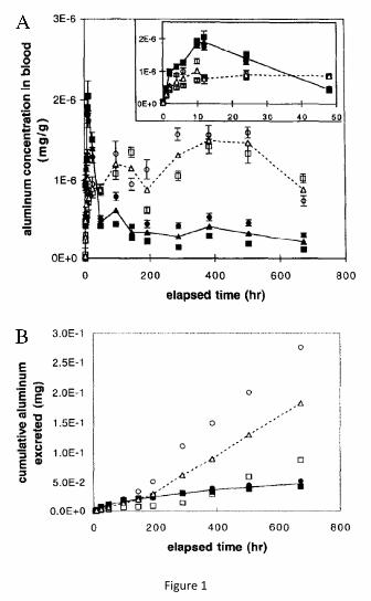
Figure 1

Figure 2

Figure 3