cranial deformation as the cause of death for a … · cranial deformation as the cause of death...
TRANSCRIPT

41Cranial deformation as the cause of death for a child from the Chillon River Valley, PeruVolumen 40, Nº 1, 2008. Páginas 41-53
Chungara, Revista de Antropología Chilena
CRANIAL DEFORMATION AS THE CAUSE OF DEATH FOR A CHILD FROM THE CHILLON RIVER VALLEY, PERU
DEFORMACIÓN CRANEANA COMO CAUSA DE MUERTE DE UN NIÑO DEL VALLE DE CHILLÓN, PERÚ
Sheila M.F. Mendonça de Souza1, Karl J. Reinhard2 and Andrea Lessa1
Two small mummy bundles, found in a tomb at the Chillon River Valley, Lima Plains, Peru exist in the collections of the Museu Nacional, Rio de Janeiro, Brazil. They were donated to the collections at the beginning of the 20th century. A multidisciplinary team is now curating and studying them. CT scans confirmed both skeletons were of individuals less than one year old at death. One of the bundles is intact and will be preserved and displayed. The other one was partly decomposed and the authors received permission to unwrap it to analyze the bones in details. Mites and pollen analysis were performed and the bones and artifacts were described. The cranium of a four to six month-old child, had a perforated necrotic occipital bone that possibly related to cause of death. Endosteal new bone reaction, a flattened area at the occipital squamae and a parietal sulcus associated with a zone of frontparietal resorption were also found. A pathognomonic periosteal reaction at the occipital and parietals, along the lambdoid suture, pointed to the compression of the skull that could explain the trauma, ischemic necrosis and subsequent infection. The cultural context suggests that the bundle could belong to a late Inca burial. Key words: Paleopathology, skull necrosis, skull deformation, prehistory, Chillon River, Peru.
Dos fardos de infantes hallados en una tumba en el valle del Río Chillón (Lima, Perú) fueron incorporados a la colección del Museo Nacional de Rio de Janeiro, Brasil, a comienzos del siglo XX. Actualmente, un equipo multidisciplinario está encargado de la curaduría y del estudio de los fardos. El análisis realizado por tomografía computadorizada confirmó que ambos esqueletos tienen menos de un año de edad. Uno de los fardos está intacto y será preservado y exhibido. El otro estaba parcialmente deterio-rado y se obtuvo permiso para desenfardarlo y hacer el análisis de los huesos. Se realizó análisis de ácaros y de polen, además de la descripción de los huesos y de los artefactos. El cráneo presentó una perforación necrosada en el occipital, interpretada como la posible causa de muerte. Se observó también una reacción endosteal de hueso nuevo, un área allanada en la escama occipital y un surco en el parietal asociado a una zona de reabsorción frontoparietal. Una reacción patognomónica del periostio en el occipital y en los parietales a lo largo de la sutura lambdoide apuntan a una compresión del cráneo, lo que podría explicar el trauma, la necrosis isquémica y la subsiguiente infección. El contexto cultural sugiere que el fardo pertenecería a un enter-ratorio de época incaica. Palabras claves: paleopatología, necrosis craneana, deformación craneana, prehistoria, Río Chillón, Perú.
1 Departamento de Endemias, Escola Nacional de Saúde Pública Sérgio Arouca, Fundação Oswaldo Cruz, Rio de Janeiro, Rio de Janeiro, Brasil. Escola Nacional de Saúde Pública - FIOCRUZ, Rua Leopoldo Bulhões Nº 1480 - Térreo. Manguinhos - Rio de Janeiro - Brasil - Cep 21041-210 Tel. 55 21 25982683, Fax 55 21 25982610. [email protected]; [email protected]
2 309 Biochemistry Hall, School of Natural Resources, University of Nebraska, Lincoln, NE 685830758. [email protected]
Recibido: mayo 2006. Aceptado: abril 2008
This paper contributes to the discussion of the health impact of intentional modification of the skull. Intentional modification of the skull is a world wide practice found in prehistoric as well as historic periods. Usually it causes no pathological consequence. The end result is simply an unusual cranial morphology.
The fortuitous finding of the skeleton of a child among the Peruvian objects of the collection of National Museum, Rio de Janeiro, Brazil, offered
the opportunity to study one unusual pathologic condition associated with the intentional modifica-tion of a skull. It also presents an opportunity to discuss severe bone necrosis associated with the compression of the occipital area. In addition, this case brings some additional insights to the discus-sion of the physiological reactions of the skull to compression bandages and to the explanation of the porotic appearance of the bone in some modified skulls described in archaeological collections.

Sheila M.F. Mendonça de Souza, Karl J. Reinhard and Andrea Lessa42
Intentional Skull Modification in America
Intentional modification of the body, especially the skull, was very common among American Indians. Many people from different periods, including the Inca, deformed the skulls in various ways including daily hand compression or cradleboarding. Textiles, leather or cotton straps, and pads were used to flatten heads in different shapes, or give them a tower-like appearance. The deforming apparatus was generally used on newborns a few days after birth (Dembo and Imbelloni 1938; Munizaga 1987) and sometimes it was used until the third year of life (Aufderheide and Rodriguez-Martin 1998). The use of cradleboards started at the same age, and generally lasted until the child was able to walk (Rowe 1946). In some cases, wearing bandages around the head or cradleboards for babies did not represent intentional cranial deformation. Some authors consider skull deformation being just an unintended consequence of some cultural practices, even in Peru (Cheverud et al. 1992). The shaping practice had important relations to ethnic identity. The intentional or incidental modification of the head brings additional difficulties to the craniomet-ric studies, specially in areas where this practice was very common (Cocilovo et al. 1995). Cranial deformation extended to men and women, and was still in use in Brazilian lowlands at the first half of the XX century (Mendonça de Souza 1994). There, it marked ethnic differences between populations and social groups. Interesting ethnographic data about this cultural practice in modern groups is available in recent medical literature (Simmons et al. 1998).
Cranial deformation, imposed on the normal anatomy of the human head, has long been a focus of special interest for anthropologists. Detailed descriptions and classifications of intentional or unintentional deformed skulls are abundant in the literature (Dembo and Imbelloni 1938; Imbelloni 1925; Munizaga 1987; Soto-Heim 2004). Consequences of this practice, including premature suture fusion, have been discussed by Dembo and Imbelloni (1938), White (1996) and others. More recently, the indirect consequences of skull deformation to the living have been dis-cussed (Aufderheide and Rodriguez-Martin 1998; Guillén 1992; Holliday 1993; Manríquez et al. 2006). Despite the description of modified skulls in collections both from North and South America,
only a few papers address the pathology associated with deformation (Holliday 1993).
Thanks to neural plasticity, the normal growth and function of the brain are generally not affected even in the most extreme changes of the skull morphology (White 1996; Soto-Heim 2004). However, in some cases the compression of the bones can stimulate changes in the bone inner structure, such as reduc-tion of the spaces in diploe, thickness of the outer table under the compressed areas, and premature fusion of the sutures causing cranial hypertension (Aufderheide and Rodriguez-Martin 1998; Guillén 1992; Soto-Heim 2004). Morphological modifica-tion of the facial bones has also being investigated (Cheverud et al. 1992; Manríquez et al. 2006). Beyond this, the bones can be affected in different ways including the side expression of Wormian bones (El-Najjar and Dawson 1977), depressed anomalous sutures, and bone inflammation and necrosis at the occiput (Holliday 1993). In the last several decades the careful description of deformed children’s skulls allowed researchers to associate the modification practices to a peculiar periosteal reaction along the occipital bone parallel to the lambda (Aufderheide and Rodriguez-Martin 1998; Guillén 1992). According to Manríquez et al. (2006) the area between the inion and the lambda is generally the most affected by the head shaping. This porosity can easily be misinterpreted as porotic hiperostosis, especially in Peruvian prehistoric populations where both the conditions are common (Blom et al. 2005).
Hrdlicka (1908), who studied modern Indian groups, described large occipital scars in children of the Pima population, a North American group that used cradleboards to confine or immobilize their babies. Inadvertently, this practice caused flattened heads. However, Hrdlicka associated the occurrence of scalp scars with impetigo and poor hygiene and never attributed the scars to cradleboarding. Dingwall (1913 in Holliday 1993) had associated the French use of cephalic tight bands to some diseases of the scalp. Stewart (1976) described an active inflamma-tory kind of lesion in two children’s skulls, one of them from Peru, and proposed its association with artificial compression. But it was Holliday (1993) who proposed and discussed a direct association between occipital lesions in Mogollon skulls to prehistoric deforming practices. Her paper adds interesting new information to the knowledge of pathologic consequences of skull-deforming in North America.

43Cranial deformation as the cause of death for a child from the Chillon River Valley, Peru
Figure 1. The mummy bundle of the Chillon child after cleaning (Photo K. Reinhard).Fardo funerario de infante del río Chillón, después de la higienización (Fotografía de K. Reinhard).
Material: The Mummy Bundle
In 2000, a new curatorial program commenced for the archaeological collections in the National Museum of Rio de Janeiro, Federal University of Rio de Janeiro, Brazil. Mummy bundles were found in the old repositories among thousands of archaeological specimens. This provided us with an opportunity to analyze those mummy bundles.
When found, the bundles were decompos-ing and after cleaning the volume of wrappings and debris in the museum box the whole volume including the box was CT scanned to confirm two independent bundles containing one skeleton each. One of them was intact, and the skeleton inside was that of a human child not older than six months. No pathologic changes were seen, but we noted some disorganization of the loose bones inside the bundle, probably from past handling and transport of the bundle. The second bundle was partly destroyed by humidity and apparently fungal activity. The skull had been crushed the legs and feet were lost, the human child was younger than the previous one, and the CT revealed abnormalities in the bones. Because the bundle was already partially destroyed, a curatorial staff authorized the authors to unwrap and analyze the bones inside so that any information remaining in the remains could be salvaged.
The remains of the bundle measured about 40 cm long and 18 cm wide. Insects and mechanical stress had destroyed the upper and lower extremities, one femur and the skull bones of the infant were partially exposed. The skeleton was cushioned by
a mass of soft light brown fibers, probably cotton. This material was also found by Silva (1930) inside some of the ceramic pots of the same grave. Different textiles surrounded the body. Inside the bundle there was a small rectangular textile, decorated in different colors and relief, and with eccentric green dots. The outer layer of the bundle was made with a wool textile sewed to close the bundle. In addi-tion, the textile was firmly tied with two stripes of wool (Figure 1).
Methodology
The superimposed badly preserved bundle had to be separated from the well preserved bundle. CT scan study was used to check preservation and to plan the separation and subsequent curato-rial and analytic procedures. A Pioneer (General Electric) helicoidal apparatus was used for the computed tomography of the contiguous bundles before separating and securing them. Images were acquired with 3 mm intervals in transverse planes, along the object. An optic disc (DEC 702) was used for saving images. It was possible to identify the different bones, their anatomical position in the bundles, and associated biocultural characteristics, like the textiles and objects inside. Some changes suggested taphonomic and pathological processes. Once the two mummified bundles were separated, it was finally possible to open and describe the skeleton in the decomposed bundle.
The bundle was opened without cutting the textiles. The outer blanket was badly decomposed

Sheila M.F. Mendonça de Souza, Karl J. Reinhard and Andrea Lessa44
and therefore it was easy to loosen it to expose the bundle’s interior without damaging it. Each layer of wrapping or textile was carefully removed and, documentation of the objects inside the bundle was made. An archaeological approach, surveying from the surface to the inside, allowed proper documenta-tion for future reconstruction. Associated material, especially the powder resulting from decomposition of the soft tissues, was removed for pollen, parasite (Souza et al. 2006) and microresidue analysis. Plant remains and textile samples were also collected. The bones were finally exposed in their original position.
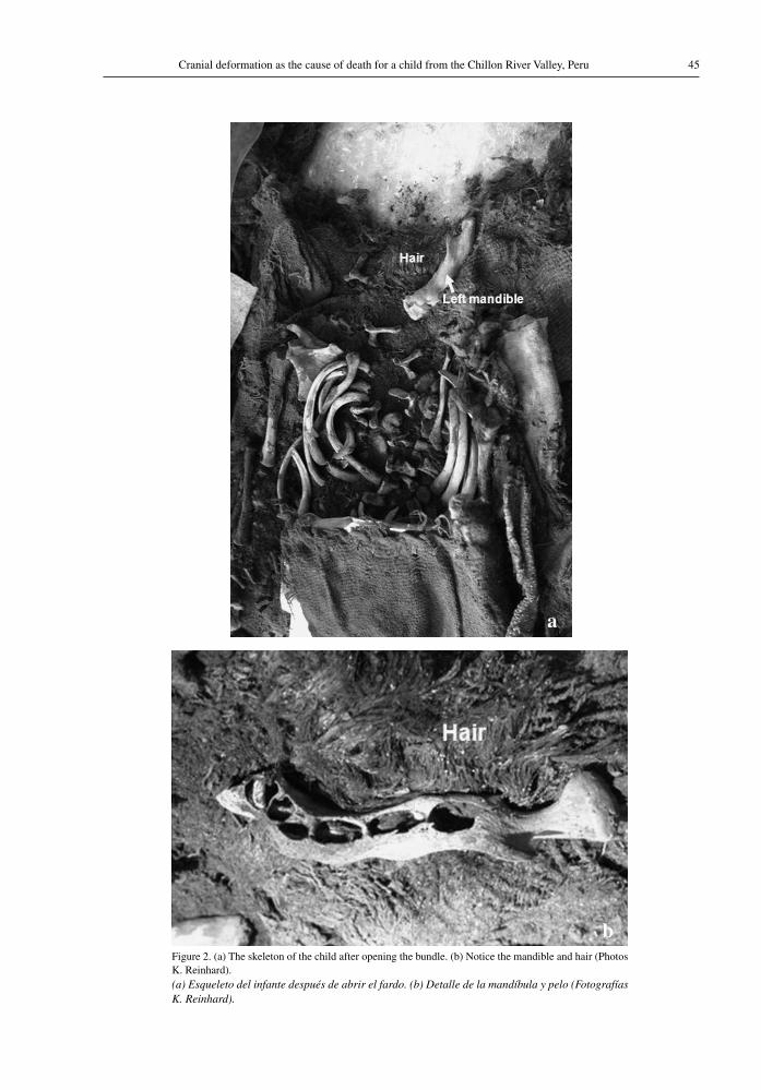
The direct visualization of the well-preserved teeth and bones in anatomical order (Figure 2a) confirmed age estimation based on Ubelaker (1978). Taphonomic changes and pathologic lesions, as well as hair and other associated evidences (Figure 2b) were also described and documented. A binocu-lar microscope was used for detailed analysis. A digital camera (Nikon/Coolpix 990) was used for documentation.
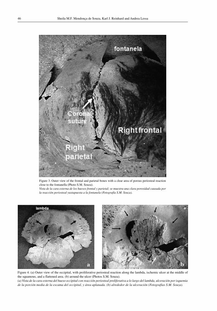
The preserved bones of the skull were found partially crushed, anatomical restoration had to be performed before the analysis. All the debris and fragments were collected inside the bundle, and the coherent parts of the parietal and occipital were reassociated based on the morphology of the borders, and the inner and the outer surface details. The pathological lesions helped to position some of the fragments based on the anatomical continuity of the structures (Figure 3). The correct anatomical coherence of the final reconstruction can be seen in Figures 4a, 4b and 5.
The differential diagnosis for abnormalities and pathologic lesions was based on morphologic comparative method and paleopathological data ac-cording to Guillén (1992), Holliday (1993), Ortner and Putshar (1997) and Aufderheide and Rodriguez-Martin (1998). The final interpretation benefits from the anatomical information concerning ontogenesis of skull (Warwick and Williams 1979).
Differential diagnosis against porotic hyper-ostosis was based on the concept that PH is the consequence of diploe expansion meaning we expect a thicker skull with thin outer table, not the apposition of bone on the skull. Differential diag-nosis against scurvy was based in the symmetry and regular development of the bone expansion follow-ing the strength lines of the skull, and not a casual ossification following sub-periosteal hemorrhages.
The same is true for cribra orbitalia which is never consistent with simple apposition and/or erosion with exposure of the diploe.
Although it was not possible to reconstruct the whole skull, the remaining bones (both frontals, reconstructed right parietal, reconstructed occipital) were complete enough to allow the identification of local compressive forces affecting the head.
Results: Head Molding, Periostitis and Death
The computed tomography suggested that the upper diaphysis of the right humerus and tibia had undergone changes. Despite the suggestion of interior rarefaction, the bones appeared super-ficially normal in direct examination, suggesting a taphonomic process could explain the image. The tomography also showed a double contour of some of the skull bones, suggesting a pathologic proliferative condition. The direct examination of the same bones revealed intense periosteal reaction, among other changes.
Fine coffee-like powder, resulting from de-composition of the soft tissues, permeated the textiles, suggesting that the body had been wrapped without evisceration. Decomposition of the parts of textiles in direct contact with the abdominal region reinforced that interpretation (Cockburn et al. 1998). No parasites or pollen could be found in the powder samples, but only acari (Souza et al. 2006). Microresidue analysis is still in progress. The hair was still in place, adherent to the bones and textiles surrounding the head. No fleas, lice, or other ectoparasites could be found. The skeleton was deeply stained by the brown color of decomposition. Textile fragments and partially mummified tissue adhered to parts of the skeleton. The teeth, still inside the maxilla and mandible, as well as bone development, allowed an age estimation of four to six months at death (Ubelaker 1978).
The skull bones were disarticulated and partly broken. The best-preserved bones were the frontal, left temporal, zygomatic bones, upper maxillas and left part of the mandible. Most of the right parietal and occipital could be recovered and restored. Other bones of the skull base and face were highly fragmented. The skull examination resulted in six different kinds of lesions:
(1) A well-defined zone of clear and porous bone, measuring about 15 x 60 mm was observed
45Cranial deformation as the cause of death for a child from the Chillon River Valley, Peru
Figure 2. (a) The skeleton of the child after opening the bundle. (b) Notice the mandible and hair (Photos K. Reinhard).(a) Esqueleto del infante después de abrir el fardo. (b) Detalle de la mandíbula y pelo (Fotografías K. Reinhard).
a
b
Sheila M.F. Mendonça de Souza, Karl J. Reinhard and Andrea Lessa46
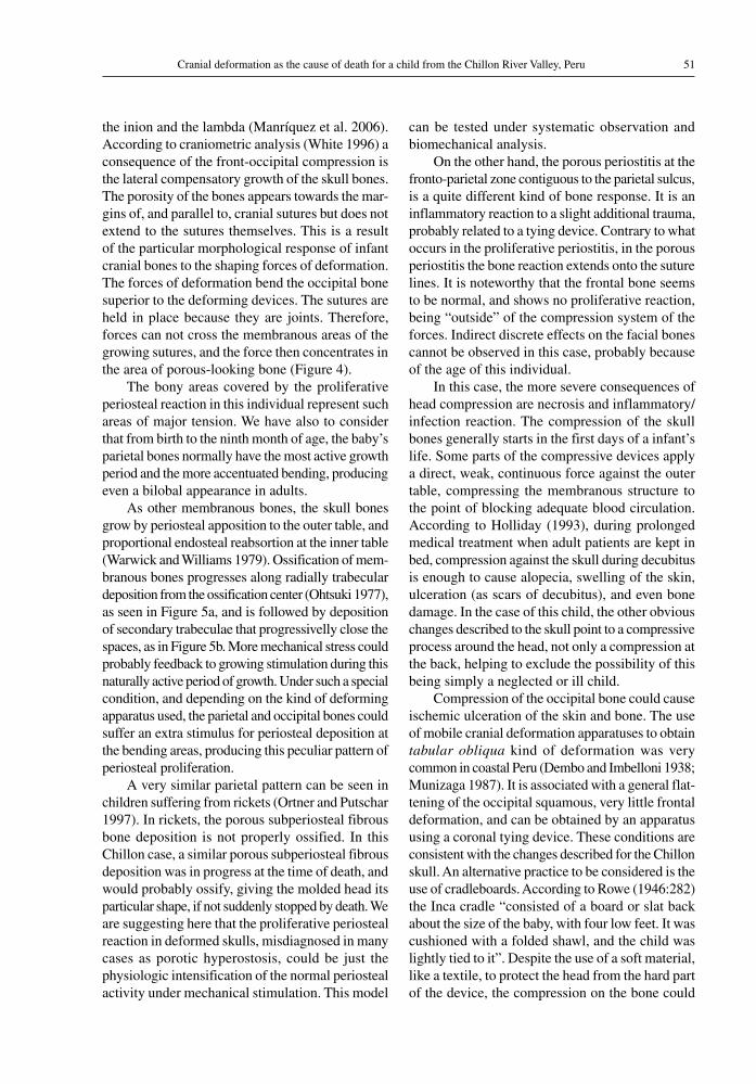
Figure 4. (a) Outer view of the occipital, with proliferative periosteal reaction along the lambda, ischemic ulcer at the middle of the squamous, and a flattened area. (b) around the ulcer (Photos S.M. Souza).(a) Vista de la cara externa del hueso occipital con reacción periosteal proliferativa a lo largo del lambda, ulceración por isquemia de la porción media de la escama del occipital, y área aplanada. (b) alrededor de la ulceración (Fotografías S.M. Souza).
a
Figure 3. Outer view of the frontal and parietal bones with a clear area of porous periosteal reaction close to the fontanella (Photo S.M. Souza).Vista de la cara externa de los huesos frontal y parietal; se muestra una clara porosidad causada por la reacción periosteal yuxtapuesta a la fontanela (Fotografía S.M. Souza).
a b

47Cranial deformation as the cause of death for a child from the Chillon River Valley, Peru
Figure 5. (a) Outer view of the right parietal bone with an apposition of (c) proliferative periosteal reaction around the bossa. (b) Notice the normal mature bone at the middle of the parietal and the similar growth pattern to normal bone in (Photos S.M. Souza).(a) Vista de la cara externa del hueso parietal donde se ve una deposición (c) de tipo periostites proliferativo alrededor de la bossa. (b) Nótese la maduración normal del tejido en la porción media del parietal, semejante al patrón de crecimiento del hueso normal (Fotografías S.M. Souza).
b
a c
b

Sheila M.F. Mendonça de Souza, Karl J. Reinhard and Andrea Lessa48
along the coronal suture, at the upper frontal (Figure 3). Asymmetrically distributed on both sides of the fontanel, the lesion also extended back some millimeters over the coronal suture, onto the right parietal bone (the left parietal, almost completely destroyed, could not be examined). The lesion is consistent with a local increase in the vascularization from a porous periosteal reaction. The limits of the porosity were clear and the lower third of the bones along the suture, on both sides, was normal. The continuous contour of the lesion, its well-defined margins and its trajectory on the skull are coherent with the trajectory of a cord, or textile band, crossing the head.
(2) Another kind of periosteal reaction, represented by proliferative periostitis, was superimposed on the outer table of the occipital and parietal bones. The layer of new bone was 1.0 to 1.5 mm thick, with the surface penetrated by shallow cavities of different diameters. It was tightly adherent to the outer table. At the occipital bone, two areas of periosteal reaction extended close to the lambdoid suture, both to the left and to the right, measuring a maximum of 11 mm wide (Figure 4a-b). At the right parietal bone a large, crescent-shaped area of periosteal reaction was distributed around the parietal eminence, covering about two thirds of the bone (Figure 5a-c). Detached parts of new bone, on the parietal bone, confirmed that the outer table was normal under the proliferative reaction. A clear pattern of fibrous periosteal new bone spread out from the parietal eminence to the lambdoid and sagittal sutures leaving the parietal ossification center with no periosteal reaction. A small fragment, with lesions that were symmetrical in relation to the opposite side, represented the left parietal bone.
(3) A destructive lesion in the middle of the oc-cipital squamous was formed by an excavated area of lobulated contour, measuring about 17x18 mm, with healing bone at the bottom (Figure 4). The outer table had been more destroyed than the inner table. The lesion was deeper at some points, and some perforations with regular borders penetrated the inner table. The larger perforated area, measuring 8 mm, corresponded to the left occipital lobe of the brain. Restoration of the occipital bone made it possible to see the continuity of the lesion at
the lower occipital squamous. We were able to as well as estimate the extension of the ulcer-ated area. The lesion was not at the midline, but more to the left side. Its gross shape was a triangle with the base turned to the lambda. The process of bone destruction clearly came from the outer to the inner table, and the absence of healing activity at the borders suggested that the perforations could be perimortem, even representing a taphonomic effect of differential breakage of a very thin remaining bone. The minor bone reaction and the expressive bone loss suggest an ischemic process causing bone necrosis followed by some amount of inflam-matory response (Kosiak 1961).
(4) A well-defined flattened area, about 2 mm wide, encircled the necrotic area of the oc-cipital (Figure 4a). At the left and upper part, the flat bone was tangent to the main surface of the occipital squamous, at the right and lower part it was more inclined to the bottom of the lesion. The flattening cannot be misinterpreted for any other normal anatomical feature of the occipital bone and its presence strongly suggests the existence of a compression force modeling the bone. The direct association between the contour of the flattened area and the ulceration reinforces the interpretation of ischemic necrosis caused by compression. Another indication of compression was the abnormally plane occipital squamous as a whole, when compared to the pronounced bowing generally found in normal baby’s skulls.
(5) A discrete inflammatory reaction, at the inner face of the occipital bone, covered an oblong area between the lambda and the internal oc-cipital protuberance (Figure 6). Along this area, the inner table was covered by fine-grained clear new bone indicating endosteal reaction. The lesion was contiguous to the perforations, reinforcing the hypothesis that the bone table was not perforated at the time of the death. Otherwise, the endosteal reaction, at the midline, was superimposed to an important cephalic venous sinus, suggesting that it could be vascular-related.
(6) Finally, a slightly depressed transverse sulcus crossed the upper part of the right parietal bone, parallel to the coronal suture, measuring about 5 mm (Figure 7). This second area of compres-sion is parallel to the fronto-parietal periosteal

49Cranial deformation as the cause of death for a child from the Chillon River Valley, Peru
resorption previously described. Considering the hypothesis of the use of deforming apparatus or cradleboard, a tying device probably caused this modeling.
Discussion
The use of different skull deforming appara-tuses is largely associated with Peruvian prehistoric people, including the Inca (Dembo and Imbelloni 1938; Munizaga 1987). The literature seldom refers to infection and necrosis as possible severe patho-logic consequences of skull deformation. Guillén (1992) and Holliday (1993) address this theme. The present case offers the opportunity to present in more detail the physiology and pathology of deformed skulls. We propose that the lesions and death of this infant resulted from cranial deforma-tion. However, we must also present differential diagnostic considerations.
The proliferative lesions at the occipital and parietal bones could be a physiologic response to
cranial deformation, but porotic hyperostosis, sub-periosteal hemorrhage, scurvy and rickets should also be considered (Ortner et al. 2001; Warwick and Williams 1979). This is especially true for the Peruvian skeleton samples when considering the high prevalence of anemia in the area (Blom et al. 2005). In the present case the distribution of the porous area in parietals and occipital, the occipital reaction parallel to lambda, the superimposition of the new bone to the outer table, the absence of woven bone at the inner side of the vault and the fibrous appearance of the new bone indicates that ossification followed the normal pattern of growth (Aufderheide and Rodriguez-Martin 1998; Ortner and Putschar 1997). The absence of lesions in the other fragments or entire bones was important evidence, including the absence of cribra orbitalia in the well-preserved orbital roofs. The rest of the skeleton was carefully examined. The long bones were normal. Porotic hyperostosis was excluded because the lesion was clearly superimposed to the outer table. Subperiosteal hemorrhage was
Figure 6. Inner view of the occipital bone with endosteal reaction covering an oblong area. Notice the perforations at the inner table (Photo S.M. Souza).Vista de la cara interna del hueso occipital con reacción endosteal en forma oblonga. Nótense los agujeros en la tabla interna (Fotografía S.M. Souza).

Sheila M.F. Mendonça de Souza, Karl J. Reinhard and Andrea Lessa50
Figure 7. Lateral view of the articulated right frontal and parietal, showing the slight depressed sulcus at the coronal suture (Photo S.M. Souza).Vista lateral derecha de los huesos frontal y parietal en articulación anatómica, evidenciando una discreta depresión o surco sobrepuesto a la sutura coronal (Fotografía S.M. Souza).
excluded because of the perfectly fibrous symmetric and regular pattern of new bone proliferation, seen at the occipital bone and suggested at the parietal bones, but and also because of the pattern of os-sification that is equal to the pattern of the normal bone growth. The normal pattern of growth was checked using skeletons of the same age available in the anatomical collection of Museu Nacional, and also in the literature. Scurvy was excluded because cranial involvement without long bones involvement is unusual in scurvy (Ortner and Putshar 1997). In addition from an epidemiologic point of view would be unlikely because such an infant, still breastfeeding, would not develop true C vitamin deficiency. Since no other porous lesion was found either in the skull bones or at the other bones, rickets, as well as scurvy, were excluded because they should be associated with systemic lesions. It is important to mention that only the endosteal lesion was found in direct association to the severe occipital scar.
Considering the results, three main processes can be proposed to explain what occurred to this
individual. Two of them could be considered physi-ologic responses to mechanical trauma of deforming the skull, and the last one, a pathologic response to the same trauma. The first one is the direct me-chanical responses to the head compression, the occipital flattening and the fronto-parietal sulcus. The second one is the indirect mechanical responses to the compression, the proliferative and the porous periosteal reactions. The third one is the pathologic response to the head compression, the occipital bone necrosis and the endocranial inflammation, probably causing death. The continued compression had a direct modeling effect on the bones, mechanically stopping the periosteal proliferation and subsequent bone growth, on those parts of the skull directly in contact with the hard or tight parts of the deforming apparatus. The same continued compression had an indirect effect on the skull bones, forcing the free parts to bend, especially the parietal and upper oc-cipital bones. This created zones of major tension trapped between frontal and occipital compressing forces with resulting compensatory bone growth. The occipital area of major modification is between
51Cranial deformation as the cause of death for a child from the Chillon River Valley, Peru
the inion and the lambda (Manríquez et al. 2006). According to craniometric analysis (White 1996) a consequence of the front-occipital compression is the lateral compensatory growth of the skull bones. The porosity of the bones appears towards the mar-gins of, and parallel to, cranial sutures but does not extend to the sutures themselves. This is a result of the particular morphological response of infant cranial bones to the shaping forces of deformation. The forces of deformation bend the occipital bone superior to the deforming devices. The sutures are held in place because they are joints. Therefore, forces can not cross the membranous areas of the growing sutures, and the force then concentrates in the area of porous-looking bone (Figure 4).
The bony areas covered by the proliferative periosteal reaction in this individual represent such areas of major tension. We have also to consider that from birth to the ninth month of age, the baby’s parietal bones normally have the most active growth period and the more accentuated bending, producing even a bilobal appearance in adults.
As other membranous bones, the skull bones grow by periosteal apposition to the outer table, and proportional endosteal reabsortion at the inner table (Warwick and Williams 1979). Ossification of mem-branous bones progresses along radially trabecular deposition from the ossification center (Ohtsuki 1977), as seen in Figure 5a, and is followed by deposition of secondary trabeculae that progressivelly close the spaces, as in Figure 5b. More mechanical stress could probably feedback to growing stimulation during this naturally active period of growth. Under such a special condition, and depending on the kind of deforming apparatus used, the parietal and occipital bones could suffer an extra stimulus for periosteal deposition at the bending areas, producing this peculiar pattern of periosteal proliferation.
A very similar parietal pattern can be seen in children suffering from rickets (Ortner and Putschar 1997). In rickets, the porous subperiosteal fibrous bone deposition is not properly ossified. In this Chillon case, a similar porous subperiosteal fibrous deposition was in progress at the time of death, and would probably ossify, giving the molded head its particular shape, if not suddenly stopped by death. We are suggesting here that the proliferative periosteal reaction in deformed skulls, misdiagnosed in many cases as porotic hyperostosis, could be just the physiologic intensification of the normal periosteal activity under mechanical stimulation. This model
can be tested under systematic observation and biomechanical analysis.
On the other hand, the porous periostitis at the fronto-parietal zone contiguous to the parietal sulcus, is a quite different kind of bone response. It is an inflammatory reaction to a slight additional trauma, probably related to a tying device. Contrary to what occurs in the proliferative periostitis, in the porous periostitis the bone reaction extends onto the suture lines. It is noteworthy that the frontal bone seems to be normal, and shows no proliferative reaction, being “outside” of the compression system of the forces. Indirect discrete effects on the facial bones cannot be observed in this case, probably because of the age of this individual.
In this case, the more severe consequences of head compression are necrosis and inflammatory/infection reaction. The compression of the skull bones generally starts in the first days of a infant’s life. Some parts of the compressive devices apply a direct, weak, continuous force against the outer table, compressing the membranous structure to the point of blocking adequate blood circulation. According to Holliday (1993), during prolonged medical treatment when adult patients are kept in bed, compression against the skull during decubitus is enough to cause alopecia, swelling of the skin, ulceration (as scars of decubitus), and even bone damage. In the case of this child, the other obvious changes described to the skull point to a compressive process around the head, not only a compression at the back, helping to exclude the possibility of this being simply a neglected or ill child.
Compression of the occipital bone could cause ischemic ulceration of the skin and bone. The use of mobile cranial deformation apparatuses to obtain tabular obliqua kind of deformation was very common in coastal Peru (Dembo and Imbelloni 1938; Munizaga 1987). It is associated with a general flat-tening of the occipital squamous, very little frontal deformation, and can be obtained by an apparatus using a coronal tying device. These conditions are consistent with the changes described for the Chillon skull. An alternative practice to be considered is the use of cradleboards. According to Rowe (1946:282) the Inca cradle “consisted of a board or slat back about the size of the baby, with four low feet. It was cushioned with a folded shawl, and the child was lightly tied to it”. Despite the use of a soft material, like a textile, to protect the head from the hard part of the device, the compression on the bone could
Sheila M.F. Mendonça de Souza, Karl J. Reinhard and Andrea Lessa52
be enough to cause damage, especially if the child was firmly tied. It is important to remember that in the case of cradles, this restriction lasted until the child was able to walk (around a year, or even more). In some cases pathology may have ensued for several reasons: daily care of the children might have been neglected, inadequate experience of the mother with the devices, or some children could have been especially frail and susceptible to trauma. Holliday (1993) and Stewart (1976) described healed occipital scars demonstrating that some infants survived minor lesions. Perhaps the cranial depressions, usually found above the inion of some skulls (Holliday 1993), are a sign of skull-deforming trauma as well. Other care-ful reexamination of skull collections from areas where cranial deformation existed could bring more information about morbidity and lethality of these practices and their scars.
Conclusion
Six changes were observed in the bones of a four to six months Chillon Valley child from a probable Inca mummy bundle: porous periosteal reaction at
the fronto-parietal zone, peculiar proliferative pe-riosteal reaction at the parietal and occipital bones, ischemic ulcer at the occipital squamous, endocranial inflammatory reaction close to ulcer, gross occipital flattening and strong occipital flattening around the ulcer, and a parietal sulcus back to the porous periosteal reaction zone. The six conditions, as well as their association, are consistent with the use of a deforming apparatus that caused physiologic and pathologic changes, encephalic complications, and probably death. The absence of other pathologic changes in the skeleton, as well as the character-istics of the bone changes helped to exclude other pathologic explanation for this case.
Acknowledgments: The authors thank the Department of Anthropology and the Department of Museology of the Museu Nacional, Universidade Federal do Rio de Janeiro, for the support and help during this investigation. We also thanks Dr. Vander Campanati of the Centro de Diagnosticos Avançados, Rio de Janeiro, for kindly providing the CT scanning images, and all the colleagues and reviewers who discussed the results and the final form of this manuscript with the authors.
References Cited
Aufderheide, A.C. and C. Rodriguez-Martin, editors 1998 The Cambridge Encyclopaedia of Human Paleopathology.
Cambridge University Press, Cambridge.Blom, D.E., J.E. Buikstra, L. Keng, P.D. Tomaczak, E. Shoreman
and D. Stevens-Tuttle 2005 Anemia and childhood mortality: Latitudinal patterning
along the coast of Precolumbian Peru. American Journal of Physical Anthropology 127:152-169.
Cheverud, J.M., L.A.P Kohn, L.W Konigsberg and S.R. Leigh
1992 Effects on fronto-occipital artificial cranial vault modi-fication on the cranial base and face. American Journal of Physical Anthropology 88:323-345.
Cocilovo J.A., H.H. Varela and S. Quevedo 1995 La deformación artificial del cráneo en la población
prehistórica de San Pedro de Atacama, Chile. Chungara 27:117-124.
Cockburn, A., E. Cockburn and T.A. Reyman, editors 1998 Mummies, Diseases & Ancient Cultures. Cambridge
University Press, Cambridge.Dembo, A. and J. Imbelloni 1938 Deformaciones intencionales del cuerpo humano de carácter
étnico. Humanior (Buenos Aires), Sección A, Tomo 3:1-348.El-Najjar, M.Y. and G.L. Dawson 1977 The effect of artificial cranial deformation on the inci-
dence of Wormian bones in the lambdoid suture. American Journal of Physical Anthropology 46:155-160.
Guillén, S. 1992 The Chinchorro Culture: Mummies and Crania in the
Reconstruction of Prehistoric Coastal Adaptation in the South Central Andes. Ph.D. Dissertation. Department of Anthropology. University of Michigan, USA.
Holliday, D.Y. 1993 Occipital lesions: A possible cost of cradleboards.
American Journal of Physical Anthropology 90:283-290.Hrdlicka, A. 1908 Physiological and medical observations among the in-
dians of Southwestern New Mexico. Papers of the Peabody Museum Bureau of Archaeology and Ethnology 15:115-170. Harvard University Press, Cambridge.
Kosiak, M. 1961 Ethiology of decubitus ulcers. Archives of Physical
Medicine and Rehabilitation 42:19-29.Manríquez, G., F.E. González-Bergás, J.C. Salinas and O.
Espoueys 2006 Deformación intencional del cráneo en poblaciones
arqueológicas de Arica, Chile: análisis preliminar de morfometría geométrica con uso de radiografías cra-neofaciales. Chungara Revista de Antropología Chilena 38:13-34.
Mendonça de Souza, S.M.F. 1994 Deformações cranianas entre os índios Karitiana: Análise
de fotos de arquivo. Boletim do Museu Paraense Emílio Goeldi (Série Antropologia) 10:43-56.

53Cranial deformation as the cause of death for a child from the Chillon River Valley, Peru
1 The two mummy bundles were brought to Brazil in 1925 by Dr. Simões da Silva, a famous traveler and early South American ethnographer, who excavated with Max Uhlle, Lechmann, Salvador Debeneditti and others in archaeo-logical projects in Peru. He had Brazilian diplomatic permission to collect archaeological and ethnographical objects for museums. The recovery of the objects from Chillon tomb was done under the supervison of Simões da Silva himself. However, the excavation followed the standards of the time and therefore was not as techni-cally sophisticated as we would expect today. Like many excavations of the early 20th century, the excavation goal was to collect museum-quality objects for exhibi-tion. The recovery of these and other objects in Peru are reported in Silva (1930). However, he does not provide either cultural description, or detailed archaeological information. Only part of his objects, such as the mummy bundle described in this paper, are in the Museu Nacional “Simões da Silva” collection.
During his trip, the owner of the Marques cotton farm, Mr. Ramon Geng suggested to Simões da Silva that the Chillon site would be a productive area for excavation. Mr. Geng personally drove da Silva to the area where the tombs were located. According to Simões da Silva´s book, the place was a desert area at the Chillon River Valley. For one day da Silva supervised the efforts of some employees of the farm who were asked to dig one of the tombs to expose the burials. According to his report, he wrote notes and helped to identify and collect bundles and many different objects. Part of this material was given to Brazilian museums. His original notes did not survive and are not in the collection.
However, some information can be gleaned from his 1930 memoirs.
To Simões da Silva, the first sign of the burial was the tip of a long cane (more than 3 m long) coming out of the soil. This could have been an ushnus marking the place of the burial. The burial contained the more or less well preserved bundles of adults and children. Basketry, pots, raw cotton, textiles, maize, beans, peanuts, wool threads, wooden rods, silver needles, silver pins, and gourds containing coca leaves and small crustaceans were associated with the mummy bundles. Differences between the objects beside each one of the individuals were noted and a wooden mask with the eyes made of shells was also found. The excavated tomb was circular and measured about 4 m deep. The bundles of 6 individuals (three very young children, one younger individual, one adult male and one adult female, possibly from a nuclear familly) were found inside the tomb in as-sociation. The woman was wearing two long silver pins with discoidal heads in her long black hair and a necklace of shell beads. Fusiforms silver objects were inside the mouth of the adult woman and the younger individual. Textiles and dressings associated to the younger individual were made of cotton and decorated with feathers. The mummy bundle of the younger individual was not deposited in a museum so we have not had any opportunity to see him/her.
The site was not excavated systematically at that time, there are only a few lines of description in the book reporting Simões da Silva trip along Chile and Peru. No fine contextual information could be recovered, and no former investiga-tion in the Museum provide better description or cultural affiliation
Munizaga, J.R. 1987 Deformación craneana intencional en América. Revista
Chilena de Antropología 6:113-147.Ohtsuki, F. 1977 Developmental change of the cranial bone thikness
in the human fetal period. American Journal of Physical Anthropology 46:141-154.
Ortner, D.J., W.J. Butler, J. Cafarella and L. Milligan 2001 Evidence of probable scurvy in subadults from ar-
chaeological sites in North America. American Journal of Physical Anthropology 114:343-351.
Ortner, D.J. and W.G.J. Putshar 1997 Identification of Pathological Conditions in Human
Skeletal Remains. Smithsonian Institution, Washington.Rowe, J.H. 1946 The Inca culture at the time of the Spanish conquest. In
Handbook of South American Indian, edited by J. Stewart, 2:183-330. Smithsonian Institution, Washington.
Silva, A.C.S. 1930 Viagens Ethnographicas Sul-Americanas. “PERU”.
Imprensa Nacional, Rio de Janeiro.Simmons, E.F., J.H. Prost and S. Peniston 1998 Infant head molding. A cultural practice. Archives of
Familiar Medicine 7:88-89.
Soto-Heim, P. 2004 Une marque d’identité imprimée sur l’os: la déformation
crânienne intentionnelle. Biométrie Humaine et Anthropologie 22:81-98.
Stewart, T.D. 1976 Are supra-inion depressions evidence of prophy-
lactic trephination? Bulletin of History of Medicine 50:414-434.
Souza, D.L. de, R. de M.S.N. de C. Guerra, S.M. de Souza, L.F. Ferreira and A. Araújo
2006 Acari found in a mummy bundle from the Chillon River Valley, Peru. Paleopathology Newsletter 136:11-16.
Ubelaker, D.H. 1978 Human skeletal remains. Excavation, analysis, interpreta-
tion. Manuals on Archaeology Nº 2. Smithsonian Institution, Washington.
Warwick, R. and P.L. Williams 1979 Gray Anatomia. Translated and Edited by Guanabara
Koogan, Rio de Janeiro.White, C.D. 1996 Sutural effects of front-occipital cranial modifications.
American Journal of Physical Anthropology 100:397-410.
Note
