cpia de disser. mest. v. atual 2 97-2003 · e aos meus filhos diogo bezerra de barros, diêgo...
TRANSCRIPT
UNIVERSIDADE FEDERAL DE PERNAMBUCO CENTRO DE CIÊNCIAS BIOLÓGICAS
DEPARTAMENTO DE BIOQUÍMICA PROGRAMA DE PÓS-GRADUAÇÕ EM BIOQUÍMICA
AVALIAÇÃO DA ATIVIDADE ANTITUMORAL EM EXTRATO DE
Indigofera suffruticosa Mill
CLEIDEANA BEZERRA DA SILVA
RECIFE-PE FEVEREIRO-2008

UNIVERSIDADE FEDERAL DE PERNAMBUCO
CENTRO DE CIÊNCIAS BIOLÓGICAS DEPARTAMENTO DE BIOQUÍMICA
PROGRAMA DE PÓS-GRADUAÇÕ EM BIOQUÍMICA
AVALIAÇÃO DA ATIVIDADE ANTITUMORAL EM EXTRATO DE Indigofera suffruticosa Mill
Cleideana Bezerra da Silva Orientadora: Profa. Dra. Vera Lúcia de Menezes Lima

RECIFE-PE FEVEREIRO – 2008
Dissertação apresentada para o cumprimento das exigências para obtenção do título de Mestre em Bioquímica pela Universidade Federal de Pernambuco.
ii
Silva, Cleideana Bezerra da
Avaliação da atividade antitumoral em extrato de Indigofera suffruticosa Mill./ Cleideana Bezerra da Silva – Recife: A Autora, 2008.
xii; 94 fls. .: il. Dissertação (Mestrado em Bioquímica e Fisiologia) – UFPE. CCB
1. Fabaceae 2. Anil 3. Indigofera suffruticosa 4. Carcinoma de Ehrlich 5. Atividade antitumoral I.Título 582.738 CDU (2ª. Ed.) UFPE 583.3 CDD (22ª. Ed.) CCB – 2008 – 101


FICHA DE APROVAÇÃO
Dissertação defendida e apresentada à Banca Examinadora:
______________________________________________________ Profa. Dra. Vera Lúcia de Menezes Lima (Presidente)
______________________________________________________ Profa. Dra. Ana Maria Mendonça de Albuquerque Melo (Membro externo)
_____________________________________________________ Profa. Dra. Maria Patrícia Guedes Paiva (Membro interno)
_____________________________________________________ Profa. Dra. Maria Bernadete Sousa Maia (Membro interno)
Fevereiro de 2008
iii

Aos meus pais Pedro Bezerra da Silva e Maria das Dôres Ferreira da Silva (In memorian)

pelo carinho e por minha formação. E aos meus filhos Diogo Bezerra de Barros, Diêgo Bezerra de Barros e Tiago Bezerra de Barros pelo amor, compreensão e incentivo.
Dedico.
iv AGRADECIMENTOS
À Deus, por estar presente em minha vida, derramando sobre mim as suas bênçãos e dando-me energia para ser sempre perseverante na busca do objetivo almejado. Aos meus pais, Pedro Bezerra da Silva e Maria das Dôres Ferreira da Silva (In memorian), pelo esforço desprendido, apoio em todos os momentos e por acreditarem em mim. Aos meus filhos Diogo, Diêgo e Tiago pelo amor, compreensão nos momentos de ausência, motivação e por todas as contribuições dadas para realização deste trabalho. As minhas irmãs Fátima, Cleonice, Clemilda, e aos meus irmãos Argemiro, Gildo, Edmilson e em especial a José Cícero pelo incentivo e carinho. A minha cunhada Alice Maria pelo incentivo, carinho e amizade. A minha orientadora, profa. Dra. Vera Lúcia de Menezes Lima, por me aceitar como sua orientanda, por todas as oportunidades ofertadas, pela orientação e ensinamentos. À professora Ivone, do Departamento de Antibiótico, pela realização dos testes antitumorais e toxicológicos e sua equipe de laboratório Aldo César, Ruth Sampaio, Isla, Sérgio, Antonioni, Elis e Cynthia pela contribuição na realização dos ensaios. A Ethiene e Fernando pela colaboração neste trabalho. À professora Ana Maria Mendonça e ao professor Amâncio do Departamento de Biofísica e Radiobiologia pela ajuda, gentileza e por permitir a realização de ensaio com Artemia no laboratório e a Ronaldo, Rebeca, Luana Clarice e Eliane. À professora Marilene do Departamento de Micologia e a Thais, Ludimila e Viviane pelo incentivo e amizade. Aos técnicos João Virgínio e Rejane pela contribuição na preparação do extrato. A Maria e Albérico pelo auxílio e colaboração concedidos. Aos Professores do Departamento de Bioquímica da Universidade Federal de Pernambuco, pelo incentivo e conhecimentos proporcionados durante este curso. Aos funcionários do Departamento de Bioquímica, Ademar, Helena, Iolanda, Djalma e Miron pela simpatia, amizade e disponibilidade demonstradas. À Rosana, uma grande amiga, pelo companheirismo e apoio. Aos colegas do mestrado pela amizade. Aos amigos do laboratório de Química e Metabolismo de Lipídeos: Caique, Adenor, Janaina, Luciana, Gabriella, Amanda, Aline, Gisele, Thiago e Tiago Araújo pela ajuda, companheirismo e por todas as horas compartilhadas nessa jornada e
v em especial a Olivá e a Bianka pelo apoio, pela valiosa colaboração para finalização deste trabalho. A todos que de alguma forma contribuiu para a realização de mais um sonho, meus sinceros agradecimentos.

vi
SUMÁRIO
AGRADECIMENTOS ............................................................................ v LISTA DE FIGURAS ............................................................................. viii LISTA DE TABELAS ............................................................................ ix LISTA DE ABREVIATURAS ................................................................ x RESUMO ...............................................................................................xi ABSTRACT ...........................................................................................xii 1. INTRODUÇÃO ..................................................................................14 2. OBJETIVOS ......................................................................................33 2.1. Geral ...............................................................................................33 2.2. Específico .......................................................................................33 3. REFERÊNCIAS BIBLIOGRÁFICAS ................................................ 35 4. CAPÍTULO I ......................................................................................46 4.1. Abstract .......................................................................................... 47 4.2. Introdução ...................................................................................... 48 4.3. Material e Métodos .........................................................................49 4.4. Resultados ..................................................................................... 54 4.5. Discussão e Conclusão ..................................................................55 Agradecimentos .................................................................................... 60 Referências ...........................................................................................61 5. CONCLUSÕES .................................................................................74 6. ANEXOS ...........................................................................................76 6.1. Trabalhos Enviados para Congressos ...........................................77 6.2. Guide for Authors – Ethnopharmacology .......................................85
vii LISTA DE FIGURAS
1. INTRODUÇÃO Figura 1. Indigofera suffruticosa Mill. A: ramos com folhas e inflorescência; B: ramos com folhas; C: ramos floridos; D: ramos com folhas e sementes. ..................22 Figura 2. Indigofera suffruticosa Mill - Vista parcial de um indivíduo adulto. .............23 Figura 3. Fórmula estrutural do indigo. ......................................................................25 Figura 4. Processo de múltiplos estágios de Carcinogênese ....................................31 4. CAPÍTULO I Figure 1. Effect of the administration (i.p.) of the extract of I. suffruticosa on solid tumor induced by Ehrlich ascites carcinoma cell line in male mice ……………...…………..7

viii
LISTA DE TABELAS
4. CAPITULO I Table 1. Mains effect observed in acute toxicity after intraperitoneal administration of extract of Indigofera suffruticosa in mice. ……………………………………………….68 Table 2. Mains effect observed in acute toxicity after oral administration of extract Indigofera suffruticosa in mice. …………………………………………………………..69 Table 3. Determination of the DL
50 of the aqueous extract of leaves of I. suffruticosa ….. 70
Table 4. Evaluation of the effect of the aqueous extract of leaves of Indigofera suffruticosa on total cholesterol and triglyceride of normal mice.…………………..72
ix LISTA DE ABREVIATURAS
UFPE – Universidade Federal de Pernambuco ANVISA – Agência Nacional de Vigilância Sanitária OMS – Organização Mundial de Saúde % - Percentagem D
1 – Maior dose que não mata nenhum animal.
D2 – Menor dose que mata todos os animais.
DL50
– Dose letal para 50% da população I.P. – Via intraperitoneal V.O. – Via oral. mg – miligramas Kg – Quilogramas mL – Mililitro ° C – Graus Celsius < - Menor que EC – Ehrlich Carcinoma TC – Colesterol total TG – Triglicerídeos SD – Desvio padrão dl – decilitro x

RESUMO
Indigofera suffruticosa Mill é uma arbusto da família Fabaceae conhecido popularmente como anil. É uma planta distribuída mundialmente, sendo utilizada na medicina popular contra diversos problemas de saúde. Este estudo avaliou a ação toxicológica do extrato aquoso de folhas de I. suffruticosa, bem como a atividade antitumoral sobre células de carcinoma de Ehrlich (EC) e a influência desse extrato sobre os níveis séricos de colesterol total e triglicerídeos em camundongos. Foram realizados ensaios de toxicidade aguda por via oral e via intraperitoneal em camundongos machos, onde se verificou efeitos estimulantes seguidos de efeitos depressores, após a administração do extrato. As doses utilizadas foram de 490 a 850 mg/kg por via intraperitoneal, onde foi estimado o valor da DL
50 em 750,6 mg/kg e 1000 a
5000 mg/kg por via oral, entretanto, nenhuma morte foi notificada por esta via de administração, somente alguns sinais tóxicos. A toxicidade do extrato também foi testada frente à Artemia salina, que mostrou uma CL
50 de 127µg/mL. Tratamento de camundongos albinos suíços
normais (n = 5) com extrato de I. suffruticosa com dose de 50 e 100 mg/kg, administrada intraperitonealmente, não apresentou diferença significante nos níveis séricos de colesterol total e triglicerídeos. Após o tratamento com I. suffruticosa por via intraperitoneal, notou-se uma redução significativa (p < 0,001) no tamanho dos tumores ( 75,5% e 63,9%), quando utilizou-se doses de 50 e 100 mg/kg, respectivamente. A I. suffruticosa possui moderada toxicidade e exibiu uma significante atividade antitumoral em camundongos portadores de EC, indicando que a mesma possui propriedades medicinais. Palavras-chave: Fabaceae; Anil; Indigofera Suffruticosa; Carcinoma de Ehrlich; Atividade antitumoral

ABSTRACT
Indigofera suffruticosa Mill is a shrub of the family Fabaceae popularly known as anil. Is a plant distributed worlwidely, and is utilized in the popular medicine against diverse problem of health. This study evaluated toxicological actions of the aqueous extract of leaves of I. suffruticosa the antitumor activity on carcinoma de Ehrlich (EC) cell and the influence of this extract on serum total cholesterol and triglycerides levels in mice. Were realized assays of acute toxicity by oral and intraperitoneal (i.p.) route in male mice, where was observed stimulant effects followed of depressors effects, after of the extract administration. The doses utilized were of 490 to 850 mg/kg by intraperitoneal route, where was esteemed the LD
50 values in 750,6 mg/kg and
1000 to 5000 mg/kg by oral route, nevertheless, no death was notify by this administration route, only some sings toxic. The toxicity of the extract also was tested front the Artemia salina that showed a LC
50 of 127 µg/mL. Treatment (i.p.) of normal Swiss white mice (n = 5) with extract of
I. suffruticosa, with dose of 50 and 100 mg/kg, not presented significant differences in the serum of total cholesterol and triglycerides levels. After, the treatment (i.p.) with the extract, was observed a significant (p < 0.001) reduction by 75.5% and 63.9%, on the size tumor of Ehrlich in mice, when was utilized doses of 50 e 100 mg/kg, respectively. The I. suffruticosa presented toxicity moderate and exhibited significant antitumor activity in EC – bearing mice, indicating that the same has medicinal propriety. Key-words: Fabaceae; Indigofera suffruticose; Ehrlich carcinome; Antitumor activity;
Cleideana Bezerra da Silva Avaliação da Atividade Antitumoral em...
Indigofera suffruticosa
13
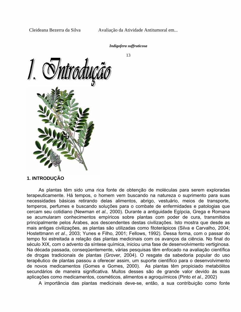
1. INTRODUÇÃO As plantas têm sido uma rica fonte de obtenção de moléculas para serem exploradas terapeuticamente. Há tempos, o homem vem buscando na natureza o suprimento para suas necessidades básicas retirando delas alimentos, abrigo, vestuário, meios de transporte, temperos, perfumes e buscando soluções para o combate de enfermidades e patologias que cercam seu cotidiano (Newman et al., 2000). Durante a antiguidade Egípcia, Grega e Romana se acumularam conhecimentos empíricos sobre plantas com poder de cura, transmitidos principalmente pelos Árabes, aos descendentes destas civilizações. Isto mostra que desde as mais antigas civilizações, as plantas são utilizadas como fitoterápicos (Silva e Carvalho, 2004; Hostettmann et al., 2003; Yunes e Filho, 2001; Fellows, 1992). Dessa forma, com o passar do tempo foi estreitada a relação das plantas medicinais com os avanços da ciência. No final do século XIX, com o advento da síntese química, iniciou uma fase de desenvolvimento vertiginosa. Na década passada, conseqüentemente, várias pesquisas têm enfocado na avaliação científica de drogas tradicionais de plantas (Grover, 2004). O resgate da sabedoria popular do uso terapêutico de plantas passou a oferecer assim, um suporte científico para o desenvolvimento de novos medicamentos (Gomes e Gomes, 2000). As plantas têm propiciado metabólitos secundários de maneira significativa. Muitos desses são de grande valor devido às suas aplicações como medicamentos, cosméticos, alimentos e agroquímicos (Pinto et al., 2002) A importância das plantas medicinais deve-se, então, a sua contribuição como fonte
natural de fármacos amplamente utilizados na clínica como a emetina, vincristina, colchicina, rutina, foscolina, taxol e a artemisinina (Filho, 1998), e por proporcionar grandes chances de obter-se uma molécula protótipo devido à diversidade de constituintes presentes nelas (Yunes e Calixto, 2001). As plantas podem ser classificadas de acordo com sua ordem de importância, iniciando-se pelas plantas empregadas diretamente na terapêutica, seguidos daquelas que constituem matéria-prima para manipulação e, por último, as empregadas na indústria para obtenção de princípios ativos ou como precursores em semi-síntese (Silva e Carvalho, 2004; Calixto, 2000). Portanto, a medicina alternativa através da utilização das ervas medicinais permanece como uma das formas mais comuns de terapia disponível às populações de todo mundo. Pode-se considerar como planta medicinal aquela planta administrada sob qualquer forma e por alguma via ao ser humano, exercendo algum tipo de ação farmacológica (Silva e Carvalho, 2004; Calixto,2000). De acordo com a Organização Mundial de Saúde (OMS) aproximadamente três quartos da população mundial usam atualmente ervas e outras formas de medicina tradicional na solução dos problemas básicos de saúde (Rao et al., 2005; Cragg e Newman, 1999). Embora, há tempos as plantas venham sendo usadas na medicina popular como instrumento de cura para o tratamento de diversas enfermidades (Rebecca et al., 2002), o cuidado no uso é imprescindível, devido à existência de plantas medicinais que podem causar toxicidade e certos efeitos colaterais danosos (Lapa, 2000). Cleideana Bezerra da Silva Avaliação da Atividade Antitumoral em...
14 Do reino vegetal se obtém substâncias extremamente tóxicas como a estricnina, digitoxina, tubocurarina, cocaína, entre outras (Lapa, 2000), não havendo assim razão para acreditar na inocuidade dos vegetais, mesmo que a incidência dos efeitos colaterais seja aparentemente menor com produtos fitoterápicos do que com drogas sintéticas (Drew e Myers, 1997). A toxicidade de uma planta pode ser definida como a capacidade de causar dano grave ou morte a um dado organismo (Draize et al., 1944). A toxicidade aguda produz efeitos adversos, provocando uma resposta rápida dentro de um curto período de tempo, após a administração de uma única dose ou doses múltiplas de um composto, no período de 24h, ocasionando geralmente uma elevada mortalidade (Abel, 1989). A dose única é utilizada para determinar a potência da substância (Oga, 2003). Os testes de toxicidade são elaborados com o objetivo de avaliar ou prever os efeitos tóxicos nos sistemas biológicos e averiguar a toxicidade relativa das substâncias (Forbes e Forbes, 1994; Loomis et al., 1996). A DL
50 (dose letal 50) corresponde à
dose capaz de causar a morte de 50% dos indivíduos de uma população, foi introduzida por Trevan, J.W. (1927), com a finalidade de estimar o potencial tóxico de substâncias que poderiam ser utilizadas em humanos como digitalis e insulina (Botham, 2003). Cleideana Bezerra da Silva Avaliação da Atividade Antitumoral em...
15 A preparação e uso apropriado de plantas na medicina trazem muitos benefícios, porém,
seus efeitos fisiológicos, genotóxicos e mutagênicos no organismo necessitam de maiores investigações (Nunes e Araújo, 2003). O panorama para a fitoquímica é muito mais importante e decisivo para o Brasil, ao considerarmos sua grande riqueza vegetal ainda sem estudo e as

possibilidades para o desenvolvimento de novos medicamentos. Em todo o mundo, apenas 17% das plantas têm sido estudadas quanto ao seu emprego medicinal e, na maioria dos casos, sem grande aprofundamento nos aspectos fitoquímicos, farmacológicos e toxicológicos. Esses dados demonstram o enorme potencial das plantas para a descoberta de novos fitoterápicos e fitomedicamentos (Hostettmann et al., 2003; Nodari e Guerra,1999; Cragg e Newman, Cleideana Bezerra da Silva Avaliação da Atividade Antitumoral em...
16 1999; Hamburger et al., 1991). A fitoterapia, pela sua capacidade de transformar e fornecer um saldo positivo quanto aos aspectos sócio-político-econômico, constitui-se uma valiosa opção para todos na América Latina, notadamente para o Brasil. Nos dias atuais em torno de 50% dos medicamentos utilizados são de origem sintética e cerca de 25% são de origem vegetal, isolados ou produzidos por semi-síntese (Calixto, 2000). Esse percentual é maior, quando restringido apenas aos fármacos antineoplásicos e antibióticos (Yue – Zhoug Shu, 1998). A pesquisa na busca da cura das neoplasias remonta desde tempos do antigo Egito (Kardinal e Yarbro, 1979), entretanto, somente após a descoberta, por Ehrlich em 1908, da transplantabilidade de tumores experimentais originados espontaneamente permitindo o teste de “screening” - é que teve início a busca científica de substâncias eficazes no tratamento dos mesmos. Recentemente tem crescido bastante o interesse no uso de tumores experimentais na pesquisa do câncer, especialmente na citologia e bioquímica de células tumorais, bem como no estudo da quimioterapia (Connors, 1969; Fávaro, 1990). De acordo com Cragg e Newman (2000), 50% de drogas em teste clínicos para atividade anticâncer foram isoladas da fonte natural ou estão relacionadas com elas. Produtos naturais derivados de plantas como flavonóides, terpenos, alcalóides (Osawa et al.,1990; Keith et al., 1990), têm recebido considerada atenção nos recentes anos, devido às suas diversas propriedades farmacológicas, incluindo efeitos citotóxicos, antioxidante e quimioprofilático de câncer (Roja e Heble, 1994; Defeudis et al., 2003; Takeoka e Dao, 2003). Cleideana Bezerra da Silva Avaliação da Atividade Antitumoral em...
17 O avanço do desenvolvimento na produção de novos agentes com atividade quimioterápica contra o câncer tem encontrado algumas dificuldades, principalmente quanto à inespecificidade destas drogas, o que acarreta danos excessivos às células normais (Moore e Erhlichman, 1987). Ainda que vários progressos venham sendo realizados no estudo das neoplasias, o estágio atual de conhecimento da biologia tumoral e da química médica tem aumentado pouco à possibilidade de uma nova classe de moléculas com ação antitumoral mais potente e sem efeitos colaterais. Assim continuam abertas novas linhas de pesquisa para novas drogas antineoplásicas, e a avaliação em vários sistemas tumorais e culturais de tecido, de onde poderão ser selecionados os compostos mais efetivos (Geran et al., 1972). Dessa forma, estudos com as diversas famílias de plantas medicinais devem prosseguir em busca de novos conhecimentos. A família Fabaceae, também conhecida como Leguminoseae, pertence à ordem Fabalis,

subclasse Rosidae e possui cerca de 670 gêneros com 18.000 espécies (Oliveira e Paiva, 2005), subordinadas a três subfamílias: Mimosoideae, Caesalpinioideae, Papilionoideae (Barroso 1984 e Lewis 1987). É a terceira maior família das angiospermas (Chappill, 1995), muitas delas possuindo importância econômica pela produção de alimentos como soja, ervilha, feijão, alfafa (Tucker, 2003), sendo suas espécies uma das maiores dentro do grupo das dicotiledôneas (Barroso, 1984).
Uma característica típica da família Fabaceae é apresentar em quase todas as espécies, simbiose de suas raízes com bactérias do gênero Rhizobium e semelhantes, Cleideana Bezerra da Silva Avaliação da Atividade Antitumoral em...
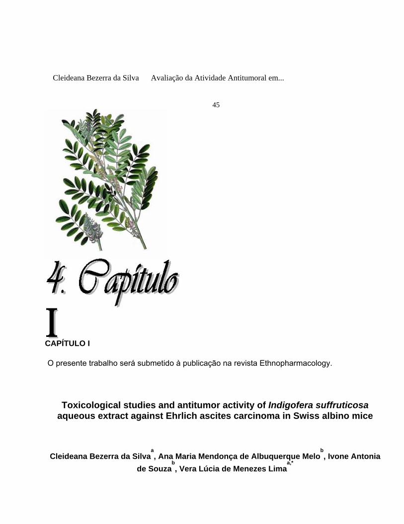
18 que fixam o nitrogênio da atmosfera, e também apresentam o fruto do tipo legume (algumas exceções), conhecido como vagem (Watson e Dallwitz, 1992). No semi-árido nordestino a subfamília Papilionoideae é um táxon bem representado e apresenta um grande número de gêneros dentro da Família Fabaceae, e entre eles está incluído o gênero Indigofera (Emparaire, 1989; Rodal, 1992; Alcoforado Filho, 1993; Oliveira et al., 1997; Ferraz et al., 1998; Lemos, 1999). A origem do nome Indigofera provém da palavra alemã Índigo, que significa produção de pigmento azul (Índigo Blue); que pode ser extraído (principalmente das sementes) de I. suffruticosa Mill, I. truxillensis (Pesavento, 2005), I. tinctoria L., I. arrecta Hoschst, I. argentea L. e I. suffruticosa guatemalensis de Kort y Thijise (Thomas, 1998). Portanto o uso mais conhecido e antigo do gênero Indigofera é a produção de pigmento azul, indigo blue, (Pesavento, 2005). A importância econômica do pigmento fez com que a planta fosse cultivada intensamente nos países asiáticos. A indústria usou o indigo até o início do nosso século quando a produção sintética de anilina substituiu o pigmento natural. Ainda hoje, as comunidades do interior do Brasil fabricam e usam o pigmento para colorir roupas de lã e algodão (Allen e Allen, 1981). O gênero Indigofera compreende aproximadamente 700 espécies herbáceas e arbustivas fitogeograficamente distribuídas na África tropical, Ásia, Austrália e América do Norte e Sul (Hassen et al., 2007). No Brasil há registro das espécies: I. suffruticosa, I. truxillensis, I. hirsuta (Pesavento, 2005) e I. microcarpa Desv. (Correa, 1984). São grupos de plantas silvestres (não cultivadas) que crescem espontaneamente em todos os solos agrícolas, principalmente nas imediações de cidades e vilas e em outras áreas de interesse do homem (Pesavento, 2005). Possui abundante ramagem, verde esbranquiçada, revestidas de pêlos e folhas elípticas compridas e em forma de palmas. Suas flores, róseas e miúdas, desabrocham em pequenos cachos; os frutos, em forma de vagens arredondadas e recurvas, contêm sementes parecidas com feijão. Possui raiz, principalmente sobre suas ramificações (Barroso, 1984), [figura 1]. Cleideana Bezerra da Silva Avaliação da Atividade Antitumoral em...
19 Típicos das leguminosas as Indigoferas têm alto teor em proteínas, têm habilidade para tolerar seca, inundações e elevadas salinidade tornando-as assim agronomicamente muito desejáveis (Sherman, 1982). Muitas espécies são usadas em países como África e Ásia, como forrageiras (I. hirsuta, I. pilosa, I. schimperi Sym, I. oblongifolia, I. spicat, I. subulata Sym e I. trita), também como adubo verde e cobertura de solo no caso da I. hirsuta e I. trita (Fröman, 1975). No Brasil, a

espécie I. hirsuta é usada como adubo verde e forragem e tem sido recomendada como potencialmente controladora de nematóides. Como forragem é bem aceita pelos animais após a fenação (Allen e Allen, 1981; Aylward et al., 1987). Informações etnofarmacológicas indicam que espécies do gênero Indigofera são utilizadas para o tratamento de afecções gastrointestinais. Outros atributos significantes, os quais espécies arbustivas como I.spinosa, valiosa como planta forrageira, é o seu ciclo de vida perene, palatabilidade, resistência para herbívoros e habilidade para resposta a pequenos eventos de chuvas (Coppock et al., 1986, 1988; Bamberg, 1986; Coughenour, et al., 1990). Esta combinação de peculiaridades é ideal para adaptação para ambiente árido e semi-árido. A espécie I. suffruticosa Mill (figura 1) é uma planta originária da Antilha e América Central (Almeida, 1993), encontra-se distribuída por toda a América tropical, (Cesário, 1980). No Brasil há registro da espécie nos estados do Mato Grosso (Fernandes, 1987), Alagoas (Ribeiro, 1984), Paraíba (Riet-Correa, 2000), Ceará, Rio Grande do Norte, Pará e Pernambuco (Neto et al., 2001). Cleideana Bezerra da Silva Avaliação da Atividade Antitumoral em...
20 Trata-se de uma planta arbustiva, medindo de 1-2 m de altura (figura 2), com ramos pubescentes, propagando-se por sementes. Folhas pinadas, com 7-15 folíolos oblongos ou ovais, glabros na face e no verso. Apresenta flores miúdas, numerosas, albo-rósea ou amarelada, em racemas axilares. Possui Pequena vagem falciforme com 6-10 sementes com aparência de feijão (Braga, 1976). I. suffruticosa Mill possui sinonímias tais como: anali tinctoria Var. Vera Kuntze, Indigofera anil L. Popularmente é conhecida como anil do campo, anileira, anileira-da-índia, anileira verdadeira, caá-chica, caá-chira, índigo, timbó-mirim, timbozinho, indigueira, indigófera. Cleideana Bezerra da Silva Avaliação da Atividade Antitumoral em...
21

Fonte: Wikipedia. Org Fonte: Silva, C. B. da, 04/2007
A B D C Fonte: Wikipedia. Org Fonte: Silva, C. B. da, 04/2007 Figura 1 – Indigofera suffruticosa Mill. A: ramos com folhas e inflorescência; B: ramos com folhas; C: ramos floridos; D: ramos com folhas e sementes. Cleideana Bezerra da Silva Avaliação da Atividade Antitumoral em...
22


Fonte: Silva, C. B. da, 04/2007.
Figura 2: Indigofera suffruticosa Mill - Vista parcial de um indivíduo adulto. Cleideana Bezerra da Silva Avaliação da Atividade Antitumoral em...
23

Miller e Smith, 1973, fizeram as primeiras investigações a respeito da composição
química do gênero Indigofera, utilizando extratos de sementes da espécie I. suffruticosa, sendo atribuída à mesma, como uma fonte rica em aminoácidos com prováveis ações tóxicas. Ésteres de glicose de ácido 3-nitropropanóico foram isolados das espécies: I. oblongifolia, I. linnaei, I. spicata (Syn. I. endecaphylla) e na I.suffruticosa (Lohda et al., 1997; Majak et al., 1992; Garcez et al., 1989, 2002), os quais evidenciam em outros membros de leguminosas , efeitos tóxicos desse composto em animais domésticos, insetos e outros animais devido à sua conversão para ácido 3-nitropropanóico, uma toxina respiratória que inibe enzimas mitocondriais (Anderson et al., 1998). Foram, também, isolados três furano-flavonóides e um raro flavonol glicosideo da I. tinctoria (Narender et al., 2006). Paiva (1987) realizou análise quantitativa de proteína e fibra bruta de I. suffruticosa e considerou a mesma um bom indicativo para forragem de ruminantes. Kamal e Mangla (1993) identificaram, caracterizaram e quantificaram seis rotenóides de diferentes órgãos como raiz, caule, semente e folhas de I. suffruticosa e verificaram que essa planta tem bioeficácia contra larvas de Anopheles e Callosobruchus chinensis adultos. Estudo fitoquímico preliminar das folhas, caule e sementes de I. suffruticosa, demonstrou abundante presença de alcalóides apenas nas folhas, polifenóis (cumarina e ácido clorogênico) e flavonóides também presentes apenas nas folhas; triterpenóides e/ou esteróides (abundantes nas folhas e, em menor extensão, no caule e sementes) e oses redutoras (em todas as partes estudas do vegetal) [Leite, 2003]. Também foi constatada a presença do indigo (figura 3) principalmente nas

sementes, apresentando-se como uma molécula com forte fluorescência, sugerindo algo bastante singular e, de certa forma, a presença dessa molécula confirma esse táxon. se como uma molécula com forte fluorescência, sugerindo algo bastante singular e, de certa forma, a presença dessa molécula confirma esse táxon. Cleideana Bezerra da Silva Avaliação da Atividade Antitumoral em...
24 Figura 3: Fórmula estrutural do indigo Figura 3: Fórmula estrutural do indigo I. suffruticosa Mill é popularmente usada em diferentes países contra diversos problemas da saúde. Suas folhas têm sido usadas como problemas estomacais (Agra et al., 1996), antiespasmódica, sedativo, diuréticos, purgativos, enquanto a raiz tem sido usada como odontálgica (Lorenzi, 1982) e útil na cura da icterícia (Lainetti e Brito, 1980 e Correa, 1984). Empregada também nas mordeduras de cobras, e como insetífuga, sendo esta última propriedade extensiva às sementes depois de pulverizadas (Pio Correia, 1926). Possui propriedades medicinais no tratamento de febres, parasitoses, doenças de pele e problemas cardíacos (Allen e Allen, 1981). Estudos farmacológicos mostraram que extratos de I. suffruticosa apresentaram atividade anticonvulsivante (Alejo et al., 1996), antigenotóxica (Badell et al., 1998) e antiepiléptica (Roig e Mesa, 1974). I. suffruticosa Mill é popularmente usada em diferentes países contra diversos problemas da saúde. Suas folhas têm sido usadas como problemas estomacais (Agra et al., 1996), antiespasmódica, sedativo, diuréticos, purgativos, enquanto a raiz tem sido usada como odontálgica (Lorenzi, 1982) e útil na cura da icterícia (Lainetti e Brito, 1980 e Correa, 1984). Empregada também nas mordeduras de cobras, e como insetífuga, sendo esta última propriedade extensiva às sementes depois de pulverizadas (Pio Correia, 1926). Possui propriedades medicinais no tratamento de febres, parasitoses, doenças de pele e problemas cardíacos (Allen e Allen, 1981). Estudos farmacológicos mostraram que extratos de I. suffruticosa apresentaram atividade anticonvulsivante (Alejo et al., 1996), antigenotóxica (Badell et al., 1998) e antiepiléptica (Roig e Mesa, 1974). Recentes estudos com extrato aquoso de folhas de I.suffruticosa demonstraram possuir atividade antimicrobiana contra a bactéria gram-positiva Staphulococcus aureus e contra os fungos dermatófitos Microsporium canis e Recentes estudos com extrato aquoso de folhas de I.suffruticosa demonstraram possuir atividade antimicrobiana contra a bactéria gram-positiva Staphulococcus aureus e contra os fungos dermatófitos Microsporium canis e Trichophyton rubrum (Leite et al., 2006); atividade citotóxica em células embrionárias de ratos, que inibiu a reprodução celular a partir do estágio de mórula e blástula (Leite et al., 2004) e atividade antiinflamatória na redução de edema de pata de camundongos Cleideana Bezerra da Silva Avaliação da Atividade Antitumoral em...

25 5
(Leite et al., 2003). Ações biológicas, bem como a toxicidade de plantas medicinais, vêm sendo avaliadas em pesquisas através de bioensaios de laboratórios usando a Artemia salina (Mathews, 1995; Fumaral e Garchitorena,1996). A Artemia salina Leach é um microcrustáceo, componente da fauna aquática salina e salobra do ecossistema marinho, sendo considerada cosmopolita e, portanto, adequada a vários ambientes. É conhecida como “camarão de salmoura” e internacionalmente como “brine shrimp”; e é um dos organismos-alimentos mais utilizados no mundo (Barbieri-Junior e Neto, 2001). A Artemia salina é um invertebrado que possui de 8 a10 mm de comprimento, pertence ao filo Arthropoda, classe Crustácea, subclasse Brachiopoda, ordem Anostraca, família Artemiidae (Barbieri-Junior e Neto, 2001). Nada sempre de dorso, direcionada para a luz ou claridade do ambiente em que se encontra (fototaxia positiva). Reproduz-se com muita facilidade e rapidez, apresentando duas rotas reprodutivas. Em condições normais de baixa salinidade e elevada abundância de alimentos, os animais são ovovivíparos (Barbieri-Junior e Neto, 2001). Porém, se as condições ambientais se tornam, por qualquer motivo, demasiadamente estressante, os animais adotam um modelo de reprodução ovíparo, ou seja, produzem cistos de resistências. Seus ovos possuem um mecanismo fisiológico da eclosão disparado na presença da luz. É de fácil cultivo, estudo e pode viver por mais de 4 meses, possuindo quatro estágios de desenvolvimento: náuplio, metanáuplio, pré-adulto e adulto (Barbieri-Junior e Neto, 2001). Náuplio é a larva recém-eclodida. Caracteriza-se por possuir elevadas quantidades de reservas vitelinas (lipídeos e carboidratos) e por não apresentar segmentos no corpo. Assim que eclode, a larva é muito rica em vitelo, apresentando uma forte coloração alaranjada. No estágio de metanáuplio, o corpo da larva apresenta-se bastante segmentado. O vitelo já foi consumido e a larva depende de alimento externo para sobreviver. O corpo do pré-adulto apresenta 11 segmentos e as antenas sofrem modificações que possibilitam identificar o sexo do animal. Nos machos, as antenas ficam maiores e mais fortes. Nas fêmeas, as antenas são bem menores e adquirem o formato de folha. No adulto, o dimorfismo sexual é facilmente perceptível e os animais são capazes de se reproduzir (Barbieri-Junior e Neto, 2001). Cleideana Bezerra da Silva Avaliação da Atividade Antitumoral em...
26 O microcrustáceo, A. salina, é amplamente usado em aplicações toxicológicas e pesquisas para estabelecer a toxicidade de produtos químicos e naturais através da estimativa da concentração letal média, valor da CL
50 como parâmetro da avaliação da atividade biológica
(McLaughlin et al., 1991; Lewan et al., 1992; Barahona e Sanchez-Fórtun, 1996; Parra et al.,
2001). Além do mais, o teste com Artemia tem sido considerado como bioindicador de contaminação ambiental por traços como de arsênio, chumbo, cobre, zinco, cadmium, mercúrio e selenium; pela susceptibilidade desses microcrustáceos frente a esses elementos. Dentre as vantagens deste método está a facilidade de execução, baixo custo, reprodutibilidade, rapidez, disponibilidade comercial dos ovos, a não exigência de equipamentos especiais e ainda a necessidade de pequenas quantidades da amostra-teste para a realização dos experimentos (Ohno, et al., 1997; Calow, 1993). Cleideana Bezerra da Silva Avaliação da Atividade Antitumoral em...
27 A importância de bioensaios com as larvas de Artemia salina se deve ao fato de que diversos trabalhos buscam correlacionar a toxidade sobre esta espécie com atividades como antifúngica, antiviral, antimicrobiana, parasiticida, inseticida, tripanossomicida (Meyer et al., 1982; McLaughlin et al., 1995; Macrae et al., 1988; Sahpaz et al.,1994; Alves et al., 2000, Sanchez et al., 1993; Ojala et al., 1999). Além disso, o ensaio de letalidade de brine shrimp é considerado um dos mais eficientes instrumentos da taxa preliminar de toxicidade geral e tem mostrado boa correlação com atividade citotóxica contra alguns tumores sólidos (McLaughlin et al., 1991). Tumor é um termo mais geral, que significa uma elevação do tecido e pode ser encontrado em processo inflamatório, infeccioso, entre outros e, certamente, em neoplasia. Neoplasia (neo = novo; plasia = tecido) se constitui em um processo patológico resultante da formação e crescimento de uma massa de células neoplásicas (tumor). As neoplasias malignas recebem a designação de câncer (Anderson et al.,1982; Body e Sheldon, 1984; Contran et al.,2000). O termo câncer (do latim Karkinos = caranguejo) foi empregado pela primeira vez por Galeno, aproximadamente 138 – 201 a.C. ao descrever um tumor maligno de mama. O órgão apresentava as veias superficiais túrgidas e ramificadas, assemelhando-se às patas de caranguejo (Filho, 2000; Fleck, 1992; Murrad, 1996). Câncer é um tumor maligno e segundo Organização Mundial de Saúde (WHO, 2005) é um importante problema de saúde que afetou em 2005 a vida de mais de 7,6 milhões de pessoas pelo mundo inteiro. O tumor é formado por uma proliferação local de clones celulares atípicos com reprodução desordenada e que tendem a se modificar de forma a se tornar autônomo dos habituais controles de crescimento, bem como apresentar alterações da sua diferenciação (Baracat et al., 2000). A perda do controle do desenvolvimento de alguma das células do corpo e sua proliferação excessiva resultam em tumores do tipo benigno e maligno, segundo derivação de suas matrizes. Os tumores benignos são bem diferenciados e constituídos por células semelhantes às células do tecido de origem. Enquanto os tumores benignos são de crescimento lento, expansivo e são bem tolerados pelo organismo do hospedeiro, os tumores malignos têm crescimento rápido, de forma invasiva e produzem metástases (Nobre e Junqueira, 1967; Franks e Teich, 1990; Murrad, 1996). O poder de invadir os tecidos vizinhos, assim como migrar pelo organismo, provocando metástases são os grandes responsáveis por levar o indivíduo ao óbito. Cleideana Bezerra da Silva Avaliação da Atividade Antitumoral em...
28 Os tumores malignos derivados de tecidos de origem mesenquimal são chamados de sarcomas

– a palavra deriva do grego Sarkos, e os oriundos de tecido epitelial, são denominados de carcinomas (Contran et al., 2000). O Carcinoma de Ehrlich provavelmente surgiu na glândula mamária de camundongos fêmea, transplantado inicialmente na forma sólida. Ele foi introduzido por Paul Ehrlich em 1896, e descrito em 1906. Lowenthal e Jahn, em 1932, desenvolveram sua forma ascítica, através do transplante seriado de seu líquido ascítico, fluido leitoso, com células tumorais em suspensão, inoculadas intraperitonealmente (Dagli, 1989; Young e Hallowes, 1973; Sigiura, 1965). Voet, 1990; Murrad, 1996, estimaram que 80% das neoplasias malignas têm origem através de estímulos ambientais. Desses estímulos contribuem para a carcinogênese: agentes biológicos (como alguns tipos de vírus), carcinógenos químicos (hormônios, metais, aminas aromáticas, etc.) e físicos (radiação ionizante, raios ultravioletas) [Volgelstein et al., 1993]. Cleideana Bezerra da Silva Avaliação da Atividade Antitumoral em...
29 O desenvolvimento neoplásico é um processo dinâmico caracterizado por etapas denominadas de múltiplos estágios da carcinogênese, que são divididos em: iniciação, promoção e progressão (figura 4) [Harris, 1991; Hursting et al., 1999; Tennant et al.,1997]. O estágio de iniciação se origina com danos ao DNA em função da exposição de uma população de células ao carcinógeno. O DNA danificado produz mutações genéticas, constituindo o primeiro passo para o desenvolvimento neoplásico (Jakóbisiak et al., 2003). No entanto, apenas essa etapa é insuficiente para formação de tumor. Para o estabelecimento do processo é preciso que as células alteradas por carcinógenos sofram pelo menos um ciclo de proliferação, de maneira que se torne fixa ou permanente (Contran et al., 2000). O estágio de promoção é a segunda etapa da carcinogênese, pode ser definida como um processo de expressão clonal de células iniciadas que resultam na formação de células pré-neoplásicas, produzindo nódulos, pólipos ou papilomas (Jakóbisiak et al., 2003). A etapa de progressão compreende a fase de evolução da neoplasia maligna que já está expressa fenotipicamente a nível histológico, sendo caracterizada pela transformação de células pré-neoplásicas em tumor maligno invadindo tecido circunvizinho e formando metástases (Jakóbisiak et al., 2003). Cleideana Bezerra da Silva Avaliação da Atividade Antitumoral em...
30

INICIAÇÃO
- Dano ao DNA - Indução de mutação em genes alvos críticos - Ativação de protooncogêneses - Iniciação de genes supressores de tumor - Replicação celular e fixação da mutação.
PROMOÇÃO - Expansão clonal de células tronco - Desenvolvimento de tumor benigno
PROGRESSÃO
- Expressão alterada de enzimas - Proteólises - Adesão - Invasão - Migração - Metástase Fonte: Jakóbisiak, et al., / Immunology Letters, 90(2003) 103-122. Figura 4: Processo de múltiplos estágios de Carcinogênese

Cleideana Bezerra da Silva Avaliação da Atividade Antitumoral em...
31

Cleideana Bezerra da Silva Avaliação da Atividade Antitumoral em...
32
2. OBJETIVOS 2.1. Objetivo Geral Estudar os efeitos do extrato aquoso de folhas de I. suffruticosa e o seu potencial como agente

antitumoral por meio do Carcinoma de Ehrlich. 2.2. Objetivo Específico Definir protocolo para obtenção de componentes ativos da folha de I. suffruticosa. Determinar a toxicidade aguda e estimar o valor da DL
50 em camundongos por via oral e
intraperitoneal. Avaliar a toxicidade e determinar a CL
50 de I. suffruticosa através do bioensaio com Artemia
salina. Analisar a atividade antineoplásica do extrato aquoso de I. suffruticosa no Carcinoma de
Ehrlich em camundongos. Determinar os efeitos da administração por via intraperitoneal do extrato aquoso de folhas
de I. suffruticosa nos níveis séricos de colesterol total e triglicerídeos em camundongos. Cleideana Bezerra da Silva Avaliação da Atividade Antitumoral em...
33

Cleideana Bezerra da Silva Avaliação da Atividade Antitumoral em...
34
3. REFERÊNCIAS BIBLIOGRÁFICAS ABEL, P. D. Water Pollution Biology. Ellis Horwood Ltda, Publishers. Chichester. p. 231, 1989. AGRA, M.F., LOCATELLI, E., Rocha, E. A., BARACHO, G. S., FORMIGA, S. C. Plantas medicinais dos Cariris Velhos, Paraíba, Brasil, parte II: subclasses Magnoliidae, Caryophyliidae, Dilleniidae e Rosidae. Revista Brasileira de Farmácia 77, pp. 97-102, 1996. ALCOFORADO-FILHO, F.G. Composição floristica e fitossociologia de uma área de caatinga arbórea no município de Caruaru-PE, Recife: UFPE, p. 220, Dissertação de Mestrado, 1993. ALEJO, J. P., MIRANDA, R., RODRIGUES, G. Actividad Anticonvulsivante; (Antiepileptica) del extracto fluido de Indigofera suffruticosa (Anil Cimarrón). Rev Cubana Plant Med, 1 (2): pp. 7-10, 1996. ALLEN e ALLEN, RODRIGUES-KABANA. Leguminosae. University of Wisconsin Press, Madison; Nematropica 18 (1): pp. 137-142; 1981. ALMEIDA, de E. R. Plantas Medicinais Brasileiras: Conhecimentos populares e científicos, Hemus Editora LTDA. São Paulo, 341p.; 1993. ALVES, T. M. D., SILVA, A. F., BRANDÃO, M. GRANDI, T. S. M.; SMÂNIA, E. F. A.; SMÂNIA, A.; ZANI, C. L. Biologicy screening of Brazilian medicinal plants. Mem Inst Oswaldo Cruz, V. 95, pp. 367-373; 2000.

ANDERSON, W. A. D.; JOHN, M.; KISSANE, M. D. Patologia. Volumes 1 e 2. 7ª edição. Editora Guanabara Koogan. Rio de Janeiro. 1796 p. 1982. ANDERSON, R. C., MAJAK, W. RASMUSSEN, M.A., ALLISON, M. J. Detoxification potential of a new species of ruminal bacteria that metabolize nitrate and naturally occurring nitrotoxins. In: Garland, T., Bar, A.C. (Eds.), Toxic Plants and Other Natural Toxicants. CAB International, Wallington, Oxon, pp. 154-158; 1998. AYLWARD, J.H., COURT, R.D., HGVLOCK, K. P., STRLCKLO, R.W., HEGARTY, M.P. Indigofera species with agronomic potential in the tropics. Rat toxicity studies. Aust. J. Agric Res. 38 (1), pp. 177-88; 1987. BADELL, J. B., RUIZ, A. R., PARRA, A. B., MICHELENA, M. D., MICHELENA, M. J. M., ARMENTEROS, A. E. Evaluación genotóxica del extracto fluido de Indigofera suffruticosa Mill (Anil Cimarrón) mediante el ensayo de anomalias en la cabeza de los espermatozoides. Rev Cubana Plant Med, 3(2), pp. 58-61; 1998. Cleideana Bezerra da Silva Avaliação da Atividade Antitumoral em...
35 BAMBERG, I. E. Effect of clipping andwatering frequency on production of the Africandwarf shrub, Indigofera spinosa. M.Sc. Thesis. Colorado State University, Fort Collins, CO, USA. 1986. BARACAT, F. F., FERNANDES JR. H. J., SILVA, M. J. Cancerologia Atual. Um enfoque multidisciplinar. Editora Roca, São Paulo. 2000. BARBIERE-JUNIOR, R. C. e Neto, A. O. Camarões Marinhos: Reprodução, Maturação e Larvicultura. Editora Aprenda Fácil, Viçosa – MG. V.1, 2001. BARROSO, G.M. Sistemática das Angiospermas do Brasil. São Paulo: EDUSP, Vol.2. 1984. BARAHONA, M. V., SÁNCHEZ-FORTUN, S. Comparative sensitivy of three age classes of Artemia salina larvae to several phenolic compounds. Bulletin of environmental contamination and toxicology. v. 56, pp. 271-278; 1996. BODY, W. and SHELDON, H. 8ª edição. Editora Guanabara Koogan. Rio de Janeiro. 512 p. 1984. BOTHAM, P.A. Acute systemic toxicity- prospects for tiered testing strategies. Toxicology in Vitro. v 17, pp. 1-4; 2003. BRAGA, R. Plantas do Nordeste Especialmente do Ceará. 3ª edição. Mossoró Escola Superior de Agricultura. p. 452, 1976. CALIXTO, J.B. Efficacy, safety, quality, control, marketing and regulatoryguidelines for herbal

medicines (phytoterapeutic agents). Braz J med Biol Res, 33, pp. 179-189; 2000. CALOW, P. Marine and estuarine invertebrate toxicity tests in HOFFMAN, D.; RATTNER, B.; BURTON, A. & CARNS, J. (ed.) Handbook in Ecotoxicology. Vol.l. Blackwell Scientific Publication. London. pp. 1-5; 1993. CESÁRIO DE MELLO, A. Presença de uma substância com atividade semelhante à acetilcolina na folha de Indigofera suffruticosa (vulgarmente conhecida como anil), anais do VI simpósio de plantas medicinais do Brasil, setembro, Fortaleza-CE. 1980. CONNORS, T.A. Anti-cancer agents. Their detection by screening tests and their mechanism of action. In: S cientific basis of cancer chemotherapy. v. 21 New York, Springer – Verlag., (Recents Results in Cancer Research) pp. 1-17; 1969. CONTRAN, R.S., KUMAR, V., COLLINS, T. Neoplasia. In: CONTRAN, R.S.; KUMAR, V., COLLINS, T. (Eds.) ROBBINS. Patologia estrutural e funcional. Rio de Janeiro: Editora Guanabara Koogan S.A., pp. 233-295. 2000. Cleideana Bezerra da Silva Avaliação da Atividade Antitumoral em...
36 COPOOCK, D. L., ELLIS, J. E., SWIFT, D. M. A comparative in vitro digestion trial using inocula of livestock from Turkana and Kitale, Kenya. J. Agr. Sci. (Cambridge) 110, pp. 61–63; 1988. COPOOCK, D. L., ELLIS, J. E., SWIFT, D. M. Seasonal nutritional characteristics of livestock diets in a nomadic pastoral ecosystem. J. Appl. Ecol. 23, pp. 573–583; 1986. CORREA, M. P. Dicionário das plantas úteis do Brasil e das exóticas cultivadas. IBDF. MA. Rio de Janeiro, RJ. 1: 130-3. 1984. COUGHENOUR, M.B., COPPOCK, D.L., ROWLAND, M.; ELLIS, J.E. Dwarf shrub ecology in Kenya’s arid zone: Indigofera spinosa as a key forage resource. J. Arid Env. 18, pp. 301–312; 1990. CHAPPILL, J.A. Cladistic analysis of the Leguminosae: The development of an explicit hypothesis. In: CRISP, M.D.; DOYLE, J.J. (Ed.). Advances in legumes systematic. Richmond: Royal Botanic Gardens, Kew, pp. 1-10; 1995. CRAGG, G. M., NEWMAN, D.J. Antineoplastic agents from natural sources: achievements and future direction. Excpert Opinion on Investigational Drugs, 9: 1-15. 2000. CRAGG, G. M., NEWMAN, D.J. Discovery and development of antineoplastic agents from natural sources. Cancer Invest., v. 17 (2), pp. 153-163; 1999. DAGLI, M.L.Z. Disseminação linfática do tumor de Ehrlich: estudo experimental. Dissertação (Mestrado) Patologia Experimental Comparada, Faculdade de Medicina Veterinária e Zootecnia

da Universidade de São Paulo (USP), São Paulo, 1989. DEFEUDIS, F.V., PAPADOUPOULOS, V., DRIEU, K. Ginkgo biloba extracts and cancer: a research area in its infancy. Fundam Clin. Pharmacol. 17: pp. 405-17. 2003. DRAIZE, J.H., WOODARD, G., CALVERY, H.O. Method for the study of irritation and toxicity of substances applied topically to the skin and mucous membranes. J. Pharmacol. Exp. Ther., v. 82, pp. 337- 90; 1944. DREW, A., MYERS, S.P. Safety issues in herbal medicine: implications for the health professions. Med. J. Australia, v. 166, pp. 538-541; 1997. EMPERAIRE, L. Vegetation et gestion des ressources naturelles dans la caatinga du sud-est du Piauí (Brésil). Doctorat d’Etat ès Sciences Naturelles: Université Pierre et Marie Curie, Paris. 1989. FÁVARO, O. C. N.; OLIVEIRA, M. M.; ROSSIMI, M. A. A. Seleção por meio de células KB de substâncias e extratos potencialmente ativos em quimioterapia do câncer. Anais de Academia Brasileira de Ciências, Rio de Janeiro, v. 62, (3), pp. 217-224; 1990. Cleideana Bezerra da Silva Avaliação da Atividade Antitumoral em...
37 FELLOWS, L.E. Pharmaceuticals from traditional medical plants and other: Future prospects: In: J.D. Coombers, ed.New drugs from natural sources. London, IBC technical Services. 1992. FERNANDES, A. Noções de Toxicologia e Plantas tóxicas. 2 ed. Fortaleza: BNB, 80p. Monografia. 1987. FERRAZ, E.M.N., RODAL, M.J.N.; SAMPAIO, E.V.B. e PEREIRA, R. DE C.A. Composição floristica em trechos de vegetação de caatinga e brejo de altitude na região do vale do Pajeú, Pernambuco. Revista Brasileira de Botânica, 21, pp. 7-15; 1998. FILHO, G.B. Patologia. 6º ed. Rio de Janeiro: Guanabara-Koogan, pp. 157-159; 2000. FILHO, C.V. Estratégias para a obtenção de compostos farmacologicamente ativos a partir de plantas medicinais. Conceitos sobre modificação estrutural para otimização da atividade, Química Nova; 21, 1. 1998. FORBES, V.E. e FORBES, T.L. Ecotoxicolgy in Theory and Practice. Chapman and Hall. Londres. p. 247; 1994. FLECK, J. Câncer – integração clínico-biológica. Rio de Janeiro: Médica e Científica. 1992. FRANKS, J. e TEICH Introdução à biologia celular e molecular do câncer. São Paulo, Roca cap. pp.1- 24. O que é câncer? 1990.

FROMAN, B. An illustrated guide to the pasture legumes of Ethiopia. Rural Development Studies No 3. Rural Development Section and Department of Plant Husbandary, College of Agriculture, S-750 07, Uppsala, Sweden. 1975. FUMARAL, F., GARCHITORENA, M.: Artemia salina, Recolección, descapsulación y desarrollo. Revista Aquamar. Ano 4 nº3: 22-4, 1996. GARCEZ, W. S., GARCEZ, F.R., BARISON, A. Additional 3-nitropropanoyl esters of glucose from Indigofera suffruticosa (Leguminosae). Biochemical systematics and ecology. V. 31, pp. 207–209; 2002. GARCEZ, W.S., GARCEZ, F.R., HONDA, N.K., SILVA, A.J.R. Phytochemistry 28, 1251. 1989. GERAN, R.I., GREEBERG, N.H. MCDONALD, M.M. Protocols for screening chemical agents and natural products against animal tumors and other biological systems, Cancer Chemother, 3, pp. 1-103; 1972. Cleideana Bezerra da Silva Avaliação da Atividade Antitumoral em...
38 GOMES, L.J. e GOMES, M.A.O. Extrativismo e Biodiversidade: o caso da fava d’anta. Ciência Hoje, n. 161, pp. 66-69; 2000. GROVER, J.K., YADAV, S.P. Pharmacological actions and potential uses of Momordica charantia: a review. Journal of Ethnopharmacology, 93, pp. 123–132; 2004. HAMBURGER, M., MARSTON, A., HOSTETMANN, K. Search for new drugs of plant origin. Advances in Drug Research, 20, pp.167-169; 1991. HARRIS, C.C. Chemical and physical carcinogenesis: advances and perspectives for the 1990s. Cancer Res., V.51, p. 5023s – 5044s. 1991. HASSEN, A., RETHMAN, N.F.G., NIEKERK, W.A. VAN, TIELELE, T.J. Influence of season/year and species on chemical composition and in vitro digestibility of five Indigofera accessions. Animal Feed science and Technology, 136, pp. 312-322; 2007. HOSTETTMAN, K., QUEIROZ, E.F., VIEIRA, P.C. A importância das plantas medicinais: Princípios ativos de plantas superiores. Série de textos da Escola de Verão em Química-IV, São Carlos, SP, EdUFSCar, 152 p. 2003. HURSTING, S.D. et al. Mechanism-Based Cancer Preventio Approaches: Targets, Examples, and the Use of Transgenic Mice. J. Natl.Cancer Inst., v.91, pp. 215–225; 1999. JAKOBISIAK, M., LASEK, W., GOLAB, J. Natural mechanisms protecting against cancer. Immunology Letters,v. 90, pp. 103–122; 2003.

KADINAL, C.G., YARBO, J.W. A Conceptual History of Cancer, Seminars in Oncology, 6, pp. 396-408. 1979. KAMAL, R.,MANGLA, M. In vivo In vitro, Investigation on retenoids from Indigofera suffruticosa and their biofficacy against the larvas of Anopheles stephensi and adults of Callosobruchus chinesis. J Biosc. 18 (1): pp. 93-110. 1993. KEITH, M. W., SALLY, A. L., MICHAEL, W. S., THOMAS, J. G., GARRY, M. M. Taxus Spp. Needles contain amounts of taxol comparable to the stem bark of taxus brevifolia: analysis and isolation. J. Nat Prod. 53, pp. 1249-1255; 1990. LAINETTI, R. e BRITO, N. R. S. A saúde pelas plantas e ervas do mundo inteiro. Ed. Tecnoprint. Rio de Janeiro, RJ. 37p. 1980. LAPA, A. J. Farmacologia e toxicologia de produtos naturais. In: Simões, C.M.O. (org.) Farmacognosia da planta ao medicamento. 2° ed. Porto Alegre/Florianópolis: Editora da UFRGS/ Editora da UFSC, pp. 181-196. 2000. Cleideana Bezerra da Silva Avaliação da Atividade Antitumoral em...
39 LEITE, S. P., VIEIRA, J. R. C., MEDEIROS, P. L., LEITE, R. M. P., LIMA, V. L. M., XAVIER, H. S., LIMA, E. O. Antimicrobial activity of Indigofera suffruticosa. Evidence- Based Complementary and Alternative Medicine, v. 3, n. 2, pp. 261-265. 2006. LEITE, S. P., MEDEIROS, P. L., SILVA, E. C., MAIA, M. B. S., LIMA, V. L. M., SAUL, D. E. Embryotoxicity in vitro with extract of Indigofera suffruticosa leaves. Reproductive Toxicology, 18: 701-705. 2004. LEITE, S. P. Indigofera suffruticosa Mill: Ensaio fitoquímico e ações biológicas. Tese de doutorado da Universidade Federal da Paraíba, João Pessoa, 90. 2003. LEITE, S. P., SILVA, L. L. S., CATANHO, M. T. J. A., LIMA, E. O., LIMA, V. L. M. Atividade antiinflamatória do extrato de Indigofera suffruticosa. Revista Brasileira de Ciências da Saúde, 7: 47–52. 2003. LOHDA, V.; KHAN, H.A.; GHANIM, A. J. Indian Chem. Soc. 74, 425. 1997. LOEWENTHAL, H.; JAHN, G. Ubertrangungversuche mit carcinomatoser mause-Ascites flussigkeit und ihr Verhalten gegen physikalische und Chemische Einwirkungen. Ztschr.f. Krebsforsch,v.37, p. 439, 1932. LEMOS, J. R. Fitossociologia do componente lenhoso de um trecho de vegetação arbustiva caducifólia espinhosa no parque Nacional Serra da Capivara, Piauí, Brasil. Recife: UFPE, 55 p. Dissertação de Mestrado. 1999.

LEWAN, L., ANDERSON, M., MORALES – GOMES, P. The use of Artemia salina in toxicity testing. Alternatives To Laboratory Animals. V. 20: 297-301. 1992. LEWIS, J. P. Legumes of Bahia. Kew: Royal Botanic Garden. 1987. LOOMIS, M. D., HAYES, A. W. Loomis´s essentials of toxicology. 4° ed. California: Academic Press, 1996. LORENZI, H. Plantas daninhas do Brasil: terrestres, aquáticas, parasitas, tóxicas e medicinais. Nova Odessa, SP. Edição do Autor, p. 271-2. 1982. MAJAK, W., BENN, M., MCEWAL, D., PASS, M.A. Three nitropropanoyl esters of glucose from Indigofera linnae. Phytochemistry 31, 2393-5. 1992. MACRAE, W.D.; HUDSON, J. B.; J.Of Ethnopharmacology. V. 22, 143. 1988. MATHEWS, R.S.:Artemia salina as a test organism for measuring superoxide-mediate toxicity. Free Radia Biol. Med. 18 (5): 919-22. 1995. MCLAUGHLIN, J.L., SAIZARBITORI, T. C., ANDERSON, J. E. Três bioensayos simples para químicos de productos naturales. Rev. Soc. Venez Quim. V. 18, 13-18; 1995. Cleideana Bezerra da Silva Avaliação da Atividade Antitumoral em...
40 MCLAUGHLIN, J.L., CHANG, C.J., SMITH, D.L. Bench-top bioassays for the discovery of bioactivity natural products: an update. In: Rahman, A.U. (Ed.), studies in natural products chemistry. Elsevier, Amsterdam. 1991. MEYER, B. M., FERRIGNI, N. R., PUTNAM, J. E., JACOBSEN, L. B., NICHOLS, D. E., MCLAUGHLIN, J. L. Brine shrimp, a convenient general bioassay for active – plant constituents. Planta Med. V. 45: 31-34. 1982. MILLER, R. W., SMITH, J.R. Seeds of Indigofera species: Their content of amino acids that may be deleterious. J. Agr Food Chem 21 (5): 909-912. 1973. MOORE, M. J. and ERHLICHMAN, C. Therapeutc Drug Monitoring in Oncology Problems and Potencial in Antineoplastc Therapy, Clin. Pharmacokinetics, 13: 205-227. 1987. MURRAD, A. M. & KATZ, A. Oncologia: Bases Clínicas do Tratamento. Guanabara Koogan, Rio de Janeiro. Cap. 1. p. 3-89. Aspectos Etiobiológicos do Câncer. 1996. NARENDER, T., KHALIG, T., PURIB, A., CHANDER, R. Antidyslipidemic activity of furano-flavonoids isolated from Indigofera tinctoria. Bioorganic & Medicinal Chemistry Letters. V. 16, 3411–3414. 2006. NETO, B.D.J., OLIVEIRA, C.M.C., PEIXOTO, V.P., BARBOSA, P.B.I., AVILAR, C.S.,

TOKARNAI, H.C. Anemia hemolítica causada por Indigofera suffruticosa (Leg. Papilonoideae) em bovinos. Pesq. Rev. Bras. 21 (1), 18-22. 2001. NEWMAN, D.J., CRAGG, G.M., SNADER, K.M. The influence of natural products upon drug discovery. Nat. Prod. Rep., v. 17, 215 – 234. 2000. NOBRE, M.O.R., JUNQUEIRA, C. Etiopatologia dos tumores. In: Cancerologia Prática. V.1 São Paulo: Fundo editorial Procienx, cap.2, 23-57. Anatomia patológicatumores malignos, 1967. NODARI, R.O., GUERRA, M.P. Biodiversidade: Aspectos Biológicos, geográficos, legais e éticos, Apud: Simões, O.M.C., Schenkel, R.P., Gosmann, G., Mello, P.C.J.; Mentz, A.L.; Petrovick, P.R. Farmacognosia da planta medicamento. Editora da Universidade Federal do Rio Grande do Sul. 1999. NUNES, A.P.M., ARAÚJO, A.C. Ausência de genotoxicidade do esteviosídeo em E. coli. In. X Semana de Iniciação Cientifica da UERJ, Anais. p.15. Rio de Janeiro. 2003. OGA, S. Fundamentos de Toxicologia. 2 ed. São Paulo: Atheneu Editora. Oliveira, D.M.T., Paiva, E.A.S. 2005. Anatomy and ontogeny of pterodon emarginatus seed. Braz. J. Biol., v. 65, n. 3, 483-494. 2003. Cleideana Bezerra da Silva Avaliação da Atividade Antitumoral em...
41 OHNO, Y.,KANEKO , T., INOVE, T., MORIKAWA, T., YOSHIDA, A., FUJII, A., MASUDA, M., OHNO, T., HAYASHI, M., MUMMA, J., AKIYAMA, J., HAGAKI, H., OHKOSHI, K., OKUMURA, H., KAKISHIMA, H., KASAI, Y., KURISHITA, A., KUJIMA, H., SAIJO, K.,SAKAMOTO, K., SUGIURA, S., SUNOUCHI, M., TANI, N., CHIBA, K., NAKAMURA, T., MATSUKAWA, K. and MATSUSHIGE, C.: Interlaboratory validation of alternative methods to the eye irritation test for the safety evaluation of cosmetic ingredients: an overview of the plan and the results. Animals Alternatives, Welfare and Ethics. 27: 1155-8, 1997. OJALA, T., VUORELA, P., KIVIRANTA, J.,VUOPRELA, H., HILTUNEM, R. A. Bioassay using Artemia salina for derecting phtotoxicity of plant coumarins. Planta Médica, V. 65, I. 8, 715-118. 1999. OLIVEIRA, D.M.T.; PAIVA, E. A. S. Anatomy And Ontogeny OF Pterodon emarginatus SEED. Braz. J. Biol.,v. 65, n. 3, p. 483-494, 2005. OLIVEIRA, M. E. A., SAMPAIO, E. V. S. B., CASTRO, A. A. J. F., RODAL, M. J. N. Flora e fitossociologia de uma transição carrasco-caatinga de areia em Padre Marcos, Piauí Naturalia, 22, 131-150. 1997. OSAWA, T.,KAWAKISHI, S., NAMIKI, M. In: KUROBA, Y., SHANKEL, D.M., WSTERS, M.D. editors. Antitumutagenesios and anticarcinogeneis mechanism II. New York: Plenum; p. 139-

153. 1990. PAIVA, A. M. S., BARBOSA, A. C. D., ALVES, H. L. J. Indigofera suffruticosa Mill (Leguminosae) com potencial forrageiro em uma região da Caatinga no semi-árido de Pernambuco (Alagoinha). XXXVIII Congresso Nacional de Botânica. São Paulo, Resumo 422. 1987. PARRA, A.L., YHEBRA, R.S., SARDINANS, I.G., BUELA, I. Comparative study of the assay of Artemia salina L. and the estimate of the medium lethal dose (LD50 value) in mice, to determine oral acute toxicity of plant extracts. Rev Cubana Plant Méd, 8, 395-400. 2001. PESAVENTO, F. O azul fluminense: Um estudo sobre o comércio do anil no Rio de Janeiro Colonial, 1749-1818. Econômica, Rio de Janeiro, v.7, (1), 207-231. 2005. PIO CORREIA, M. Dicionário de plantas úteis no Brasil e das exóticas cultivadas. Rio de Janeiro, Imprensa Nacional, v. 1. 1926. PINTO, A.C., SILVA, D. H. S., BOLZANI, V.S., LOPES, N. P., EPIFANIO, R. A. Produtos naturais: atualidade, desafios e perspectivas. Quim. Nova, 25, 45-61. 2002. RAO, Y.K., SHIH-HUA FANG., YEW-MIN, TZENG. Anti-inflammatory activities of flavonoids isolated from Caesalpinia pulcherrima. Journal of Ethnopharmacology, v.100. 249–253. 2005. Cleideana Bezerra da Silva Avaliação da Atividade Antitumoral em...
42 REBECCA, M.A., et al. Toxicological studies on Stryphnodendron adstringens. Journal of Ethnopharmacology, v.83, 101-104. 2002. RIBEIRO, I. M. S., SILVA, M. A., RANGEL, J. H. A. R. Levantamento Botânico de Leguminosas Forrageiras Nativas da Bacia Leiteira do Estado de Alagoas. Maceió, EPEAL, 5p. 1984. RIET-CORREA, F. Comunicação pessoal (Lab. Diagnóstico UFPel, Pelotas, RS). 2000. RODAL, M. J. N. Fitossociologia da vegetação arbustiva-arborea em quatros áreas de caatinga em Pernambuco. Campinas: UNICAMP, 224 p. Tese de Doutorado. 1992. ROIG Y MESA, J. T. Plantas Medicinales aromáticas y venenosas de Cuba. La Habana; Editorial Ciência y Técnica, 163-5. 1974. ROJA, G., HEBLE, M.R. The quinoline alkaloid Camptothecin and 9-methoxy camptothecin from tissue cultures and mature trees of Nathapodytes foetida. Phytochemistry. 36, 65-66. 1994. SANCHEZ, C., GUPTA, M.,VASQUEZ, M., De NORIEGA, Y.M., MONTENEGRO, G. Bioassay with brine Artemia to predict antibacterial and pharmacologic activity. Revista médica de Panama, V. 18, 62-69. 1993. SIGIURA, K. Tumor transplantation. Apud Gay, W. I. Methods of animal Experimentation. Vol.

Cap3 Academic Press. New York London. P.17121, 1965. SILVA, M. C., CARVALHO, J. C. T. Plantas Medicinais: In: CARVALHO, J. C. T. Fitoterápicos, antiinflamatórios, aspectos químicos, farmacológicos e aplicações terapêuticas. Ribeirão Preto, SP, Tecmedd, 480 p. 2004. SHAHPAZ, S., BORIS, C. H., LOIEAU, P. M., CORTES, D. Planta Médica, V. 60, 538. 1994. SHERMAN, P.J. Tropical Forage Legumes. Food and Agricultural Organization, Rome, Italy. 1982. TAKEOKA, G.R.; DAO, L.T. Antioxidant constituent of almond [Prunus dulcis (Mill) D.A. Webb] hulls. J. Agric Food Chem, 51, 496-501. 2003. TENNANT, R. et al. Chemical Carcinogenesis. In: FRANKS, L.M.; TEICH, N.M. (eds). Uma Introdução a Biologia Celular e Molecular do Câncer, 3 ed., Ed. Oxford, Oxford University Press, p.106. 1997. TUCKER, S.C. Floral development in legumes. Plant Physiology, v.131, 911-926, 2003. Cleideana Bezerra da Silva Avaliação da Atividade Antitumoral em...
43 THOMAS, Y. Etude et mise au point de differentes methods d’analyses des pignts indigoides presents dans Indigofera suffruticosa sufruticosa M. Rapport de stage. Lab. Sciences des Agroressoursces, ENSACT, INPT. 90p. 1998. TREVAN, J. The error of determination of toxicity. Proceedings of the Royal Society. V. 101B, 483-514. 1927. VOET, D., VOET, J.G. Biochemistry. Eukariotic gene expression. New York, Jonh Wiley e Sons, cap 33, 1032-1085. 1990. VOGELSTEIN, B., KINZLER, K.W. The multistep nature of cancer. Nature, 9: 138-141. 1993. WATSON, L., DALLWITZ, M.J. In: Leguminosae. The families of flowering plants: descriptions, illustrations, identification, information retrieval. 1992.
WHO - World Health Organization. Stop the global epidemic of chronic disease. www.who.int/cancer/en. 2005. YOUNG, S. & HALLOWES, R.C. Tumours of mammary gland. In: Pathology of tumours inlaboraty animals. V.1, pt.1 Iyon: World Health Organization, p.31-55. (I arc Scientific Publications nº 5), 1993. YUE-ZHONG SHU. Recent natural products based drug development: a pharmaceutical industry

perspective. J. Nat. Prod., v. 61, n. 8, 1053-1071. 1998. YUNES, R. A., CECHINEL FILHO, V. Breve análise histórica da química de plantas medicinais: Sua importância na atual concepção de fármaco segundo os paradigmas Ocidental e Oriental: In: R. A. Yunes, J. B. Calixto, Plantas Medicinais sob a ótica da Química Medicinal Moderna. Chapecó-SC, Argus, 523p. 2001. YUNES, R. A., CALIXTO, J. B. Plantas medicinais sob a ótica da química medicinal moderna, Chapecó-SC, Argos, 120p. 2001. Cleideana Bezerra da Silva Avaliação da Atividade Antitumoral em...
44
Cleideana Bezerra da Silva Avaliação da Atividade Antitumoral em...
45
CAPÍTULO I
O presente trabalho será submetido à publicação na revista Ethnopharmacology.
Toxicological studies and antitumor activity of Indigofera suffruticosa aqueous extract against Ehrlich ascites carcinoma in Swiss albino mice
Cleideana Bezerra da Silvaa, Ana Maria Mendonça de Albuquerque Melo
b, Ivone Antonia
de Souzab, Vera Lúcia de Menezes Lima
a,*

aDepartment of Biochemistry,
bDepartment of Biophysics and Radiobiology,
cDepartment of Antibiotic. Federal University of Pernambuco. Recife. PE. Brazil.
* Corresponding author Vera Lúcia de Menezes Lima Avenida Professor Moraes Rêgo, S/N. Cidade Universitária, Recife, Pernambuco, Brazil. Cep: 50670–420. Fone: (+55 81) 2126-8540 E-mail address: [email protected]
Cleideana Bezerra da Silva Avaliação da Atividade Antitumoral em...
46 Toxicological studies and antitumor activity of Indigofera suffruticosa
aqueous extract against Ehrlich ascites carcinoma in Swiss albino mice
Cleideana Bezerra da Silvaa, Ana Maria Mendonça de Albuquerque Melo
b, Ivone Antonia
de Souzac, Vera Lúcia de Menezes Lima
a,*
aDepartment of Biochemistry,
bDepartment of Biophysics and Radiobiology,
cDepartment of Antibiotic. Federal University of Pernambuco. Recife. PE. Brazil.
______________________________________________________________________ Abstract The aqueous extract of leaves of Indigofera suffruticosa Mill (Fabaceae) was evaluated for antitumour activity against Ehrlich Carcinoma (EC) – bearing Swiss white mice. Toxicity of the extract was tested in mice and larvae of brine shrimp, Artemia salina. The acute toxicity test of the aqueous extract in mice showed that by oral administration the LD
50 > 5000 mg/Kg, but by
intraperitoneal administration (i.p.) the LD50
was 750.6 mg/kg. The lethality assay for Artemia salina showed that LC
50 was 127 µg/mL. Therefore, depending on the way of administration the
aqueous extract of this plant can be considered as moderately toxic. Treatment of normal mice, for eight days with extract of I. suffruticosa in the dose of 50 and 100mg/kg (i.p.), not presented significant differences in the levels of serum total cholesterol and levels of triglycerides indicating that the doses administered of extract not caused alteration in the lipid profile with eight days of treatment. Furthermore, the extract induced a significant (P < 0.001) decrease by 75.5% and 63.9% on the tumor size in EC – bearing mice, after seven days of treatment with same dose of 50 and 100 mg/kg/day (i.p.), respectively. In conclusion, the resuts suggest that the aqueous

extract of leaves of I. suffruticosa exhibited significant antitumor activity in EC – bearing mice, and the treatment do not affected the of serum total cholesterol and triglycerides levels. Keywords: Indigofera suffruticosa; antitumor activity; acute toxicity; Ehrlich carcinoma. Cleideana Bezerra da Silva Avaliação da Atividade Antitumoral em...
47 1. Introduction Cancer continues to represent the main reason of morbidity and mortality in the worldwide, affecting the live of more than 7.6 million people in 2005, according to the World Health Organization (WHO, 2005). Plants have been a rich source of obtainment of molecules explored in the folk and traditional medicine, as recourse of cancer chemoprevention drug various discovery and development (Abdullaev, 2001; Gupta et al., 2004). Clinical trials for anticancer activity in 50% of the drugs were isolated of natural product or are related to them (Cragg and Newman, 2000; Costa-Lotufo et al., 2005). In the recent years has been shown substantial increase in the knowledge of various bioactive compounds and their derivatives capable of inhibiting, retarding or reversing the process of multistage carcinogenesis in a number of experimental systems involving cancer initiation, promotion and progression (Ho et al., 1994; Rajeshkumar et al., 2002). In Brazil as in other parts of the world exists a great popular tendency of increase in the utilization of phytotherapeutic compounds (Brasil, 1998; Falcão et al., 2005 and Barbosa-Filho et al., 2005). However, many plants are known to be toxic. For this reason is growing the interest in the pharmacological evaluation and toxicity of various medicinal plants. The determination of the toxicity is extremely important for the establishment of a safe dose (Balliga et al., 2004) considering the undue use on the population. Indigofera suffruticosa Mill, commonly known as anil, belong to the family Fabaceae and is a shrubby plant found in tropical and subtropical areas and adapted to growth in semi-arid and soil of low fertility (Paiva et al., 1987). Alkaloids, flavanoids, steroids, proteins, carbohydrates and indigo were previously reported as chemical constituents from this plant (Napralert, 2003). Due to its medicinal properties this plant has been used in the folk medicine for the treatment of fever, skin diseases, parasitic diseases and cardiac problem (Allen and Allen, 1981). The leaves of the plant are used to problem of stomach (Agra et al., 1996), as antispasmodic, sedative, diuretic, purgative, while the root has been used for odontalgia (Lorenzi, 1982) and in the jaundice (Lainetti and Brito, 1980; Correa, 1984). Some pharmacological studies of the plant revealed that the aqueous extract of leaves and root of I. suffruticosa presented anticonvulsant (Alejo et al., 1996), antigenotoxic (Badell et al., 1998) and antiepileptic (Roig et al., 1974) activities. Recent studies from our laboratory have demonstrated that aqueous extract of this plant possesses embryotoxic effect (Leite et al., 2004) and antimicrobial activity (Leite et al., 2006). Cleideana Bezerra da Silva Avaliação da Atividade Antitumoral em...
48 The present study aims was to determine the antitumor activity, the acute oral and intraperitoneal toxicity in mice, and the toxicological effect in Artemia salina of the aqueous

extract made with leaves of I. suffruticosa. In addition, the effect of the administration of the plant in serum total cholesterol levels and triglycerides levels were also evaluated. 2. Materials and methods 2.1. Plant material The leaves of Í. suffruticosa were collected in São Caitano a city of Pernambuco - Brazil. The plant was taxonomically identified by the Dr. Marlene Carvalho de Alencar Barbosa from the Departament of Botany, Federal University of Pernambuco (UFPE), Brazil, institution where a Voucher specimen has been at the deposited in the Herbarium UFP Geraldo Mariz (number 45.217). Cleideana Bezerra da Silva Avaliação da Atividade Antitumoral em...
49 2.2. Preparation of the I. suffruticosa extract
Dry leaves of I. suffruticosa were triturated in liquefier and subjected to two consecutive extractions in distilled water for 16 h, under constant moving, at room temperature, and the ratio of solvent volume to the weight of the plant material in the first step of extraction was 4:1 followed by second extraction with a rate of 2:1. The material was filtrated, congealed, and after lyophilization, the plant extract was stored at –20 °C until being used.
2.3. Animal experiment Male Swiss white mice (60 days old, 30 ± 5 g) donated from the Biotery Antibiotic Departament, Federal University of Pernambuco, Brazil, were selected for the present investigation. The animals were maintained at temperature 22 ± 2
0C, fed with a standard pellet diet and drinking
water ad libitum. The assay was approved by the Animals Ethical Committee – Biological Science Center of Federal University of Pernambuco, approval number 137-2003, CEEA-CCB. Cleideana Bezerra da Silva Avaliação da Atividade Antitumoral em...
50 2.4. Acute toxicity study in mice Groups of five male Swiss white mice were used for each dose. The animals were private of eat, but not water 12 hours before being submitted to the experiment. The aqueous extract of leaves of I. suffruticosa was diluted in saline (0.9 %) and the doses were administered intraperitoneally (i.p.) in different doses of 490, 660, 710, 735, 773, 800, 810 e 850 mg/kg. In another experiment the aqueous extract was administered by gavage (1000, 1500 e 5000 mg/kg). The control group was treated only with saline solution (0.5 mL). The general behavior of

the mice was observed during the 1 hour, and continued for 48 hours after administration of extract. The number of deaths during the study was recorded for each group and the LD
50 value
was calculate second the method proposed for Karber and Benhrens (1964). 2.5. Brine Shrimp lethality assay The plant extract was evaluated for lethality to brine shrimp larvae (Artemia salina Leach) according to the procedure described else were (Meyer et al., 1982; McLaughlin et al., 1995). Briefly, toxicity of the extract was determined in 25 mg of class “A” eggs of brine shrimp hatched in natural seawater at temperatures from 25 to 30 ºC, and the pH was adjusted between 8.0 and 9.0 with Na
2CO
3 in order to avoid risk of death of A. salina larvae by decreasing pH during the
incubation period (Lewan et al., 1992). The eggs were placed into container with two compartments, one of which was covered so that the eggs were kept in a dark ambient, and the other compartment was illuminated and the A. salina were attracted through perforations at the boundary plate. After 24 h, the phototropic brine shrimp which went to the illuminated compartment were collected by pasteu pipette. Stock solution was made with 50 mg of plant extract in 5 mL of sea water, and it was used at concentration of 50, 100, 150, 200, 250, 500, 750 and 1000 μg/mL. Larvae of brine shrimp (12 to 15) ware placed into 5 mL sample solution for 24 h, and a control group was set with sea water. After this time, survivors were counted and the percentage of deaths at each sample concentration was recorded, and the LC
50 -value was
estimated using the statistical method of Probits (Finney, 1971). The assays were carried out in triplicate, and repeated in three new experiments. Cleideana Bezerra da Silva Avaliação da Atividade Antitumoral em...
51 2.6. Blood analysis
Groups of male Swiss white mice (n = 5) were treated with aqueous extract of I. suffruticosa (diluted in salina, 0.9%) for administration intraperitoneal (i.p.), at doses of 50 mg/kg and 100 mg/kg; in the control groups, saline solution (0.5 mL) was similarly administered to mice. The mice were fasted overnight prior to blood collection without anticoagulants, before and with 8 days of treatment. The serum was obtained after centrifugation at 2.500 rpm during 15 minute. Total cholesterol (TC) and triglycerides (TG) levels were quantified by enzymatic colorimetric methods by using Standardized diagnostic kits (Labtest Diagnostic S.A.). Cleideana Bezerra da Silva Avaliação da Atividade Antitumoral em...
52 2.7. Antitumor activity assays

Ehrlich tumor cells were aspirated from the peritoneal cavity of mice donor and were injected subcutaneously in region axillar of receiver male Swiss while mice (Stock and Sugiura, 1954 modificated by Komiyama and Funayama, 1992). Six group (n=5) of the animals were daily treated with aqueous extract of I. suffruticosa with dose 50 and 100 mg/kg/day by intraperitoneal and oral administration. The control group was treated only with solution saline (0.5 mL). The treatment was started 48h after inoculation of tumor and continued daily for 7 days. After treatment, tumors were removed and measured. The efficacy of treatment was estimated by the tumor inhibition (TWI%) for each group, and antitumor activity of the I. suffruticosa extract was compared with the control group using the formula percent TWI% = (C – T) / C x 100, where “T” and “C” are the weigh mean of the tumor of the treated and control mice, respectively (Machon et.al, 1981). 2.8. Statistical analysis The experimental results are expressed as mean ± S.D., statistical analysis was performed utilizing one-way Anova and the “t” Student Test to evaluate significant differences between the groups, using 4.0 verson of Graphpad Prism Computer Software. Differences were considered statistically significant by P< 0.001. Cleideana Bezerra da Silva Avaliação da Atividade Antitumoral em...
53 3. Results 3.1. Acute toxicity studies and determination of LD
50 in mice
In the acute toxicity test of the aqueous extract of leaves of I. suffruticosa by oral and intraperitoneal administration, were observed stimulant behavioral changes in the first minutes of the administration of the extract and after 30 minutes initials of the administration of the extract was presented effect depressors, which were reversible before 24 hours. The doses expressing the behavioral reactions of the intraperitoneal and oral administration of the extract of the I. suffruticosa (i.p.) are showed in table 1 and 2, respectively. Lethal effect not were produced after the oral administration of the extract, however the intraperitoneally administration of I. suffruticosa, caused dose-dependent lethality (table 3). The rate of mortality of 0% with the 660 mg/kg dose was high progressively by 100% with 810 mg/kg dose. Thus, the LD
50 value for intraperitoneal administration of I. suffruticosa extract was
evaluated in 750.6 mg/kg. 3.2. Brine shrimp lethality assay Aqueous extract of leaves of I. suffruticosa showed a positive result in this assay of toxicity after 24 hours of exposition of the larvae. The toxic effect of the extract against the brine shrimp caused concentration-dependent mortalities with the median lethal concentration (LC
50) of 127
µg/mL obtained by method of Probits.
Cleideana Bezerra da Silva Avaliação da Atividade Antitumoral em...
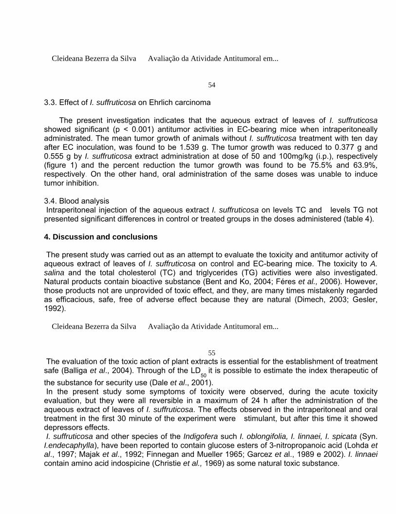
54 3.3. Effect of I. suffruticosa on Ehrlich carcinoma The present investigation indicates that the aqueous extract of leaves of I. suffruticosa showed significant (p < 0.001) antitumor activities in EC-bearing mice when intraperitoneally administrated. The mean tumor growth of animals without I. suffruticosa treatment with ten day after EC inoculation, was found to be 1.539 g. The tumor growth was reduced to 0.377 g and 0.555 g by I. suffruticosa extract administration at dose of 50 and 100mg/kg (i.p.), respectively (figure 1) and the percent reduction the tumor growth was found to be 75.5% and 63.9%, respectively. On the other hand, oral administration of the same doses was unable to induce tumor inhibition. 3.4. Blood analysis Intraperitoneal injection of the aqueous extract I. suffruticosa on levels TC and levels TG not presented significant differences in control or treated groups in the doses administered (table 4). 4. Discussion and conclusions The present study was carried out as an attempt to evaluate the toxicity and antitumor activity of aqueous extract of leaves of I. suffruticosa on control and EC-bearing mice. The toxicity to A. salina and the total cholesterol (TC) and triglycerides (TG) activities were also investigated. Natural products contain bioactive substance (Bent and Ko, 2004; Féres et al., 2006). However, those products not are unprovided of toxic effect, and they, are many times mistakenly regarded as efficacious, safe, free of adverse effect because they are natural (Dimech, 2003; Gesler, 1992). Cleideana Bezerra da Silva Avaliação da Atividade Antitumoral em...
55 The evaluation of the toxic action of plant extracts is essential for the establishment of treatment safe (Balliga et al., 2004). Through of the LD
50 it is possible to estimate the index therapeutic of
the substance for security use (Dale et al., 2001). In the present study some symptoms of toxicity were observed, during the acute toxicity evaluation, but they were all reversible in a maximum of 24 h after the administration of the aqueous extract of leaves of I. suffruticosa. The effects observed in the intraperitoneal and oral treatment in the first 30 minute of the experiment were stimulant, but after this time it showed depressors effects. I. suffruticosa and other species of the Indigofera such I. oblongifolia, I. linnaei, I. spicata (Syn. I.endecaphylla), have been reported to contain glucose esters of 3-nitropropanoic acid (Lohda et al., 1997; Majak et al., 1992; Finnegan and Mueller 1965; Garcez et al., 1989 e 2002). I. linnaei contain amino acid indospicine (Christie et al., 1969) as some natural toxic substance.

Extract of I. suffruticosa when administered by intraperitoneal route, showed LD50
value of 750.6 mg/kg, which, according with the classification of Schuartsman (1980), indicate a moderate acute toxicity. Nevertheless, there was no mortality recorded after the acute oral administration of the extract up to 5.000 mg/kg, which is a maximum dose considered for evaluating oral acute toxicological effects (Brasil, 2004; Barros and Davino, 2003). The absence of lethality by oral route may to be attributed to the biotransformation and/or low bioavailability of any constituents of the plant extract that might cause toxicity. Also, showed be considered the mechanism of absorption, since as absorption at the intestinal level or modification of the plant compound when in contact with the acid pH of the stomach. On the other had, the death of mice observed when the extract- substances were injected by i.p., which can be tentatively linked to a faster rate of absorption and to a first pass liver metabolism ( Obici, et al., 2008) Cleideana Bezerra da Silva Avaliação da Atividade Antitumoral em...
56 A. salina Leach, the brine shrimp, can be used in a laboratory bioassay in order to determine toxicity through the estimation of the mean lethal concentration (LC
50 values).
The larvae of the brine shrimp are very sensible and the death of this organism when exposed to various concentrations (< 10
3 µg/mL) forms the basis of the toxicity test. The bioassays are
simple, quick, low-cost economic and allows a great number of samples to be evaluated processed adequately (Ohno et al., 1997), and also has show a good correlation with various biological activity with pesticide activity and antitumor activity (McLaughlin, et al., 1991), and thus, the brine shrimp lethality assay has been used in several studies for detecting relative toxicity or toxic effect of bioactive compounds from plant extracts or from other substances (Meyer et al., 1982; Forbes and Forbes, 1994; Nick et al., 1995). To Knowledge, this first time that extract of I. suffruticosa are used to assays of toxicity with A. salina and the result of this experiment (LC
50 of 127 μg/mL) corroborate with other studies.
Déciga-campos et al., 2007, reported that the value of extract of extract of Piper sanctum is a little above of 200 μg/mL. Second Meyer et al., (1982) plant extract with LC
50 value lower than
1000 μg/mL is considered bioactive. Thus, the result of brine shrimp bioassay, suggest biological activity of the aqueous extract of I. suffruticosa. Cleideana Bezerra da Silva Avaliação da Atividade Antitumoral em...
57 Previous studies indicate that aqueous extract of I. suffruticosa were able to stop the cell development of embryos (Leite et al., 2004). In this study, intraperitoneal treatment of mice with the aqueous extract of I. suffruticosa 50 mg/kg and 100 mg/kg doses was able to reduce the solid tumor induced by Ehrlich ascites carcinoma cell. According to the literature, this tumor is of rapid growth and aggressive behavior (Ajith et al., 2003; Kaneno, 2004). No statistical significant difference was observed between the groups treated either with 50 mg/kg or with 100 mg/kg dose of aqueous extract. Nevertheless, the 50 mg/kg dose is lower than 10% of the acute toxicity (LD
50 of 750.6 mg/kg) and is effective to reduce 75.5% of the Ehrlich tumor. On the other hand,
oral treatment of mice with similar dose of the extract (50 mg/kg and 100 mg/kg), was ineffective

to inhibit the tumor, when compared with control group (data not shown). Similar result was reported by Amida el al., (2007), in aqueous extract of Sanseviera liberica, whereas, orally administered doses of the extract did not produced mortality in mice, but intraperitoneally administered caused dose-dependent mortalities (LD
50 of 668.3 mg/kg).
The action mechanism by which compounds present in the aqueous extract of leaves of I. suffruticosa inhibit tumoral cell remains to be elucidated. However, since embryogeny stoped, the inhibition of Ehrlich carcinoma cell by the I. suffruticosa extract may by related to its interference with cell development in process of initiation or promotion of the carcinogenis. Cleideana Bezerra da Silva Avaliação da Atividade Antitumoral em...
58 Furthermore, determination of serum TC and TG is important in this study in order to identify whither the use of I. suffruticosa with therapeutic purpose id affecting the animal lipid metabolism. Analysis of the TC and TG levels in serum of mice treated with the doses of 50 and 100 mg/kg (i.p.) were similar to that found before treatment, this indicate that the treatment of mice with I. suffruticosa extract by using this doses do not cause alteration in the main serum plasma lipid, with eight days of treatment. Whilst, by using the same doses and same period of treatment, the extract of I. suffruticosa has a potential effect in inhibiting the tumor size in EC – bearing mice. Finally, this study revealed that aqueous extract of leaves of I. suffruticosa is relatively safe when given orally, but it is considered moderately toxic in mice when administered by intraperitoneal injection and front the Artemia salina. Furthermore, I. suffruticosa extract also presents significant antitumor activity on Ehrlich ascites carcinoma in mice when using dose considered as therapeutical safe (< 10% of DL
50). Further studies are necessary by isolate and
characterize in the extract, the compound responsible for the anticancer activity, to establish the mechanism of action of the extract that leads to anticarcinoma activity. Cleideana Bezerra da Silva Avaliação da Atividade Antitumoral em...
59 Acknonledgments This work was financially supported by MCT CNPq/PADCT, FACEPE/PRONEX, CNPq, CAPES.

Cleideana Bezerra da Silva Avaliação da Atividade Antitumoral em...
60 References Abdullaev, F.I. 2001. Plant-drives agents against cancer. In: Gupta SK, editor. Pharmacology and the new millennium. New Delhi: Narosa Puplising Ilouse; p 345-54. Agra, M.F., Locatelli, E., Rocha, E. A., Baracho, G. S., Formiga, S. C., 1996. Plantas medicinais dos Cariris Velhos, Paraíba, Brasil, parte II: subclasses Magnoliidae, Caryophyliidae, Dilleniidae e Rosidae. Revista Brasileira de Farmácia 77, 97-102. Ajith, T. A., Janardhanan, K. K., 2003. Cytotoxic and antitumor activities of a polypore macrofungus, hellinusrimosus (Berk) Pilat. Journal of Ethnopharmacology, 84: 157-162. Alejo, J.P., Miranda, R., Rodrigues, G., 1996. Actividad Anticonvulsivante (Antiepileptica) del extracto fluido de Indigofera suffruticosa (Anil Cimarrón). Rev Cubana Plant Med, 1 (2), 7-10, Allen and Allen; Rodrigues-Kabana., 1981. Leguminosae, University of Wisconsin Press, Madison; Nematropica 18 (1): 137-142. Amida, M. B., Yemitan, O. K., Adeyemi, O. O., 2007. Toxicological assessment of the aqueous root extract of Sanseviera liberica gerome and Labroy (Agavaceae). Journal Ethnopharmacological 113, 171-175. Badell, J.B., Ruiz, A.R., Parra, A.B., Michelena, M.D., Michelena, M.J.M., Armenteros, A.E., 1998. Evaluación genotóxica del extracto fluido de Indigofera suffruticosa Mill (anil cimarrón) mediante el ensayo de anomalías em la cabeza de los espermatozoides. Rev Cubana Plant Med, 3 (2), 58-61. Balliga, S.M., Jagetia, C.G., Ulloor, J.N., 2004. The evaluation of the acute toxicity and long term
safety of hydroalcoholic extract of Sapthaparna (Alstonia scholaris) in mice and rats. Toxicologicals Letters 151, 317-326. Cleideana Bezerra da Silva Avaliação da Atividade Antitumoral em...
61 Barbosa-Filho, J. M., Vasconcelos, T. H. C., Alencar, A. A., Batista, L. M., Oliveira, R. A. G., Guedes, D. N., Falcão, H. S., Moura M. D., Diniz, M. F. F. M., Modesto- Filho, J., 2005. Plants and their active constituents from South, Centro, and North America with hypoglycemic activity. Rev Bras Farmacogn 15, 392-413. Barros, S. B. M., Davino, S. C., 2003. Avaliação da toxicidade. In: S. Oga. Fundamentos de toxicologia. 2ed. São paulo: Atheneup.57-67. Bent, S., Ko, R., 2004. Commonly used herbal medicines in the United States: a review. The American Journal of Medicine 116, 478– 485. Brasil 1998. Primeiro relatório nacional para a conservação sobre diversidade biológica: Brasil. Ministério do Meio Ambiente, dos Recursos Hídricos e da Amazônia Legal, Brasília. Brasil 2004. Ministério da Saúde. Agência Nacional de Vigilância Sanitária (ANVISA). Resolução-RE nº 90/2004. Normas para estudos toxicológicos de produtos fitoterápicos. Diário Oficial da república Federativa do Brasil, Poder Executivo, Brasília, DF, 12 de março de 2004. Costa-Lotufo, L.V, Khan, M. T. H., Ather, A., Wilke, D. V., Jimenez, P. C., Pessoa, C., Moraes, M. E. A., Moraes, M. O., 2005. Studies of the anticancer potencial of plants used in Bangladeshi folk medicine. Journal of Ethnopharmacology 99, 21-30. Correa, M. P., 1984. Dicionário das plantas úteis do Brasil e das exóticas cultivadas. IBDF. MA. Rio de Janeiro, 1,130-3. Cleideana Bezerra da Silva Avaliação da Atividade Antitumoral em...
62 Christie, G.S, Madsen, N.P., Hegarty, M.P., 1969. Acute biochemical changes in rat liver induced by the naturally occurring amino acid indospicine. Biochem Phannacol 18, 693-700. Cragg, G.M., Newman, D.J., 2000. Antineoplastic agents from natural sources: achievements and future directions. Expert Opinion on Investigational Drugs 9, 1-15. Dale, M.M., Rang, H.P., Ritter, J.M., 2001. Farmacologia, 4º edição: Rio de Janeiro: Guanabara Koogan.

Déciga-Campos, M., Rivero-Cruz, I., Arriaga-Alba, M., Castañeda-Corral, G., Angeles-López, G. E., Navarrete, A., Mata, R., 2007. Acute toxicity and mutagenic acrivity of Mexican plants used in traditional medicine. Journal of Ethnopharmacology, 110, 334-342. Dimech, G. S. 2003. Avaliação toxicológica pré-clínica do extrato bruto da Mentha crispa. Dissertação (Mestrado em Fisiologia) – Laboratório de Farmacologia e Toxicologia Pré-Clínica de Compostos Bioativos, Universidade Federal de Pernambuco, Recife. Falcão, H. S., Lima I. O., Santos, V. L., Dantas, H. F., Diniz, M. F. F. M., Barbosa-filho, J. M., Batista, L. M., 2005. Review of the plants with anti-infl ammatory activity studied in Brazil. Rev Bras farmacogn 15, 381-391. Féres, C. A. O., Madalosso, R. C., Rocha, O. A., Leite , J. P. V., Guimarães ,T. M. D. P., Toledo , V. P. P., Tagliati, C. A., 2006. Acute and chronic toxicological studies of Dimorphandra mollis in experimental animals. Journal of Ethnopharmacology 108, 450-456. Finnegan, R.A., Mueller, W.H.J. 1965. Pharm. Sci. 54,1136. Finney, D. J. 1971. Probits analysis. 3
rd ed. Cambridge University Press.
Cleideana Bezerra da Silva Avaliação da Atividade Antitumoral em...
63 Forbes, V. E. & Forbes, T. L. 1994. Ecotoxicology in Theory and Practice. Champman and Hall. Londres. Pp. 247. Garcez, W. S., Garcez, F. R., Barison, A., 2002. Additional 3-nitropropanoyl esters of glucose from Indigofera suffruticosa (Leguminosae). Biochemical systematics and ecology. V. 31, 207–209. Garcez, W. S., Garcez, F. R., Honda, N. K., Silva, A. J. R. 1989. Phytochemistry 28, 1251. Gesler, W.M., 1992. Therapeutic landscapes: medical issues in light of the new cultural geography. Social Science and Medicine 34, 735–746. Gupta, M., Mazumder, U. K., Kumar, R. S., Kumar, T. S. 2004. Antitumor activity and antioxident role of Bauhinia racemosa against Ehrlich ascites carcinoma in Swiss albino mice, Acta Pharmacol; 25, 1070-1076 Ho, C. T., Osawa, T., Huang., M.T., Rosen, R. T., (Eds.), 1994. Food Phytochemicals for Cancer Prevention II Teas, Spices, and Herbs. ACS Symposium 547 Washington DC. American Cancer Society. Karber, G., Behrens, B., 1964. Statistical Methods in Biological Assays. London: Ed. Griffin Ch. and C.

Kaneno R. et al., 2004. Effects of extracts from Brazilian sun-mushroom (Agaricus blazei) on the NK activity and lymphoproliferative responsiveness of Ehrlich tumor-bering mice. Food and Chemical Toxicology, 42 (6), 909-916. Komiyama, K. & Funayama, S., 1992. Antitumor agents. In: Omura, S. (edt). The search for bioactive compounds from microorganisms, New York: Spriger-Verlag, 79-97. Cleideana Bezerra da Silva Avaliação da Atividade Antitumoral em...
64 Lainetti, R., Brito, N.R.S., 1980. A saúde pelas plantas e ervas do mundo inteiro. Ed. Tecnoprint. Rio de Janeiro, RJ. 37p. Leite, S. P., Vieira, J. R. C., Medeiros, P. L., Leite, R. M. P., Lima, V. L. M., Xavier, H. S., Lima, E. O., 2006. Antimicrobial activity of Indigofera suffruticosa. Evidence- Based Complementary and Alternative Medicine, v. 3, n. 2, p. 261-265. Leite, S. P., Medeiros, P. L., Silva, E. C., Maia, M. B. S., Lima, V. L. M., Saul, D. E., 2004. Embryotoxicity in vitro with extract of Indigofera suffruticosa leaves. Reproductive Toxicology, 18, 701-705. Lewan, L.,Anderson, M., Morales–Gomez, P., 1992. The use of Artemia salina in toxicity testing. Alternatives To Laboratory Animals. 20, 297-301. Lohda, V.,Khan, H.A., Ghanim, A.J. 1997. Indian Chem. Soc. 74, 425. Lorenzi, H., 1982. Plantas daninhas do Brasil: terrestres, aquáticas, parasitas, tóxicas e medicinais. Nova Odessa, SP. Edição do Autor, p. 271-2. Machon, S., kuozynski, l., Cieldaowsaki, Y., Weiczsek., Zimecki, M.,Blasczy, B., Mordanki, M., Wiczorsk., Fiozerraliszewaska, L., 1981. Chemical and biological properties of 2 pyridielbenzil-carbinol. Achiv in Imunologiae it Therapie Experimentalis, 2, 29, 217-233. Majak, W., Benn, M., Mcewan, D., Pass, M. A., 1992. Three nitropropanoyl esters of glucose from Indigofera linnae. Phytochemistry 31, 2393-5 Meyer, B. M., Ferrigni, N. R., Putnam, J. E., Jacobsen, L. B., Nichols, D. E., Mclaughlin, J. L., 1982. Brine shrimp, a convenient general bioassay for active – plant constituents. Planta Med. 45, 31-34. Cleideana Bezerra da Silva Avaliação da Atividade Antitumoral em...
65 McLaughlin JL, Saizarbitori T-C, Anderson JE 1995. Tres bioensayos simples para quimicos de

productos naturales. Ver Soc Venez Quim 18: 13-18. McLaughlin, J.L., Chang, C.J., Smith, D.L. 1991. Bench-top bioassays for the discovery of bioactivity natural products: an update. In: Rahman, A.U. (Ed.), studies in natural products chemistry. Elsevier, Amsterdam. Napralert- Natural Products Alert. Chicago: Illinois University, 2003. Available at: www. c as.org/ONLINE/UG/napralert. Nick, A., Rali, T., Sticher, O. 1995. Biology screening of traditional medicinal plants from Papua New Guinea. J Ethnopharmacol 49: 147-156. Obici, S., Otobone, F. J., Sela, V.R.S., Ishida, K., Silva, J.C., Nakamurab, C.V., Cortez, D.A.G., Audi, E.A. 2008. Preliminary toxicity study of dichloromethane extract of Kielmeyera coriacea stems in mice and rats. Journal of Ethnopharmacology 115, 131–139. Ohno, Y., Kaneko, T., Inove, T., Morikawa, T., Yoshida, A., Fujii, A., Masuda, M., Ohno, T., Hayashi, M., Mumma, J., Akiyama, J., Hagaki, H., Ohkoshi, K., Okumura, H., Kakishima, H., Kasai, Y., Kurishita, A., Kujima, H., Saijo, K.,Sakamoto, K., Sugiura, S., Sunouchi, M., Tani, N., Chiba, K., Nakamura, T., Matsukawa, K. and Matsushige, C.1997. Interlaboratory validation of alternative methods to the eye irritation test for the safety evaluation of cosmetic ingredients: an overview of the plan and the results. Animal Alternatives, Welfare and Ethics. 27: 1155-8. Paiva, M. A. S., Barbosa A. C. D., Alves H. L. J., 1987. Indigofera suffruticosa Mill (leguminosae) com potencial forrageiro em uma região da caatinga no semi-árido de Pernambuco (Alagoinha). Sociedade Nacional de Botânica, 422. Cleideana Bezerra da Silva Avaliação da Atividade Antitumoral em...
66 Rajeshkumar, N. V., Joy, K. L., Kuttan, G., Ramsewak, R. S., Muraleedharan, G. N., Kuttan, R. 2002. Antitumour and anticarcinogenic activity of Phyllanthus amarus extract, journal of Ethnopharmacology 81, 17-22. Schuartsman, S. 1980. Produtos químicos de uso domiciliar – segurança e ricos toxicológicos. São Paulo: Almed, pp. 92. Stock. C.C., Sugiura K, 1954. The effect of phosphoramides on the growth of a variety of mouse and rat tumors. Cancer Research. 2, 38-50.
WHO - World Health Organization. Stop the global epidemic of chronic disease. www.who.int/cancer/en. 2005.

Cleideana Bezerra da Silva Avaliação da Atividade Antitumoral em...
67
Table 1
g
Mains effect observed in acute toxicity after intraperitoneal administration of extract of I. suffruticosa in mice.
Dose mg/kParameters
490 660 710 735 773 800
Stimulant ↑Frequency respiratory
++ ++ ++ ++ ++ +++ ++
+++
Agitation - + - + + + + ++Piloerection ++ +++ ++ ++ +++ +++ +++ +++
Leap + - - ++ - ++ + +

Tremors - +++ ++ +++ +++ +++ +++ +++ Stereotyping movement
+++ +++ +++ +++ +++ +++ +++ +++
Circular movement
+++ +++ +++ ++ + ++ +++ +++
Posture of attack
+ - ++ +
- - + -
Depressors Dyspnoea - - + ++ + ++ ++ ++Alteration of march
+++ + +++ +++ +++ +++ +++ +++
Lowering of the member
+ + ++
posterior
++ ++ ++ ++
+++
Sleepiness + - - + + + + +Prostration ++ + +++ ++ +++ +++ +++ +++
↓Frequency respiratory
- - + + + ++ ++
++
Other Reaction of flight
+++ +++ +++ +++ +++ +++ +++ +++
Contortions +++ +++ +++ +++ +++ +++ +++ +++
Diuresis - + + + + + - +Palaness - +++ - - + - ++ ++Edema of muzzle
+++ +++ - ++ +++ +++ ++ +++
Itching ++ ++ ++ +++ +++ ++ +++ +++ ________________________________________________________________________________ D
1 = Greater dose that not provoke lethality.
D2 = Less dose that provoke lethality.
Grade of effect observed: (-) without effect, (+) discreet effect, (++) moderately increased effect, (+++) intensively increased effect. Observation: No signs of toxicity were observed in the control group.

Table 2 Mains effect observed in acute toxicity after oral administration of extract of I. suffruticosa in mice.
Parameters Dose mg/kg
1000
1500
5000
Stimulant ↑Frequency respiratory
+++
+++
+++ Agitation
++
+
++ Piloerection
+
+++

+ Leap
++ -
+++ Tremors
+++
++
+++ Stereotyping movement
+++
+++
+++ Circular movement
+ - -
Posture of attack
-
+++
+++ Depressors

Dyspnoea
- - -
Alteration of march ++
++
++
Lowering of the member ++
++
++
posterior
Sleepiness
++ - -
Prostration
+++
++
++ ↓Frequency respiratory
- - -
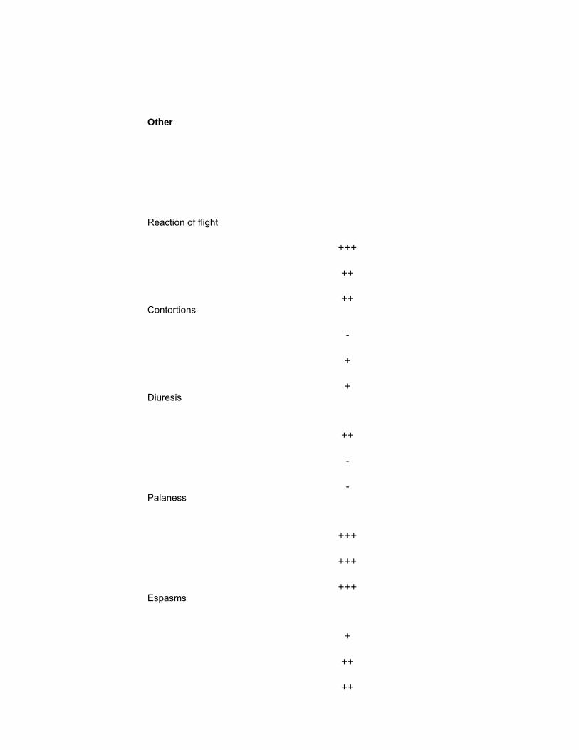
Other Reaction of flight
+++
++
++ Contortions
-
+
+ Diuresis
++ - -
Palaness
+++
+++
+++ Espasms
+
++
++

Itching
- - -
_____________________________________________________________________ Grade of effect observed: (-) without effect, (+) discreet effect, (++) moderately increased effect, (+++) intensively increased effect.
Observation: No signs of toxicity were observed in the control group.
Cleideana Bezerra da Silva Avaliação da Atividade Antitumoral em...
68
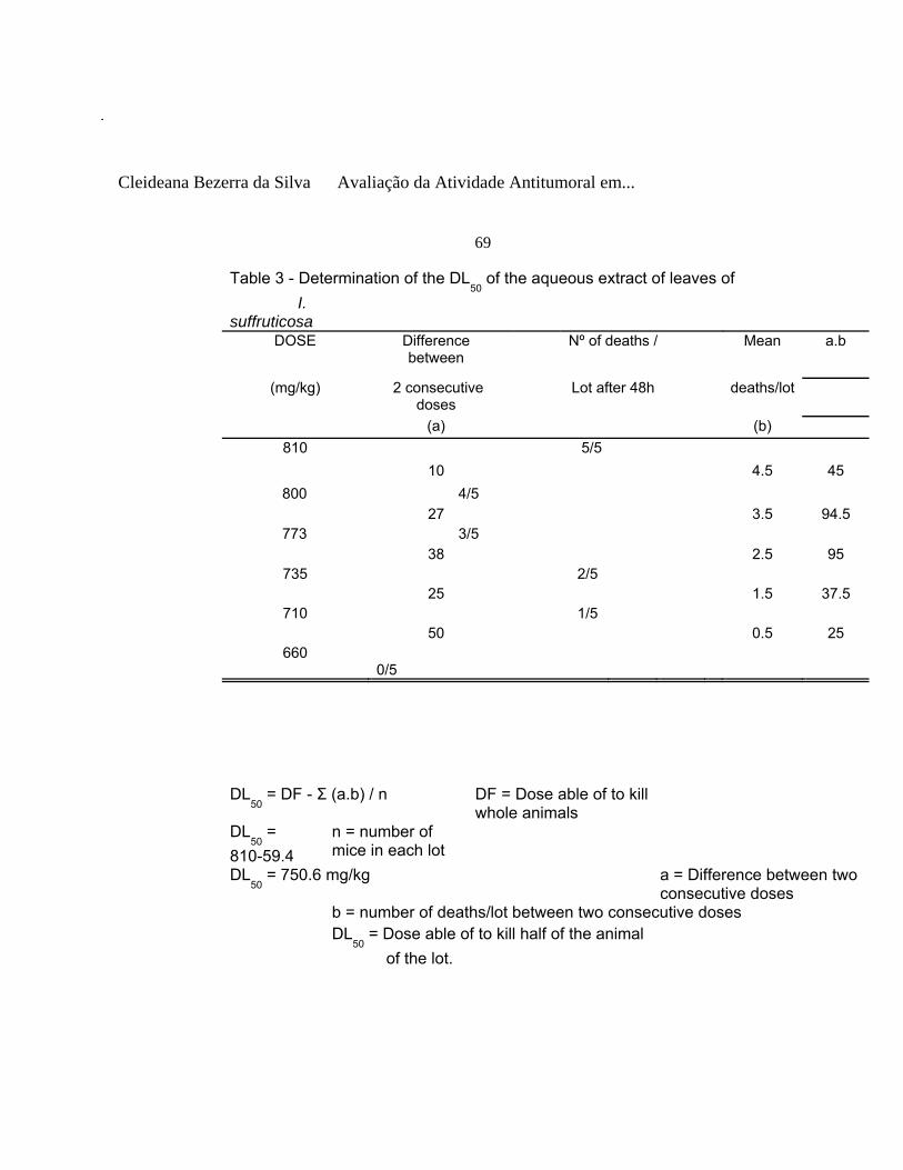
Cleideana Bezerra da Silva Avaliação da Atividade Antitumoral em...
69
Table 3 - Determination of the DL50
of the aqueous extract of leaves of I. suffruticosa
DOSE Difference between
Nº of deaths / Mean a.b
(mg/kg) 2 consecutive doses
Lot after 48h deaths/lot
(a) (b) 810 5/5
10 4.5 45 800 4/5
27 3.5 94.5 773 3/5
38 2.5 95 735 2/5
25 1.5 37.5 710 1/5
50 0.5 25 660
0/5
DL50
= DF - Σ (a.b) / n DF = Dose able of to kill whole animals
DL50
= 810-59.4
n = number of mice in each lot
DL50
= 750.6 mg/kg a = Difference between two consecutive doses
b = number of deaths/lot between two consecutive doses DL
50 = Dose able of to kill half of the animal
of the lot.

Cleideana Bezerra da Silva Avaliação da Atividade Antitumoral em...
70
***o Figure 1 Effect of the administration (i.p.) of the extract of I. suffruticosa on solid tumor induced by Ehrlich ascites carcinoma cell line in male mice. Values are mean ± S.D., n=5.
***P<0.001, extract treated
group compared with the control group.

Cleideana Bezerra da Silva Avaliação da Atividade Antitumoral em...
71
Table 4 Evaluation of the effect of the aqueous extract of leaves of I. suffruticosa on total cholesterol and triglyceride of normal mice
Dose (i.p.) T0 T8 (mg/kg) TC(mg/dL) TG(mg/dL) TC(mg/dL) TG(mg/dL) Control (saline)
94.18±11.80 184.4±25.21 115.5±12.02 161.18±33.01

50 96.56±8.06 181.33±40.80 96.5±18.04 151.51±36.36 100 90.26±6.62 165.33±51.86 94.79±20.99 205.28±54.63
Data are expressed as mean ± S.D., n=5 no statistical difference between extract-treated groups and groups control (p > 0.05) IP = intraperitoneal route ; T
0 = Before of the treatment
T
8 =8 days of treatment
Cleideana Bezerra da Silva Avaliação da Atividade Antitumoral em...
72
73
5. Conclusões Com base nos resultados obtidos, foi possível concluir que:
• O extrato aquoso de folhas de I. suffruticosa possui potente atividade antitumoral contra o modelo experimental “carcinoma ascite de Ehrlich” em camundongos.
• O extrato aquoso de folhas de I. suffruticosa é relativamente seguro quando ingerido
oralmente.
• Extrato aquoso de I. suffruticosa apresentou atividade tóxica moderada.
• Aparentemente, o tratamento com extrato de I. suffruticosa não interferiu nos níveis de
parâmetros bioquímicos como colesterol total e triglicerídeos, tão importantes para a
composição das membranas celulares, formação de hormônios e metabolismo energético.
74
75

6. ANEXO 6.1. TRABALHOS ENVIADOS PARA CONGRESSOS 6.2. GUIDE FOR AUTHORS
76

TRABALHOS ENVIADOS PARA CONGRESSOS
77
A REUNIÃO DA FEDERAÇÃO DA SOCIEDADE DE BIOLOGIA EXPERIMENTAL, 2007 – FESBE, 2007
78
AVALIAÇÃO DA ATIVIDADE ANTITUMORAL DO EXTRATO AQUOSO DE FOLHAS DE INDIGOFERA SUFFRUTICOSA MILL EM CARCINOMA DE EHRLICH.
1Silva, C. B. da;
2Pereira, D. R.;
3Ramos. A. L. G.;
4Souza, I. A.;
5Lima, V. L. M.;
1, 2, 3,
5Departamento de Bioquímica, UFPE;
4Departamento de Antibióticos, UFPE;
Objetivo:

Indigofera suffruticosa Mill (Fabeceae) é um leguminosa encontrada no Nordeste do Brasil e tem intensivo uso popular no tratamento de infecções e em outros processos inflamatórios, apresentando várias propriedades medicinais. O presente trabalho tem como objetivo investigar a atividade anticarcinogênica do extrato aquoso de folhas de I. suffruticosa, utilizando o tumor maligno denominado Carcinoma de Ehrlich.
Métodos e Resultados:
50g de folhas da planta foram trituradas e adicionadas a 200ml de água destilada. Esse material foi submetido a agitação constante por 16h, a temperatura ambiente para a obtenção do extrato aquoso. O ensaio antitumoral foi realizado em camundongos albinos suíços, machos (Mus musculus) com cerca de 60 dias de nascidos, pesando entre 25 a 35g. A linhagem tumoral do Carcinoma de Ehrlich foi mantida através de transplante de um fragmento tumoral de cerca de 3mm de diâmetro de um animal doador, inoculado subcutaneamente na região axilar. O teste de inibição tumoral foi desenvolvido empregando-se grupos de 5 animais para o extrato a ser testado, incluindo o grupo controle correspondente a animais transplantados e sem tratamento. A quimioterapia experimental teve início 48h após o implante do tumor. O extrato aquoso de folhas de I. suffruticosa foi dissolvida em solução salina estéril e aplicado diariamente por 7 dias, via intraperitoneal, com dose de 50mg/kg de peso do animal. Ao término do tratamento, os animais tratados e controle foram sacrificados, os tumores retirados, dissecados e pesados. A inibição do tumor foi calculada seguindo a fórmula de taxa de inibição tumoral (TWI%). O resultado do experimento da atividade antitumoral mostrou que a substância não revelou efeitos colaterais. Análise da atividade antitumoral demonstrou que para o tumor Carcinoma de Ehrlich a inibição foi de 75,5% com o extrato de folhas de I. suffruticosa.
Conclusões:
O extrato aquoso do vegetal pesquisado apresentou uma atividade antitumoral importante para o tipo característico de malignidade do referido tumor. Sendo assim, aumenta a nossa busca para conseguirmos através de estudos pré-clinicos encontrarmos um produto que possa minimizar a doença ate hoje considerada incurável e que abala a humanidade.
Apoio Financeiro: MCT/CNPq/PADCT, FACEPE/PRONEX, CNPq, CAPES
Data de Apresentação: 02/06/2007
79

XXXVI REUNIÃO ANULA DA SOCIEDADE BRASILEIRA DE BIOQUÍMICA E BIOLOGIA MOLECULAR – SBBq, NA FORMA DE COMUNICAÇÃO ORAL
80
PRELIMINARY MOLLUSCICIDAL ACTIVITY IN AQUEOUS EXTRACT FROM LEAVES OF Indigofera suffruticosa.
Cleideana B. Silva1; Ana M. M. A. Melo2; José O. Apolinário-Segundo
1; Vera L. M.
Lima1
1Departamento de Bioquímica, CCB, UFPE, Brazil;
2Departamento de Biofísica e
Radiobiologia, CCB, UFPE, Brazil
Indigofera suffruticosa Mill. (Fabaceae) is a plant found in Northeast Brazil. Recently, in vitro studies indicate that leaves extract of Indigofera suffruticosa is toxic to mice embryos. The molluscicidal effect of some plants has been searched as an attempt to break the Schistosoma mansoni life cycle. In this study, it was assayed the toxicity of an aqueous extract made with leaves of Indigofera suffruticosa against Biomphalaria glabrata adult snail and egg. Several groups of 5 adult mollusk were exposed during 24 hours to 80 e 100 ppm of the aqueous extract. The results showed a significant decrease on the snail fertility. The viable snail embryos were significantly reduced by 26.6% and 52.7% when exposed to 80 ppm and to 100 ppm of the plant extract, respectively, while 9.4% of embryos from the control group was unviable. The results suggest that the toxic effect of the aqueous extract against the mollusk is dose dependent, and that further studies are important to better evaluate these activity. Therefore, the extract of Indigofera suffruticosa is a natural product with potential to be used as a molluscicidal agent.

This work was supported by: MCT/CNPq/PADCT, FACEPE/PRONEX, CNPq, CAPES. Keywords: Indigofera suffruticosa, Biomphalaria glabrata, Molluscicidal activity.
81
XX CONGRESSO BRASILEIRO DE PARASITOLOGIA - 2007

82
ESTUDOS PRELIMINARES DOS EFEITOS DO EXTRATO AQUOSO DE FOLHAS DE INDIGOFERA SUFFRUTICOSA EM BIOMPHALARIA GLABRATA (SAY,1818).
Cleideana Bezerra da Silva (UFPE); Eliane Souza Figueiredo (UFPE); Ana Maria Mendonça de Albuquerque Melo (UFPE); Vera Lucia de Menezes Lima (UFPE)
Introdução: A esquistossomose mansônica é um dos mais importantes problemas de saúde pública. A doença é provocada pelo Schistosoma mansoni ,que tem como hospedeiro intermediário caramujos do gênero Biomphalaria, sendo o Biomphalaria glabrata, o principal vetor nas Américas do Sul e Central. Portanto, a busca de novas substâncias naturais, com efeito moluscicida, é fundamental para controlar o vetor desse parasita na tentativa de promover o controle da esquistossomose. Objetivos: Neste estudo utilizou-se o extrato aquoso de folhas de I. suffruticosa com o intuito de avaliar o grau de toxicidade dessa substância em caramujos adultos e embriões na fase de blástula de Biomphalaria glabrata. Metodologia: Grupos de 5 moluscos adultos e 100 embriões na fase de blástula foram expostos durante 24 horas às concentrações de 80ppm e 100ppm do extrato aquoso de folhas de Indigofera suffruticosa. Resultado: Os embriões viáveis dos caramujos expostos foram significativamente reduzidos para 26,6% e 52,7% quando exposto a 80ppm e 100ppm do extrato da planta, respectivamente, enquanto apenas 9,4% de embriões do grupo controle foram inviáveis. As desovas com embriões na fase de blástula não apresentaram resultados significativos. A concentração de 100ppm do extrato apresentou maior toxicidade para adultos de Biomphalaria glabrata que a concentração de 80ppm. Conclusão: Os resultados preliminares sugerem que o efeito tóxico do extrato de I. suffruticosa contra o molusco é dose dependente.
Palavras-chave: Biomphalaria, Indigofera suffruticosa, moluscicida
83
ESTUDO DA EMBRIOTOXICIDADE DE INDIGOFERA SUFFRUTICOSA SOBRE BIOMPHALARIA GLABRATA
Eliane Souza Figueiredo (UFPE); Cleideana Bezerra da Silva (UFPE); Vera Lúcia de Menezes Lima (UFPE); Ana Maria Mendonça de Albuquerque Lima (UFPE)

Introdução: A esquistossomose é uma doença endêmica em 76 países e o controle da transmissão dessa enfermidade envolve medidas de educação, saneamento, quimioterapia dos indivíduos infectados e o uso localizado de substâncias que contenham atividades moluscicidas e/ou cercaricidas. A necessidade de substâncias alternativas e ecologicamente eficientes tem impulsionado a pesquisa de plantas que contenham essas atividades. Na região nordeste do Brasil nota-se a presença da Indigofera suffruticosa, planta utilizada na medicina popular para tratamento de inflamações e de doenças tais como epilepsia em animais e humanos. Entretanto, testes de toxicidade, atividades moluscicida e cercaricida, ainda necessitam ser investigados. Objetivos: Avaliar a toxicidade do extrato aquoso da Indigofera suffruticosa sobre embriões de Biomphalaria glabrata em estágio de blástula. Metodologia: Embriões de B. glabrata no estágio de blástula foram expostos por 48 horas ao extrato aquoso de I. suffruticosa, nas concentrações de 0 (controle), 3mg/mL, 5mg/mL e 10mg/mL. Para cada concentração do extrato, foram utilizados cem embriões. A embriotoxicidade foi avaliada por meio da análise de embriões inviáveis (mortos e malformados). Resultado: Após a exposição ao extrato aquoso da I. suffruticosa, foi constatado a inviabilidade de 8,33% dos embriões expostos a 3mg/mL; 92,85% dos embriões expostos a 5mg/ml e 100% dos embriões expostos a 10mg/mL. Conclusão: Os resultados preliminares mostraram que o extrato aquoso de Indigofera suffruticosa foi letal para os embriões de B. glabrata no estágio de blástula quando expostos às concentrações de 5mg/mL e 10mg/mL. Palavras-chave: Indigofera suffruticosa, moluscicida, Biomphalaria glabrata
84
GUIDE FOR AUTHORS – JOURNAL OF ETHNOPHARMACOLOGY

85
GUIDE FOR AUTHORS The Journal of Ethnopharmacology is dedicated to the exchange of information and understandings about people's use of plants, fungi, animals, microorganisms and minerals and their biological and pharmacological effects based on the principles established through international conventions. Early people, confronted with illness and disease, discovered a wealth of useful therapeutic agents in the plant and animal kingdoms. The empirical knowledge of these medicinal substances and their toxic potential was passed on by oral tradition and sometimes recorded in herbals and other texts on materia medica. Many valuable drugs of today (e.g., atropine, ephedrine, tubocurarine, digoxin, reserpine) came into use through the study of indigenous remedies. Chemists continue to use plant-derived drugs (e.g., morphine, taxol, physostigmine, quinidine, emetine) as prototypes in their attempts to develop more effective and less toxic medicinals. In recent years the preservation of local knowledge, the promotion of indigenous medical systems in primary health care, and the conservation of biodiversity have become even more of a concern to all scientists working at the interface of social and natural sciences but especially to ethnopharmacologists. Recognizing the sovereign rights of States over their natural resources, ethnopharmacologists are particularly concerned with local people's rights to further use and develop their autochthonous resources. Accordingly, today's Ethnopharmacological research embraces the multidisciplinary effort in the documentation of indigenous medical knowledge, scientific study of indigenous medicines in order to contribute in the long-run to improved health care in the regions of study, as well as search for pharmacologically unique principles from existing indigenous remedies. The Journal of Ethnopharmacology publishes original articles concerned with the observation and experimental investigation of the biological activities of plant and animal substances used in the traditional medicine of past and present cultures. The journal will particularly welcome interdisciplinary papers with an ethnopharmacological, an ethnobotanical or an ethnochemical approach to the study of indigenous drugs. Reports of anthropological and ethnobotanical field studies fall within the journal's scope. Studies involving pharmacological and toxicological mechanisms of action are especially welcome. Clinical studies on efficacy will be considered if contributing to the understanding of specific ethnopharmacological problems. The journal welcomes review articles in the above mentioned fields especially those highlighting the multi-disciplinary nature of ethnopharmacology. Commentaries are by invitation only. All reviews and commentaries are fully peer-reviewed. Potential authors are strongly encouraged to contact the Reviews Editor [email protected] prior to writing a review. A one-page outline and a short C.V. of the (senior) author should also be included.
86
THE "RULES OF 5" The Editors and Editorial Board have developed the "Rules of 5" for publishing in JEP. We have produced five clear criteria that each author needs to think about before submitting a manuscript and setting the whole process of editing and reviewing at work.
II. Preparation of manuscripts Authors who want to submit a manuscript should consult and peruse carefully recent issues of the journal for format and style. Authors must include the following contact details on the title page of their submitted manuscript: full postal address; fax; e-mail. All manuscripts submitted are subject to peer review. The minimum requirements for a manuscript to qualify for peer review are that it has been prepared by strictly following the format and style of the journal as mentioned, that it is written in good English, and that it is complete. Manuscripts that have not fulfilled these requirements will be returned to the author(s). Contributions are accepted on the understanding that the authors have obtained the

necessary authority for publication. Submission of multi-authored manuscripts implies the consent of each of the authors. The publisher will assume that the senior or corresponding author has specifically obtained the approval of all other co-authors to submit the article to this journal. Submission of an article is understood to imply that it is not being considered for publication elsewhere and that the author(s) permission to publish his/her article in this journal implies the exclusive authorization to the publisher to deal with all issues concerning copyright therein. Further information on copyright can be found on the Elsevier website. In the covering letter, the author must also declare that the study was performed according to the international, national and institutional rules considering animal experiments, clinical studies and biodiversity rights. See below for further information. The ethnopharmacological importance of the study must also be explained in the cover letter. Animal and clinical studies - Investigations using experimental animals must state in the Methods section that the research was conducted in accordance with the internationally accepted principles for laboratory animal use and care as found in for example the European Community guidelines (EEC Directive of 1986; 86/609/EEC) or the US guidelines (NIH publication #85-23, revised in 1985). Investigations with human subjects must state in the Methods section that the research followed guidelines of the Declaration of Helsinki and Tokyo for humans, and was approved by the institutional human experimentation committee or equivalent, and that informed consent was obtained. The Editors will reject papers if there is any doubt about the suitability of the animal or human procedures used. Biodiversity rights - Each country has its own rights on its biodiversity. Consequently for studying plants one needs to follow the international, national and institutional rules concerning the biodiversity rights. 1.Manuscript types The Journal of Ethnopharmacology will accept the following contributions:
87
1. Original research articles - whose length is not limited and should include Title, Abstract, Methods and Materials, Results, Discussion, Conclusions, Acknowledgements and References. As a guideline, a full length paper normally occupies no more than 10 printed pages of the journal, including tables and illustrations
2. Ethnopharmacological communications (formerly Short Communications) - whose average length is not more than 4 pages in print (approx. 2000-2300 words, including abstract and references). A maximum of 2 illustrations (figures or tables) is allowed. See paragraph below for description and format.
3. Letters to the Editors;
4. Reviews - Authors intending to write review articles should consult and send an outline to the Reviews Editor (see inside front cover for contact information) before preparing their manuscripts. The organization and subdivision of review articles can be arranged at the author's discretion. Authors should keep in mind that a good review sets the trend and direction of future research on the subject matter being reviewed. Tables, figures and references are to be arranged in the same way as research articles in the journal. Reviews on topics that address cutting-edge problems are particularly welcome.
5. Book reviews - Books for review should be sent to the Reviews Editor.
6. Commentaries - invited, peer-reviewed, critical discussion about crucial aspects of the field but most importantly methodological and conceptual-theoretical developments in the field and should also provide a standard, for example, for pharmacological methods to be used in papers in the Journal of Ethnopharmacology. The scientific dialogue differs greatly in the social / cultural and natural sciences, the discussions about the common

foundations of the field are ongoing and the papers published should contribute to a transdisciplinary and multidisciplinary discussion. The length should be a maximum of 2-3 printed pages or 2500 words. Please contact the Reviews [email protected] with an outline.
7. Conference announcements and news. 2. General procedures The language of the Journal is English. Manuscripts should be neatly typed, double-spaced throughout, including tables, on pages of uniform size with at least 2.5 cm margins on all sides. Use one font type and size throughout the manuscript. Author(s) should not break or hyphenate words. When using an electronic printer, the right-hand margin should not be justified. Footnotes in text are not permitted. The text of the manuscript must be paginated, the first page being the title page. The manuscript, typed with double spacing and ample margins, should be submitted with a cover letter (containing the declaration that the study was performed according to the international, national and institutional rules considering animal experiments, clinical studies and biodiversity rights and a clear explanation of the ethnopharmacological importance of the study) and a completed Author Checklist . The following format and order of presentation is suggested. 2.1. Title, author(s), address(es) The title should be no longer than 100 letters, including spaces. Initials or first and middle names followed by last name of the author or authors must be given (not last name followed by initials). If there are two or more authors with different addresses, use a superscripted letter (a, b, c etc.), not a number, at the end of the last name of each author to indicate his her corresponding address. The full address of the corresponding author (the way the author wishes to be contacted) should be provided. The corresponding (usually, the senior) author, to whom correspondence and proofs will be sent, must be indicated by an asterisk and footnoted, and in the footnote, his/her the telephone and fax numbers, and e-mail address must be indicated. Address(es) should be underlined or italicised. 2.2. Abstract The abstract should present a summary of the problem, scientific method, major findings and conclusions, in no more than 200 words and in one paragraph and presented at the beginning of the paper. Unsubstantiated speculation should not be included. Footnotes may not be used. References, if cited, must provide complete publication data. 2.3. Text layout The text of a research paper should be divided into the following headings: Introduction, Methodology (or Materials and Methods), Results, and Discussion and conclusions. Each heading (and subheading) must be numbered using the convention established in the journal. Acknowledgements should come after Discussion and conclusions and before References; Acknowledgements and References are not to be numbered. Headings must be bold-faced and written in an upper-and-lower case style [not in caps], while subheadings should be underlined or italicised. Tables and figures are to be placed at the end of the text, after References. Authors are required to include: (i) the chemical structure, formula and proprietary name of novel or ill-defined compounds; (ii) the w/w yield of prepared extracts in terms of starting crude material; (iii) complete formulation details of all crude drug mixtures; (iv) the voucher herbarium specimen number of the plant(s) studied in case of less well known plants, cited using the collector and collection number (e.g., Doe 123), and indicating the name of the herbarium institution where it has been deposited. All plant materials must be fully identified as in the following illustration: Catharanthus roseus (L.) G. Don f. albus Pich. (Apocynaceae) as authenticated by Dr. John Doe, Department of Botany, University of Connecticut. 2.4. Guidelines for Plant and Animal Names All scientific names (Latin binomials) must be underlined or italicised throughout the text and in the tables and figures. For plant and animal species, full or complete scientific names, genus-species and the correct authority citation, must be used, when that name appears for the first time in text. The authority citation may be dropped in subsequent mention of that name throughout the text. The family name must follow the scientific name in parentheses when the name appears for the first

time in the text. Full scientific names and the family name of the subject plants/animals must be used in the Abstract. Synonyms must be indicated in parentheses and preceded by the word "syn." followed by a colon. Authors are advised to consult the International Plant Name Index (IPNI) ( http://www.ipni.org and W3Tropicos (http://www.mobot.org ) web-based databases to determine the correct spelling of full plant scientific names. Generic names may be abbreviated (e.g., C. roseus for Catharanthus roseus), provided such practice does not lead to confusion; generic names, however, must not be abbreviated when the name appears for the first time in the text. Specific epithets must never be abbreviated; thus, the use of Catharanthus r. is not allowed. 2.5. Keywords Authors are requested to assign 3-6 keywords to the manuscript, preferably taken from Index Medicus or Excerpta Medica Index, for abstracting and indexing purposes. These keywords should be typed at the end of the Abstract. Each keyword should start with a capital letter and be separated from each other by a semi-colon. 2.6. Tables, illustrations and graphs Tables should be on separate sheets, one table per sheet, and should bear a short descriptive title. Footnotes in tables should be indicated by consecutive superscript letters, not numbers. Figures should be original ink drawings, photographs or computer drawn figures in the original, and of high quality, ready for direct reproduction. Xerox copies are unacceptable as they give unsatisfactory results after final printing. Figures should be drawn in such a way that they can be reduced to 8 cm in width (i.e., the column width); in exceptional cases a reduction to a width of 17.5 cm will be allowed. All lettering should be such that height of 1.2-1.5mm (minimum) of numbers and capital letters results after reduction. Numerical scales, scale and curve legends, and all other lettering within the figure itself should be drawn with a lettering guide (stencil) or should be done using stripletters (Letraset, etc). All figures should have captions. Each figure should be identified in the margin or at the back in a corner with the name of the author and the figure number. The figure captions should be on a separate sheet. One set of original drawings is required. Colour illustrations should be submitted as original photographs, high-quality computer prints or transparencies, close to the size expected in publication, or as 35 mm slides. Polaroid colour prints are not suitable. If, together with your accepted article, you submit usable colour figures then Elsevier will ensure, at no additional charge, that these figures will appear in colour on the web (e.g., ScienceDirect and other sites) regardless of whether or not these illustrations are reproduced in colour in the printed version. For colour reproduction in print, you will receive information regarding the total cost from Elsevier after receipt of your accepted article. The 2006 price for color figures is EUR 285 for the first page and EUR 191 for subsequent pages. For further information on the preparation of electronic artwork, please see http://www.elsevier.com/artworkinstructions . Please note: Because of technical complications which can arise by converting colour figures to 'grey scale' (for the printed version should you not opt for colour in print) please submit in addition usable black and white prints corresponding to all the colour illustrations. 2.7. References References should be referred to by name and year (Harvard system) chronologically in the text (e.g.: Brown and Penry, 1973; Stuart, 1979; Ageel et al., 1987) and listed alphabetically at the end of the paper. No ampersand should be used and the words "et al." should not be underlined or italicized. Only papers and books that have been published or in press may be cited. For papers in press, please cite the DOI article identifier. The Digital Object Identifier (DOI) is a persistent identifier which may be used to cite and link to electronic documents. The DOI consists of a unique alpha-numeric character string which is assigned to a document by the publisher upon the initial electronic publication. The DOI will never change. Therefore, it is an ideal medium for citing Articles in Press, which have not yet received their full bibliographic information. Unpublished manuscripts or manuscripts submitted to a journal but which have not been accepted may not be cited. Journal and book titles should not be underlined or italicised and should be given in full in the reference list, with no underline or italics. Examples: Journals: Britton, E.B., 1984. A pointer to a new hallucinogen of insect origin. Journal of Ethnopharmology 12, 331-333. Books: Emboden, W., 1972. Narcotic Plants. Studio Vista, London, p. 24. Multiauthor Books: Farnsworth, N.R., 1988. Screening plants for

new medicines. In: E.O. Wilson and F.M. Peter (Eds.), Biodiversity, National Academy Press, Washington, D.C., pp. 83-97. Ethnopharmacological Communications (formerly short communications) are brief contributions on: - isolation of biological active compound(s) from a traditional medicine, - screening of a series traditional medicines for biological activity, - study on a pharmacological activity of a traditional medicine, - study on the toxicology of a traditional medicine. III. Submission All manuscripts (except reviews, commentaries and book reviews) must be submitted to (http://www.elsevier.com/journals) Each Submission must include a cover letter (containing the declaration that the study was performed according to the international, national and institutional rules considering animal experiments, clinical studies and biodiversity rights and a clear explanation of the ethnopharmacological importance of the study) and a completed Author Checklist (click here). If an author cannot submit their manuscript electronically, then please send to: Professor Dr R. Verpoorte Editor-in-Chief, Journal of Ethnopharmacology Division of Pharmacognosy Institute of Biology Leiden University P.O. Box 9502 2300 RA Leiden The Netherlands IV. Copyright regulations for authors All authors must sign the "Transfer of Copyright" agreement before the article can be published. This transfer agreement enables Elsevier to protect the copyrighted material for the authors, but does not relinquish the author's proprietary rights. The copyright transfer covers the exclusive rights to reproduce and distribute the article, including reprints, photographic reproductions, microform, or any other reproductions of similar nature and translations, and includes the right to adapt the article for use in conjunction with computer systems and programs, including reproduction or publication in machine-readable form and incorporation into retrieval systems. Authors are responsible for obtaining from the copyright holder permission to reproduce any figures for which copyright exists. Transfer of copyright agreement forms will be sent to the corresponding author following acceptance of the manuscript.
88
89
90
91
92 V. Retained authors' rights As an author you (or your employer or institution) may do the following: • make copies (print or electronic) of the article for your own personal use, including for your own classroom teaching use • make copies and distribute such copies (including through e-mail) of the article to research colleagues, for the personal use by such colleagues (but not commercially or systematically, e.g., via an e-mail list or list server) • post a pre-print version of the article on Internet websites including electronic pre-print servers, and to retain indefinitely such version on such servers or sites • post a revised personal version of the final text of the article (to reflect changes made in the peer review and editing process) on your personal or institutional website or server, with a link to the journal homepage (on http://www.elsevier.com ) • present the article at a meeting or conference and to distribute copies of the article to the delegates attending such a meeting • for your employer, if the article is a 'work for hire', made within the scope of your employment, your employer may use all or part of the information in the article for other intra-company use (e.g., training) • retain patent and trademark rights and rights to any processes or procedure described in the article • include the article in full or in part in a thesis or dissertation (provided that this is not to be published commercially) • use the article or any part thereof in a printed compilation of your works, such as collected writings or lecture notes (subsequent to publication of your article in the journal) • prepare other derivative works, to

extend the article into book-length form, or to otherwise re-use portions or excerpts in other works, with full acknowledgement of its original publication in the journal VI. Correcting proofs and reprints Proofs will be sent to the corresponding author. Elsevier is now sending PDF proofs by e-mail for correction. If an author is unable to handle this process, regular print proofs will be sent. Elsevier will do everything possible to get the article corrected and published as quickly and accurately as possible. Therefore, it is important to ensure that all corrections are sent back in ONE communication. Subsequent corrections will not be possible. Only typesetting errors may be corrected; no changes in, or additions to, the accepted manuscript will be allowed. Proofs should be returned to Elsevier within 48 hours. Twenty-five offprints of each paper will be supplied free of charge to the corresponding author. Additional offprints can be ordered at prices shown on the offprint order form that accompanies the copyright form. VII. Language Polishing For authors, who require information about language editing and copyediting services pre- and post-submission, please visit www.elsevier.com/locate/languagepolishing or contact [email protected] for more information. Please note Elsevier neither endorses nor takes responsibility for any products, goods or services offered by outside vendors through our services or in any advertising. For more information please refer to our Terms & Conditions .
93 VIII. US National Institutes of Health (NIH) voluntary posting ("Public Access") policy Elsevier facilitates author posting in connection with the voluntary posting request of the NIH (referred to as the NIH "Public Access Policy"; see http://www.nih.gov/about/publicaccess/index.htm ) by posting the peer-reviewed author's manuscript directly to PubMed Central on request from the author, after formal publication. Upon notification from Elsevier of acceptance, we will ask you to confirm via e-mail (by e-mailing us at [email protected] ) that your work has received NIH funding (with the NIH award number, as well as the name and e-mail address of the Prime Investigator) and that you intend to respond to the NIH request. Upon such confirmation, Elsevier will submit to PubMed Central on your behalf a version of your manuscript that will include peer-review comments, for posting 12 months after the formal publication date. This will ensure that you will have responded fully to the NIH request policy. There will be no need for you to post your manuscript directly with PubMed Central, and any such posting is prohibited. Individual modifications to this general policy may apply to some Elsevier journals and its society publishing partners. IX. Author enquiries For enquiries relating to the submission of articles (including electronic submission where available) please visit this journal's homepage at http://www.elsevier.com/locate/jethpharm . You can track accepted articles at http://www.elsevier.com/trackarticle and set up e-mail alerts to inform you of when an article's status has changed, as well as copyright information, frequently asked questions and more. Contact details for questions arising after acceptance of an article, especially those relating to proofs, are provided after registration of an article for publication. No responsibility is assumed by the Publisher for any injury and/or damage to persons or property as a matter of products liability, negligence or otherwise, or from any use or operation of any methods, products, instructions or ideas contained in the material herein. Because of the rapid advances made in the medical sciences, independent verification of diagnoses and drug dosages should be made.
94