course 660 ohp 1 evaluation of the pupils · course 660 ohp 1 evaluation of the pupils george...
TRANSCRIPT

COURSE 660 OHP 1COURSE 660 OHP 1
EVALUATION OF EVALUATION OF
THE PUPILSTHE PUPILS
George W. Comer, O.D., M.B.A.Southern California College of Optometry
OVERVIEW OF LECTURE
• General why/when of pupil testing
• Neuro quick review
–The anatomic basis of our strategy and clinically significant anatomy
• Strategy of pupil evaluation
–Why are we doing what we are doing
• Techniques of pupil evaluation
–How to evaluate pupils
• Common pupil anomalies
WHY EVALUATE PUPILS?
• Test of neurological integrity - integrity of afferent and efferent pupil pathways.
• Pupil reflexes are important in the differential diagnosis of the cause of vision loss
• Pupil signs are important signs in ocular disease especially anterior segment disease.
Iritis → small pupils & significant photophobia
Acute angle closure glaucoma → mid-dilated, fixed pupil (pupil stuck in the mid-dilated position)
WHEN TO EVALUATE PUPILS
• All comprehensive exams
• All cases of vision loss or visual
field loss
• Always prior to pupillary dilation
• Prior to contact lens fitting
• All tests of neurovisual function and/or neurological status
WHAT IS EVALUATED IN A PUPIL EXAM?
• Neurological integrity/neurovisual function
Afferent pathway –
From eye to midbrain for pupillary light reflex. From eye to cortex for pupillary near reflex
Efferent pathways – brain to the pupils (CN III to pupillary sphincter and oculosympathetic pathway to the pupillary dilator)
• Gross external ocular health
PUPIL NEURO
Quick Review

THE PUPIL PATHWAYS!!
• Afferent pathway– From eye to brain
– Pre-geniculate visual pathways (optic nerve + anterior portion of optic tracts) + projections to midbrain
• Efferent pathways– From brain to eye
– Third cranial nerve to pupillary sphincter
– Oculosympathetic pathway to pupillary dilator
AFFERENT PUPIL PATHWAY
• Retina
–Retinal receptors
– Intra-retinal connections
–Retinal ganglion cells (RGCs)
–Ganglion cell axons (RNFL)
• Optic nerve
–Optic nervehead (RGC axons)
–Optic nerve (RGC axons)
AFFERENT PUPIL PATHWAY
(cont’d)
• Optic chiasm (RGC axons)
• Optic tract (RGC axons)
• (Brachium of superior colliculus)
• (Pretectal nuclei)
• (Posterior commissure)
Afferent Pupillary Light Pathway
CROSSOVER AT THE CHIASM
• The fibers from nasal retina (53-55% of all) from each eye cross to the opposite side in the chiasm
• The fibers from the temporal retina (only 45-47% of all from an eye) stay on the same side
So…in the right optic tract 53-55% of fibers are from the LEFT eye!

CONSENSUAL PUPILLARY LIGHT REFLEX
ANATOMIC BASIS
• Optic chiasm
• Posterior commissure
A LESION IN THE AFFERENT
PATHWAY PRODUCES
• Impaired pupillary light reflex
–But not the near reflex
• Light - near dissociation
–Near reflex is stronger, quicker than light reflex
• APD (if unilateral or asymmetric
damage)
DAMAGE TO AFFERENT
PATHWAY DOES NOT CAUSE
• Anisocoria–Difference in pupil size
• Change in near reflexes–Near reflex not changed if tested as
described in this lecture
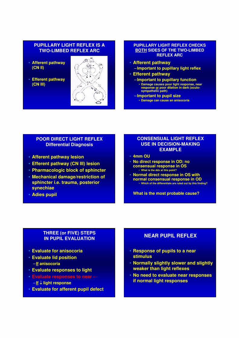
PUPILLARY LIGHT REFLEX IS A TWO-
LIMBED REFLEX ARC
• Afferent pathway
• Efferent pathway (CN III)
EFFERENT PUPIL PATHWAYS
• Third cranial nerve
(parasympathetic to pupillary
sphincter - CN III)
• Oculosympathetic pathway
(sympathetics to pupillary dilator)
EFFERENT PATHWAY DAMAGE
CAUSES
• Change in pupil size
– If asymmetric or unilateral →anisocoria
• Reduced pupil function
– If CN III damage → reduced response to light and nearor
– If oculosympathetic pathway damage → reduced or slower dilation in dark

The
Oculosympathetic
Pathway
OCULOSYMPATHETIC PATHWAY
First Order Neuron
• Hypothalamus
• Brainstem
• Upper spinal cord
• Ciliospinal center of Budge(C8, T1, T2)
Oculosympathetic Pathway OCULOSYMPATHETIC PATHWAY
Second Order Neuron
• White rami communicantes
• Stellate ganglion (no synapse)
• Over the apex of the lung
• Joins common carotid artery (CCA)
• Superior cervical ganglion (synapse)
SYMPATHETIC PATHWAYTO FACIAL SWEATING
Third Order Neuron
• External carotid artery
• Also goes to sudomotor (sweating)
and vasomotor control in face
OCULOSYMPATHETIC PATHWAY
Third Order Neuron to Eye/Globe
• Follows internal carotid artery
• Cavernous sinus
• Cranial nerve V, ophthalmic
division, nasociliary
• Long ciliary nerves

SYMPATHETIC INNERVATION IN HEAD/EYE & FUNCTIONS
• Pupillary dilator - pupillary dilation
• Mueller’s muscles - lid retraction– Upper & lower lid
• Facial sweating
• Lacrimal glands - lacrimation
• Ciliary muscle →accommodation (?)
• Vessels of conjunctiva -vasoconstriction
• Iris melanin development in early life
Oculosympathetic dysfunction (Horners’ syndrome)
• From www.thyroid-eyes.com/ptosis
• From: www.wikidoc.org/iindex.php/Horner%27s_syndrome
Based on this gross external view what is the most probable cause of this patient’s
problem? LID RETRACTORS
• Mueller’s muscle (sympathetic)
• Levator palpebrae superioris (CN
III)
Lid Retractors NEUROGENIC PTOSIS
• Third nerve palsy - large ptosis
• Oculosympathetic paresis - small
ptosis + inverse ptosis
The
Parasympathetic
Pathway to the Pupil
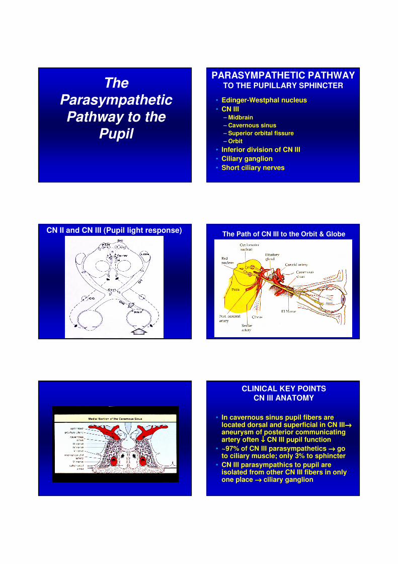
PARASYMPATHETIC PATHWAY TO THE PUPILLARY SPHINCTER
• Edinger-Westphal nucleus
• CN III– Midbrain
– Cavernous sinus
– Superior orbital fissure
– Orbit
• Inferior division of CN III
• Ciliary ganglion
• Short ciliary nerves
CN II and CN III (Pupil light response)The Path of CN III to the Orbit & Globe
CLINICAL KEY POINTS
CN III ANATOMY
• In cavernous sinus pupil fibers are located dorsal and superficial in CN III→→→→aneurysm of posterior communicating artery often ↓↓↓↓ CN III pupil function
• ~97% of CN III parasympathetics →→→→ go to ciliary muscle; only 3% to sphincter
• CN III parasympathics to pupil are isolated from other CN III fibers in only one place →→→→ ciliary ganglion

Aneurysm of Posterior Communicating Artery and its Effect on CN III
(Pupillomotor Fibers)
DAMAGE TO CN III
PARASYMPATHETICS CAUSES
• ↓↓↓↓ light response and ↓↓↓↓ near response
• ↓↓↓↓ accommodation
THIRD CRANIAL NERVE FUNCTIONS
• Inferior oblique - upgaze on adduction
• Ciliary muscle - accommodation
• Pupillary sphincter- pupil constriction
• Levator palpebrae superioris - superior lid retraction
• Superior rectus - upgaze in abduction
• Medial rectus - adduction
• Inferior rectus - downgaze in abduction
THE STRATEGY FOR CLINICAL EVALUATION OF THE PUPILS
Why you do what you do in
the pupil evaluation!
THREE (or FIVE) STEPS
IN PUPIL EVALUATION
• Evaluate for anisocoria
• Evaluate lid position
– If anisocoria
• Evaluate responses to light
• Evaluate responses to near
– If ↓↓↓↓ light response
• Evaluate for afferent pupil defect
THREE (or FIVE) STEPS
IN PUPIL EVALUATION
• Evaluate for anisocoria←
• Evaluate lid position
– If anisocoria
• Evaluate responses to light
• Evaluate responses to near
– If ↓↓↓↓ light response
• Evaluate for afferent pupil defect
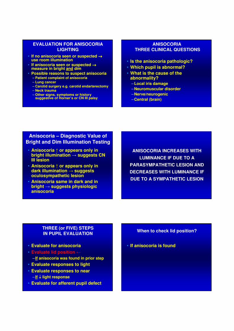
EVALUATION FOR ANISOCORIA
LIGHTING
• If no anisocoria seen or suspected →→→→use room illumination
• If anisocoria seen or suspected →→→→measure in bright and dim
• Possible reasons to suspect anisocoria– Patient complaint of anisocoria
– Lung cancer– Carotid surgery e.g. carotid endarterectomy– Neck trauma– Other signs, symptoms or history
suggestive of Horner’s or CN III palsy
ANISOCORIA
THREE CLINICAL QUESTIONS
• Is the anisocoria pathologic?
• Which pupil is abnormal?
• What is the cause of the abnormality?–Local iris damage
–Neuromuscular disorder
–Nerve/neurogenic
–Central (brain)
Anisocoria – Diagnostic Value of
Bright and Dim Illumination Testing
• Anisocoria ↑ or appears only in bright illumination → suggests CN III lesion
• Anisocoria ↑ or appears only in dark illumination → suggests oculosympathetic lesion
• Anisocoria same in dark and in bright → suggests physiologic anisocoria
ANISOCORIA INCREASES WITH
LUMINANCE IF DUE TO A
PARASYMPATHETIC LESION AND
DECREASES WITH LUMINANCE IF
DUE TO A SYMPATHETIC LESION
THREE (or FIVE) STEPS
IN PUPIL EVALUATION
• Evaluate for anisocoria
• Evaluate lid position ←
– If anisocoria was found in prior step
• Evaluate responses to light
• Evaluate responses to near
– If ↓↓↓↓ light response
• Evaluate for afferent pupil defect
When to check lid position?
• If anisocoria is found

WHY CHECK LID POSITION?
If anisocoria is present and is pathological it may be due to damage to either CNIII or to oculosympathetic pathway
Superior Lid Position
Neurological Control
Superior lid position controlled by:1. Levator
CN III innervationPrimary retractor of the upper lid
2. Superior tarsal muscle (Mueller’smuscle)Sympathetic innervationSecondary lid (both upper and lowerlid) retractor
NEUROGENIC PTOSIS
• Third nerve palsy - large (usually ≥
5mm) ptosis possible
• Oculosympathetic paresis
(Horner’s syndrome) - small (1 to 2
mm) ptosis of superior lid usually +
inverse ptosis (inferior lid up slightly)
NORMAL LID POSITION
• Patient should look in primary gaze and judge lid position relative to the limbus
• Upper lid crosses limbus at 2:00 and 10:00
• Upper lid covers 1 to 2 mm of superior cornea
• Lower lid just grazes inferior limbal region
• Do NOT judge lid position relative to the pupil margin
• Watch out for frontalis contraction
Normal lid position?

Normal lid position? WARNING!!
Do not measure/evaluate lid position relative to the pupil
margin. Use the distance from limbus to lid margin.
LID - PUPIL RELATIONS
• CN III - superior lid down (a lot
usually), pupil larger
• Horner’s - superior lid down a little;
pupil smaller
–May not detect ptosis if lid margin evaluated/measured from pupil margin!! Use the limbus as the landmark.
SIGNIFICANCE OF LID POSITION
EVALUATION
• Helps to confirm presence of
efferent pathway lesion especially
Horner’s (88% of Horner’s have ptosis)
• Helps to localize a lesion within
pathway especially CN III
• Helps to differentiate type of lesion
THREE (or FIVE) STEPS
IN PUPIL EVALUATION
• Evaluate for anisocoria
• Evaluate lid position
– If anisocoria
• Evaluate responses to light ←
• Evaluate responses to near
– If ↓↓↓↓ light response
• Evaluate for afferent pupil defect
PUPILLARY LIGHT REFLEX IS A
TWO-LIMBED REFLEX ARC
• Afferent pathway(CN II)
• Efferent pathway (CN III)
PUPILLARY LIGHT REFLEX CHECKS BOTH SIDES OF THE TWO-LIMBED
REFLEX ARC
• Afferent pathway– Important to pupillary light reflex
• Efferent pathway
– Important to pupillary function • Damage causes poor light response, near
response or poor dilation in dark (oculo-sympathetic path)
– Important to pupil size • Damage can cause an anisocoria
POOR DIRECT LIGHT REFLEX
Differential Diagnosis
• Afferent pathway lesion
• Efferent pathway (CN III) lesion
• Pharmacologic block of sphincter
• Mechanical damage/restriction of
sphincter i.e. trauma, posterior synechiae
• Adies pupil
CONSENSUAL LIGHT REFLEX USE IN DECISION-MAKING
EXAMPLE
• 4mm OU
• No direct response in OD; no consensual response in OS
• What is the ddx at this point?
• Normal direct response in OS with normal consensual response in OD
• Which of the differentials are ruled out by this finding?
What is the most probable cause?
THREE (or FIVE) STEPS
IN PUPIL EVALUATION
• Evaluate for anisocoria
• Evaluate lid position
– If anisocoria
• Evaluate responses to light
• Evaluate responses to near ←
– If ↓↓↓↓ light response
• Evaluate for afferent pupil defect
NEAR PUPIL REFLEX
• Response of pupils to a near
stimulus
• Normally slightly slower and slightly
weaker than light reflexes
• No need to evaluate near responses
if normal light responses

THE NEAR PUPIL REFLEX
Why check it?
• To bypass the afferent pathway in
order to prove that the efferent (CN
III) pathway is normal
• An afferent path lesion affects the
light response but not the near
response (if proprioception is used to drive the near response)
LIGHT-NEAR DISSOCIATION
Near reflex is stronger/faster than light reflex
DDx of LIGHT-NEAR DISSOCIATION
• Afferent pathway lesion
• Adies pupil
• Aberrant regeneration of CN III
• Diabetes
• Argyll-Robertson pupils
• Midbrain lesion
NEAR REFLEX USE IN DECISION-
MAKING - EXAMPLE
• 4mm OU
• No direct response in OD
• Normal direct response in OS
• Normal near response OU
What does the normal near response in OD indicate?
What is the most probable cause of this?
THREE (or FIVE) STEPS
IN PUPIL EVALUATION
• Evaluate for anisocoria
• Evaluate lid position
– If anisocoria
• Evaluate responses to light
• Evaluate responses to near
– If ↓↓↓↓ light response
• Evaluate for afferent pupil defect ←
How to check for an afferent
pupillary defect?
Compare the direct pupil response in
one eye to the direct pupil
response in the other eye
Compare speed, magnitude and
presence of “escape”

What is an afferent pupil defect
(APD)?
The light response in one eye is less
than the light response in the other
eye due to an afferent pathway lesion (not some other cause).
An APD is due to unilateral or
asymmetric afferent pathway damage.
EVALUATE FOR AFFERENT
PUPIL DEFECT
• How? Compare direct light
response in OD to that in OS by
checking the light responses in rapid succession
• Why? To detect very subtle
difference between the pupillary light reflex of OD compared to OS
AFFERENT PUPIL DEFECT
Other Names
• APD or +APD
• Relative afferent pupil defect (RAPD)
• Marcus-Gunn pupil
• + Swinging flashlight test (+ SFLT)
• Pupillary escape
AFFERENT PUPIL DEFECT
Differential Diagnoses
• Optic nerve disease
• Macular disease
Very gross macular lesion
• Retinal detachment Large RD
• Chiasmal disease
• Optic tract lesion
Very extensive or complete tract lesion produces only a mild (1+ or 2+) APD
AFFERENT PUPIL DEFECT
Most common causes:
• Optic nerve disease
– Only a little ON damage, especially inflammation of
ON (optic neuritis), causes large APD
– May or may not (retrobulbar damage) be able to
visualize the ON damage on ophthalmoscopy →
depends on where the damage is and how soon you
look after the damage has occurred
• Extensive retinal damage
– Large macular/retinal lesion causes little APD
– You should easily see the fundus lesion
CLINICAL TIP
If a large APD is present with macular/ retinal disease or
amblyopia →→→→ rule out ON disease!

OTHER CAUSES OF APD
• Chiasmal disease
• Contralateral extensive optic tract lesion
• Gross macular disease
• Large, extensive retinal lesion–RD
–BRAO/CRAO
– Ischemic CRVO
Case Example
30 y/o male
CVC: ↓ vision and side vision OU for over 1 year, getting worse
Hab VAs: 20/50, 20/50
CF confrontations: Very dim in superior temporal quadrant OU
Pupils: 5 mm OU, 2+ direct OU, 4+ near OU, 1+ APD OS
D.O. see slides

Case Example Will this patient have a pupil anomaly?
Bilateral APDs???Is it possible?
Bilateral APDs do not exist!!!
Bilateral L-N dissociation does.
An APD only occurs when the light
response is worse in one eye than
the other eye.
CLINICAL TIP
There will be ophthalmoscopic
evidence of visual pathway damage
for most lesions causing APD
Look for: ON pallor/atrophy, RNFL
dropout, macular lesion, RD etc.
CLINICAL TIP
Cataracts rarely produce
an afferent pupillary defect
(A dense cataract can produce an APD in the other eye!!)

CLINICAL TIP
If an APD is present on the same side as a cataract rule out afferent pathway damage
especially ON disease
CLINICAL TIP
If a patient has both
anisocoria and an APD →→→→there are two different
causes/lesions.
ANISOCORIA IS VERY
RARELY RELATED VIA A
SINGLE DISEASE OR
LESION TO AN APD
APD GRADING SYSTEM
4+ little, if any, light response in affected eye → big difference in light reflexes between the two eyes
3+ some light response (near normal) but quicker-than-normal pupil escape
2+ slight response to light in one eye
1+ very slight difference between the two eyes light responses. Affected eye has near normal light response but quicker escape.
TECHNIQUE OF CLINICAL
EVALUATION OF THE PUPILS
How to test pupils!
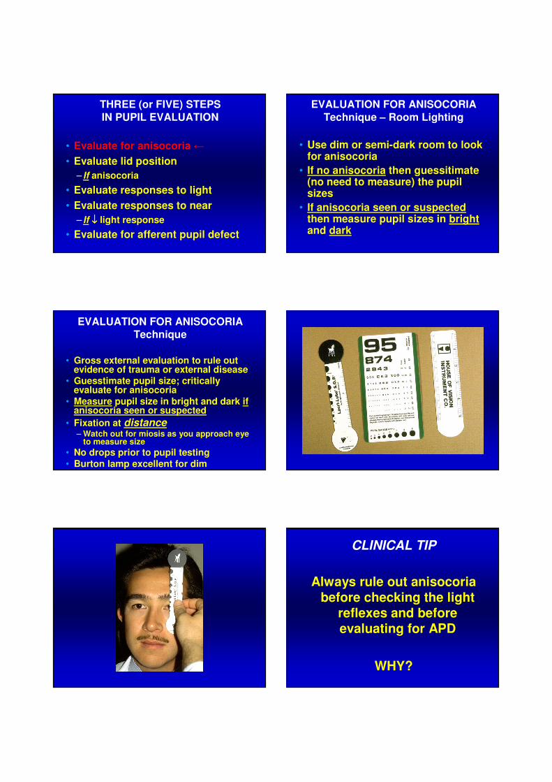
THREE (or FIVE) STEPS
IN PUPIL EVALUATION
• Evaluate for anisocoria ←
• Evaluate lid position
– If anisocoria
• Evaluate responses to light
• Evaluate responses to near
– If ↓↓↓↓ light response
• Evaluate for afferent pupil defect
EVALUATION FOR ANISOCORIA
Technique – Room Lighting
• Use dim or semi-dark room to look for anisocoria
• If no anisocoria then guessitimate (no need to measure) the pupil sizes
• If anisocoria seen or suspectedthen measure pupil sizes in brightand dark
EVALUATION FOR ANISOCORIA
Technique
• Gross external evaluation to rule out evidence of trauma or external disease
• Guesstimate pupil size; critically evaluate for anisocoria
• Measure pupil size in bright and dark ifanisocoria seen or suspected
• Fixation at distance– Watch out for miosis as you approach eye
to measure size
• No drops prior to pupil testing • Burton lamp excellent for dim
CLINICAL TIP
Always rule out anisocoria before checking the light
reflexes and before evaluating for APD
WHY?

THREE (or FIVE) STEPS
IN PUPIL EVALUATION
• Evaluate for anisocoria
• Evaluate lid position ←
– If anisocoria
• Evaluate responses to light
• Evaluate responses to near
– If ↓↓↓↓ light response
• Evaluate for afferent pupil defect
EVALUATION OF LID POSITION
Technique
• Distance fixation
• Gross external exam
• Note lid positions relative to limbus
–Don’t judge relative to pupil margin
Normal lid positon?? Measure lid position/ptosis relative to the limbus
THREE (or FIVE) STEPS
IN PUPIL EVALUATION
• Evaluate for anisocoria
• Evaluate lid position
– If anisocoria
• Evaluate responses to light ←
• Evaluate responses to near
– If ↓↓↓↓ light response
• Evaluate for afferent pupil defect
PUPILLARY LIGHT REFLEX EVALUATION
Technique
• No prior drops or tonometry• Identify any anisocoria FIRST• Subdued room illumination• Patient fixates a distant target• Use bright, cool light source• Direct source into eye from just below
the visual axis• Evaluate direct response (2-3 trials)• Grade response from 4+ (strong, brisk)
to 0 (no response)• View from side, not on patient’s line of
sight

NO DIRECT LIGHT REFLEX
(Or no light perception claimed) Strategy to Detect Minimal Light
Response
• Transilluminator
• BIO on maximum
• Slit lamp on maximum
THE NORMAL DIRECT LIGHT
REFLEX
1. Pupil constricts briskly
2. Slow dilation to intermediate size
3. Pupillary unrest (Hippus)
POOR OR NO DIRECT LIGHT RESPONSEPOOR OR NO DIRECT LIGHT RESPONSE
Potential causes:
• Afferent pathway lesion
• CN III lesion
• Pharmacologic block of sphincter
• Local mechanical iris/sphincter
damage
• Adies pupil
THREE (or FIVE) STEPS
IN PUPIL EVALUATION
• Evaluate for anisocoria
• Evaluate lid position
– If anisocoria
• Evaluate responses to light
• Evaluate responses to near ←
– If ↓↓↓↓ light response
• Evaluate for afferent pupil defect
EVALUATION OF THE NEAR REFLEX(Evaluation for Light-Near Dissociation)
• Distance fixation
• Patient holds finger on nose & looks at it
– Strong proprioceptive stimulus is best→
patient’s finger on patient’s nose
• Evaluate for near miosis
• Direct fixation to distance
• Evaluate for redilation on distance fixation
– Redilation may be much easier to see

CLINICAL TIP
In testing the near reflex, the redilation on viewing
distance (after near viewing) is very often much easier to detect than the near miosis.
THREE (or FIVE) STEPS
IN PUPIL EVALUATION
• Evaluate for anisocoria
• Evaluate lid position
– If anisocoria
• Evaluate responses to light
• Evaluate responses to near
– If ↓↓↓↓ light response
• Evaluate for afferent pupil defect ←
SWINGING FLASHLIGHT TEST
TECHNIQUE
• Identify any anisocoria first
• Room illumination: subdued to dark
• Patient fixation: distant object
• Light source: transilluminator
• Direct beam into OD from just below visual axis
• Observe direct light reflex (in OD) until maximal miosis
• At maximal miosis swing to OS
• Observe direct light response in OS
• Swing at moment of maximal miosis
• Watch carefully for “escape”
CLINICAL TIP
Common causes of pseudo-APD
• Anisocoria
• Penlight (poor light source)

EXAMPLE OF APD
MILD (1-2+) APD OD
• Light into OD →→→→ constriction followed more quickly than normal by dilation (pupillary escape)
• Light into OS →→→→ constriction followed by dilation
EXAMPLE OF APD
SEVERE APD OD
• Light into OD →→→→ minimal or no constriction
• Light into OS →→→→ strong, rapid constriction
SWINGING FLASHLIGHT TEST
Comparison of direct response in OD
to that of OS to detect very subtle
difference.
Go to UC Davis home page and search “eye simulator”
UC Davis Eye Simulator Watch demo, test pupils yourself or take quiz!!!

Bilateral APDs???
Is it possible?
Bilateral APDs do not exist!!!
(Bilateral L-N dissociation does.)
An APD occurs when the light
response is worse in one eye than
the other eye.
CLINICAL TIPReverse SFLT
Only one working pupil is
needed to check for APD.
So use the reverse swinging
flashlight test anytime only one
pupil is functional or visible or if
you don’t trust one!
REVERSE SWINGING
FLASHLIGHT TEST
TechniqueDirect light into normal pupil, note its responseUpon maximal pupillary constriction quickly direct light into abnormal pupil while noting the response (consensual response) in the normal pupilAgain direct light into normal pupil, note its response
REVERSE SWINGING
FLASHLIGHT TEST
Indications:
• Abnormal or possibly abnormal pupillary light reflex
• Corneal scar
• Pharmacological block of the sphincter
• Traumatic sphincter damage
• Posterior synechiae
• Old CN III, old Adies
• You don’t “trust” the pupil
SUBJECTIVE MARCUS GUNN TEST
• Subjective, unilateral reduction in
perceived brightness due to
afferent pathway lesion
• Relative to the other eye
• Not as reliable as regular swinging flashlight test
SUBJECTIVE MARCUS GUNN TEST
Technique:
• Present the light source before each eye in
sequence
• Direct patient fixation toward the light source
• Ask for the patient to note any apparent
interocular difference in perceived brightness
• Try to quantify - “If this is 100% bright (light
source before eye with greater perceived
brightness) what would this be worth (light
source before other eye)?”

CLINICAL TIP
Biggest problem with the subjective Marcus Gunn
test →→→→ its subjective!!
PUPIL EVALUATION
Recording the results
Minimum necessary on a normal pupil
• Pupil size (guessitimate)
• Light reflexes (graded)
• Presence or absence of afferent
defect
MINIMUM RECORDING -
NORMAL PUPILS
P RRL -APD
In Examwriter:
5 mm OU, 4+ OU, -APD
55
4+4+
PUPIL EVALUATION - RECORDING
Additional recording as needed:• Anisocoria
– Record pupil size in dark and in bright
• Abnormal lid position– Describe position relative to the normal
position
• Abnormal light reflex– Perform and record near reflex
• Regular SFLT not possible– Record reverse SFLT
P RRL -APD3→→→→45→→→→8
4+4+
Slight (1-2 mm) right ptosis
What is the most probable cause of this?

ANISOCORIA
THREE CLINICAL QUESTIONS
• Is the anisocoria pathologic?
• Which pupil is abnormal?
• What is the cause of the abnormality?–Local
–Neuromuscular
–Nerve
–Central (brain)
P RRL A 2+ APD (OS)44
4+2+
4+4+
(reverse)
P RRL A - Rev APD8→→→→85→→→→2
0+4+
0+4+
(Rt ptosis 3-4 mm)
P RRL A - APD (rev)8→→→→85→→→→2
0+4+
0+4+
(No ptosis, normal CN III)
NORMAL PUPIL FINDINGS
Size: 3 to 7 mm, reducing with
age
Shape: Round
Equality: 80% of normals - equal
~20% of normals have physiologic anisocoria
PHYSIOLOGIC ANISOCORIA
• Anisocoria - always rule out
efferent pathway lesion
• No lesion in efferent pathways
• Mimics more significant causes of
anisocoria i.e., Horner’s
–Horner’s can be life-threatening!

PHYSIOLOGIC ANISOCORIA
• 20% of population under age 17
• 30% of population over age 60
• 4% of general population
anisocoria > 1mm
PHYSIOLOGICAL ANISOCORIA
• Often transient
• Often switches eyes
• No neurological signs– Normal pupil function, normal EOMs, no
ptosis
• Anisocoria roughly equal in bright and dark
• Rarely > 1 mm (~4% of population); generally < 1/2 mm
PHYSIOLOGICAL ANISOCORIA
vs.
HORNER’S SYNDROME
• No dilation lag
• No ptosis, no inverse ptosis
• Watch out for dermatochalasis +
physiological anisocoria – it will mimic
the appearance of Horner’s
PUPIL SIZE
BILATERAL INCREASED PUPILS
• Normal: Young, blue-eyed/blond,
myopes, anxiety
• Other factors:
–Hyperthyroidism, OTC meds (sympathomimetics or anti-cholinergics), recreational drugsi.e., cocaine
PUPIL SIZE
BILATERAL PUPIL CONSTRICTION
• Infants
• Elderly
• Parasympathomimetics (miotics)
• Iritis
• Garden poisons (cholinesterase)
• Diabetes

HORNER’S SYNDROME
(Oculosympathetic paresis)
Disruption of the 1st, 2nd or 3rd order sympathetic neuron in the
oculosympathetic pathway
HORNER’S SYNDROME
• Miosis
• Dilation lag– Check within 5within 5--6 seconds6 seconds of turning lights
out then again 10-15 seconds after
• Slight ptosis
• Inverse ptosis
• Apparent enophthalmos
• Facial anhydrosis (if pre-ganglionic)
• Heterochromia irides (if congenital)
• Conjunctival hyperemia
• Increased accommodative amp?
Oculosympathetic Pathway
• Check (measure) pupils in bright
• Turn off lights (dark)
• Remeasure pupils in dark (Burton lamp) within 5 seconds
• Remeasure pupils after 15 seconds
• Anisocoria increases (due to slow dilation) within 5 seconds then decreases at > 15 seconds
HORNER’S SYNDROME
DILATION LAG
HORNER’S SYNDROME
CLINICAL SIGNIFICANCE
• Many preganglionic Horner’s are
due to malignancies!!
• Most post-ganglionic (3rd order)
Horner’s are benign
HORNER’S SYNDROME
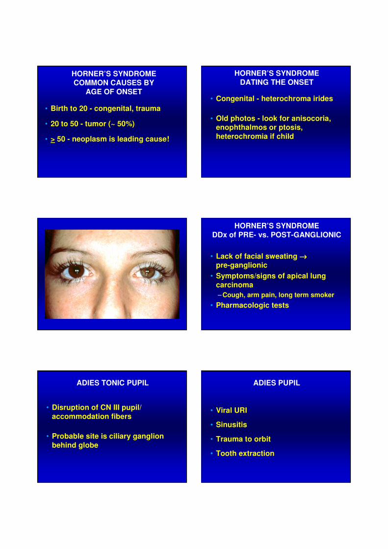
COMMON CAUSES BY
AGE OF ONSET
• Birth to 20 - congenital, trauma
• 20 to 50 - tumor (~ 50%)
• > 50 - neoplasm is leading cause!
HORNER’S SYNDROME
DATING THE ONSET
• Congenital - heterochroma irides
• Old photos - look for anisocoria, enophthalmos or ptosis,
heterochromia if child
HORNER’S SYNDROME
DDx of PRE- vs. POST-GANGLIONIC
• Lack of facial sweating →→→→pre-ganglionic
• Symptoms/signs of apical lung
carcinoma
–Cough, arm pain, long term smoker
• Pharmacologic tests
ADIES TONIC PUPIL
• Disruption of CN III pupil/
accommodation fibers
• Probable site is ciliary ganglion
behind globe
ADIES PUPIL
• Viral URI
• Sinusitis
• Trauma to orbit
• Tooth extraction

ADIES PUPIL
TYPICAL PATIENT
• Middle-aged
• Female
• Recent URI (viral)
ADIES PUPIL
• Anisocoria greatest in bright– Due to sphincter (CN III parasympathetics)
paresis
• 80-90% are unilateral
• Acutely - poor or no light response
• Tonic (slow) near response– Light - near dissociation
• Partial accommodative paresis
• Segmental iris sphincter paresis →→→→segmental miosis
• Possibly ↓↓↓↓ DTRs (Adies syndrome)
ADIES PUPIL
DDX From CN III
• Light response ↓↓↓↓ or absent but better near response
• Normal EOMs - SR, MR, IO, IR
• Normal levator (no ptosis)
ADIES PUPIL
Long-term Changes
• Usually small & sluggish to light
• Pupil may be normal in size (but not responsive to light)
• Near reflex improves
• Accommodation usually normal within 2 years
• DTRs ↓↓↓↓