course 4. biomolecules and their interactions objective
TRANSCRIPT
1
Course 4. Biomolecules and their interactions
Module 19: Structures and conformations of polysaccharide cellulose, amylase, chitin,
carbohydrate conjugates
OBJECTIVE
The main aim of this module is to introduce the students
the importance of polysaccharides
their structures and
to provide insights into how the physical properties of polysaccharides are relevant
for their biological functions
1. INTRODUCTION
Two or more monosaccharides linked to each other by glycosidic bond generate polysaccharides, also
referred to as glycans. Homopolysaccharides consist of one type of monosaccharide while
heteropolysaccharides contain more than one type of monosaccharide. Polysaccharides, as opposed to
proteins and nucleic acids, form branched and linear polymers. The reason for this is that glycosidic
linkage can form between any of the hydroxyl groups of the monosaccharide. Exoglycosidases and
endoglycosdiases are enzymes that hydrolyze monosaccharide units from a polysaccharide.
1.1 Disaccharides
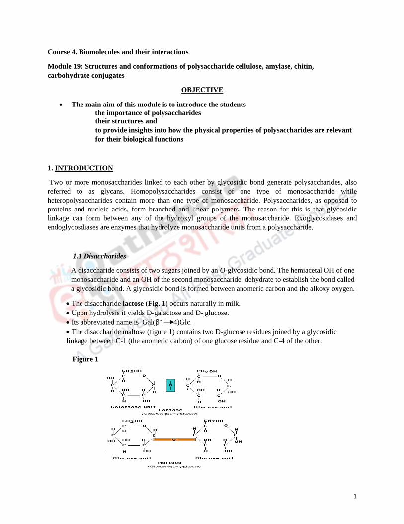
A disaccharide consists of two sugars joined by an O-glycosidic bond. The hemiacetal OH of one
monosaccharide and an OH of the second monosaccharide, dehydrate to establish the bond called
a glycosidic bond. A glycosidic bond is formed between anomeric carbon and the alkoxy oxygen.
The disaccharide lactose (Fig. 1) occurs naturally in milk.
Upon hydrolysis it yields D-galactose and D- glucose.
Its abbreviated name is Gal(β1 4)Glc.
The disaccharide maltose (figure 1) contains two D-glucose residues joined by a glycosidic
linkage between C-1 (the anomeric carbon) of one glucose residue and C-4 of the other.
Figure 1

2
1.1.1 Formation of maltose
Maltose is formed when two molecules of glucose are linked to each other by an O-
glycosidic bond. This glycosidic bond is formed when a hydroxyl group of one
glucose molecule (the one on the right in Fig. 2) reacts or condenses with the
intramolecular hemiacetal of the glucose molecule (on the left). A water molecule is
eliminated during this condensation process, resulting in the formation of a glycosidic
bond. On the contrary, if the glycosidic bond is attacked by a water molecule, it is
referred to as hydrolysis. Glycsodic bonds can be attacked by acids but not by bases,
hence disaccharides can be hydrolyzed to release their monosaccharide components by
boiling with dilute acid. N-glycosidic bonds link the anomeric carbon of one sugar to a
nitrogen atom in glycoproteins and nucleotides.
Sugars that can be oxidized by cupric ion (as discussed in module 18) are referred to
as reducing sugars. This reaction can only occur with the linear forms of sugars, which
though exist in equilibrium with the cyclic forms of sugars in aqueous solution. When
the anomeric carbon is involved in the formation of a glycosidic bond, that sugar
cannot exist in the linear form and is referred to as a non-reducing sugar. In the case of
maltose, as the C-1 of the glucose molecule on the left is not involved in forming the
glycosidic bond, it can undergo reactions typical of reducing sugars. Sucrose on the
other hand is defined as a non-reducing sugar.
Figure 2
2. Polysaccharides
Polysaccharides also known as glycans consists of monosaccharides linked together by glycosidic
bonds.
They generally do not have defining molecular weights like proteins. Unlike proteins,
polysaccharides do not require a template for their synthesis.
Polysaccharides can be classified as homopolysaccharides or heteropolysaccharides.
Hydrolysis
Condensation
H2O
H2O
OH
HO
H
OH
CH2OH
H
H
H
HO
OH
H
OH
CH2OH
HOH
H
H
HO
O
Hemiacetal Acetal
OH
HO
H
OH
CH2OH
HH
OH
H
HO
OH
HO
H
OH
CH2OH
HOH
H
HO
α-D-Glucose β-D-Glucose
Hemiacetal
Alcohol
Maltose
α-D-glucopyranosyl-(1 4)-D-glucopyranose
H

3
Homopolysaccharide contains only a single type of monomeric unit whereas
hetropolysaccharides consists of two or more different kinds of monomeric units.
2.1 Homopolysaccharides/Storage polysaccharides
Starch and glycogen are considered as storage homopolysaccharides, while cellulose and chitin
serve structural roles in plant cell walls and the exoskeletons of animals.
2.1.1 Starch
Starch is considered the most vital storage polysaccharide in plants while glycogen
serves the same purpose in animal cells. These polysaccharides are hydrated and
stored as large granule inside the cells.
Starch is the storage form of glucose in plants, where it is predominantly stored
in cholorplasts.
It is a branched chain of D-glucose.
It contains a mixture of amylose and amylopectin (Fig. 3).
Amylose is a linear unbranched polymer of α-D-glucose units in a repeating
sequence of α1 4 glycosidic linkages.
Amylose is an isomer of cellulose but these differ from each other in their
structural properties.
Cellulose is also a glucose polymer where glucose residues are linked via β
glycosidic linkages causing it to attain a fully extended conformation that can be
tightly packed.
Amlyose on the other hand, due to the α glycosidic linkages attains a helical
coiled structure that can aggregate irregularly (Fig. 3B).
Amylopectin is a branched polymer of α1 4 glycosidic linkages and with α1
6 branching points that occur at intervals of approximately 25-30 α-D-glucose
residues.
Amylopectin is considered as one of the largest molecules that exist in nature due
to their extensive branching.
It is a reducing sugar and is present abundantly in potatoes and in seed.
If starch were to be stored as monomers, it would increase the intracellular
osmotic pressure. Hence, storing glucose as starch keeps the osmotic pressure inside
the cell under check, preventing the cells from lysis.
Figure 3A
(α1 6) branch point
Reducing endNonreducing end

4
Fig. 3B depicts the arrangement of amylose and amylopectin in starch
granules. A double helical structure is formed when amylopectin (red) coils
with itself or with amylose strands Amylose (blue). Starch is mobilized for
energy production when glucose residues are enzymatically cleaved from the
nonreducing ends.
Figure 3B
2.1.2 Glycogen
It is the major storage form of carbohydrate in animals, stored as granules largely
in the liver and in muscle.
The granules consist of several clusters of smaller granules. Each granule is
comprised of one branched glycogen molecule.
It is a highly branched form similar to amylopectin but is more extensively
branched than amylopectin as the α1 6 branching occurs every 8 to 14 D-glucose
residues (Fig.4).
Glycogen has many non-reducing ends, on which glycogen phophorylase can act
to mobilize the breakdown of glycogen to generate free glucose molecules.
Glycogen debranching enzyme acts on the branches.
Both these enzymes are associated, in a tightly bound form with the glycogen
granules making the mobilization of glycogen more efficient.
Glycogen granules are more tightly packed and have more branches than starch.
Figure 4

5
2.2 Homopolysaccharides/Structural polysaccharides
2.2.1 Cellulose
It is a linear, unbranched homopolysaccharide of D- glucose units joined
by β1 4 glycosidic linkages (Fig. 5).
It is a structural polysaccharide of plant cell.
Although cellulose forms a part of the human diet, it is not hydrolyzed by
human enzyme system.
Herbivores contain symbiotic microorganisms that secrete cellulases that
hydrolyzes this β1 4 linkage.
Figure 5
2.2.1 Chitin
It is a linear homopolysaccharide composed of N-acetylglucosamine residues joined
by β1 4 linkages (Fig. 6).
It is the principal component of the exoskeleton of insect and crustaceans.
It is the second most abundant polysaccharide in nature.
Figure 6
2.3 Homopolysaccharide folding
The three dimensional structure of polysaccharides are stabilized by hydrogen bonds,
hydrophobic and van der Waals interactions and electrostatic interactions. Hydrogen

6
bonding has an important influence on polysaccharide folding due to the presence of
multiple OH in its structure.
The three-dimensional structures of these molecules can be described in terms of the
dihedral angles, ϕ and ψ, about the glycosidic bond. The bulkiness of the pyranose ring
and its substituents, and electronic effects at the anomeric carbon, place constraints on
the angles ϕ and ψ. Thus certain conformations are much more stable than others.
The tightly coiled helix is the most stable structure for starch and glycogen stabilized
by interchain hydrogen bonds (Fig. 7).
Figure 7
2.2 Hetropolysaccharides
Glycosaminoglycans (GAGs)
The extracellular matrix is a gel like material present in the extracellular space of the
tissues. It holds the cells together and provides a porous pathway for the diffusion of
nutrients and oxygen to individual cells. Glycosaminoglycans form an important
constituent of the extacellular matrix (ECM).
They are unique to bacteria and animals and are not present in plants.
These are negatively charged linear polymers with repeating disaccharide units.

7
The repeating disaccharide unit consists of acidic sugar (uronic acid) and an
amino sugar (N-acetylglucosamine or N-acetylgalactosamine).
Some glycosaminoglycans also contain esterified sulphate groups.
They attain a extended conformation in solution forming a rodlike helix that
provides maximum separation between the negatively charged sulfate groups.
Specific pattern of sulfated and nonsulfated sugar residues are recognition sites
for variety of protein ligands.
GAGs provide high viscosity and elasticity that is the basis of their providing
strength to the ECM.
Hyaluronic acid
It is an important component of connective tissue, synovial fluid and vitreous
humor of the eye.
Is composed of about 50,000 repeating disaccharide units of D-glucuronic acid
and N-acetyl-D-glucosamine linked by β1 3 bond (Fig. 9).
Forms clear, viscous solution that enables it to act as shock absorber and lubricant
in the synovial fluid of the joints.
Hyaluronidase is an enzyme secreted by certain pathogenic bacteria that
hydrolyzes the hyaluronic acid, making the tissue more susceptible to bacterial
invasion.
All the glycosaminoglycans except hyaluronic acid are found covalently attached
to protein forming proteoglycan.
Figure 9
Because of a large number of anionic groups within hyaluronic acid molecule,
it assumes a extended rod like structure to reduce the repulsive forces amongst
the anionic groups.
The anionic groups bind to cations and water molecules.
Other examples of glycosaminoglycans include chondroitin sulfate, keratan
sulfate, dermatan sulfate, heparin sulfate etc.

8
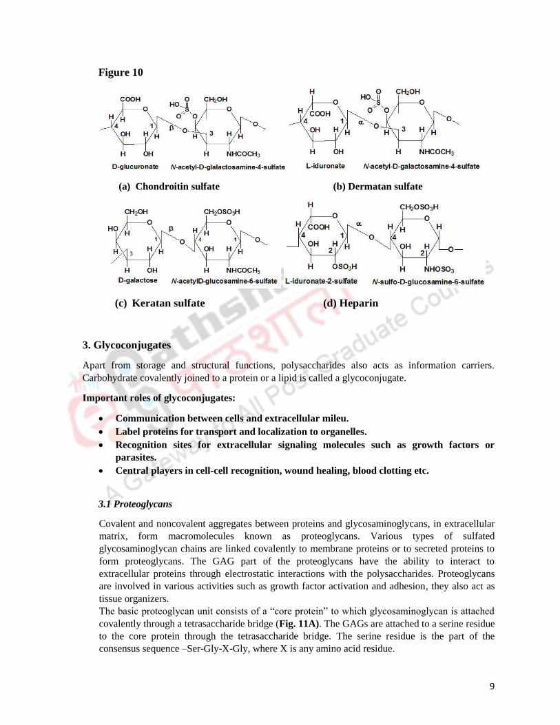
Chondroitin sulfate: consist of N-acetyl-D-galactosamine with sulphate
group, D-glucuronic acid (Figure 10). Provides tensile strength to cartilage,
tendons etc.
Dermatan sulfate: provides elasticity to skin and is present in blood vessels
and heart valves. L-iduronate substitutes the glucuronate residues that
constitute chondroitin sufate. The epimerization of the C5 of glucuronate
residue generates iduronate.
Keratan sulfate: present in cornea, cartilage, horns, hair, nails etc. Acidic
sugar (uronic acid) is not present in keratan sulfate and their sulfate content is
also variable. Repeating disaccharide unit consists of D-galactose and N-
acetyl-D-glucosamine.
The other GAGs differ from hyaluronan in the following aspects:
Other GAGs are shorter polymers.
They are linked to specific proteins resulting in the formation of proteoglycans.
One or more monomeric units differ from that of hyaluronan.
Heparin
Heparin is not a constituent of connective tissue but occurs exclusively in the
intracellular granules of the mast cells that occur in the arterial walls. It has the highest
negative charge density amongst all known biological macromolecule.
It is vital for healing wounds.
Complex formed between heparin sulfate, growth factor and growth factor receptors
initiates the proliferation and differentiation of cells.
The sulfate groups impart a certain pattern to heparin sulfate and this pattern is specific
for specific growth factors.
It is used as an anticoagulant agent. It inhibits coagulation by binding to antithrombin- a
protease inhibitor (Fig. 10).
Heparin is a protease mandatory for clotting of blood and by binding to antithrombin, its
activity is inhibited, thereby preventing blood clotting.
Heparin is added in blood samples obtained for clinical analysis and during blood
transfusion to prevent clotting of blood.
9
Figure 10
(a) Chondroitin sulfate (b) Dermatan sulfate
(c) Keratan sulfate (d) Heparin
3. Glycoconjugates
Apart from storage and structural functions, polysaccharides also acts as information carriers.
Carbohydrate covalently joined to a protein or a lipid is called a glycoconjugate.
Important roles of glycoconjugates:
Communication between cells and extracellular mileu.
Label proteins for transport and localization to organelles.
Recognition sites for extracellular signaling molecules such as growth factors or
parasites.
Central players in cell-cell recognition, wound healing, blood clotting etc.
3.1 Proteoglycans
Covalent and noncovalent aggregates between proteins and glycosaminoglycans, in extracellular
matrix, form macromolecules known as proteoglycans. Various types of sulfated
glycosaminoglycan chains are linked covalently to membrane proteins or to secreted proteins to
form proteoglycans. The GAG part of the proteoglycans have the ability to interact to
extracellular proteins through electrostatic interactions with the polysaccharides. Proteoglycans
are involved in various activities such as growth factor activation and adhesion, they also act as
tissue organizers.
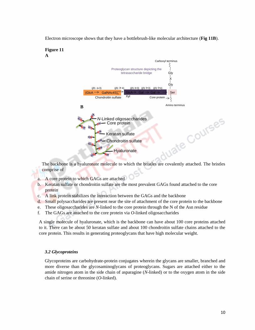
The basic proteoglycan unit consists of a “core protein” to which glycosaminoglycan is attached
covalently through a tetrasaccharide bridge (Fig. 11A). The GAGs are attached to a serine residue
to the core protein through the tetrasaccharide bridge. The serine residue is the part of the
consensus sequence –Ser-Gly-X-Gly, where X is any amino acid residue.
10
Electron microscope shows that they have a bottlebrush-like molecular architecture (Fig 11B).
Figure 11
A
B
The backbone is a hyaluronate molecule to which the bristles are covalently attached. The bristles
comprise of
a. A core protein to which GAGs are attached
b. Keratan sulfate or chondroitin sulfate are the most prevalent GAGs found attached to the core
protein
c. A link protein stabilizes the interaction between the GAGs and the backbone
d. Small polysaccharides are present near the site of attachment of the core protein to the backbone
e. These oligosaccharides are N-linked to the core protein through the N of the Asn residue
f. The GAGs are attached to the core protein via O-linked oligosaccharides
A single molecule of hyaluronate, which is the backbone can have about 100 core proteins attached
to it. There can be about 50 keratan sulfate and about 100 chondroitin sulfate chains attached to the
core protein. This results in generating proteoglycans that have high molecular weight.
3.2 Glycoproteins
Glycoproteins are carbohydrate-protein conjugates wherein the glycans are smaller, branched and
more diverse than the glycosaminoglycans of proteoglycans. Sugars are attached either to the
amide nitrogen atom in the side chain of asparagine (N-linked) or to the oxygen atom in the side
chain of serine or threonine (O-linked).
(GlcA GalNAc4S)n GlcA Gal Gal
Xyl
Ser
Gly
X
Gly
(β1 3) (β1 4) (β1 3) (β1 3) (β1 4)
Chondroitin sulfate
Carboxyl terminus
Amino terminus
Core protein
Proteoglycan structure depicting the
tetrasaccharide bridge
N-Linked oligosaccharides
Keratan sulfate
Chondroitin sulfate
Hyaluronate
Core protein

11
N-linked glycoproteins
Glycoproteins, in which the GLcNAc is attached to the amide of an Asn residue, are referred to as
N-linked glycoproteins. The Asn is part of the sequence Asn-X-Ser/Thr, where X can be any
amino acid except Pro or Asp.
N-glycosylation occurs as the polypeptide chain is being synthesized (cotranslationally) while still
attached to the ribosome.
The synthesis is N-linked proteins is as depicted in Fig. 12.
Figure 12
The synthesis of N-linked glycoproteins is initiated when 9 mannose residues, 3 glucose and 2
GlcNac residues are attached to the Asn of a polypeptide chain that is being synthesized.
This is followed by removal of some of the sugar molecules and this process occurs in the
endoplasmic reticulum lumen. The removal of the sugar molecules continues in the Golgi
apparatus by glucosidases and mannosidases.
Glycosyltransferases present in the Golgi then enzymatically add GlcNac, galactose, fucose and
sialic acid residues to the already existing core oligosaccharide chain.
The processing is either limited, generating high mannose oligosaccharides or is extensive
generating large oligosaccharides containing different kinds of sugar molecules. This results in
the diversity observed in the oligosaccharides of the N-linked glycoproteins.
O-linked glycoproteins
These glycoproteins have oligosaccharides attached to the protein via Ser or Thr residues. All the
O-linked glycoproteins share a disaccharide core β-galactosyl-(1-3)-α-N-acteylgalactosamine to
the hydroxyl group of either Ser or Thr. Other sugar residues that can be rarely attached to Ser or
Thr are galactose, mannose and xylose.
14-residue
oligosaccharide is
attached to Asn of
a polypeptide.
Removal of monosaccharide
units produces a
(mannose)3(GlcNAc)2
oligosaccharide. This
(mannose)3(GlcNAc)2 core
is found in all N-linked
oligosaccharides.
Further trimming and addition of other
sugars yields a variety of N-linked
oligosaccharides.
Core
pentasaccharide

12
The O-linked oligosaccharides are attached to the polypeptide chain after it has been synthesized,
that is, posttranslationally in the Golgi apparatus.
The transfer of GalNAc to a Ser or Thr residue on the polypeptide is the initiation step for the
synthesis of the O-linked oligosaccharides.
The Ser and Thr residues are not a part of a consensus sequence unlike the Asn of the N-linked
oligosaccharides. The point of attachment of the oligosaccharides is dictated by the secondary and
tertiary structures of the polypeptide. Glycosyltransferases add sugars in a stepwise manner to
increase the length of the chain.
Summary
Monosaccharides link through glycosidic linkages to generate polysaccharides.
Cellulose and chitin are structural polysaccharides with rigid and extended structures with β-
glycosidically linked glucose residues.
Starch and glycogen are storage polysaccharides with α-glycosidically linked glucose residues.
Glycosaminoglycans are unbranched polysaccharides that consist of uronic acid and amino sugars
that are often sulfated.
Proteoglycans are components of the extracellular matrix and consist of hyaluronate with attached
core proteins.
There are two types of glycoproteins; N-linked and O-linked glycoproteins.