correlation between imaging and clinical picture
TRANSCRIPT
II Spine Surgery DepartmentI.R.C.C.S. Istituto Ortopedico GaleazziChief: Claudio Lamartina, MDProfessor EFM Universities of Milan and Turin - ItalyCo-Chief: Roberto Bassani, MD
Correlation between imaging and clinical picture
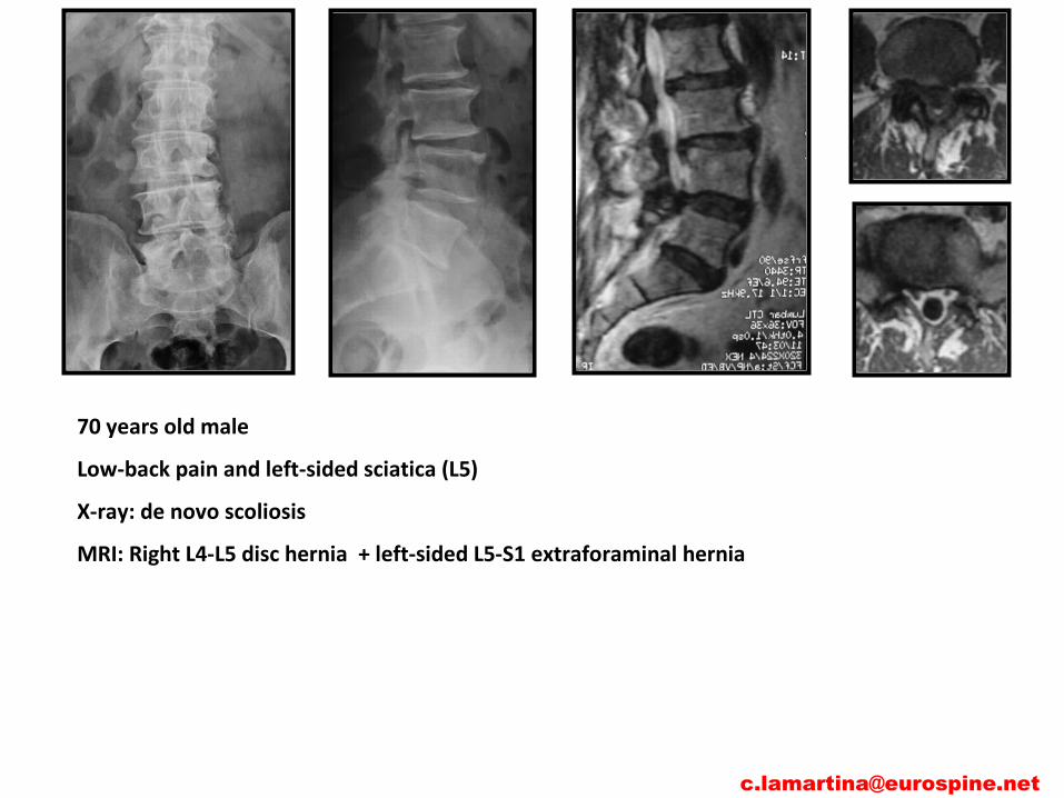
70 years old male
Low-back pain and left-sided sciatica (L5)
X-ray: de novo scoliosis
MRI: Right L4-L5 disc hernia + left-sided L5-S1 extraforaminal hernia

Frymoyer JD. Back pain and sciatica. N Engl J Med. 1988;318:291–300
• approximately 90 percent of adults experience back pain at some time in life
• Fewer than 2 percent of patients have disc herniation
Scavone JG, Latshaw RF, Rohrer GV. Use of lumbar spine films. Statistical evaluation at a university teaching hospital. JAMA. 1981;246:1105–8.
• plain-film radiographs were normal or demonstrated changes of equivocal clinical significance in more than 75 percent of patients with low back pain
Scavone JG, Latshaw RF, Weidner WA. Anteroposterior and lateral radiographs: an adequate lumbar spine examination. AJR Am J Roentgenol. 1981;136:715–7.
• oblique views of the spine uncovered useful information in fewer than 3 percent of patients
Jensen MC, Brant-Zawadzki MN, Obuchowski N, Modic MT, Malkasian D, Ross JS. Magnetic resonance imaging of the lumbar spine in people without back pain. N Engl J Med. 1994; 331:69–73.
•MRI scans revealed herniated discs in approximately 25 percent of asymptomatic persons less than 60 years of age and in 33 percent of those more than 60 years of age

Boden SD, Davis DO, Dina TS, Patronas NJ, Wiesel SW. Abnormal magnetic-resonance scans of the lumbar spine in asymptomatic subjects. A prospective investigation. J Bone Joint Surg [Am]. 1990;72:403–8.
• Magnetic resonance imaging (MRI) has been found to demonstrate abnormalities in “normal” asymptomatic people
Wiesel SW, Tsourmas N, Feffer HL, Citrin CM, Patronas N. A study of computer-assisted tomography. I. The incidence of positive CAT scans in an asymptomatic group of patients. Spine. 1994;9:549–51.
• computed tomographic (CT) scanning has been found to demonstrate abnormalities in “normal” asymptomatic people

70 years old male
Low-back pain and left-sided sciatica (L5)
X-ray: de novo scoliosis
MRI: Right L4-L5 disc hernia + left-sided L5-S1 extraforaminal hernia
Neurological examination:
left L5 deficit (2/5 BMRC)
Walking on left heel was not possible
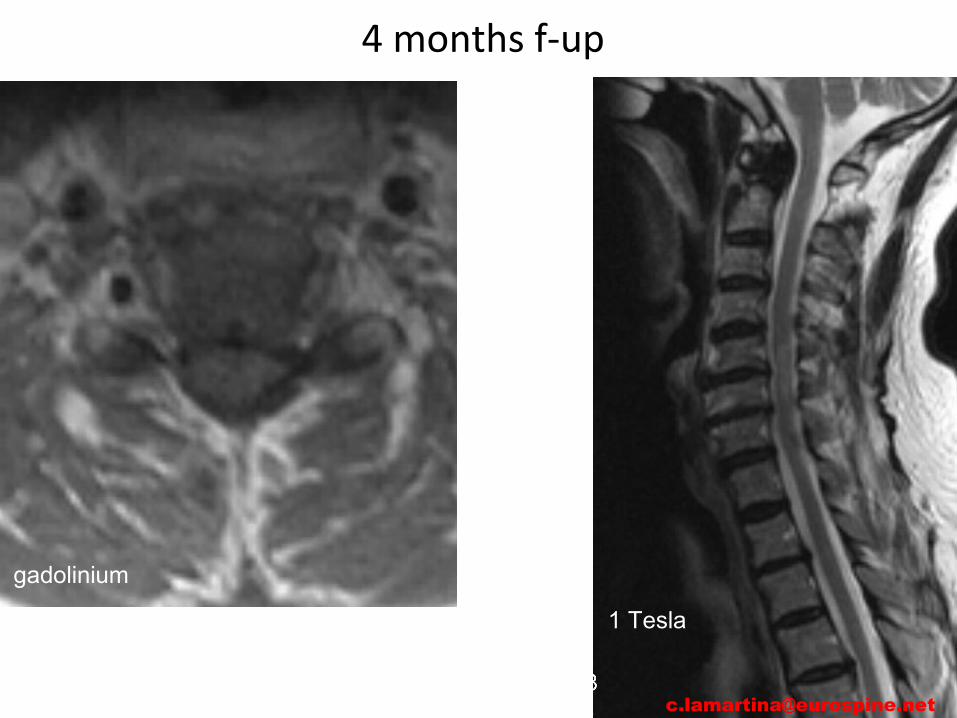
Brisk knee jerks, bilateral knee clonus, positive Babinski [email protected]

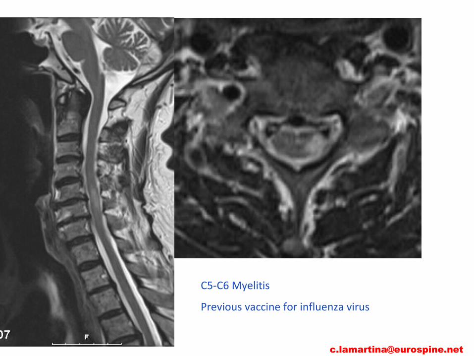
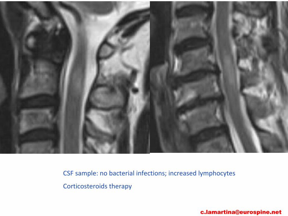
CSF sample: no bacterial infections; increased lymphocytes
Corticosteroids therapy

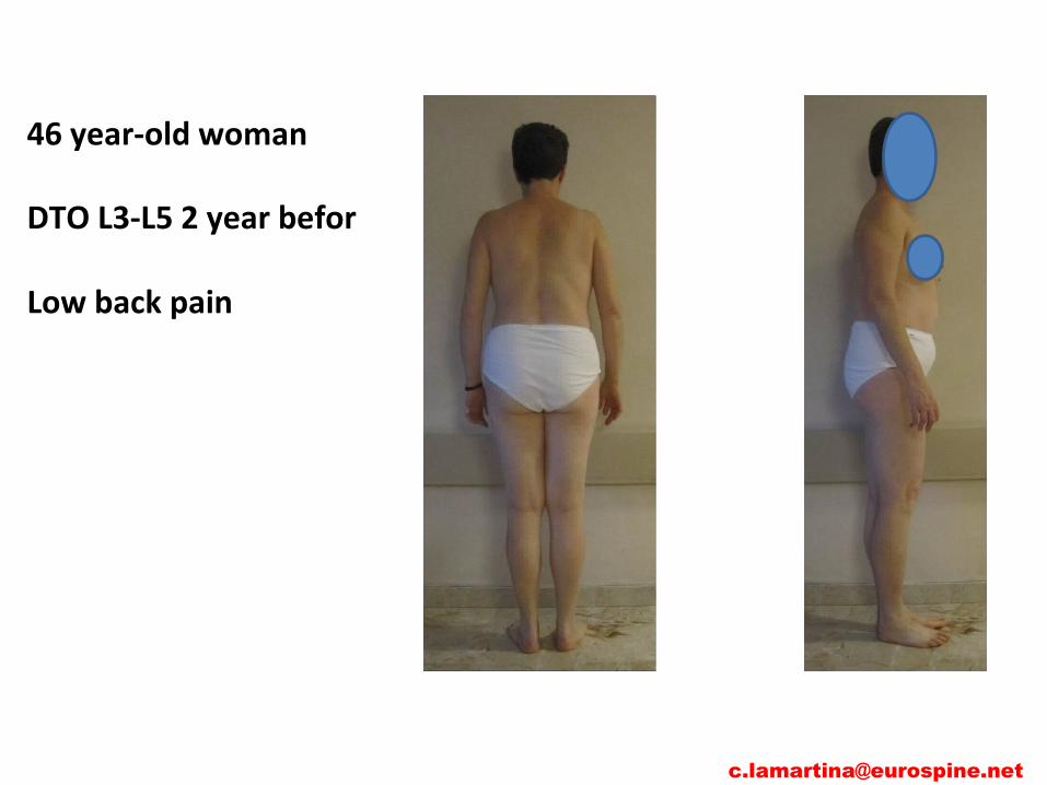
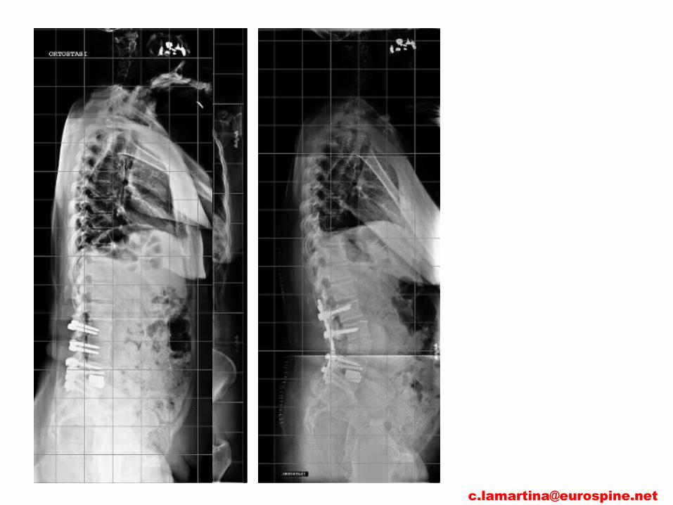
65 year-old woman low back pain (more than 5 years) increasing gait disturbance in the last months (claudicatio after 30 meters)
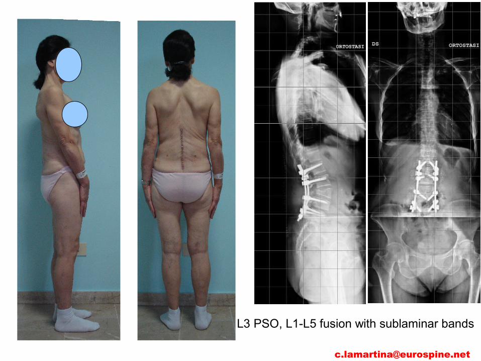
unbalanced spine with compensatory mechanisms
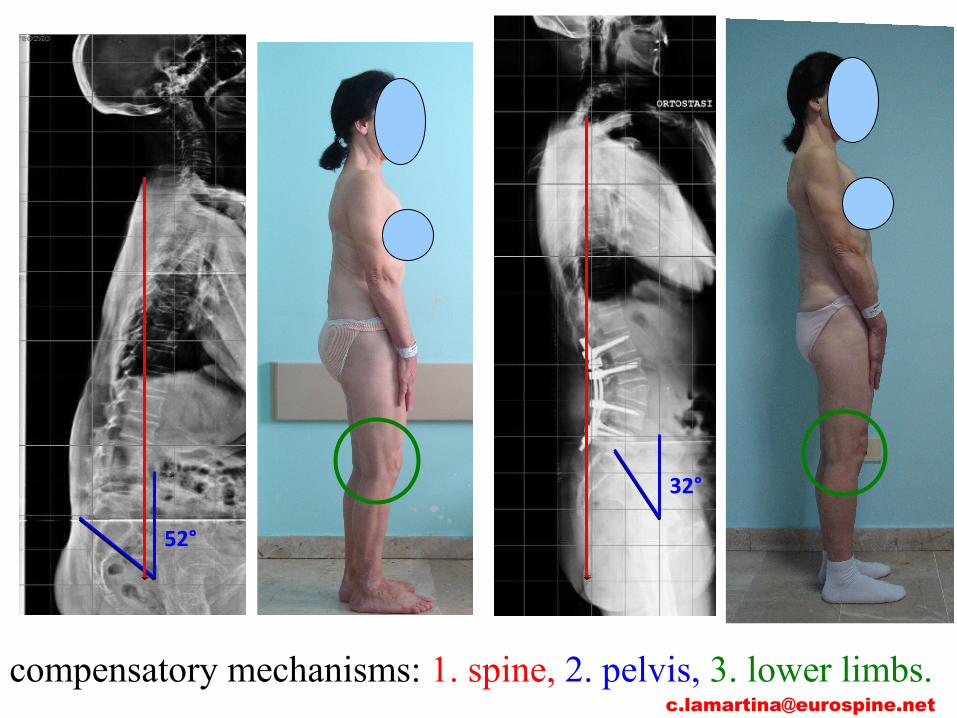

Conclusion
Clinical evaluation has priority over1.imaging2.instrumental tests
report clinical picture
clinical picture imaging report