correia ... · 1laboratório de imunologia mário arala chaves, departamento de imuno-fisiologia e...
TRANSCRIPT

VETERINARY RESEARCHCorreia et al. Veterinary Research 2013, 44:69http://www.veterinaryresearch.org/content/44/1/69
RESEARCH Open Access
Mucosal and systemic T cell response in miceintragastrically infected with Neospora caninumtachyzoitesAlexandra Correia1,2, Pedro Ferreirinha1,2, Amanda A Costa1, Joana Dias1, Joana Melo1, Rita Costa1, Adília Ribeiro1,2,Augusto Faustino3, Luzia Teixeira4,5, António Rocha1 and Manuel Vilanova1,2*
Abstract
The murine model has been widely used to study the host immune response to Neospora caninum. However, inmost studies, the intraperitoneal route was preferentially used to establish infection. Here, C57BL/6 mice wereinfected with N. caninum tachyzoites by the intragastric route, as it more closely resembles the natural route ofinfection through the gastrointestinal tract. The elicited T-cell mediated immune response was evaluated in theintestinal epithelium and mesenteric lymph nodes (MLN). Early upon the parasitic challenge, IL-12 production byconventional and plasmacytoid dendritic cells was increased in MLN. Accordingly, increased proportions andnumbers of TCRαβ+CD8+IFN-γ+ lymphocytes were detected, not only in the intestinal epithelium and MLN, but alsoin the spleen of the infected mice. In this organ, IFN-γ-producing TCRαβ+CD4+ T cells were also found to increasein the infected mice, however later than CD8+ T cells. Interestingly, splenic and MLN CD4+CD25+ T cells sorted frominfected mice presented a suppressive activity on in vitro T cell proliferation and cytokine production above that ofcontrol counterparts. These results altogether indicate that, by producing IFN-γ, TCRαβ+CD8+ cells contribute forlocal and systemic host protection in the earliest days upon infection established through the gastrointestinal tract.Nevertheless, they also provide substantial evidence for a parasite-driven reinforcement of T regulatory cell functionwhich may contribute for parasite persistence in the host and might represent an additional barrier to overcometowards effective vaccination.
IntroductionNeospora caninum is a protozoan parasite found in awide range of domestic and wild animal hosts [1], and isresponsible for clinical infections in dogs and cattle [2],having a major impact in dairy and beef industry [3]. Ex-perimentally, the murine model has been the one pre-ferred to study neosporosis, as it presented similarfeatures to the infection occurring naturally in permis-sive hosts such as brain lesions [4], reproductive loss [5]and mother to fetus parasite transmission [6]. AlthoughN. caninum is transplacentally transmitted in cattle withhigh efficiency, significant postnatal transmission also
* Correspondence: [email protected]ório de Imunologia Mário Arala Chaves, Departamento de Imuno-Fisiologia e Farmacologia, ICBAS-UP, Instituto de Ciências Biomédicas deAbel Salazar – Universidade do Porto, Rua de Jorge Viterbo Ferreira nº 228,Porto , 4050-313, Portugal2IBMC - Instituto de Biologia Molecular e Celular, Porto, PortugalFull list of author information is available at the end of the article
© 2013 Correia et al.; licensee BioMed CentralCommons Attribution License (http://creativecreproduction in any medium, provided the or
occurs in these animals [1], likely through oocyst inges-tion [7]. Even though neosporosis can thus be estab-lished through the gastrointestinal (GI) tract, moststudies on the host immune response have been carriedout in hosts infected via the intraperitoneal (i.p.) or sub-cutaneous routes. Consequently, the mucosal immuneresponse to this parasite in infected hosts was barelystudied. As mucosal immunizations have been alreadyattempted in experimental models of neosporosis [8-10],the characterization of the immune response to N.caninum in the mucosa and associated lymphoid tissueswill be helpful to further understand the immunobiologyof this parasitic disease. Therefore, a murine model ofneosporosis established by intragastric (i.g.) administra-tion of N. caninum tachyzoites was used here to studythe immune response elicited by this parasite in the gutand associated lymphoid tissue of the infected hosts.
Ltd. This is an Open Access article distributed under the terms of the Creativeommons.org/licenses/by/2.0), which permits unrestricted use, distribution, andiginal work is properly cited.

Correia et al. Veterinary Research 2013, 44:69 Page 2 of 13http://www.veterinaryresearch.org/content/44/1/69
Materials and methodsMiceFemale C57BL/6 mice, 8–10 weeks old, were pur-chased from Charles River (Barcelona, Spain) and keptunder specific pathogen-free conditions at the AnimalFacility of Instituto de Ciências Biomédicas Abel Salazar(ICBAS), Porto, Portugal. Female p40−/− C57BL/6 mice,7–11 weeks old, were purchased from Jackson Labora-tories (Bar Harbor, Maine, USA) and housed and bredalso at ICBAS in individual ventilated cages. Nestingand housing material was provided as enrichment. Allprocedures involving mice were performed accordingto the European Convention for the Protection of Ver-tebrate Animals used for Experimental and other Sci-entific Purposes (ETS 123), 86/609/EEC Directive andPortuguese rules (DL 129/92). Authorization to per-form the experiments was issued by the competent na-tional board authority, Direcção Geral de Veterinária(0420/000/000/2008).
ParasitesNeospora caninum tachyzoites (NC-1 isolate) were cul-tured and serially passaged in VERO cells maintained at37 °C in Minimum Essential Medium (MEM) containingEarle’s salts (Gibco: Invitrogen Corporation, Carlsbad,CA, USA) supplemented with 10% fetal bovine serum(FBS), L-glutamine (2 mM), penicillin (200 IU/mL) andstreptomycin (200 μg/mL) (all from Sigma, St Louis,USA) in a humidified atmosphere of 5% CO2 in air. Freeparasitic forms of N. caninum were obtained as previ-ously described [11] with slight modifications. InfectedVERO cells were cultured until the host cell monolayerwas 90% destroyed. Culture supernatants and adherentcells, harvested using a cell scraper, were centrifuged at1500 × g for 15 min. The pellet was passed through a25G needle and then washed three times in PhosphateBuffered Saline (PBS). The obtained pellet wassuspended in 3 mL of PBS and passed through a PD-10column filled with Sephadex™ G-25 M (Amersham Bio-sciences Europe GmbH, Freiburg, Germany). Parasiteconcentration was determined with a haemocytometer.
Challenge infectionsN. caninum infections in C57BL/6 mice were performedby the i.g. route using a previously described protocol[11]. Briefly, 5 h before infection mice were deprived offood. Mice were then anaesthetized by intramuscular in-jection of 20 μL of a 4:5 mixture containing xylazine(RompumW, Bayer Portugal, S.A., Carnaxide) and keta-mine (Imalgéne 1000, Bayer Portugal, S.A., Carnaxide).Stomach acidity was neutralized by directly administer-ing into the stomach, with a gavage feeding needlelinked to a 1-mL syringe, 50 μL of a 10% sodium bicar-bonate solution in water. The same procedure was used
to inoculate N. caninum tachyzoites 15 min later. Micewere i.g. challenged with 5 × 10⁷ tachyzoites in 0.2 mL ofPBS or similarly inoculated with 0.2 mL of PBS andsacrificed at 6 h, 12 h, 18 h, 48 h, and 4, 7 and 21 daysafter challenge.
Sample collectionAt the different time points, mice were sacrificed uponisoflurane anesthesia by cervical dislocation. Spleens andmesenteric lymph nodes (MLN), from infected mice andnon-infected controls, were aseptically removed and ho-mogenized to single cell suspensions in HBSS for theirusage in cell culture experiments and flow cytometryanalysis. Additionally, brain, liver, MLN, and intestinaltissue samples were collected and either frozen (DNAisolation) or formalin-fixed (histology and immunohisto-chemistry). Small intestines were alternatively collectedfor IEL isolation. The number of animals per group perexperiment is indicated in the respective figure legends.
Histopathology and immunohistochemistryHistopathology of intestinal tissue samples was assessedin formalin-fixed, paraffin-embedded 4 μm sections,mounted on amino-propyl-tri-ethoxy-silane (Sigma-Aldrich, St Louis, MO, USA) coated slides, of the gastro-intestinal tract of mice 6, 12 and 18 h upon i.g.infection, stained with haematoxylin-eosin. The presenceof N. caninum parasitic forms was assessed in similartissue sections by immunohistochemistry, performed aspreviously described with slight changes [11]. Tissue sec-tions were deparaffinized in xylene, rehydrated by gradedwashes of ethanol in water, ending in a final rinse in de-ionized water. Antigen retrieval was performed by incu-bating the slides in 10 mM citrate buffer (pH = 6) for3 min in a pressure cooker. The slides were cooled andrinsed three times in Tris-buffered saline (TBS; 50 mMTris, 150 mM NaCl, 0.1% Tween 20, pH = 7.6) for5 min. Endogenous peroxidase activity was blocked byimmersing slides in methanol containing 3% hydrogenperoxide for 10 min, followed by TBS washing. Toreduce non-specific antibody binding, slides were in-cubated with normal rabbit serum (Dako, Glostrup,Denmark) diluted at 1:5 in TBS containing 10% bovineserum albumin (BSA), in a humidified chamber for20 min at room temperature. Excess normal serum wasremoved and replaced by the goat anti-N. caninum anti-serum (VMRD, Pullman, WA, USA) diluted at 1:2000.After one hour incubation at room temperature, slideswere washed with TBS and incubated for 30 min with a1:1000 dilution of peroxidase-labeled rabbit anti-goatsecondary antibody (Millipore, Billerica, MA, USA).Slides were then washed with TBS and detection wasperformed for 3 to 5 min with 0.05% 3,3 diaminoben-zidinetetrahydrochloride (DAB) freshly prepared in

Correia et al. Veterinary Research 2013, 44:69 Page 3 of 13http://www.veterinaryresearch.org/content/44/1/69
0.05 M Tris/hydroxymethylaminomethane buffer, pH 7.6,containing 0.1% hydrogen peroxide (Dako). Finally, sec-tions were lightly counterstained with Mayer’s haematoxy-lin, dehydrated and mounted in EntellanW mountingmedium (Merck, Darmstadt, Germany). Dilution of pri-mary antibody and peroxidase-labeled secondary antibodywere made with TBS containing 5% BSA. Positive controlsections of N. caninum-infected IL-12−/− mouse organswere included. Negative controls were performed by omit-ting the primary antibody incubation. Slides were evalu-ated under light microscopy.
Real-time PCR analysisDNA from intestinal tissue sections, MLN, liver and brainwas isolated as previously described [11]. Detection of N.caninum DNA in infected tissue samples was assessed bya quantitative real-time PCR (qRT-PCR) analysis per-formed in a Corbett rotor gene 6000 system (Corbett lifescience, Sydney, Australia), using Express Sybr green ERqPCR supermix universal (Invitrogen, Carlsbad, CA,USA), for the amplification of a 337 bp sequence of theNc5 region of N. caninum genome using the primersNp21plus 5’ CCCAGTGCGTCCAATCCTGTAAC 3’ andNp6plus 5’ CTCGCCAGTCAACCTACGTCTTCT 3’(TIB-Molbiol, Berlin, Germany), both at a final con-centration of 0.5 μM. The DNA samples were ampli-fied using the following program: 95 °C for 10 min,followed by 45 cycles of 95 °C for 30 s, 63 °C for 20 s,and 72 °C for 45 s with fluorescence acquisition. Amelting curve was performed in each run in orderto confirm specificity of the amplicon: from 65 °C to95 °C, with increments of 1 °C for 5 s. Parasite quanti-fication was determined by interpolation of a standardcurve, ranging from 10 to 10-4 ng of DNA extractedfrom N. caninum tachyzoites included in each run.Data were analyzed in the Rotor gene 6000 softwarev1.7 (Corbett life science).
Intraepithelial lymphocyte isolationGut intraepithelial lymphocytes (IELs) were isolated aspreviously described [12]. Briefly, mice small intestineswere removed and flushed with 20 mL of cold CMF(Ca++, Mg++ free Hank’s Balanced Salt Solution (HBSS)with 1 mM HEPES and 2% FBS, all from Sigma) usinga syringe and needle. The Peyer’s Patches, fat andremaining mucous were removed along the intestine.The intestine was opened lengthwise, cut into 5 mmpieces and placed in a conical tube with 40 mL CMF.The pieces of tissue were washed twice with CMF byinverting the tube 10 times and letting the pieces settlebefore removing the supernatant. The intestine pieceswere incubated in 25 mL CMF/DTE (CMF with 10%FBS and 1 mM dithioerythritol) (Sigma) at 37 °C and100 rpm in an orbital incubator (GFL 3031, GFL,
Burgwedel, Germany) for 20 min. The tube was vor-texed at maximum speed for 15 s and the supernatantremoved to a new tube. 25 mL of CMF/DTE wereadded to the tube containing the pieces of tissue andthe vortexing step and collection of supernatant weredone once more. All the incubation and supernatantcollection steps were repeated. Supernatants from eachintestine were pooled and centrifuged at 400 × g, 4 °Cfor 20 min. The pellet was suspended in 5 mL HBSSwith 2% FBS and passed through a nylon wool columnpre-wet with HBSS with 2% FBS (0.15 g teased nylonwool in a 5 cc syringe) and the column was washedwith 20 mL HBSS with 2% FBS. The collected cell sus-pension was centrifuged and suspended in 16 mL of44% Percoll™ (GE Healthcare Bio-Sciences AB, Upp-sala, Sweden), adding 8 mL per 14 mL polystyreneround bottom tube. 5 mL of 67% Percoll were under-laid per tube and the 44%/67% Percoll gradients werecentrifuged for 20 min at room temperature, 1400 × g,with the brake off. Cells from the interface werecarefully removed with a Pasteur pipette and washedtwice with 40 mL cold complete RPMI (RPMI 1640supplemented with 50 U/mL penicillin, 50 μg/mLstreptomycin, 1% HEPES buffer, 10% FCS and 5 μM 2-mercaptoethanol, all from Sigma). Pellet was sus-pended in complete RPMI.
Flow cytometric analysisThe assessment of cell surface and cytoplasmic lineageor activation markers on different splenic leukocytepopulations was performed by flow cytometric analysis(FACS). From spleen and MLN cell suspensions, preparedas described above, a number of 1 × 10⁶ leucocytes werestained per sample. The following monoclonal anti-bodies (mAbs), along with the respective isotypecontrols were used (at previously determined optimaldilutions) for immunofluorescence cytometric dataacquisition in a Coulter EPICS XL flow cytometer(Beckman Coulter, Inc., Brea, CA, USA): fluoresceinisothiocyanate (FITC) anti-mouse/rat Foxp3 (FJK-16 s),phycoerythrin (PE) anti-mouse TCR β (H57-597)and PE-Cy5 rat anti-mouse CD4 (L3T4) (RM4-5) (allfrom eBioscience, San Diego, CA, USA); Biotin anti-mouse PDCA-1 (JF05-1C2.4.1) (Miltenyi Biotech, Inc.Auburn, CA, USA); FITC hamster anti-mouse CD11c(HL3), PE-Cy5 rat anti-mouse CD8a (53–6.7), PE anti-mouse CD25 (PC61), PE rat anti-mouse IL-4 (BVD4-1D11), Biotin hamster anti-mouse γδ T-cell receptor(GL3), FITC anti-mouse IFN-γ (XMG1.2), FITC ratanti-mouse IL-17A (TC11-18H10), PE rat anti-mouseIL-10 (JES5-2A5) (all from BD Pharmingen, San Diego,CA, USA). Biotin conjugated mAbs were revealed withStreptavidin-PE-Cy7 (BD Pharmingen). Cells werepreincubated for 15 min with anti-FcγR (a kind gift of

Correia et al. Veterinary Research 2013, 44:69 Page 4 of 13http://www.veterinaryresearch.org/content/44/1/69
Dr Jocelyne Demengeot, Gulbenkian Institute of Sci-ence, Oeiras, Portugal) before CD11c and Foxp3 stain-ing. The Foxp3 Staining Buffer Set (eBioscience) wasused for fixation and permeabilization of splenocytespreviously surface stained with CD4 and CD25 mAbs.Data were analysed by using CELLQUEST software(Becton-Dickinson, San Jose, CA, USA).
Intracellular stainingThe intracellular expression of the cytokine IFN-γ wasdetected in splenic and MLN CD8+ and CD4+ T lym-phocytes, as well as in IELs. IL-4, IL-17A and IL-10expression was also evaluated in splenic and MLNCD4+T cells. Splenocytes and MLN cells were obtainedas described above. Red blood cell lysis was performedin spleen suspensions by incubation with 0.15 M am-monium chloride. Cells were washed and suspended incomplete RPMI medium. Spleen, MLN or IEL 1 × 10⁶
cells were transferred to 96-well tissue culture plates(Nunc, Roskilde, Denmark) and stimulated for 4.5 hwith 20 ng/mL phorbol myristate acetate and 200ng/mL ionomycin in the presence of 10 ng/mL of bref-eldin A (all from Sigma). Staining of cell surface mar-kers CD4, CD8, TCR β and TCRγδ was performed asdescribed above, after a preincubation step of 15 minwith anti-FcγR, followed by fixation with 2% formalde-hyde. Cells were permeabilized with 0.5% saponin inflow cytometric buffer (PBS containing 1% BSA and0.01 M sodium azide) and, subsequently, cells were in-cubated for 15 min with anti-FcγR and stained for30 min at room temperature with the appropriate anti-body. The intracellular expression of the cytokinesIL-12 and IL-10 was detected in splenic and MLN con-ventional and plasmacytoid dendritic cells (cDC andpDC, respectively). Spleen and MLN suspensions wereenriched with DC by magnetic sorting using anti-CD11c beads (Miltenyi), according to the manufac-turer’s instructions. 1 × 10⁶ cells were then placed in96-well tissue culture plates (Nunc) and incubated for4.5 h with 10 ng/mL of brefeldin A (Sigma). Cells weresurface stained with PE-Cy5.5 hamster anti-mouseCD11c (HL3) (BD Pharmingen) and Biotin anti-mousePDCA-1 (Miltenyi Biotech) revealed with PE-Cy7 con-jugated streptavidin (BD Pharmingen). Intracellularstaining was performed with PE rat anti-mouse IL-12(p40/p70) (C15.6) and FITC rat anti-mouse IL-10(JES5-2A5) (all from BD Pharmingen). Intracellularstaining with the isotypic controls was performed toconfirm the specificity of antibody binding.
Cell cultures and suppression assaysFor anti-CD3 mAb-stimulated cultures, antigen pre-senting cells (APC) were prepared from naïve splenicor MLN single cell suspensions by layering 5 mL onto
2.5 mL of a polysucrose-sodium ditrizoate solution(Histopaque 1083W, Sigma) and centrifuging at 800 × gfor 20 min at room temperature. Mononuclear cellscollected from the medium-Histopaque interface werewashed, suspended in RPMI complete medium and ir-radiated at 3000 rad in a Gammacell 1000 Elite irradia-tor (Nordion International, Inc., Ottawa, Canada).Total CD4+ and the T cell subsets CD4+CD25- andCD4+CD25+ from non-infected and infected mice wereisolated from pooled spleen or MLN cells of five miceper group, by using a magnetic cell sorting CD4+CD25+
T-cell isolation kit (Miltenyi Biotech, Inc., Auburn, CA,USA) following the manufacturer’s instructions. Purityof the sorted cells routinely ranged between 92-98%.Naïve CD4+CD25- T cells (responder cells) were platedat 2.5 × 10⁴/well in U-shape 96-well plates togetherwith 105 APC without stimulus or stimulated with1 μg/mL anti-CD3 mAb (145.2C11) (BD Pharmingen).CD4+CD25+ T cells sorted from control and N.caninum-infected mice were added to the naïve CD4+
CD25- T cells in a 1:1 proportion. Each condition wasset in sextuplicates and culture was maintained for72 h. Supernatants from these cell cultures were col-lected and stored at −80 °C until further use. TheCellTraceTM CFSE Cell Proliferation Kit (MolecularProbes, Invitrogen, Eugene, OR, USA) was used for celllabelling. A CFSE (5-(and-6)-carboxyfluorescein dia-cetate succinimidyl ester) stock solution (10 mM inDMSO) stored at −20 °C was thawed and diluted in PBSwith 0.1% BSA to a final concentration of 10 μM. NaïveCD4+CD25- T cells (responder cells) were suspended at2 × 106/mL in PBS with 0.1% BSA and further incu-bated with an equal volume of the diluted CFSE solu-tion, for 7 min at room temperature. Excess CFSE wasquenched by adding 1/5 of the volume of heat in-activated FBS. Cells were washed three times withcomplete RPMI medium. Responder cells were plated at2.5 × 104/well in U-shape 96-well plates together with105 APC and 1 μg/mL anti-CD3 mAb. In order toevaluate Treg cell suppressive function, CD4+CD25+ Tcells from control and infected mice were added at dif-ferent responder: CD4+CD25+ T cell ratios (1:1, 2:1, and10:1). Responder cells without anti-CD3 stimulus wereused as the negative control. Stimulated responder cellswith no suppressor populations added were used as thepositive control. Unlabelled stimulated responder cellswere used to define cell auto fluorescence. Othercontrols consisted of stimulated responder cells co-cultured with CD4+CD25- T cells from the different ani-mal groups tested, to exclude suppression due to cellnumber/well. Each condition was set in sextuplicatesand cultures were maintained for 72 h at 37 °C and 5%CO2. Proliferation/suppression was determined basedon CFSE fluorescence by flow cytometric analysis.

Correia et al. Veterinary Research 2013, 44:69 Page 5 of 13http://www.veterinaryresearch.org/content/44/1/69
For antigen stimulated cultures, bone marrow-deriveddendritic cells (BMDC) were prepared by a granulocytemacrophage colony-stimulating factor (GM-CSF)-basedmethod, as described by Lutz et al. [13]. Upon differenti-ation, BMDC were antigen-loaded by overnight incubationwith 100 μg/mL of N. caninum sonicates prepared aspreviously described [11], or cultured without antigen inthe presence of 50 ng/mL of lipopolysaccharide from E.coli (Sigma). BMDC APC were then washed twice withPBS and suspended in complete RPMI. CFSE-labelledresponder cells (CD4+CD25- cells isolated, as describedabove, from infected mice, 7 days upon infection) wereplated at 2.5 × 10⁴/well in U-shape 96-well plates togetherwith 105 BMDC and were used as the positive control. Toevaluate Treg suppression capacity, CD4+CD25+ T cellsfrom control and infected mice were added to the culturesat 1:1 and 2:1 responder: CD4+CD25+ T cell ratios.
IFN-γ, IL-4, and IL-10 measurementsThe concentration of IFN-γ, IL-4 and IL-10 in cell cul-ture supernatants from N. caninum-infected mice andfrom non-infected controls were quantified with theMouse IFN-γ, IL-4 and IL-10 ELISA Ready-Set-Go!W kits(eBioscience), according to manufacturer’s instructions.
Statistical analysisUnless otherwise indicated, statistical significance of re-sults was determined by unpaired Student t-test, usingthe GraphPad Prism 4 Software (GraphPad Software,Inc., La Jolla, CA, USA). Results were considered statis-tically significant with P values of less than 0.05.
ResultsInfection of C57BL/6 mice with N. caninum tachyzoitesadministered intragastricallyWe have previously reported that neosporosis could beestablished in mice i.g. challenged with N. caninumtachyzoites. However, in the i.g.-challenged mice, theimmune response elicited in the gut associated lymph-oid tissue was studied only in the Peyer’s Patches [11].Here, C57BL/6 mice were challenged i.g. with N.caninum tachyzoites to evaluate the elicited immuneresponse in the gut epithelium and draining lymphnodes. Early after the i.g. challenge, parasitic DNA wasdetected by qRT-PCR in intestinal tissue samples (1/4,2/4 and 1/4 mice at 6, 12 and 18 h, respectively) andMLN (1/4 mice at 6 h). The presence of tachyzoiteswithin the intestinal tissue was confirmed by immuno-histochemistry (Additional file 1). No evident signs ofinflammation were observed in intestinal tissue sam-ples of the infected mice up to 18 h upon infection, asevaluated by microscopic observation of haematoxilin/eosin stained paraffin sections (data not shown). In theinfected mice, the presence of N. caninum DNA was
assessed in the liver and brain at 4 and 7 days upon in-fection, in pooled samples of two independent expe-riments. Parasitic DNA was detected by qRT-PCR inboth organs in 4/10 mice and 6/10 mice, 4 and 7 daysafter infection, respectively. Similarly infected C57BL/6mice still presented parasitic DNA in the brain 21 daysafter infection (2/4) and 5/5 survived infection for atleast six months. A more exhaustive analysis wouldnevertheless be necessary to determine whether thesurviving mice were chronically infected. These resultsshow that in C57BL/6 mice N. caninum tachyzoites candisseminate in the host from the GI tract. Results alsoshow that the parasite i.g.-challenged mice controlacute infection.
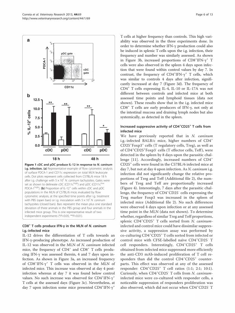
MLN cDC and pDC produce IL-12 early upon i.g.challenge with N. caninum tachyzoitesAs we have shown in a previous report a large pro-portion of splenic conventional and plasmacytoid den-dritic cells (cDC and pDC, respectively) produce IL-12in BALB/c mice infected i.p. with N. caninum tachyzoites [14]. This cytokine is a crucial factor in mediat-ing host immune protection against neosporosis[15-17]. As shown in Figure 1, an increased frequencyof IL-12-expressing cDC and pDC was detected in theMLN of infected mice, 18 and 48 h upon the parasiticchallenge. The frequency of IL-10-producing MLNcDC and pDC was also evaluated and did not signi-ficantly change upon infection (data not shown). Theseresults show that N. caninum tachyzoites administeredi.g. induce IL-12 production by host dendritic cells(DC) in the draining lymph nodes.
Increased frequencies of TCRβ+CD8+IFN-γ+ IEL wereobserved in C57BL/6 mice challenged i.g. with N. caninumtachyzoitesMurine gut IEL comprise both αβ and γδ TCR+ cells[18] which have been shown to mediate host protectionagainst enteric infections, including those caused by pro-tozoans [19]. Here, an increased frequency of TCRβ+
CD8+IFN-γ+ IEL was observed in C57BL/6 mice com-paratively to mock-infected controls, 48 h upon i.g.challenge with N. caninum tachyzoites (Figure 2). Con-versely, no differences were found between infected miceand controls in the frequencies of IFN-γ-producingTCRγδ+ (1,30 ± 0,27 vs 1,13 ± 0,29) or TCRβ+CD4+
(2,70 ± 0,82 vs 1,84 ± 0,76) IEL. Production of IL-17Aby TCRγδ+ IEL was not detected either in controls orinfected mice. These results indicate that in the gut,CD8+αβTCR+, but not γδTCR+, IEL are activated by i.g.-administered N. caninum tachyzoites and produce thehost protective cytokine IFN-γ.
0.0
2.5
5.0
7.5
10.0
12.5
% I
L-1
2+ cel
ls
cDC pDC cDC pDC
18 h 48 h
** ** **
*
a
b
CD11c
PD
CA
-1
Figure 1 cDC and pDC produce IL-12 in response to N. caninumi.g. infection. (a) Representative example of flow cytometric analysisof surface PDCA-1 and CD11c expression on total MLN leukocytecells. Dot plots represent cells collected from C57BL/6 mice 18 hafter i.g. challenge with 5 × 10⁷ N. caninum tachyzoites. Gates wereset as shown to delineate cDC (CD11chigh) and pDC (CD11clow
PDCA-1high). (b) Proportion of IL-12+ cells within cDC and pDCpopulations in the MLN of C57BL/6 mice, evaluated by flowcytometric analysis, at the specified time points after i.g. treatmentwith PBS (open bars) or i.g. inoculation with 5 × 10⁷ N. caninumtachyzoites (closed bars). Bars represent the mean plus one standarddeviation of three animals in the PBS group and four animals in theinfected mice group. This is one representative result of twoindependent experiments (*P<0.05; **P<0.01).
Correia et al. Veterinary Research 2013, 44:69 Page 6 of 13http://www.veterinaryresearch.org/content/44/1/69
CD8+ T cells produce IFN-γ in the MLN of N. caninumi.g.-infected miceIL-12 drives the differentiation of T cells towards anIFN-γ-producing phenotype. As increased production ofIL-12 was observed in the MLN of N. caninum infectedmice, the frequency of CD4+ and CD8+ T cells produ-cing IFN-γ was assessed therein, 4 and 7 days upon in-fection. As shown in Figure 3a, an increased frequencyof CD8+IFN-γ+ T cells was observed in the MLN ofinfected mice. This increase was observed at day 4 post-infection whereas at day 7 it was found below controlvalues. No such increase was observed for CD4+IFN-γ+
T cells at the assessed days (Figure 3c). Nevertheless, atday 7 upon infection some mice presented CD4+IFN-γ+
T cells at higher frequency than controls. This high vari-ability was observed in the three experiments done. Inorder to determine whether IFN-γ production could alsobe induced in splenic T cells upon the i.g. infection, theirfrequency and number was similarly assessed. As shownin Figure 3b, increased proportions of CD8+IFN-γ+ Tcells were also observed in the spleen 4 days upon infec-tion that were found within control values by day 7. Incontrast, the frequency of CD4+IFN-γ+ T cells, whichwas similar to controls 4 days after infection, signifi-cantly increased at day 7 (Figure 3d). The frequency ofCD4+ T cells expressing IL-4, IL-10 or IL-17A was notdifferent between controls and infected mice at bothassessed time points and lymphoid tissues (data notshown). These results show that in the i.g. infected miceCD8+ T cells are early producers of IFN-γ, not only atthe intestinal mucosa and draining lymph nodes but alsosystemically, as detected in the spleen.
Increased suppressive activity of CD4+CD25+ T cells frominfected miceWe have previously reported that in N. caninumi.g.-infected BALB/c mice, higher numbers of CD4+
CD25+Foxp3+ cells (T regulatory cells, Treg), as well asof CD4+CD25+Foxp3- cells (T effector cells, Teff ), weredetected in the spleen by 8 days upon the parasitic chal-lenge [11]. Accordingly, increased numbers of CD4+
CD25+ cells were found in the C57BL/6 infected mice atday 7, but not at day 4 upon infection. As N. caninum i.g.infection did not significantly change the relative pro-portions of Treg and Teff (Additional file 2), the num-bers of Treg and Teff are proportionally increased(Figure 4). Interestingly, 7 days after the parasitic chal-lenge, the frequency of CD4+CD25- cells expressing theTreg marker Foxp3 was increased in the spleen ofinfected mice (Additional file 2). No such differenceswere observed 4 days upon infection or at any assessedtime point in the MLN (data not shown). To determinewhether, regardless of similar Treg and Teff proportions,splenic CD4+CD25+ T cells sorted from N. caninum-infected and control mice could have dissimilar suppres-sive activity, a suppression assay was performed byco-culturing CD4+CD25+ Tcells sorted from infected orcontrol mice with CFSE-labelled naïve CD4+CD25- Tcell responders. Interestingly, CD4+CD25+ T cellsobtained from infected mice suppressed more efficientlythe anti-CD3 mAb-induced proliferation of T cell re-sponders than did the control CD4+CD25+ counter-parts. This effect was observed at any of the assessedresponder: CD4+CD25+ T cell ratios (1:1; 2:1; 10:1).Curiously, when CD4+CD25- T cells from N. caninum-infected mice were co-cultured with responder cells, anoticeable suppression of responders proliferation wasalso observed, which did not occur when CD4+CD25- T

Figure 2 Increased expression of IFN-γ by TCRβ+CD8+ IEL in N. caninum infected mice. Representative dot plots of (a) cells isolated fromthe small intestines of C57BL/6 mice (IEL were gated as shown); (b) TCRγδ and TCRβ IEL. (c) Gated TCRβ CD8+ IEL expressing IFN-γ in control(PBS) and N. caninum-infected mice (NcT). (d) Frequency of TCRβ+CD8+ IFN-γ+ IEL in control and N. caninum i.g.-infected mice, 48 h afterchallenge. Bars represent mean plus one standard deviation of three animals in the PBS group and four animals in the infected mice group. Thisis one representative result of three independent experiments (*P<0.05).
Correia et al. Veterinary Research 2013, 44:69 Page 7 of 13http://www.veterinaryresearch.org/content/44/1/69
cells from controls were used (Figure 5a). This might bea consequence of the increased frequency of Foxp3-expressing cells within this cell subset detected in the 7-day infected mice, which in mice are known to have aregulatory function [20]. In order to determine whether
Figure 3 Expression of IFN-γ in splenic and MLN CD8+ and CD4+ T ceT cells (c and d), as indicated, in the MNL (a and c) and spleen (b and d) of contEach symbol represents an individual mouse; horizontal bars correspond to meanrepresentative result of three independent experiments. Statistical significance bet
the observed disparate suppressive effect of Treg frominfected and control mice could also occur in N. caninum-antigen stimulated cultures, antigen-loaded BMDCwere used as APC to stimulate T cell proliferation/sup-pression. Activation of BMDC upon incubation with N.
lls. Scatter plots of frequency of CD8+IFN-γ+ T cells (a and b) or CD4+IFN-γ+
rol (PBS) or N. caninum-infected mice (NcT), 4 and 7 days upon i.g. challenge.values of the respective group. Five mice per group were used. This is oneween groups is indicated above symbols (*P<0.05; **P<0.01).

Figure 4 Spleen and MLN Treg and Teff cell numbers. Scatterplots of CD4+CD25+ Treg (Foxp3+) and Teff (Foxp3-) cell numbers inthe MLN and spleen of control (open squares) or N. caninum-infectedmice (closed triangles), 4 and 7 days upon the i.g. challenge. Eachsymbol represents an individual mouse; horizontal bars correspond tomean values of the respective group. Five mice per group were used.This is one representative result of three independent experiments.Statistical significance between groups is indicated above symbols(*P<0.05; **P<0.01).
Correia et al. Veterinary Research 2013, 44:69 Page 8 of 13http://www.veterinaryresearch.org/content/44/1/69
caninum antigen extracts was confirmed by up-regulatedexpression of surface MHC class II, CD40 and CD86,detected by using flow cytometry (data not shown). Asshown in Figure 5b, antigen-driven T cell proliferation wasalso more effectively suppressed by Treg from infectedmice than by Treg from non-infected controls.Decreased levels of IFN-γ and also of IL-10 were
detected in the supernatants of anti-CD3 mAb-stimulatedco-cultures of splenic naïve CD4+CD25- T cells whenco-cultured with splenic CD4+CD25+ T cells frominfected and control mice. CD4+CD25+ T cells fromboth N. caninum-infected mice and non-infected con-trols, efficiently suppressed the production of IFN-γ
and IL-10 to levels similar to those of non-stimulatedcells. The equivalent suppression of cytokine pro-duction by Treg from both groups, when a higher sup-pressive activity on T cell proliferation was observedfor Treg from infected mice might result from the highTreg: T responder ratio (1:1) which may have pre-vented differences to show up. Interestingly, in similarco-cultures of cells sorted from the MLN, CD4+CD25+
T cells from the MLN of infected mice suppressedmore efficiently the production of IFN-γ by MLN re-sponder cells than CD4+CD25+ T cell counterpartsfrom non-infected controls. In contrast, in the lattercultures, no suppression of IL-10 production wasobserved likely because IL-10 levels in the superna-tants of stimulated cultures were no significantly differ-ent from the ones found in non-stimulated cultures(Figure 6). No differences were found in the levels ofIL-4 in any of the conditions tested (data not shown).Altogether, these results show that CD4+CD25+ T cellsfrom N. caninum-infected mice display an enhancedsuppressive activity when compared with the equiva-lent T cell population from non-infected controls.
DiscussionAlthough N. caninum can infect its natural hoststhrough the GI tract [7], very little is known about thelocal immune response in the intestinal mucosa and as-sociated lymphoid tissue. We have previously reportedthat neosporosis could be established in mice i.g. chal-lenged with N. caninum tachyzoites [11]. Although thisparasitic form may present antigenic differences fromoocysts and sporozoites, this model may neverthelessbetter mimic the natural infection route in horizontallytransmitted neosporosis, than the intraperitoneal or sub-cutaneous routes. The i.g. infection model was used hereto study the immune response elicited in the intestinalmucosa and MLN. In infected mice, tachyzoites couldbe detected within the intestinal tissue early after theparasitic challenge. Also soon after infection, parasiticDNA was detected in the MLN of one infected mouse.This might explain the stimulatory effect on MLN cDCand pDC observed in the infected mice, and their in-creased expression of IL-12, a key cytokine in mediatinghost immune protection against N. caninum infections[14-16]. On the other hand, as DC have been shown tohelp systemic dissemination of N. caninum [21] and ofthe closely-related protozoan T. gondii [22,23], it wouldbe interesting to determine whether DC may transportthe parasites from the gut to the MLN, contributing forparasite dissemination within the host. The detection ofN. caninum DNA in the MLN of an i.g.-infected mouseas early as 6 h after infection might support this hypo-thesis, though further studies must be carried out toconfirm such a role of DC in this infection model. As

Figure 5 High suppressive activity of Treg from N. caninum-infected mice. (a) Flow cytometric evaluation of anti-CD3 mAb (1 μg/mL)induced proliferative response of 2.5 × 10⁴ CFSE-labelled naïve CD4+CD25- T (responder) cells cultured for 72 h with 105 irradiated APC/well, inthe absence (Pos. control) or presence of CD4+CD25+ T cells obtained from control mice or mice infected with N. caninum. Neg. controlcorresponds to responder cells with no mAb added. Histograms correspond to 1:1, 2:1 and 10:1 responder: CD4+CD25+ cell ratios or 1:1responder: CD4+CD25- T cell ratio, as indicated. (b) Flow cytometric evaluation of N. caninum antigen-induced proliferative response of 2.5 × 10⁴
CFSE-labelled splenic CD4+CD25- T (responder) cells obtained from 7 day-infected mice, cultured for 72 h with 105 antigen-loaded BMDC/well, inthe absence (Pos. control) or presence of CD4+CD25+ T cells obtained from control mice or mice infected with N. caninum. Non-specificproliferation control (Neg. control) corresponds to responder cells cultured with LPS (50 ng/mL) activated BMDC, without N. caninum antigen.Histograms correspond to 1:1 and 2:1 responder: CD4+CD25+ cell ratios, as indicated. Numbers within histograms correspond to the percentagesof cells that divided at least once. CD4+CD25+ cells added in each condition were sorted from pooled splenic cells of 5 mice per group. Resultsare a representative example out of three (a) or one out of two (b) independent experiments.
Correia et al. Veterinary Research 2013, 44:69 Page 9 of 13http://www.veterinaryresearch.org/content/44/1/69
parasite DNA was detected in the brain of infected mice21 days upon infection, this confirmed previous resultsshowing that N. caninum tachyzoites might cross the in-testinal epithelial barrier and disseminate to other or-gans [11]. Lack of evident signs of disease in the infectedmice indicates that mice are able to control neosporosisestablished by the i.g. route. Nevertheless, it cannot beexcluded that this control might in part be due to a lownumber of parasites successfully crossing the intestinalepithelial barrier.Our results, by showing that MLN DC produce IL-12
in response to N. caninum infection are in agreement
with our previous observation that both cDC and pDCproduced IL-12 in the spleen of mice infected i.p. withN. caninum tachyzoites [14] and with other reportsshowing in vivo [15] and in vitro [24,25] production ofthis cytokine upon DC stimulation with this parasiticform. Both cDC and pDC populations were demon-strated to be early sources of IL-12 in mice infected withT. gondii [26,27] and their importance for host protec-tion against this parasite has been recently highlighted[28,29]. A similar protective role of these cell popu-lations may also be important for host resistance againstN. caninum infection. Our results indicate that such a

Figure 6 Suppression of cytokine production by splenic andMLN Treg. IFN-γ and IL-10 cytokine concentration in thesupernatants of MLN or splenic naïve 2.5 × 10⁴ CD4+CD25- T cellscultured for 72 h with anti-CD3 mAb (1 μg/mL) and 105 APC, aloneor in the presence of 1:1 CD4+CD25+ MLN or splenic T cells fromcontrol or N. caninum-infected mice, as indicated. Each symbolrepresents an individual culture well. Horizontal bars represent themean values of the respective group. CD4+CD25+ cells added ineach condition were sorted from pooled MLN or splenic cells of 5mice per group, as indicated. Results are a representative exampleout of three independent experiments. Statistical significance amonggroups was determined by one-way ANOVA, followed by aBonferroni post-test, and is indicated above symbols (**P<0.01;***P<0.001).
Correia et al. Veterinary Research 2013, 44:69 Page 10 of 13http://www.veterinaryresearch.org/content/44/1/69
protective immune response may be triggered already atthe mucosal immune system in hosts challenged withthis parasite in the GI tract. The IL-12 productiondetected in the MLN of the infected mice maycontribute for the differentiation of IFN-γ-producingCD8+αβTCR+ IEL, found in higher proportions in thesemice. Previous works have reported the importance ofMLN [30], and of MLN DC in particular [31], in gener-ating CD8+αβTCR+ IEL. Primed IEL have been shown tomediate protective immunity to oral T. gondii infectionin adoptive cell transfer experiments [32,33]. Thereforeit could be expected that these cells would have a similarrole in N. caninum-infected mice.In bovine neosporosis, the study of CD8+ T cells mainly
addressed their possible participation in the immune re-sponse associated with foetal loss [34-36]. Nevertheless,CD8+ T cells have been extensively demonstrated to have
a host protective role against parasitic infections [37],including neosporosis [34]. The production of IFN-γ byCD8+ T cells, which was also detected in calves experi-mentally infected with N. caninum [35], is an importantmechanism in their host protective role against parasiteinfections [38-42]. It has been particularly shown thatCD8+ T cells were the main early producers of IFN-γ inmurine toxoplasmosis and were involved in resistanceto acute primary infection [43]. Our results indicate thatin murine i.g.-established neosporosis, CD8+ T cells arealso early major producers of the protective cytokineIFN-γ. These cells were found elevated in the MLN ofinfected mice but also in the spleen. It would be inter-esting to determine whether these cells were locally ac-tivated in the spleen and identify the antigen-presentingcells responsible for this activation. Elevated propor-tions of IFN-γ-producing CD4+ T cells were detectedlater than CD8+ counterparts in the spleen and MLN ofinfected mice and not at all in the intestinal epithelium.A predominant response of CD8+IFN-γ+ IEL as com-pared to CD4+ counterparts has been also observed inmice orally infected with T. gondii [44]. Why CD8+ Tcells apparently respond faster than CD4+ T cells in thegut mucosa and draining lymph nodes remains to be de-termined. A hypothesis worth to explore could be thatN. caninum differentially affect the class I vs class IImajor histocompatibility complex antigen presentationpathways.Production of IL-12 by MLN DC elicited in mice or-
ally infected with T. gondii oocysts was shown to dependon bacterial translocation, promoted by the inflamma-tory reaction in the gut that followed the oocyst admin-istration [45]. As no evidence of significant intestinalinflammation was found in the N. caninum-infectedmice, IL-12 production may depend mostly on the para-sitic antigens. Production of the pro-inflammatory cyto-kine IL-17 was associated with intestinal inflammatorypathology [46-48]. Our results, by showing that produc-tion of this cytokine was not detected in elevated pro-portions of IEL or MLN T cells, are thus in agreementwith the lack of evident inflammation in the gut of theinfected animals.Control of microbial induced inflammation, including
that caused by protozoans, largely depends on the ac-tion of Treg cells [49,50]. The observation reportedhere, by showing a high suppressive function of Tregobtained from N. caninum-infected mice may providean additional explanation for the success of N. caninumin colonizing its natural hosts, where it can persist ina symptomless condition [51]. This highly suppressivefunction was more evident when antigenic instead ofpolyclonal stimulation was used in the Treg in vitrosuppression assay of T cell proliferation. It is thus plau-sible that N. caninum, as demonstrated for other protozoan

Correia et al. Veterinary Research 2013, 44:69 Page 11 of 13http://www.veterinaryresearch.org/content/44/1/69
parasites [52], might manipulate natural Treg functionin order to favour its persistence within the host. Inter-estingly, a recent report on persistent Salmonellainfection showed that Treg suppressive potency de-creased from the acute to the chronic phase, signifi-cantly affecting bacterial burden [53]. It would beworthwhile examining if the suppressive function ofTreg later in N. caninum infection could be diminishedwhen the acute phase of infection is overcome. Here,the immunosuppressive function was revealed by theinhibition of both in vitro T-cell proliferation and cyto-kine production. Curiously, no significant suppressionof IFN-γ production was observed in co-cultures ofTreg from the MLN of non-infected mice and MLN re-sponder cells whereas the Treg spleen counterpartswere highly suppressive. Particular environmental con-ditions of the MLN [54] might have conditioned bothTreg and T conventional cells responsiveness, as maybe suggested by the lower cytokine production of theMLN responder cells upon induction with anti-CD3and irradiated APC, when compared with similarlystimulated spleen counterparts. As a decrease of spleenand MLN CD8+IFN-γ+ T cells proportions and num-bers to underneath basal levels by day 7 of infectionwas observed, it would be interesting to determinewhether it may reflect Treg function. In fact, other re-ports on apicomplexan parasite infections show thatTreg, apart from suppressing CD4+ T cell proliferationand cytokine production, similarly affect CD8+ T cells[55,56]. It would be also interesting to assess whethersuch a high suppressive activity could be induced inmice infected by using other parasite administrationroutes. Moreover, it would be interesting to evaluatewhether N. caninum infection would affect Treg sup-pressive activity along the gestational period and its in-fluence in the cytokine environment, since higherlevels of IFN-γ were detected in infected pregnantdams carrying live foetuses [57].The suppressive activity of T regulatory cells may
pose an additional difficulty to overcome infection bymeans of vaccination, as previous remarked [58]. As apreferential involvement of CD8+ T cells in the muco-sal immune response to N. caninum was shown herein,the stimulation of parasite-specific effector and mem-ory CD8+ T cell responses at mucosal sites may be aprivileged target to achieve in vaccination against hori-zontally transmitted neosporosis.In conclusion, intragastric infection of C57BL/6 mice
with N. caninum tachyzoites preferentially activates mu-cosal and splenic CD8+ T cells, resulting in the produc-tion of the host protective cytokine IFN-γ. Nevertheless,the highly suppressive Treg present in the spleen of N.caninum-i.g.-infected mice may contribute to the estab-lishment of a chronic infection.
Additional files
Additional file 1: Detection of N. caninum in the intestinal tissue ofmice infected by the i.g. route. Representative images showing a N.caninum tachyzoite in the murine intestinal tissue (a and b), 12 h upon i.g. infection, detected by immunohistochemistry. N. caninum tachyzoite(brown colour, denoted by arrow). The selected area in (a) is presentedat higher magnification in (b). Bar=100 μm. Results are representative ofdata from two independent experiments.
Additional file 2: Proportions of Treg within splenic and MLN CD4+
CD25+ T cells. Flow cytometry analysis of intracellular Foxp3 expressionin splenic and MLN CD4+ T cells from C57BL/6 mice, 4 and 7 days after i.g. challenge with PBS or 5 × 10⁷ N. caninum tachyzoites (NcT), asindicated. (a) Gating of CD4+CD25+ and of CD4+CD25- T cells. (b)Numbers within dot plots correspond to mean ± one SD of Treg (Foxp3+
cells) frequency within gated CD4+CD25+ T cell population. (c) Numberswithin dot plots correspond to mean ± one SD of the frequency of CD4+
CD25- T cells expressing Foxp3, in the spleen of non-infected or infectedmice, 7 days upon the parasitic challenge. In each panel, results are of arepresentative experiment out of at least three independent experiments(n=5 in each group). Statistical significance between groups in panel c isindicated (*P<0.05). No statistically significant differences were observedin the frequencies of Treg and Teff between control and infected mice.
Competing interestsThe authors declare that they have no competing interests.
Authors’ contributionsAC and MV conducted and supervised the experiments, analysed the data,and wrote the manuscript. AF, LT and AR assisted in the experimental designand data analysis, and contributed to the interpretation of results andmanuscript writing. PF, JD, AAC, and RC conducted the experiments,analysed data and contributed to the interpretation of results. JMparticipated in the experiments on Figures 1 and 2 and contributed to theanalysis and interpretation of results therein. AR participated in dataacquisition in the experiments involving mice. All authors read and approvedthe final manuscript.
AcknowledgmentsSupported by FCT/MCTES (PIDDAC) and co-funded by FEDER throughCOMPETE, PTDC/CVT/115126/2009 and FCOMP‐01‐0124‐FEDER‐014679.Pedro Ferreirinha was supported by FCT grant SFRH/BD/76900/2011. LuziaTeixeira was supported by FSE and MCTES through POPH-QREN-Tipologia4.2.
Author details1Laboratório de Imunologia Mário Arala Chaves, Departamento de Imuno-Fisiologia e Farmacologia, ICBAS-UP, Instituto de Ciências Biomédicas deAbel Salazar – Universidade do Porto, Rua de Jorge Viterbo Ferreira nº 228,Porto , 4050-313, Portugal. 2IBMC - Instituto de Biologia Molecular e Celular,Porto, Portugal. 3Departamento de Patologia e Imunologia Molecular, ICBAS-UP, Instituto de Ciências Biomédicas de Abel Salazar – Universidade doPorto, Rua de Jorge Viterbo Ferreira nº 228, Porto 4050-313, Portugal.4Departamento de Anatomia, ICBAS-UP, Instituto de Ciências Biomédicas deAbel Salazar – Universidade do Porto, Rua de Jorge Viterbo Ferreira nº 228,Porto 4050-313, Portugal. 5UMIB-Unidade Multidisciplinar de InvestigaçãoBiomédica, Porto, Portugal.
Received: 30 January 2013 Accepted: 6 August 2013Published: 10 August 2013
References1. Dubey JP, Schares G: Neosporosis in animals – the last five years. Vet
Parasitol 2011, 180:90–108.2. Dubey JP, Schares G, Ortega-Mora LM: Epidemiology and control of
neosporosis and Neospora caninum. Clin Microbiol Rev 2007, 20:323–367.3. Reichel MP, Alejandra Ayanegui-Alcérreca M, Gondim LF, Ellis JT: What is
the global economic impact of Neospora caninum in cattle - the billiondollar question. Int J Parasitol 2013, 43:133–142.

Correia et al. Veterinary Research 2013, 44:69 Page 12 of 13http://www.veterinaryresearch.org/content/44/1/69
4. Lindsay DS, Lenz SD, Cole RA, Dubey JP, Blagburn BL: Mouse model forcentral nervous system Neospora caninum infections. J Parasitol 1995,81:313–315.
5. Long MT, Baszler TV: Fetal loss in BALB/c mice infected with Neosporacaninum. J Parasitol 1996, 82:608–611.
6. Liddell S, Jenkins MC, Dubey JP: Vertical transmission of Neosporacaninum in BALB/c mice determined by polymerase chain reactiondetection. J Parasitol 1999, 85:550–555.
7. McCann CM, McAllister MM, Gondim LF, Smith RF, Cripps PJ, Kipar A,Williams DJ, Trees AJ: Neospora caninum in cattle: experimental infectionwith oocysts can result in exogenous transplacental infection, but notendogenous transplacental infection in the subsequent pregnancy. Int JParasitol 2007, 37:1631–1639.
8. Debache K, Hemphill A: Differential effects of intranasal vaccination withrecombinant NcPDI in different mouse models of Neospora caninuminfection. Parasite Immunol 2013, 35:11–20.
9. Debache K, Kropf C, Schütz CA, Harwood LJ, Käuper P, Monney T, Rossi N,Laue C, McCullough KC, Hemphill A: Vaccination of mice with chitosannanogel-associated recombinant NcPDI against challenge infection withNeospora caninum tachyzoites. Parasite Immunol 2011, 33:81–94.
10. Debache K, Guionaud C, Alaeddine F, Hemphill A: Intraperitoneal andintra-nasal vaccination of mice with three distinct recombinant Neosporacaninum antigens results in differential effects with regard to protectionagainst experimental challenge with Neospora caninum tachyzoites.Parasitology 2010, 137:229–240.
11. Teixeira L, Botelho AS, Batista AR, Meireles CS, Ribeiro A, Domingues HS,Correia Da Costa JM, Castro AG, Faustino AM, Vilanova M: Analysis of theimmune response to Neospora caninum in a model of intragastricinfection in mice. Parasite Immunol 2007, 29:23–36.
12. Lefrançois L, Lycke N: Isolation of mouse small intestinal intraepitheliallymphocytes, Peyer’s patch, and lamina propria cells. Curr Protoc Immunol2001, 3:3.19.1–3.19.16.
13. Lutz MB, Kukutsch N, Ogilvie AL, Rössner S, Koch F, Romani N, Schuler G: Anadvanced culture method for generating large quantities of highly puredendritic cells from mouse bone marrow. J Immunol Methods 1999,223:77–92.
14. Teixeira L, Botelho AS, Mesquita SD, Correia A, Cerca F, Costa R, Sampaio P,Castro AG, Vilanova M: Plasmacytoid and conventional dendritic cells areearly producers of IL-12 in Neospora caninum-infected mice. Immunol CellBiol 2010, 88:79–86.
15. Mineo TW, Benevides L, Silva NM, Silva JS: Myeloid differentiation factor 88is required for resistance to Neospora caninum infection. Vet Res 2009,40:32.
16. Baszler TV, Long MT, McElwain TF, Mathison BA: Interferon-γ andinterleukin-12 mediate protection to acute Neospora caninum infectionin BALB/c mice. Int J Parasitol 1999, 29:1635–1646.
17. Khan IA, Schwartzman JD, Fonseka S, Kasper LH: Neospora caninum: rolefor immune cytokines in host immunity. Exp Parasitol 1997, 85:24–34.
18. Moens E, Veldhoen M: Epithelial barrier biology: good fences make goodneighbours. Immunology 2012, 135:1–8.
19. Cheroutre H, Lambolez F, Mucida D: The light and dark sides of intestinalintraepithelial lymphocytes. Nat Rev Immunol 2011, 11:445–456.
20. Shevach EM: Mechanisms of foxp3+ T regulatory cell-mediatedsuppression. Immunity 2009, 30:636–645.
21. Collantes-Fernandez E, Arrighi RB, Alvarez-García G, Weidner JM, Regidor-Cerrillo J,Boothroyd JC, Ortega-Mora LM, Barragan A: Infected dendritic cellsfacilitate systemic dissemination and transplacental passage of theobligate intracellular parasite Neospora caninum in mice. PLoS One 2012,7:e32123.
22. Bierly AL, Shufesky WJ, Sukhumavasi W, Morelli AE, Denkers EY: Dendriticcells expressing plasmacytoid marker PDCA-1 are Trojan horses duringToxoplasma gondii infection. J Immunol 2008, 181:8485–8491.
23. Lambert H, Hitziger N, Dellacasa I, Svensson M, Barragan A: Induction ofdendritic cell migration upon Toxoplasma gondii infection potentiatesparasite dissemination. Cell Microbiol 2006, 8:1611–1623.
24. Feng X, Zhang N, Tuo W: Neospora caninum tachyzoite- and antigen-stimulated cytokine production by bone marrow-derived dendritic cellsand spleen cells of naive BALB/c mice. J Parasitol 2010, 96:717–723.
25. Dion S, Germon S, Guiton R, Ducournau C, Dimier-Poisson I: Functionalactivation of T cells by dendritic cells and macrophages exposed to theintracellular parasite Neospora caninum. Int J Parasitol 2011, 41:685–695.
26. Pepper M, Dzierszinski F, Wilson E, Tait E, Fang Q, Yarovinsky F, Laufer TM,Roos D, Hunter CA: Plasmacytoid dendritic cells are activated byToxoplasma gondii to present antigen and produce cytokines. J Immunol2008, 180:6229–6236.
27. Liu CH, Fan YT, Dias A, Esper L, Corn RA, Bafica A, Machado FS, Aliberti J:Cutting edge: dendritic cells are essential for in vivo IL-12 productionand development of resistance against Toxoplasma gondii infection inmice. J Immunol 2006, 177:31–35.
28. Mashayekhi M, Sandau MM, Dunay IR, Frickel EM, Khan A, Goldszmid RS,Sher A, Ploegh HL, Murphy TL, Sibley LD, Murphy KM: CD8α+ dendritic cellsare the critical source of interleukin-12 that controls acute infection byToxoplasma gondii tachyzoites. Immunity 2011, 35:249–259.
29. Koblansky AA, Jankovic D, Oh H, Hieny S, Sungnak W, Mathur R, Hayden MS,Akira S, Sher A, Ghosh S: Recognition of profilin by Toll-like receptor 12 iscritical for host resistance to Toxoplasma gondii. Immunity 2013,38:119–130.
30. Svensson M, Marsal J, Ericsson A, Carramolino L, Brodén T, Márquez G,Agace WW: CCL25 mediates the localization of recently activatedCD8αβ+ lymphocytes to the small-intestinal mucosa. J Clin Invest 2002,110:1113–1121.
31. Johansson-Lindbom B, Svensson M, Wurbel MA, Malissen B, Márquez G,Agace W: Selective generation of gut tropic T cells in gut-associatedlymphoid tissue (GALT): requirement for GALT dendritic cells andadjuvant. J Exp Med 2003, 198:963–969.
32. Buzoni-Gatel D, Lepage AC, Dimier-Poisson IH, Bout DT, Kasper LH:Adoptive transfer of gut intraepithelial lymphocytes protects againstmurine infection with Toxoplasma gondii. J Immunol 1997, 158:5883–5889.
33. Lepage AC, Buzoni-Gatel D, Bout DT, Kasper LH: Gut-derived intraepitheliallymphocytes induce long term immunity against Toxoplasma gondii.J Immunol 1998, 161:4902–4908.
34. Orozco MA, Morales E, Salmerón F: Characterization of the inflammatoryresponse in the uteri of cows infected naturally by Neospora caninum.J Comp Pathol 2013, 148:148–156.
35. Rosbottom A, Gibney H, Kaiser P, Hartley C, Smith RF, Robinson R, Kipar A,Williams DJ: Up regulation of the maternal immune response in theplacenta of cattle naturally infected with Neospora caninum. PLoS One2011, 6:e15799.
36. Maley SW, Buxton D, Macaldowie CN, Anderson IE, Wright SE, Bartley PM,Esteban-Redondo I, Hamilton CM, Storset AK, Innes EA: Characterization ofthe immune response in the placenta of cattle experimentally infectedwith Neospora caninum in early gestation. J Comp Pathol 2006,135:130–141.
37. Jordan KA, Hunter CA: Regulation of CD8+ T cell responses to infectionwith parasitic protozoa. Exp Parasitol 2010, 126:318–325.
38. Tanaka T, Hamada T, Inoue N, Nagasawa H, Fujisaki K, Suzuki N, Mikami T:The role of CD4+ or CD8+ T cells in the protective immune response ofBALB/c mice to Neospora caninum infection. Vet Parasitol 2000,90:183–191.
39. Gazzinelli RT, Hakim FT, Hieny S, Shearer GM, Sher A: Synergistic role ofCD4+ and CD8+ T lymphocytes in IFN-γ production and protectiveimmunity induced by an attenuated Toxoplasma gondii vaccine.J Immunol 1991, 146:286–292.
40. Tarleton RL: Depletion of CD8+ T cells increases susceptibility andreverses vaccine-induced immunity in mice infected with Trypanosomacruzi. J Immunol 1990, 144:717–724.
41. Weiss WR, Sedegah M, Beaudoin RL, Miller LH, Good MF: CD8+ T cells(cytotoxic/suppressors) are required for protection in mice immunizedwith malaria sporozoites. Proc Natl Acad Sci USA 1988, 85:573–576.
42. Schofield L, Villaquiran J, Ferreira A, Schellekens H, Nussenzweig R,Nussenzweig V: Gamma interferon, CD8+ T cells and antibodies requiredfor immunity to malaria sporozoites. Nature 1987, 330:664–666.
43. Shirahata T, Yamashita T, Ohta C, Goto H, Nakane A: CD8+ T lymphocytesare the major cell population involved in the early gamma interferonresponse and resistance to acute primary Toxoplasma gondii infection inmice. Microbiol Immunol 1994, 38:789–796.
44. Lee YH, Shin DW: T cell phenotype and intracellular IFN-γ production inperitoneal exudate cells and gut intraepithelial lymphocytes during acuteToxoplasma gondii infection in mice. Korean J Parasitol 2002, 40:119–129.
45. Benson A, Pifer R, Behrendt CL, Hooper LV, Yarovinsky F: Gut commensalbacteria direct a protective immune response against Toxoplasmagondii. Cell Host Microbe 2009, 6:187–196.

Correia et al. Veterinary Research 2013, 44:69 Page 13 of 13http://www.veterinaryresearch.org/content/44/1/69
46. Zhang L, Liu R, Song M, Hu Y, Pan B, Cai J, Wang M: Eimeria tenella:Interleukin 17 contributes to host immunopathology in the gut duringexperimental infection. Exp Parasitol 2013, 133:121–130.
47. Schaefer JS, Montufar-Solis D, Vigneswaran N, Klein JR: ICOS promotes IL-17synthesis in colonic intraepithelial lymphocytes in IL-10−/− mice. J LeukocBiol 2010, 87:301–308.
48. Park SG, Mathur R, Long M, Hosh N, Hao L, Hayden MS, Ghosh S: Tregulatory cells maintain intestinal homeostasis by suppressing γδ Tcells. Immunity 2010, 33:791–803.
49. Belkaid Y, Tarbell K: Regulatory T cells in the control of host-microorganism interactions. Annu Rev Immunol 2009, 27:551–589.
50. Demengeot J, Zelenay S, Moraes-Fontes MF, Caramalho I, Coutinho A:Regulatory T cells in microbial infection. Springer Semin Immunopathol2006, 28:41–50.
51. Buxton D, McAllister MM, Dubey JP: The comparative pathogenesis ofneosporosis. Trends Parasitol 2002, 18:546–552.
52. Belkaid Y, Rouse BT: Natural regulatory T cells in infectious disease.Nat Immunol 2005, 6:353–360.
53. Johanns TM, Ertelt JM, Rowe JH, Way SS: Regulatory T cell suppressivepotency dictates the balance between bacterial proliferation andclearance during persistent Salmonella infection. PLoS Pathog 2010,6:e1001043.
54. Pabst O, Wahl B, Bernhardt G, Hammerschmidt SI: Mesenteric lymph nodestroma cells in the generation of intestinal immune responses. J Mol Med(Berl) 2009, 87:945–951.
55. Tenorio EP, Fernández J, Castellanos C, Olguín JE, Saavedra R: CD4+ Foxp3+
regulatory T cells mediate Toxoplasma gondii-induced T-cell suppressionthrough an IL-2-related mechanism but independently of IL-10. Eur JImmunol 2011, 41:3529–3541.
56. Abel S, Lückheide N, Westendorf AM, Geffers R, Roers A, Müller W,Sparwasser T, Matuschewski K, Buer J, Hansen W: Strong impact of CD4+Foxp3+ regulatory T cells and limited effect of T cell-derived IL-10 onpathogen clearance during Plasmodium yoelii infection. J Immunol 2012,188:5467–5477.
57. Bartley PM, Wright SE, Maley SW, Macaldowie CN, Nath M, Hamilton CM,Katzer F, Buxton D, Innes EA: Maternal and foetal immune responses ofcattle following an experimental challenge with Neospora caninum atday 70 of gestation. Vet Res 2012, 43:38.
58. Joosten SA, Ottenhoff TH: Human CD4 and CD8 regulatory T cells ininfectious diseases and vaccination. Hum Immunol 2008, 69:760–770.
doi:10.1186/1297-9716-44-69Cite this article as: Correia et al.: Mucosal and systemic T cell responsein mice intragastrically infected with Neospora caninum tachyzoites.Veterinary Research 2013 44:69.
Submit your next manuscript to BioMed Centraland take full advantage of:
• Convenient online submission
• Thorough peer review
• No space constraints or color figure charges
• Immediate publication on acceptance
• Inclusion in PubMed, CAS, Scopus and Google Scholar
• Research which is freely available for redistribution
Submit your manuscript at www.biomedcentral.com/submit