correction ...a major risk factor of erf in the clinical settings are insufficiently developed....
TRANSCRIPT

CORRECTION
Correction: Secretion of ERP57 is important for extracellular matrixaccumulation and progression of renal fibrosis, and is an early signof disease onset (doi:10.1242/jcs.125088)Hassan Dihazi, Gry Helene Dihazi, Asima Bibi, Marwa Eltoweissy, Claudia A. Mueller, Abdul R. Asif,Diana Rubel, Radovan Vasko1 and Gerhard A. Mueller
There was an error published in J. Cell Sci. (2013) 126, 3649–3663 (doi:10.1242/jcs.125088).
The affiliations for Marwa Eltoweissy were incorrect. The correct affiliations are as given below.
Hassan Dihazi1, Gry Helene Dihazi1, Asima Bibi1, Marwa Eltoweissy1,2, Claudia A. Mueller3, Abdul R. Asif 4, Diana Rubel1,Radovan Vasko1 and Gerhard A. Mueller1
1Department of Nephrology and Rheumatology, Georg-August University Goettingen, Robert-Koch-Strasse 40, 37075 Goettingen,Germany. 2Department of Zoology, Faculty of Science, Alexandria University, Alexandria, Egypt. 3Section for Transplantation,Immunology and Immunohematology, ZMF, Eberhard-Karls-University Tuebingen, Germany. 4Department of Clinical Chemistry,Georg-August University Goettingen, Goettingen, Germany
Marwa Eltoweissy apologises to the readers for any confusion that this error might have caused.
1
© 2018. Published by The Company of Biologists Ltd | Journal of Cell Science (2018) 131, jcs219014. doi:10.1242/jcs.219014
Journal
ofCe
llScience

Journ
alof
Cell
Scie
nce
Secretion of ERP57 is important for extracellular matrixaccumulation and progression of renal fibrosis, and isan early sign of disease onset
Hassan Dihazi1,*, Gry Helene Dihazi1, Asima Bibi1, Marwa Eltoweissy1, Claudia A. Mueller2, Abdul R. Asif3,Diana Rubel1, Radovan Vasko1 and Gerhard A. Mueller1
1Department of Nephrology and Rheumatology, Georg-August University Goettingen, Robert-Koch-Strasse 40, 37075 Goettingen, Germany2Section for Transplantation, Immunology and Immunohematology, ZMF, Eberhard-Karls-University Tuebingen, Germany3Department of Clinical Chemistry, Georg-August University Goettingen, Goettingen, Germany
*Author for correspondence ([email protected])
Accepted 20 May 2013Journal of Cell Science 126, 3649–3663� 2013. Published by The Company of Biologists Ltddoi: 10.1242/jcs.125088
SummaryRenal fibrosis is characterized by excessive accumulation of extracellular matrix (ECM), which compromises organ function byreplacing normal organ tissue. The molecular mechanisms leading to renal fibrosis are not fully understood. Here we demonstrated thatTGFb1, AGT or PDGF stimulation of renal cells resulted in endoplasmic reticulum (ER) stress followed by activation of the protectiveunfolded protein response pathway and a high secretory level of protein disulfide isomerase ERP57 (also known as PDIA3). The
TGFb1-induced impairment of ER function could be reversed by treatment with BMP7, suggesting a specific involvement in renalfibrosis. A clear correlation between the degree of fibrosis, ER stress and the level of ERP57 could be seen in fibrosis animal models andin biopsies of renal fibrosis patients. Protein interaction studies revealed that secreted ERP57 exhibits a strong interaction with ECM
proteins. Knockdown of ERP57 or antibody-targeted inhibition of the secreted form significantly impaired the secretion andaccumulation of ECM. Moreover, ERP57 was excreted in the early stages of chronic kidney disease, and its level in urine correlated withthe degree of renal fibrosis, suggesting that the secretion of ERP57 represents one of the first signs of renal fibrosis onset and
progression.
Key words: ECM, ERP57, ER stress, Renal fibrosis, UPR
IntroductionRenal diseases are rapidly increasing in western populations due
to a continuous rise of common illnesses such as diabetes
mellitus and hypertension. Numerous risk factors (e.g.
inflammation, elevated arterial blood pressure, hyperglycemia,
proteinuria, toxins, drugs, hypoxia and hyperfiltration) are known
to induce renal diseases, which often lead to endstage renal
failure (ERF) through a common pathway of renal fibrosis. Until
now, diagnostic tools for the early recognition of renal failure as
a major risk factor of ERF in the clinical settings are
insufficiently developed. Also, discrimination of irreversibly
progredient renal diseases from occasionally observed reversible
damages are not defined. There is still an incomplete
understanding of the process of renal failure in addition to a
lack of effective preventative therapy. Renal failure is a process
that is characterized by the excessive accumulation of
extracellular matrix (ECM) deposition in the tubulo-interstitial
space and is associated with declining excretory renal function
(Hirschberg, 2005). The molecular mechanisms of fibrosis are
not fully understood, but imbalance of ECM synthesis and
degradation was described as a common final pathway. In healthy
kidney, the formation of ECM and its degradation are in
homeoestatic equilibrium, whereas in fibrotic kidney this
balance is disturbed owing to a high rate of ECM synthesis and
a low level of its degradation or to a combination of both
(Branton and Kopp, 1999; Eitner and Floege, 2005; Liu, 2006). It
appears to result from pathological, dysregulated repair
mechanisms of diverse renal injuries within the interstitial space.
Interstitial renal fibroblasts at different stages of differentiation are
key players in this process. Like in wound healing, different
cytokines (e.g. TGFb1 and BMP7) and enzymes [e.g. matrix
metallo-proteases (MMPs)] that regulate tissue remodelling seem
to be involved. New insights into renal failure suggest that this
process is related to complex local interactions of cells of the
interstitial and vascular space as well as interactions of tubuli, but
also correlates in part with cellular feeding from blood-borne
progenitor cells (Harris and Neilson, 2006; Iwano and Neilson,
2004). Genetic factors predisposing to renal failure might be
relevant for specific external or internal deteriorating stimuli, such
as in diabetic nephropathy. At present, the relative importance of
each of these components in the development of renal failure or in
physiological renal repair is still unknown. Among the
mechanisms involved in renal failure, stress pathways were
reported to inhibit ECM degradation through upregulation of the
plasminogen activator inhibitor-1 system (PAI-1), which has an
important function in modulating the degradation of ECM (Lassila
et al., 2007; Lee et al., 2005; Nangaku, 2006). Despite the progress
reported in this field (Djamali, 2007; Iwano and Neilson, 2004; Qi
et al., 2006), the mechanisms leading to renal fibrosis still remain
poorly understood.
Research Article 3649

Journ
alof
Cell
Scie
nce
The knowledge of molecular mechanisms involved in ECM
turnover might contribute to a better understanding of the
pathological process to improve therapeutic strategies. Like all
proteins destined for secretion, ECM proteins are translocated into
the endoplasmic reticulum (ER) where folding takes place before
secretion through the Golgi complex. Because of the high protein
concentration in the ER, lumen cells express molecular chaperones
to assist folding by blocking nonspecific intermolecular
interactions, in particular aggregation (Bedard et al., 2005; Hebert
and Molinari, 2007). The levels of the chaperons are continuously
adjusted to cope with stress conditions and to satisfy the needs of
the cell (Dobson, 2003; Goldberg, 2003). An overload of ER with
nascent proteins results in ER stress and activation of the unfolded
protein response (UPR) pathway, leading to upregulation of ER-
stress proteins to improve the folding procedure. ER stress has been
linked to a large number of diseases including cancer, diabetes,
cardiac disease and muscle degeneration (Oyadomari and Mori,
2004). The investigation of the mechanisms connecting ER stress to
the pathophysiology of renal fibrosis is of great interest in order to
explore the potential of key proteins of this pathway in drug
discovery and therapeutic aspects.
In the present study, we performed functional proteomics of
model cell lines to identify new pathways involved in renal
fibrosis. We demonstrate a strong involvement of ER-stress
proteins in the synthesis and secretion of ECM. Moreover, we
identify ERP57 (also known as PDIA3) as key protein in ECM
protein synthesis and accumulation, and present it as a potential
diagnostic marker for renal fibrosis and therapeutic target.
ResultsTGFb1-induced cell transformation impacts the renal
cell proteome
Despite the complex molecular mechanisms involved in renal
failure, one of the central regulators of tissue fibrosis is the
cytokine transforming growth factor-beta (TGFb), including at
least three highly homologous mammalian isoforms TGFb1, 2
and 3, which have similar profibrogenic effects in vitro. Also in
vivo, the profibrotic effect of TGFb1 is well established, because
it is overexpressed in most fibrotic tissues. Furthermore, TGFb1
transgenic mice develop progressive fibrosis in multiple organs,
especially the kidneys. In order to identify new molecular factors
and pathways potentially associated with renal failure at the cellular
level, we used cell models with established inducible profibrogenic
phenotypes (Bechtel et al., 2010; Zeisberg et al., 2005). For this
purpose, cell extracts were prepared from the control and from the
TGFb1-treated renal fibroblast cell line TK173 (72 h). Proteins
were separated by two-dimensional gel electrophoresis (2-DE). The
protein expression patterns from the two cell extracts were
compared with each other using the Delta2D software (Decodon
GmbH). Two-dimensional protein maps derived from the cell
extracts analyzed with conventional 2-DE in the pH ranges 3–10
and 5–8 identified more than 2350 protein spots in each analysed
cell extract when 20,000 pixels were used as the filter limit. The
pixel volume of each spot provides the basis for comparison of the
protein expression patterns between control and TGFb1-treated
TK173 cells. A representative 2-DE protein map from TGFb1-
treated TK173 for pH 5–8 is shown in supplementary material Fig.
S1A,B. To ensure solid quantification of differentially expressed
proteins, 2D-difference gel electrophoresis (DIGE) analysis was
performed with the two cell extracts using Cydyes, as described in
Materials and Methods. The 2D-DIGE images were analyzed using
the Delta2D software (Decodon); interesting protein spots were
excised and analyzed by mass spectrometry. The proteins
were identified using the MASCOT Database. A total of 114
differentially expressed proteins were identified (supplementary
material Table S1). Comparing the treated cells with the control
revealed that treatment with TGFb1 resulted in the alteration of 62
non-redundant proteins (P,0.05). Among these proteins, 13 were
found to be downregulated (Table 1), whereas 49 were upregulated
(Table 2). The identified proteins were found to be associated with
different biological functions and a large number of them could
be classified into three functional categories: the first category
grouped the proteins that are known to be involved in fibrogenesis
(COL1A1, FIN, ACTA2, VIN, VIM, DES), confirming a
profibrotic effect of the cytokine TGFb1 on TK173. The other
two categories involved protein markers of the ER stress- and the
UPR (unfolded protein response)-pathways (GRP78, GRP94,
ERP57, ERP72 and CALR) (Fig. 1A–C), and proteins of the
oxidative stress pathway (PRDX1, PRDX2, PRDX6, SOD2,
PARK7, HYOU1), which were highly upregulated in TGFb-
treated TK173 cells (Fig. 1A–C).
Upregulation of ER-stress proteins as a cell response to
cytokine-induced transformation
To further determine whether the profibrotic cell phenotype with the
continuous presence of TGFß1 could be accompanied by the
Table 1. List of proteins found to be downregulated in the TGFb1-treated TK173 cell line compared with untreated controls
Protein name Gene symbol Swiss Prot Acc. No. Mass PMF MS/MS score
Low molecular weight phosphotyrosine protein phosphatase ACP1 P24666 18,031 103Chloride intracellular channel protein 1 CLIC1 O00299 26,906 98Cystatin-B CSTB P04080 11,133 92Eukaryotic translation initiation factor 1 EIF1 P41567 12,725 78Histidine triad nucleotide-binding protein 1 HINT1 P49773 13,793 45Inosine-59-monophosphate dehydrogenase 2 IMPDH2 P12268 55,770 42NADH dehydrogenase [ubiquinone] flavoprotein 2, mitochondrial NDUFV2 P19404 27,374 109PEST proteolytic signal-containing nuclear protein PCNP Q8WW12 18,913 96Peptidyl-prolyl cis-trans isomerase A PPIA P62937 18,001 123Pyruvate kinase isozymes M1/M2 PKM2 P14618 57,937 145Splicing factor, arginine/serine-rich 1 SFRS1 Q07955 27,728 96Tubulin-specific chaperone A TBCA O75347 12,847 89Ubiquitin-conjugating enzyme E2 N UBE2N P61088 17,127 141
The accession number in Swiss-Prot, the peptide mass fingerprinting (PMF) and MS/MS information are given. The spots where MS/MS-data are missing wereonly identified with PMF.
Journal of Cell Science 126 (16)3650
Journ
alof
Cell
Scie
nce
activation of UPR and alteration in the expression of the ER-stress
proteins at the cellular level, TK173 as model system was stimulated
for different periods of time with the profibrotic cytokine TGFb1,
and the expression of the proteins of interest was analysed by western
blot analysis and immunofluorescence staining. The activation ofTK173 into a profibrotic phenotype upon treatment with TGFb1 was
accompanied by a progressive and time-dependent upregulation of
the ER-stress proteins (GRP78, GRP94, ERP57, ERP72, and CALR)
(Fig. 1D). In parallel to the increase in ER-stress protein expression,
a substantial and progressive upregulation in fibrosis and
myofibroblast markers (e.g. FIN, ACTA2, VIM, DES) expression
and secretion was confirmed in cell extracts and supernatants,
respectively (Fig. 1E,F). The extensive upregulation of ER-stress
proteins was observed in the first week of treatment. Long-term
exposure to TGFb1 resulted in cell adaptation and regeneration ofnormal levels of ER-stress proteins but not of the fibrosis markers,indicating a persistent cell alteration. The profibrotic effects ofTGFb1 on the fibroblast cell line TK173 were accompanied by a
progressive increase in the expression of the key proteins of ER-stress protein. Western blot and immunofluorescence datastrengthened the proteomics results and suggested an important
role of the UPR pathway and ER-stress proteins in TGFb1 signallingand in the pathogenesis of renal fibrosis.
Upregulation of ER-stress proteins and activation of theUPR pathway play an important role in renal fibrosis
To ensure that activation of UPR and the alteration in expression ofER-stress proteins is not restricted to TGFb1 and activated renal
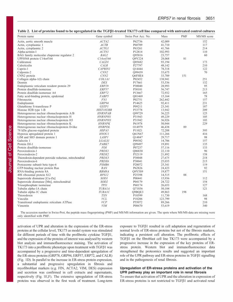
Table 2. List of proteins found to be upregulated in the TGFb1-treated TK173 cell line compared with untreated control cultures
Protein name Gene symbol Swiss Prot Acc. No. Mass PMF MS/MS score
Actin, aortic smooth muscle ACTA2 P62736 42,009 152Actin, cytoplasmic 1 ACTB P60709 41,710 117Actin, cytoplasmic 2 ACTG1 P63261 41,766 214Alpha-actinin-1 ACTN1 P12814 102,993 110BAG family molecular chaperone regulator 2 BAG2 Q95816 23,757 60UPF0568 protein C14orf166 C14orf166 Q9Y224 28,068 81Caldesmon CALD1 Q05682 93,194 173Calreticulin CALR P27797 48,142 210Caprin-1 CAPRIN1 Q14444 78,318 122Calponin-2 CNN2 Q99439 33,675 70CNN2 protein CNN2 Q6FHE4 33,709 151Collagen alpha-1(I) chain COL1A1 P02452 138,941 251Desmin DES P17661 53,536 132Endoplasmic reticulum resident protein 29 ERP29 P30040 28,993 95Protein disulfide-isomerase ERP57 P30101 56,747 213Protein disulfide-isomerase A4 ERP72 P13667 72,932 165Fatty acid-binding protein, epidermal FABP5 Q01469 15,164 78Fibronectin FN1 P02751 262,442 157Endoplasmin GRP94 P14625 92,411 231Glutathione S-transferase P GSTP1 P09211 23,341 187Histone H2B type 1-B HIST1H2BB P33778 13,942 209Heterogeneous nuclear ribonucleoprotein A/B HNRNPAB Q99729 36,225 125Heterogeneous nuclear ribonucleoprotein H HNRNPH1 P31943 49,229 105Heterogeneous nuclear ribonucleoprotein H3 HNRNPH3 P31942 36,926 105Heterogeneous nuclear ribonucleoprotein K HNRNPK P61978 50,944 229Heterogeneous nuclear ribonucleoprotein D-like HNRPDL O14979 46,438 8978 kDa glucose-regulated protein HSPA5 P11021 72,288 395Hypoxia upregulated protein 1 HYOU1 Q61N67 111,266 416LIM and SH3 domain protein 1 LASP1 Q14847 29,717 98Galectin 1 LGALS1 P09382 14,706 157Protein DJ-1 PARK7 Q99497 19,891 135Protein disulfide-isomerase PDI P07237 57,116 133Peroxiredoxin-1 PRDX1 Q06830 22,110 96Peroxiredoxin-2 PRDX2 P32119 21,892 158Thioredoxin-dependent peroxide reductase, mitochondrial PRDX3 P30048 27,675 218Peroxiredoxin-6 PRDX6 P30041 25,035 215Proteasome subunit beta type-6 PSMB6 P28072 25,341 119GTP-binding nuclear protein Ran RAN P62826 24,423 92RNA-binding protein 8A RBM8A Q9Y5S9 19,877 15140S ribosomal protein S12 RPS12 P25398 14,515 88Superoxide dismutase [Cu-Zn] SOD1 P00441 15,936 112Superoxide dismutase [Mn], mitochondrial SOD2 P04179 24,722 154Triosephosphate isomerase TPI1 P60174 26,653 127Tubulin alpha-1A chain TUBA1A Q71036 50,104 121Tubulin alpha-1C chain TUBA1C Q9BQE3 49,863 198Thioredoxin TXN P10599 11,730 168Vinculin VCL P18206 123,799 98Transitional endoplasmic reticulum ATPase VCP P55072 89,266 210Vimentin VIM P08670 53,619 340
The accession number in Swiss-Prot, the peptide mass fingerprinting (PMF) and MS/MS information are given. The spots where MS/MS-data are missing wereonly identified with PMF.
ERP57 in renal fibrosis 3651

Journ
alof
Cell
Scie
nce
fibroblasts, two renal cell lines derived from different parts of the
kidney (from interstitial part the TK173 and from proximal Tubule
HK-2) were treated with three different profibrotic cytokines
(TGFb1, AGT and PDGF) for 72 h. The expression of the protein
of interest was investigated using western blot analysis. A
substantial upregulation of CALR, GRP78, ERP57 and ERP72
Fig. 1. See next page for legend.
Journal of Cell Science 126 (16)3652
Journ
alof
Cell
Scie
nce
was observed in renal cells treated with the three cytokines.
Moreover, overexpression of CHOP and EIF2A together with
increased levels of phosphorylated EIF2A were detected under all
three treatments (Fig. 2A,B). To ensure the involvement of the
UPR pathway in fibrosis, activation of UPR-key proteins PERK,
IREa1 and ATF6 was investigated in renal cell lines treated with
TGFb1, tunicymycin and thapsigargin. In a similar manner to
tunicamycin, IREa1 and PERK were phosphorylated upon
treatment with TGFb1. Moreover, activation of ATF6, as shown
by the cleaved form, was observed in tunicamycin and TGFb1-
treated cells (Fig. 2C). The phosphorylation of IREa1 and PERK,
and the cleavage of ATF6 suggest that the UPR pathway activation
and the subsequent alteration in the expression of ER-stress
proteins are common pathways in the progression of renal fibrosis.
Our suggestion was supported by the fact that treatment with
BMP7, a TGFb1 antagonist, significantly reversed the TGFb1-
induced ER-stress and activation of the UPR pathway (Fig. 2D,E).
The induction of ER-stress proteins is proportional to the
progression of renal fibrosis
To investigate the involvement of ER-stress proteins in renal
fibrosis, the Col4a3 knockout mouse, an animal model for
chronic progressive renal fibrosis, was used. Animals from
different stages of the disease (4, 6, 7 and 9 weeks) were killed
and the kidneys were harvested. After lysis of the kidneys and
preparation of the protein extracts, the changes of the ER-stress
protein expression were monitored by western blot analysis.
Parallel to the progression of renal fibrosis (Fig. 3A), there was a
significant increase in ER-stress and UPR revealed by the
increase in expression of the ER-stress proteins (Fig. 3B).
ERP57 is secreted into the extracellular medium upon
cytokine treatment
The results presented above point to an involvement of ER-stress
proteins in renal fibrosis. These proteins also have an important
role in the folding of secretory proteins (e.g. ECM). Besides its
chaperone function, the protein disulfide isomerase ERP57
catalyses the formation and breakage of disulfide bridges, which
are often vital for the stability of the final protein structure. The
incorrect pairing of cysteine residues usually prevents the foldingof a protein into its native conformation and leads to its
degradation (Ahamed et al., 2006). The formation ofbiosynthetic disulfide bonds is an important step in thematuration of ECM proteins (COL1A1, FIN and LAMN) ineukaryotic cells. The preservation of the protein conformation and
the disulfide bridges is crucial for the accumulation of ECM in theextracellular milieu. Secreted proteins are exposed to a highlyreducing extracellular environment, often leading to impairment of
the disulfide bridges and destruction of the ECM network. Wehypothesized that, in case of renal fibrosis, the maintenance ofstable ECM and its protection from the reducing extracellular
environment requires the involvement of secretory protectingproteins, and that this process is regulated by profibrotic cytokines.To prove our assumption we investigated whether treatment withprofibrotic cytokines can cause the secretion of folding proteins.
TK173 and HK-2 cells were cultured in FCS-free medium for24 h. Subsequently, the cells were treated with TGFb1, PDGF orAGT for 24 h. Control cells were incubated in FCS-free medium
without treatment. The medium with the cytokines was changed toavoid any minimal impact of dead cells on the supernatantproteome, and the cells were incubated for an additional 48 h with
the three cytokines separately. The supernatant (with almost nodead cells) was collected and the proteins were extracted, asdescribed in Materials and Methods, and analyzed by 2-DE.
Surprisingly, the image analysis and identification by massspectrometry of the proteins showed that among the ER-stressprotein ERP57 was secreted in higher amounts in both cell linesunder all three treatment conditions, suggesting a potential
function in ECM stabilization (Fig. 4A,B). Moreover, our 2Dgels revealed that compared with the cytosolic form, the excretedERP57 protein exhibited post-translational modifications as shown
by the presence of several spots (up to five) with different pIs on 2-D gels (Fig. 4A,B).
To ensure that the secretory effect of ERP57 is correlated to a
profibrotic phenotype, renal cells were treated with tunicamycin,an antibiotic that blocks the first step of glycoprotein synthesisand activates the UPR pathway leading to ER stress. In a similarmanner to TGFb1, treatment with tunicamycin resulted in
upregulation of ERP57 (Fig. 4Ci,ii). In contrast to TGFb1, theprotein was not detected in the supernatant of cells treated withtunicamycin, confirming that ER stress and activation of the
UPR-pathway do not automatically result in secretion of ERP57(Fig. 4D). This confirms that the secretion of ERP57 upontreatment with TGFb1 is not the result of only ER-stress and
UPR-pathway activation.
Excretion of ERP57 in urine as an early diagnostic markerfor renal fibrosis
The stimulated renal cells react by secreting ERP57 uponcytokine treatment. This response can be one of the first signs offibrosis, and if so, the protein has the potential to be an earlier
marker for renal fibrosis. To investigate our hypothesis and toprove the usefulness of the secreted ERP57 as a marker for renalfibrosis, urine samples from 15 diabetic patients with lower
microalbuminuria (20–50 mg/l), 15 urine samples from AKIpatients and 15 samples from healthy controls were collected.After protein precipitation and estimation, the ERP57 excretion
levels in urine were quantified using western blot analysis. Theimmunoblot data could clearly confirm the exclusive excretion ofERP57 in diabetic patients in the earlier stages of nephropathy
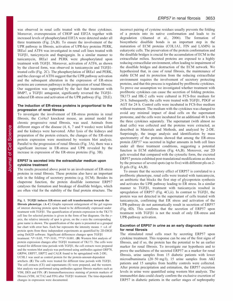
Fig. 1. TGFb1 induces ER-stress and cell transformation towards the
fibrosis phenotype. (A–C) Graphs represent enlargement of the gel regions
of interest showing protein spots found to be differentially expressed under
treatment with TGFb1. The quantification of protein expression in the TK173
cell line for selected proteins is given in the form of bar diagrams. On the y-
axis, the relative intensity of spot is given, on the x-axis the corresponding
gene name is shown. The quantification of the spots is presented as a grouped
bar chart with error bars. Each bar represents the intensity means 6 s.d. of
protein spots from three independent experiments as quantified by 2D-DIGE
using Delt2D software. Significant differences changes upon TGFb1
treatment: *P,0.05, **P,0.01, ***P,0.001. (D) Western blot analysis of
protein expression changes after TGFb1 treatment of TK173. The cells were
treated for different time periods with TGFb1, the cell extracts were prepared
and the western blot analysis was performed using antibodies against GRP78,
GRP94, ERP57, ERP72 and CALR found to be upregulated with 2-DE;
UCHL1 was used as control protein for the protein-amount-dependent
artefacts. (E) The cells were treated for different time periods with TGFb1.
The cell extracts (CE) and supernatants (S) were prepared, and the western
blot analysis was performed using antibodies against fibrosis markers such as
VIM, DES and FIN. (F) Immunofluorescence staining of protein markers of
fibrosis (VIM, ACTA2 and FIN) after TGFb1 treatment. The time-dependent
changes in expression were documented.
ERP57 in renal fibrosis 3653

Journ
alof
Cell
Scie
nce
Fig. 2. ER Stress and UPR as common pathways for
fibrosis. Western blot analysis of protein expression changes
after treatment of renal cells with TGFb1, PDGF or AGT. The
cells were treated for 72 h with one of the cytokines, the cell
extracts were prepared and the western blot analysis was
performed using antibodies against (A) GRP78, ERP57, ERP72
and CALR, and (B) EIF2A, phosphorylated EIF2A and CHOP.
(C) Western blot analysis of the UPR pathway activation. The
cells were treated for 72 h with tunicamycin, thapsigargin or
TGFb1, the cell extracts were prepared as described in the
Materials and Methods, and western blot analysis was
performed using antibodies against IRE1A, phosphorylated
IRE1A, PERK, phosphorylated PERK, ATF6 and cleaved
ATF6 (D,E) renal cells were subjected for 48 h to treatment
with TGFb1. A part of the cells was then incubated for
additional 72 h with BMP7 (5 ng/ml). Western blot analysis
was performed using the antibodies described above. ACTB was
detected as a protein loading control. The western blot
quantification is presented as a grouped bar chart with error
bars. Each bar represents the intensity means 6 s.d. of blots
from three independent experiments. Significant differences:
*P,0.05, **P,0.01, ***P,0.001.
Journal of Cell Science 126 (16)3654

Journ
alof
Cell
Scie
nce Fig. 3. ER-stress proteins induction is proportional to the renal fibrosis progression. Col4a3 knockout mice from different stage of the diseases (4, 6, 7 and 9
weeks) were killed and the kidneys were harvested. (A) Western blot analysis was performed with the protein extracts. The expression quantification is presented
as grouped bar chart. Each bar represents the intensity means 6 s.d. of blots from three independent animal groups. ACTB was used as control protein for the
protein-amount-dependent artefacts. (B) HE staining of kidney from Col4a3 mice presenting different stages of the disease. The western blot quantification is
presented as a grouped bar chart with error bars. Each bar represents the intensity means 6 s.d. of blots from three independent experiments. Significant
differences: *P,0.05, **P,0.01, ***P,0.001.
Fig. 4. ERP57 is secreted to extra cellular
medium upon cytokine treatment. 2-DE map of
cell supernatant after treatment with TGFb1,
PDGF or AGT. TK173 or HK-2 cells were treated
for 72 h with one of the cytokines. The proteins
were prepared from the supernatant as described in
the Materials and Methods, and separated by 2-
DE. The protein maps were compared with the
control, and the differentially secreted proteins
were identified by mass spectrometry.
Enlargement of the gel regions of interest showing
spots from the secreted ERP57 (A) TK173 and
(B) HK-2. (C) Renal cells were treated with
tunicamycin and the expression of ERP57 was
analyzed by western blot. (D) In contrast to
TGFb1, the tunicamycin-induced upregulation of
ERP57 did not result in its secretion.
ERP57 in renal fibrosis 3655

Journ
alof
Cell
Scie
nce
(Fig. 5). The analysis of ERP57 excretion in urine from Col4a3
knockout mice at different stages of fibrosis clearly confirmed a
correlation between the ERP57 excretion level and the progression
of renal fibrosis in the first 7 weeks (supplementary material Fig.
S2). The decrease in excretion level in later stages could be
explained by the relatively higher increase in excretion of plasma
proteins (e.g. albumin).
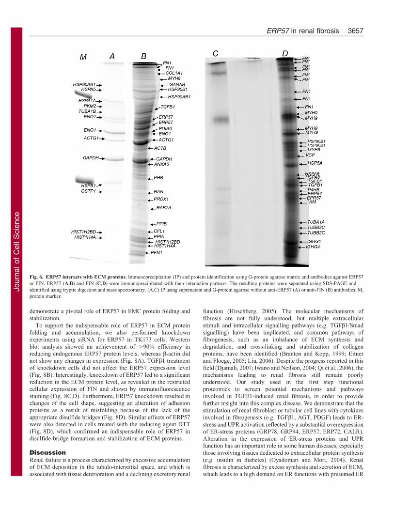
The secreted ERP57 interacts with ECM proteins
To investigate the role of ERP57 in ECM protein folding and
stabilization, protein interaction studies were performed.
Immunoprecipitation using antibodies against ERP57 or FIN
combined with 1D gel and mass spectrometry analysis confirmed a
clear interaction between the secreted protein disulfide isomerase
ERP57 and ECM proteins, especially FIN and COL1A1 (Fig. 6A–
D; supplementary material Tables S2, S3). This suggests an
important role of the secreted ERP57 in ECM stabilization.
Effect of protein disulfide isomerase inhibition on ECM
synthesis
To test the effect of protein disulfide isomerase inhibitors on
ERP57 secretion and on ECM synthesis and accumulation, TK173
was treated with TGFb1 supplemented with bacitracin (protein
disulfide isomerase inhibitor) and incubated for 72 h. To confirm
the inhibition of the enzyme activity, the protein disulfide
isomerase activity assay was performed. The activity of the
purified recombinant protein disulfide isomerase was compared
with its activity in extracts of TK173 cell cultures with and without
bacitracin, according to the procedure of Lundstrom and Holmgren
(Lundstrom and Holmgren, 1990). The time course and
concentration of the reduced and precipitated insulin in the
TK173 cell extract showed that the increase in disulfide isomerase
activity was proportional to the protein concentration and
incubation time, as expected (Fig. 7A,B). Moreover, the insulin-
reduction assay showed that bacitracin at 200 mM caused a total
inhibition of protein disulfide isomerase activity in the TK173 cell
extract (Fig. 7C). In contrast to bacitracin, T3 did not significantly
reduce the protein disulfide isomerase activity (Fig. 7C). Activity
assays performed using anti-ERP57 antibodies, or a combination
of anti-ERP57 and anti-ERP72 antibodies as inhibitors, resulted
only in partial inhibition of the protein activity in cell extracts (data
not shown), indicating the existence of additional protein disulfide
isomerases (PDIs) in the analysed cell extracts.
To assess the toxic effect of protein disulfide isomerase
inhibitors on TK173 cells, we monitored the response of the cells
stimulated with different concentrations of bacitracin and T3 over
24 h using the MTT cell viability assay method. TK173 cells
were initially seeded in 96-well plates and grown in normal
culture medium. The cells were then stimulated in FCS-free
medium with various concentrations of bacitracin or T3 and the
percentage viability was compared with an untreated control
culture after 24 h and 48 h. As shown in Fig. 7D, cell survival
strongly correlated with the concentration of the inhibitors. No
significant changes, however, were observed when the incubation
time was prolonged. Consequently, experimental conditions for
an optimal inhibition of the protein disulfide isomerase
experiments over 72 h were designed using concentrations at a
maximum of 250 mM in the case of bacitracin and 100 mM in the
case of T3.
The effect of the of protein disulfide isomerase inhibition
activity on ECM accumulation was then monitored by analysis of
the excretion of two components of the ECM, FIN and LAMA1, in
the cell culture supernatant of TK173 treated with TGFb1
combined with either bacitracin or T3. Western blot analysis of
the proteins isolated from the supernatants or cell extracts showed
a clear decrease in ECM protein synthesis and excretion in cells
treated with bacitracin compared with the control and T3
(Fig. 7Eii,iii). Moreover, the expression of ERP57 was also
reduced in cells upon treatment with bacitracin, revealing a
possible feedback control between the function of ERP57 in the
cell and in extracellular medium. Both in vitro and in vivo
experiments showed that T3 had no efficient inhibitory effect on
the protein disulfide isomerase activity similar to results reported
earlier (Primm and Gilbert, 2001). The impact of ERP57 inhibition
on ECM accumulation was also confirmed by specific inhibition
with the monoclonal antibody against ERP57. FIN was not able to
form dimers as a result of the inhibition of ERP57 (Fig. 7F) and the
hindering of disulfide bridge synthesis. These results clearly
Fig. 5. ERP57 is a potential marker for earlier detection
of renal injury. Urine samples from 15 diabetic patients
with lower microalbuminuria (20–50 mg/l), 15 urine
samples from AKI patients and 15 from healthy controls
were collected, and proteins were prepared as described in
the Materials and Methods. ERP57 excretion levels in urine
were quantified using western blot analysis. Western blot
quantification: on the y-axis the relative intensity is given
and the x-axis shows distribution of the intensity thought the
corresponding urine group where the protein was analyzed.
The detection of proteins within experimental groups on the
blotting membrane was presented below the graph.
Statistical analysis was performed using Prizma4 software
(***P,0.001).
Journal of Cell Science 126 (16)3656
Journ
alof
Cell
Scie
nce
demonstrate a pivotal role of ERP57 in EMC protein folding andstabilization.
To support the indispensable role of ERP57 in ECM proteinfolding and accumulation, we also performed knockdownexperiments using siRNA for ERP57 in TK173 cells. Western
blot analysis showed an achievement of .90% efficiency inreducing endogenous ERP57 protein levels, whereas b-actin didnot show any changes in expression (Fig. 8A). TGFb1 treatment
of knockdown cells did not affect the ERP57 expression level(Fig. 8B). Interestingly, knockdown of ERP57 led to a significantreduction in the ECM protein level, as revealed in the restrictedcellular expression of FIN and shown by immunofluorescence
staining (Fig. 8C,D). Furthermore, ERP57 knockdown resulted inchanges of the cell shape, suggesting an alteration of adhesionproteins as a result of misfolding because of the lack of the
appropriate disulfide bridges (Fig. 8D). Similar effects of ERP57were also detected in cells treated with the reducing agent DTT(Fig. 8D), which confirmed an indispensable role of ERP57 in
disulfide-bridge formation and stabilization of ECM proteins.
DiscussionRenal failure is a process characterized by excessive accumulationof ECM deposition in the tubulo-interstitial space, and which isassociated with tissue deterioration and a declining excretory renal
function (Hirschberg, 2005). The molecular mechanisms of
fibrosis are not fully understood, but multiple extracellular
stimuli and intracellular signalling pathways (e.g. TGFb1/Smad
signalling) have been implicated, and common pathways of
fibrogenesis, such as an imbalance of ECM synthesis and
degradation, and cross-linking and stabilization of collagen
proteins, have been identified (Branton and Kopp, 1999; Eitner
and Floege, 2005; Liu, 2006). Despite the progress reported in thisfield (Djamali, 2007; Iwano and Neilson, 2004; Qi et al., 2006), the
mechanisms leading to renal fibrosis still remain poorly
understood. Our study used in the first step functional
proteomics to screen potential mechanisms and pathways
involved in TGFb1-induced renal fibrosis, in order to provide
further insight into this complex disease. We demonstrate that the
stimulation of renal fibroblast or tubular cell lines with cytokines
involved in fibrogenesis (e.g. TGFb1, AGT, PDGF) leads to ER-
stress and UPR activation reflected by a substantial overexpression
of ER-stress proteins (GRP78, GRP94, ERP57, ERP72, CALR).
Alteration in the expression of ER-stress proteins and UPRfunction has an important role in some human diseases, especially
those involving tissues dedicated to extracellular protein synthesis
(e.g. insulin in diabetes) (Oyadomari and Mori, 2004). Renal
fibrosis is characterized by excess synthesis and secretion of ECM,
which leads to a high demand on ER functions with presumed ER
Fig. 6. ERP57 interacts with ECM proteins. Immunoprecipitation (IP) and protein identification using G-protein agarose matrix and antibodies against ERP57
or FIN. ERP57 (A,B) and FIN (C,D) were immunoprecipitated with their interaction partners. The resulting proteins were separated using SDS-PAGE and
identified using tryptic digestion and mass spectrometry. (A,C) IP using supernatant and G-protein agarose without anti-ERP57 (A) or anti-FIN (B) antibodies. M,
protein marker.
ERP57 in renal fibrosis 3657

Journ
alof
Cell
Scie
nce
Fig. 7. An alternation in ERP57 expression impairs ECM synthesis and accumulation. Time and concentration course of insulin reduction and precipitation
for (A) recombinant PDI (0–20 mg/ml) and (B) TK173 cell extract. Cell extracts of 4, 8, 16, 24, 32 and 40 mg/ml concentration and GSH were used for
insulin reduction. The reaction was followed for each cell extract concentration for 35 min. (C) Bacitracin or T3 inhibition of the PDI activity. Reactions were
performed using 20 mg cell extract for 40 min, the bacitracin concentration was 250 mM, T3 concentration was 100 mM. Each bar represents the OD650 intensity
means 6 s.d. of at least three independent experiments. (D) The effect of bacitracin and T3 on cell proliferation and viability. TK173 treated with variable
bacitracin (100–500 mM) (i) or T3 (50–200 mM) (ii) concentrations for different incubation times and the cell viability was assessed using the MTT [(3-(4,5-
dimethylthiazol-2-yl)-2,5-diphenyltetrazolium bromide)] assay. Western blot analyses of ECM components (FIN and laminin) expression and excretion changes
in TK173 after TGFb1 treatment combined either with bacitracin or T3 (E) or with anti-ERP57 antibody. (F) The cells were first treated with the inhibitor then
with TGFb1 (7 ng/ml) for 72 h, the proteins were isolated from cell culture or supernatant as described in Materials and Methods and the western blots were
performed with antibodies against FIN and laminin.
Journal of Cell Science 126 (16)3658

Journ
alof
Cell
Scie
nce
stress and UPR at the cellular level during the course of the disease.
The data presented here also suggest a crucial role for ER and ER-
stress proteins in ECM synthesis, and thus, most probably in the
pathogenesis of renal fibrosis. ER is the site of synthesis and
folding of proteins intended to be secreted or displayed on the cell
surface (Kim et al., 2008). Important modifications of these
secreted proteins, such as glycosylation and disulfide-bond
formation, necessitate a special environment outside of the
confines of the cytosol. The membranous ER network provides
such optimal and isolated conditions to fulfil this function inside
the cells. An efficient functioning of the ER is crucial for most
cellular activities and survival. Perturbation of ER function results
in accumulation and aggregation of unfolded secretory proteins,
thus provoking ER stress, which is detected by ER transmembrane
receptors and leads to initiation of the UPR to restore normal ER
function (Szegezdi et al., 2006). It is well known that stimulation
with TGFb1 activates TGFb1–Smad signalling, resulting in an
increase in the expression of genes encoding proteins involved in
renal fibrosis and those that are important for ECM formation such
as fibronectins, procollagens and laminins. Our data suggest that
the cytokine-activated transcription machinery in renal cells
provokes an overloading of the ER with ECM proteins, inducing
ER-stress and activating UPR. The UPR attempts to resolve ER
stress by inhibiting protein translation, increasing production of
chaperone proteins and enhancing protein degradation, allowing
the restoration of ER homeostasis and its normal function
(Schroder and Kaufman, 2005). To achieve this goal, the UPR
stimulates transcription factors, which in turn activate the
expression of genes to increase the folding capacity and to
improve the ER-assisted degradation (ERAD) (Rao and Bredesen,
2004; Schroder and Kaufman, 2005). We demonstrate that renal
cells under cytokine treatment prevent the accumulation of
misfolded proteins in the ER by improving the protein folding
through the activation of expression of ER chaperones.
Secretory proteins require special post-translational modifications
taking place in the ER. Among these modifications, the formation,
isomerization and reduction of disulfide bonds are crucial. These
modifications are catalyzed by thiol-oxidoreductases of the PDI
family. The PDIs are determined by the presence of at least one
thioredoxin-like domain with the catalytic activity within the motif
CXXC (Frand et al., 2000; Freedman, 1989). ERP57 is a well-
characterized member of the PDIs, which forms, breaks and
rearranges disulfide bonds in nascent glycoproteins reaching the ER.
In the case of ERP57, the redox function is based on the presence of
Fig. 8. Downregulation of ERP57 resulted in an alteration in the accumulation of ECM. (A) Western blot and immunofluorescence analysis of ERP57 in
non-transfected control (Ctr) cells and cells transfected with siRNA vector for ERP57 showing the knockdown of ERP57. (B) siRNA-transfected cells and the
control were subjected to TGFb1 stimulation. In contrast to the control, the expression of ERP57 was almost not affected in siRNA-transfected cells. (C) siRNA
transfected cells showed less ECM expression and accumulation. (D) In siRNA cells, FIN expression was restricted to intracellular compartments; FIN
accumulation in the extracellular milieu was completely disturbed.
ERP57 in renal fibrosis 3659

Journ
alof
Cell
Scie
nce
two active sites of the CGHC (CXXC) motif. The cleavage orformation of the disulfide bond depends on the status of the
cysteines in CGHC. The oxidized cysteine forms the disulfide bond,whereas reduced cysteine leads to cleavage of neighboring disulfide
bonds. We proved that renal cell lines stimulated with profiboticcytokines increase the expression of ERP57 parallel to theirtransition to a profibrotic phenotype, as shown by the high
expression of fibrosis markers and excretion of a high level ofECM proteins. For the synthesis and correct folding of ECM
proteins, especially COL IV, LAMA1/2 and FIN, the disulfidebridges play a crucial role. Without formation of these disulfide
bridges, the proteins are misfolded and degraded through the ERAD,which involves the ubiquitin-proteasome system for elimination ofunfolded proteins. Upregulation of ERP57 during the progression of
renal fibrosis suggests an important role in the formation of disulfidebridges and also protein folding of ECM proteins in the extracellular
space. Several interaction partners for ERP57 were identified(Jessop et al., 2007). Among these proteins, laminin and collagenwere described as substrates for ERP57. This provides further
support that ERP57 directly might interact with EMC proteins inorder to form crucial disulfide bridges and to help with the correct
folding of these proteins. Downregulation of ERP57 using siRNAhas a substantial impact on EMC protein synthesis and secretion. In
contrast to other PDIs, ERP57 interacts with the lectins CANX andCALR, and forms a complex, which is part of the glycoprotein-specific quality control machinery operating in the lumen of the ER
(Ellgaard and Frickel, 2003). The main target of the complex is toensure that newly synthesized proteins are correctly folded (Hebert
and Molinari, 2007). Overexpression of ERP57 in renal cells uponcytokine treatment might serve to overcome the limiting step in
ECM folding to accelerate their proper synthesis and secretion.
It was supposed that because of the KDEL (endoplasmicreticulum retention motif) the PDIs are limited to the ER and the
Golgi body (Lewis et al., 1990). However, a number of studiesemerged during the last decade, which demonstrated that despite
the integrity of the KDEL signal, protein disulfide isomerases canshow additional localizations including the Golgi, secretoryvesicles and the cell-surface, where they can have an important
function in stabilization and the correct folding of secretedproteins (Akagi et al., 1988). The localization and function of
PDIs at the cell surface have been proven in a number ofinvestigations including viral fusion and entry (Fenouillet et al.,2001; Markovic et al., 2004; Ryser et al., 1994), gamete fusion
(Ellerman et al., 2006), integrin ligation and activation (Lahavet al., 2002; Lahav et al., 2003), inhibition of thrombin generation
and fibrin deposition, and initiation of coagulation at sites ofvascular damage (Reinhardt et al., 2008; Schott et al., 2005).
PDIs of the cell surface and ER are biochemically andimmunologically identical, and changes in the expression ofintracellular PDIs are mostly reflected in the expression of
surface PDIs. ERP57 is the only member of the PDI with a C-terminal motif (QEDL) that seems to be ineffective for ER
retrieval (Raykhel et al., 2007), explaining a possible way ofescaping the ER retention control. When it is secreted, ECM is
exposed to environmental conditions that differ to the highlyoxidizing ER environment. This might result in the disruption ofdisulfide bonds and destabilization of protein structure. We
showed that ERP57 was the only member of the PDIs that wasfound to be secreted in high amounts upon cytokine treatment,
even though the renal cells express several other members of thePDI family. Our data suggest that secreted ERP57 is a leading
candidate among the PDI family members, which could
participate in ECM synthesis and stabilization, thus potentially
characterizing a progressive renal fibrosis. We demonstrated that
secreted ERP57 interacts with ECM proteins, such as fibronectin
and collagen, and is supposed to prevent disulfide bond reduction
and misfolding of these proteins, thus supporting regular
extracellular accumulation. Bacitracin is poorly transported into
mammalian cells, and even at high concentrations, it has minimal
effects on the proliferative capacity of the cell. The combined
treatment of the cells with TGFb1 and bacitracin (for the
inhibition of ERP57 secretion) substantially impaired ECM
synthesis and extracellular accumulation. Moreover, inhibition of
secretory ERP57 using specific monoclonal anti-ERP57 antibody
resulted in a lower level of ECM proteins. The excretion of
ERP57 in the urine of patients in earlier stages of renal fibrosis
reveals the potential role of the secreted form as an earlier urinary
marker for renal fibrosis before the onset of visible kidney
damage. Furthermore, the amount of ERP57 excreted in urine
was found to correlate with the level of tissue damage in an
animal model for progressive renal fibrosis.
We demonstrated that rapid growing fibroblasts upon cytokine
stimulation induce ER stress followed by protective UPR and
increased expression of folding chaperones. This strongly
supports the notion that the modulation of ER-stress proteins
results in a reduction of ECM production and could have a
substantially decreased fibrogenesis. Compounds that selectively
inhibit UPR or stimulate ERAD activity could also be envisioned
for renal fibrosis in which UPR seems to be pathologically
activated. The ability of ERP57 inhibitors to impact disulfide
oxidation and to abolish ECM accumulation strongly supports the
potential of ERP57 as a therapeutic target in renal fibrosis.
Materials and MethodsDMEM (Dulbecco’s modified Eagle’s medium) was from Gibco. L-glutamine,urea and DTT were from Sigma. Culture flasks were from Falcon, CHAPS [(3-Cholamidopropyl)dimethylammonio]-1-propanesulfonate] was from MERCK.Precision plus protein marker and Bio-LyteH were from Bio-Rad. BSA wasfrom Roche. Protease inhibitor Mix 100 was from GE Healthcare. Lambda2 UV/VIS spectrophotometer was from Perkin-Elmer. SequazymeTM Peptide MassStandards Kit was from Applied Biosystems. Colloidal Coomasie blue staining(Roti-Blue) was from Carl-Roth. Monoclonal mouse anti-vimentin-antibody wasfrom DAKO. Mouse anti-ERP57 monoclonal antibody and rabbit anti-ERP72polyclonal antibody were from Stressgen. Mouse anti-b-actin monoclonal antibodyand rabbit anti-GRP78/BiP polyclonal antibody were from Sigma. Mouse anti-CALR monoclonal antibody was from BD Bioscience. Rabbit anti-ERP57, anti-EIF2A mouse monoclonal, anti-pEIF2A mouse monoclonal and anti-CHOP mousemonoclonal antibodies were from Abcam. Bacitracin, T3 and AGT were fromSigma. TGFb-1 and PDGF were from R&D Systems.
Animals
Col4a3-deficient and wild-type littermate mice in identical SvJ129 geneticbackgrounds were selected from the available Alport disease models (JacksonImmunoresearch Laboratories, Westgrove, PA, USA) because of their consistentphenotype leading to progressive renal fibrosis and to uraemic death atapproximately 10 weeks old (Cosgrove et al., 1996). All mice were bred underspecific pathogen-free housing conditions and were genotyped using thepolymerase chain reaction as described previously (Ninichuk et al., 2005). Allexperimental procedures were performed according to the German animal care andethics legislation (NIH standards), and were approved by the local governmentauthorities.
Cell line and culture procedure
Human renal fibroblasts cell line (TK173) used in these experiments was derivedfrom a normal human kidney. The cells were immortalized by transfection with theplasmid pSV3gpt from SV4O and have typical morphological and biochemicalproperties of renal interstitial fibroblasts (Muller et al., 1995). The TK173 cell linewas routinely maintained as a monolayer culture in 75 cm2 tissue culture flasks(Falcon) in Dulbecco’s modified Eagle’s medium (Gibco), supplemented with
Journal of Cell Science 126 (16)3660

Journ
alof
Cell
Scie
nce
10% fetal calf serum (FCS, Gibco), 1% L-glutamine (PAA) and 1% penicillin/streptomycin (PAA). The second cell line culture consists of a renal epithelial celldesignated HK-2 (human kidney-2). HK-2 was derived from a normal adult humanrenal cortex (Ryan et al., 1994). Cultured cells were exposed to a recombinantretrovirus containing the HPV 16 E6/E7 genes. The HK-2 cell line was maintainedas a monolayer culture in Quantum 286 medium for epithelial cells (PAA) with 1%Penicillin/Streptomycin (Gibco). Cells were passaged at 85–90% confluency.Before the start of each experiment, normal growing cells were harvested withtrypsin, and cultured in 7 ml medium at a density of 56104 cells per flask andallowed to attach and grow overnight at 37 C in a humidified atmosphere with 5%CO2.
FCS-free cell culture and cell treatment
To identify new mechanisms and pathways associated with renal fibrosis, TGFb1-mediated transition of renal cells to myofibroblasts was used as model. For thisreason, TK173 or HK-2 cells were grown to subconfluency (,70% confluency)75-cm2 culture flasks. Medium was removed, and after washing in phosphate-buffered saline (PBS) the cells were incubated for a further 24 h in 10 ml serum-free DMEM. Purified human TGFb1, PDGF (7 ng/ml) (R&D Systems,Minneapolis, MN, USA), AGT (Sigma-Aldrich, St Louis, MO, USA) ortunicamycin (5 mM) were added to the medium, and the transformation offibroblasts to myofibroblasts was monitored at different times (24, 48, 72, 96 h and2 weeks) in 10 ml serum-free DMEM. In parallel, supernatants and cell extractswere collected, and the proteins were processed as described below for furtheranalyses.
To investigate the effect of BMP7 on TGFb1-induced impairment of proteinexpression, renal cells were subjected for 48 h to treatment with TGFb1.Subsequently a portion of the cells was treated with BMP7 (5 ng/ml) for anadditional 48 h. The protein expression changes were examined using western blotanalysis.
To investigate the proteome of the secreted proteins, TK173 and HK-2 cellswere cultured in FCS free medium for 24 h. Subsequently the cells were treatedwith TGFb1, PDGF or AGT for 24 h. The medium with the cytokines was changedto avoid any impact of dead cells on supernatant proteome, and the cells wereincubated for additional 48 h with the three cytokines separately. The supernatantwas collected and processed as described below.
Protein extraction and precipitation
The protein extraction was performed as described previously (Dihazi et al., 2011).To reduce the salt contamination and to enrich the proteins, chloroform-methanolprecipitation was performed according to Wessel and Flugge (Wessel and Flugge,1984).
For enrichment of the proteins before precipitation in supernatant samples, anultrafiltration step using Vivacell 100 centrifugal filter device (5 kDa molecularweight cut-off; Sartorius, Goettingen, Germany) was performed. Centrifugationwas carried out at 3000 g to reduce the volume of the supernatant to 500 ml. Theresulting samples were subjected to chloroform-methanol precipitation asdescribed above. For the western blot analysis of ECM proteins, the reducingagents (e.g. DTT) were avoided in lysis buffer to keep disulfide bridges intact.
Total protein concentration was estimated using the Bio-Rad protein assay (Bio-Rad, Hercules, CA, USA) according to Bradford (Bradford, 1976). BSA (Sigma,Steinheim, Germany) was used as a standard.
2-D gel electrophoresis
Large scale 2-DE and gel staining were carried out as described previously (Dihaziet al., 2011). For protein identification, 2-DE gels were additionally stained withcollodial Coomassie blue, Roti-Blue (Roth, Karlsruhe, Germany) overnight.
2D-DIGE
Protein extraction and chloroform-methanol precipitation were performed asdescribed above. The resulting pellet was solubilized in labelling buffer (30 mMTris/HCl pH 8.5, 9.5 M urea, 2% CHAPS, 10 mM PMSF), centrifuged (5 min,13,000 g), and the protein concentration of the supernatant was determined asdescribed above. The protein labelling with CyDyes and the 2-DE were performedas described previously (Dihazi et al., 2011). Fluorescent images were captured in16-bit TIFF files format. Spot matching across gels and normalization based on theinternal standard was performed using Delta2D software (Decodon, Greifswald,Germany). To analyze the significance of protein regulation, a Student’s t-test wasperformed, and statistical significance was assumed for P,0.01. For proteinidentification, 2-DE gels were post-stained with colloidal Coomassie blue (Roti-Blue) overnight. Differentially regulated proteins were excised and processed foridentification by MS.
Protein identification from 2-DE gels
In-gel digestion and peptide extraction were carried out as described previously(Dihazi et al., 2011). Subsequently the extracted peptides were subjected to peptidesequence analysis. The samples were dissolved in 0.1% formic acid and processed
as described previously (Dihazi et al., 2011). Processed data were searched againstMSDB and Swissprot databases through the Mascot search engine using a peptidemass tolerance of 50 ppm (parts per million) and fragment tolerance of 100 mmu(millimass unit). Protein identifications with at least two peptides sequenced wereconsidered significant.
Indirect immunofluorescence staining
For the indirect immunofluorescence staining 30,000 cells were cultivatedovernight in eight-well chamber slides. The medium was removed and the cellswere washed twice with PBS-buffer. Fixation of the cells was carried out for15 min at room temperature with 4% paraformaldehyde in PBS. Afterpermeabilization with 0.1% Triton X-100 in PBS for 1 h, the fixed cells wereblocked with 1% BSA (Sigma-Aldrich, St Louis, MO, USA) and PBS-buffer for30 min and incubated overnight separately with the appropriate primary antibody.Alexa Fluor 488-conjugated goat anti-mouse antibodies or donkey anti-rabbitantibodies were used as secondary antibodies. They were incubated for 60 min atroom temperature in the dark. The unbounded secondary antibody was removedwith three successive wash steps with PBS-buffer for 10 min each. Thereafter, thesamples were counterstained with DAPI in PBS-buffer for 5 min. Afterwards,samples were analyzed on an immunofluorescence Zeiss Axiophot microscope(Carl Zeiss, Jena, Germany) using the AnalySIS software (Soft Imaging Systems,Leinfelden, Germany).
Western blot analysis
Western blot analysis was performed according to Towbin and colleagues (Towbinet al., 1989; Towbin et al., 1992). 50 mg of the cell extracts were loaded per lane ona 12% SDS-gel after denaturation with Laemmli buffer (in the case of ECMproteins without reducing agents). Antibodies against rabbit ERP57, rabbit GRP78,rabbit CALR, mouse EIF2A, phosphorylated EIF2A, mouse CHOP, rabbit IRE1A,rabbit phosphorylated EREA, rabbit PERK, rabbit phosphorylated PERK, rabbitATF6A and mouse cleved ATF6, used as the primary antibodies, were diluted inblocking buffer, then added to the membrane and allowed to incubate for 60 min.Molecular ProbesH Alexa Fluor 647 goat anti-mouse IgG antibody or Alexa Fluor647 goat anti-rabbit IgG antibody, and Alexa Fluor 680 goat anti-mouse IgGantibody or Alexa Fluor 680 goat anti-rabbit antibody were used as secondaryantibodies. Before imaging, the blots were dried in the dark. The blot membraneswere scanned at a resolution of 50 mm on a Fuji FLA-5100 scanner (Fuji Photo,Kanagawa, Japan) with single laser-emitting excitation light at 635 nm and670 nm, respectively.
In the case of Col4a3 knockout mice, three animals per group were killed atweeks 4, 6, 7 and 9, and as a control three wild-type animals were used. Thekidneys from all animals were harvested. Aliquots of tissue extracts from threekidneys of different animals (40 mg protein) were dissolved in SDS-sample buffer,separated by electrophoresis in a SDS-polyacrylamide gel (12.5%) under reducingconditions, transferred to a nitrocellulose membrane, and blocked for 60 min atroom temperature with 5% milk-powder in a 0.2 mol/L Tris-HCl buffer, pH 7.6,containing 0.1% Tween 20 solution (TBST buffer). Antibodies against rabbitERP57, rabbit GRP78, and rabbit CALR as the primary antibodies were diluted inblocking buffer, then added to the membrane and allowed to incubate for 60 min.The addition of secondary antibodies, membrane scanning and analysis wereperformed as described above.
Urine collection and analysis of ERP57 excretion
The collection of urine samples and analysis were approved by the local InstitutionalEthics Review Committee of the University Medical Centre, Georg-AugustUniversity, Goettingen (Germany). All patients had given their informed consentbefore the study. Urine samples (20 ml) were collected from 15 diabetic patientswith lower microalbuminuria (20–50 mg/l), 15 patients with AKI and 15 healthyindividuals. The collection and preparation of urine samples was performedaccording to the EuroKup/HKUPP urine protocol and recommendation (http://www.eurokup.org/). Second morning midstream urine was used. Urine samples werecollected from the examinees, centrifuged at 1000 g for 10 min at 4 C to remove celldebris and casts. The supernatant was aliquoted (1 mL aliquots) without disturbingthe pellets and was stored at 280 C until use.
In case of animal model of fibrosis, urine was sampled by placing mice inmetabolic cages. Samples from five different wild-type mice and from five micefrom each group from 4, 6, 7 and 9 weeks Col4a3 knockout mice were collected.30 ml of urine were used for western blot analysis. Protein precipitation andwestern blot analysis were performed as described above.
Protein immunoprecipitation
For protein–protein interaction studies, immunoprecipitation (IP) using protein G-Agarose matrix was performed. The cell secretome was harvested from treatedcells as described above and concentrated to 500 ml. 200 mg secretome proteinswere used for immunoprecipitation. The samples were adjusted to 1 ml with PBSbuffer and 5 ml monoclonal anti-ERP57 or monoclonal anti-FIN antibodies wereadded. The samples were incubated overnight at 4 C under agitation. 70 ml protein
ERP57 in renal fibrosis 3661
Journ
alof
Cell
Scie
nce
G-Agarose matrix (Roche) was added to each sample. The mixture was incubatedat 4 C under rotary agitation for 4 h. The samples were centrifuged and thesupernatant removed. The agarose matrix was washed three times with PBS buffer.The supernatant was removed and 30 ml of 26loading buffer were added, thesamples were boiled at 95 C for 5 min to denature the proteins and separate themfrom the protein G-Agarose, and then centrifuged. The supernatant was used to runSDS-PAGE and identify the bind proteins.
MTT cell viability assayFor the cell proliferation assay the cell proliferation Kit I (MTT) from Roche wasused according to the manufacturer’s instructions. To investigate the effect of PDIinhibitors bacitracin and tri-iodothyronine (T3) on cell viability and proliferation,8000–10,000 cells were grown in a 96-well tissue culture plate in FCS-freemedium as a control, or supplemented with either bacitracin (100–500 mM) or T3(50–200 mM). All analyses were performed in triplicate.
PDI activity in insulin turbimetric assayThe PDI activity assay was performed by measuring insulin disulfide reductionaccording to Lundstrom and Holmgren (Lundstrom and Holmgren, 1990). Theeffect of the inhibitors on PDI activity was tested by adding bacitracin (250 mM),triiodothyronine (T3) (100 mM), and antibodies against ERP72 or ERP57 (10 mg/ml) to the reagent mixture 10 min before starting the reaction. The change inactivity was monitored by measuring the insulin precipitation.
Effect of protein disulfide isomerase inhibition on ECM synthesisTo test the effect of PDI inhibitors on ECM synthesis and accumulation, TK173were cultured in FCS-free medium as described above. PDI inhibitors (bacitracin,T3, and antibodies against ERP57 or ERP72) were added at the concentration withthe maximum effect 4 h before treatment with TGFb1. Subsequently, the cellswere incubated for 72 h. The impact of the inhibition on TK173 transformationand on ECM synthesis was monitored using western blot analysis.
siRNA construct and transfectionsiRNA oligonucleotides specific for the knockdown of ERP57 expression (sensestrand: 59-ACCTCGTCCTTCACATCTCACTAACATCAAGAGTGTTAGTG-AGATGTGAAGGACTT-39), (antisense strand: 39-CAAAAAGTCCTTC-ACATCTCACTAACACTCTTGATGTTAGTGAGATGTGAAGGACG-59) weredesigned in our laboratory and synthesized by Eurofins MWG Operon (Germany).siRNA vector was constructed by ligating oligonucleotides in psiRNAh7SK neovector (Invitrogen). All constructs were verified by sequencing. TK173 cellscultured to ,80% confluency were transfected with the vector containing siRNA forthe ERP57 knockdown using transfection reagent Lipofectamine 2000TM
(Invitrogen) according to the standard protocol of the manufacturer. Cellstransfected with psiRNAh7SK without siRNA for ERP57 were used as a positivecontrol. The transfection medium was removed after 24 h and replaced with normalculture media supplemented with 0.5 mg/ml G-418 (Invitrogen) as a selection factorfor stable transfection. Cells were maintained in the selective medium for 14 days toachieve stable transfection. The expression of ERP57 was assessed by western blotanalysis and immunofluorescence staining. The impact of ERP57 knockdown onECM protein folding and accumulation was monitored by western blot analysis andimmunofluorescence staining. For comparison, the cells were treated with thereducing agent dithiothreitol (1 mM) for 30 min before staining.
Statistical analysisFor 2-DE the digitalized images were analyzed; spot matching across gels andnormalization were performed using Delta2D 3.4 (Decodon, Braunschweig,Germany). Delta2D computes a ‘spot quality’ value for every spot detected. Thisvalue shows how closely a spot represents the ‘ideal’ 3D Gaussian bell shape. On thebasis of the average spot volume ratio, spots whose relative expression is changed atleast twofold (increase or decrease) between the compared samples were consideredto be significant. To analyze the significance of protein regulation, Student’s t-testwas performed, and statistical significance was assumed for P,0.01.
All blots were quantified using the ImageJ software. For comparison betweentwo measures (in the same group) the paired t-test was used. The unpaired t-test(for comparing two groups) or one-way ANOVA (comparing three or moregroups) were used. The data were compiled using the software package GraphPadPrism, version 4. The software was used for graphical presentation. Results arepresented as the mean 6 s.d. of at least three independent experiments. Differenceswere considered statistically significant when P,0.05.
AcknowledgementsWe thank Elke Brunst-Knoblich for technical assistance.
Author contributionsH.D. designed the study performed the majority of experiments andwrote the article, and revised the article critically; G.H.D. performed
the proteomics analysis, wrote the article and helped with datainterpretation; A.B. and M.E. performed the experiments involvingthe animal model; C.A.M. provided the cell lines and was alsoinvolved in revising the manuscript; A.R.A. was responsible for theacquisition of the mass spectrometry data; D.R. was involved inanimal model experiments and urine collection; R.V. assisted in theexperiments involving cytokine treatments; G.A.M. helped in thestudy design and manuscript writing, and revised the articlecritically.
FundingThis research received no specific grant from any funding agency inthe public, commercial or not-for-profit sectors.
Supplementary material available online at
http://jcs.biologists.org/lookup/suppl/doi:10.1242/jcs.125088/-/DC1
ReferencesAhamed, J., Versteeg, H. H., Kerver, M., Chen, V. M., Mueller, B. M., Hogg, P. J.
and Ruf, W. (2006). Disulfide isomerization switches tissue factor from coagulationto cell signaling. Proc. Natl. Acad. Sci. USA 103, 13932-13937.
Akagi, S., Yamamoto, A., Yoshimori, T., Masaki, R., Ogawa, R. and Tashiro,Y. (1988). Localization of protein disulfide isomerase on plasma membranes of ratexocrine pancreatic cells. J. Histochem. Cytochem. 36, 1069-1074.
Bechtel, W., McGoohan, S., Zeisberg, E. M., Muller, G. A., Kalbacher, H., Salant,
D. J., Muller, C. A., Kalluri, R. and Zeisberg, M. (2010). Methylation determinesfibroblast activation and fibrogenesis in the kidney. Nat. Med. 16, 544-550.
Bedard, K., Szabo, E., Michalak, M. and Opas, M. (2005). Cellular functions ofendoplasmic reticulum chaperones calreticulin, calnexin, and ERp57. Int. Rev. Cytol.
245, 91-121.
Bradford, M. M. (1976). A rapid and sensitive method for the quantitation ofmicrogram quantities of protein utilizing the principle of protein-dye binding. Anal.
Biochem. 72, 248-254.
Branton, M. H. and Kopp, J. B. (1999). TGF-beta and fibrosis. Microbes Infect. 1,1349-1365.
Cosgrove, D., Meehan, D. T., Grunkemeyer, J. A., Kornak, J. M., Sayers, R.,Hunter, W. J. and Samuelson, G. C. (1996). Collagen COL4A3 knockout: a mousemodel for autosomal Alport syndrome. Genes Dev. 10, 2981-2992.
Dihazi, H., Dihazi, G. H., Jahn, O., Meyer, S., Nolte, J., Asif, A. R., Mueller, G. A.
and Engel, W. (2011). Multipotent adult germline stem cells and embryonic stemcells functional proteomics revealed an important role of eukaryotic initiation factor5A (Eif5a) in stem cell differentiation. J. Proteome Res. 10, 1962-1973.
Djamali, A. (2007). Oxidative stress as a common pathway to chronic tubulointerstitialinjury in kidney allografts. Am. J. Physiol. 293, F445-F455.
Dobson, C. M. (2003). Protein folding and misfolding. Nature 426, 884-890.
Eitner, F. and Floege, J. (2005). Therapeutic targets for prevention and regression ofprogressive fibrosing renal diseases. Curr. Opin. Investig. Drugs 6, 255-261.
Ellerman, D. A., Myles, D. G. and Primakoff, P. (2006). A role for sperm surfaceprotein disulfide isomerase activity in gamete fusion: evidence for the participation ofERp57. Dev. Cell 10, 831-837.
Ellgaard, L. and Frickel, E. M. (2003). Calnexin, calreticulin, and ERp57: teammatesin glycoprotein folding. Cell Biochem. Biophys. 39, 223-247.
Fenouillet, E., Barbouche, R., Courageot, J. and Miquelis, R. (2001). The catalyticactivity of protein disulfide isomerase is involved in human immunodeficiency virusenvelope-mediated membrane fusion after CD4 cell binding. J. Infect. Dis. 183, 744-752.
Frand, A. R., Cuozzo, J. W. and Kaiser, C. A. (2000). Pathways for protein disulphidebond formation. Trends Cell Biol. 10, 203-210.
Freedman, R. B. (1989). Protein disulfide isomerase: multiple roles in the modificationof nascent secretory proteins. Cell 57, 1069-1072.
Goldberg, A. L. (2003). Protein degradation and protection against misfolded ordamaged proteins. Nature 426, 895-899.
Harris, R. C. and Neilson, E. G. (2006). Toward a unified theory of renal progression.Annu. Rev. Med. 57, 365-380.
Hebert, D. N. and Molinari, M. (2007). In and out of the ER: protein folding, qualitycontrol, degradation, and related human diseases. Physiol. Rev. 87, 1377-1408.
Hirschberg, R. (2005). Wound healing in the kidney: complex interactions in renalinterstitial fibrogenesis. J. Am. Soc. Nephrol. 16, 9-11.
Iwano, M. and Neilson, E. G. (2004). Mechanisms of tubulointerstitial fibrosis. Curr.
Opin. Nephrol. Hypertens. 13, 279-284.
Jessop, C. E., Chakravarthi, S., Garbi, N., Hammerling, G. J., Lovell, S. and
Bulleid, N. J. (2007). ERp57 is essential for efficient folding of glycoproteins sharingcommon structural domains. EMBO J. 26, 28-40.
Kim, I., Xu, W. and Reed, J. C. (2008). Cell death and endoplasmic reticulum stress:disease relevance and therapeutic opportunities. Nat. Rev. Drug Discov. 7, 1013-1030.
Lahav, J., Jurk, K., Hess, O., Barnes, M. J., Farndale, R. W., Luboshitz, J. andKehrel, B. E. (2002). Sustained integrin ligation involves extracellular freesulfhydryls and enzymatically catalyzed disulfide exchange. Blood 100, 2472-2478.
Journal of Cell Science 126 (16)3662

Journ
alof
Cell
Scie
nce
Lahav, J., Wijnen, E. M., Hess, O., Hamaia, S. W., Griffiths, D., Makris, M.,Knight, C. G., Essex, D. W. and Farndale, R. W. (2003). Enzymatically catalyzeddisulfide exchange is required for platelet adhesion to collagen via integrinalpha2beta1. Blood 102, 2085-2092.
Lassila, M., Fukami, K., Jandeleit-Dahm, K., Semple, T., Carmeliet, P., Cooper,
M. E. and Kitching, A. R. (2007). Plasminogen activator inhibitor-1 production ispathogenetic in experimental murine diabetic renal disease. Diabetologia 50, 1315-1326.
Lee, E. A., Seo, J. Y., Jiang, Z., Yu, M. R., Kwon, M. K., Ha, H. and Lee, H. B.
(2005). Reactive oxygen species mediate high glucose-induced plasminogen activatorinhibitor-1 up-regulation in mesangial cells and in diabetic kidney. Kidney Int. 67,1762-1771.
Lewis, M. J., Sweet, D. J. and Pelham, H. R. (1990). The ERD2 gene determines thespecificity of the luminal ER protein retention system. Cell 61, 1359-1363.
Liu, Y. (2006). Renal fibrosis: new insights into the pathogenesis and therapeutics.Kidney Int. 69, 213-217.
Lundstrom, J. and Holmgren, A. (1990). Protein disulfide-isomerase is a substrate forthioredoxin reductase and has thioredoxin-like activity. J. Biol. Chem. 265, 9114-9120.
Markovic, I., Stantchev, T. S., Fields, K. H., Tiffany, L. J., Tomic, M., Weiss, C. D.,Broder, C. C., Strebel, K. and Clouse, K. A. (2004). Thiol/disulfide exchange is aprerequisite for CXCR4-tropic HIV-1 envelope-mediated T-cell fusion during viralentry. Blood 103, 1586-1594.
Muller, G. A., Frank, J., Rodemann, H. P. and Engler-Blum, G. (1995). Human renalfibroblast cell lines (tFKIF and tNKF) are new tools to investigate pathophysiologicmechanisms of renal interstitial fibrosis. Exp. Nephrol. 3, 127-133.
Nangaku, M. (2006). Chronic hypoxia and tubulointerstitial injury: a final commonpathway to end-stage renal failure. J. Am. Soc. Nephrol. 17, 17-25.
Ninichuk, V., Gross, O., Reichel, C., Khandoga, A., Pawar, R. D., Ciubar, R.,
Segerer, S., Belemezova, E., Radomska, E., Luckow, B. et al. (2005). Delayedchemokine receptor 1 blockade prolongs survival in collagen 4A3-deficient mice withAlport disease. J. Am. Soc. Nephrol. 16, 977-985.
Oyadomari, S. and Mori, M. (2004). Roles of CHOP/GADD153 in endoplasmicreticulum stress. Cell Death Differ. 11, 381-389.
Primm, T. P. and Gilbert, H. F. (2001). Hormone binding by protein disulfideisomerase, a high capacity hormone reservoir of the endoplasmic reticulum. J. Biol.
Chem. 276, 281-286.
Qi, W., Chen, X., Poronnik, P. and Pollock, C. A. (2006). The renal cortical fibroblastin renal tubulointerstitial fibrosis. Int. J. Biochem. Cell Biol. 38, 1-5.
Rao, R. V. and Bredesen, D. E. (2004). Misfolded proteins, endoplasmic reticulumstress and neurodegeneration. Curr. Opin. Cell Biol. 16, 653-662.
Raykhel, I., Alanen, H., Salo, K., Jurvansuu, J., Nguyen, V. D., Latva-Ranta, M. and
Ruddock, L. (2007). A molecular specificity code for the three mammalian KDELreceptors. J. Cell Biol. 179, 1193-1204.
Reinhardt, C., von Bruhl, M. L., Manukyan, D., Grahl, L., Lorenz, M., Altmann, B.,Dlugai, S., Hess, S., Konrad, I., Orschiedt, L. et al. (2008). Protein disulfideisomerase acts as an injury response signal that enhances fibrin generation via tissuefactor activation. J. Clin. Invest. 118, 1110-1122.
Ryan, M. J., Johnson, G., Kirk, J., Fuerstenberg, S. M., Zager, R. A. and Torok-
Storb, B. (1994). HK-2: an immortalized proximal tubule epithelial cell line fromnormal adult human kidney. Kidney Int. 45, 48-57.
Ryser, H. J., Levy, E. M., Mandel, R. and DiSciullo, G. J. (1994). Inhibition of humanimmunodeficiency virus infection by agents that interfere with thiol-disulfideinterchange upon virus-receptor interaction. Proc. Natl. Acad. Sci. USA 91, 4559-4563.
Schott, P., Singer, S. S., Kogler, H., Neddermeier, D., Leineweber, K., Brodde,
O. E., Regitz-Zagrosek, V., Schmidt, B., Dihazi, H. and Hasenfuss, G. (2005).Pressure overload and neurohumoral activation differentially affect the myocardialproteome. Proteomics 5, 1372-1381.
Schroder, M. and Kaufman, R. J. (2005). ER stress and the unfolded protein response.Mutat. Res. 569, 29-63.
Szegezdi, E., Logue, S. E., Gorman, A. M. and Samali, A. (2006). Mediators ofendoplasmic reticulum stress-induced apoptosis. EMBO Rep. 7, 880-885.
Towbin, H., Staehelin, T. and Gordon, J. (1989). Immunoblotting in the clinicallaboratory. J. Clin. Chem. Clin. Biochem. 27, 495-501.
Towbin, H., Staehelin, T. and Gordon, J. (1992). Electrophoretic transfer of proteinsfrom polyacrylamide gels to nitrocellulose sheets: procedure and some applications.1979. Biotechnology 24, 145-149.
Wessel, D. and Flugge, U. I. (1984). A method for the quantitative recovery of proteinin dilute solution in the presence of detergents and lipids. Anal. Biochem. 138, 141-143.
Zeisberg, M., Shah, A. A. and Kalluri, R. (2005). Bone morphogenic protein-7 inducesmesenchymal to epithelial transition in adult renal fibroblasts and facilitatesregeneration of injured kidney. J. Biol. Chem. 280, 8094-8100.
ERP57 in renal fibrosis 3663