coronal height after expansion using a damon® system · 2 ducroz b., brézulier d., bertaud-gounot...
TRANSCRIPT

This is an Open Access article distributed under the terms of the Creative Commons Attribution License (http://creativecommons.org/licenses/by/4.0),
which permits unrestricted use, distribution, and reproduction in any medium, provided the original work is properly cited. 1
Article received: 17-04-2017.Accepted for publication: 07-06-2017.
Address for correspondence:
Bertrand Ducroz Patis Tatelin – 35700 RennesE-mail: [email protected]
J Dentofacial Anom Orthod 2017;20:403© The authors
DOI: 10.1051/odfen/2017020
The treatment philosophies of the 21st century tend to preserve dental tissues and try to limit extractions as much as possible. It is well established that orthopedic expan-sion systems such as disjunctors or even, to a lesser extent, dentoalveolar expansion systems increase the transverse diameter, thereby decreasing in the number of pre-molar extractions. The Damon® system’s
main purpose is to limit the use of extrac-tions. It uses the incisivocanine congestion to expand the posterior sectors. Studies have shown that there is indeed an expan-sion in these areas. Expansion is a common phenomenon in orthodontic treatment but whose noniatrogenic behavior remains con-troversial, both at the periodontal and dental levels. To date, no studies have examined
INTRODUCTION
Coronal height after expansion using a Damon® system
B. Ducroz1, D. Brézulier2, V. Bertaud-Gounot3, O. Sorel4
1 Qualified Specialist in Dentofacial Orthopedics, University Hospital Assistant, University of Rennes
2 Qualified Specialist in Dentofacial Orthopedics, University Hospital Assistant, University of Rennes
3 University Professor - Hospital Practitioner, University of Rennes 4 Qualified Specialist in Dentofacial Orthodontics, Professor of Universities - Hospital Practitioner,
University of Rennes
SUMMARY
Damon’s philosophy is based on gradual tooth movements generated by weak forces. It aims to help surrounding bone to remodel favorably. This way of thinking justifies expansion as an alternative to ex-traction. However, it is well established that teeth can be moved according to their myo-functional equi-librium and periodontium condition. The aim of this study is to analyze coronal height on various teeth both before and after treatment with the Damon system. Measures have been carried out on virtual models. Results showing a link between expansion and coronal height which increase by 0.65 mm when both the maxillary and mandibular arches are taken into account.
KEY WORDS
Expansion, Damon, Bracket device, Periodontum status, Coronal height
Article available at https://www.jdao-journal.org or https://doi.org/10.1051/odfen/2017020

Ducroz B., Brézulier D., Bertaud-Gounot V., Sorel O. Coronal height after expansion using a Damon® system2
B. DUCROZ, D. BREZULIER, V. BERTAUD-GOUNOT, O. SOREL
Figure 1Coronal height measurements are performed three times by varying the inclination of the
model. A. Measure at +45°. B. Measure at −45°. C. Measurement at 0°.
the periodontal implications of expan-sion following treatment with a Damon® system. The objective of this study is to
analyze the correlation between coronal height and transverse expansion with the Damon® technique.
Number of required subjects
The number of required subjects was calculated on the basis of the compari-son of a quantitative variable between two groups of subjects. According to the data from a preliminary study, the stand-ard deviations of the heights measured vary from 0.29 to 1.50 ( retained value) depending on the teeth.
To show a difference in height of 1 mm between the beginning and the end of the treatment with a power of 0.80; thus, the number of subjects necessary for this study was set at 35.
Inclusion criteria
Fifty consecutively treated cases were selected according to the following cri-teria: increase of the transverse diam-eters during the treatment, existence of three sets of models (pretreatment, during treatment, and post-treatment).
Post-treatment models are those per-formed at the end of contention, that is, at least 1 year after the device is re-moved. Cases of 30 girls and 20 boys were recorded. The treatment starting ages are between 8 and 14.5 years and the ending ages corresponding to the date of registration are between 12 and 19 years. The patients were treated without extractions. Thirty-eight cases were treated exclusively with Damon® multi-attachment therapeutics, where-as 12 underwent an interceptive phase. However, none were treated by an expansive system, such as a Hyrax or Quad-Helix device.
Exclusion criteria
All cases with virtual models that were incomplete or unusable were not included. Similarly, subjects whose erupting teeth rendered the measures unusable were excluded.
MATERIALS AND METHODS
J Dentofacial Anom Orthod 2017;20:403 3
CORONAL HEIGHT AFTER EXPANSION USING A DAMON® SYSTEM
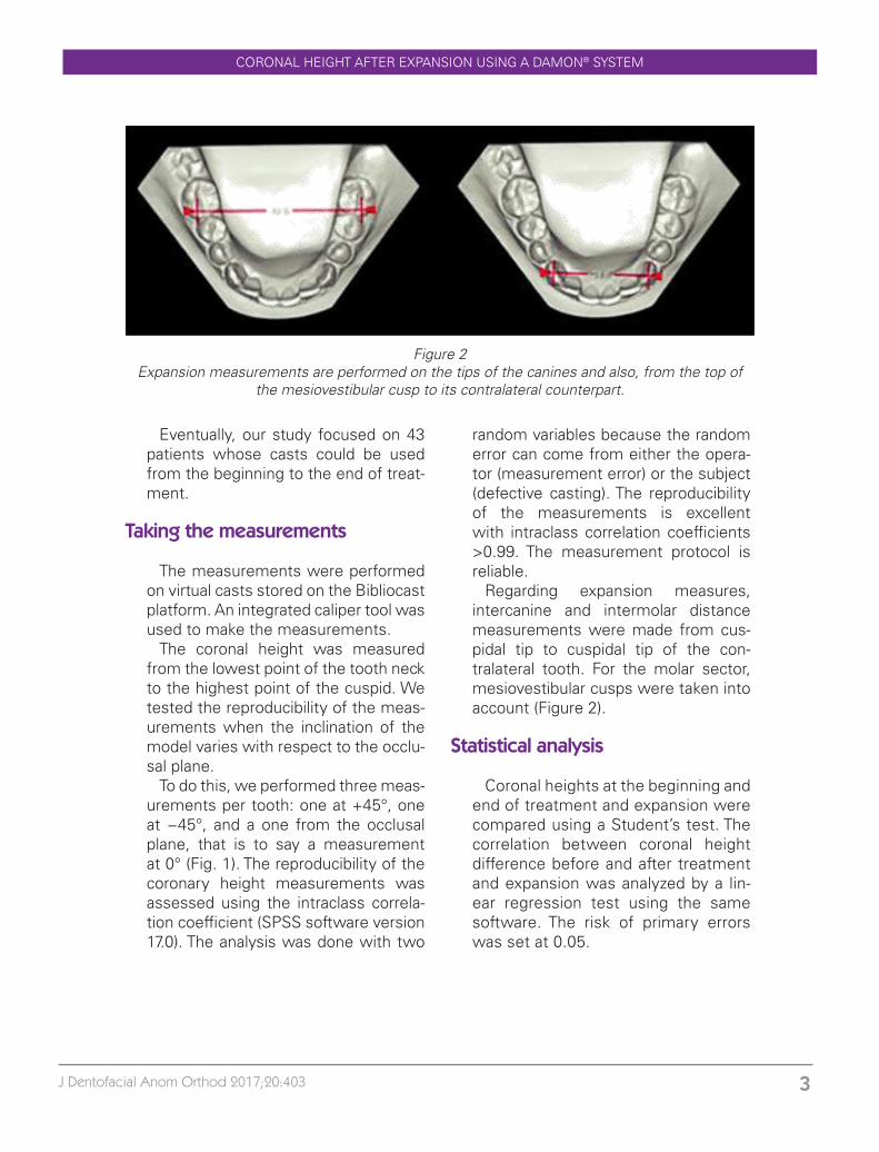
Figure 2Expansion measurements are performed on the tips of the canines and also, from the top of
the mesiovestibular cusp to its contralateral counterpart.
Eventually, our study focused on 43 patients whose casts could be used from the beginning to the end of treat-ment.
Taking the measurements
The measurements were performed on virtual casts stored on the Bibliocast platform. An integrated caliper tool was used to make the measurements.
The coronal height was measured from the lowest point of the tooth neck to the highest point of the cuspid. We tested the reproducibility of the meas-urements when the inclination of the model varies with respect to the occlu-sal plane.
To do this, we performed three meas-urements per tooth: one at +45°, one at −45°, and a one from the occlusal plane, that is to say a measurement at 0° (Fig. 1). The reproducibility of the coronary height measurements was assessed using the intraclass correla-tion coefficient (SPSS software version 17.0). The analysis was done with two
random variables because the random error can come from either the opera-tor (measurement error) or the subject (defective casting). The reproducibility of the measurements is excellent with intraclass correlation coefficients >0.99. The measurement protocol is reliable.
Regarding expansion measures, intercanine and intermolar distance measurements were made from cus-pidal tip to cuspidal tip of the con-tralateral tooth. For the molar sector, mesiovestibular cusps were taken into account (Figure 2).
Statistical analysis
Coronal heights at the beginning and end of treatment and expansion were compared using a Student’s test. The correlation between coronal height difference before and after treatment and expansion was analyzed by a lin-ear regression test using the same software. The risk of primary errors was set at 0.05.

Ducroz B., Brézulier D., Bertaud-Gounot V., Sorel O. Coronal height after expansion using a Damon® system4
B. DUCROZ, D. BREZULIER, V. BERTAUD-GOUNOT, O. SOREL
Cross-sectional expansion
The intercanine and intermolar dis-tances at the beginning and at the end of the treatment were compared initially. The measurements given here are the average mandibular and maxillary in-tercanine and intermolar distances. On the maxillary arch, we note that the av-erage arch width increases from 42.51 to 45.26 mm. This 2.7-mm increase in the transversal dimension is statistically significant (p < 0.0001). At the man-dibular arch, the expansion results in an average width that goes from 35.76 to 37.28 mm. The 1.5-mm expansion is statistically significant (p < 0.003).
The average of these values was calculated, it served as a basis for the correlation test with overall gingival height (see below). Thus, the average arch width (maxillary and mandibular combined) increases from 39.14 to 41.27 mm. Note that the difference of 2.13 mm is statistically significant (p = 0.0002) (Figure 3).
Coronal heights
Similarly, maxillary and mandibular cor-onal height measurements were taken individually, and the mean was calculated to test the coronal expansion-height correlation.
RESULTS
Overall Maxillary
Before
After
Mandibular
Figure 3Mean transverse distance in mm of the maxillary and mandibular arches before and after treat-ment and algebraic averages of these values. *Statistically significant difference with p < 0.05.

J Dentofacial Anom Orthod 2017;20:403 5
CORONAL HEIGHT AFTER EXPANSION USING A DAMON® SYSTEM
The coronal height of the maxillary teeth increased from 7.02 to 7.65 mm, an increase of 0.63 mm (p = 0.0001). On the mandible, the height variation of 0.68 mm (7.14 mm at the start of treatment and 7.82 mm at the end of treatment) is statistically significant (p < 0.0001).
With the maxilla and mandible com-bined, the mean increase in coronal height is 0.65 mm (from 7.08 to 7.73 mm) which is also statistically signifi-cant (p < 0.0001) (Fig. 4).
Correlation test between expansion and coronal height
The quantitative variables extracted previously are decreased in percent-ages (percentage of increase relative
to the initial dimension). Therefore, the measures of expansion and cor-onal height have a common order of magnitude. Average expansion of both arches is 5.8%, while coronary heights increase by 9.6%.
A linear association between increas-es in coronal height and expansion (in percentages) is clearly visible (p < 0.008). The correlation coefficient is small (0.16) but not zero (Figure 5).
Considering only the mandibular arch, the linear regression rule does not show any link between the increase in coronal height and the increase of the arch width (p > 0.05), the coefficient of correlation is 0.09. There is no statis-tically significant relationship between the increase in coronal height and man-dible expansion (Fig. 6).
Coronal height
Overall Maxillary Mandibular
Before
After
Figure 4Mean coronary height in mm of maxillary and mandibular arches before and after treatment and
algebraic averages of these values. *Statistically significant difference with p < 0.0001.

Ducroz B., Brézulier D., Bertaud-Gounot V., Sorel O. Coronal height after expansion using a Damon® system6
B. DUCROZ, D. BREZULIER, V. BERTAUD-GOUNOT, O. SOREL
Average of both arches
Percentage of expansion
Per
cent
age
incr
ease
Figure 5There is a correlation between expansion and increased coronal height. The correlation
coefficient is low.
Correlation between expansion and mandibular crown height
Percentage of expansion
Per
cent
age
incr
ease
Figure 6In the mandibule, no correlation can be demonstrated.
J Dentofacial Anom Orthod 2017;20:403 7
CORONAL HEIGHT AFTER EXPANSION USING A DAMON® SYSTEM
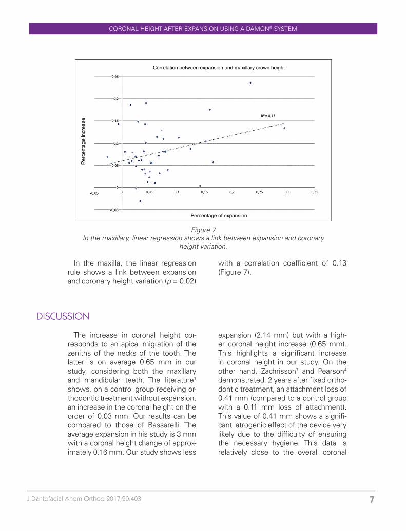
Correlation between expansion and maxillary crown height
Percentage of expansion
Per
cent
age
incr
ease
Figure 7In the maxillary, linear regression shows a link between expansion and coronary
height variation.
In the maxilla, the linear regression rule shows a link between expansion and coronary height variation (p = 0.02)
with a correlation coefficient of 0.13 (Figure 7).
The increase in coronal height cor-responds to an apical migration of the zeniths of the necks of the tooth. The latter is on average 0.65 mm in our study, considering both the maxillary and mandibular teeth. The literature1 shows, on a control group receiving or-thodontic treatment without expansion, an increase in the coronal height on the order of 0.03 mm. Our results can be compared to those of Bassarelli. The average expansion in his study is 3 mm with a coronal height change of approx-imately 0.16 mm. Our study shows less
expansion (2.14 mm) but with a high-er coronal height increase (0.65 mm). This highlights a significant increase in coronal height in our study. On the other hand, Zachrisson7 and Pearson4 demonstrated, 2 years after fixed ortho-dontic treatment, an attachment loss of 0.41 mm (compared to a control group with a 0.11 mm loss of attachment). This value of 0.41 mm shows a signifi-cant iatrogenic effect of the device very likely due to the difficulty of ensuring the necessary hygiene. This data is relatively close to the overall coronal
DISCUSSION

Ducroz B., Brézulier D., Bertaud-Gounot V., Sorel O. Coronal height after expansion using a Damon® system8
B. DUCROZ, D. BREZULIER, V. BERTAUD-GOUNOT, O. SOREL
Figure 8CT scan at the level of the first molars. Note the fineness of the maxillary external cortex and the short distance separating it from the
tooth.
height increase observed in our study (0.65 mm). Therefore, the iatrogenic effect of the sealed or bonded multiring orthodontic devices is an important fac-tor in increasing coronal height.
At this level, it is necessary to differ-entiate between the maxillary arch and the mandibular arch. In the mandible, the average change in coronal height is 0.68 mm. This variation is not corre-lated with the amount of expansion. It is however greater than that observed without treatment on the one hand, and also greater than that observed in the control group by Bassarelli. The changes in the coronal height in the maxillary teeth differ from those ob-served in the mandible. The evolution of the coronal height is statistically re-lated to the degree of expansion but in a moderate way. The maxillary perio-dontium appears to be more sensitive to expansion than its mandibular coun-terpart.
Maxillary expansion is not compa-rable with mandibular expansion. The position before treatment in the lateral
sectors is either a normal third-order inclination, or in compensation by a coronovestibular version. In this study, no account was taken of tooth version movements that may affect coronal height and the extent of expansion6. However, it is necessary to consider the third-order inclination of the teeth to definitively conclude on the poten-tial effects of expansion, but few stud-ies focus on this point. Nevertheless, the observation of the casts shows in a very large number of cases in occlusal view, vestibular surfaces which, appar-ent at the beginning of treatment, dis-appear after treatment. Endoalveolia, marked by a palatal version of alveolar processes, is the least encountered. The expansion movement does not correspond to the one observed in the mandible where the vestibular version is most often encountered. On the oth-er hand, the maxillary arch is directly affected by the jugal musculature and the buccinators, due in particular to its greater distance from the mandible. This phenomenon is particularly notice-able in the lyre-shaped arches2.
Although coronal height measure-ment may be questionable in estimat-ing attachment loss, the fact remains that its increase reflects a decrease in periodontal coverage. When choosing the measurement method, it was as-sumed that there is a bias due to gin-gival recessions that increase the cor-onal height of the teeth between the casts before and after treatment. Sim-ilarly, the measurement of the casts is likely to be less precise. Indeed, it does not take into account attrition or even gingival hyperplasia. In addition, it does not take into consideration the pres-

J Dentofacial Anom Orthod 2017;20:403 9
CORONAL HEIGHT AFTER EXPANSION USING A DAMON® SYSTEM
ence of periodontal pockets. Therefore, even if the periodontal tissue appears intact, there may be underlying bone loss that is detectable only during the clinical examination3.
The natural development of teeth, from childhood to adulthood, results in the growth of dento-alveolar pro-cesses with an increase in coronal height. Once the tooth is occlud-ed, during adolescence, the coronal height increases from 0.58 to 0.85 mm depending on the teeth and the individual5.
Gingival maturation in adults leads to a coronal height increase at the molar level of 0.4 mm and 0.19 mm at the in-cisal level over the course of 10 years5. The phenomenon is described as the passive eruption that compensates for the phenomena of attrition. The order of magnitude of the change in coronal
height in adolescence, which is esti-mated between 0.58 and 0.85 mm corresponds to that of our study (0.65 mm), which takes place over the course of 2–4 years.
The hypothesis that can be formulat-ed is the following: the increase in cor-onal height is only related to maxillary expansion. The anatomical conditions of the dentoalveolar bone environment should be considered.
The analysis of tomodensitomet-ric sections at the level of the molars shows great differences between the two arches (Figure 8).
The cortex is thicker at the mandible and the distance separating it from the tooth is greater. In the maxilla where the cortex is thinner, we cannot pre-vent fenestration phenomena by bone remodeling.
The average increase in coronal height is 0.65 mm in our study. This value following orthodontic treatment may appear relatively significant.
On the other hand, even if the aver-age increase in maxillary coronal height is lower in the mandible, it is only in the maxilla that we find a weak correlation with the measured expansion. The ini-tial position of the teeth in the maxilla and the mandible is not equal. It seems that the extent of the vestibular version is, therefore, greater in the mandible at the end of treatment.
A longer-term study is needed to determine whether the increase in coronal height after treatment is an indication of the future development of the periodontium or an irreversible iatrogenic effect.
In addition, the study does not take into account differences in the amount of dental crowding between patients, but this parameter is affected when us-ing the Damon technique®, especially when there is a possible indication of avulsion.
CONCLUSION

Ducroz B., Brézulier D., Bertaud-Gounot V., Sorel O. Coronal height after expansion using a Damon® system10
B. DUCROZ, D. BREZULIER, V. BERTAUD-GOUNOT, O. SOREL
The authors thank Dr. Bernard Nourry for having made available 50 cases treated consecutively with the Damon technique®.
Conflict of interest: The authors declare that they have no conflict of interest.
ACKNOWLEDGMENTS
1. Bassarelli T, Dalstra M, Melsen B. Changes in Clinical Achievement in Transverse Expan-sion of the Maxilla in Adults. Eur J Orthod 2005;27(2):121-128.
2. Joss-Vassalli I, Grebenstein C, Topouzelis N, Sculean has, Katsaros C. Orthodontic therapy and gingival recession: a systematic review. Orthod Craniofac Res 2010;(3):127-141.
3. Karring T, Nyman S, Thilander B, Magnusson I. Bone regeneration in orthodontically pro-duced alveolar bone dehiscences. Dr J Res 1982;17(3):309-315.
4. Pearson the Gingival height of lower central incisors, orthodontically treated and untreated. Angle Orthod 1968;38(4):337-339.
5. THEYTAZ GA, P Christou, Kiliaridis S. Gingival changes and secondary tooth eruption in adolescents and adults: a longitudinal retrospective study. Orthod Orthof Dentofacial Or-thop 2011;139:129-132.
6. Wennström JL. Mucogingival considerations in orthodontic treatment. Semin Orthod 1996;2(1):46-54.
7. Zachrisson BU, Alnæs L. Periodontal condition in orthodontically treated and untreated individuals II. Alveolar bone loss: radiographic findings. Angle Orthod 1974;44(1):48-55.
BIBLIOGRAPHY