corneal cross-linking at the slit lamp
TRANSCRIPT

Copyright © SLACK Incorporated78
O R I G I N A L A R T I C L E
Corneal cross-linking (CXL) using riboflavin and ultraviolet-A (UVA) light is a commonly per-formed treatment and the only method to arrest
corneal ectasias such as keratoconus and postoperative ectasia.1,2 CXL involves saturating the corneal stroma with riboflavin, followed by a period of stromal irra-diation with UVA light.3 The resulting photochemical reaction creates reactive oxygen species that stiffen the cornea by predominantly covalently binding collagen fibril surface and the surrounding protein network.4 This reaction also induces cell death of any living cells (and pathogens) by damaging cell membranes and nu-cleic acids.5-7 This latter reaction has led to a second indication for CXL called photoactivated chromophore
for keratitis-CXL (PACK-CXL) as a treatment for corneal infection of bacterial and/or fungal origin.8-12
To date, CXL and PACK-CXL are commonly per-formed in operating rooms, but with two major limita-tions. First, the use of CXL tends to be limited to areas that have access to an operating room infrastructure, restricting the procedure in more remote regions of the world. Second, the associated costs with the use of an operating room increase the overall price of the procedure.
To safely and effectively transfer CXL technology to be used at the slit lamp would greatly increase the accessibility to treatment globally and reduce overall costs related to the procedure. We present how this
From the Laboratory for Ocular Cell Biology, Center for Applied Biotechnology and Molecular Medicine, University of Zurich, Zurich, Switzerland (FH, EAT-N); ELZA Institute, Dietikon/Zurich, Switzerland (FH, EAT-N, MH, NLH); the Faculty of Medicine, University of Geneva, Geneva, Switzerland (FH); the Department of Ophthalmology, University of Southern California, Los Angeles, California (FH); the Department of Ophthalmology, Wenzhou Medical University, Wenzhou, China (FH); EHC Centre Médical du Simplon, Renens, Switzerland (OR); and Federal University of São Paulo, Paulista School of Medicine, Department of Ophthalmology and Visual Sciences, São Paulo, Brazil (EAT-N).
Submitted: July 24, 2020; Accepted: November 9, 2020
Supported by Light for Sight Foundation, Zurich, Switzerland, and Velux Stiftung, Zurich, Switzerland.
Dr. Farhad Hafezi holds a patent on a UV light source (PCT/CH 2012/000090). Dr. Nikki Hafezi is CEO of EMAGine AG, a company producing a CXL device. The remaining authors have no financial or proprietary interest in the materials presented herein.
Correspondence: Farhad Hafezi, MD, PhD, ELZA Institute, Webereistrasse 2 8953, Dietikon/Zurich, Switzerland. Email: [email protected]
doi:10.3928/1081597X-20201123-02
Corneal Cross-linking at the Slit LampFarhad Hafezi, MD, PhD; Olivier Richoz, MD, PhD, MBA; Emilio A. Torres-Netto, MD; Mark Hillen, PhD; Nikki L. Hafezi, MAS IP ETHZ
ABSTRACT
PURPOSE: To describe a new surgical technique where corneal cross-linking (CXL) (to treat corneal ectasias) and photo-activated chromophore for keratitis-CXL (PACK-CXL) are performed while the patient is seated in an upright posi-tion at the slit lamp.
METHODS: Topical anesthesia is applied in the waiting room, 10 minutes before the procedure. Once in the office or proce-dure room, eyelids and periorbital areas are disinfected with chloramphenicol and the patient is seated at the slit lamp. Epi-thelial debridement is performed with a cotton swab soaked in freshly prepared 40% ethanol, using 70 seconds of tapping, fol-lowed by gentle pressure to remove the epithelium. The patient is placed in the supine position for riboflavin application for 10 minutes. Stromal thickness is assessed using ultrasound
pachymetry after 5 and 10 minutes. Finally, the patient is re-turned to the slit lamp to receive ultraviolet irradiation.
RESULTS: CXL at the slit lamp is an easy-to-perform tech-nique that substantially reduces the infrastructure needed to perform CXL and PACK-CXL procedures.
CONCLUSIONS: A significant advantage of allowing CXL treatment at the slit lamp is that CXL technology can now be used in clinics that do not have easy access to an operating room infrastructure. Slit-lamp CXL can also reduce proce-dure costs by eliminating the technical fees related to the use of an operating room, making this treatment not only more accessible for patients, but also affordable.
[J Refract Surg. 2021;37(2):78-82.]

• Vol. 37, No. 2, 2021 79
corneal surgical technique can be safely performed us-ing only the slit lamp for the required infrastructure.
SURGICAL TECHNIQUECXL was performed with all parties wearing per-
sonal protective equipment per local legislation en-acted to prevent the spread of coronavirus 2 (SARS-CoV-2) in medical facilities.
PreParing the CXL DeviCePrior to surgery, the UVA illumination device
(C-Eye; EMAGine AG) was charged and calibrated by fully opening the aperture using a rotating dial, then placing the device onto a charging and calibration base (C-Base). A magnetic dock for the cross-linking device was fitted on the central mount present on the slit lamp and secured into place with a thumbscrew. Video 1 (available in the online version of this article) describes every step of the procedure.
anesthesia anD Patient PreParationFor epithelium-off CXL and PACK-CXL proce-
dures, topical anesthesia is administered in the wait-ing room, with one drop of oxybruprocaine hydro-chloride (4 mg/mL, Théa Pharma SA) followed by one drop of tetracaine 1% (Théa Pharma SA), applied three times each over a 10-minute period. For the various epithelium-on procedures, the application of anesthesia is adapted according to the protocol of choice.
The patient is brought to the slit lamp (SL9900; CSO Italia), where the height of the chair and slit lamp are adjusted to ensure the patient’s maximum comfort. This step is important because once com-fortably seated, the patient will be able to more eas-ily keep a steady position during irradiation. For that purpose, we also use a chair with two armrests rather than a simple stool. While the patient is in the sitting
position, the eye and periorbital region are thorough-ly disinfected with sterile cotton wool buds soaked in octenidine hydrochloride (Octenisept; Schülke & Mayr GmbH). A lightweight open-wire speculum (Kratz speculum, enclosed in the C-Eye Procedure Kit; EMAGine AG) is inserted and sterile surgical gauze is taped laterally to the temporal canthus to collect any riboflavin solution run-off (Figure 1A).
abrasionFor epithelium-off CXL, several different approach-
es can be used to remove the epithelium, such as by means of a hockey knife or an Amoils brush. However, these surgical tools may be challenging to maneuver in the upright position. Additionally, special attention should be taken to not injure Bowman’s membrane during the epithelial removal process. Therefore, we used an alternative approach to remove the epi-thelium. This approach is a modified laser epithelial keratomileusis (LASEK) approach13 using a sterile cot-ton swab soaked with 40% ethanol. The epithelium-off process using the cotton swab and 40% ethanol is easy to perform, without potential harm to Bowman’s membrane, rapid, and safe. Specifically, a sterile cot-ton swab is dipped in freshly prepared 40% ethanol solution, then gently tapped on the center and periph-ery of the cornea in a circular fashion for 70 seconds. After approximately 45 seconds of tapping, a loosen-ing and folding of the epithelium can be seen (Figure 1B). After 70 seconds, gentle pressure is applied to the cornea with the cotton swab tip to wipe away the epi-thelium in a circular motion. An erosion of approxi-mately 8 mm will appear (Figure 1C). Particular care must be taken that the 40% ethanol is not exposed to the air for prolonged periods of time prior to use. The evaporation of the ethanol from the solution will rap-idly change the ethanol content within the solution and, ultimately, the effectiveness of the epithelium re-
Figure 1. Corneal abrasion at the slit lamp. (A) An open-wire lightweight speculum is placed, and abrasion is performed using a sterile cotton swab first soaked in 40% ethanol. The cornea is tapped in a circular motion for 70 seconds. (B) After approximately 45 seconds, the first folds in the cor-neal epithelium appear. (C) After 70 seconds, gentle pressure is applied using a triangular sponge to remove the epithelium in a circular movement. Corneal cross-linking was performed with all parties wearing personal protective equipment per local legislation enacted to prevent the spread of severe acute respiratory syndrome coronavirus 2 (SARS-CoV-2) in medical facilities.
Copyright © SLACK Incorporated80
moval process. Finally, the cornea is rinsed with bal-anced salt solution using a syringe with an irrigation canula. For a PACK-CXL treatment, epithelial debris is removed over and/or around the infiltrate using a dry sterile triangular sponge.
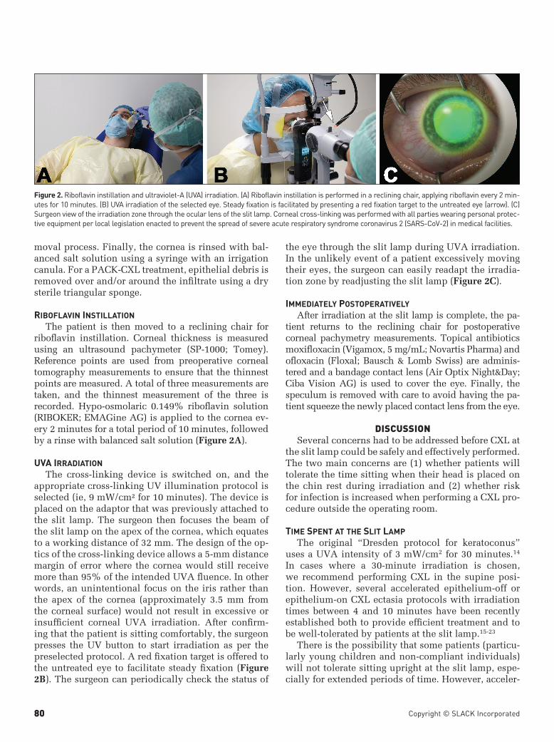
ribofLavin instiLLationThe patient is then moved to a reclining chair for
riboflavin instillation. Corneal thickness is measured using an ultrasound pachymeter (SP-1000; Tomey). Reference points are used from preoperative corneal tomography measurements to ensure that the thinnest points are measured. A total of three measurements are taken, and the thinnest measurement of the three is recorded. Hypo-osmolaric 0.149% riboflavin solution (RIBOKER; EMAGine AG) is applied to the cornea ev-ery 2 minutes for a total period of 10 minutes, followed by a rinse with balanced salt solution (Figure 2A).
Uva irraDiationThe cross-linking device is switched on, and the
appropriate cross-linking UV illumination protocol is selected (ie, 9 mW/cm² for 10 minutes). The device is placed on the adaptor that was previously attached to the slit lamp. The surgeon then focuses the beam of the slit lamp on the apex of the cornea, which equates to a working distance of 32 mm. The design of the op-tics of the cross-linking device allows a 5-mm distance margin of error where the cornea would still receive more than 95% of the intended UVA fluence. In other words, an unintentional focus on the iris rather than the apex of the cornea (approximately 3.5 mm from the corneal surface) would not result in excessive or insufficient corneal UVA irradiation. After confirm-ing that the patient is sitting comfortably, the surgeon presses the UV button to start irradiation as per the preselected protocol. A red fixation target is offered to the untreated eye to facilitate steady fixation (Figure 2B). The surgeon can periodically check the status of
the eye through the slit lamp during UVA irradiation. In the unlikely event of a patient excessively moving their eyes, the surgeon can easily readapt the irradia-tion zone by readjusting the slit lamp (Figure 2C).
immeDiateLy PostoPerativeLyAfter irradiation at the slit lamp is complete, the pa-
tient returns to the reclining chair for postoperative corneal pachymetry measurements. Topical antibiotics moxifloxacin (Vigamox, 5 mg/mL; Novartis Pharma) and ofloxacin (Floxal; Bausch & Lomb Swiss) are adminis-tered and a bandage contact lens (Air Optix Night&Day; Ciba Vision AG) is used to cover the eye. Finally, the speculum is removed with care to avoid having the pa-tient squeeze the newly placed contact lens from the eye.
DISCUSSIONSeveral concerns had to be addressed before CXL at
the slit lamp could be safely and effectively performed. The two main concerns are (1) whether patients will tolerate the time sitting when their head is placed on the chin rest during irradiation and (2) whether risk for infection is increased when performing a CXL pro-cedure outside the operating room.
time sPent at the sLit LamPThe original “Dresden protocol for keratoconus”
uses a UVA intensity of 3 mW/cm2 for 30 minutes.14 In cases where a 30-minute irradiation is chosen, we recommend performing CXL in the supine posi-tion. However, several accelerated epithelium-off or epithelium-on CXL ectasia protocols with irradiation times between 4 and 10 minutes have been recently established both to provide efficient treatment and to be well-tolerated by patients at the slit lamp.15-23
There is the possibility that some patients (particu-larly young children and non-compliant individuals) will not tolerate sitting upright at the slit lamp, espe-cially for extended periods of time. However, acceler-
Figure 2. Riboflavin instillation and ultraviolet-A (UVA) irradiation. (A) Riboflavin instillation is performed in a reclining chair, applying riboflavin every 2 min-utes for 10 minutes. (B) UVA irradiation of the selected eye. Steady fixation is facilitated by presenting a red fixation target to the untreated eye (arrow). (C) Surgeon view of the irradiation zone through the ocular lens of the slit lamp. Corneal cross-linking was performed with all parties wearing personal protec-tive equipment per local legislation enacted to prevent the spread of severe acute respiratory syndrome coronavirus 2 (SARS-CoV-2) in medical facilities.

• Vol. 37, No. 2, 2021 81
ated CXL protocols, such as the 10 minutes of 9 mw/cm² UV irradiation used in the initial cases performed at the slit lamp, help minimize the time a patient has to sit at the slit lamp. Such an accelerated protocol only loses a negligible amount of corneal stiffening efficacy when compared to the Dresden protocol and can potentially be used in most keratoconus cases.23-27 In any event, traditional cross-linking in the supine position can still be performed by docking the device into a table mount should it be necessary.
risk for infeCtionDuring every CXL procedure (standard CXL or
PACK-CXL), the keratocytes of the cornea and patho-gens (bacteria and fungi) are killed to a certain depth, depending on the protocol used.9 The killing is due to two mechanisms: generation of oxidative stress (re-active oxygen species) and intercalation of photoacti-vated riboflavin with the DNA of pathogens, disabling replication.12
In CXL for keratoconus, the irradiated cornea must be free of pathogens at the end of the procedure. Ad-ditionally, prophylactic antibiotics and a bandage contact lens are administered. Therefore, it is highly unlikely that there is a sterility advantage of perform-ing CXL in the operating room instead of an office or procedure room because the CXL procedure ultimate-ly creates an antiseptic treatment.
In PACK-CXL for infectious keratitis, the cornea is already septic. As already mentioned with CXL for keratoconus, PACK-CXL is specifically used as an an-tiseptic procedure with an aim of reducing the patho-genic load on the cornea.8,9 Thus, it is unnecessary and counterproductive to treat a septic eye in a sterile op-erating room to create an antiseptic procedure.
CXL in the sUPine PositionUltimately, CXL for keratoconus requires three fac-
tors: UVA light, oxygen, and a chromophore (eg, ribo-flavin). The propagation of UV light and the diffusion of oxygen are not impinged by the patient’s position. For riboflavin, though, there was a theoretical concern that riboflavin might settle in the inferior part of the cornea due to gravity. However, experimental investi-gation has shown that even over the course of 1 hour (twice the duration of the classic “Dresden protocol”), the effect that gravity places on the riboflavin in the cornea is negligible.24 Finally, another concern was that the supine position of the patient may theoreti-cally entail a less steady fixation. We observed that by using a comfortable chair and providing a red fixation light on the untreated eye, the patient was able to fix-ate more easily than in a supine position.
We do not expect any difference in surgical out-comes between CXL performed in a sitting position at the slit lamp when compared to CXL performed in a supine patient. Notwithstanding, we do expect that this modification could facilitate the access of CXL technology to more patients, extending coverage in remote areas, and also allow treatments with reduced related costs and administrative burden of reserving, using, and maintaining an operating room.
AUTHOR CONTRIBUTIONSStudy concept and design (FH); data collection
(OR, EAT-N, NLH); analysis and interpretation of data (FH, OR, EAT-N, MH, NLH); writing the manuscript (FH, MH); critical revision of the manuscript (FH, OR, EAT-N, MH, NLH); supervision (FH)
REFERENCES1. Raiskup F, Theuring A, Pillunat LE, Spoerl E. Corneal colla-
gen crosslinking with riboflavin and ultraviolet-A light in pro-gressive keratoconus: ten-year results. J Cataract Refract Surg. 2015;41(1):41-46. doi:10.1016/j.jcrs.2014.09.033
2. Richoz O, Mavrakanas N, Pajic B, Hafezi F. Corneal collagen cross-linking for ectasia after LASIK and photorefractive kera-tectomy: long-term results. Ophthalmology. 2013;120(7):1354-1359. doi:10.1016/j.ophtha.2012.12.027
3. Spoerl E, Huhle M, Kasper M, Seiler T. Ophthalmologe. 1997;94(12):902-906. doi:10.1007/s003470050219
4. Hayes S, Kamma-Lorger CS, Boote C, et al. The effect of ribofla-vin/UVA collagen cross-linking therapy on the structure and hy-drodynamic behaviour of the ungulate and rabbit corneal stroma. PLoS One. 2013;8(1):e52860. doi:10.1371/journal.pone.0052860
5. Chan TC, Agarwal T, Vajpayee RB, Jhanji V. Cross-linking for microbial keratitis. Curr Opin Ophthalmol. 2016;27(4):348-352. doi:10.1097/ICU.0000000000000271
6. Goodrich RP, Edrich RA, Li J, Seghatchian J. The Mirasol PRT system for pathogen reduction of platelets and plasma: an over-view of current status and future trends. Transfus Apheresis Sci. 2006;35(1):5-17. doi:10.1016/j.transci.2006.01.007
7. Martins SA, Combs JC, Noguera G, et al. Antimicrobial efficacy of riboflavin/UVA combination (365 nm) in vitro for bacterial and fungal isolates: a potential new treatment for infectious keratitis. Invest Ophthalmol Vis Sci. 2008;49(8):3402-3408. doi:10.1167/iovs.07-1592
8. Knyazer B, Krakauer Y, Tailakh MA, et al. Accelerated cor-neal cross-linking as an adjunct therapy in the management of presumed bacterial keratitis: a cohort study. J Refract Surg. 2020;36(4):258-264. doi:10.3928/1081597X-20200226-02
9. Kling S, Hufschmid FS, Torres-Netto EA, et al. High fluence increases the antibacterial efficacy of PACK cross-linking. Cor-nea. 2020;39(8):1020-1026.
10. Knyazer B, Krakauer Y, Baumfeld Y, Lifshitz T, Kling S, Hafezi F. Accelerated corneal cross-linking with photoactivated chromo-phore for moderate therapy-resistant infectious keratitis. Cor-nea. 2018;37(4):528-531. doi:10.1097/ICO.0000000000001498
11. Tabibian D, Richoz O, Riat A, Schrenzel J, Hafezi F. Acceler-ated photoactivated chromophore for keratitis-corneal collagen cross-linking as a first-line and sole treatment in early fungal keratitis. J Refract Surg. 2014;30(12):855-857. doi:10.3928/1081597X-20141113-06

Copyright © SLACK Incorporated82
12. Tabibian D, Mazzotta C, Hafezi F. PACK-CXL: corneal cross-linking in infectious keratitis. Eye Vis (Lond). 2016;3(1):11. doi:10.1186/s40662-016-0042-x
13. Browning AC, Shah S, Dua HS, Maharajan SV, Gray T, Bra-gheeth MA. Alcohol debridement of the corneal epithelium in PRK and LASEK: an electron microscopic study. Invest Oph-thalmol Vis Sci. 2003;44(2):510-513. doi:10.1167/iovs.02-0488
14. Wollensak G, Spoerl E, Seiler T. Riboflavin/ultraviolet-a-in-duced collagen crosslinking for the treatment of keratoconus. Am J Ophthalmol. 2003;135(5):620-627. doi:10.1016/S0002-9394(02)02220-1
15. Shetty R, Nagaraja H, Jayadev C, Pahuja NK, Kurian Kummelil M, Nuijts RM. Accelerated corneal collagen cross-linking in pediatric patients: two-year follow-up results. BioMed Res Int. 2014;2014:894095. doi:10.1155/2014/894095
16. Marino GK, Torricelli AA, Giacomin N, Santhiago MR, Espin-dola R, Netto MV. Accelerated corneal collagen cross-linking for postoperative lasik ectasia: two-year outcomes. J Refract Surg. 2015;31(6):380-384. doi:10.3928/1081597X-20150521-04
17. Gatzioufas Z, Richoz O, Brugnoli E, Hafezi F. Safety profile of high-fluence corneal collagen cross-linking for progressive keratoconus: preliminary results from a prospective cohort study. J Refract Surg. 2013;29(12):846-848. doi:10.3928/1081597X-20131023-03
18. Hashemi H, Miraftab M, Seyedian MA, et al. Long-term results of an accelerated corneal cross-linking protocol (18 mW/cm2) for the treatment of progressive keratoconus. Am J Ophthalmol. 2015;160(6):1164-1170.e1. doi:10.1016/j.ajo.2015.08.027
19. Agca A, Tülü B, Yasa D, et al. Accelerated corneal crosslink-ing in children with keratoconus: 5-year results and compari-son of 2 protocols. J Cataract Refract Surg. 2020;46(4):517-523. doi:10.1097/j.jcrs.0000000000000101
20. Mazzotta C, Traversi C, Paradiso AL, Latronico ME, Rechichi
M. Pulsed light accelerated crosslinking versus continuous light accelerated crosslinking: one-year results. J Ophthalmol. 2014;2014:604731. doi:10.1155/2014/604731
21. Mazzotta C, Bagaglia SA, Sgheri A, et al. Iontophoresis corneal cross-linking with enhanced fluence and pulsed UV-A light: 3-year clinical results. J Refract Surg. 2020;36(5):286-292. doi:10.3928/1081597X-20200406-02
22. Belviranli S, Oltulu R. Efficacy of pulsed-light accelerated crosslinking in the treatment of progressive keratoconus: two-year results. Eur J Ophthalmol. 2019;1120672119872375. doi:10.1177/1120672119872375
23. Lang PZ, Hafezi NL, Khandelwal SS, Torres-Netto EA, Hafezi F, Randleman JB. Comparative functional outcomes after cor-neal crosslinking using standard, accelerated, and accelerated with higher total fluence protocols. Cornea. 2019;38(4):433-441. doi:10.1097/ICO.0000000000001878
24. Salmon B, Richoz O, Tabibian D, Kling S, Wuarin R, Hafezi F. CXL at the slit lamp: no clinically relevant changes in corneal riboflavin distribution during upright UV irradiation. J Refract Surg. 2017;33(4):281. doi:10.3928/1081597X-20161219-03
25. Akkaya S, Ulusoy DM, Duru Z, Demirtas AA. Long-term out-comes of accelerated corneal cross-linking in the treatment of keratoconus: comparison of hypotonic riboflavin solution with standard riboflavin solution. J Refract Surg. 2020;36(2):110-117. doi:10.3928/1081597X-20191218-01
26. Kobashi H, Tsubota K. Accelerated versus standard corneal cross-linking for progressive keratoconus: a meta-analysis of random-ized controlled trials. Cornea. 2020;39(2):172-180. doi:10.1097/ICO.0000000000002092
27. Richoz O, Hammer A, Tabibian D, Gatzioufas Z, Hafezi F. The biomechanical effect of corneal collagen cross-linking (cxl) with riboflavin and UV-A is oxygen dependent. Transl Vis Sci Technol. 2013;2(7):6. doi:10.1167/tvst.2.7.6