control of kidney function
TRANSCRIPT

UNITSixAlterations in theUrinary System
CHAPTER
22Control of Kidney Function
Kidney Structure and FunctionGross Structure and LocationThe Nephron
The GlomerulusTubular Components of the NephronNephron Blood Supply
Urine FormationGlomerular FiltrationTubular Reabsorption and Secretion
Regulation of Renal Blood FlowNeural and Humoral Control MechanismsAutoregulation
Elimination Functions of the KidneyRenal ClearanceRegulation of Sodium and Potassium EliminationRegulation of pHpH-Dependent Elimination of Organic IonsUric Acid EliminationUrea Elimination
Drug EliminationEndocrine Functions of the Kidney
The Renin-Angiotensin-Aldosterone MechanismErythropoietinVitamin D
401
Tests of Renal FunctionUrine TestsGlomerular Filtration RateBlood Tests
Serum CreatinineBlood Urea Nitrogen
■ It is no exaggeration to say that the composition ofthe blood is determined not so much by what themouth takes in as by what the kidneys keep.
■ Homer Smith, From Fish to Philosopher
T he kidneys are remarkable organs. Each is smaller thana person’s fist, but in a single day the two organs processapproximately 1700 L of blood and combine its waste
products into approximately 1.5 L of urine. As part of theirfunction, the kidneys filter physiologically essential substan-ces, such as sodium and potassium ions, from the blood andselectively reabsorb those substances that are needed to main-tain the normal composition of internal body fluids. Substan-ces that are not needed for this purpose or are in excess passinto the urine. In regulating the volume and composition ofbody fluids, the kidneys perform excretory and endocrine func-tions. The renin-angiotensin mechanism participates in the reg-ulation of blood pressure and the maintenance of circulatingblood volume, and erythropoietin stimulates red blood cellproduction.
KIDNEY STRUCTURE AND FUNCTION
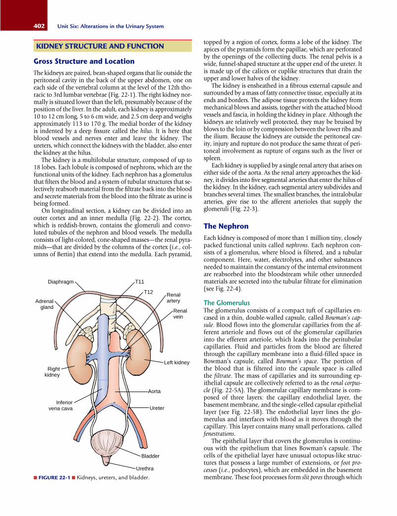
Gross Structure and LocationThe kidneys are paired, bean-shaped organs that lie outside theperitoneal cavity in the back of the upper abdomen, one oneach side of the vertebral column at the level of the 12th tho-racic to 3rd lumbar vertebrae (Fig. 22-1). The right kidney nor-mally is situated lower than the left, presumably because of theposition of the liver. In the adult, each kidney is approximately10 to 12 cm long, 5 to 6 cm wide, and 2.5 cm deep and weighsapproximately 113 to 170 g. The medial border of the kidneyis indented by a deep fissure called the hilus. It is here thatblood vessels and nerves enter and leave the kidney. Theureters, which connect the kidneys with the bladder, also enterthe kidney at the hilus.
The kidney is a multilobular structure, composed of up to18 lobes. Each lobule is composed of nephrons, which are thefunctional units of the kidney. Each nephron has a glomerulusthat filters the blood and a system of tubular structures that se-lectively reabsorb material from the filtrate back into the bloodand secrete materials from the blood into the filtrate as urine isbeing formed.
On longitudinal section, a kidney can be divided into anouter cortex and an inner medulla (Fig. 22-2). The cortex,which is reddish-brown, contains the glomeruli and convo-luted tubules of the nephron and blood vessels. The medullaconsists of light-colored, cone-shaped masses—the renal pyra-mids—that are divided by the columns of the cortex (i.e., col-umns of Bertin) that extend into the medulla. Each pyramid,
402 Unit Six: Alterations in the Urinary System
topped by a region of cortex, forms a lobe of the kidney. Theapices of the pyramids form the papillae, which are perforatedby the openings of the collecting ducts. The renal pelvis is awide, funnel-shaped structure at the upper end of the ureter. Itis made up of the calices or cuplike structures that drain theupper and lower halves of the kidney.
The kidney is ensheathed in a fibrous external capsule andsurrounded by a mass of fatty connective tissue, especially at itsends and borders. The adipose tissue protects the kidney frommechanical blows and assists, together with the attached bloodvessels and fascia, in holding the kidney in place. Although thekidneys are relatively well protected, they may be bruised byblows to the loin or by compression between the lower ribs andthe ilium. Because the kidneys are outside the peritoneal cav-ity, injury and rupture do not produce the same threat of peri-toneal involvement as rupture of organs such as the liver orspleen.
Each kidney is supplied by a single renal artery that arises oneither side of the aorta. As the renal artery approaches the kid-ney, it divides into five segmental arteries that enter the hilus ofthe kidney. In the kidney, each segmental artery subdivides andbranches several times. The smallest branches, the intralobulararteries, give rise to the afferent arterioles that supply theglomeruli (Fig. 22-3).
The NephronEach kidney is composed of more than 1 million tiny, closelypacked functional units called nephrons. Each nephron con-sists of a glomerulus, where blood is filtered, and a tubularcomponent. Here, water, electrolytes, and other substancesneeded to maintain the constancy of the internal environmentare reabsorbed into the bloodstream while other unneededmaterials are secreted into the tubular filtrate for elimination(see Fig. 22-4).
The GlomerulusThe glomerulus consists of a compact tuft of capillaries en-cased in a thin, double-walled capsule, called Bowman’s cap-sule. Blood flows into the glomerular capillaries from the af-ferent arteriole and flows out of the glomerular capillariesinto the efferent arteriole, which leads into the peritubularcapillaries. Fluid and particles from the blood are filteredthrough the capillary membrane into a fluid-filled space inBowman’s capsule, called Bowman’s space. The portion of the blood that is filtered into the capsule space is called the filtrate. The mass of capillaries and its surrounding ep-ithelial capsule are collectively referred to as the renal corpus-cle (Fig. 22-5A). The glomerular capillary membrane is com-posed of three layers: the capillary endothelial layer, thebasement membrane, and the single-celled capsular epitheliallayer (see Fig. 22-5B). The endothelial layer lines the glo-merulus and interfaces with blood as it moves through thecapillary. This layer contains many small perforations, calledfenestrations.
The epithelial layer that covers the glomerulus is continu-ous with the epithelium that lines Bowman’s capsule. Thecells of the epithelial layer have unusual octopus-like struc-tures that possess a large number of extensions, or foot pro-cesses (i.e., podocytes), which are embedded in the basementmembrane. These foot processes form slit pores through which
Left kidney
Renalvein
Renalartery
Aorta
Ureter
Bladder
Urethra
Inferiorvena cava
Rightkidney
Adrenalgland
Diaphragm T11
T12
■ FIGURE 22-1 ■ Kidneys, ureters, and bladder.

probable that the epithelial cells are active in forming newbasement membrane material throughout life. Alterations inthe structure and function of the glomerular basement mem-brane are responsible for the leakage of proteins and bloodcells into the filtrate that occurs in many forms of glomerulardisease.
Another important component of the glomerulus is themesangium. In some areas, the capillary endothelium and thebasement membrane do not completely surround each cap-illary. Instead, the mesangial cells, which lie between the capillary tufts, provide support for the glomerulus in theseareas (see Fig. 22-5B). The mesangial cells produce an inter-cellular substance similar to that of the basement membrane.This substance covers the endothelial cells where they are notcovered by basement membrane. The mesangial cells possess(or can develop) phagocytic properties and remove macro-molecular materials that enter the intercapillary spaces. Mes-angial cells also exhibit contractile properties in response toneurohumoral substances and are thought to contribute to theregulation of blood flow through the glomerulus. In normalglomeruli, the mesangial area is narrow and contains only asmall number of cells. Mesangial hyperplasia and increasedmesangial matrix occur in a number of glomerular diseases.
Tubular Components of the NephronThe nephron tubule is divided into four segments: a highlycoiled segment called the proximal convoluted tubule, whichdrains Bowman’s capsule; a thin, looped structure called theloop of Henle; a distal coiled portion called the distal convolutedtubule; and the final segment called the collecting tubule, whichjoins with several tubules to collect the filtrate (Fig. 22-4). Thefiltrate passes through each of these segments before reachingthe pelvis of the kidney.
Nephrons can be roughly grouped into two categories.Approximately 85% of the nephrons originate in the superficialpart of the cortex and are called cortical nephrons. They have
403Chapter 22: Control of Kidney Function
Renalcortex
Renal column (Bertin)
Calyx (cut edge)
Calyx
Capsule
Ureter
Renal artery
Renal pelvis
Renal blood vesselRenal medulla
Renal papillae
■ FIGURE 22-2 ■ Internal structure of the kidney.
■ FIGURE 22-3 ■ Simplified illustration of the arterial supply of the kidney. (Cormack D.H. [1987]. Ham’s histology [9th ed.].Philadelphia: J.B. Lippincott)
Interlobarartery
Renalartery
UreterPyramid
Interlobularartery
Intralobularartery
Arcuateartery
the glomerular filtrate passes. The basement membrane consistsof a homogeneous acellular meshwork of collagen fibers, gly-coproteins, and mucopolysaccharides (see Fig. 22-5C).Because the endothelial and the epithelial layers of theglomerular capillary have porous structures, the basementmembrane determines the permeability of the glomerularcapillary membrane. The spaces between the fibers that makeup the basement membrane represent the pores of a filter anddetermine the size-dependent permeability barrier of theglomerulus. The size of the pores in the basement membranenormally prevents red blood cells and plasma proteins frompassing through the glomerular membrane into the filtrate.There is evidence that the epithelium plays a major role inproducing the basement membrane components, and it is

short, thick loops of Henle that penetrate only a short distanceinto the medulla. The remaining 15% are called juxtamedullarynephrons. They originate deeper in the cortex and have longerand thinner loops of Henle that penetrate the entire length ofthe medulla. The juxtamedullary nephrons are largely con-cerned with urine concentration.
The proximal tubule is a highly coiled structure that dipstoward the renal pelvis to become the descending limb of theloop of Henle. The ascending loop of Henle returns to the re-gion of the renal corpuscle, where it becomes the distal tubule.The distal convoluted tubule, which begins at the juxtaglomer-ular complex, is divided into two segments: the diluting segmentand the late distal tubule. The late distal tubule fuses with thecollecting tubule. Like the distal tubule, the collecting duct isdivided into two segments: the cortical collecting tubule and theinner medullary collecting tubule.
Throughout its course, the tubule is composed of a singlelayer of epithelial cells resting on a basement membrane. Thestructure of the epithelial cells varies with tubular function.
404 Unit Six: Alterations in the Urinary System
■ FIGURE 22-4 ■ Nephron, showing the glomerular and tubular structures along with the blood supply.
Proximal convoluted tube
Efferent arteriole
Afferent arteriole
Bowman's capsule
Glomerulus
Cortex
Medulla
Descending limb
Ascending limb
Loop of Henle
To papilla
Peritubular capillary
Collecting tubule
Distal convoluted tubule
Interlobular vein
Interlobular artery
Juxtaglomerularapparatus
KEY CONCEPTS
THE NEPHRON
■ The nephron, which contains a glomerulus and tu-bular structures, is the functional unit of the kidney.
■ Each nephron is closely associated with two capillarybeds: the glomerulus, where water-soluble nutrients,wastes, and other small particles are filtered from theblood, and the peritubular capillaries that surroundthe tubular structures.
■ Tubular structures process the glomerular (urine) fil-trate, selectively reabsorbing substances from thetubular fluid into the peritubular capillaries and se-creting substances from the peritubular capillariesinto the urine filtrate.
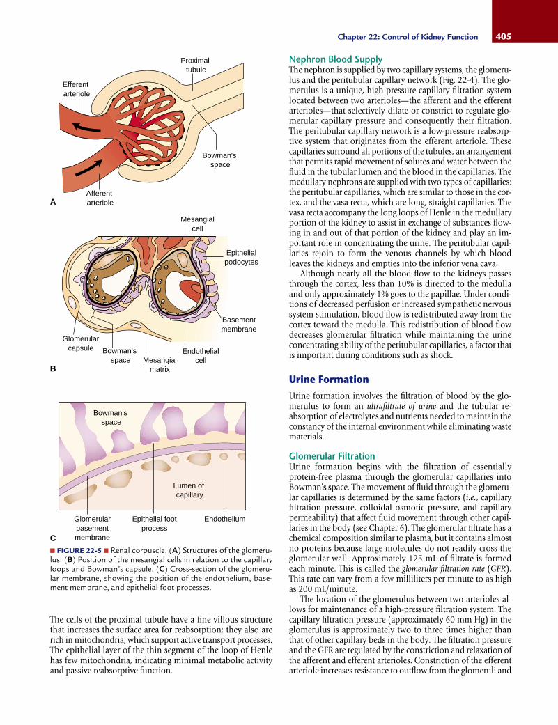
The cells of the proximal tubule have a fine villous structurethat increases the surface area for reabsorption; they also arerich in mitochondria, which support active transport processes.The epithelial layer of the thin segment of the loop of Henlehas few mitochondria, indicating minimal metabolic activityand passive reabsorptive function.
Nephron Blood SupplyThe nephron is supplied by two capillary systems, the glomeru-lus and the peritubular capillary network (Fig. 22-4). The glo-merulus is a unique, high-pressure capillary filtration systemlocated between two arterioles—the afferent and the efferentarterioles—that selectively dilate or constrict to regulate glo-merular capillary pressure and consequently their filtration.The peritubular capillary network is a low-pressure reabsorp-tive system that originates from the efferent arteriole. Thesecapillaries surround all portions of the tubules, an arrangementthat permits rapid movement of solutes and water between thefluid in the tubular lumen and the blood in the capillaries. Themedullary nephrons are supplied with two types of capillaries:the peritubular capillaries, which are similar to those in the cor-tex, and the vasa recta, which are long, straight capillaries. Thevasa recta accompany the long loops of Henle in the medullaryportion of the kidney to assist in exchange of substances flow-ing in and out of that portion of the kidney and play an im-portant role in concentrating the urine. The peritubular capil-laries rejoin to form the venous channels by which bloodleaves the kidneys and empties into the inferior vena cava.
Although nearly all the blood flow to the kidneys passesthrough the cortex, less than 10% is directed to the medullaand only approximately 1% goes to the papillae. Under condi-tions of decreased perfusion or increased sympathetic nervoussystem stimulation, blood flow is redistributed away from thecortex toward the medulla. This redistribution of blood flowdecreases glomerular filtration while maintaining the urineconcentrating ability of the peritubular capillaries, a factor thatis important during conditions such as shock.
Urine FormationUrine formation involves the filtration of blood by the glo-merulus to form an ultrafiltrate of urine and the tubular re-absorption of electrolytes and nutrients needed to maintain theconstancy of the internal environment while eliminating wastematerials.
Glomerular FiltrationUrine formation begins with the filtration of essentiallyprotein-free plasma through the glomerular capillaries intoBowman’s space. The movement of fluid through the glomeru-lar capillaries is determined by the same factors (i.e., capillaryfiltration pressure, colloidal osmotic pressure, and capillarypermeability) that affect fluid movement through other capil-laries in the body (see Chapter 6). The glomerular filtrate has achemical composition similar to plasma, but it contains almostno proteins because large molecules do not readily cross theglomerular wall. Approximately 125 mL of filtrate is formedeach minute. This is called the glomerular filtration rate (GFR).This rate can vary from a few milliliters per minute to as highas 200 mL/minute.
The location of the glomerulus between two arterioles al-lows for maintenance of a high-pressure filtration system. Thecapillary filtration pressure (approximately 60 mm Hg) in theglomerulus is approximately two to three times higher thanthat of other capillary beds in the body. The filtration pressureand the GFR are regulated by the constriction and relaxation ofthe afferent and efferent arterioles. Constriction of the efferentarteriole increases resistance to outflow from the glomeruli and
405Chapter 22: Control of Kidney Function
Bowman'sspace
Bowman'sspace
Bowman'sspace
Glomerularcapsule
Mesangialmatrix
Endothelialcell
Basementmembrane
Efferentarteriole
Afferentarteriole
Epithelialpodocytes
Mesangialcell
Proximaltubule
Lumen of capillary
EndotheliumEpithelial footprocess
Glomerularbasementmembrane
■ FIGURE 22-5 ■ Renal corpuscle. (A) Structures of the glomeru-lus. (B) Position of the mesangial cells in relation to the capillaryloops and Bowman’s capsule. (C) Cross-section of the glomeru-lar membrane, showing the position of the endothelium, base-ment membrane, and epithelial foot processes.
A
B
C

increases the glomerular pressure and the GFR. Constriction ofthe afferent arteriole causes a reduction in the renal blood flow,glomerular filtration pressure, and GFR. The afferent and the ef-ferent arterioles are innervated by the sympathetic nervous sys-tem and also are sensitive to vasoactive hormones, such asangiotensin II. During periods of strong sympathetic stimula-tion, such as occurs during shock, constriction of the afferentarteriole causes a marked decrease in renal blood flow and thusglomerular filtration pressure. Consequently, urine output canfall almost to zero.
Tubular Reabsorption and SecretionFrom Bowman’s capsule, the glomerular filtrate moves intothe tubular segments of the nephron. In its movement throughthe lumen of the tubular segments, the glomerular filtrate ischanged considerably by the tubular transport of water andsolutes. Tubular transport can result in reabsorption of sub-stances from the tubular fluid into the blood or secretion of substances from the blood into the tubular fluid (Fig. 22-6).Segments of the renal tubule are adapted to reabsorb or se-crete specific substances, using particular modes of transport.
The basic mechanisms of transport across the tubular ep-ithelial cell membrane are similar to those of other cell mem-branes in the body and include active and passive transportmechanisms (see Chapter 1). Water and urea are passivelyabsorbed along concentration gradients. Sodium, potassium,chloride, calcium, and phosphate ions, as well as urate, glu-cose, and amino acids are reabsorbed using primary or sec-
ondary active transport mechanisms to move across the tubularmembrane. Some substances, such as hydrogen, potassium,and urate ions, are secreted into the tubular fluids. Undernormal conditions, only approximately 1 mL of the 125 mL ofglomerular filtrate that is formed each minute is excreted in the urine. The other 124 mL is reabsorbed in the tubules. Thismeans that the average output of urine is approximately 60 mL/hour.
Renal tubular cells have two membrane surfaces throughwhich substances must pass as they are reabsorbed from thetubular fluid. The side of the cell that is in contact with thetubular lumen and tubular filtrate is called the luminal mem-brane. The outside membrane that lies adjacent to the inter-stitial fluid and the peritubular capillaries is called the baso-lateral membrane. In most cases, substances move from thetubular filtrate into the tubular cell along a concentrationgradient, but they require facilitated transport or carrier sys-tems to move across the basolateral membrane into the in-terstitial fluid, where they are absorbed into the peritubularcapillaries.
The bulk of energy used by the kidney is for active transportmechanisms that facilitate sodium reabsorption and cotrans-port of other electrolytes and substances such as glucose andamino acids (Fig. 22-7). Cotransport uses a carrier system inwhich two substances move in the same direction. The activetransport of one substance such as sodium is coupled to themovement of a second substance such as glucose or an aminoacid. A few substances, such as hydrogen, are secreted into thetubule using countertransport, in which the movement of onesubstance, such as sodium, enables the movement of a secondsubstance in the opposite direction.
406 Unit Six: Alterations in the Urinary System
Bowman'scapsule
Glomerularfiltrate
TubuleSecretion
To urine Toblood
Peritubularcapillary
Reabsorption
■ FIGURE 22-6 ■ Reabsorption and secretion of substances be-tween the renal tubules and peritubular capillaries.
■ FIGURE 22-7 ■ Mechanism for secondary active transport or co-transport of glucose and amino acids in the proximal tubule. Theenergy-dependent sodium-potassium pump on the basal lateralsurface of the cell maintains a low intracellular gradient that fa-cilitates the downhill movement of sodium and glucose or aminoacids (cotransport) from the tubular lumen into the tubular celland then into the peritubular capillary.
Interstitialspace
Tubularfluid
Proximal tubular cell
Peritubularcapillary
GlucoseAmino acids
GlucoseAmino acids
Na
NaNa+
H2O
H+
Luminal cell membrane
K+K+
ATP
Basolateralcell membrane

Proximal Tubule. Although tubular transport occurs through-out the renal tubule, most of it occurs in the proximal tubule.Approximately 65% of all reabsorptive and secretory processesthat occur in the tubular system take place in the proximaltubule. There is almost complete reabsorption of nutritionallyimportant substances, such as glucose, amino acids, lactate,and water-soluble vitamins. Electrolytes, such as sodium,potassium, chloride, and bicarbonate, are 65% to 80% re-absorbed. As these solutes move into the tubular cells, theirconcentration in the tubular lumen decreases, providing a con-centration gradient for the osmotic reabsorption of water andurea. The proximal tubule is highly permeable to water, and theosmotic movement of water occurs so rapidly that the concen-tration difference of solutes on either side of the membrane sel-dom is more than a few milliosmoles.
Many substances, such as glucose, are freely filtered in theglomerulus and reabsorbed by energy-dependent cotransportcarrier mechanisms. The maximum amount of substance thatthese transport systems can reabsorb per unit time is called thetransport maximum. The transport maximum is related to thenumber of carrier proteins that are available for transport andusually is sufficient to ensure that all of a filtered substance suchas glucose can be reabsorbed, rather than being eliminated inthe urine. The plasma level at which the substance appears inthe urine is called the renal threshold (Fig. 22-8). Under somecircumstances, the amount of substance filtered in the glome-rulus exceeds the transport maximum. For example, when theblood glucose level is elevated in uncontrolled diabetes melli-tus, the amount that is filtered in the glomerulus often exceedsthe transport maximum (approximately 320 mg/minute), andglucose spills into the urine.
The Loop of Henle. The loop of Henle is divided into three seg-ments: the thin descending segment, the thin ascending seg-ment, and thick ascending segment. The loop of Henle, taken
as whole, always reabsorbs more sodium and chloride thanwater. This is in contrast to the proximal tubule, which re-absorbs sodium and water in equal proportions.
The thin descending limb is highly permeable to water andmoderately permeable to urea, sodium, and other ions. As theurine filtrate moves down the descending limb, water movesout of the filtrate into the surrounding interstitium (Fig. 22-9).Thus, the osmolality of the filtrate reaches its highest point atthe elbow of the loop of Henle. In contrast to the descendinglimb, the ascending limb of the loop of Henle is impermeableto water. In this segment, solutes are reabsorbed, but water can-not follow; as a result, the tubular filtrate becomes more andmore dilute, often reaching an osmolality of 100 mOsm/kg ofH2O as it enters the distal convoluted tubule, compared withthe 285 mOsm/kg of H2O in plasma. This allows for excretionof free water from the body. For this reason, it is often calledthe diluting segment.
The thick segment of the loop of Henle begins in the as-cending limb where the epithelial cells become thickened. Thebeginning of the thick ascending limb marks the border be-tween the outer and inner medulla; thus, the thick ascendinglimb is found only in the cortex and outer medulla. As with thethin ascending limb, this segment is relatively impermeable to
407Chapter 22: Control of Kidney Function
Cortex
Outermedulla
Innermedulla
285285 100 285
100
H2O
H2O
H2OH2O
H2O
H2O
NaCl
NaCl
NaCl
NaCl
NaCl
K+ Na+
H2O
600
400
200
NaClUrea
NaClUrea
NaClUrea
NaClUrea
H2O
UreaH2O
600
800
1200
Osmolality(mOsm)
■ FIGURE 22-9 ■ Summary of movements of ions, urea, and waterin the kidney during production of a maximally concentrated urine(1200 mOsm/kg H2O). Solid arrows indicate active transport;dashed arrows indicate passive transport. The heavy outlining alongthe ascending limb of Henle’s loop indicates decreased water permeability in that tubule segment. Note the osmotic gradient inthe medulla from the outer to the inner medulla. (Modified fromRhoades R.A., Tanner G.A. [1996]. Medical physiology [p. 441].Boston: Little, Brown)

water. The epithelium of the thick segment has a Na+-K+-2Cl–
cotransport system that moves these ions out of the urine fil-trate into the surrounding interstitium and peritubular capil-laries (Fig. 22-10). Approximately 20% to 25% of the filteredload of sodium, potassium, and chloride is reabsorbed in thethick loop of Henle. This transport system is selectively blockedby diuretic agents known as loop diuretics. The Na+-K+-2Cl– co-transport system also provides the electrochemical gradientneeded for the passive reabsorption of the divalent magnesiumand calcium ions. Thus, the inhibition of sodium transport inthe thick loop of Henle by the loop diuretics causes an increasein the urinary excretion of these divalent ions in addition tosodium chloride.
In approximately one fifth of the juxtamedullary nephrons,the loops of Henle and special hairpin-shaped capillaries calledthe vasa recta descend into the medullary portion of the kidney.A countercurrent mechanism controls water and solute move-ment so that water is kept out of the area surrounding thetubule and sodium and urea are retained (see Fig. 22-9). Theterm countercurrent refers to a flow of fluids in opposite direc-tions in adjacent structures. In this case, there is an exchange ofsolutes between the adjacent descending and ascending loopsof Henle and between the ascending and descending sectionsof the vasa recta. Because of these exchange processes, a highconcentration of osmotically active particles (approximately1200 mOsm/kg of H2O) collects in the interstitium of the kid-ney medulla. The presence of these osmotically active particlesin the interstitium surrounding the medullary collectingtubules facilitates the antidiuretic hormone (ADH)-mediatedreabsorption of water.
Distal Convoluted Tubule. Like the thick ascending loop ofHenle, the distal convoluted tubule is relatively impermeableto water, and reabsorption of sodium chloride from this seg-ment further dilutes the tubular fluid. Sodium reabsorption oc-curs through a sodium and chloride cotransport mechanism.Approximately 10% of filtered sodium chloride is reabsorbed
in this section of the tubule. Unlike the thick ascending loop ofHenle, neither calcium nor magnesium is passively absorbedin this segment of the tubule. Instead, calcium ions are activelyreabsorbed in a process that is largely regulated by parathyroidhormone and possibly by vitamin D. The thiazide diureticsexert their action by inhibiting sodium chloride reabsorptionin this segment of the renal tubules.
Distal Tubule and Cortical Collecting Tubule. The late distaltubule and the cortical collecting tubule constitute the sitewhere aldosterone exerts its action on sodium and potassiumreabsorption. Although responsible for only 2% to 5% of so-dium chloride reabsorption, this site is largely responsible fordetermining the final sodium concentration of the urine. Thelate distal tubule with the cortical collecting tubule also is themajor site for regulation of potassium excretion by the kidney.When the body is confronted with a potassium excess, as oc-curs with a diet high in potassium content, the amount ofpotassium secreted at this site may exceed the amount filteredin the glomerulus.
The mechanism for sodium reabsorption and potassium se-cretion by this section of the kidney is distinct from other tubu-lar segments. This tubular segment is composed of two types ofcells, the intercalated cells, where potassium is reabsorbed andhydrogen is secreted, and the principal cells, where aldosteroneexerts its action. The secretion of hydrogen ions into the tubu-lar fluid by the intercalated cells is accompanied by the re-absorption of bicarbonate ions. The intercalated cells can alsoreabsorb potassium ions. The principal cells reabsorb sodiumand facilitate the movement of potassium into the urine fil-trate. Under the influence of aldosterone, sodium moves fromthe urine filtrate into principal cells; from there it moves intothe surrounding interstitial fluid and peritubular capillaries.Potassium moves from the peritubular capillaries into the prin-cipal cells and then into the urine filtrate.
Medullary Collecting Duct. The epithelium of the inner med-ullary collecting duct is well designed to resist extreme changesin the osmotic or pH characteristics of tubular fluid, and it ishere that the urine becomes highly concentrated, highly di-luted, highly alkaline, or highly acidic. During periods of waterexcess or dehydration, the kidneys play a major role in main-taining water balance.
ADH exerts its effect in the medullary collecting ducts.ADH maintains extracellular volume by returning water to thevascular compartment and leads to the production of a con-centrated urine by removing water from the tubular filtrate.Osmoreceptors in the hypothalamus sense the increase inosmolality of extracellular fluids and stimulate the release ofADH from the posterior pituitary gland (see Chapter 6). Thepermeability of the collecting ducts to water is determinedmainly by the concentration of ADH. In exerting its effect,ADH, also known as vasopressin, binds to vasopressin recep-tors on the blood side of the tubular cells. Binding of ADH to the vasopressin receptors leads to the opening of waterchannels on the luminal side of the tubular cells, producinga marked increase in water permeability. After the permeabil-ity of the collecting tubules has been established, water movesout of the tubular lumen and into the hyperosmotic inter-stitium of the medullary area, where it enters the peritubu-lar capillaries for return to the vascular system. In the absence
408 Unit Six: Alterations in the Urinary System
■ FIGURE 22-10 ■ Sodium, chloride, and potassium reabsorptionin the thick segment of the loop of Henle.
Interstitialspace
Tubularfluid
Peritubularcapillary
Na+ Na+
H2O
K+
K+ K+ATP
Thick ascending limb of Henle
2Cl–
Cl–

of ADH, the water channels are closed, the tubular cells losetheir water permeability, and a dilute urine is formed.
Regulation of Renal Blood FlowIn the adult, the kidneys are perfused with 1000 to 1300 mL ofblood per minute, or 20% to 25% of the cardiac output. Thislarge blood flow is mainly needed to ensure a sufficient GFR forthe removal of waste products from the blood, rather than forthe metabolic needs of the kidney. Feedback mechanisms in-trinsic to the kidney normally keep blood flow and GFR con-stant despite changes in arterial blood pressure.
Neural and Humoral Control MechanismsThe kidney is richly innervated by the sympathetic nervous sys-tem. Increased sympathetic activity causes constriction of theafferent and efferent arterioles and thus a decrease in renalblood flow. Intense sympathetic stimulation such as occurs inshock and trauma can produce marked decreases in renalblood flow and GFR, even to the extent of causing blood flowto cease altogether.
Several humoral substances, including angiotensin II, ADH,and endothelins, produce vasoconstriction of renal vessels. Theendothelins are a group of peptides released from damagedendothelial cells in the kidney and other tissues. Although notthought to be important regulators of renal blood flow duringeveryday activities, endothelin I may play a role in reduction ofblood flow in conditions such as postischemic acute renal fail-ure (see Chapter 24).
Other substances such as dopamine, nitric oxide, andprostaglandins (i.e., E2 and I2) produce vasodilation. Nitricoxide, a vasodilator produced by the vascular endothelium,appears to be important in preventing excessive vasoconstric-tion of renal blood vessels and allowing normal excretion of sodium and water. Prostaglandins are a group of mediatorsof cell function that are produced locally and exert theireffects locally. Although prostaglandins do not appear to be of major importance in regulating renal blood flow andGFR under normal conditions, they may protect the kidneysagainst the vasoconstricting effects of sympathetic stimula-tion and angiotensin II. Salicylates and the nonsteroidal anti-inflammatory drugs that inhibit prostaglandin synthesis maycause reduction in renal blood flow and GFR under certainconditions.
AutoregulationThe constancy of renal blood flow is maintained by a processcalled autoregulation (see Chapter 14). Normally, autoregula-tion of blood flow is designed to maintain blood flow at a levelconsistent with the metabolic needs of the tissues. In the kid-ney, autoregulation of blood flow also must allow for preciseregulation of water and solute secretion. For autoregulation tooccur, the resistance to blood flow through the kidneys mustbe varied in direct proportion to the arterial pressure. The exactmechanisms responsible for the intrarenal regulation of bloodflow are unclear. One of the proposed mechanisms is a directeffect on vascular smooth muscle that causes the blood vesselsto relax when there is an increase in blood pressure, and to con-strict when there is a decrease in pressure. A second proposedmechanism is the feedback regulation exerted by the juxta-glomerular complex.
The Juxtaglomerular Complex. The juxtaglomerular complexis thought to represent a feedback control system that linkschanges in the GFR with renal blood flow. The juxtaglomeru-lar complex is located at the site where the distal tubule extendsback to the glomerulus and then passes between the afferentand efferent arteriole (Fig. 22-11). The distal tubular site that isnearest the glomerulus is characterized by densely nucleatedcells called the macula densa.
In the adjacent afferent arteriole, the smooth muscle cells ofthe media are modified as special secretory cells called juxta-glomerular cells. These cells contain granules of inactive renin,an enzyme that functions in the conversion of angiotensinogento angiotensin. Renin functions by means of angiotensin II toproduce vasoconstriction of the efferent arteriole as a means ofpreventing serious decreases in the glomerular filtration rate(see Chapter 16). Angiotensin II also increases sodium re-absorption indirectly by stimulating aldosterone secretion fromthe adrenal gland and directly by increasing sodium reabsorp-tion by the proximal tubule cells.
Because of its location between the afferent and efferentarteriole, the juxtaglomerular complex is thought to play an es-sential feedback role in linking the level of arterial blood pres-sure and renal blood flow to the GFR and the composition ofthe distal tubular fluid. The juxtaglomerular complex monitorsthe systemic blood pressure by sensing the stretch of the affer-ent arteriole, and it monitors the concentration of sodiumchloride in the tubular filtrate as it passes through the maculadensa. This information is then used in determining how muchrenin should be released to keep the arterial blood pressure inits normal range and maintain a relatively constant GFR.
Elimination Functions of the KidneyThe functions of the kidney focus on elimination of water,waste products, excess electrolytes, and unwanted substancesfrom the blood. Blood tests can provide valuable information
409Chapter 22: Control of Kidney Function
Glomerulus
Basementmembrane
Afferentarteriole
Efferentarteriole
MaculadensaDistal
tubule
Juxtaglomerularcells
■ FIGURE 22-11 ■ Juxtaglomerular apparatus, showing the closecontact of the distal tubule with the afferent arteriole, the maculadensa, and the juxtaglomerular cells.
about the kidneys’ ability to remove metabolic wastes from theblood and maintain normal electrolyte and pH compositionof the blood. As renal function declines, serum levels of sub-stances such urea, creatinine, phosphate, and potassium in-crease. The effect of renal failure on the concentration of serumelectrolytes and metabolic end products is discussed in Chap-ter 24.
Renal ClearanceRenal clearance is the volume of plasma that is completelycleared each minute of any substance that finds its way into theurine. It is determined by the ability of the substance to be fil-tered in the glomeruli and the capacity of the renal tubules toreabsorb or secrete the substance. Every substance has its ownclearance rate, the units of which are expressed as the volumeof plasma that is cleared per unit time. It can be determined bymeasuring the amount of a substance that is excreted in theurine (i.e., urine concentration × urine flow rate in millilitersper minute) and dividing by its plasma concentration. Inulin,a large polysaccharide, is freely filtered in the glomeruli andneither reabsorbed nor secreted by the tubular cells. After intra-venous injection, the amount that appears in the urine is equalto the amount that is filtered in the glomeruli (i.e., the clear-ance rate is equal to the GFR). Because of these properties,inulin can be used as a laboratory measure of the GFR.
Creatinine is a product of creatine metabolism in muscles;its formation and release are relatively constant and propor-tional to the amount of muscle mass present. Creatinine isfreely filtered in the glomeruli, is not reabsorbed from thetubules into the blood, and is only minimally secreted into thetubules from the blood; therefore, its serum values dependclosely on the GFR. Serum creatinine levels are often used as ameasure of renal function. A normal serum creatinine levelusually indicates normal renal function.
Some substances, such as urea, are freely filtered in theglomeruli, but the volume that is cleared from the plasma isless than the GFR, indicating that at least some of the substanceis being reabsorbed. At normal plasma levels, glucose has aclearance of zero because it is reabsorbed in the tubules andnone appears in the urine.
Regulation of Sodium and Potassium EliminationElimination of sodium and potassium is regulated by the GFRand by humoral agents that control reabsorption. Aldosteronefunctions in the regulation of sodium and potassium elimina-tion. Atrial natriuretic peptide (ANP) contributes to the regula-tion of sodium elimination.
Aldosterone. Sodium reabsorption in the distal tubule and col-lecting duct is highly variable and depends on the presence ofaldosterone, a hormone secreted by the adrenal gland. In thepresence of aldosterone, almost all the sodium in the distaltubular fluid is reabsorbed, and the urine essentially becomessodium free. In the absence of aldosterone, virtually no sodiumis reabsorbed from the distal tubule. The remarkable ability ofthe distal tubular and collecting duct cells to alter sodium re-absorption in relation to changes in aldosterone allows the kid-neys to excrete urine with sodium levels that range from a fewtenths of a gram to 40 g per day. Like sodium, potassium isfreely filtered in the glomerulus, but unlike sodium, potassiumis reabsorbed from and secreted into the tubular fluid. The se-
cretion of potassium into the tubular fluid occurs in the distaltubule and, like that of sodium, is regulated by aldosterone.Only approximately 70 mEq of potassium is delivered to thedistal tubule each day, but the average person consumes thismuch and more potassium in the diet. Excess potassium that isnot filtered in the glomerulus and delivered to the collectingtubule therefore must be secreted (i.e., transported from theblood) into the tubular fluid for elimination from the body. Inthe absence of aldosterone (as in Addison’s disease; see Chap-ter 31), potassium secretion becomes minimal. In these cir-cumstances, potassium reabsorption exceeds secretion, andblood levels of potassium increase.
Atrial Natriuretic Peptide. Atrial natriuretic peptide, discoveredin 1981, is a hormone believed to have an important role in saltand water excretion by the kidney. It is synthesized in musclecells of the atria of the heart and released when the atria arestretched. The actions of ANP include vasodilation of the affer-ent and efferent arterioles, which results in an increase in renalblood flow and glomerular filtration rate. ANP also inhibitssodium reabsorption from the collecting tubules through its in-hibition of aldosterone secretion and through direct action onthe tubular cells. It also inhibits ADH release from the posteriorpituitary gland, thereby increasing excretion of water by thekidneys. ANP also has vasodilator properties. Whether these ef-fects are sufficient to produce long-term changes in blood pres-sure is uncertain.
Regulation of pHThe kidneys regulate body pH by conserving base bicarbonate(HCO3
–) and eliminating hydrogen ions (H+). Neither theblood buffer systems nor the respiratory control mechanismsfor carbon dioxide elimination can eliminate H+ from thebody. This is accomplished by the kidneys. The average NorthAmerican diet results in the liberation of 40 to 80 mmol of H+
each day. Virtually all the H+ excreted in the urine is secretedinto the tubular fluid by means of tubular secretory mecha-nisms. The lowest tubular fluid pH that can be achieved is 4.4to 4.5. The ability of the kidneys to excrete H+ depends onbuffers in the urine that combine with the H+. The three majorurine buffers are HCO3
–, phosphate (HPO4–), and ammonia
(NH3). The HCO3– ions, which are present in the urine filtrate,
combine with H+ ions that have been secreted into the tubu-lar fluid; this results in the formation of carbon dioxide andwater. The carbon dioxide is then absorbed into the tubularcells, and bicarbonate is regenerated. The HPO4
– ion is ametabolic end product that is filtered into the tubular fluid; itcombines with a secreted H+ ion and is not reabsorbed.Ammonia is synthesized in tubular cells by deamination ofthe amino acid glutamine; it diffuses into the tubular fluidand combines with the H+ ion. An important aspect of thisbuffer system is that the deamination process increases whenthe body’s H+ ion concentration remains elevated for 1 to 2 days. These mechanisms for pH regulation are describedmore fully in Chapter 6.
pH-Dependent Elimination of Organic IonsThe proximal tubule actively secretes large amounts of differ-ent organic anions. Foreign anions (e.g., salicylates, penicillin)and endogenously produced anions (e.g., bile acids, uric acid)are actively secreted into the tubular fluid. Most of the anions
410 Unit Six: Alterations in the Urinary System

that are secreted use the same transport system, allowing thekidneys to rid the body of many different drugs and environ-mental agents. Because the same transport system is shared bydifferent anions, there is competition for transport such thatelevated levels of one substance tend to inhibit the secretionof other anions. The proximal tubules also possess an activetransport system for organic cations that is analogous to thatfor organic ions.
Uric Acid EliminationUric acid is a product of purine metabolism (see Chapter 43).Excessively high blood levels (i.e., hyperuricemia) can causegout, and excessive levels in the urine can cause kidney stones.Uric acid is freely filtered in the glomerulus and is reabsorbedand secreted into the proximal tubules. Uric acid is one of theanions that uses the previously described anion transport sys-tem in the proximal tubule. Tubular reabsorption normally ex-ceeds secretion, and the net effect is removal of uric acid fromthe filtrate. Although the rate of reabsorption exceeds secretion,the secretory process is homeostatically controlled to maintaina constant plasma level. Many persons with elevated uric acidlevels secrete less uric acid than do persons with normal uricacid levels.
Uric acid uses the same transport systems as other anions,such as aspirin, sulfinpyrazone, and probenecid. Small dosesof aspirin compete with uric acid for secretion into the tubu-lar fluid and reduce uric acid secretion, and large doses com-pete with uric acid for reabsorption and increase uric acid ex-cretion in the urine. Because of its effect on uric acid secretion,aspirin is not recommended for treatment of gouty arthritis.Thiazide and loop diuretics (i.e., furosemide and ethacrynicacid) also can cause hyperuricemia and gouty arthritis, pre-sumably through a decrease in extracellular fluid volume andenhanced uric acid reabsorption.
Urea EliminationUrea is an end product of protein metabolism. The normaladult produces 25 to 30 g/day; the quantity rises when a high-protein diet is consumed, when there is excessive tissue break-down, or in the presence of gastrointestinal bleeding. With gas-trointestinal bleeding, the blood proteins are broken down toform ammonia in the intestine; the ammonia is then absorbedinto the portal circulation and converted to urea by the liver be-fore being released into the bloodstream. The kidneys, in theirrole as regulators of blood urea nitrogen (BUN) levels, filterurea in the glomeruli and then reabsorb it in the tubules. Thisenables maintenance of a normal BUN, which is in the rangeof 8 to 20 mg/dL. During periods of dehydration, the bloodvolume and GFR drop, and BUN levels increase. The renaltubules are permeable to urea, which means that the longer thetubular fluid remains in the kidneys, the greater is the re-absorption of urea into the blood. Only small amounts of ureaare reabsorbed into the blood when the GFR is high, but rela-tively large amounts of urea are returned to the blood when theGFR is reduced.
Drug EliminationMany drugs are eliminated in the urine. These drugs are selec-tively filtered in the glomerulus and reabsorbed or secreted intothe tubular fluid. Only drugs that are not bound to plasma pro-
teins are filtered in the glomerulus and therefore able to beeliminated by the kidneys.
Many drugs are weak acids or weak bases and are presentin the renal tubular fluid partly as water-soluble ions andpartly as nonionized lipid-soluble molecules. The nonionizedlipid-soluble form of a drug diffuses more readily through thelipid membrane of the tubule and then back into the blood-stream. The water-soluble ionized form remains in the urinefiltrate. The ratio of ionized to nonionized drug depends onthe pH of the urine. For example, aspirin is highly ionized inalkaline urine and in this form is rapidly excreted in the urine.Aspirin is largely nonionized in acid urine and is reabsorbed,rather than excreted. Alkaline or acid diuresis may be used toincrease elimination of drugs in the urine, particularly in sit-uations of drug overdose.
Endocrine Functions of the KidneyIn addition to their function in regulating body fluids andelectrolytes, the kidneys function as an endocrine organ inthat they produce chemical mediators that travel through theblood to distant sites where they exert their actions. The kid-neys participate in control of blood pressure by way of therenin-angiotensin mechanism, in calcium metabolism by acti-vating vitamin D, and in regulating red blood cell productionthrough the synthesis of erythropoietin.
The Renin-Angiotensin-Aldosterone MechanismThe renin-angiotensin-aldosterone mechanism plays an im-portant part in the short-term and long-term regulation ofblood pressure (see Chapter 16). Renin is synthesized andstored in the juxtaglomerular cells of the kidney. This enzymeis released in response to a decrease in renal blood flow or achange in the composition of the distal tubular fluid, or as theresult of sympathetic nervous system stimulation. Renin itselfhas no direct effect on blood pressure. Rather, it acts enzy-matically to convert a circulating plasma protein called angio-tensinogen to angiotensin I. Angiotensin I, which has fewvasoconstrictor properties, leaves the kidneys and enters thecirculation; as it is circulated through the lungs, angiotensin-converting enzyme catalyzes the conversion of angiotensin I to angiotensin II. Angiotensin II is a potent vasoconstrictor,and it acts directly on the kidneys to decrease salt and water
411Chapter 22: Control of Kidney Function
KEY CONCEPTS
FUNCTIONS OF THE KIDNEY
■ Long-term regulation of blood pressure is facilitatedthrough the kidney’s activation of the renin-angiotensin system and regulation of sodium and water balance.
■ The activation of vitamin D, which is important for intestinal absorption of calcium, occurs in the kidney.
■ The kidney synthesizes erythropoietin, which stimu-lates bone marrow production of red blood cells.

excretion. Both mechanisms have relatively short periods ofaction. Angiotensin II also stimulates aldosterone secretion bythe adrenal gland. Aldosterone acts on the distal tubule to in-crease sodium reabsorption and exerts a longer-term effect onthe maintenance of blood pressure. Renin also functions bymeans of angiotensin II to produce constriction of the efferentarteriole as a means of preventing a serious decrease inglomerular filtration pressure.
ErythropoietinErythropoietin is a polypeptide hormone that regulates the dif-ferentiation of red blood cells in the bone marrow (see Chap-ter 13). Between 89% and 95% of erythropoietin is formed in the kidneys. The synthesis of erythropoietin is stimulated bytissue hypoxia, which may be brought about by anemia, resi-dence at high altitudes, or impaired oxygenation of tissuescaused by cardiac or pulmonary disease. Persons with end-stage kidney disease often are anemic because of an inabilityof the kidneys to produce erythropoietin. This anemia usuallyis managed by the administration of epoetin-alfa, a syntheticform of erythropoietin produced through DNA technology, tostimulate erythropoiesis.
Vitamin DActivation of vitamin D occurs in the kidneys. Vitamin D in-creases calcium absorption from the gastrointestinal tract andhelps to regulate calcium deposition in bone. It also has a weakstimulatory effect on renal calcium absorption. Although vita-min D is not synthesized and released from an endocrinegland, it often is considered as a hormone because of its path-way of molecular activation and mechanism of action.
It exists in several forms: natural vitamin D (cholecalcif-erol), which results from ultraviolet irradiation of the skin,and synthetic vitamin D (ergocalciferol), which is derivedfrom irradiation of ergosterol. The active form of vitamin D is 1,25-dihydroxycholecalciferol. Cholecalciferol and ergocal-ciferol must undergo chemical transformation to become ac-tive: first to 25-hydroxycholecalciferol in the liver and then to1,25-dihydroxycholecalciferol in the kidneys. Persons withend-stage renal disease are unable to transform vitamin D toits active form and must rely on pharmacologic preparationsof the active vitamin (calcitriol) for maintaining mineraliza-tion of their bones.
TESTS OF RENAL FUNCTION
The functions of the kidney are to filter the blood and selec-tively reabsorb those substances that are needed to maintainthe constancy of body fluids and excrete metabolic wastes.Laboratory tests of the urine and blood can provide valuableinformation about kidney pathology and the adequacy of renalfunction.
Urine TestsUrine is a clear, amber-colored fluid that is approximately 95%water and 5% dissolved solids. The kidneys normally produceapproximately 1.5 L of urine each day. Normal urine containsmetabolic wastes and few or no plasma proteins, blood cells,or glucose molecules.
Urine tests can be performed on a single urine specimen oron a 24-hour urine specimen. First-voided morning specimensare useful for qualitative protein and specific gravity testing. Afreshly voided specimen is most reliable. Urine specimens thathave been left standing may contain lysed red blood cells, dis-integrating casts, and rapidly multiplying bacteria.
Casts are molds of the distal nephron lumen. A gel-like sub-stance called Tamm-Horsfall mucoprotein, which is formed in thetubular epithelium, is the major protein constituent of urinarycasts. Casts composed of this gel but devoid of cells are calledhyaline casts. These casts develop when the protein concentra-tion of the urine is high (as in nephrotic syndrome), urine os-molality is high, and urine pH is low. The inclusion of granulesor cells in the matrix of the protein gel leads to the formationof various other types of casts.
Because of the glomerular capillary filtration barrier, lessthan 150 mg of protein is excreted in the urine during 24 hoursin a healthy person. Qualitative and quantitative tests to deter-mine urinary protein content are important tools to assess theextent of glomerular disease. pH-sensitive reagent strips areused to test for the presence of proteins, whereas immunoassaymethods are used to test for microalbuminuria (30 to 300 mgalbumin/24 hours).
412 Unit Six: Alterations in the Urinary System
In summary, the kidneys perform excretory and en-docrine functions. In the process of excreting wastes, the kid-neys filter the blood and then selectively reabsorb those mate-rials that are needed to maintain a stable internalenvironment. The kidneys rid the body of metabolic wastes,regulate fluid volume, regulate the concentration of elec-trolytes, assist in maintaining acid-base balance, aid in theregulation of blood pressure through the renin-angiotensin-aldosterone mechanism and control of extracellular fluid vol-ume, regulate red blood cell production through erythropoi-etin, and aid in calcium metabolism by activating vitamin D.
The kidneys selectively eliminate water, waste products,excess electrolytes, and other substances that are not neededto maintain the constancy of the internal environment. Renalclearance is the volume of plasma that is completely clearedeach minute of any substance that finds its way into the urine.
It is determined by the ability of the substance to be filtered in the glomeruli and the capacity of the renal tubules to re-absorb or secrete the substance. The GFR is the amount offiltrate that is formed each minute as blood moves throughthe glomeruli. It is regulated by the arterial blood pressureand renal blood flow in the normally functioning kidney. Thejuxtaglomerular complex is thought to represent a feedbackcontrol system that links changes in the GFR with renal bloodflow.
In addition to their function in regulating body fluids andelectrolytes, the kidneys function as an endocrine organ inthat they produce chemical mediators that travel through theblood to distant sites where they exert their actions. The kid-neys participate in control of blood pressure by way of therenin-angiotensin mechanism, in calcium metabolism by acti-vating vitamin D, and in regulating red blood cell productionthrough the synthesis of erythropoietin.

The specific gravity (or osmolality) of urine varies with itsconcentration of solutes. Urine specific gravity provides a valu-able index of the hydration status and functional ability of thekidneys. Healthy kidneys can produce a concentrated urinewith a specific gravity of 1.030 to 1.040. During periods ofmarked hydration, the specific gravity can approach 1.000.With the loss of nephrons and diminished renal function, thereis a loss of renal concentrating ability, and the urine specificgravity may fall to levels of 1.006 to 1.010 (usual range is 1.010to 1.025 with normal fluid intake). These low levels are partic-ularly significant if they occur during periods that follow a de-crease in water intake (e.g., during the first urine specimen onarising in the morning). The ability to concentrate urine alsodepends on the availability of and renal response to ADH. Theurine specific gravity is decreased when ADH levels are de-creased, such as in diabetes insipidus, and it is increased whenADH levels are inappropriately elevated, such as in the syn-drome of inappropriate ADH.
Glomerular Filtration RateThe GFR provides a gauge of renal function. It can be measuredclinically by collecting timed samples of blood and urine.Creatinine, a product of creatine metabolism by the muscle, isfiltered by the kidneys but not reabsorbed in the renal tubule.Creatinine levels in the blood and urine can be used to mea-sure GFR. The clearance rate for creatinine is the amount thatis completely cleared by the kidneys in 1 minute. The formulais expressed as C = UV/P, in which C is the clearance rate(mL/minute), U is the urine concentration (mg/dL), V is theurine volume excreted (mL/minute or 24 hours), and P isplasma concentration (mg/dL).
Normal creatinine clearance is 115 to 125 mL/minute. Thisvalue is corrected for body surface area, which reflects the mus-cle mass where creatinine metabolism takes place. The test maybe done on a 24-hour basis, with blood being drawn when theurine collection is completed. In another method, two 1-hoururine specimens are collected, and a blood sample is drawn inbetween.
Blood TestsBlood tests can provide valuable information about the kid-neys’ ability to remove metabolic wastes from the blood andmaintain normal electrolyte and pH composition of the blood.Normal blood values are listed in Table 22-1. Serum levels ofpotassium, phosphate, BUN, and creatinine increase in renalfailure. Serum pH, calcium, and bicarbonate levels decrease inrenal failure. The effect of renal failure on the concentration ofserum electrolytes and metabolic end products is discussed inChapter 24.
Serum CreatinineSerum creatinine levels reflect the glomerular filtration rate.Because these measurements are easily obtained and relativelyinexpensive, they often are used as a screening measure of renalfunction. Creatinine is a product of creatine metabolism inmuscles; its formation and release are relatively constant andproportional to the amount of muscle mass present. Creatinineis freely filtered in the glomeruli, is not reabsorbed from thetubules into the blood, and is only minimally secreted into the
413Chapter 22: Control of Kidney Function
Normal Blood Chemistry Levels
TABLE 22-1
Blood urea nitrogen 8.0–20.0 mg/dL (2.9–7.1 mmol/L)Creatinine 0.6–1.2 mg/dL (50–100 µmol/L)Sodium 135–145 mEq/L (135–148 mmol/L)Chloride 98–106 mEq/L (98–106 mmol/L)Potassium 3.5–5 mEq/L (3.5–5 mmol/L)Carbon dioxide 24–29 mEq/L (24–29 mmol/L)
(CO2 content)Calcium 8.5–10.5 mg/dL (2.1–2.6 mmol/L)Phosphate 2.5–4.5 mg/dL (0.77–1.45 mmol/L)Uric acid 1.4–7.4 mg/dL (0.154–0.42 mmol/L)pH 7.35–7.45
*Values may vary among laboratories, depending on the method of analysisused.
Substance Normal Value*
Percentage of normal renal function(average adult male)
Ser
um c
reat
inin
e (m
g/dl
)
0
123456789
10
20 30 40 50 60 70 80 90 10010
■ FIGURE 22-12 ■ Relation between the percentage of renal func-tion and serum creatinine levels.
tubules from the blood; therefore, its blood values dependclosely on the GFR.
The normal creatinine value is approximately 0.6 mg/dL ofblood for a woman with a small frame, approximately 1.0 mg/dLof blood for a normal adult man, and approximately 1.2 mg/dLof blood for a muscular man. There is an age-related decline increatinine clearance in many elderly persons because musclemass and the GFR decline with age (see Chapter 24). A normalserum creatinine level usually indicates normal renal function.In addition to its use in calculating the GFR, the serum creati-nine level is used in estimating the functional capacity of thekidneys (Fig. 22-12). If the serum creatinine value doubles, theGFR—and renal function—probably has fallen to one half ofits normal state. A rise in the serum creatinine level to threetimes its normal value suggests that there is a 75% loss of renalfunction, and with creatinine values of 10 mg/dL or more, itcan be assumed that approximately 90% of renal function hasbeen lost.
Blood Urea NitrogenUrea is formed in the liver as a by-product of protein metab-olism and is eliminated entirely by the kidneys. Therefore,

BUN is related to the GFR but, unlike creatinine, also is in-fluenced by protein intake, gastrointestinal bleeding, and hy-dration status. Increased protein intake and gastrointestinalbleeding increase urea by means of protein metabolism. Ingastrointestinal bleeding, the blood is broken down by the in-testinal flora, and the nitrogenous waste is absorbed into theportal vein and transported to the liver, where it is convertedto urea. During dehydration, elevated BUN levels result fromincreased concentration. Approximately two thirds of renalfunction must be lost before a significant rise in the BUN leveloccurs.
The BUN is less specific for renal insufficiency than creati-nine, but the BUN–creatinine ratio may provide useful diagnos-tic information. The ratio normally is approximately 10:1.Ratios greater than 15:1 represent prerenal conditions, such ascongestive heart failure and upper gastrointestinal tract bleed-ing, that produce an increase in BUN but not in creatinine. Aratio of less than 10:1 occurs in persons with liver disease andin those who receive a low-protein diet or chronic dialysis be-cause BUN is more readily dialyzable than creatinine.
tubular components of the nephron in terms of filtering andreabsorbing nutrients, eliminating waste products, and main-taining the acid-base and electrolyte composition of theextracellular fluid.
■ Describe the kidney blood supply and mechanisms for regu-lating blood flow.
■ Use the concept of the countercurrent mechanism and theactions of the antidiuretic hormone to explain how the kidneyproduces a concentrated or dilute urine.
■ Characterize the function of the juxtaglomerular complex.
■ Explain the endocrine functions of the kidney.
■ Use the concepts of glomerular filtration, tubular reabsorp-tion, and tubular secretion to explain why serum creatinine is abetter indicator of the glomerular filtration rate than blood ureanitrogen.
Visit the Connection site at connection.lww.com/go/porthfor links to chapter-related resources on the Internet.
BIBLIOGRAPHYCormack D.H. (1993). Essential histology (pp. 322–333). Philadelphia: J.B.
Lippincott.Guyton A.C., Hall J.E. (2000). Textbook of medical physiology (10th ed.,
pp. 279–311). Philadelphia: W.B. Saunders.Koeppen B.M., Stanton B.A. (1997). Renal physiology (2nd ed.). St. Louis:
Mosby.Price C.P., Finney H. (2000). Developments in the assessment of glomeru-
lar filtration rate. Clinica Chimica Acta 297, 55–66.Rahn K.H., Heidenreich S., Bruckner D. (1999). How to assess glomerular
function and damage in humans. Journal of Hypertension 17, 309–317.Rhoades R.A., Tanner G.A. (1996). Medical physiology (pp. 417–445).
Boston: Little, Brown.Smith H. (1953). From fish to philosopher (p. 4). Boston: Little, Brown.Vander A.J. (1995). Renal physiology (5th ed.). New York: McGraw-Hill.
414 Unit Six: Alterations in the Urinary System
In summary, urinalysis and blood tests that measure lev-els of by-products of metabolism and electrolytes provide in-formation about renal function. Serum creatinine reflects theglomerular filtration rate and can be used as an estimate ofrenal function. Measurements of BUN, which is formed inliver as a by-product of protein metabolism and eliminatedalmost entirely by the kidney, are also a measure of renalfunction.
REVIEW QUESTIONS■ Describe the location and gross structure of the kidney andexplain why kidney injury does not produce peritonitis.
■ Explain the structure and function of the capillary structuresof the nephron (glomerulus and peritubular capillaries) and the