control of electrostatic interactions between f-actin and genetically
TRANSCRIPT

Control of electrostatic interactions between F-actinand genetically modified lysozyme in aqueous mediaLori K. Sanders*, Wujing Xian*, Camilo Guaqueta*, Michael J. Strohman*, Chuck R. Vrasich*, Erik Luijten*†‡§,and Gerard C. L. Wong*†‡§¶
Departments of *Materials Science and Engineering, †Physics, and ¶Bioengineering, ‡The Beckman Institute for Advanced Science and Technology,University of Illinois at Urbana–Champaign, Urbana, IL 61801-2920
Edited by Michael J. Welsh, University of Iowa College of Medicine, Iowa City, IA, and approved August 21, 2007 (received for review June 26, 2007)
The aim for deterministic control of the interactions betweenmacroions in aqueous media has motivated widespread experi-mental and theoretical work. Although it has been well establishedthat like-charged macromolecules can aggregate under the influ-ence of oppositely charged condensing agents, the specific condi-tions for the stability of such aggregates can only be determinedempirically. We examine these conditions, which involve an inter-play of electrostatic and osmotic effects, by using a well definedmodel system composed of F-actin, an anionic rod-like polyelec-trolyte, and lysozyme, a cationic globular protein with a chargethat can be genetically modified. The structure and stability ofactin–lysozyme complexes for different lysozyme charge mutantsand salt concentrations are examined by using synchrotron x-rayscattering and molecular dynamics simulations. We provide evi-dence that supports a structural transition from columnar arrange-ments of F-actin held together by arrays of lysozyme at thethreefold interstitial sites of the actin sublattice to marginallystable complexes in which lysozyme resides at twofold bridgingsites between actin. The reduced stability arises from stronglyreduced partitioning of salt between the complex and the sur-rounding solution. Changes in the stability of actin–lysozymecomplexes are of biomedical interest because their formation hasbeen reported to contribute to the persistence of airway infectionsin cystic fibrosis by sequestering antimicrobials such as lysozyme.We present x-ray microscopy results that argue for the existence ofactin–lysozyme complexes in cystic fibrosis sputum and demon-strate that, for a wide range of salt conditions, charge-reducedlysozyme is not sequestered in ordered complexes while retainingits bacterial killing activity.
antimicrobial � cystic fibrosis � self-assembly �small-angle x-ray scattering � x-ray microscopy
In the presence of multivalent cations, strongly charged anionicpolyelectrolytes such as DNA and F-actin can assemble into
densely packed aggregates (1). The existence of this counterin-tuitive ‘‘like-charge attraction,’’ which cannot be explainedwithin the framework of mean-field approaches such as thePoisson–Boltzmann theory (2, 3), has motivated widespreadexperimental and theoretical work (4–6). In the case of F-actinbundles induced by multivalent ions, it has been shown that theions within the bundles are correlated and hierarchically orga-nized into density waves at polymer-length scales (7) but remainliquid-like at molecular-length scales (8). The problem becomesmore complex when the mediating multivalent cations arereplaced by spatially extended macroions, such as globularproteins. A macroion is characterized by a surface chargedistribution and has its own counterions, as well as a significantexcluded volume. In addition, in the presence of salt there is astrong interplay between electrostatic and osmotic effects. Forexample, in previous work, we have found that actin–lysozymecomplexes are stable up to considerably higher salt concentra-tions than would be expected, because of osmotic pressure fromion partitioning that is constitutive of electrostatic bindingbetween oppositely charged objects (9, 10).
The stability of these oppositely charged polyelectrolyte–protein complexes has important biomedical consequences. Ac-cumulation of viscous mucus in pulmonary airways is the pri-mary cause of long-term bacterial infections and eventuallydeath in cystic fibrosis (CF) (11). Anionic polyelectrolytes suchas F-actin (12) and DNA (13, 14) are released into the airwaysurface liquid when neutrophils and other cells lyse during theinflammatory response. The concentration of F-actin in theairway is reported to be 0.1–5 mg/ml and comprises �10% oftotal leukocyte protein (12). Together with anionic mucins, thesepolyelectrolytes cause the electrostatic assembly of large aggre-gates stabilized by cationic ligands, which commonly is observedin CF sputum (15–17). It has been suggested that endogenousantibacterial proteins, which are mostly cationic (e.g., lysozyme,lactoferrin, �-defensin, LL-37), constitute at least a portion ofthe ligands holding these anionic polyelectrolytes together (18).The concentration of the antibacterial lysozyme is estimated at1 mg/ml in the CF airway (19). Because of their sequestration inpolyelectrolyte complexes, the availability of antimicrobial pro-teins such as lysozyme is diminished significantly, and antimi-crobial function in the airway is impaired correspondingly (20,21). A biophysical understanding of antimicrobial polyelectro-lyte binding thus can contribute to the rational design oftherapeutic strategies.
In this article, we use genetically engineered lysozyme (22)with different monodisperse net charges (�9e, �5e, �3e) tounderstand and manipulate the stability of self-assembled actin–lysozyme complexes. Results from high-resolution synchrotronsmall-angle x-ray scattering (SAXS) and molecular dynamics(MD) simulations indicate that, as the lysozyme charge isreduced, actin–lysozyme complexes evolve from a high-stabilityphase (�9e lysozyme) to a low-stability phase (�5e and �3elysozyme): The evidence suggests that the system evolves fromcomplexes composed of hexagonally coordinated columnar ar-rangements of F-actin rods held together by 1D arrays ofwild-type (WT) lysozyme at the threefold interstitial sites of theactin sublattice to complexes in which the 1D arrays of charge-reduced lysozyme are arranged at bridging sites between pairs ofactin rods. Synchrotron x-ray microscopy using Fresnel zone-plate optics suggests that the high-stability phase of actin–lysozyme complexes may occur in sputum collected from CFpatients, consistent with the expected behavior of stronglysequestered endogenous lysozyme with �9e charge. Interest-
Author contributions: L.K.S., W.X., E.L., and G.C.L.W. designed research; L.K.S., W.X., C.G.,M.J.S., and C.R.V. performed research; L.K.S., C.G., E.L., and G.C.L.W. analyzed data; andL.K.S., E.L., and G.C.L.W. wrote the paper.
The authors declare no conflict of interest.
This article is a PNAS Direct Submission.
Abbreviations: CF, cystic fibrosis; SAXS, small-angle x-ray scattering; MD, moleculardynamics.
§To whom correspondence may be addressed at: Department of Materials Science andEngineering, University of Illinois at Urbana–Champaign, 1304 W. Green Street, Urbana,IL 61801-2920. E-mail: [email protected] or [email protected].
© 2007 by The National Academy of Sciences of the USA
15994–15999 � PNAS � October 9, 2007 � vol. 104 � no. 41 www.pnas.org�cgi�doi�10.1073�pnas.0705898104
ingly, the low-stability phase containing charge-reduced ly-sozyme dissociates at a broad range of salt conditions, includingthe range reported for the airway, so that they are not seques-tered by actin in close-packed complexes. This finding is inde-pendently confirmed by spectroscopic measurements of the freelysozyme concentration in actin–lysozyme mixtures with ly-sozyme of different charge. Moreover, bacterial killing assaysshow that the charge-reduced lysozyme mutants can retain mostof the antimicrobial activity compared with the WT lysozymeagainst the PAO1 strain of Pseudomonas aeruginosa, a commonopportunistic pathogen in CF, which suggests that it is possibleto simultaneously minimize adventitious binding and retainactivity in charge-optimized antimicrobials.
Results and DiscussionWT Lysozyme and F-Actin Self-Assemble into Stable Complexes atPhysiological Conditions. First, we examine the complexationbehavior of F-actin with WT bacteriophage T4 lysozyme. F-actinis an anionic rod-like cytoskeletal polymer (linear charge density�e/0.25 nm, persistence length 10 �m). WT T4 lysozyme(dimensions 30 � 30 � 50 Å3) is a cationic globular protein witha net charge of �9e at physiological pH. In previous work (9, 10),we have demonstrated that F-actin forms ordered aggregates inthe presence of hen egg-white lysozyme, which has a 3D struc-ture different from WT T4 lysozyme (23). We confirm that WTT4 lysozyme also organizes F-actin into bundles. Fig. 1a showsa representative 2D SAXS diffraction pattern for partiallyaligned isoelectric lysozyme–actin bundles probed with a 300 �300 �m2 synchrotron x-ray beam, with associated 1D integratedintensity slices along the qz and qr directions shown in Fig. 1b. Aninspection of the equatorial slice (qr) shows a correlation peak atq � 0.071 Å�1 that corresponds to inter-actin packing in thecomposite actin–lysozyme bundles. Assuming the generic case ofhexagonal coordination, this peak corresponds to an inter-actinspacing of �102 Å (Fig. 1c). The inter-actin space afforded bythis structure corresponds well to the size required by interstitiallysozyme aligned with its long axis parallel to the actin rods.Other arrangements of lysozyme with different orientations andat different high-symmetry sites cannot generate peaks at this qposition. In addition to this diffraction feature and weak mosaic-smeared intensity from the actin form factor (�0.113 Å�1), anew correlation peak at �0.128 Å�1 is observed along themeridional (qz) direction, which corresponds to a correlationdistance of �49.1 Å, roughly the length of T4 lysozyme along itslong axis and consistent with the orientation and position oflysozyme inferred from the equatorial peak, indicating thatlysozyme is close-packed along the actin filaments. These find-ings are in good accord with those for complexes of actin and henegg-white lysozyme (9, 10), supporting the generic nature ofactin–lysozyme complexation.
These x-ray observations are confirmed by a combination ofgrand-canonical Monte Carlo simulations and MD simulationsof F-actin bundles to which a neutralizing amount of lysozymehas been added (10). Such complexes are found to have avanishing net osmotic pressure (indicating stability) at an inter-actin spacing of 100 Å, in good agreement with the SAXS results.Moreover, the simulations reveal the spatial distribution oflysozyme within this complex (Fig. 1d). Under conditions ofthermodynamic equilibrium, the proteins are concentrated inthe threefold interstitial sites between the filaments, in fullagreement with the structure inferred from the SAXS data (Fig.1c). The numerical calculations also explicitly confirm that aneven more expanded lattice structure, in which WT lysozyme issituated between pairs of actin filaments, is thermodynamicallyunfavorable and will contract. In addition, the simulationsreproduce the close-packed arrangement of lysozyme with itslong axis parallel to the actin axis.
Experimentally, we find that this actin–lysozyme structure also
is stable in the presence of mucins, such as purified humanMUC5B fragments, at physiological concentrations and thatMUC5B does not form complexes with lysozyme (Fig. 1e). The
q r
q z
d
102 Å
Horiz./Vert.Slits
Sample
Si(111)Monochromator
Fresnel ZonePlate Order Sorting
Aperature
CCDDetector
g
a
tnI
gnir
etta
cS
ytis
ne
bra(
)sti
nu
yrarti
0.05 0.150.10q(Å-1)
2
1qz
qr
b
gnir
etta
cS
sn
etnI
ytibr
a()
stin
u yr
arti
q(Å-1)0.05 0.10 0.15
f
3.1 g/l
2.6 g/l
2.1 g/l
0 g/l
gnir
etta
cS
sn
etnI
ytiyr
artibr
a()
stin
u
q(Å-1)0.05 0.10 0.15
e
c
Fig. 1. Synchrotron x-ray measurements and MD simulations demonstrate thatF-actin and WT lysozyme form stable, ordered complexes. (a) Synchrotron 2Dx-ray diffraction pattern of partially aligned actin–WT lysozyme bundles self-assembled in a solution containing 100 mM NaCl. (b) 1D integrated slices along qz
(red line) and qr (black line) directions with arrows marking the actin–actinclose-packed bundling peak (1) and the lysozyme–lysozyme correlation peak (2).(c) Proposed model of actin–WT lysozyme bundle (end view) with WT lysozyme(orange) packed between actin filaments (blue). The x-ray data (b) indicate aninter-actin spacing of 102 Å, providing sufficient space for the presence ofinterstitial lysozyme. (d)Densityplots fromMDsimulations showingthe lysozymedistribution in the actin–WT lysozyme complexes without added salt. The ly-sozyme is located predominantly in the threefold interstitial regions. (e) Inte-grated diffraction data show that actin–lysozyme complexes are not significantlyaffected by the presence of human MUC5B fragments [at concentrations of 0mg/ml (control) up to 3.1 mg/ml]. (f) 1D integration from x-ray microdiffractionmeasurements of CF sputum (red line), which shows a diffraction peak at q � 0.07Å, consistent with actin–lysozyme complexes, along with peaks corresponding toself-assembly from other mucus components. The local character of the aggre-gates is demonstrated by the black curve, which is sampled at �200 �m from thesample shown in red but displays no signs of ordered structure. (g) Schematicdiagram of the microdiffraction experiment at the Advanced Photon Source,Argonne, IL.
Sanders et al. PNAS � October 9, 2007 � vol. 104 � no. 41 � 15995
APP
LIED
PHYS
ICA
LSC
IEN
CES

addition of up to 3 mg/ml of MUC5B mucin fragments has nosignificant effect on the diffraction signature of actin–lysozymebundles. A more stringent test of whether such complexes canexist in CF airway conditions, however, is a direct examinationof sputum samples from CF patients. To investigate orderedstructures within sputum, we employ x-ray microscopy tech-niques (Fig. 1 f and g) in which a 0.5 � 0.5 �m2 beam is rasteredacross sputum samples, and the resultant spatially resolveddiffraction data are collected. In addition to F-actin and ly-sozyme, CF sputum also consists of water, salts, DNA, variousspecies of mucins, and other polymeric components and celldebris (24). Surprisingly, ordered structures indeed can beobserved in CF sputum, despite its complex and heterogeneousnature. We clearly observe the presence of a correlation peak atq � 0.07 Å�1, which is close to the characteristic diffraction peakfrom actin–lysozyme complexes (Fig. 1f, red line). Diffractionfeatures near q � 0.110 Å�1 may be attributable to the helix form
factor of moderately twist-distorted actin (25, 26) or may indi-cate possible additional self-assembly from other mucus com-ponents. The diffraction features are highly spatially localized:Examination of a representative grid of microdiffraction datashows that ordered aggregates have typical sizes of �100–200�m and are embedded in a disordered matrix that only exhibitslow-q scattering from density fluctuations but otherwise does notshow discernable diffraction peaks (Fig. 1f, black line). Thisevidence argues for the existence of ordered actin–lysozymecomplexes in disordered and heterogeneous CF mucus, theoccurrence of which is unexpected. Because good surfacecharge-matching between antimicrobials and F-actin enhancesthe stability of bound complexes in salt solutions through themaximization of counterion release, a potential way to dissolvesuch complexes is to destroy the charge-matching by modifyingthe charge on lysozyme.
MD Simulations Predict That Charge-Reduced Lysozyme Forms aLow-Stability Phase with Lysozyme at Twofold ‘‘Bridging’’ Sites. Toexamine this hypothesis, we first employ MD simulations of acoarse-grained mixture of F-actin and lysozyme (10), where thenet lysozyme charge is reduced from �9e (the value of WTlysozyme at physiological pH) to �5e. To maintain a charge-neutral mixture, the number of lysozyme molecules in thesimulation cell is increased by a factor 9/5. We find that, in theabsence of external salt, the charge-reduced lysozyme still is ableto bundle the F-actin. However, instead of the threefold coor-dination observed for the WT lysozyme (Fig. 1d), we now finda strikingly different arrangement in which each lysozyme has atwofold coordination and forms a bridge between pairs of actinrods (Fig. 2d). This shift in geometry is accompanied by a shiftin the inter-actin separation, resulting in a swelling of the bundle.The swelling significantly reduces the partitioning of any addedmonovalent salt and thus greatly diminishes the stabilizing effectobserved for WT lysozyme (9), as is confirmed in Fig. 3 (grayline), where the complex is found to be unstable upon additionof even small amounts of monovalent salt. Indeed, already in asalt-free environment (Fig. 3, black line), the net osmoticpressure only barely reaches zero upon variation of the actinspacing and displays a very shallow negative region, indicating alow stability of the actin–lysozyme complex. It is instructive tocompare the ionic distributions within complexes that are heldtogether by WT and charge-reduced lysozyme, respectively (Fig.4). Although the cross-sectional distribution of anions is ratherhomogeneous and similar in both cases (Fig. 4 a and b), thecation distributions are different. In the compact bundles con-taining WT lysozyme, the cations are concentrated betweenpairs of actin rods (Fig. 4c), allowing them to contribute to theeffective binding, but in the swollen bundles with mutant ly-sozyme of charge �5e, the cations are localized completelyaround the actin rods (Fig. 4d), preventing them from exertingany binding effect. Whereas the present model can provide aqualitative picture, future improvements to these simulationscould be made with computational models that include addi-tional degrees of freedom to obtain a more accurate descriptionof the swelling behavior in the presence of significant concen-trations of salt.
These findings suggest a promising route toward destabiliza-tion of the actin–lysozyme complexes. Thus, we reduce theoverall charge of lysozyme through site-directed mutagenesisand examine how this affects the electrostatic complexes. Themonodispersity of these tailored macroions in charge as well asin size makes them particularly suited for comparison to modelsystems and greatly enhances the predictive capabilities of thesimulations.
75 mM
Mm 05
25 mM
0 mM
0.05 0.10 0.15 q(Å-1)
ytis
net
nI g
ni rett
ac
S)
stin
u yr
artibr
a(
e
111 Å
qr
qz
a
0.150.100.05q(Å-1)
tnI
gnir
etta
cS
ytis
ne
artibr
a()
stin
u yr
b
c d
Fig. 2. Charge-reduced lysozyme forms complexes with F-actin that arestructurally distinct from and less stable than those observed for WT lysozyme.(a) Synchrotron 2D x-ray diffraction pattern of partially aligned actin–mutantlysozyme bundles. (b and c) 1D integrated slices of actin–WT qz (red line),actin–mutant qz (black line), and actin–mutant qr (gray line) directions. A shiftin the position of the actin–actin bundling peak to lower q, as indicated by thearrows (b), suggests a change in lysozyme coordination in the complex fromthreefold sites (Fig. 1c) to twofold sites (c), thus shifting the inter-actin spacingfrom �102 Å to �111 Å. (d) Density plots showing the MD-simulated lysozymedistribution in the actin–lysozyme complexes without added salt. In contrastto the WT–lysozyme distribution (Fig. 1d), in the mutant lysozyme–actincomplexes (d) the lysozyme is found strongly concentrated around the actinfilaments with enhanced concentration in the twofold bridging sites with thethreefold sites being nearly devoid of lysozyme. (e) 1D integrated slices ofactin–triple-mutant lysozyme (�3e) in the qr direction showing the actin–actinclose-packed bundling peak at q � 0.063 Å�1, which corresponds to aninter-actin spacing of �114 Å. This phase dissociates upon the addition of 25mM NaCl.
15996 � www.pnas.org�cgi�doi�10.1073�pnas.0705898104 Sanders et al.
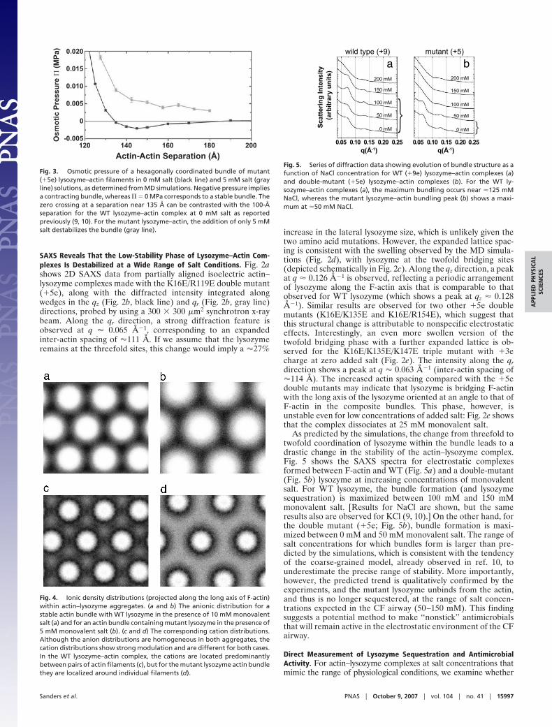
SAXS Reveals That the Low-Stability Phase of Lysozyme–Actin Com-plexes Is Destabilized at a Wide Range of Salt Conditions. Fig. 2ashows 2D SAXS data from partially aligned isoelectric actin–lysozyme complexes made with the K16E/R119E double mutant(�5e), along with the diffracted intensity integrated alongwedges in the qz (Fig. 2b, black line) and qr (Fig. 2b, gray line)directions, probed by using a 300 � 300 �m2 synchrotron x-raybeam. Along the qr direction, a strong diffraction feature isobserved at q � 0.065 Å�1, corresponding to an expandedinter-actin spacing of �111 Å. If we assume that the lysozymeremains at the threefold sites, this change would imply a �27%
increase in the lateral lysozyme size, which is unlikely given thetwo amino acid mutations. However, the expanded lattice spac-ing is consistent with the swelling observed by the MD simula-tions (Fig. 2d), with lysozyme at the twofold bridging sites(depicted schematically in Fig. 2c). Along the qz direction, a peakat q � 0.126 Å�1 is observed, reflecting a periodic arrangementof lysozyme along the F-actin axis that is comparable to thatobserved for WT lysozyme (which shows a peak at qz � 0.128Å�1). Similar results are observed for two other �5e doublemutants (K16E/K135E and K16E/R154E), which suggest thatthis structural change is attributable to nonspecific electrostaticeffects. Interestingly, an even more swollen version of thetwofold bridging phase with a further expanded lattice is ob-served for the K16E/K135E/K147E triple mutant with �3echarge at zero added salt (Fig. 2e). The intensity along the qrdirection shows a peak at q � 0.063 Å�1 (inter-actin spacing of�114 Å). The increased actin spacing compared with the �5edouble mutants may indicate that lysozyme is bridging F-actinwith the long axis of the lysozyme oriented at an angle to that ofF-actin in the composite bundles. This phase, however, isunstable even for low concentrations of added salt: Fig. 2e showsthat the complex dissociates at 25 mM monovalent salt.
As predicted by the simulations, the change from threefold totwofold coordination of lysozyme within the bundle leads to adrastic change in the stability of the actin–lysozyme complex.Fig. 5 shows the SAXS spectra for electrostatic complexesformed between F-actin and WT (Fig. 5a) and a double-mutant(Fig. 5b) lysozyme at increasing concentrations of monovalentsalt. For WT lysozyme, the bundle formation (and lysozymesequestration) is maximized between 100 mM and 150 mMmonovalent salt. [Results for NaCl are shown, but the sameresults also are observed for KCl (9, 10).] On the other hand, forthe double mutant (�5e; Fig. 5b), bundle formation is maxi-mized between 0 mM and 50 mM monovalent salt. The range ofsalt concentrations for which bundles form is larger than pre-dicted by the simulations, which is consistent with the tendencyof the coarse-grained model, already observed in ref. 10, tounderestimate the precise range of stability. More importantly,however, the predicted trend is qualitatively confirmed by theexperiments, and the mutant lysozyme unbinds from the actin,and thus is no longer sequestered, at the range of salt concen-trations expected in the CF airway (50–150 mM). This findingsuggests a potential method to make ‘‘nonstick’’ antimicrobialsthat will remain active in the electrostatic environment of the CFairway.
Direct Measurement of Lysozyme Sequestration and AntimicrobialActivity. For actin–lysozyme complexes at salt concentrations thatmimic the range of physiological conditions, we examine whether
erusser
P cito
msO
)aP
M(
-0.005
0
0.005
0.010
0.015
0.020
Actin-Actin Separation (Å)120 140 160 180 200
Fig. 3. Osmotic pressure of a hexagonally coordinated bundle of mutant(�5e) lysozyme–actin filaments in 0 mM salt (black line) and 5 mM salt (grayline) solutions, as determined from MD simulations. Negative pressure impliesa contracting bundle, whereas � � 0 MPa corresponds to a stable bundle. Thezero crossing at a separation near 135 Å can be contrasted with the 100-Åseparation for the WT lysozyme–actin complex at 0 mM salt as reportedpreviously (9, 10). For the mutant lysozyme–actin, the addition of only 5 mMsalt destabilizes the bundle (gray line).
Fig. 4. Ionic density distributions (projected along the long axis of F-actin)within actin–lysozyme aggregates. (a and b) The anionic distribution for astable actin bundle with WT lysozyme in the presence of 10 mM monovalentsalt (a) and for an actin bundle containing mutant lysozyme in the presence of5 mM monovalent salt (b). (c and d) The corresponding cation distributions.Although the anion distributions are homogeneous in both aggregates, thecation distributions show strong modulation and are different for both cases.In the WT lysozyme–actin complex, the cations are located predominantlybetween pairs of actin filaments (c), but for the mutant lysozyme actin bundlethey are localized around individual filaments (d).
}
a
tisnet
nI g
nirettacS
y )stin
u yrartibra(
0.05 0.10 0.15 0.20 0.25q(Å-1)
wild type (+9)
200 mM
150 mM
100 mM
50 mM
0 mM }0.05 0.10 0.15 0.20 0.25
q(Å-1)
mutant (+5)
200 mM
150 mM
100 mM
50 mM
0 mM
b
Fig. 5. Series of diffraction data showing evolution of bundle structure as afunction of NaCl concentration for WT (�9e) lysozyme–actin complexes (a)and double-mutant (�5e) lysozyme–actin complexes (b). For the WT ly-sozyme–actin complexes (a), the maximum bundling occurs near �125 mMNaCl, whereas the mutant lysozyme–actin bundling peak (b) shows a maxi-mum at �50 mM NaCl.
Sanders et al. PNAS � October 9, 2007 � vol. 104 � no. 41 � 15997
APP
LIED
PHYS
ICA
LSC
IEN
CES

the computational and SAXS findings are reflected by the degreeof lysozyme sequestration. Microequilibrium dialysis experimentsare used to measure the extent to which WT and mutant lysozymeremain sequestered by F-actin (Fig. 6 a–c). As a baseline measure-ment, WT and mutant lysozyme are dialyzed for 120 h againstbuffer in the absence of F-actin (Fig. 6a). In this case, the relativeconcentration difference �Clysozyme � (Cexperimental � Ctheoretical)/Ctheoretical is very small, indicating that both WT and mutantlysozyme are nearly fully dialyzed. In contrast, if lysozyme isdialyzed from actin–lysozyme complexes (Fig. 6b), �Clysozyme showsa large decrease in the amount of dialyzed WT lysozyme, reflectingthe strong sequestration of WT lysozyme by the actin filaments. Onthe other hand, the much weaker binding of mutant lysozyme isconfirmed by the weak increase of �Clysozyme. In Fig. 6c, wenormalize the dialysis results obtained in the presence of actin (Fig.6b) by those for lysozyme only (Fig. 6a), which shows that for WTlysozyme the degree of sequestration as measured by the deviationfrom a homogeneous distribution is �17 times larger than that forthe mutant lysozyme.
The reduced adhesion of genetically engineered lysozyme toactin results in an increased availability of lysozyme. To assesswhether this can translate into increased antimicrobial activity,it is necessary to evaluate whether the structural modificationsof charge-reduced mutant lysozyme have not ablated its anti-bacterial activity. We perform bacterial killing assays on thePAO1 strain of P. aeruginosa, a Gram-negative bacterium com-
monly found in CF airway infections. Fig. 6d shows the averagedresults from a series of 20 trials in which bacterial survival ismonitored as a function of increasing protein concentration. Thekilling efficiency for the �5e mutant charge-reduced lysozyme(Fig. 6d, gray line) is comparable to, albeit slightly lower than,that of the WT �9e lysozyme (Fig. 6d, black line). Moreover, ata concentration of 100 �g/ml, the �5e mutant kills nearly all ofthe bacteria. These results suggest that it is indeed possible tooptimize the charge distribution in antimicrobials to simulta-neously minimize adventitious binding to inflammatory poly-mers and maintain antimicrobial activity.
Materials and MethodsThe methodology of the MD and grand-canonical simulationshas been provided in detail in ref. 10. The mutant lysozyme wasmodeled identically to the WT lysozyme (9, 10) but with a �2.5echarge on each of the two subunits.
Bacteriophage T4 pseudo WT lysozyme carried a charge of�9e at neutral pH and had dimensions of 30 � 30 � 50 Å3 anda molecular weight of 18,700 (22). Expression and purificationprocedures of the WT lysozyme and its charge-reduced mutantswere performed as reported (22). The charge mutants includedthree double mutants (K16E/R119E, K16E/R135E, and K16E/R154E) and one triple mutant (K16E/K135E/K147E); K, E, andR denote the amino acids lysine, glutamic acid, and arginine,respectively, and, for example, R154E denotes a mutation wherethe arginine at position 154 was replaced with glutamic acid.
Monomeric actin (G-actin) (Mr 43,000) was prepared from alyophilized powder of rabbit skeletal muscle (Cytoskeleton,Denver, CO) as previously reported (9, 10).
The F-actin–lysozyme isoelectric point was at a molar ratio of1.85:1 for F-actin:WT and 1:1 for F-actin:double mutant. Typicalfinal concentrations were: F-actin, 5.6 mg/ml; WT lysozyme, 3.0mg/ml; and double mutant, 5.4 mg/ml. These values are close tothose found in the airway. A series of samples was prepared withthe final monovalent salt concentration ranging from 0 mM to200 mM. Lysozyme–actin complexes were sealed in 1.5-mmquartz capillaries (Hilgenberg, Malsfeld, Germany) and mixedthoroughly by centrifugation. The approximate sample volumein the capillary was 30 �l.
SAXS measurements were performed with beamline 4–2 atthe Stanford Synchrotron Radiation Laboratory (Palo Alto, CA)and beamline 12-ID-C at the Advanced Photon Source (ArgonneNational Laboratory, Argonne, IL). For the Stanford Synchro-tron Radiation Laboratory experiments (incident x-ray wave-length � � 1.3806 Å), the scattered radiation was collected byusing a MAR Research (Evanston, IL) charge-coupled device(CCD) camera (pixel size � 79 � 79 �m2). For the AdvancedPhoton Source experiments (incident x-ray wavelength � � 1.033Å), the scattered x-rays were collected by using a MAR Research2D mosaic CCD detector (pixel size � 79 � 79 �m2). The 2DSAXS data from both setups were checked for mutual consis-tency. Additional details have been reported elsewhere (9, 10).
In collaboration with Donald Davidson (Carle Clinic Associa-tion, Urbana, IL) and in accordance with institutional reviewboard-approved protocols at both Carle Clinic and at the Universityof Illinois at Urbana–Champaign, we collected mucus from CFpatients who had not been treated with DNase (Pulmozyme;Genentech, Inc., South San Francisco, CA) for study by synchrotronx-ray microdiffraction. Patients voluntarily expectorated �5–10 mlof sputum during respiratory therapy. After collection, the sputumsamples were rapidly frozen and stored at �80°C until use. Frozensamples were ultramicrotomed and sealed between kapton poly-amide film (Dupont, Wilmington, DE).
Microdiffraction experiments of CF sputum samples were per-formed at beamline 2-ID-D at the Advanced Photon Source byusing a beam size of 0.5 � 0.5 �m2 focused with a Fresnel zone platein conjunction with an order-sorting aperture. The sample-to-
cre
Pt
ne
la
vivr
uS
oc f
o % ,
ufc(
lort
n)
d100
60
0
80
40
20
20 40 60 80 1000Concentration (µg/ml)
wild-type mutant
aC
∆l
os
ym
yze
0.35
0.25
0.15
0.05
0
0.10
0.20
0.30
lysozyme only
mutantwild-type
bactin-lysozyme
0
3.0
1.5
1.0
2.0
2.5
0.5
wild-type mutant
cnormalized
C∆
dezil
amr
oN
Fig. 6. Microequilibrium dialysis experiments indicate that charge-reducedlysozyme is drastically less susceptible to actin-induced sequestration than WTlysozyme is, which is measured by �Clysozyme � (Cexperimental � Ctheoretical)/Ctheoretical, where Ctheoretical is the expected concentration of a fully dialyzedsolution (0.36 mg/ml) or 1⁄2 of the initial lysozyme concentration (0.72 mg/ml).�Clysozyme is shown for dialysis of 0.72 mg/ml lysozyme solutions in the absenceof actin (a) and in the presence of 1.4 mg/ml F-actin (b). (c) Results for theactin–lysozyme system normalized by those for the lysozyme-only system. Thisnormalization corrects for potential differential adhesion of the differentlysozyme to the dialysis membrane and shows a 17-fold decrease in seques-tration for the mutant (light gray) relative to the WT (dark gray). (d) Resultsfrom antibacterial killing assays showing the percentage survival of PAO1bacteria in the presence of increasing WT lysozyme (black line) and double-mutant lysozyme (gray line) concentrations. These results indicate that, de-spite structural changes, the double mutant retains most of the WT antibac-terial activity.
15998 � www.pnas.org�cgi�doi�10.1073�pnas.0705898104 Sanders et al.
detector distance was �420 mm corresponding to q � 0.3 �1. Gridscans were taken about a center point with a 100-�m step size inboth the horizontal and vertical directions.
Equilibrium dialysis was performed in microdialyzers (Har-vard Apparatus, Boston, MA) containing two 100-�l chambersseparated by a 50,000-Da cutoff cellulose acetate membrane.The ‘‘sample’’ chamber contained protein solutions in 100 mMNaCl/2 mM Tris buffer; the ‘‘assay’’ chamber contained only the100 mM NaCl/2 mM Tris buffer. The protein solutions wereeither actin–lysozyme complexes (1.4 mg/ml actin�0.72 mg/mllysozyme) or lysozyme-only controls (0.72 mg/ml). Protein so-lutions were allowed to dialyze against the NaCl/Tris buffer for6 days to ensure complete dialysis. Protein concentrations weremeasured by using UV-visible spectroscopy at 280 nm.
The bactericidal activities of WT and mutant lysozyme weretested by using a microdilution killing assay on P. aeruginosastrain PAO1. Bacteria were grown in cation-adjusted Mueller–Hinton broth from an overnight culture (1:100 dilution) tomid-log phase at 37°C, harvested by centrifuging at 9,250 � g for10 min, and resuspended in PBS (10 mM Na2HPO4/100 mMNaCl, pH 7.4). Bacteria were diluted so that 105 cfu were presentin a final volume of 150 �l of the assay buffer, PBS. Bacteria wereincubated with lysozyme in sterile 96-well f lat-bottom polypro-pylene dishes while shaking for 3 h at 37°C. After the incubation
period, bacteria were serially diluted, dropped on Mueller–Hinton agar plates, and incubated 18 h at 37°C, and colony-forming units were determined by using plate-counting methods.
We thank Dr. Brian W. Matthews (University of Oregon, Eugene, OR)for providing the T4 lysozyme plasmids and Dr. John K. Sheehan and Dr.C. William Davis (University of North Carolina, Chapel Hill, NC) forproviding human mucins. We also gratefully acknowledge Dr. MichaelJ. Welsh and Dr. Pradeep K. Singh for insightful discussions and Dr.Zhonghou Cai, Dr. Barry Lai, Dr. Donald Davidson, Janice Douglas,Blaise Bowles, Leslie Gay, Scott C. Slimmer, John C. Butler, Thomas E.Angelini, Jae-Wook Lee, and Evelyn Huang for technical assistance.This material is based on work supported by the National Institutes ofHealth under Grant 1R21DK6843-01 (to G.C.L.W.), the Cystic FibrosisFoundation (G.C.L.W.), and the National Science Foundation underGrants DMR-0346914 (to E.L.), DMR-0409769 (to G.C.L.W.), andCTS-0120978 (to E.L. and G.C.L.W.) via the WaterCAMPWS Scienceand Technology Center. Portions of this research were carried out at theStanford Synchrotron Radiation Laboratory and at the Advanced Pho-ton Source. The Stanford Synchrotron Radiation Laboratory StructuralMolecular Biology Program is supported by the U.S. Department ofEnergy, Office of Biological and Environmental Research, and by theNational Institutes of Health, National Center for Research Resources,Biomedical Technology Program. Use of the Advanced Photon Sourceis supported by the U.S. Department of Energy, Office of Basic EnergySciences, under Contract DE-AC02-06CH11357.
1. Wong GCL (2006) Curr Opin Col Int Sci 11:310–315.2. Grosberg AY, Nguyen TT, Shklovskii BI (2002) Rev Mod Phys 74:329–345.3. Levin Y (2002) Rep Prog Phys 65:1577–1632.4. Bloomfield VA (1996) Curr Opin Struct Biol 6:334–341.5. Gelbart WM, Bruinsma RF, Pincus PA, Parsegian VA (2000) Phys Today
53:38–44.6. Olvera de la Cruz M, Belloni L, Delsanti M, Dalbiez JP, Spalla O, Drifford M
(1995) J Chem Phys 103:5781–5791.7. Angelini TE, Liang H, Wriggers W, Wong GCL (2003) Proc Natl Acad Sci USA
100:8634–8637.8. Angelini TE, Golestanian R, Coridan RH, Butler JC, Beraud A, Krisch M, Sinn
H, Schweizer KS, Wong GCL (2006) Proc Natl Acad Sci USA 103:7962–7967.9. Sanders LK, Guaqueta C, Angelini TE, Lee J-W, Slimmer SC, Luijten E, Wong
GCL (2005) Phys Rev Lett 95:108302.10. Guaqueta C, Sanders LK, Wong GCL, Luijten E (2006) Biophys J 90:4630–
4638.11. Welsh MJ, Smith AE (1995) Sci Am 273(6):52–59.12. Vasconcellos CA, Allen PG, Wohl ME, Drazen JM, Janmey PA, Stossel TP
(1994) Science 263:969–971.13. Brandt T, Breitenstein S, von der Hardt H, Tummler B (1995) Thorax
50:880–882.
14. Shak S, Capon DJ, Hellmiss R, Marsters SA, Baker CL (1990) Proc Natl AcadSci USA 87:9188–9192.
15. Sheils CA, Kas J, Travassos W, Allen PG, Janmey PA, Wohl ME, Stossel TP(1996) Am J Pathol 148:919–927.
16. Felgentreff K, Beisswenger C, Griese M, Gulder T, Bringmann G, Bals R(2006) Peptides 27:3100–3106.
17. Tang JX, Wen Q, Bennett A, Kim B, Sheils CA, Bucki R, Janmey PA (2005)Am J Physiol 289:L599–L605.
18. Travis SM, Singh PK, Welsh MJ (2001) Curr Opin Immunol 13:89–95.19. Brogan TD, Ryley HC, Neale L, Yassa J (1975) Thorax 30:72–79.20. Weiner DJ, Bucki R, Janmey PA (2003) Am J Respir Cell Mol Biol 28:738–745.21. Bucki R, Byfield FJ, Janmey PA (2007) Eur Respir J 29:624–632.22. Sun DP, Soderlind E, Baase WA, Wozniak JA, Sauer U, Matthews BW (1991)
J Mol Biol 221:873–887.23. Matthews BW, Remington SJ (1974) Proc Natl Acad Sci USA 71:4178–4182.24. Broughton-Head VJ, Smith JR, Shur J, Shute JK (September 9, 2006) Pulm
Pharmacol Ther, 10.1016/j.pupt.2006.08.008.25. Angelini TE, Liang H, Wriggers W, Wong GCL (2005) Eur Phys J B
16:389–400.26. Purdy KR, Bartles JR, Wong GCL (2007) Phys Rev Lett 98:058105.
Sanders et al. PNAS � October 9, 2007 � vol. 104 � no. 41 � 15999
APP
LIED
PHYS
ICA
LSC
IEN
CES