contribution of active site dynamics to enzyme catalysis: study on a
TRANSCRIPT

University of IowaIowa Research Online
Theses and Dissertations
2012
Contribution of active site dynamics to enzymecatalysis: study on a series of mutants ofdihydrofolate reductaseVanja StojkovicUniversity of Iowa
Copyright 2012 Vanja Stojkovic
This dissertation is available at Iowa Research Online: http://ir.uiowa.edu/etd/5062
Follow this and additional works at: http://ir.uiowa.edu/etd
Part of the Chemistry Commons
Recommended CitationStojkovic, Vanja. "Contribution of active site dynamics to enzyme catalysis: study on a series of mutants of dihydrofolate reductase."PhD (Doctor of Philosophy) thesis, University of Iowa, 2012.http://ir.uiowa.edu/etd/5062.

CONTRIBUTION OF ACTIVE SITE DYNAMICS TO ENZYME CATALYSIS:
STUDY ON A SERIES OF MUTANTS OF DIHYDROFOLATE REDUCTASE
by
Vanja Stojković
An Abstract
Of a thesis submitted in partial fulfillment of the requirements for the Doctor of Philosophy degree
in Chemistry in the Graduate College of The University of Iowa
December 2012
Thesis Supervisor: Professor Amnon Kohen

1
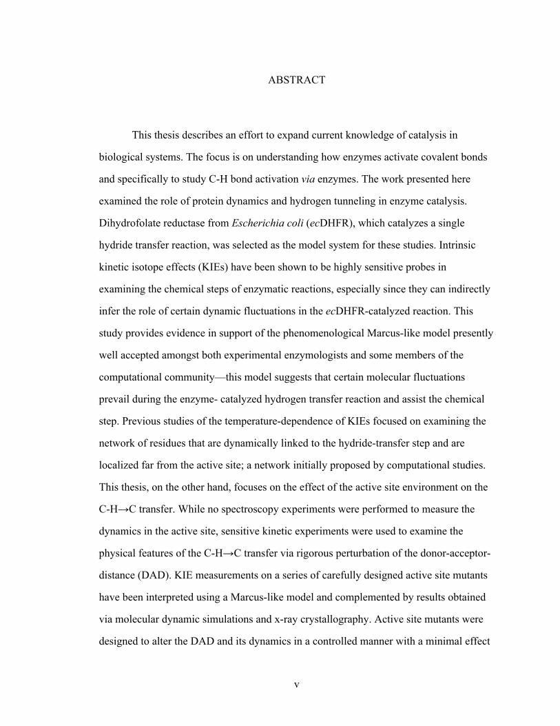
ABSTRACT
This thesis describes an effort to expand current knowledge of catalysis in
biological systems. The focus is on understanding how enzymes activate covalent bonds
and specifically to study C-H bond activation via enzymes. The work presented here
examined the role of protein dynamics and hydrogen tunneling in enzyme catalysis.
Dihydrofolate reductase from Escherichia coli (ecDHFR), which catalyzes a single
hydride transfer reaction, was selected as the model system for these studies. Intrinsic
kinetic isotope effects (KIEs) have been shown to be highly sensitive probes in
examining the chemical steps of enzymatic reactions, especially since they can indirectly
infer the role of certain dynamic fluctuations in the ecDHFR-catalyzed reaction. This
study provides evidence in support of the phenomenological Marcus-like model presently
well accepted amongst both experimental enzymologists and some members of the
computational community—this model suggests that certain molecular fluctuations
prevail during the enzyme- catalyzed hydrogen transfer reaction and assist the chemical
step. Previous studies of the temperature-dependence of KIEs focused on examining the
network of residues that are dynamically linked to the hydride-transfer step and are
localized far from the active site; a network initially proposed by computational studies.
This thesis, on the other hand, focuses on the effect of the active site environment on the
C-H→C transfer. While no spectroscopy experiments were performed to measure the
dynamics in the active site, sensitive kinetic experiments were used to examine the
physical features of the C-H→C transfer via rigorous perturbation of the donor-acceptor-
distance (DAD). KIE measurements on a series of carefully designed active site mutants
have been interpreted using a Marcus-like model and complemented by results obtained
via molecular dynamic simulations and x-ray crystallography. Active site mutants were
designed to alter the DAD and its dynamics in a controlled manner with a minimal effect

2
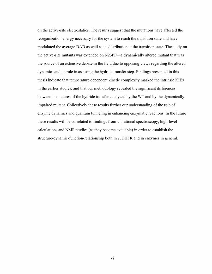
on the active-site electrostatics. The results suggest that the mutations have affected the
reorganization energy necessary for the system to reach the transition state and have
modulated the average DAD as well as its distribution at the transition state. The study on
the active-site mutants was extended on N23PP—a dynamically altered mutant that was
the source of an extensive debate in the field due to opposing views regarding the altered
dynamics and its role in assisting the hydride transfer step. Findings presented in this
thesis indicate that temperature dependent kinetic complexity masked the intrinsic KIEs
in the earlier studies, and that our methodology revealed the significant differences
between the natures of the hydride transfer catalyzed by the WT and by the dynamically
impaired mutant. Collectively these results further our understanding of the role of
enzyme dynamics and quantum tunneling in enhancing enzymatic reactions. In the future
these results will be correlated to findings from vibrational spectroscopy, high-level
calculations and NMR studies (as they become available) in order to establish the
structure-dynamic-function-relationship both in ecDHFR and in enzymes in general.
Abstract Approved:________________________________
Thesis Supervisor
________________________________ Title and Department
________________________________ Date

1
CONTRIBUTION OF ACTIVE SITE DYNAMICS TO ENZYME CATALYSIS:
STUDY ON A SERIES OF MUTANTS OF DIHYDROFOLATE REDUCTASE
by
Vanja Stojković
A thesis submitted in partial fulfillment of the requirements for the Doctor of
Philosophy degree in Chemistry in the Graduate College of
The University of Iowa
December 2012
Thesis Supervisor: Professor Amnon Kohen

2
Copyright by
VANJA STOJKOVIĆ
2012
All Rights Reserved

Graduate College The University of Iowa
Iowa City, Iowa
CERTIFICATE OF APPROVAL
_______________________
PH.D. THESIS
_______________
This is to certify that the Ph.D. thesis of
Vanja Stojković
has been approved by the Examining Committee for the thesis requirement for the Doctor of Philosophy degree in Chemistry at the December 2012 graduation.
Thesis Committee: ___________________________________ Amnon Kohen, Thesis Supervisor
___________________________________ Maxwell Geng
___________________________________ James Gloer
___________________________________ Ernesto Fuentes
___________________________________ Daniel Quinn

ii
2
To my parents

iii
3
Kod unutarnjih borba koje čovjek vodi sa samim sobom i sa nepoznatim silama u sebi, važi više nego igdje pravilo: ne predaj se nikad! — Ni predaje, ni ustupanja! A prije
svega, što kažu u Bosni: ne veži tugu za srce!
Ivo Andrić “Znakovi pored puta”

iv
4
ACKNOWLEDGMENTS
I am sincerely grateful and indebt to all the scientists and scholars I encountered
during my PhD studies, especially: Dr. Sonya Franklin, Dr. Samantha Nolting and Dr.
Jennifer Wong-Deyrup from the Franklin research group; Dr. Arundhuti Sen, Eric Koehn,
Daniel Roston, Zhen Wang, Thelma Abeysinghe from Kohen research group at
University of Iowa. Many thanks to Dr. Lokesh Gahkar from the protein crystallography
facility at the University of Iowa for all the assistance and support in my protein
crystallography endeavors. I would also like to thank all the present and past members of
the Kohen group for providing a great working environment. Thanks go also to all the
staff at Department of Chemistry and Department of Biochemistry who facilitated this
research.
I am indebt to my advisor Dr. Amnon Kohen for the mentorship, guidance and
support through out the years. Amnon, thank you for both the criticism and
encouragement, for always keeping me focused on the big picture and for believing in
me.
Big thanks to my wonderful parents, Stojanka and Bratislav, who even though
being physically far away, were always there to give me support and strength to keep on
going. No words can describe my gratitude. Many thanks to my sister Bojana, Ian and
Lana, and many friends who were my family away from home: my co-housemates Nina,
Ana, Tomislav and Juan, and many others both near and far, especially Jelena, Frane,
Modei, and my Gaia’s girls. I would not be able to do it without any of you. Thank you!
v
5
ABSTRACT
This thesis describes an effort to expand current knowledge of catalysis in
biological systems. The focus is on understanding how enzymes activate covalent bonds
and specifically to study C-H bond activation via enzymes. The work presented here
examined the role of protein dynamics and hydrogen tunneling in enzyme catalysis.
Dihydrofolate reductase from Escherichia coli (ecDHFR), which catalyzes a single
hydride transfer reaction, was selected as the model system for these studies. Intrinsic
kinetic isotope effects (KIEs) have been shown to be highly sensitive probes in
examining the chemical steps of enzymatic reactions, especially since they can indirectly
infer the role of certain dynamic fluctuations in the ecDHFR-catalyzed reaction. This
study provides evidence in support of the phenomenological Marcus-like model presently
well accepted amongst both experimental enzymologists and some members of the
computational community—this model suggests that certain molecular fluctuations
prevail during the enzyme- catalyzed hydrogen transfer reaction and assist the chemical
step. Previous studies of the temperature-dependence of KIEs focused on examining the
network of residues that are dynamically linked to the hydride-transfer step and are
localized far from the active site; a network initially proposed by computational studies.
This thesis, on the other hand, focuses on the effect of the active site environment on the
C-H→C transfer. While no spectroscopy experiments were performed to measure the
dynamics in the active site, sensitive kinetic experiments were used to examine the
physical features of the C-H→C transfer via rigorous perturbation of the donor-acceptor-
distance (DAD). KIE measurements on a series of carefully designed active site mutants
have been interpreted using a Marcus-like model and complemented by results obtained
via molecular dynamic simulations and x-ray crystallography. Active site mutants were
designed to alter the DAD and its dynamics in a controlled manner with a minimal effect
vi
6
on the active-site electrostatics. The results suggest that the mutations have affected the
reorganization energy necessary for the system to reach the transition state and have
modulated the average DAD as well as its distribution at the transition state. The study on
the active-site mutants was extended on N23PP—a dynamically altered mutant that was
the source of an extensive debate in the field due to opposing views regarding the altered
dynamics and its role in assisting the hydride transfer step. Findings presented in this
thesis indicate that temperature dependent kinetic complexity masked the intrinsic KIEs
in the earlier studies, and that our methodology revealed the significant differences
between the natures of the hydride transfer catalyzed by the WT and by the dynamically
impaired mutant. Collectively these results further our understanding of the role of
enzyme dynamics and quantum tunneling in enhancing enzymatic reactions. In the future
these results will be correlated to findings from vibrational spectroscopy, high-level
calculations and NMR studies (as they become available) in order to establish the
structure-dynamic-function-relationship both in ecDHFR and in enzymes in general.

vii
7
TABLE OF CONTENTS
LIST OF TABLES ...............................................................................................................x
LIST OF FIGURES ........................................................................................................... xi
CHAPTER 1. INTRODUCTION .......................................................................................1! 1.1 Scope of Research .......................................................................................1!1.2 Thesis overview ..........................................................................................2!1.3 Background .................................................................................................3!
1.3.1 Dihydrofolate reductase (DHFR) .....................................................3!1.3.2 Kinetic isotope effect (KIE) .............................................................6!1.3.3 Swain-Schaad exponent ....................................................................7!1.3.4 Kinetic complexity ...........................................................................9!1.3.5 Temperature dependence of KIEs as a probe for enzymatic quantum mechanical tunneling: semi-classical models with tunneling correction .................................................................................10!1.3.6 Temperature dependence of KIEs as a probe for dynamics: Marcus-like models (full tunneling model) .............................................13!
CHAPTER 2. SYNTHESIS OF RADIOLABELED NICOTINAMIDE COFACTORS FROM LABELED PYRIDINES: VERSATILE PROBES FOR ENZYME KINETICS ............................................................24! 2.1 Introduction ...............................................................................................24!2.2 Materials and Methods .............................................................................28!
2.2.1 Materials .........................................................................................28!2.2.2 Analytical methods .........................................................................29!2.2.3 Preparation of porcine brain NADase ............................................29!2.2.4 Synthesis of [carbonyl-14C]-NADPH .............................................30!2.2.5 Synthesis of 4R-[carbonyl-14C,4-2H]-NADPH from [carbonyl-14C]-NADP+ ............................................................................32!2.2.6 Synthesis of [carbonyl-14C,4-2H2]-NADPH from [carbonyl-14C]-NADP+ .............................................................................................32!2.2.7 Determination of 1° H/T and D/T KIEs for EcDHFR using synthesized [carbonyl-14C] derivatives ....................................................33!2.2.8 Measurement of γ - 2° 14C KIE due to 14C isotopic labeling at the amide carbonyl of NADPH ...........................................................34!
2.3 Results and Discussion .............................................................................34!2.3.1 Synthesis of [carbonyl-14C]-NADPH, 4R- [carbonyl-14C, 4-2H]-NADPH and [carbonyl-14C, 4-2H2]-NADPH ...................................34!2.3.2 14C-labeling at the nicotinamide carbonyl carbon of NADPH does not result in a significant γ -2° 14C KIE ..........................................37!2.3.3 Testing the viability of synthesized materials using KIEs: Comparison of intrinsic KIEs measured using [carbonyl-14C]-NADPH vs. [Ad-14C]-NADPH ..............................................................37!2.3.4 Comparison of cost, effort, and yield of [Ad-14C]-NADPH 95 vs. [carbonyl-14C]-NADPH syntheses. ....................................................38!
2.4 Conclusion ................................................................................................38!

viii
8
CHAPTER 3. EFFECTS OF THE DONOR ACCEPTOR DISTANCE AND DYNAMICS ON HYDRIDE TUNNELING IN THE DIHYDROFOLATE REDUCTASE CATALYZED REACTION ................45! 3.1 Introduction ...............................................................................................45!3.2 Materials and Methods .............................................................................51!
3.2.1 Synthesis of labeled cofactors for 1˚KIEs ......................................51!3.2.2 Construction of expression vectors ................................................51!3.2.3 Expression and purification of I14V DHFR, I14A DHFR and I14G DHFR .............................................................................................52!3.2.4 Competitive and intrinsic primary kinetic isotope effect ...............52!3.2.5 MD simulation ................................................................................54!
3.2 Presteady-state kinetics .............................................................................56!3.3 Results and Discussion .............................................................................57!
3.3.1 Competitive KIEs and Their Temperature Dependence ................57!3.3.2 MD Simulations on mutants of DHFR ...........................................60!
3.4 Conclusions ...............................................................................................64!Supplementary Information ............................................................................72!
CHAPTER 4. STRUCTURAL STUDY ON A SERIES OF DIHYDROFOLATE REDUCTASE MUTANTS ............................................................................76! 4.1 Introduction ...............................................................................................76!4.2 Materials and methods ..............................................................................79!
4.2.1 Preparation of I14V, I14A and I14G DHFR ternary complexes for crystallization ...................................................................79!4.2.2 Crystallization conditions ...............................................................80!4.2.3 Data collection and structure determination ...................................81!4.2.4 Calculation of the normalized B-factors .........................................82!
4.3 Results and Discussion .............................................................................82!4.3.1 Crystal structures of the I14A and I14V ecDHFR. ........................82!4.3.2 Crystal structure of the I14G ecDHFR. ..........................................87!
4.4 Conclusion ................................................................................................89!CHAPTER 5. LOOKING ON THE OTHER SIDE: ROLE OF THE RESIDUE F31
IN THE DHFR CATALYZED HYDRIDE-TRANSFER ............................101! 5.1 Introduction .............................................................................................101!5.2 Materials and methods ............................................................................104!
5.2.1 Construction of expression vector. ...............................................104!5.2.2 Preparation of F31V DHFR ternary complexes for crystallization. .......................................................................................105!5.2.3 Crystallization conditions. ............................................................105!5.2.4 Data collection and structure determination. ................................106!5.2.5 Synthesis of Labeled Cofactors for 1˚KIEs. .................................107!5.2.6 Kinetic isotope effect measurements and determination of the intrinsic KIEs. ........................................................................................108!5.2.7 Presteady-state kinetics. ...............................................................109!5.2.8 Determination of temperature stability using dynamic light scattering (DLS) ....................................................................................110!5.2.9 Numerical modeling .....................................................................111!5.2.10 MD simulations. .........................................................................111!
5.3 Results and Discussion ...........................................................................113!
ix
9
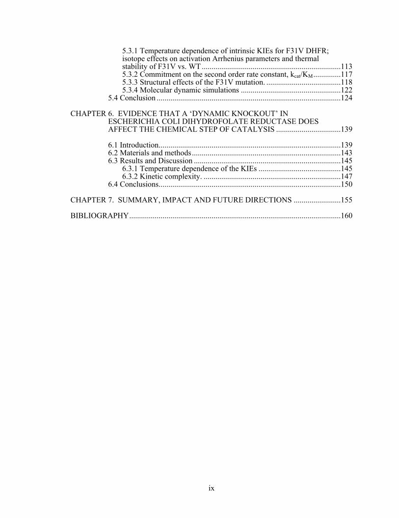
5.3.1 Temperature dependence of intrinsic KIEs for F31V DHFR; isotope effects on activation Arrhenius parameters and thermal stability of F31V vs. WT .......................................................................113!5.3.2 Commitment on the second order rate constant, kcat/KM ..............117!5.3.3 Structural effects of the F31V mutation. ......................................118!5.3.4 Molecular dynamic simulations ...................................................122!
5.4 Conclusion ..............................................................................................124!CHAPTER 6. EVIDENCE THAT A ‘DYNAMIC KNOCKOUT’ IN
ESCHERICHIA COLI DIHYDROFOLATE REDUCTASE DOES AFFECT THE CHEMICAL STEP OF CATALYSIS .................................139! 6.1 Introduction .............................................................................................139!6.2 Materials and methods ............................................................................143!6.3 Results and Discussion ...........................................................................145!
6.3.1 Temperature dependence of the KIEs ..........................................145!6.3.2 Kinetic complexity. ......................................................................147!
6.4 Conclusions .............................................................................................150!CHAPTER 7. SUMMARY, IMPACT AND FUTURE DIRECTIONS ........................155!BIBLIOGRAPHY ............................................................................................................160!!
x
10
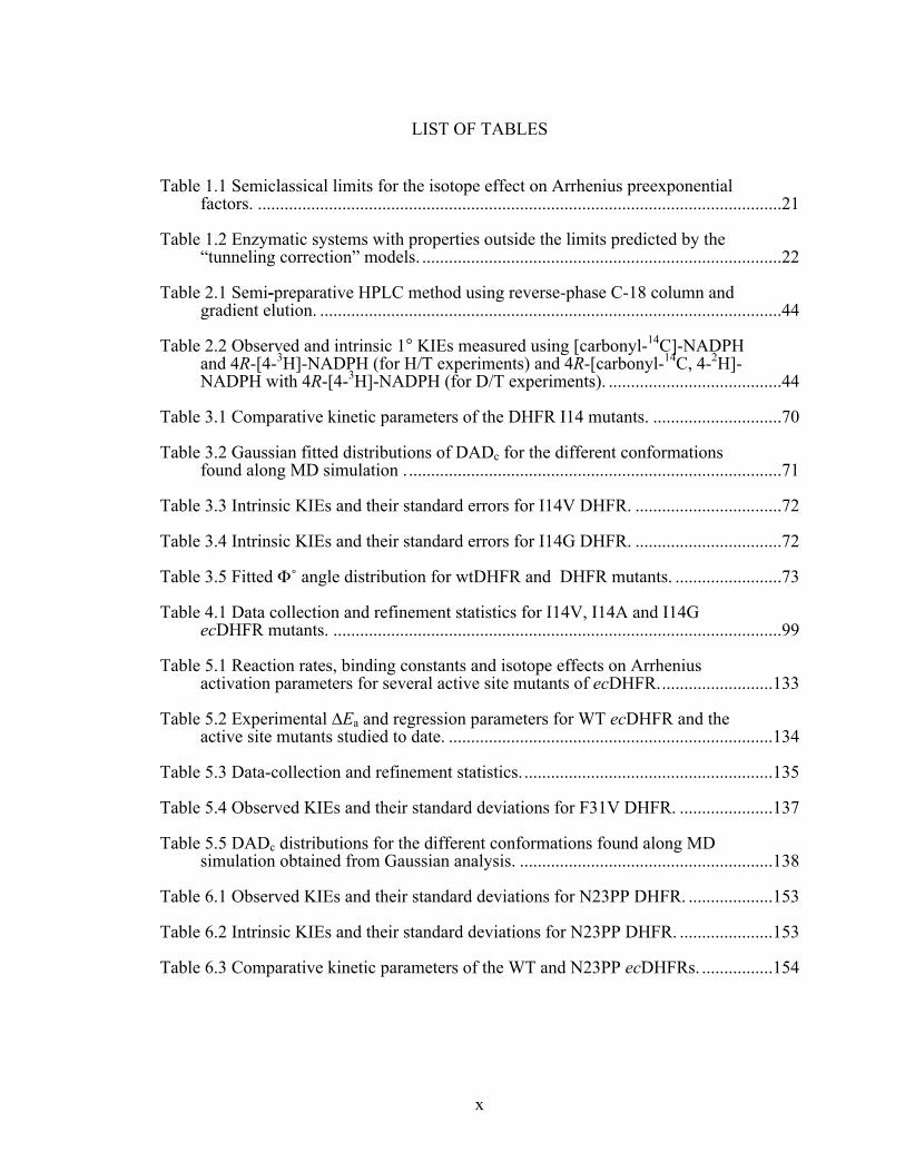
LIST OF TABLES
Table 1.1 Semiclassical limits for the isotope effect on Arrhenius preexponential factors. ......................................................................................................................21!
Table 1.2 Enzymatic systems with properties outside the limits predicted by the “tunneling correction” models. .................................................................................22!
Table 2.1 Semi-preparative HPLC method using reverse-phase C-18 column and gradient elution. ........................................................................................................44!
Table 2.2 Observed and intrinsic 1° KIEs measured using [carbonyl-14C]-NADPH and 4R-[4-3H]-NADPH (for H/T experiments) and 4R-[carbonyl-14C, 4-2H]-NADPH with 4R-[4-3H]-NADPH (for D/T experiments). .......................................44!
Table 3.1 Comparative kinetic parameters of the DHFR I14 mutants. .............................70!Table 3.2 Gaussian fitted distributions of DADc for the different conformations
found along MD simulation . ....................................................................................71!Table 3.3 Intrinsic KIEs and their standard errors for I14V DHFR. .................................72!Table 3.4 Intrinsic KIEs and their standard errors for I14G DHFR. .................................72!Table 3.5 Fitted Φ˚ angle distribution for wtDHFR and DHFR mutants. ........................73!Table 4.1 Data collection and refinement statistics for I14V, I14A and I14G
ecDHFR mutants. .....................................................................................................99!Table 5.1 Reaction rates, binding constants and isotope effects on Arrhenius
activation parameters for several active site mutants of ecDHFR. .........................133!Table 5.2 Experimental ∆Ea and regression parameters for WT ecDHFR and the
active site mutants studied to date. .........................................................................134!Table 5.3 Data-collection and refinement statistics. ........................................................135!Table 5.4 Observed KIEs and their standard deviations for F31V DHFR. .....................137!Table 5.5 DADc distributions for the different conformations found along MD
simulation obtained from Gaussian analysis. .........................................................138!Table 6.1 Observed KIEs and their standard deviations for N23PP DHFR. ...................153!Table 6.2 Intrinsic KIEs and their standard deviations for N23PP DHFR. .....................153!Table 6.3 Comparative kinetic parameters of the WT and N23PP ecDHFRs. ................154!!

xi
11
LIST OF FIGURES
Figure 1.1 (a) Dihydrofolate reductase catalyzed reaction; R= para-aminobenzoyl polyglutamate moiety, R’= 2’-monophosphoadenosine-5’-diphosphoribose. (b) The catalytic cycle of ecDHFR. The five primary intermediates and the pH-independent rate constants at 25˚C are shown; E = ecDHFR, H2F = 7,8-dihydrofolate, H4F = 5,6,7,8-tetrahydrofolate , NADPH = reduced nicotinamide adenine dinucleotide phosphate, NADP+ = oxidized nicotinamide adenine dinucleotide phosphate. .........................................................16!
Figure 1.2 Structure of ecDHFR in a ternary complex with Folate and NADP+ crystallized in P212121space group (PDB ID 1RX2). The active cleft divides the protein into two subdomains: adenosine binding subdomain (residues 38-88) and the loop subdomain (major subdomain). Three flexible loops mentioned in the main text are: M20 (shown in red), F-G (shown in blue) and G-H loop (shown in mangenta). In this particular crystal structure, which is considered to represent the active Michaelis complex, the M20 loop is in the closed conformation. .................................................................................................17!
Figure 1.3 Graphical representation of the semi-classical model indicating that the difference in the energy of activation (∆Ea) for H, D, and T, result from their different zero-point energies (ZPE) in the ground state (GS) and transition state (TS). The GS-ZPE is constituted by all degrees of freedom but mostly by the C-H stretching frequency and the TS-ZPE is constituted by all degrees of freedom orthogonal to the reaction coordinate. . ..................................................18!
Figure 1.4 An example of the ground state tunneling along the reaction coordinate. The picture depicts the probability of a particle to tunnel, where the blue and red lines represent the probability functions for lighter and heavier isotopes, respectively. As noticeable from the figure, lighter isotope has a higher probability of tunneling than the heavier one. ..........................................................18!
Figure 1.5 An Arrhenius plot of a H-transfer applicable for semi-classical models with tunneling correction. Top panel represents the Arrhenius plot of the reaction rates for two isotopes. Bottom panel represents the Arrhenius plot for their KIEs. The KIE on the Arrhenius preexponential factor is an intercept of the tangent to curve at different experimental temperatures. Highlighted regions indicate three distinct possibilities: I) No tunneling contribution; where AL/AH is close to unity and falls in the semi-classical limit; II) moderate tunneling contribution; results in AL/AH below the lower semi-classical limit; III) extensive tunneling contribution; where AL/AH is above the semi-classical limit; system exhibits very large intrinsic KIEs which are temperature independent. ..........................................................................................19!
Figure 1.6 Graphical representation of a Marcus-like model. The heavy-atoms position coordinate represents the heavy-atom motions that modulate the average energies of reactant and product shown here as a double-well potential (in blue and red respectively). The heavy-atom motions will eventually bring the system to a degenerate state, TRS(‡), where tunneling is plausible. The H-position coordinate represents the double-well potential for the H-tunneling which is modulated by the heavy-atoms throughout the reaction. Tunneling probability of each isotope, at the TRS, is proportional to

xii
12
the overlap of the probability functions in the reactant and product states, which is dependable on the DAD. The range of the possible DADs at the TRS is determined by the heavy-atom motions, which depends on the Boltzmann distribution (as presented by the donor-acceptor distance coordinate). The thermal fluctuations of the DADs determine the temperature dependence of the KIEs for the H-transfer. ......................................................................................20!
Figure 2.1 The reaction catalyzed by DHFR. R = adenine dinucleotide 2’ phosphate and R’ = (p-aminobenzoyl) glutamate. .....................................................................40!
Figure 2.2 Synthesis of [carbonyl-14C]-NADPH from [carbonyl-14C]-nicotinamide. R = 2'- monophosphoadenosine-5'-diphosphate ribose, where the asterisks denote the position of the 14C-label. .........................................................................40!
Figure 2.3 Labeled NADPH derivatives. Asterixes indicate the position of isotopic labeling with 14C (red), 2H (blue) or 3H (green). HR=1H for NADPH, [Ad-14C]-NADPH and [carbonyl-14C]-NADPH; HR=2H for 4R-[Ad-14C, 4-2H]-NADPH and 4R-[carbonyl-14C, 4-2H]-NADPH; HR=3H for 4R-[4-3H]-NADPH. For the sake of clarity, 3H-labeling on [Ad-3H]-NADPH and its derivatives is not shown. ...........................................................................................41!
Figure 2.4 Summary of the results for the first synthetic step - synthesis of [carbonyl-14C]-NADP+. A) Reverse-phase HPLC-flow radiogram of the reaction mixture at different time points. The main peaks correspond to [carbonyl-14C]-NADP+, ~12.5 min, and [carbonyl-14C] nicotinamide, ~20.5 min. The middle peak represents an unidentified by-product, which accounts for <1.6% of total radioactivity, and was easily separated from the NADP+. B) Percent of each compound (from panel A) during the course of 60 min reaction, where [carbonyl-14C]-NADP+ is presented by closed circles, [carbonyl-14C]-nicotinamide by squares, and the 14C-labeled by-product by diamonds. ..................................................................................................................42!
Figure 2.5 Reverse-phase HPLC-flow radiogram of the reaction mixture after the [carbonyl-14C]-NADP+ reduction. Radiogram indicates a complete conversion (>99.5%) of NADP+ to NADPH. ..............................................................................43!
Figure 3.1 Illustration of a Marcus-like model. The reorganization coordinate represents the heavy-atom motions that carry the system to the TRS(‡). The blue and green correspond to the reactant and product states, respectively. The DHA coordinate (donor-hydrogen-acceptor) represents the fluctuations of the DADe. The red curve represents the wave functions of the hydrogen nucleus. .....................................................................................................................66!
Figure 3.2 The active-site of DHFR from E. coli (PDB ID 1RX2) emphasizing the role of Ile14 (metallic blue) as a support of the nicotinamide ring of NADP+. The nicotinamide ring is highlighted in light blue and the folate in magenta. Several other residues that form hydrogen bonding with the amide of NADPH are highlighted as well as I14 and A7. Three distinct hydrogen bonds, labeled in red, are: (a): NADPH(O-amide)-Ala-7(H); (b): NADPH(H72)-Ala-7(O); and (c): NADPH(H71)-Ile-14(O). The pterin ring is also immobilized in the active site via tight van der Waals interactions with F31, and strong hydrogen bonds to D27 and I5. .................................................................................................66!

xiii
13
Figure 3.3 Arrhenius plot of intrinsic H/T KIEs (on a log scale) for wild-type (red), I14V DHFR mutant (green), I14A DHFR (blue) and I14G DHFR (purple). The lines represent the nonlinear regression to an exponential equation. ................67!
Figure 3.4 Structures comparing WT (red) to the different conformations found for the three mutants (see color code of each mutant on the left) together with the corresponding DADc and F average values (DAD in the figure refers to DADc). (I) A wt-like conformation; (II) A conformation with almost the same DADc average distance as the conformation I, but with the nicotinamide almost perpendicular to the H2F pterin ring; (III) A conformation where the nicotinamide is partially twisted to the right towards residue 14; (IV) A conformation similar to III but with a broader distribution of distances; and (V) A conformation where the nicotinamide is completely twisted towards residue 14. Inset: definition for DADc distance, F angle, and average values found for WT DHFR. Structures I and II display positive values for F, whereas structures III, IV and V (nicotinamide twisted to the right, towards residue 14) display negative values. .........................................................................68!
Figure 3.5 Correlation plot between the DADc (angstroms) and the relative orientation of donor and acceptor (Φ, degrees) for WT DHFR (red), I14V (green), I14A (blue), I14G (magenta). I, II, III, IV and V indicate the different populations identified for each DADc and Φ values. Overlaid wt and mutants DADc and Φ distributions are shown on the y and x axis respectively. .....69!
Figure 3.6 Time evolution for several hydrogen bonds between the nicotinamide ring and surrounding residues, for wt DHFR. Average values with the standard deviations presented in the parentheses are: Ala7-NH---O-amide(NADPH): 2.02(0.21); Ala7-O---NH1-amide(NADPH): 2.53(0.62); Ile14-O---NH2-amide(NADPH): 2.17(0.27). ...........................................................74!
Figure 3.7 Time evolution for several hydrogen bonds between the nicotinamide ring and surrounding residues, for a couple of I14G runs. Average values are presented together with the standard deviations in the following table. ...................75!
Figure 4.1 Structural details of ecDHFR. a) Structure of ecDHFR in complex with NADP+ and folate crystallized in P212121 space group (PDB ID 1RX2). Two subdomains are labeled together with three flexible loops. b) A graphic representation of the active site for WT ecDHFR with bound NADP+ and folate (PDB ID 1RX2). The hydrophobic residue of interest is presented as sticks. c) Active site of DHFR with bound NADP+ and folate. Several residues that have been identified as being part of the network of coupled promoting motions in ecDHFR are presented as sticks. The yellow arc and arrows indicate the coupled promoting motions. ......................................................90!
Figure 4.2 Comparison of the normalized B-factors for all the non-hydrogen atoms belonging to residues of the open I14A ecDHFR conformation. Residues of molecule A are presented on the left, and residues of molecule B are presented on the right. ...............................................................................................91!
Figure 4.3 Structure of the active site for I14A ecDHFR. a) The 2Fo-Fc maps of the substrate and cofactor binding regions of the I14A ecDHFR. The density corresponding to the bound folate, NADP+ and several active-site residues is shown in dark blue and light blue, respectively. The maps are contoured at

xiv
14
1.5σ levels. b) Comparison of the active site structures of WT (PDB ID 1RB2; red) and I14A (blue) enzymes. ......................................................................91!
Figure 4.4 Interactions of NADP+ with active site residues in I14A mutant (left) and WT (PDB ID 1RB2, right). Hydrogen bonds with their respective values are shown as dotted lines. The overlapping residues are highlighted. The figure was generated using LigPlus. .........................................................................92!
Figure 4.5 Putty cartoon of B-factor variation on the I14A (left) and WT (right) ternary structures, colored from low to high (blue to red). .......................................92!
Figure 4.6 Comparison of the normalized B-factors for all the non-hydrogen atoms in the NADP+ molecule starting from the adenine ring to the nicotinamide ring. Details about the normalization of the B-factors are presented in the materials and methods. Different colors are representation of normalized B-factors for different crystal structures in the following order: 1RX2 (WT ecDHFR, P212121; red), 1RB2 (WT ecDHFR, P21; molecule A – green; molecule B – blue), I14A (molecule A - light blue; molecule B - yellow), I14V (molecule A – black; molecule B – grey), and I14G (purple). Since I14V and I14A crystallize as two molecules per asymmetric unit, we present the normalized B-factors for molecule A in which the nicotinamide ring of NADP+ is bound in the active site. ...........................................................................93!
Figure 4.7 Comparison of the normalized B-factors for all the non-hydrogen atoms belonging to residues of the open I14V ecDHFR conformation. Residues of molecule A are presented on the left, and residues of molecule B are presented on the right. ...............................................................................................94!
Figure 4.8 Comparison of the normalized B-factors for all the non-hydrogen atoms belonging to residues of the open WT ecDHFR conformation (PDB ID 1RB2). Residues of molecule A are presented on the left, and residues of molecule B are presented on the right. .....................................................................95!
Figure 4.9 The 2Fo-Fc maps of the substrate and cofactor binding regions of the I14G ecDHFR complexed with NADP+ and folate. Maps are contoured in blue at 1.5σ levels. Difference maps are contoured in green and red at 3σ levels. Ligand models and protein backbone are shown in pink. .............................96!
Figure 4.10 2Fo-Fc OMIT Fourier map for the I14G-Folate-NADP+ ternary complex. The map is contoured at 1σ within 1.6 Å from the ligands and residues of M20 and F-G loop. No electron density is observed for either the side chains or backbone of residues A9-P20. ...........................................................97!
Figure 4.11 Comparison of normalized B-factors for several active-site residues in direct contact with either pterin ring of folate or nicotinamide ring of NADP+. The inserted figure of the active site indicates the residues of interest. Their respective normalized B-factors are presented in the clockwise manner starting with D27. Correlation between an increase in the normalized B-factors and the reduction in the size of the residue 14 is clearly observed for A7, and is implied for Y100. ....................................................................................98!
Figure 5.1 The active-site of ecDHFR emphasizing the role of F31 as a support of the pterin ring of H2F. The nicotinamide ring is highlighted in light blue, and
xv
15
the folate in magenta. Several other residues that form important hydrogen bonds with either nicotinamide ring or pterin ring are highlighted. .......................126!
Figure 5.2 Temperature dependence of intrinsic KIEs and commitments. Panel A presents Arrhenius plot of intrinsic H/T KIEs on the hydride transfer catalyzed by WT ecDHFR (red), series of I14 mutants, and F31V ecDHFR (grey). The KIEs are presented as average values and standard deviations, and the lines are the nonlinear fittings of all intrinsic-KIEs to the Arrhenius equation. Panel B presents Arrhenius plot of the forward commitment (Cf) to catalysis on kcat/KM for WT and F31V DHFR. The figure presents the data as average values with standard deviations. ................................................................127!
Figure 5.3 F31V mutant in comparison to WT ecDHFR. A) Active site structure of F31V-Folate-NADP+ ternary complex illustrating the binding of the folate in the relation to D27, I94, and T113. The folate is presented with electron density 2Fo-Fc map contoured at 1.5 σ within 1.6 Å of the ligand, and shown in blue. For better visibility of the folate we omitted showing residue I5 that also forms an important H-bond with pterin ring, and residue 31. B) Structural comparison of the active site of F31V-Folate-NADP+ (grey) and WT-Folate-NADP+ (pink; 1RB2). Several active site residues are shown as sticks together with both folate and NADP+. Structures have been aligned in PyMol (residues exhibiting low B-factors: 1-45, 90-100, 109-116; this alignment gave lowest RMS value). Slight rotation of the p-amino-benzoyl moiety towards the residue 31 is observed. C) Active site structure of F31V-MTX-NADP+ ternary complex illustrating the binding of the MTX and NADP+ in the relation to D27, I94, and T113. MTX and NADP+ are presented with electron density 2Fo-Fc map contoured at 1.2 σ within 1.6 Å of the ligand with several active site residues. NADP+ is bound in a dual conformation. D) Structural comparison of the active site of F31V-MTX-NADP+ (grey) and WT-MTX-NADP+ (pink-1RB3; purple-1RX3). Several active site residues are shown as sticks together with MTX and NADP+. Structures have been aligned in PyMol as described in panel B. ...........................128!
Figure 5.4 2Fo-Fc OMIT Fourier map for the F31V-MTX-NADP+ ternary complex contoured at 1σ within 1.6 Å from the ligands for the molecule A (left) and molecule B (right). ..................................................................................................129!
Figure 5.5 B-factor putty (PyMol) comparison between the molecule A of F31V-MTX-NADP+ (left panel), WT (PDB ID 1RX3; middle panel), and molecule A of WT (PDB ID 1RB3; right panel). ...................................................................129!
Figure 5.6 A 2D representation of the MTX bound in the active site of molecule A of F31V-NADP+ (left panel), and WT-NADP+ (PDB ID 1RB3; right panel) generated from the LigPlot+ analysis. Most interactions are preserved between the mutant and the WT; the major difference is an increase in the two H-bonds between MTX and residues I94 and I5 in F31V complex, due to rotation of the pterin ring. Several H-bonds are conserved such as between the pterin ring of MTX and residues Asp27, and glutamyl moiety and Arg57. ...........130!
Figure 5.7 MD studies of the relative orientation of the H-donor and acceptor. Correlation plot between DADc and the relative orientation of donor and acceptor (φ) for WT (red) and F31V (black). Overlaid WT and F31V DADc and φ distributions are shown on the y and x-axis respectively. Inset represents the comparison of the average structures for the WT and F31V. For

xvi
16
F31V mutant we observe two distinct populations that both have slightly longer DADc and where pterin ring is twisted towards the residue 31 (for detail definition of populations I-V and DADc and φ please refer to Figure 3.4). The black box (φ-value between 15°-45° and DADc below 3.5 Å) encapsulates the estimated 90% of the reactive conformers. That box is analogues to the “Near Attack Conformation” concept, and while it does not represent a meaningful energetic or kinetic space of states, it illustrates the geometrical bottleneck through which reactive conformation have to pass on their way to the TRS. Larger population of ground states in that box indicates larger fraction of reactive states. .............................................................................131!
Figure 5.8 RMS fluctuation for all residues of wtDHFR, presented in black, and average of ten 6 ns runs for F31V shown in red. ....................................................132!
Figure 6.1 The reaction catalyzed by DHFR. R = adenine dinucleotide 2’ phosphate and R’ = (p-aminobenzoyl) glutamate. It was shown previously that the protonation of the N5 position occurs prior to hydride transfer. ............................151!
Figure 6.2 Arrhenius plot of intrinsic H/T KIEs on the hydride transfer catalyzed by WT ecDHFR (red)83 and N23PP DHFR (blue). The figure shows the KIEs as average values and standard deviations, and the lines are the nonlinear fittings of all calculated intrinsic KIEs to the Arrhenius equation. .....................................151!
Figure 6.3 Comparison of the Arrhenius plot of C. a) C for WT ecDHFR determined for the observed V/K KIEs at pH 9 (blue circles), and observed KIEs obtained under single turnover conditions at pH 7 (red circles) and at pH 9 (purple circles). b) C for N23PP determined for the observed V/K KIEs at pH 9 (blue squares), and observed KIEs obtained under single turnover conditions at pH 7 (red squares) and at pH 9 (purple squares). The figure presents the data as average values with standard deviations. The lines are an interpolation of the data and do not represent any fitting. ......................................152!
1
CHAPTER 1.
INTRODUCTION
1.1 Scope of Research
The broad scope of research presented here is to find a better understanding of C-
H bond activation in enzymes. Our investigation focused on several important topics: (i)
exploration of the role of enzyme dynamics in enhancing the chemical reaction, (ii)
examination of the physical features of a C-H-C transfer and lastly (iii) the assessment of
the role of the specific active site residues in assisting chemical transformations in
enzymes. In order to investigate these issues, the nature and the molecular mechanism of
the specific chemical transformation were assessed experimentally, through kinetic
studies and X-ray crystallography, and computationally by classical mechanics molecular
dynamic (MD) simulations. We decided to focus on the C-H bond cleavage and hydride
transfer in a well-studied C-H→C system of biological and medical importance –
dihydrofolate reductase from Escherichia coli (ecDHFR).
EcDHFR is a small, flexible enzyme that catalyzes a single C-H→C transfer. Its
structure and kinetic mechanism have been vastly studied throughout the years, making it
a paradigm in numerous experimental studies as well as in theoretical investigation.
Based on the large scope of knowledge available for this enzyme and the fact that it is
easy to control, manipulate, simulate, and determine its structure, make it an excellent
model for the investigations in hand. The main focus of our study was to examine the
possible role of several active site residues in assisting the hydride transfer. The nature of
the hydride transfer was studied via perturbation of the potential surface through active
site mutations. Specifically, we focused on two hydrophobic active-site residues, I14 and
F31, situated behind the cofactor and substrate, respectively. Systematic reduction of

2
these residues, to smaller hydrophobic ones, enabled us to make a control change in the
active site with minimal alteration of the active-site electrostatics. Such controlled change
is a holy grail and an extremely difficult task to achieve in most relevant studies of
enzymes and solution reactions. Additionally, we extended the study on the active site
mutants to a distal ecDHFR mutant (N23PP) that was designed to dynamically resemble
human DHFR and was shown to have impaired active-site dynamics on micro-
millisecond timescale.1 All mutants were studied via temperature dependence of kinetic
isotope effects (KIEs); the dynamics of their respective donor-acceptor distances through
MD simulations, and the ground state structures were assessed through X-ray structures
of relevant ternary complexes. This experimental approach was aimed at developing a
framework for the examination of the structure-function-dynamics relationship in
enzymatic C-H-C transfer in particular, and solution in general.
1.2 Thesis overview
The work presented in this thesis focused on several different aspects of DHFR
studies. In chapter II, we present novel chemoenzymatic syntheses of radiolabeled
nicotinamide adenine compounds that can be used in kinetic studies as well as for the
determination of isotope effects in NAD(P)+/NAD(P)H-dependent enzymes. The
microscale synthesis of these sterospecifically labeled nicotinamides allowed for an
alternative and cheaper way to produce radiolabeled NAD(P)+/NAD(P)H. Chapter III
focuses on implementing radiolabeled cofactors in measuring the temperature
dependence of kinetic isotope effects (KIEs), as well as MD simulation studies on a
series of mutants of residue 14 in order to determine the role of this residue in assisting
hydride transfer. This work was extended in chapter IV, which includes a detailed
structural study on the same series of active site mutants. Chapter V extends the work
presented in chapters III and IV and is comprised of the structural/kinetic/computational

3
studies on another active site residue, F31. Chapter VI is somewhat different from the
other chapters in that it is concerned with probing the effect of the distal mutation N23PP
on the nature of the hydride transfer.
1.3 Background
1.3.1 Dihydrofolate reductase (DHFR)
EcDHFR (E.C. 1.5.1.3) is a small, monomeric enzyme (18 kDa) that plays a
central role in maintenance of the cellular pools of tetrahydrofolate, which is essential for
the synthesis of purines and therefore is crucial for cell growth and proliferation. Due to
its pivotal role in nucleotide biosynthesis in many organisms, DHFR has been the target
for many antibiotic and chemotherapeutic agents2 and an excellent platform for genetic
and evolutionary analysis.3 Consequently, DHFR has been studied by different
experimental methods in order to gain understanding of its structure,4 dynamics,5 and
kinetics.6
This NADPH-dependent oxidoreductase catalyzes the reduction of 7,8-
dihydrofolate (H2F) to 5,6,7,8-tetrahydrofolate (H4F) by stereospecific transfer of a
hydride from the pro-R C4 position of NADPH to the re face of C6 atom of the pterin
ring (Figure 1.1.a). It was initially suggested that the hydride transfer occurs concomitant
to protonation at N5 position on the pterin ring of H2F,7 however, certain experimental
and computational work showed that hydride transfer is subsequent to protonation at the
N5 position (Figure 1.1).6,8-10 The only ionizable group within the active site that could
act as a Lewis acid and donate hydrogen to the N5 is Asp 27. This has been supported by
kinetic studies that showed that the rate of the hydride transfer is pH dependent, with a
pKa of 6.5 – that has been attributed to Asp 27.6 However, some computational work11
coupled with experimental evidence12 suggested that protonation occurs through a solvent
molecule. Extensive kinetic studies on the ecDHFR have shown that its kinetic cascade is

4
quite complex, where the rate limiting step at pH 7 is release of the product H4F from the
abortive complex with NADPH (Figure 1.1.b). At this pH and 25˚C, kcat is 12 s-1 and the
hydride transfer rate, under pre-steady state conditions is measured to be 220 s-1.6 At pH
values higher than 8 hydride transfer becomes more rate-determining.
The biological importance of ecDHFR has prompted numerous structural
investigations over the years. Currently, there are more than 40 structures of ecDHFR
available in various ligated states. This enzyme has an α/β structure consisting of a
central eight-stranded β-sheet and four α-helices connected with several loop regions
(Figure 1.2). The active site region divides the protein into two subdomain regions:
adenosine binding subdomain and major subdomain. The major subdomain is dominated
by three loops on the ligand binding face that surround the active site. The loops are
termed: M20 (residues 9-24), F-G (residues 116-132), and G-H (residues 142-150).
Extensive X-ray studies showed that M20 loop can adopt four characteristic
conformations and its movement is coordinated with different stages of the catalytic
cycle. NMR relaxation experiments confirmed the ground state conformational
heterogeneity to be a common feature of DHFRs from different sources. It also suggested
that the movement of the M20 loop might modulate the turnover rate by limiting the rate
of product dissociation, as the flexibility of the M20 loop occurs on a relevant timescale
(~2-40 s-1).13 Subsequent NMR relaxation experiments suggested that binding of different
ligands resulted in dynamic changes in the regions both proximal and far removed from
the active-site, including the portions of the three flexible loops. Moreover, it was
suggested that the M20 loop may also contribute to catalysis through active site
compression and stabilization of the transition state.14 NMR studies of the ternary
enzyme-NADP+-folate complex (mimic of Michaelis complex; Figure 1.2) have revealed
dynamic rotamer averaging about the χ1 dihedral angle for several threonine, isoleucine
and valine residues that are in the interface between the M20 and F-G loops.15 One of
such residues is residue I14, which populates both gauche and trans rotameric states in

5
solution, and is situated just behind the nicotinamide ring of the cofactor. Since the
optimal geometry for hydride transfer involves sub-van der Waals contacts between the
hydride donor and acceptor it was proposed that dynamic sampling of the trans rotamer
of I14 could play a role in transition state stabilization, thereby facilitating the hydride
transfer.15
A number of theoretical studies over the years on ecDHFR provided a way to
identify motional correlations between different parts of the protein, and to examine the
dynamics of transient complexes. For example, molecular dynamic (MD) simulations
identify motions of the several residues in the F-G loop to be coupled to motions of a
number of active site residues, including I14 (part of the M20 loop) and F31.16 Both of
these residues are in direct contact with cofactor and substrate, respectively. The
aforementioned NMR relaxation experiments suggested rotamer averaging of residue I14
in solution, a phenomenon which was not observed in the X-ray crystal structures, but
which was confirmed by the MD simulations.17 Quantum/classical molecular dynamics
approach allowed for the identification of the network of coupled motions extending
throughout the protein and ranging from the femtosecond to millisecond timescale.18,19
“Promoting motions” refers to the thermally averaged structural changes that occur as the
system proceeds from the ground state Michaelis complex to transition state to product.
The simulation indicated that several active site residues, including the already mentioned
I14 and F31, participate in the network of promoting motions. More specifically, the side
chains of these residues were suggested to aid in directing cofactor or substrate towards
each other, as was previously suggested through experimental studies. The experimental
approach that was used to indirectly infer the role of the fast-timescale dynamics in
assisting catalyzed reaction is temperature dependence of KIEs (presented in detail in
Chapter 3, for mutants of I14, and Chapter 5 for the F31V mutant).
6
1.3.2 Kinetic isotope effect (KIE)
An isotope effect is the ratio of any property between two compounds that differ
only in their isotopic composition (isotopologues). Isotope effects are among the most
powerful tools available to both physical organic chemists as well as mechanistic
enzymologists due to the amount and different types of information one can obtain.
Isotope effects are very useful for studying the reaction coordinate of the enzymatic
reaction because they are isosteric and isoelectronic and therefore nonperturbing.
However, isotope effects do reflect changes in vibrational frequencies of reactants as they
are converted to products in the rate-determining transition states. Application of isotope
effects is vast, as they can probe kinetic mechanisms, yielding both quantitative and
qualitative information, as well as give information about regulatory kinetic mechanisms
such as chemical interconversions, reactant release, etc.20
The KIE is the ratio of reaction rates of two isotopologues. Two types of KIEs can
be defined: primary (1˚) KIEs that refer to an isotopic substitution of an atom that is
being transferred during the reaction, and secondary (2˚) KIEs, where the labeled atom is
proximal to the atom being transferred. 2˚ KIEs have been used extensively in studies of
H-tunneling,21-25 however, in work presented here, only 1˚ KIEs have been employed. 1˚
hydrogen KIEs proved to be particularly interesting because: (i) the mass ratio of its
isotopes is larger than for any other element, leading to large KIE values, and (ii) the
mass of hydrogen is small enough so that the quantum effects are more likely to be
significant.25-28 If non-classical behavior, such as quantum mechanical (QM) tunneling,
does not contribute to the reaction rate, the rate of the transfer as well as activation
energies between different isotopes will mostly differ based on their zero point energies
(ZPE) between the ground state vs. transitions state (Figure 1.3). A detailed treatment of
transition-state theory (TST) leads to the Bigeleisen equation, in which the KIE is simply
a ratio of the TST rate constant for the light and the heavy isotopes:

7
HHHH
LLLLTSTH
TSTL
ZPEEXCMMIZPEEXCMMI
kkKIE
κκ
== (1)
where κ, MMI, EXC, and ZPE refer to the transmission coefficient, mass moment of
inertia (rotational and translational partition functions), contributions from excited state
populations, and zero point energies, respectively, and the subscripts indicate the light (L)
and heavy (H) isotopes. In general, computing the complete partition function for a
system is infeasible, therefore, certain approximations are deemed useful. For example,
the MMI term, in the most enzymatic systems, is negligible due to the overall large mass
of the system. Moreover, since most enzymes are active only over a small temperature
range, the excited states do not contribute significantly to the overall rate. Therefore,
equation (1) reduces to the isotopic difference in the ZPE in the ground state vs. transition
state:
€
kLkH
≈κL
κH
expΔGH − ΔGL
RT (2)
where ΔG‡ represents the activation energy. The transmission coefficient is often
assumed to be equal to unity, however, quantum mechanical tunneling and dynamic
effects such as barrier recrossing can lead to large isotopic differences in κ.
This model predicts the a primary kH/kD KIE of 6.9 for C-H cleavage at 298 K, if
we assume a stretching frequency of ~3000 cm-1 for a C-H bond and 2200 cm-1 for a C-D
bond29. A similar frequency of 1800 cm-1 for C-T gives primary kH/kT=18 at the same
temperature.
1.3.3 Swain-Schaad exponent
In the instances where quantum mechanical tunneling does not contribute to the
reaction rate, and where the difference in the reaction rates between different isotopes
depends on the difference in ZPE, the H/D/T KIEs follow a semiclassical pattern denoted
as the Swain-Schaad relationship. These relationships are derived from the semi-classical

8
description of the rate (vide infra), and are determined from the reduced masses of the
participating molecules. For the simple single-barrier hydride transfer reaction, the
Swain-Schaad relationship can be described by the following equation:
EXP
D
H
T
H
kk
kk
)(= (3)
where ki is the rate-constant for particular isotope (in the above equation, the reference
isotope is hydrogen) and EXP is the Swain-Schaad exponent (SSE) and is calculated
from the reduced masses of the reactants. For equation (3), where hydrogen is used as a
reference isotope, the SSE will be presented by the equation (4):
42.1/1/1/1/1
)ln(
)ln(=
−
−==
DH
TH
D
H
T
H
kkkk
SSEµµ
µµ (4)
On the other hand, when tritium is used as an isotope of reference, then we obtain the
following value:
34.3/1/1/1/1
)ln(
)ln(=
−
−==
TD
TH
T
D
T
H
kkkk
SSEµµ
µµ (5)
where µi is the reduced mass of isotope i and ki is the rate for the same isotope. Semi-
classically, the SSE is a constant for all reactions. For many reactions, the measured KIEs
fall well within the semi-classical range (Table 1.1), however, the deviation of the SSE
from its semi-classical value has served as a benchmark for tunneling.30
A very sensitive probe for studying tunneling and coupled motion is the mixed
labeled SSE (mSSE).22,31,32 In mSSE experiments, one measures the 2˚ H/T KIE, with
hydrogen at the primary position, and 2˚ D/T KIE, with deuterium at the primary
position. Semi-classically, there are no isotope effects on isotope effects (the rule of the
geometric mean),33 so the mSSE is:

9
)ln(
)ln(
DT
DD
HT
HH
kkkk
mSSE = (6)
where k!!indicates the rate with isotope i at the 1° position and j at the 2° position.
Deviation from this value has been used as an indication of existence of tunneling and
coupled motions in reactions.23
1.3.4 Kinetic complexity
Interpretation of data from KIE experiments proves to be quite challenging due to
kinetic complexity, a feature common to enzymatic systems where the slower isotopically
insensitive kinetic steps (e.g., substrate binding, conformational changes of intermediates,
product release, etc.) mask the intrinsic KIE.34,35 As a result, under steady state
conditions, for both kcat and kcat/Km, the observed KIE (KIEobs) is often smaller than the
intrinsic KIE (KIEint). In situations where only one step in an enzymatic reaction is
isotopically sensitive, and KIEs are measured by a competitive method (see Chapter 2 for
details), the equation for the KIE on kcat/Km can be defined by the following equation:
rf
rfobs CC
EIECCKIEKIE
++
⋅++=
1int (7)
where Cf and Cr are forward and reverse commitments, respectively.36 Several methods
have been used to unmask KIEint from KIEobs and to provide direct information about the
chemical step in the context of a kinetic cascade that involves other steps.34,35,37 Three of
these methods, which have been designed according to the specific system under study
and the instrumentation available, are: pre-steady state measurements, the Northrop
method, and the multiple KIEs method. The first and last methods are described
elsewhere,36, however we have shown (as presented in Chapter 6) that the former one has
some disadvantages since even under the single-turnover conditions, the rates measured

10
for some enzymatic systems are not commitment-free. The Northrop method, developed
for hydrogen isotopes, requires measurements of KIEs for all three isotopes of hydrogen.
This method can be used only when the equilibrium isotope effect (EIE) is close to unity
or when the reaction is irreversible. Under these assumptions, equation (7) can be
rewritten to contain single commitment C. Moreover, if one measures the deuterium and
tritium KIEs on kcat/Km, KIEint will be related by equation (8):
Ckk
kk D
H
obsD
H
+
−
=−1
1)(1)(
int
and C
kk
kk T
H
obsT
H
+
−
=−1
1)(1)(
int
so that
1)(
1)(
1)(
1)(
int
int
−
−
=−
−
T
H
D
H
obsT
H
obsD
H
kkkk
kkkk
(8)
Assuming the Swain-Schaad relationship between the isotopes as presented in equations
(4) and (5), one can obtain equation (9).
€
D (kcatKm
) −1
T (kcatKm
) −1=
Dk −1(Dk)1.43 −1
(9)
One can extract the intrinsic values from equation (9) by finding the numerical solution
from reference tables38 or by computer programs, such as one available free of charge
here: http://cricket.chem.uiowa.edu/~wang11/temp/intrin.html
1.3.5 Temperature dependence of KIEs as a probe for
enzymatic quantum mechanical tunneling: semi-classical
models with tunneling correction
One of the first models that addressed the phenomenon of hydrogen QM
tunneling, observed initially in organic solutions, was the Bell model. In general terms,
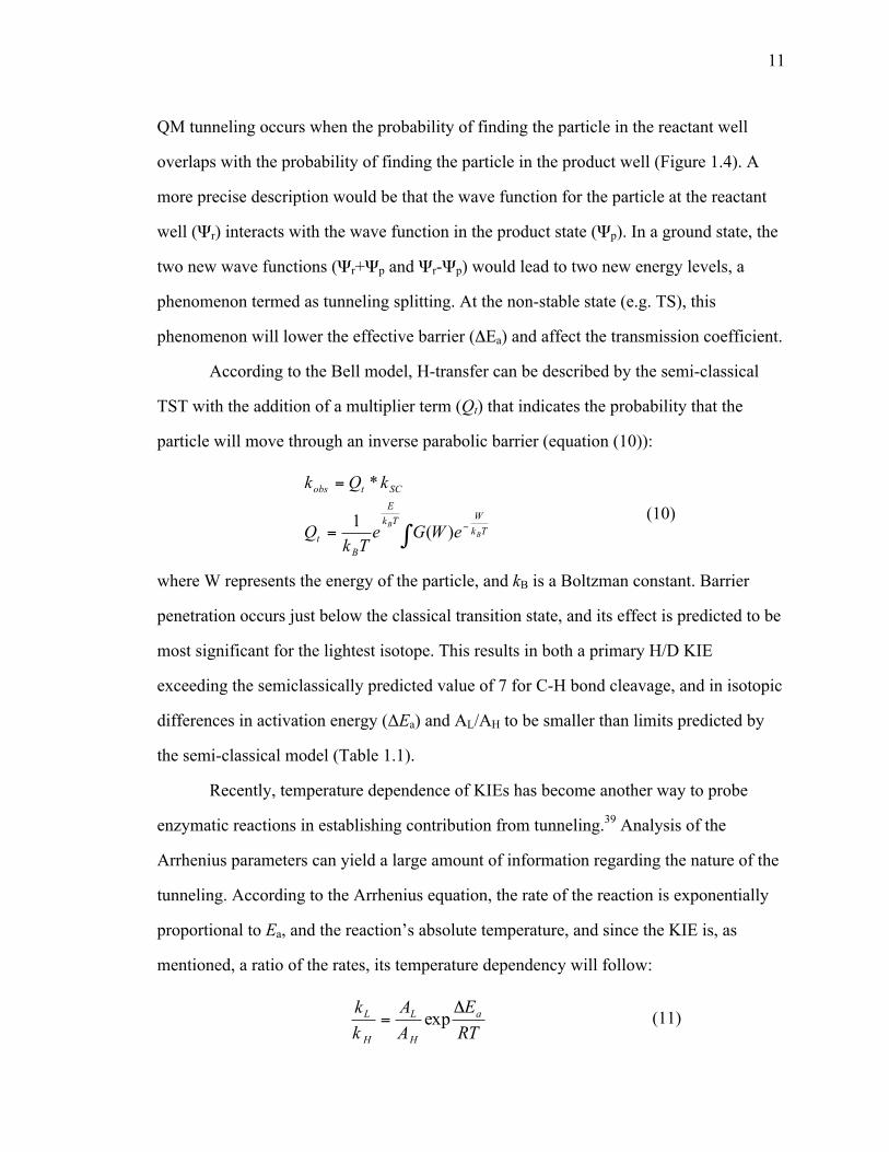
11
QM tunneling occurs when the probability of finding the particle in the reactant well
overlaps with the probability of finding the particle in the product well (Figure 1.4). A
more precise description would be that the wave function for the particle at the reactant
well (Ψr) interacts with the wave function in the product state (Ψp). In a ground state, the
two new wave functions (Ψr+Ψp and Ψr-Ψp) would lead to two new energy levels, a
phenomenon termed as tunneling splitting. At the non-stable state (e.g. TS), this
phenomenon will lower the effective barrier (ΔEa) and affect the transmission coefficient.
According to the Bell model, H-transfer can be described by the semi-classical
TST with the addition of a multiplier term (Qt) that indicates the probability that the
particle will move through an inverse parabolic barrier (equation (10)):
∫ −=
=
TkWTk
E
Bt
SCtobs
BB eWGe
TkQ
kQk
)(1
*
(10)
where W represents the energy of the particle, and kB is a Boltzman constant. Barrier
penetration occurs just below the classical transition state, and its effect is predicted to be
most significant for the lightest isotope. This results in both a primary H/D KIE
exceeding the semiclassically predicted value of 7 for C-H bond cleavage, and in isotopic
differences in activation energy (ΔEa) and AL/AH to be smaller than limits predicted by
the semi-classical model (Table 1.1).
Recently, temperature dependence of KIEs has become another way to probe
enzymatic reactions in establishing contribution from tunneling.39 Analysis of the
Arrhenius parameters can yield a large amount of information regarding the nature of the
tunneling. According to the Arrhenius equation, the rate of the reaction is exponentially
proportional to Ea, and the reaction’s absolute temperature, and since the KIE is, as
mentioned, a ratio of the rates, its temperature dependency will follow:
RTE
AA
kk a
H
L
H
L Δ= exp (11)

12
where A is the Arrhenius pre-exponential factor and ΔEa is the isotopic difference in
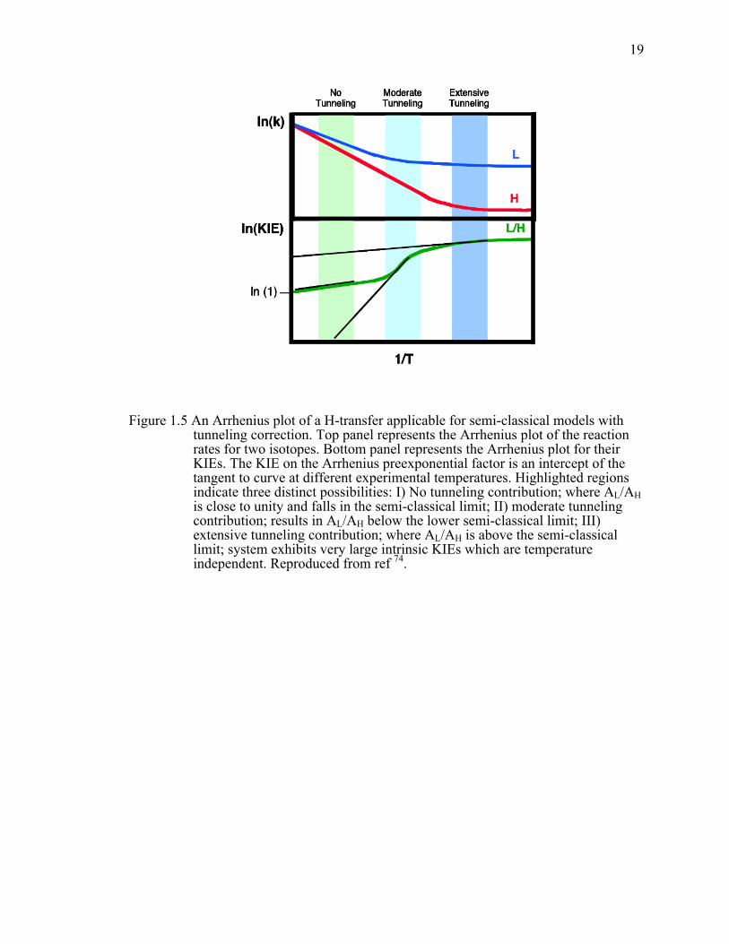
activation energy. As evident from Figure 1.5, Arrhenius plots of a reaction in different
temperature regimes can yield vastly different results for both AL/AH and ∆Ea. For
majority of the enzymatic systems, only a narrow experimental temperature range is
available (0-80˚C), thus the plot of ln(k) vs. 1/T often appears linear. At the high
temperature end, the slope of the Arrhenius plot is exponentially proportional to ∆Ea
(region I). At very low temperatures, only tunneling contributes significantly to rates due
to the lack of thermal energy (region III). Consequently, the rates are temperature
independent, and KIEs are very large and temperature independent. As a result, AL/AH,
which are intercept values of the tangent to the plot in Figure 1.5, are expected to be
inflated in comparison to the high temperature limit. In region II, which includes the
experimental temperature for the majority of enzymatic systems, Arrhenius plots of the
KIEs will be curved as the lighter isotope has the higher probability to tunnel at the
higher temperature than the heavier one. As a result, the plot will appear very steep and
AL/AH is expected to be smaller than unity.40-42 The value of using isotope effects on
Arrhenius preexponential factor as means to determine the extent of tunneling in
enzymatic systems has been reviewed extensively, and the semi-classical limits for these
values are presented in Table 1.1.40
TST-like models with or without tunneling correction, could, in several instances
rationalize temperature-dependent KIEs,43 assuming a rigid 1D potential surface, and
large temperature independent KIEs for systems which showed no Ea for the isotopically
sensitive step. However, these models fall short in explaining temperature independent
small KIEs, with significant Ea for the isotopically sensitive step. One of the
shortcomings of the Bell tunneling correction and similar TST-like models is that they
exclusively focus on the hydrogen reaction coordinate and ignore the contributory
motions of the heavy-atom environment. It is important to note that theoreticians have
tried, somewhat successfully, to explain and predict KIEs using models that are in
13
essence based on the tunneling corrections to TST.44 The few such examples include
high-level hybrid QM/MM simulations of well-characterized model enzymes such
alcohol dehydrogenase,45-48 dihydrofolate reductase,49 and thymidylate synthase,50,51
among others.
1.3.6 Temperature dependence of KIEs as a probe for
dynamics: Marcus-like models (full tunneling model)
During the past two decades, the simple Bell correction model proved to be
insufficient in rationalizing data for several enzymatic systems (few such examples are
presented in Table 1.2, however, this list is not meant to be exhaustive). As mentioned
above, instances where KIEs greatly exceeded the semi-classical limit and where AL/AH
surpassed unity (e.g. methylamine dehydrogenase and soybean lipooxygenase), or cases
where systems exhibited non zero Ea for the H-transfer process with small temperature
independent KIEs (e.g. DHFR, thymidylate synthase, alcohol dehydrogenase, etc.)
required a new theoretical model to be developed. Such a model appeared under various
names such as “vibrationally enhanced tunneling” model,52 “environmentally coupled
tunneling” model,53 “ protein promoting vibration” model,54 and others. All of these
models are based on Marcus’ theory for the electron transfer, and therefore are commonly
referred as Marcus-like models, as is the case in the following chapters,(e.g., Borgis and
Hynes,55,56 Kuznetzov and Ulstrup,57 Scrutton,52 Warshel,58 Schwartz,54 Klinman39) A
common graphical representation of this model is given in Figure 1.6. This approach
involves a Born-Oppernheim-like approximation which considers H-vibration to occur on
a much faster timescale than the heavy atom motion in the solvent or in the protein.
These motions are separately presented in Figure 1.6 as heavy-atom coordinate and
hydrogen coordinate, which together constitute the reaction coordinate of the system. In
these models, as the reaction proceeds, the heavy atom motions will bring the system to a

14
point, called tunneling ready state (TRS), where the potential surfaces for the reactant and
product are degenerate, allowing the wavefunction of the transferred particle to spread
from a donor well to an acceptor well without surmounting the intervening barrier. A
general form of these models is given by the following equation:
€
k = C(T)e(ΔG
+λ)2
4RTλ)
eF (m,DAD )eE (DAD ) / kBTdDADDAD0
DAD1∫ (12)
where C is an isotope-independent term, representing the fraction of reactive conformers
in solution. The first exponential term is taken directly from Marcus’s theory and is
therefore referred as the “Marcus term”, which is dependent on the reorganization energy
(λ) and the driving force of the reaction (∆G˚). The first integrated exponential, the
“Franck-Condon” term is a nuclear overlap integral between the donor and acceptor
states at a given donor acceptor distance (DAD). Therefore, it is an isotopically sensitive
term. The second integrated term is a Boltzmann factor giving the probability of being at
any particular DAD. These two exponents are integrated over all possible DADs to give
the total tunneling probability. As evident from equation (12), at different temperatures
heavy atom motions can affect the DAD distribution at TRS resulting in temperature
dependent KIEs. The advantage of this model is that it was possible to explain
temperature dependent rates and small temperature independent KIEs. Most mature wild
type enzymes, like (WT ecDHFR), exhibit temperature independent KIEs, which indicate
that heavy atom motions bring the system to an optimal TRS where DAD distribution
does not change significantly over the experimental temperature range. Deviations from
the physiological temperature and pressure, as well as certain site-specific mutations, can
lead to temperature dependent KIEs (as it was studied in Chapters 3, 5 and 6).
In recent years, there have been modest attempts to model the temperature
dependence of KIEs in enzymes using a form of Marcus-like model.39 High-level
QM/MM calculations have given some explanations of KIEs for soybean
lipoxygenase,59,60 alcohol dehydrogenase,61,62 dihydrofolate reductase,63,64 and
15
thymidylate synthase.65 However, even lower level modeling has helped experimentalists
to quantitatively link the experimental data to the potential energy surface describing the
hydrogen coordinate (since the model represented in Equation (12) is a phenomological
model).48,66-68 However, due to the complexity of the DAD’s potential surface, and the
fact that the DAD’s fluctuations do not constitute a normal mode, there are certain
limitations, e.g. in determining a precise DAD and its fluctuations.69 In addition, even
though lower level modeling has helped experimentalists in understanding the KIEs in
soybean lipoxygenase70,71 and alcohol dehydrogenase,72 in some cases these models
required heavy parameterization in order to replicate experimental data. KIE data
obtained for the mutants of residue 14, presented in Chapter 3, has been recently fitted to
a form of Equation (12),73 using a program available free of charge at
http://chemmath.chem.uiowa.edu/webMathematica/kohen/marcuslikemodel.html. KIE
data for both I14 mutants and F31V DHFR have been fitted using that program and
presented in reference 73 and Chapter 5, respectively.
a
b

16
Figure 1.1 (a) Dihydrofolate reductase catalyzed reaction; R= para-aminobenzoyl polyglutamate moiety, R’= 2’-monophosphoadenosine-5’-diphosphoribose. (b) The catalytic cycle of ecDHFR. The five primary intermediates and the pH-independent rate constants at 25˚C are shown6; E = ecDHFR, H2F = 7,8-dihydrofolate, H4F = 5,6,7,8-tetrahydrofolate , NADPH = reduced nicotinamide adenine dinucleotide phosphate, NADP+ = oxidized nicotinamide adenine dinucleotide phosphate.

17
Figure 1.2 Structure of ecDHFR in a ternary complex with Folate and NADP+ crystallized in P212121space group (PDB ID 1RX2). The active cleft divides the protein into two subdomains: adenosine binding subdomain (residues 38-88) and the loop subdomain (major subdomain). Three flexible loops mentioned in the main text are: M20 (shown in red), F-G (shown in blue) and G-H loop (shown in mangenta). In this particular crystal structure, which is considered to represent the active Michaelis complex, the M20 loop is in the closed conformation.

18
Figure 1.3 Graphical representation of the semi-classical model indicating that the difference in the energy of activation (∆Ea) for H, D, and T, result from their different zero-point energies (ZPE) in the ground state (GS) and transition state (TS). The GS-ZPE is constituted by all degrees of freedom but mostly by the C-H stretching frequency and the TS-ZPE is constituted by all degrees of freedom orthogonal to the reaction coordinate. Reproduced from ref 42.
Figure 1.4 An example of the ground state tunneling along the reaction coordinate. The picture depicts the probability of a particle to tunnel, where the blue and red lines represent the probability functions for lighter and heavier isotopes, respectively. As noticeable from the figure, lighter isotope has a higher probability of tunneling than the heavier one. Reproduced from ref 74.
19
Figure 1.5 An Arrhenius plot of a H-transfer applicable for semi-classical models with tunneling correction. Top panel represents the Arrhenius plot of the reaction rates for two isotopes. Bottom panel represents the Arrhenius plot for their KIEs. The KIE on the Arrhenius preexponential factor is an intercept of the tangent to curve at different experimental temperatures. Highlighted regions indicate three distinct possibilities: I) No tunneling contribution; where AL/AH is close to unity and falls in the semi-classical limit; II) moderate tunneling contribution; results in AL/AH below the lower semi-classical limit; III) extensive tunneling contribution; where AL/AH is above the semi-classical limit; system exhibits very large intrinsic KIEs which are temperature independent. Reproduced from ref 74.

20
Figure 1.6 Graphical representation of a Marcus-like model. The heavy-atoms position coordinate represents the heavy-atom motions that modulate the average energies of reactant and product shown here as a double-well potential (in blue and red respectively). The heavy-atom motions will eventually bring the system to a degenerate state, TRS(‡), where tunneling is plausible. The H-position coordinate represents the double-well potential for the H-tunneling which is modulated by the heavy-atoms throughout the reaction. Tunneling probability of each isotope, at the TRS, is proportional to the overlap of the probability functions in the reactant and product states, which is dependable on the DAD. The range of the possible DADs at the TRS is determined by the heavy-atom motions, which depends on the Boltzmann distribution (as presented by the donor-acceptor distance coordinate). The thermal fluctuations of the DADs determine the temperature dependence of the KIEs for the H-transfer. This figure has been reproduced from ref 75.

21
Table 1.1 Semiclassical limits for the isotope effect on Arrhenius preexponential factors29,43.
AH/AT AH/AD AD/AT
Upper limit 1.7 1.4 1.2
Lower limit 0.3 0.5 0.7

22
Table 1.2 Enzymatic systems with properties outside the limits predicted by the “tunneling correction” models.
Enzyme Type of transfer
Temperature dependence kH/kD AH/AD Ref.
AADH Proton No 55 NR 76
MADH Proton No 16.8 13.3 77
TMADH Proton No 4.6 7.8 78
SRO Proton No 7.3 5.8 79
PTR Hydride No ~4.1 4.1 80
EAAL H radical ND 4.4-4.7 ND 81
GLM H radical ND 2.4 ND 82
SBL-1 H radical Yes 81 18 70
SBL-1 mutants H radical Yes 93-112 4-0.12 70
Enzyme Type of transfer
Temperature dependence kH/kT AH/AT Ref.
DHFR Hydride No 6.1 7.4 83
G121V-M42W DHFR Hydride Yes 5.9 0.1 84
I14G DHFR Hydride Yes 6.1 0.024 85
R67 DHFR Hydride Yes 7.4 1.36 86
FDH Hydride No 5.9 6.1 87
TSase Hydride No 7.4 at 20˚C 6.8 88
TSase Proton Yes 6.2 8.3*10-6 89
bsADH above 30 ˚C Hydride No 5.6 at
30˚C 4.3 90
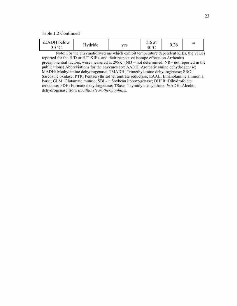
23
Table 1.2 Continued
bsADH below 30 ˚C Hydride yes 5.6 at
30˚C 0.26 90
Note: For the enzymatic systems which exhibit temperature dependent KIEs, the values reported for the H/D or H/T KIEs, and their respective isotope effects on Arrhenius preexponential factors, were measured at 298K. (ND = not determined; NR= not reported in the publications) Abbreviations for the enzymes are: AADH: Aromatic amine dehydrogenase; MADH: Methylamine dehydrogenase; TMADH: Trimethylamine dehydrogenase; SRO: Sarcosine oxidase; PTR: Pentaerythritol tetranitrate reductase; EAAL: Ethanolamine ammonia lyase; GLM: Glutamate mutase; SBL-1: Soybean lipooxygenase; DHFR: Dihydrofolate reductase; FDH: Formate dehydrogenase; TSase: Thymidylate synthase; bsADH: Alcohol dehydrogenase from Bacillus stearothermophilus.
24
CHAPTER 2.
SYNTHESIS OF RADIOLABELED NICOTINAMIDE COFACTORS
FROM LABELED PYRIDINES: VERSATILE PROBES FOR ENZYME
KINETICSi
2.1 Introduction
An enormous number of metabolic enzymes depend on the nicotinamide
cofactors, viz. NAD+, NADP+, or their reduced forms, NADH and NADPH 91. Chemical
and isotopic modification of nicotinamide cofactors have enabled enzymological studies
for several decades now (see refs.92, 93, 94 and 95 for selected examples, and 96 for a
comprehensive review of these cofactors). The development of new biophysical
techniques drives the continued quest for novel nicotinamide derivatives prepared by
rapid, cost-effective means.97 When investigating enzymes that utilize one of these
molecules as a redox cofactor, the fate of the labeled or modified cofactor may be
followed to determine the mechanistic or kinetic course of the enzyme in question. While
this can be achieved by a number of methods (e.g., following the change in 340 nm
absorbance of the reduced forms through out the reaction), radiolabeling offers higher
sensitivity than most other techniques 98 and also facilitates the measurement of isotope
effects.
Among the many uses of isotopically labeled nicotinamide cofactors is the
measurement of kinetic isotope effects (KIEs)ii for H-transfer enzymatic reactions. This
i This chapter was published in the journal of Analytical Biochemistry [Sen, A.,
Stojković, V. and Kohen (2012), Synthesis of radiolabeled nicotinamide cofactors from labeled pyridines: versatile probes for enzyme kinetics, 430, pp123-129] (Sen and Stojković contributed equally to this work) Copyright 2012, Elsevier.
ii Abbreviations used: KIE, kinetic isotope effect; EcDHFR, Escherichia coli dihydrofolate reductase; H2F, dihydrofolate; H4F, tetrahydrofolate; T(V/K)H,obs and H/Tobs, observed tritium KIE; D(V/K)H,obs and H/Dobs, observed deuterium KIE; H/Tint, intrinsic tritium

25
is exemplified below by KIE experiments with the Escherichia coli dihydrofolate
reductase (EcDHFR) enzyme. EcDHFR is an extensively studied system and thus an
excellent benchmark with which to test new nicotinamide labeling patterns. EcDHFR
uses NADPH as a hydride source in the reduction of 7,8-dihydrofolate (H2F) to S-5,6,7,8-
tetrahydrofolate (H4F), transferring the pro-R hydrogen from C4 on the nicotinamide ring
of NADPH to the si-face of C6 of the pteridine ring of H2F (Figure 2.1). Several previous
works have used NADPH labeled with deuterium (2H) or tritium (3H) at C4 to determine
the KIE on the hydride transfer step for EcDHFR and its mutants.6,83,99-101 When such
KIEs are measured competitively, both isotopologues are allowed to compete
simultaneously for the enzyme. In these experiments the ratio of the reaction rates
between the isotopologues is measured by following the fractionation of the isotopes at
the product or the reactant, instead of determining the absolute rates.29 For example, in
competitive H/T and D/T KIE measurements [denoted also by T(V/K)H,obs and T(V/K)D,obs, respectively] it is imperative that the non-radioactive isotopologues (1H- or 2H-labeled) are tracked by a radioactive labeling other than 3H at a position on the
NADPH molecule remote from the transferred hydrogen. This remote tracer (commonly 14C) in the product thus represents the H (or 2H) that has been transferred and so permits
analytical examination of the 3H depletion relative to H (or 2H) in the product by
following the 3H/14C ratio at different fractional conversions (as explained in detail in
Materials and Methods). The same remote tracer can also be used to measure competitive
H/D KIEs [denoted also by D(V/K)H,obs], where a remote 3H label tracks one isotope (e.g., 2H) and a remote 14C label identifies the other (e.g., H). Consequently, in such
experiments it is important that the remote tracer has no measurable effect on the reaction
KIE; H/Dint, intrinsic deuterium KIE; BIE, binding isotope effect; NADase, DPN nucleosidase/NAD+ glycohydrolase; PMSF, phenylmethylsulfonate; bmGDH, Bacillus megaterium glucose dehydrogenase; tbADH, Thermoanaerobium brockii alcohol dehydrogenase; DPM, disintegrations per minute.

26
rate, and no effect on the KIEs under study. If the labeling is too close to the reactive
center, isotope effects due to the tracer isotope may lead to inflation or deflation of the
KIEs being measured. As an extreme example, the use of [4-14C]-NADPH as a labeled
cofactor in the EcDHFR reaction would add a primary (1˚, see below) 14C KIE to the
measured hydrogen KIE. While such an unwanted isotope effect would have a minor
effect on 1˚ hydrogen KIEs (due to their large size), it could have a more detrimental
impact on the much smaller secondary (2˚, see below) KIEs, or on KIEs of heavy
isotopes.
Using such a remote-labeling dual-isotope method lends itself very well to a
sequential HPLC/liquid scintillation counting (LSC) analysis, as demonstrated by a
wealth of studies over the years.20,102 For example, in the case of EcDHFR, we previously
performed competitive measurements of H/T KIEs using [Ad-14C]-NADPH in
competition with 4R-[4-3H]-NADPH (see Figure 2.3 for radiolabeling details): here, the 14C label on the adenine ring is far from the point of hydride transfer and cannot affect
the rate of this step. The H/T KIEs are measured by simultaneously following the
conversion of [Ad-14C]-NADPH to [Ad-14C]-NADP+, and of 4R-[4-3H]-NADPH to
tritiated H4F and its oxidized derivatives.103 The measured values can be used together
with D/T KIEs (measured in a similar fashion) to extract the intrinsic KIEs for the
reaction, thus providing direct insight into the nature of the chemical step in the
enzymatic reaction under consideration.35,104
The isotopic labeling pattern must be modified according to the type of KIE being
measured – i.e., either a 1° KIE, which results when the isotopic difference is in an atom
directly involved in making or breaking bonds during the reaction, or a 2° KIE, which
arises when the nature of the bonding to a labeled atom changes without formation or
cleavage of bonds to that atom. Moreover, the labeling pattern should take into
consideration whether H/T, D/T or H/D KIEs are being measured, as well as the
interaction of the labeled nicotinamide cofactor with the enzyme. Indeed, the degree of
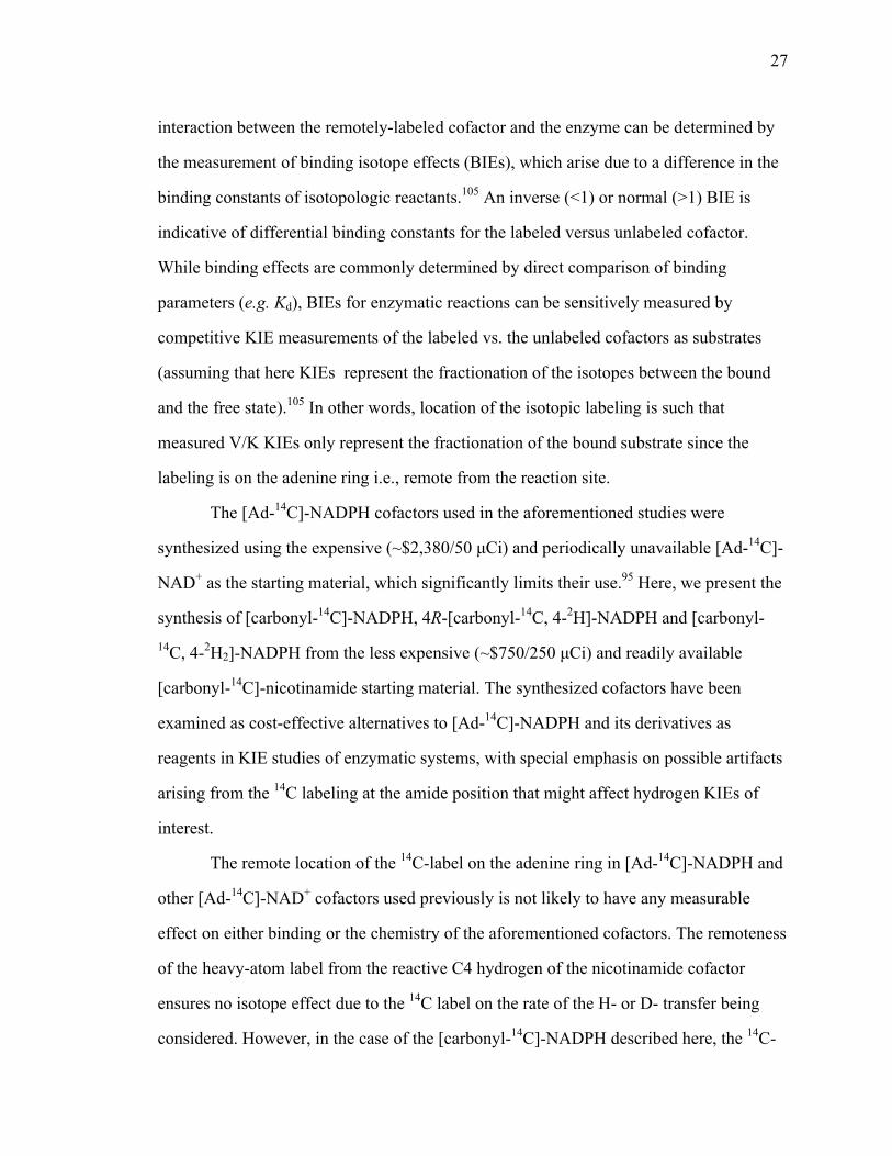
27
interaction between the remotely-labeled cofactor and the enzyme can be determined by
the measurement of binding isotope effects (BIEs), which arise due to a difference in the
binding constants of isotopologic reactants.105 An inverse (<1) or normal (>1) BIE is
indicative of differential binding constants for the labeled versus unlabeled cofactor.
While binding effects are commonly determined by direct comparison of binding
parameters (e.g. Kd), BIEs for enzymatic reactions can be sensitively measured by
competitive KIE measurements of the labeled vs. the unlabeled cofactors as substrates
(assuming that here KIEs represent the fractionation of the isotopes between the bound
and the free state).105 In other words, location of the isotopic labeling is such that
measured V/K KIEs only represent the fractionation of the bound substrate since the
labeling is on the adenine ring i.e., remote from the reaction site.
The [Ad-14C]-NADPH cofactors used in the aforementioned studies were
synthesized using the expensive (~$2,380/50 µCi) and periodically unavailable [Ad-14C]-
NAD+ as the starting material, which significantly limits their use.95 Here, we present the
synthesis of [carbonyl-14C]-NADPH, 4R-[carbonyl-14C, 4-2H]-NADPH and [carbonyl-14C, 4-2H2]-NADPH from the less expensive (~$750/250 µCi) and readily available
[carbonyl-14C]-nicotinamide starting material. The synthesized cofactors have been
examined as cost-effective alternatives to [Ad-14C]-NADPH and its derivatives as
reagents in KIE studies of enzymatic systems, with special emphasis on possible artifacts
arising from the 14C labeling at the amide position that might affect hydrogen KIEs of
interest.
The remote location of the 14C-label on the adenine ring in [Ad-14C]-NADPH and
other [Ad-14C]-NAD+ cofactors used previously is not likely to have any measurable
effect on either binding or the chemistry of the aforementioned cofactors. The remoteness
of the heavy-atom label from the reactive C4 hydrogen of the nicotinamide cofactor
ensures no isotope effect due to the 14C label on the rate of the H- or D- transfer being
considered. However, in the case of the [carbonyl-14C]-NADPH described here, the 14C-
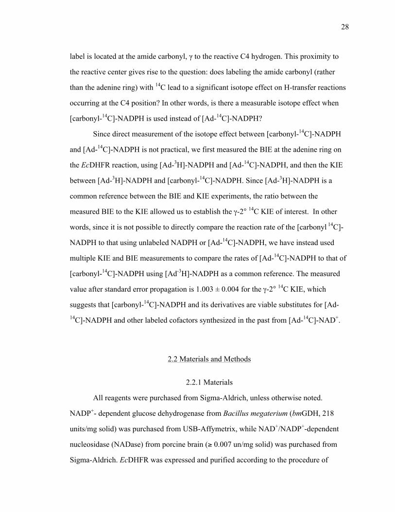
28
label is located at the amide carbonyl, γ to the reactive C4 hydrogen. This proximity to
the reactive center gives rise to the question: does labeling the amide carbonyl (rather
than the adenine ring) with 14C lead to a significant isotope effect on H-transfer reactions
occurring at the C4 position? In other words, is there a measurable isotope effect when
[carbonyl-14C]-NADPH is used instead of [Ad-14C]-NADPH?
Since direct measurement of the isotope effect between [carbonyl-14C]-NADPH
and [Ad-14C]-NADPH is not practical, we first measured the BIE at the adenine ring on
the EcDHFR reaction, using [Ad-3H]-NADPH and [Ad-14C]-NADPH, and then the KIE
between [Ad-3H]-NADPH and [carbonyl-14C]-NADPH. Since [Ad-3H]-NADPH is a
common reference between the BIE and KIE experiments, the ratio between the
measured BIE to the KIE allowed us to establish the γ-2° 14C KIE of interest. In other
words, since it is not possible to directly compare the reaction rate of the [carbonyl 14C]-
NADPH to that using unlabeled NADPH or [Ad-14C]-NADPH, we have instead used
multiple KIE and BIE measurements to compare the rates of [Ad-14C]-NADPH to that of
[carbonyl-14C]-NADPH using [Ad-3H]-NADPH as a common reference. The measured
value after standard error propagation is 1.003 ± 0.004 for the γ-2° 14C KIE, which
suggests that [carbonyl-14C]-NADPH and its derivatives are viable substitutes for [Ad-14C]-NADPH and other labeled cofactors synthesized in the past from [Ad-14C]-NAD+.
2.2 Materials and Methods
2.2.1 Materials
All reagents were purchased from Sigma-Aldrich, unless otherwise noted.
NADP+- dependent glucose dehydrogenase from Bacillus megaterium (bmGDH, 218
units/mg solid) was purchased from USB-Affymetrix, while NAD+/NADP+-dependent
nucleosidase (NADase) from porcine brain (≥ 0.007 un/mg solid) was purchased from
Sigma-Aldrich. EcDHFR was expressed and purified according to the procedure of
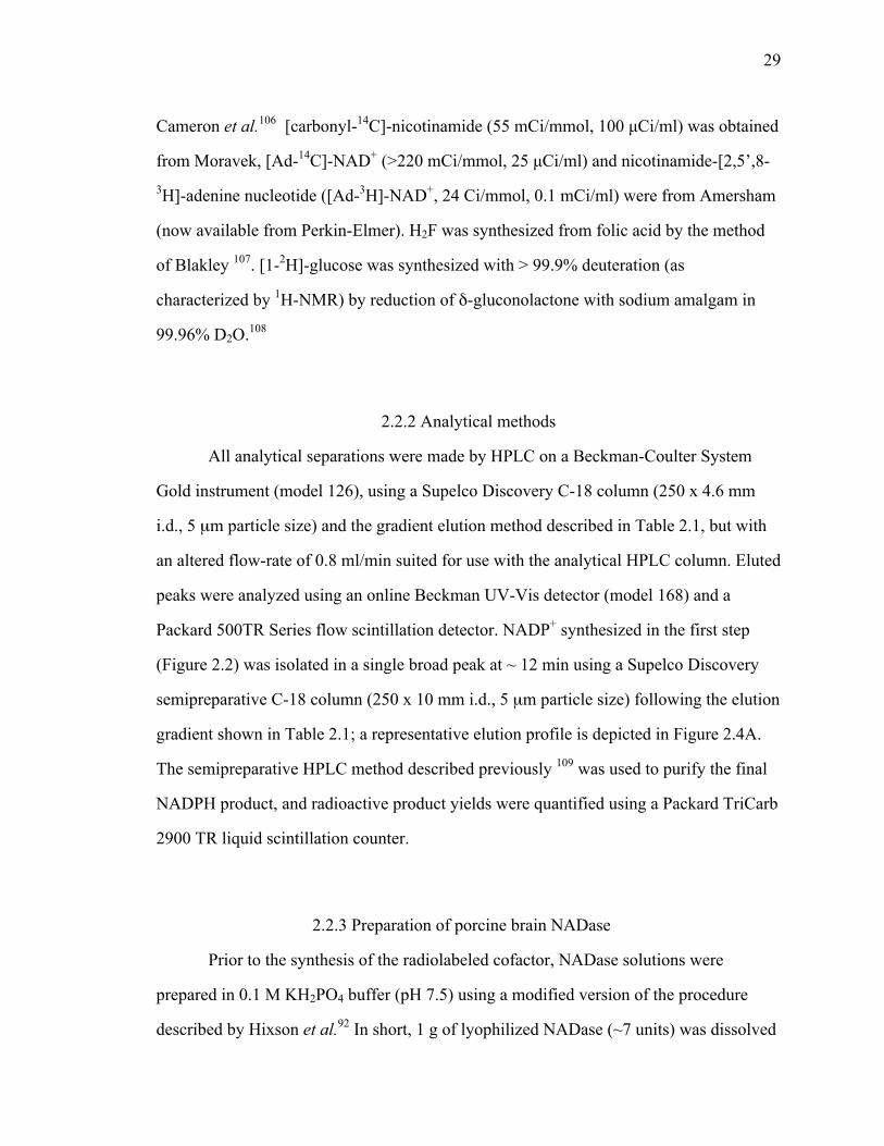
29
Cameron et al.106 [carbonyl-14C]-nicotinamide (55 mCi/mmol, 100 µCi/ml) was obtained
from Moravek, [Ad-14C]-NAD+ (>220 mCi/mmol, 25 µCi/ml) and nicotinamide-[2,5’,8-3H]-adenine nucleotide ([Ad-3H]-NAD+, 24 Ci/mmol, 0.1 mCi/ml) were from Amersham
(now available from Perkin-Elmer). H2F was synthesized from folic acid by the method
of Blakley 107. [1-2H]-glucose was synthesized with > 99.9% deuteration (as
characterized by 1H-NMR) by reduction of δ-gluconolactone with sodium amalgam in
99.96% D2O.108
2.2.2 Analytical methods
All analytical separations were made by HPLC on a Beckman-Coulter System
Gold instrument (model 126), using a Supelco Discovery C-18 column (250 x 4.6 mm
i.d., 5 µm particle size) and the gradient elution method described in Table 2.1, but with
an altered flow-rate of 0.8 ml/min suited for use with the analytical HPLC column. Eluted
peaks were analyzed using an online Beckman UV-Vis detector (model 168) and a
Packard 500TR Series flow scintillation detector. NADP+ synthesized in the first step
(Figure 2.2) was isolated in a single broad peak at ~ 12 min using a Supelco Discovery
semipreparative C-18 column (250 x 10 mm i.d., 5 µm particle size) following the elution
gradient shown in Table 2.1; a representative elution profile is depicted in Figure 2.4A.
The semipreparative HPLC method described previously 109 was used to purify the final
NADPH product, and radioactive product yields were quantified using a Packard TriCarb
2900 TR liquid scintillation counter.
2.2.3 Preparation of porcine brain NADase
Prior to the synthesis of the radiolabeled cofactor, NADase solutions were
prepared in 0.1 M KH2PO4 buffer (pH 7.5) using a modified version of the procedure
described by Hixson et al.92 In short, 1 g of lyophilized NADase (~7 units) was dissolved

30
in 0.1 M KH2PO4 buffer (pH 7.5) to a final concentration of 0.5 units/ml. The buffered
solution was sonicated for 45 min at 4 °C in the dark, then 6 mg of trypsin
(≥10,000 units/mg) was added and the reaction mixture was incubated at 37 °C for 40
min. 3.6 mmol of phenylmethylsulfonate (PMSF) was then added, and incubation
continued for an additional 5 min at 37 °C. The reaction mixture was centrifuged at 4 °C
and 4000 to 5000 x g to remove insoluble debris. The supernatant was removed and
stored at 4 °C in the dark until used. Under these conditions, NADase solution was found
to be stable for at least 2 months. Prior to every synthesis, an aliquot of NADase solution
corresponding to 0.175 units was spun down at 10,000 x g to remove any denatured or
precipitated protein that may have accumulated during storage.
2.2.4 Synthesis of [carbonyl-14C]-NADPH
Figure 2.2 illustrates the general synthetic strategy employed for the preparation
of [carbonyl-14C]-NADPH in two steps: first, the exchange reaction of NADP+ and
[carbonyl-14C]-nicotinamide to yield [carbonyl-14C]-NADP+, and the subsequent
reduction with bmGDH and glucose to yield [carbony-14C]-NADPH.
Synthesis of [carbonyl-14C]-NADP+ (Figure 2.2, first step): 125 µL of the
ethanolic solution of [carbonyl-14C]-nicotinamide (12.5 µCi, 227 nmol) was blown to
dryness with N2 or Ar gas, and combined with 0.175 units NADase, 2.3 µmol NADP+
and 0.1 M KH2PO4 (pH 7.5) buffer to a final volume of 400 µL. The reaction was
allowed to proceed at 35 °C in the dark for 60 min. Reaction progress was periodically
monitored by HPLC/flow scintillation detector analysis. It is important to note that, since
NADase catalyzes the reversible hydrolysis of NADP+ to nicotinamide and 2’-
monophosphoadenosine-5’-diphosphoribose, the reaction mixture for the first step should
contain a 10:1 excess of NADP+ over [carbonyl-14C]-nicotinamide. Different ratios lead

31
to lower yields or lower specific radioactivities of the radiolabeled NADP+ (see
discussion). It is also noteworthy that in the synthesis of radiolabeled material, the yield
is measured in terms of the radiolabeled starting material ([carbonyl-14C]-nicotinamide in
this case), which is the limiting-reagent in the synthesis.
After completion of the exchange reaction (generally after 30 min), NADase was
removed from the reaction mixture by filtration through an Amicon Ultra Ultracel-10K
(10,000 MWCO) regenerated cellulose centrifugal filter. LSC analysis of the filtrate
indicated minimal loss of radioactivity due to filtration. [carbonyl-14C]-NADP+ was then
isolated from the filtrate by semipreparative HPLC purification as described in the
Methods section, and lyophilized overnight to remove all traces of solvent. The
lyophilized NADP+ was stable at -80 °C for at least a year. Before continuing with further
synthetic steps, it was critical that the large amounts of salt (the result of lyophilization)
be removed, as high salt concentrations can inhibit certain enzymatic reactions.
Therefore, lyophilized [carbonyl-14C]-NADP+ was dissolved in 1 ml of doubly de-ionized
(DDI) H2O and loaded onto a 3 ml Sephadex C-18 desalting column. The loaded column
was washed through with 3 ml DDI H2O, and [carbonyl-14C] NADP+ was then eluted
using 3 ml of 15% MeOH/2.5 mM KH2PO4 buffer. This solution was lyophilized
overnight.
Synthesis of [carbonyl-14C]-NADPH (Figure 2.2, second step): The lyophilized
[carbonyl-14C]-NADP+ was re-suspended in 450 µl 0.1 M KH2PO4 (pH 7.5) buffer
immediately before its reduction to [carbonyl-14C]-NADPH. 1.25 µmol D-glucose and 1
mg (~218 units) bmGDH were added to the [carbonyl-14C]-NADP+ solution and the pH
was adjusted to 8.0 at 37 °C, with a final volume of 500 µl. The reaction was allowed to
proceed at 37 °C for 1 hour, when HPLC/flow scintillation analysis showed >99%
conversion of [carbonyl-14C]-NADP+ to [carbonyl-14C]-NADPH (Figure 2.5). Upon
completion of the reaction, the reaction mixture was centrifuged at room temperature and
32
13,000 x g for 5 min, and the products were purified by semipreparative HPLC as
described in the Methods section. The purified [carbonyl-14C]-NADPH was quantified by
LSC analysis, and divided into 3 million DPM aliquots. These aliquots were immediately
lyophilized and stored in the dark at -80 ˚C. Previously published data indicates that
NADPH stored under these conditions remains viable for more than 24 months 109. After
purification and lyophilization, the NADPH product was found to be more than 99.5%
pure by HPLC/radiographic flow-scintillation analysis (Figure 2.5).
2.2.5 Synthesis of 4R-[carbonyl-14C,4-2H]-NADPH from
[carbonyl-14C]-NADP+
4R-[carbonyl-14C, 4-2H]-NADPH was synthesized from [carbonyl-14C]-NADP+
using the one-step reaction catalyzed by Thermoanaerobium brockii alcohol
dehydrogenase (tbADH).110 The final product was purified, characterized and stored in
the same manner as [carbonyl-14C]-NADPH described in the previous section.
2.2.6 Synthesis of [carbonyl-14C,4-2H2]-NADPH from
[carbonyl-14C]-NADP+
[carbonyl-14C, 4-2H2]-NADPH was synthesized from [carbonyl-14C]-NADP+
following the 3-step procedure involving bmGDH, tbADH and 1-2H-glucose as described
in refs. 111 and 103. Purification, characterization and storage were the same as for
[carbonyl-14C]-NADPH.
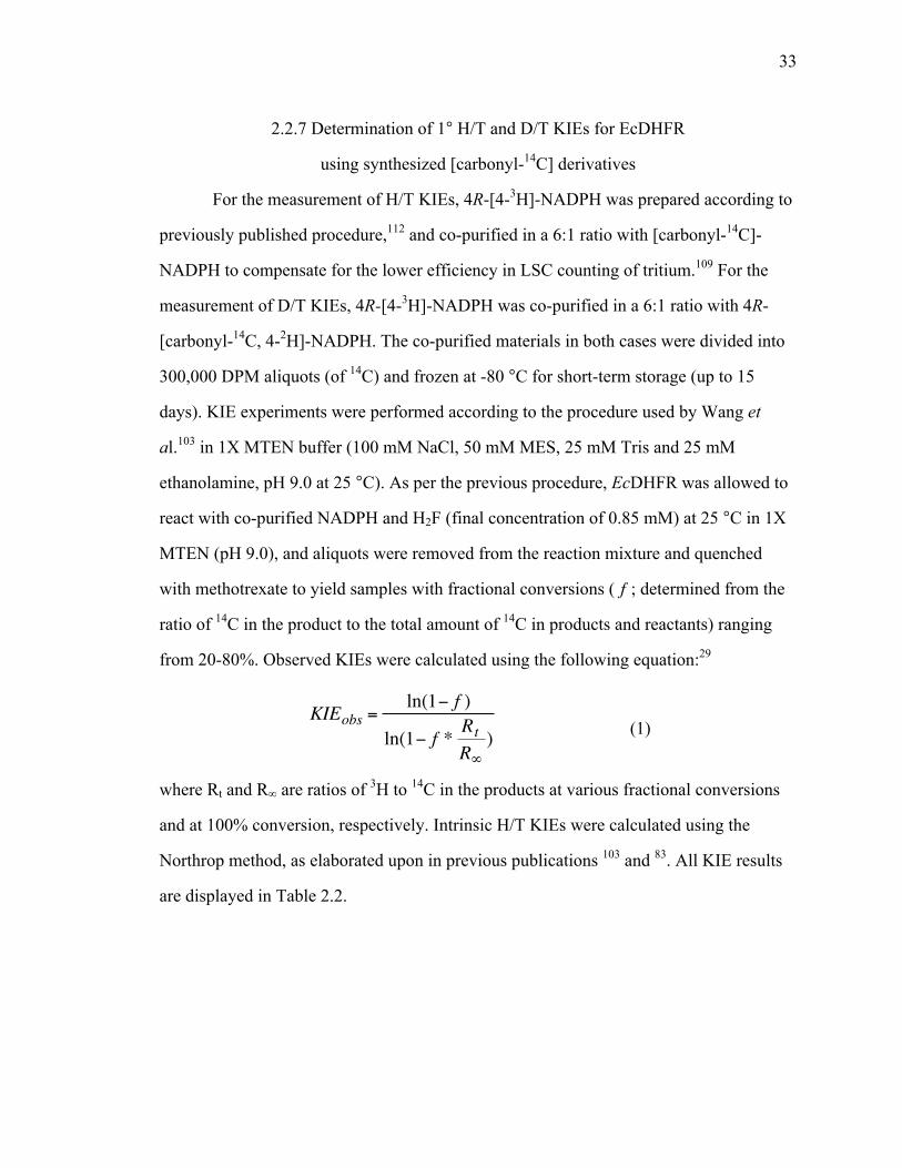
33
2.2.7 Determination of 1° H/T and D/T KIEs for EcDHFR
using synthesized [carbonyl-14C] derivatives
For the measurement of H/T KIEs, 4R-[4-3H]-NADPH was prepared according to
previously published procedure,112 and co-purified in a 6:1 ratio with [carbonyl-14C]-
NADPH to compensate for the lower efficiency in LSC counting of tritium.109 For the
measurement of D/T KIEs, 4R-[4-3H]-NADPH was co-purified in a 6:1 ratio with 4R-
[carbonyl-14C, 4-2H]-NADPH. The co-purified materials in both cases were divided into
300,000 DPM aliquots (of 14C) and frozen at -80 °C for short-term storage (up to 15
days). KIE experiments were performed according to the procedure used by Wang et
al.103 in 1X MTEN buffer (100 mM NaCl, 50 mM MES, 25 mM Tris and 25 mM
ethanolamine, pH 9.0 at 25 °C). As per the previous procedure, EcDHFR was allowed to
react with co-purified NADPH and H2F (final concentration of 0.85 mM) at 25 °C in 1X
MTEN (pH 9.0), and aliquots were removed from the reaction mixture and quenched
with methotrexate to yield samples with fractional conversions ( f ; determined from the
ratio of 14C in the product to the total amount of 14C in products and reactants) ranging
from 20-80%. Observed KIEs were calculated using the following equation:29
€
KIEobs =ln(1− f )
ln(1− f * RtR∞) (1)
where Rt and R∞ are ratios of 3H to 14C in the products at various fractional conversions
and at 100% conversion, respectively. Intrinsic H/T KIEs were calculated using the
Northrop method, as elaborated upon in previous publications 103 and 83. All KIE results
are displayed in Table 2.2.
34
2.2.8 Measurement of γ - 2° 14C KIE due to 14C isotopic
labeling at the amide carbonyl of NADPH
[Ad-3H]-NADPH and [carbonyl-14C]-NADPH for KIE measurement, and [Ad-3H]-NADPH and [Ad-14C]-NADPH for the BIE measurement, were combined and co-
purified in a DPM ratio of 5:1 (3H/14C). These two mixtures were used in the kinetic
measurements following the same procedure as described for 1˚ KIEs above. As
mentioned in the introduction, while the experimental procedure is the same for BIE and
KIE experiments, the value calculated in the BIE measurement using equation 1
corresponds to a BIE, while KIE obtained by [Ad-3H]-NADPH and [carbonyl-14C]-
NADPH reflects the product of the BIE and the γ - 2° 14C KIE of interest. Dividing the
second by the first yields a value indicative of the carbonyl γ - 2° 14C KIE.
2.3 Results and Discussion
2.3.1 Synthesis of [carbonyl-14C]-NADPH, 4R- [carbonyl-14C, 4-2H]-NADPH and [carbonyl-14C, 4-2H2]-NADPH
NADase catalyzes the conversion of NAD(P)+ to nicotinamide and 2’-
monophosphoadenosine-5’-diphosphoribose. The hydrolysis is reversible in both cases,
and the reaction equilibrium is governed by the ratio of NAD(P+) to free nicotinamide,
and the amount of ADP-ribose or 2’-monophosphoadenosine-5’-diphosphoribose in
solution. It was found that the reaction could be optimized for minimal reaction times (<
1 hour) and maximal yields of radioactive cofactor by using a 10:1 ratio of NADP+ to
[carbonyl-14C]-nicotinamide in the initial reaction mixture. Deviations from this ratio
produced deleterious effects on the time required for maximal yield, and the yield itself.
Moreover, dissimilar ratios resulted in different specific radioactivities in the product,
and in some cases led to the formation of large amounts of unidentified by-products (as
determined by HPLC analysis). For example, a lower ratio (3:1) of NADP+ to labeled

35
nicotinamide required longer reaction times and resulted in maximum yields of 37% after
1 hour, while ratios higher than 10:1 led to final products with low specific radioactivity,
thus limiting their use in KIE experiments. When a ratio of 10:1 was used, yields reached
a maximum value of 70-75% (measured as the percentage of total radioactivity in the
desired product) after ~30 minutes (Figure 2.4B). It is important to establish a point at
which the amount of formed product is at its maximum, as a slow steady decline occurs
at longer reaction times. This decline could be result of several different processes, and
not simply a reverse reaction catalyzed by NADase (as this is an equilibrium reaction).
Moreover, we observed a small amount of unidentified by-product (max ~1.6% of total
radioactivity; Figure 2.4B) that was easily separated from the NADP+, and therefore did
not present a major obstacle in either total yield or purity of the product. Since a 10:1
ratio produced satisfactory yields of [carbonyl-14C]-NADP+, the reaction was not further
optimized. However, if lower specific radioactivities are acceptable in the product, it is
possible to obtain up to 90% yields in 1 hr using higher ratios of NADP+/nicotinamide.
The relatively low price of the radioactive [carbonyl-14C]-nicotinamide starting material
was also a factor in the experimental design described above. Since the cost of this
starting material is the major contributor to the overall cost of each synthesis, the price
difference makes the [carbonyl-14C]-nicotinamide more appealing as a reagent.
The [carbonyl-14C]-NADP+ produced must be separated from the reaction mixture
that also contains [carbonyl-14C]-nicotinamide and 2’-monophosphoadenosine-5’-
diphosphoribose. Otherwise, it is possible that subsequent kinetic experiments with the
final [carbonyl-14C]-NADPH product will show artifacts due to nicotinamide
contamination. In addition, 2’-monophosphoadenosine-5’-diphosphoribose has been
shown to inhibit certain NADP+-dependent dehydrogenases, including GDH.113 This
makes isolating the [carbonyl-14C]-NADP+ from the exchange reaction mixture crucial
for successful downstream steps. A previously published method for the purification of
NADPH 95 was found to be inefficient in separating NADP+ from nicotinamide.

36
Therefore, that method was modified by reducing the percentage of methanol from 15 to
7.5% during the isocratic elution that starts at 11 min, consequently increasing separation
between NADP+ and nicotinamide peaks to 8 min. Thus, in the modified method, NADP+
elutes at 12.5 min, and nicotinamide at 20.5 min. The [carbonyl-14C] NADP+ elutes in a
broad peak over 5 min, as depicted in Figure 2.4, and is collected in at least 6 ml of 7.5%
MeOH /0.1 M KH2PO4 solution. Further reduction in the methanol percentage caused
significant peak broadening that rendered greater separation in retention times
meaningless.
The lyophilized product from the first exchange step contains [carbonyl-14C]-
NADP+ as well as significant amounts of KH2PO4. When this solid was simply
resuspended in 500 µl water (to a final concentration of ~ 1.2 M KH2PO4) and used for
the subsequent GDH reaction, a dramatic decrease in the rate of the reduction to NADPH
was observed (80% of the NADP+ still remained unreacted at 1 hour, in contrast to the
99% conversion to NADPH usually seen at 37 °C and pH 7.0 for the same reaction in the
presence of 0.1 M KH2PO4). We hypothesized that this was due to the high salt
concentration in the resuspension solution, which was either causing active GDH to
precipitate out of the solution (a ‘salting out’ effect) or directly inhibiting the GDH
reaction. To test this hypothesis, we used a Sephadex 1 ml C-18 column to remove the
excess salt from the lyophilized product, and [carbonyl-14C]-NADP+ was eluted from the
column using a 15% MeOH/2.5 mM KH2PO4 (pH 6.0) solution. The losses in yield due
to the addition of this step were minor (<2%). Addition of this step restored the reaction
rate of subsequent GDH-catalyzed reductions, providing support to the above hypothesis.
Our procedure describes the reduction of [carbonyl-14C]-NADP+ to [carbonyl-14C]-NADPH, 4R-[carbonyl-14C, 4-2H]-NADPH, or [carbonyl-14C, 4-2H2]-NADPH. If
desired, 4S-[carbonyl-14C-4-2H] NADPH could be produced by simply truncating the
procedure and reducing [carbonyl-14C]-NADP+ with [1-2H]-glucose and GDH. Also,
since NADase can accept both NAD+ and NADP+ as substrates, experimental procedures
37
similar to the ones described here can be envisioned for the preparation of [carbonyl-14C]-NAD+/NADH and their deuterated derivatives for kinetic and mechanistic studies of
NAD(H) specific enzymes.
2.3.2 14C-labeling at the nicotinamide carbonyl carbon of
NADPH does not result in a significant γ -2° 14C KIE
Due to the location of the new label near the reaction center, more specifically at
the amide carbonyl of NADPH, the possible issue of a γ-2˚ KIE artifact on hydride
transfer KIEs to be measured using this compound had to be addressed. To examine the
extent of that effect on cofactor oxidation, we designed experiments using [Ad-3H]-
NADPH in competition with either [Ad-14C]-NADPH or [carbonyl-14C]-NADPH as
substrates of the EcDHFR catalyzed reaction. The first combination ([Ad-14C]-
NADPH/[Ad-3H]-NADPH) resulted in a BIE on the adenine ring of 1.002 ± 0.002 and
the second ([carbonyl-14C]-NADPH/[Ad-3H]-NADPH) in a mixed KIE of 1.005 ± 0.002.
The ratio of these values yield a γ-2˚ 14C KIE ([carbonyl-14C]-NADPH/[Ad-14C]-
NADPH) of 1.003 ± 0.004. This value is practically unity (no KIE) as the errors usually
obtained on either 1˚ and even 2˚ hydrogen KIEs are larger than 0.01. When measuring
small KIEs (e.g. 2˚ KIEs) with other enzymes than EcDHFR, or when using other
carbonyl-labeled nicotinamide cofactors, we recommend that researchers test for such
possible artifacts for their specific system.
2.3.3 Testing the viability of synthesized materials using
KIEs: Comparison of intrinsic KIEs measured using
[carbonyl-14C]-NADPH vs. [Ad-14C]-NADPH
To validate the use of the new [carbonyl-14C]-NADPH and its deuterated
derivatives in kinetic experiments, we have compared the outcome of KIE experiments

38
that used the new synthesized materials, to previous KIE measurements that utilized
adenine labeled cofactors as substrates of EcDHFR. We conducted 1˚ H/T experiments
using [carbonyl-14C]-NADPH and 4R-3H-NADPH as substrates, and 1˚ D/T KIEs using
4R-[carbonyl-14C, 4-2H]-NADPH and 4R-3H-NADPH as substrates, for the EcDHFR
reaction. All experiments were performed at 25 °C and pH 9.0. The results, along with
the calculated intrinsic KIEs, are compared to previously published values in Table 2.2.
All previous measurements were done using [Ad-14C]-NADPH and its deuterated analogs
as substrates. The close agreement between our measured and intrinsic values and all
prior results indicates that with this enzyme the [carbonyl-14C]-NADPH materials are
excellent substitutes for the [Ad-14C]-NADPH materials in KIE experiments.
2.3.4 Comparison of cost, effort, and yield of [Ad-14C]-
NADPH 95 vs. [carbonyl-14C]-NADPH syntheses.
The new synthesis of [carbonyl-14C]-NADPH presented here is advantageous
because: (i) the [carbonyl-14C]-nicotinamide precursor is substantially cheaper starting
material than the commercially available [Ad-14C]-NAD+ precursor used to make [Ad-14C]-NADPH (by a factor of >12 based on $/Ci and synthetic yields); (ii) the new
synthesis is much faster (e.g., 30 min glycosidic exchange vs. 3 hours 2’-
phosphorilation); (iii) it requires fewer enzymes and reagents (e.g., ~ U.S. $1 NADase vs.
~ U.S. $70 NAD-kinase and phosphocreatine kinase); and (iv) has better yield than that
of [Ad-14C]-NADPH (i.e., 60-75% vs. 40-60%). In summary, the new synthetic
procedure is a cost- and effort-effective alternative to previously published procedures”.
2.4 Conclusion
The development of a new labeling pattern for 14C-labled nicotinamide cofactors
is presented along with some applications and the examination of possible experimental

39
artifacts. We have shown that [carbonyl-14C]-NADP+, [carbonyl-14C]-NADPH, 4R-
[carbonyl-14C, 4-2H]-NADPH and [carbonyl-14C, 4-2H2]-NADPH can be synthesized
quickly, in reasonable yields, and at relatively low costs, using a simple chemoenzymatic
procedure. We have utilized [carbonyl-14C]-NADPH to demonstrate that NADPH labeled
with 14C at the nicotinamide carbonyl has no significant effects on the hydrogen KIEs
measured using those isotopologues as substrates in the EcDHFR reaction. 1° H/T and
D/T KIEs measured using the newly synthesized materials are in complete agreement
with KIEs previously measured with NADPH labeled at the adenine ring, and confirm the
utility of this material. Finally, the synthesized material can easily be adapted for use in
kinetic experiments with other NADP+/NADPH/NAD+/NADH-dependent enzymes, and
as these are ubiquitous in nature the synthetic methods described herein could potentially
be of broad utility.
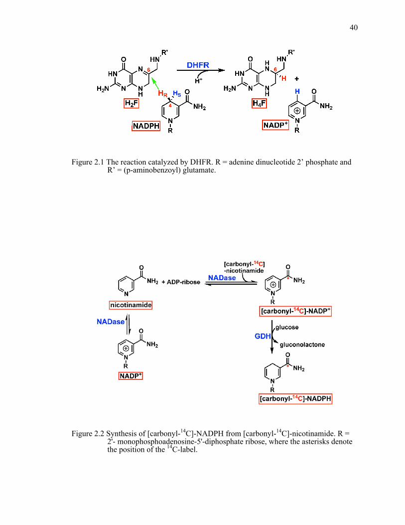
40
Figure 2.1 The reaction catalyzed by DHFR. R = adenine dinucleotide 2’ phosphate and R’ = (p-aminobenzoyl) glutamate.
Figure 2.2 Synthesis of [carbonyl-14C]-NADPH from [carbonyl-14C]-nicotinamide. R = 2'- monophosphoadenosine-5'-diphosphate ribose, where the asterisks denote the position of the 14C-label.

41
Figure 2.3 Labeled NADPH derivatives. Asterixes indicate the position of isotopic labeling with 14C (red), 2H (blue) or 3H (green). HR=1H for NADPH, [Ad-14C]-NADPH and [carbonyl-14C]-NADPH; HR=2H for 4R-[Ad-14C, 4-2H]-NADPH and 4R-[carbonyl-14C, 4-2H]-NADPH; HR=3H for 4R-[4-3H]-NADPH. For the sake of clarity, 3H-labeling on [Ad-3H]-NADPH and its derivatives is not shown.
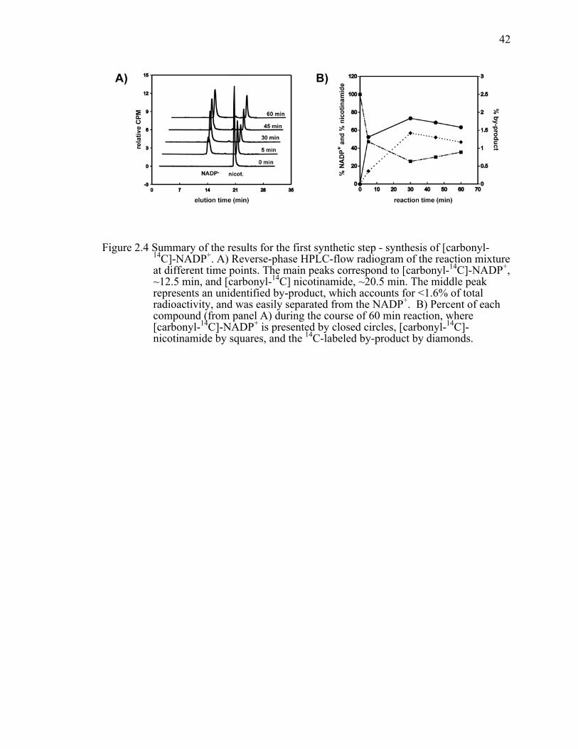
42
Figure 2.4 Summary of the results for the first synthetic step - synthesis of [carbonyl-14C]-NADP+. A) Reverse-phase HPLC-flow radiogram of the reaction mixture at different time points. The main peaks correspond to [carbonyl-14C]-NADP+, ~12.5 min, and [carbonyl-14C] nicotinamide, ~20.5 min. The middle peak represents an unidentified by-product, which accounts for <1.6% of total radioactivity, and was easily separated from the NADP+. B) Percent of each compound (from panel A) during the course of 60 min reaction, where [carbonyl-14C]-NADP+ is presented by closed circles, [carbonyl-14C]-nicotinamide by squares, and the 14C-labeled by-product by diamonds.

43
Figure 2.5 Reverse-phase HPLC-flow radiogram of the reaction mixture after the [carbonyl-14C]-NADP+ reduction. Radiogram indicates a complete conversion (>99.5%) of NADP+ to NADPH.

44
Table 2.1 Semi-preparative HPLC method using reverse-phase C-18 column and gradient elution.
Time (min) % solvent A % solvent B Flow rate (ml/min)
0 100 0 3.2
11 100 0 3.2
11.10 92.5 7.5 3.2
21 92.5 7.5 3.2
21.10 50 50 3.2
23 50 50 3.2
23.10 0 100 3.2
30 0 100 3.2 Note: Solvent A is 0.2 M NaCl/1 mM Tris-HCl (pH 8.2) and solvent B is MeOH.
Table 2.2 Observed and intrinsic 1° KIEs measured using [carbonyl-14C]-NADPH and 4R-[4-3H]-NADPH (for H/T experiments) and 4R-[carbonyl-14C, 4-2H]-NADPH with 4R-[4-3H]-NADPH (for D/T experiments).
14C-labeling
pattern
H/Tobs D/Tobs H/Tint D/Tint H/Dint
[carbonyl-14C] 4.86 ± 0.06 1.66 ± 0.01 6.13 ± 0.18 1.72 ± 0.02 3.56 ± 0.07
[Ad-14C]a 4.85 ± 0.09 1.66 ± 0.03 6.10 ± 0.42 1.72 ± 0.04 3.55 ± 0.17 a Ref. 83. Note: The results are compared to previously published values obtained using [Ad-14C] NADPH and [Ad-14C, 4-2H2] NADPH (with 4R-[4-3H]-NADPH and 4S-[4,4-3H,2H]-NADPH, respectively). 95

45
CHAPTER 3.
EFFECTS OF THE DONOR ACCEPTOR DISTANCE AND
DYNAMICS ON HYDRIDE TUNNELING IN THE DIHYDROFOLATE
REDUCTASE CATALYZED REACTIONiii
3.1 Introduction
Despite intense studies during the past century, many questions regarding enzyme
catalysis still remain unanswered. Specifically, the physical features of enzyme catalyzed
reactions continue to be part of a vigorous debate. The transition state (TS) approach
introduced by Pauling,114 which suggested that enzymes work by preferentially binding
the activated form of the substrate(s) at the TS, still remains the generally accepted
description. This static view of enzyme catalysis is likely to be correct; however it fails to
address the mechanism by which physical features that contribute to that TS stabilization
(e.g., electrostatics and steric effects) arise along the reaction coordinate. An approach
that provides a more dynamic view of the catalytic process suggests that the thermal
motions of both enzyme and substrates are essential for the chemical transformations
catalyzed by an enzyme. A large amount of experimental and theoretical evidence
supports such a view for certain enzymes, however the relationship between enzyme
dynamics115 and bond activation needs further description. Analogous issues are of equal
importance in solution reactions, and here we take advantage of the ability to control the
reactants’ relative orientation in an enzyme’s active site to examine the nature of C-H→C
transfer reactions in general. Several experimental and theoretical approaches provide an
iii This chapter was published in the Journal of American Chemical Society [Stojković,
V., Perissinoti, L.L., Willmer, D., Benkovic, S.J. and Kohen, A. (2012), Effects of the donor acceptor distance and dynamics on hydride tunneling in the dihydrofolate reductase catalyzed reaction, 134, pp1738-1745]. Copyright 2012, American Chemical Society.

46
examination of hydrogen tunneling and its environmental coupling.39,58,66,74,116-123 Here,
we focus on the temperature dependence of the intrinsic kinetic isotope effect (KIE), and
on single turnover rates, as a means to probe the nature of the hydride transfer.39,74,124,125
Experimental data collected by this approach led to the development of several
phenomenological working models, which are extensions of the Marcus theory for
electron transfer.118,126,127 They are referred to as Marcus-like models (also addressed in
literature as environmentally coupled tunneling,128 rate-promoting vibrations,129
vibrationally enhanced tunneling,52 and more. In these models, illustrated in Figure 3.1,
heavy-atom motions along the reorganization coordinate bring the system to a point
where donor and acceptor wells are degenerate and where tunneling of the hydride is
possible (i.e., a tunneling ready state, TRS). Thus, environmental reorganization occurs
before hydride transfer takes place.39,58,62,72,74,116,118,127,130 At that point, the tunneling
probability will depend on the overlap between the donor and acceptor wavefunctions
(the first exponential inside the integral of eq. 1, referred to as the Franck-Condon term),
which is sensitive to the mass of the tunneling particle, donor-acceptor distance
(DADe)131 and its fluctuation along the DHA coordinate (Figure 3.1). Eq. 1 summarizes,
in general terms, the form of the rate equation of various Marcus-like models,
( ) ∫−+Δ− °
=1
0
),(2 /),()4/()(
DADe
DADee
TkEDADmFRTGT dDADeeeCk bDADemFeλλ (1)
where C(T) represents the fraction of reactive complexes. The first exponential term is
referred to as the Marcus-term and gives the probability of reaching a TRS. This term is a
function of λ, the work associated with reorganization of the heavy-atoms, the reaction’s
exoergicity (∆G˚), and the absolute temperature T. Importantly, while C and the Marcus-
term are mostly isotopically insensitive they generate much of the temperature
dependence in the reaction’s rate. The second exponent represents the Franck-Condon
term (FC), an overlap integral that determines the tunneling probability for each isotope

47
as a function of donor-acceptor distance. The last exponent has been referred to as the
“gating” term, and represents the Boltzman distribution of DADes at a given temperature.
This term is temperature sensitive, and together with the FC term is the source of the
temperature dependency of the KIEs. The last two exponents are integrated over all the
DADes sampled by the system.
Several researchers employed these models to rationalize their experimental
results,39,66,72,84,120,125,132 where temperature-independent KIEs indicate a system wherein
the reactants’ relative orientation and electronic states are optimized for H-tunneling and
thermally-activated fluctuations of the DADe do not affect the KIE. On the other hand,
temperature-dependent KIEs indicate a system wherein the reactants’ orientation and
relative dynamics are not optimal and thermally activated gating motions have a different
effect on the transfer of H than that of D or T. These models have been instrumental in
qualitatively rationalizing trends in both the temperature and pressure dependence of
kinetic data. In recent years, there have been modest attempts to quantitatively link the
experimental data to the potential energy surface describing the gating coordinate (to link
it to Equation 1.).48,66-68 However, due to the complexity of the DADe’s potential surface,
and the fact that the DAD’s fluctuations do not constitute a normal mode, there are
certain limitations in e.g. determining a precise DADe and its fluctuations.69 MD
simulations may be necessary to visualize the atomic level structural changes that affect
the DADe in these reactions.
Here, we assess the modulation of the DADe by examining the relationship
between the H-transfer rates and temperature dependence of KIEs, and the distribution
and fluctuations of the DADc131 via MD simulations, through the targeted mutagenesis of
an active site residue. Several other recent studies examined the effects of active site
mutations, especially residues that are in direct contact with the substrates, on H-transfer
reactions. In the study of a ketoglutarate-dependent dioxygenase, residue F159, which is
in a position where the size of its side chain might affect the DADe, was systematically
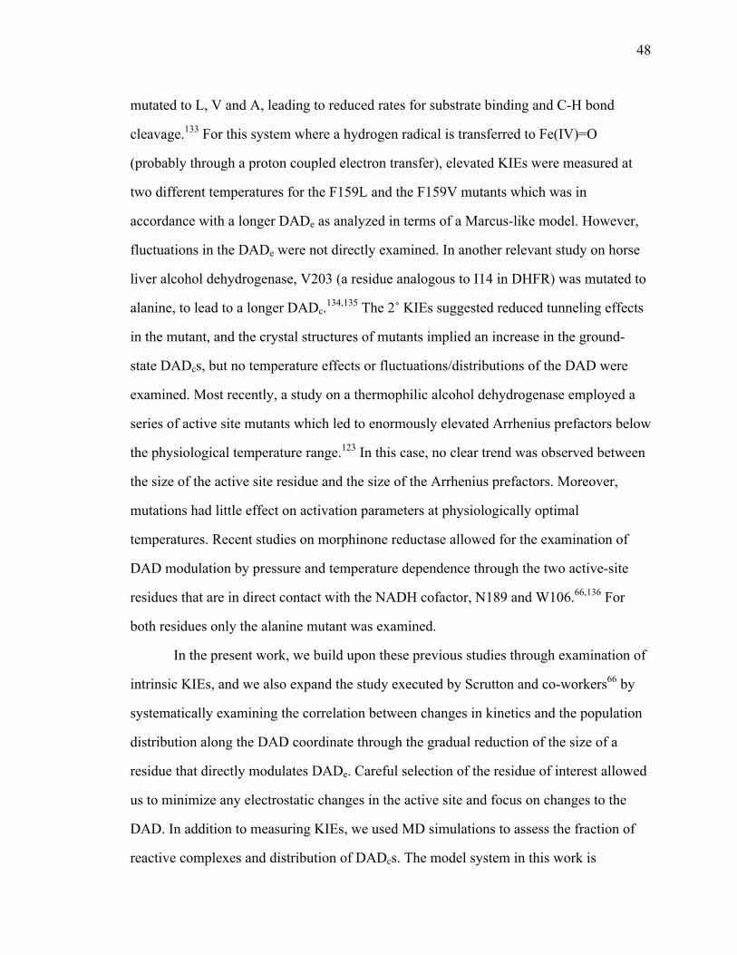
48
mutated to L, V and A, leading to reduced rates for substrate binding and C-H bond
cleavage.133 For this system where a hydrogen radical is transferred to Fe(IV)=O
(probably through a proton coupled electron transfer), elevated KIEs were measured at
two different temperatures for the F159L and the F159V mutants which was in
accordance with a longer DADe as analyzed in terms of a Marcus-like model. However,
fluctuations in the DADe were not directly examined. In another relevant study on horse
liver alcohol dehydrogenase, V203 (a residue analogous to I14 in DHFR) was mutated to
alanine, to lead to a longer DADc.134,135 The 2˚ KIEs suggested reduced tunneling effects
in the mutant, and the crystal structures of mutants implied an increase in the ground-
state DADcs, but no temperature effects or fluctuations/distributions of the DAD were
examined. Most recently, a study on a thermophilic alcohol dehydrogenase employed a
series of active site mutants which led to enormously elevated Arrhenius prefactors below
the physiological temperature range.123 In this case, no clear trend was observed between
the size of the active site residue and the size of the Arrhenius prefactors. Moreover,
mutations had little effect on activation parameters at physiologically optimal
temperatures. Recent studies on morphinone reductase allowed for the examination of
DAD modulation by pressure and temperature dependence through the two active-site
residues that are in direct contact with the NADH cofactor, N189 and W106.66,136 For
both residues only the alanine mutant was examined.
In the present work, we build upon these previous studies through examination of
intrinsic KIEs, and we also expand the study executed by Scrutton and co-workers66 by
systematically examining the correlation between changes in kinetics and the population
distribution along the DAD coordinate through the gradual reduction of the size of a
residue that directly modulates DADe. Careful selection of the residue of interest allowed
us to minimize any electrostatic changes in the active site and focus on changes to the
DAD. In addition to measuring KIEs, we used MD simulations to assess the fraction of
reactive complexes and distribution of DADcs. The model system in this work is

49
dihydrofolate reductase (DHFR) from E. coli, an enzyme that has been the subject of
extensive experimental and theoretical studies over the years, and is well suited for the
study reported here.6,137 DHFR is a small, flexible, monomeric protein (18 kDa), that
catalyzes the conversion of 7,8-dihydrofolate (H2F) to 5,6,7,8-tetrahydrofolate (H4F) with
the stereospecific transfer of a hydride from the pro-R C4 position of the nicotinamide
ring to the si face of C6 of the dihydropterin ring. The complex kinetic mechanism of
DHFR has been determined from equilibrium binding, steady-state, and presteady-state
kinetic studies.6,137
Previous measurements of intrinsic KIEs and their temperature dependence
implied quantum mechanical tunneling of the hydride in the wild-type DHFR (WT
DHFR) catalyzed reaction.83 Kinetic studies of several distal mutants of DHFR indicated
an important role of remote residues in modulating the H-tunneling process.84,103,138 The
latter studies supported the hypothesis that a set of equilibrated conformational changes
are coupled to the chemical transformation (C-H→C transfer), as predicted by hybrid
quantum/classical MD simulations.18,139 Nonetheless, the long distance and complex
relations between these remote residues and the active site prohibited a microscopic
explanation of the effect of the mutants on KIEs and on the DAD. Reproducing similar
observations via active site mutations, which have more straightforward effects on DADe
and DADc, is critical in rationalizing analogous effects caused by remote mutants or
altered conditions.
The focus of the present study is an active site residue, Ile 14, a hydrophobic
residue situated behind the nicotinamide ring of the cofactor (NADPH) that keeps the
nicotinamide ring, i.e., the H-donor, in close proximity to the pterin ring of the substrate,
i.e., the H-acceptor (Figure 3.2). Importantly, this hydrophobic side-chain is typical to
many nicotinamide dependent enzymes, which further broadens the application of the
current study. Two-dimensional heteronuclear (1H-15N) magnetic relaxation studies of
DHFR demonstrated that this residue exists as two rotamers, about the χ1 dihedral angle

50
(χ1 :N-Cα-Cβ-Cγ1), in solution; the more populated +gauche rotamer and smaller but
significantly populated trans rotamer.15 However, in both open and occluded crystal
structures of WT DHFR, I14 residue is observed as only the +gauche rotamer. Modeling
suggested that if this residue occupied the trans rotameric state, it would clash with the
nicotinamide ring. In order for residue 14 to exist as a trans rotamer, the nicotinamide
ring would be displaced towards the pterin ring. Brook and co-workers17 computed the
free energy surfaces corresponding to the χ1 dihedral angle of residue 14, along the
reaction coordinate, as part of the conformational change of the Met20 loop. They found
that the trans rotamer population is observed only in the open state and in high-energy
conformations leading to the occluded state of that loop. Only the +gauche and small
amount of –gauche populations are present in the closed state; the state in which
chemistry takes place. Moreover, they observed that the free energy barriers separating
the functional important conformational states of the Met20 loop of DHFR are small and
can be populated via thermal fluctuations.17 These two rotameric populations are of
importance in analyzing our MD simulation (see Results and Discussion).
In earlier kinetic studies of residue 14 we found an order of magnitude reduction
in the rate of the hydride transfer for I14A relative to the WT DHFR.140 In a more recent
communication we reported the temperature dependence of the intrinsic KIEs of this
mutant.141 Here, we extend these studies to a rigorous and systematic reduction of the
side chain volume of I14 without changing its electrostatic properties. We found that the
mutation leads to an increase in the magnitude of the KIEs as well as in their temperature
dependence, and is also associated with reduction in hydride-transfer rate. In addition,
classical MD simulations suggest new populations of DADcs, with larger average DADc
and broader distributions, as well as a smaller population of the active conformers with
the similar DADcs to WT DHFR. The analysis of all findings establishes a benchmark for
the interpretation of KIEs and rates at the molecular level, and has significant
applications to DHFRs and other enzymes.
51
3.2 Materials and Methods
[Ad-14C]-NAD+ (specific radioactivity of >220mCi/mmol) was purchased from
Amersham Pharmacia. 7,8-Dihydrofolate (H2F) was prepared by dithionate reduction of
folic acid as described elsewhere.107 Glucose dehydrogenase from Bacillus megaterium
(GluDH) was purchased from Affymetrix / USB. All other materials were purchased
from Sigma unless otherwise noted.
3.2.1 Synthesis of labeled cofactors for 1˚KIEs
(R)-[4-3H]NADPH (680 mCi/mmol) was synthesized by reduction of NADP+
with 2-Deoxy-D-Glucose-3H using glucose dehydrogenase from Bacillus megaterium
(GluDH), followed by oxidation of the resulting NADPH with acetone using alcohol
dehydrogenase from Thermoanaerobium brockii (tbADH), and final reduction using
GluDH and unlabeled glucose, as described previously.111 [Ad-14C]-NADPH
(50mCi/mmol) was prepared by phosphorylation of [Ad-14C]-NAD+ using NAD+ kinase
from chicken liver to produce [Ad-14C]-NADP+, followed by reduction with glucose
using GluDH as described elsewhere.95 (R)-[Ad-14C,4-2H]-NADPH (50 mCi/mmol) was
prepared from [Ad-14C]-NADP+, which was synthesized as described above, and
subsequent reduction with deuterated glucose and GluDH.142 All synthesized cofactors
have been purified by semipreparative reverse-phase HPLC on a Supelco column and
stored at -80 ˚C prior to use as discussed elsewhere.109
3.2.2 Construction of expression vectors
The sequence of the mutagenic, forward primer (I14V-for) is 5’-GCG-GTA-
GAT-CGC-GTT-GTC-GGC-ATG-GAA-AAC-GCC-3’, and the sequence of the
52
mutagenic, reverse primer (I14V-rev) is 5’-GGC-GTT-TTC-CAT-GCC-GAC-AAC-
GCG-ATC-TAC-CGC-3’, where the site of the mutation is underlined. The sequence of
the mutagenic, forward primer (I14G-for) is 5’-GCG-GTA-GAT-CGG-GTT-GGC-
GGC-ATG-GAA-AAC-GCC-3’, and the sequence of the mutagenic, reverse primer
(I14G-rev) is 5`- GGC-GTT-TTC-CAT-GCC-GCC-AAC-GCG-ATC-TAC-CGC-3,
where the site of the mutation is underlined. PCR reaction was completed using pET22b-
DHFR as a template. The original template was digested with the DpnI restriction
enzyme, and the PCR product was transformed into DH5α- cells. Plasmid was extracted
from the overnight culture and the sequence was verified by automated DNA sequencing
by the University of Iowa DNA facility. Primers were purchased from Integrated DNA
Technologies.
3.2.3 Expression and purification of I14V DHFR, I14A
DHFR and I14G DHFR
The mutants were expressed, purified and stored as reported for other DHFR
mutants.106
3.2.4 Competitive and intrinsic primary kinetic isotope
effect
For H/T 1˚KIE, (R)-[4-3H]NADPH and [Ad-14C]-NADPH, and for D/T 1˚KIE,
(R)-[4-3H]NADPH and (R)-[Ad-14C,4-2H]-NADPH were combined in a radioactivity
ratio close to 5:1. Each mixture was co-purified on an analytical reverse-phase HPLC
column, divided into aliquots containing 300,000 dpm of 14C, and frozen in liquid
nitrogen for short-term storage at -80 ˚C.
All experiments were performed in MTEN buffer (50 mM MES, 25 mM Tris, 25
mM EtOH-NH2 and 100 mM NaCl) at pH=8.0, over the temperature range of 5-45 ˚C.

53
H2F was added to a final concentration of 0.85 mM, which is around a 200-fold excess
over NADPH (final concentration of 4 µM). The pH was adjusted to 8.0 at each
experimental temperature following the calibration of the pH electrode in appropriate
buffers at that temperature. The reaction was quenched by adding an excess of
methotrexate (Kd=1 nM), and samples were stored on dry ice prior to HPLC analysis.
Before the HPLC-LSC analysis, samples were thawed and bubbled with oxygen to ensure
complete oxidation of the product tetrahydrofolate (H4F). The samples were then
separated by reverse-phase HPLC using a method described elsewhere.109 The observed
KIEs were calculated according to ref 29 using the following equation:
)](1ln[
)1ln(
∞
−
−=
RRf
fKIEt
where the fractional conversion (f) was determined from the ratio of 14C in the product
and reactant, and Rt and R∞ are the ratio 3H/14C at each time point and at infinite time,
respectively.
The intrinsic KIEs were calculated using the modified Northrop equation35,74
1)/(1)/(
1)(
1)(34.3/1
1
1
1
−
−=
−
−
−
−
−
−
TH
TH
DT
HT
kkkk
KVKV
obs
obs
where T(V/K)Hobs and T(V/K)Dobs are the observed H/T and D/T KIEs, respectively, and
kH/kT represents the intrinsic H/T KIE. The intrinsic KIEs were calculated numerically
from all possible combinations of observed H/T and D/T values. All intrinsic values were
used to fit to the Arrhenius equation in order to determine the isotope effect on the
Arrhenius preexponential factors as well as temperature dependence of the KIEs, as
described in the main text.
54
3.2.5 MD simulation
System Setup – The simulations were performed starting from the crystal structure
of wild type DHFR, determined at 1.8 Å resolution (PDB entry 1RX2). The crystal
structure contains a total of 159 amino acid residues, 153 crystallographic water
molecules, and folate and NADP+ as ligands. Missing hydrogens were added to the PDB
structure using the leap module of the Amber package.143,144 The folate and NADP+
ligands were replaced by N5 protonated H2F and NADPH respectively. The protonation
state for all ionizable residues was set for a pH of 7. Thus, histidine residues were
modeled as neutral residues with the proton on Nε or Nδ as determined on the basis of
possible hydrogen bond interactions deduced from X-ray crystallographic structure. Only
His-45 was modeled as charged, based on both NMR145 and crystallographic146 studies
which indicate a salt bridge between His-45 and the nicotinamide 5'-phosphate of
NADP+. Crystallographic studies have also established the existence of a salt bridge
between Arg-44 and Adenine 2'-phosphate.147 The estimated pKa for His-45 determined
by PropPKa148 was 8.83 in accordance with experimental observation. The resulting
system has a net charge of -15e including N5-protonated- H2folate (-1) and NADPH(-
4).149 The final protein structure was solvated with a previously equilibrated truncated
octahedral box of TIP3P water molecules. The size of the box ensured that all protein
atoms were at least 9 Å away from the edges of the box. Overlapping solvent molecules
were removed leaving 5879 water molecules. Mutations were performed in silico by
changing the corresponding amino acid in the original structure.
Simulation Parameters – All simulations were done at 300 K and 1 bar, regulated
with the Berendsen barostat and thermostat,150 using periodic boundary conditions and
Ewald sums for treating long range electrostatic interactions.151,152 The SHAKE
algorithm153 was used to keep bonds involving hydrogen atoms at their equilibrium
length. A 2 fs time step for the integration of Newton's equations was used. Nonbonded
interactions had a cutoff radius of 12 Å. The AMBER99 force field154 parameters were

55
used for all residues. The NADP+ and NADPH parameters were taken from the Amber
parameter database.155 Gaff force field156 parameters together with RESP157 charges
calculated at the HF/6-31G* level were used to generate the parameter files for H2F.
MD runs – All simulations were run with the PMEMD module of the AMBER9
package.143,144 An equilibration protocol was applied that consists of performing an
energy minimization by optimizing the initial structure, followed by a slow heating to the
desired temperature using a linear temperature ramp from 100 to 300 K during 80 ps at
constant volume and a subsequent 100 ps pressure stabilization run at 300 K and 1 bar .
Position frames, which were used for analyzing trajectories, were collected at 2 ps
intervals. Production MD simulations consisted of 15 ns simulations: (i) wtDHFR-H2F-
NADPH; (ii) I14V DHFR-H2F–NADPH, (iii) I14A DHFR-H2F–NADPH. In the case of
(iv) I14G DHFR-H2F–NADPH, more effective sampling of the conformational space was
obtained from multiple short trajectories rather than a single long trajectory. The same
sampling methodology was also applied to the (ii) and (iii) mutants in order to compare
with the long simulation and between each mutant. A set of ten independent 3 ns
simulations for (ii), (iii) and (iv) that differ only in the initial velocities were run. To
combine data from different trajectories we have given equal weight to trajectories of
equal length. For the (iv) mutant, each trajectory was extended to 6 ns without observing
significant changes in the final result. For (ii) and (iii) the distribution of DADs
calculated by both methodologies (i.e. long run versus several short runs), was similar,
whereas for (iv) it was significantly different, as discussed in the main text. Due to the
significant difference of the residue volume in the case of I14G DHFR, the nicotinamide
ring is able to sample more space, and in this case the choice of an effective way of
sampling is crucial.
MD data analysis – The ptraj module of Amber9 together with a homemade
program used to calculate the Phi angle between the planes that fit to the NADPH
nicotine amide moiety (C4N, C3N, C2N, N1N, C6N, C5N) and H2F pterin moiety (C4A,

56
N5, C6, C4, N3, C2, N1, C8A, N8, C7) were used to analyze the data extracted from the
MD simulations. Trajectories were visualized with Visual Molecular Dynamics
(VMD).158 Xmgrace and QtiPlot were used to build the graphics and figures.159,160 POV-
Ray was used for image rendering.161 DAD distributions were fitted with a combination
of Gaussian and split Gaussian functions using the open source software for non linear
curve fitting, Fityk.162
Gaussian:
!!"
#
$$%
&''(
)**+
, −−=
2
2
10210 )2ln(exp),,,(
aax
aaaaxy
where a0=height, a1=center, a2=hwhm (half width at half maximum), and
2*hwhm = FWHM = 2 2 ln2σ , where FWHM is Full Width at Half Maximum.
SplitGaussian:
!"#
$%&
>
≤=
1310
12103210 ),,,,(
),,,;(),,,;(
axaaaxGaussianaxaaaxGaussian
aaaaxy
where a0=height, a1=center, a2=hwhm1, a3=hwhm2
In all the cases the splitGaussians used were only slightly asymmetric, where hwhm1 is a
little smaller than hwhm2, so we consider valid the following approximation:
Center=a1~ mean value
σ2ln2221 ≈=+ FWHMhwhmhwmh ; hwhm stands for half width at half
maximum, and FWHM stands for Full Width at Half Maximum
3.2 Presteady-state kinetics
The concentration of the NADPH and H2F were determined
spectrophotometrically by using the extinction coefficients of 6200 M-1 cm-1 at 340 nm,
and 28000 M-1cm-1 at 282 nm respectively. Concentration of enzyme was determined
spectrophotometrically by using the extinction coefficient of 74600 M-1 cm-1 at 280 nm,

57
and by Bradford assay. All the measurements were obtained on a stopped-flow apparatus
(Applied Photophysics Ltd., Leatherhead, U.K.), with dead time of 1.6 ms, a 2-mm path
length, and a thermally regulated sample cell. The enzyme reaction was monitored by
taking advantage of fluorescent resonance energy transfer (FRET) between DHFR and
NADPH.6 Enzyme tryptophans were excited at 290 nm, and FRET results in the strong
fluorescence by the reduced cofactor at 450 nm, which is observed through a 390 nm
output filter. Since NADP+ is not fluorescent, the chemical step can be monitored by the
loss of fluorescence. In the experiment, 20 µM enzyme (either wtDHFR or the relevant
mutant) was preincubated with NADPH (10 µM) in MTEN buffer at 25˚C. The reaction
was initiated by rapid mixing with 300 µM H2F. We used the average of five runs for
data analysis. All data were analyzed by non-linear least squares fits to single exponential
functions using KaleidaGraph 3.5.
3.3 Results and Discussion
3.3.1 Competitive KIEs and Their Temperature
Dependence
We determined the temperature dependence of the intrinsic KIEs and hydride
transfer rates for a series of DHFR isoforms for which the side-chain that holds the
hydride donor (nicotinamide) close to the acceptor (dihydropterin) are systematically
reduced from isoleucine to glycine. Figure 3.3 shows Arrhenius plots of the intrinsic H/T
1˚ KIEs for all four enzymes. The same trend was observed for H/D and D/T KIEs as
presented in the SI Text. Table 3.1 summarizes the volume of these side chains, their
hydride transfer rates, the isotope effect on the Arrhenius preexponential factors (Al/Ah,
where l denotes light and h heavy isotope), and the difference in the activation energy
between light and heavy isotope (∆Eah-l=Eah-Eal).

58
The isotope effects on the preexponential Arrhenius factors (AH/AT, AH/AD, and
AD/AT) for the WT DHFR, I14V DHFR and I14A DHFR are larger than unity, and above
the semiclassical limit, whereas the values for the I14G DHFR are smaller than unity and
below the semiclassical limit. The traditional, semi-classical approach with the Bell
correction43 would suggest that hydride transfer for the first three isozymes involves
“extensive tunneling” for both heavy and light isotopes, whereas the hydride transfer for
I14G DHFR would be characterized as a case with “moderate tunneling”.27,28 According
to the Marcus-like model, AH/AT is affected by both the average DADe and its
distribution, and reveals the difference in the nature of the hydride transfer between these
mutants. A longer average DADe leads to an increase in the Al/Ah, whereas a broader
distribution of DADe-s increases the temperature dependence of the KIEs resulting in a
decrease in the Al/Ah. Because of this ambiguity, a better indicator for the nature of the
hydride-transfer is the slope of the Arrhenius plot, (Figure 3.3, i.e., the ∆Ea h-l, where h
and l are the heavy and light isotopes, respectively) that increases as the distribution of
DADes broadens. ∆Ea h-l close to zero indicates that the KIEs of WT DHFR exhibits no
temperature dependence (within the experimental error), and the larger ∆Ea h-l for the
mutants indicates that temperature dependence of the KIEs increases as the size of the
hydrophobic residue behind the nicotinamide ring decreases. In terms of the Marcus-like
model this means that in the case of the WT DHFR the average DADe is optimal for the
hydride transfer and has a narrow distribution around the average. The I14V mutant is
similar to WT DHFR, but has a slight (but statistically significant) temperature
dependence on the intrinsic KIEs. The I14A mutant has the largest intrinsic KIE at 25˚C,
but its KIEs’ temperature dependence (∆Ea h-l) falls between the I14V and I14G DHFR.
This interesting observation supports the earlier examination of distal mutants of DHFR,
which concluded that in order to assess the nature of the hydride transfer it is necessary
not only to measure the KIE at a single temperature but for the whole temperature
range.84

59
According to the Marcus-like model, the inflated temperature dependence of the
KIEs in the mutants reflects a broader distribution of DADe sampling that is not present
in the WT. The average DADe in the mutants is too long for efficient tunneling at low
temperatures, but at higher temperatures more conformations with a shorter DADe are
sampled. This affects the rate of transfer by heavy isotopes more than that of protium-
transfer because these can only tunnel from the shorter distances, thus leading to
temperature dependent KIEs. These observations are in accordance with some of the
earlier studies, e.g. a study on the soybean lipoxygenase-1 (SBL-1), which showed that a
decrease in the bulk of the active site residue (residue 553, not analogous to I14, not in
direct contact with the substrate) was correlated with an increase in the temperature
dependence of the 1˚ KIE.68 Numerical modeling using a form of equation 1 provided
more evidence that these remote mutations led to longer DADes. However, in other
studies, in particular the one on morphinone reductase, Pudney et al.66 found an increase
in temperature dependence of KIEs with an increase in the size of the active-site residue.
In that particular study, the residue of interest was V108, which is not in direct contact
with the cofactor NADH; thus the effect of the size of the residue on the DADe was hard
to assess. The other residue of interest in the same study was W106, which is in direct
contact with the cofactor; however, only the alanine mutant was examined and therefore
no trends were available. In the pressure-dependence studies on the same enzyme the
authors suggested that an increase in the force constant of the compressive mode can lead
to an increase in the magnitude of the 1˚ KIE even as the DAD decreases,69,163 which
became a source of the extensive debate.164 Our findings are in accordance with the
majority of cases where numerical modeling of the temperature dependence of the 1˚
KIEs using equation 1 results in decreasing temperature dependence with decreasing
average DADe. From these independent studies it is evident that the relationship between
the size of the side chain and the DADe might be quite complex, and might go beyond the
notion that the smaller size of the residue leads to longer average DADe. These results

60
were the driving force in the experimental approach presented here, where we determined
the effect of the DADs’ distribution on the nature of the catalyzed H-transfer.
In order to asses the effect of the mutations on the hydride transfer, we measured
the single turnover rates at 25 ˚C, pH=7.0, for all mutants and for WT DHFR.6
Fluorescence Energy Transfer (FRET) decay traces were fitted to single exponential
functions with slope. These rates indicate that hydride transfer for the I14V, I14A, and
I14G mutants are about 7, 40, and 1000 times slower than WT DHFR, respectively
(Table 3.1). There is a clear correlation between the decrease in size of the hydrophobic
residue, the rate and the nature of the hydride transfer. We would like to point out that the
reduction of the size of this residue is likely to affect the mobility of the M20 loop and
consequently affect the fraction of reactive complexes, the pKa of H2F,165 the fraction of
different M20 loop conformations, and other factors that affect the multisteps reaction’s
rate, but not the hydride transfer step under study. All these effects are included in the
prefactor C(T) in rate eq 1, but do not affect the terms in the integral, including the DAD,
its dynamics and distribution. The intrinsic KIEs, on the other hand, probe the hydride
transfer step per sé, and not the steps preceding it (such as protonation of N5 position of
H2F, and other isotopically insensitive factors).
3.3.2 MD Simulations on mutants of DHFR
In order to provide a detailed microscopic description of the active site structural
change upon mutation of residue 14, we performed MD simulations. One 15 ns
simulation was performed for WT DHFR and each mutant, as was previously done for
WT DHFR and I14A DHFR.141 In order to better sample the conformational space for
each mutant, we also performed ten independent runs of 3 ns, differing only in initial
velocities for each one of them.

61
A comparison of the conformations is presented in Figure 3.4, which also
describes two parameters of interest, DADc and Φ. DADc is defined as the distance
between C4 of NADPH and C6 of H2F (see Figure 3.4 inset). The relative donor acceptor
orientation was measured as the angle (Φ) between the planes that fit the nicotinamide
moiety of NADPH and the pterin ring of H2F (see Figure 3.4 inset). The distribution of
DADcs and the orientation of donor and acceptor (Φ angle) are displayed in Figure 3.5.
The DADcs’ and Φs’ distributions (Figure 3.5, 1D plots on y and x-axis) were fitted to
one split Gaussian function or a combination of Gaussian functions and split Gaussian
functions (Table 3.2). The correlation is presented as a 2-D plot in Figure 3.5.
The results indicate that reduction in the size of the hydrophobic residue behind
the nicotinamide ring leads to an increase in the flexibility of the nicotinamide ring. As a
result, the nicotinamide ring can not only explore more orientations at the same DADcs,
but also this increase in the flexibility leads to an increase in the average DADc, a broader
DADc distributions, as well as new DADc populations (Table 3.2 and Figures 3.4-3.5).
The different populations are labeled I, II, III, IV and V; the structures that represent
these different populations are presented in Figure 3.4 for each mutant, and are overlaid
with the average wt-DHFR structure.
The DADc distribution for WT DHFR was fitted to one split Gaussian function
and the residue I14 was only observed as the +gauche rotamer during the time scale of
the simulation. This result agrees well with Brooks’s work,17 where this rotamer
represents the major population observed for the closed conformation of WT DHFR. The
DADc distribution for the I14V mutant was fitted to a combination of one split and one
normal Gaussian functions suggesting that there are two populations present during the
simulation time. The average structure for the major population is very similar to the WT
DHFR (structure I in Figure 3.4), displaying similar DADc and Φ distributions, but with a
slight increase in the average DADc as well as a deviation of the average Φ value found
in the WT DHFR. The smaller population (structure III in Figures 3.4 and 3.5) has a
62
larger average DADc with wider distribution, where new Φ values are explored,
manifesting themselves as a partial twisting of the nicotinamide ring. In the case of the
I14A mutant, the correlation plot (Figure 3.4) shows that the conformational space
explored is bigger compared to the I14V mutant. The simulations in this case suggest
three populations: (i) the wt-like population; (ii) a new population with similar DADc to I,
but a different Φ distribution (Structure II); and (iii) the Structure III, equivalent to the
one found for the I14V mutant, but with a slightly larger average DADc and Φ, which
also exhibits broader distributions, indicating more flexibility in the location of the
nicotinamide ring (see Figure 3.4 and 3.5). Population III is not expected to contribute to
a reactive conformation (average DAD too large), therefore leading to a smaller constant
C(T), from the equation 1, which is in accordance with a reduction in rate of the hydride
transfer (Table 3.1). On the other hand the new population (II) exhibits the same average
DADc as the population I, however it is expected to be by far less reactive due to the
substantial deviation of the Φ angle from 30˚. As visible in Figure 3.4, the nicotinamide
ring of NADPH is almost perpendicular to the pterin ring of H2F, making the HOMO-
LUMO orbital overlap very poor, which has a deleterious effect on the rate.166
Consequently, population II is not likely to be reactive, but its exact reactivity (if any) is
hard to determine without high-level QM/MM calculations that are beyond the scope of
this work. Lastly, the hydrogen-bonds (two to residue A7 and one to the backbone of I14)
that hold the NADPH amide moiety (see Figure 3.2), show a broader distribution, that is
in accord with the elevated flexibility of nicotinamide ring in the I14A mutant relative to
I14V and WT DHFR.
MD simulations for the mutant with the smallest side chain, I14G DHFR,
suggested all five significant populations. In this case, the three H-bond interactions
mentioned before, ((a), (b) and (c) in Figure 3.2) are longer and weaker than in the wt and
I14V/ I14A mutants. The last population, V, has one of the H-bonds (c), completely
broken, while the other two (a) and (b), are only weakly established (see Figures 3.6-3.7
63
in the SI Text). Once this H-bond is broken the structure remains in conformation V and
doesn’t return to wt-like or other conformations during the time scale of the simulation
(the simulation was extended to 30 ns). The breakage of the H-bonds, particularly the (c)
H-bond, results from the diminishment in the side chain of the residue 14, and contributes
to the increase in the flexibility over other mutants and the ability to explore a broader
conformational space that leads to a non-reactive population (DADcs > 6 Å). Finally,
Table 3.2 lists the fraction of each population found, which appear to correlate well with
the reduced rate of H-transfer. In all the cases, the wt-like population is the predominant
one, although its relative contribution decreases in proportion to the size of the side chain,
leading to decrease in the CT term in Eq. 1. A similar effect on rates was obtained in a
recent study where modeling of the conformational sampling that lead to elevated
Arrhenius prefactors (below the physiologically optimal temperatures) for the
thermophilic alcohol dehydrogenase showed an increase in the fractional occupation of
catalytically inactive regions of conformational space.123 More importantly in this work, a
correlation was observed between the temperature dependence of the intrinsic KIEs
(∆EaT-H, Table 3.1) and the DADc, its σ values, and the Φ angle (Table 3.2) for the
reactive population (I). As the ∆EaT-H increases (Table 3.1), the average DADc of
population I increases and its distribution widens, while the average Φ angle decreases
(Table 3.2). This important correlation is also visualized in Figure 3.5, where the reactive
population (short DADc and 0˚ < Φ < 60˚) is larger for the WT (red), smaller and with
broader distribution toward longer DADs for I14V (green), and more so for I14A (blue).
The population I for I14G is too small to be clearly observed in Figure 3.5, but has the
largest DADcs distribution (σ) and smallest Φ (Table 3.2).
The MD simulations provide molecular insight and support to the interpretation of
the kinetic findings via the Marcus-like model: the smaller side chain acts to broaden the
sampling of configurations within the reactive ternary complex along the reaction
coordinate. This leads to a steeper temperature dependence of intrinsic KIEs and reduced
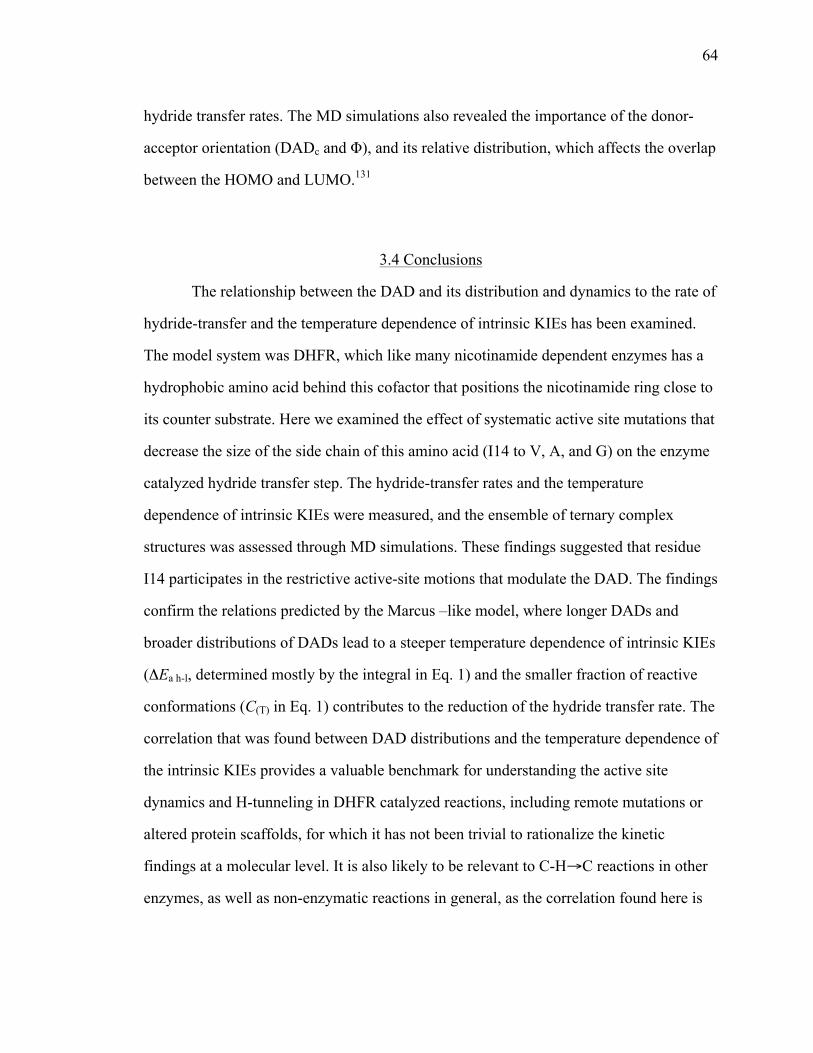
64
hydride transfer rates. The MD simulations also revealed the importance of the donor-
acceptor orientation (DADc and Φ), and its relative distribution, which affects the overlap
between the HOMO and LUMO.131
3.4 Conclusions
The relationship between the DAD and its distribution and dynamics to the rate of
hydride-transfer and the temperature dependence of intrinsic KIEs has been examined.
The model system was DHFR, which like many nicotinamide dependent enzymes has a
hydrophobic amino acid behind this cofactor that positions the nicotinamide ring close to
its counter substrate. Here we examined the effect of systematic active site mutations that
decrease the size of the side chain of this amino acid (I14 to V, A, and G) on the enzyme
catalyzed hydride transfer step. The hydride-transfer rates and the temperature
dependence of intrinsic KIEs were measured, and the ensemble of ternary complex
structures was assessed through MD simulations. These findings suggested that residue
I14 participates in the restrictive active-site motions that modulate the DAD. The findings
confirm the relations predicted by the Marcus –like model, where longer DADs and
broader distributions of DADs lead to a steeper temperature dependence of intrinsic KIEs
(ΔEa h-l, determined mostly by the integral in Eq. 1) and the smaller fraction of reactive
conformations (C(T) in Eq. 1) contributes to the reduction of the hydride transfer rate. The
correlation that was found between DAD distributions and the temperature dependence of
the intrinsic KIEs provides a valuable benchmark for understanding the active site
dynamics and H-tunneling in DHFR catalyzed reactions, including remote mutations or
altered protein scaffolds, for which it has not been trivial to rationalize the kinetic
findings at a molecular level. It is also likely to be relevant to C-H→C reactions in other
enzymes, as well as non-enzymatic reactions in general, as the correlation found here is

65
likely to hold whether the DADes are imposed by proteins’ scaffold or by solvent
molecules.
The current findings suggest that this series of DHFR mutants is suitable for more
in-depth examination by a broad variety of researchers applying a variety of methods.
Future studies of this system may include high-level hybrid classical-quantum
mechanical MD simulations,62,167 structural studies,4 examination of the systems
dynamics at millisecond to nanosecond time scales using NMR relaxation experiments,121
and femtosecond to picosecond time-scale motions using two-dimensional infrared
spectroscopy.125 Those experiments will allow more in depth examination of possible
correlations between structure, dynamics, and the nature of H-transfer in a well-defined
environment imposed by the enzyme, and should reflect on C-H→C reactions in general.

66
Figure 3.1 Illustration of a Marcus-like model. The reorganization coordinate represents the heavy-atom motions that carry the system to the TRS(‡). The blue and green correspond to the reactant and product states, respectively. The DHA coordinate (donor-hydrogen-acceptor) represents the fluctuations of the DADe. The red curve represents the wave functions of the hydrogen nucleus.
Figure 3.2 The active-site of DHFR from E. coli (PDB ID 1RX2) emphasizing the role of Ile14 (metallic blue) as a support of the nicotinamide ring of NADP+. The nicotinamide ring is highlighted in light blue and the folate in magenta. Several other residues that form hydrogen bonding with the amide of NADPH are highlighted as well as I14 and A7. Three distinct hydrogen bonds, labeled in red, are: (a): NADPH(O-amide)-Ala-7(H); (b): NADPH(H72)-Ala-7(O); and (c): NADPH(H71)-Ile-14(O). The pterin ring is also immobilized in the active site via tight van der Waals interactions with F31, and strong hydrogen bonds to D27 and I5.
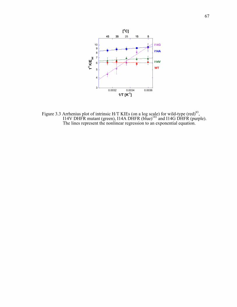
67
Figure 3.3 Arrhenius plot of intrinsic H/T KIEs (on a log scale) for wild-type (red)83, I14V DHFR mutant (green), I14A DHFR (blue)141 and I14G DHFR (purple). The lines represent the nonlinear regression to an exponential equation.
3
4
5
6
7
89
10
0.0032 0.0034 0.0036
H/Tint I14AH/Tint I14VH/Tint I14GH/Tint wtDHFRH/Tint I14A aveH/Tint I14V aveH/Tint I14G ave
I14A
I14V
WT
I14G
1/T [K-1]
525 153545 5153545
[oC]
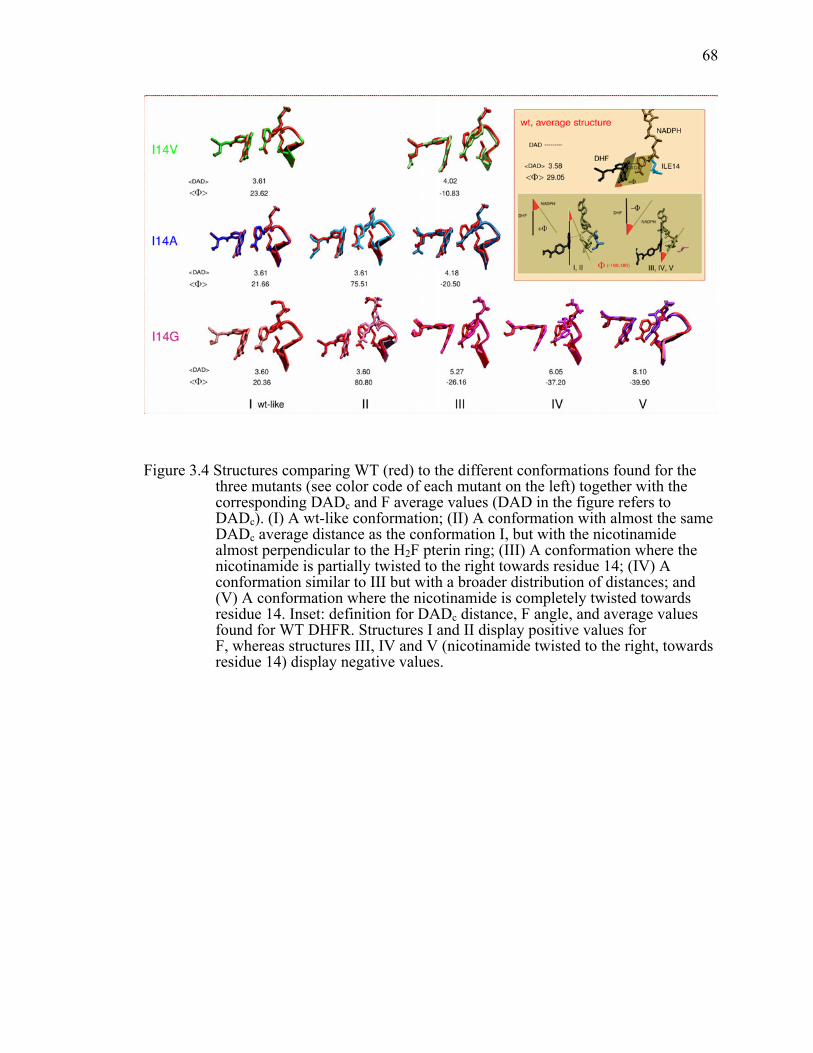
68
Figure 3.4 Structures comparing WT (red) to the different conformations found for the three mutants (see color code of each mutant on the left) together with the corresponding DADc and F average values (DAD in the figure refers to DADc). (I) A wt-like conformation; (II) A conformation with almost the same DADc average distance as the conformation I, but with the nicotinamide almost perpendicular to the H2F pterin ring; (III) A conformation where the nicotinamide is partially twisted to the right towards residue 14; (IV) A conformation similar to III but with a broader distribution of distances; and (V) A conformation where the nicotinamide is completely twisted towards residue 14. Inset: definition for DADc distance, F angle, and average values found for WT DHFR. Structures I and II display positive values for F, whereas structures III, IV and V (nicotinamide twisted to the right, towards residue 14) display negative values.

69
Figure 3.5 Correlation plot between the DADc (angstroms) and the relative orientation of donor and acceptor (Φ, degrees) for WT DHFR (red), I14V (green), I14A (blue), I14G (magenta). I, II, III, IV and V indicate the different populations identified for each DADc and Φ values. Overlaid wt and mutants DADc and Φ distributions are shown on the y and x axis respectively.

70
Table 3.1 Comparative kinetic parameters of the DHFR I14 mutants.
Parameters WT I14V I14A I14G Residue volumea (Å3)
124 105 67 48
kHb [s-1] 228 ±8d 33.3 ±3.1f 5.7 ±0.3f 0.22 ±0.04f
AH/AT c 7.0 ±1.5e 4.2 ±0.4f 4.7 ±0.5g 0.024 ±0.003f
∆EaT-Hc,
[kcal/mol] -0.1 ±0.2e 0.27 ±0.05f 0.39 ±0.06g 3.31 ±0.07f
a. Side chain volume.168
b. Presteady state rates of H transfer at 25˚C and pH 7.
c. Similar trends were observed for H/D and D/T (data not shown).
d. Ref. 169.
e. Ref. 95.
f. This work.
g. Ref. 141.

71
Table 3.2 Gaussian fitted distributions of DADc for the different conformations found along MD simulation (see text).
DHFR Active site conformation
<DAD> (Å) σ (Å)c <Φ>d (degrees)
Fraction R
wt Ia 3.58 0.16 29.0 1 0.9979 I14V Ia
IIIb 3.61 4.02!
0.17 0.31
23.6 -10.8
0.86 0.14
0.9984
I14A Ia
IIa IIIb
3.61 3.61 4.18
0.17 0.17 0.49
21.7 75.5 -20.5
0.68 0.13 0.19
0.9980
Ia
I14G IIa IIIb IVb
Vb
3.60 3.60 5.27 6.05 8.10
0.19 0.19 0.82 0.67 0.36
20.4 80.8
-26.2 -37.2 -39.9
0.33 0.26 0.09 0.22 0.1
0.9943
a. Fitted to a split-Gaussian
b. Fitted to a Gaussian.
c. For split-Gaussian σ is the average of both parts.
d. Φ separately from the DADc. See SI for the procedure and parameters (Table S3).

72
Supplementary Information
Table 3.3 Intrinsic KIEs and their standard errors for I14V DHFR.
Temp. ˚C H/T KIE H/D KIE D/T KIE 45 6.40 ±0.57 3.67 ±0.23 1.74 ±0.05 35 6.49 ±0.46 3.71 ±0.19 1.75 ±0.04 25 6.92 ±0.22 3.88 ±0.08 1.78 ±0.02 15 6.73 ±0.22 3.80 ±0.09 1.77 ±0.02 5 6.80 ±0.50 3.83 ±0.19 1.78 ±0.04 Note: These values are calculated from the observed H/T and D/T KIE data using Northrop’s method as described in the Materials and methods. The errors indicate the 95% confidence level.
Table 3.4 Intrinsic KIEs and their standard errors for I14G DHFR.
Temp. ˚C H/T KIE H/D KIE D/T KIE 45 4.39 ±0.30 2.82 ±0.13 1.56 ±0.03 35 5.10 ±0.70 3.13 ±0.30 1.63 ±0.07 25 6.13 ±0.32 3.56 ±0.16 1.72 ±0.03 15 8.17 ±0.27 4.36 ±0.17 1.88 ±0.03 5 9.15 ±0.96 4.72 ±0.34 1.94 ±0.06 Note: These values are calculated from the observed H/T and D/T KIE data using Northrop’s method as described in the Materials and methods. The errors indicate the 95% confidence level.

73
Table 3.5 Fitted Φ˚ angle distribution for wtDHFR and DHFR mutants.
Center Area Height FWHM Int. Width R
wtDHFR
29.05 0.995 0.042 22.33 23.77 0.9912
I14V
I 23.62 0.930 0.045 19.52 20.78 0.9948
III -10.84 0.063 0.004 15.01 15.97
I14A
I 21.66 0.768 0.034 21.03 22.39 0.9893
II 75.51 0.164 0.003 33.40 36.84
III -20.50 0.071 0.003 20.76 22.10
I14G
I 20.36 0.305 0.013 22.74 24.20 0.9924
II 80.80 0.234 0.007 30.27 32.22
III -26.16 0.143 0.006 22.11 23.54
IV -37.20 0.189 0.011 16.54 17.60
V -39.87 0.13 0.005 21.60 22.99
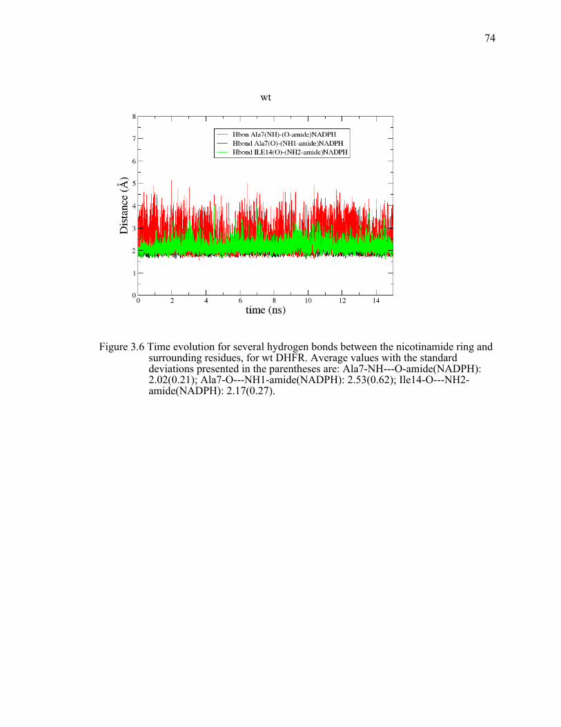
74
Figure 3.6 Time evolution for several hydrogen bonds between the nicotinamide ring and surrounding residues, for wt DHFR. Average values with the standard deviations presented in the parentheses are: Ala7-NH---O-amide(NADPH): 2.02(0.21); Ala7-O---NH1-amide(NADPH): 2.53(0.62); Ile14-O---NH2-amide(NADPH): 2.17(0.27).

75
Figure 3.7 Time evolution for several hydrogen bonds between the nicotinamide ring and surrounding residues, for a couple of I14G runs. Average values are presented together with the standard deviations in the following table.
Run 5: wt-like Run 2: structure II
Run 6: wt-like and structure III
Run 8: mainly structure V
Ala7-NH---O-amide(NADPH)
2.00(0.18) 2.10(0.19) 1.99(0.17) 4.45(1.33)
Ala7-O---NH1-amide(NADPH):
2.33(0.57) 1.98(0.44) 2.68(0.79) 2.46(0.60)
Ile14-O---NH2-amide(NADPH)
2.11(0.25) 2.38(0.56) 2.27(0.47) 2.90(0.62)

76
CHAPTER 4.
STRUCTURAL STUDY ON A SERIES OF DIHYDROFOLATE
REDUCTASE MUTANTSiv
4.1 Introduction
One of the most highly studied enzymes from the structural/dynamic/functional
viewpoint is dihydrofolate reductase from Escherichia coli (ecDHFR; EC 1.5.1.3), an
ubiquitous enzyme crucial for biosynthesis of purines, pyrimidines and certain amino
acids. This NADPH-dependent oxidoreductase catalyzes a single stereospecific hydride
transfer from the pro-R C4 position of NADPH to the re face of C6 position of 7,8-
dihydrofolate (H2F) producing 5,6,7,8-tetrahydrofolate (H4F) and NADP+. Both kinetics
and catalytic mechanism have been extensively studied,6,140,170 and numerous structural
studies of different enzymatic complexes exposed important conformations, thus
providing crucial information regarding the functionality of this enzyme.4,146,171,172 Study
of the temperature dependence of the kinetic isotope effects (KIEs) indicated that the
hydride is being transferred through quantum-mechanical tunneling.83 A
phenomenological model,39,40,44,57,66,73,85,125,127,173-177 addressed here as Marcus-like model,
used for the analysis of the temperature dependence of the KIEs suggests that protein
motions contribute to the reorganization of the protein, solvent and ligand atoms that
assist the structural changes in the active-site as the reaction proceeds from the reactant
state to the tunneling ready state (TRS). At the TRS faster motions (femto- to picosecond
timescale) control the fluctuations in the donor-acceptor distance (DAD) that affect the
H-tunneling probability. Various simulation and computational studies, based on the x-
iv This chapter is in preparation to be submitted to the journal PLOS ONE.

77
ray crystal structures, have examined the nature of the hydride transfer in ecDHFR and
addressed the role of protein motions in catalysis.17,122,178
Relating X-ray crystal structures at atomic-resolution to enzyme functionality is
not a trivial task. From the structural viewpoint it would be ideal to experimentally
observe and follow the whole hydrogen transfer event to atomic detail. However, for
most of the H-transfer enzymatic reactions that is unattainable. Nonetheless, high-
resolution crystal structures of either mimics of transition state or Michaelis complex can
provide information regarding the donor and acceptor groups relevant to the H-transfer
reaction. X-ray crystal structures are still static pictures of the enzyme, and present an
ensemble and a time-averaged picture of the protein. However, together with temperature
factors, kinetic, and computational results can give us important information for
establishing the structure-function-dynamic relationship in enzymes.
EcDHFR is a small monomeric protein that consists of central eight-stranded
beta-sheets and four alpha helices. The active site cleft divides the enzyme into two
structural subdomains: the adenosine binding subdomain and the major subdomain
(Figure 4.1a). The adenosine subdomain provides the binding cleft for the adenosine
moiety of the cofactor. The major subdomain is much larger and is dominated by the
three loops on ligand binding face that surround the active site. Those three loops are:
M20 (residues 9-24), F-G (residues 116-132) and G-H (residues 142-150) loops. M20
loop is also referred to as an active-site loop since it lays directly over the active site and
protects it from the solvent. Previous X-ray crystallization studies showed that in the
crystalline state M20 loop can be observed in the four distinct conformations: open,
closed, occluded and disordered.4 The existence of the occluded and the closed
conformations in the solution has been supported by the NMR experiments,179 and the
loop movement between these two conformations was shown to coordinate with different
stages of the catalytic cycle.4,6,180 On the other hand, the open conformation is thought to
be an intermediate state between the closed and occluded conformations and has been
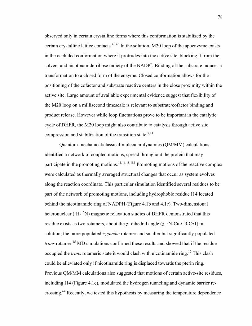
78
observed only in certain crystalline forms where this conformation is stabilized by the
certain crystalline lattice contacts.4,146 In the solution, M20 loop of the apoenzyme exists
in the occluded conformation where it protrudes into the active site, blocking it from the
solvent and nicotinamide-ribose moiety of the NADP+. Binding of the substrate induces a
transformation to a closed form of the enzyme. Closed conformation allows for the
positioning of the cofactor and substrate reactive centers in the close proximity within the
active site. Large amount of available experimental evidence suggest that flexibility of
the M20 loop on a millisecond timescale is relevant to substrate/cofactor binding and
product release. However while loop fluctuations prove to be important in the catalytic
cycle of DHFR, the M20 loop might also contribute to catalysis through active site
compression and stabilization of the transition state.5,14
Quantum-mechanical/classical-molecular dynamics (QM/MM) calculations
identified a network of coupled motions, spread throughout the protein that may
participate in the promoting motions.11,16,18,181 Promoting motions of the reactive complex
were calculated as thermally averaged structural changes that occur as system evolves
along the reaction coordinate. This particular simulation identified several residues to be
part of the network of promoting motions, including hydrophobic residue I14 located
behind the nicotinamide ring of NADPH (Figure 4.1b and 4.1c). Two-dimensional
heteronuclear (1H-15N) magnetic relaxation studies of DHFR demonstrated that this
residue exists as two rotamers, about the χ1 dihedral angle (χ1 :N-Cα-Cβ-Cγ1), in
solution; the more populated +gauche rotamer and smaller but significantly populated
trans rotamer.15 MD simulations confirmed these results and showed that if the residue
occupied the trans rotameric state it would clash with nicotinamide ring.17 This clash
could be alleviated only if nicotinamide ring is displaced towards the pterin ring.
Previous QM/MM calculations also suggested that motions of certain active-site residues,
including I14 (Figure 4.1c), modulated the hydrogen tunneling and dynamic barrier re-
crossing.64 Recently, we tested this hypothesis by measuring the temperature dependence

79
of KIEs on the series of mutants of residue I14.41,85 Temperature dependence of the KIEs
showed that decrease in the size of this hydrophobic residue leads to an increase in the
average DAD and its distribution at the TRS. MD simulations on the ternary E-NADPH-
H2F complex for the series of I14 mutants indicated that decrease in the size of this
residue leads to an appearance of the new DAD populations with longer average DAD
and broader DAD’s distribution. Moreover, mutations also distorted donor-acceptor
orientation (orientation between the pterin and nicotinamide rings).85
In order to relate the mutations’ effect on H-transfer to the mutants’ structure we
examined the ternary complexes of the ecDHFR active site mutants I14V, I14A and I14G
using X-ray crystallography. We choose to study the complex with folate and NADP+
since this is a proposed mimic of Michaelis complex, and thus has been previously used
as starting point for most published QM/MM and MD simulations. The goal of the study
presented here was to determine the structures of the mutants within single crystal
packing so that distortions due to crystal-packing effects remain constant for the series of
structures. Since crystal packing effects cause small local, and sometimes global,
deformations, we also attempted to examine the same complexes in different crystal
packings and space groups in order to get a better view of the dynamic behavior of these
mutants, as it was previously done for WT ecDHFR.4 Lastly we wanted to see if under
different crystallization conditions we would be able to trap different populations of
ground state structures that have been suggested by the MD simulations.85
4.2 Materials and methods
4.2.1 Preparation of I14V, I14A and I14G DHFR ternary
complexes for crystallization
EcDHFR mutants were expressed and purified as described before.182 Briefly, E.
coli ER2566 ΔthyA ΔfolA cells containing the DHFR genes in the pET22b vector were

80
grown in LB medium in the presence of 50 mg/l thymidine and 100 mg/ml ampicillin at
37 °C to an OD600nm of 0.6-0.8. DHFR expression was induced by the addition of the
IPTG to a final concentration of 0.4 mM, and incubation continued at 18 °C overnight.
Cells were harvested by centrifugation at 4 ˚C for 30 min. The cell pellet was re-
suspended in the lysis buffer (25 mM NaH2PO4, pH 7.0, 5 mM EDTA pH 8.0, and 10%
v/v glycerol), and lysed as described previously.182 The supernatant was purified using
methotrexate-agarose affinity column, followed by DE52 column.182 Eluted fractions
were tested for protein via UV and Bradford assay, and the purity was assessed using
SDS-PAGE gel. Only fractions containing pure protein were pooled, concentrated and
dialyzed against storage buffer. Protein was either stored at -80 °C until further use, or
loaded onto a Superdex-75 (GE) gel filtration column pre-equilibrated with the buffer
containing 20 mM Tris-HCl, pH 7.5. DHFR eluted in a single peak, which was tested for
the monodispersity using dynamic light scattering on the NanoStar (Wyatt Technology).
Concentration of the pooled fractions was measured using UV absorbance
(ε280=74.6 mM-1cm-1) and Bradford assay. Due to low solubility of folate, folate was
added to a dilute protein solution at three-fold molar excess relative to the concentration
of specific DHFR mutant. The sample was incubated on ice for 30 min, and then filtered
using Milipore 0.22 µm filter. The binary complex was concentrated to a final
concentration of 12-18 mg/ml (as determined by Bradford assay). NADP+ was added as a
solid to the concentrated binary DHFR-Folate complex at a five-fold molar excess to
DHFR.
4.2.2 Crystallization conditions
All enzyme-Folate-NADP+ ternary complexes were crystallized via the hanging
drop vapor diffusion method by mixing 400 nl well solution and 400 nl of prepared
ternary complex using TTP LabTech Mosquito. The only exception is I14G DHFR

81
ternary complex whose crystals were obtained through macroseeding into 4 µl wells (2 µl
well solution and 2 µl of protein complex).
In more detail: I14G-NADP+-Folate crystallized in the P21212 space group after
macroseeding under the following conditions: 16 mg/ml I14G DHFR, 0.1 M Tris, pH 8.5,
0.9 M LiCl, and 36% w/v PEG 6,000; at 18 °C. Crystals appeared within 7 days after
macroseeding.
I14V-NADP+-Folate ternary complex crystallized in the P21 space group under
the following conditions: 16 mg/ml I14V DHFR, 0.1 M Tris, pH 8.5, 0.1 M LiCl, and
34.2% w/v PEG 6,000; at 18 °C, within 2 weeks.
I14A-NADP+-Folate ternary complex was crystallized in P21 space group under
the following conditions: 15 mg/ml I14A DHFR, 0.2 M Lithium acetate, and 20% w/v
PEG 3,350; at 18 °C, within 7 days.
4.2.3 Data collection and structure determination
Crystals were flashed-cooled in liquid nitrogen, and data were collected at 100 K
at the 4.2.2 synchotron beamline at the Advance Light Source of the Lawrence Berkley
National Laboratory. The data were either processed using d*Trek183 (for I14G) or
XDS184 (for I14V and I14A mutants). All structures were determined by molecular
replacement in CCP4185 (with PHASER186), using the coordinates for the refined WT
ecDHFR structure (PDB 1RX2) with the ligands removed to generate the initial model.
Refinement and model building for all structures were done using REFMAC187 and
COOT188. Model bias was avoided during the initial refinement by removing the
substrate and cofactor from the model. Iterative composite OMIT maps were generated
and used where structural features were in doubt. During the early stages of the
refinement, hydrogens were included in riding positions, and isotropic temperature
factors were refined. In the final stages of the refinement, for the I14A-Folate-
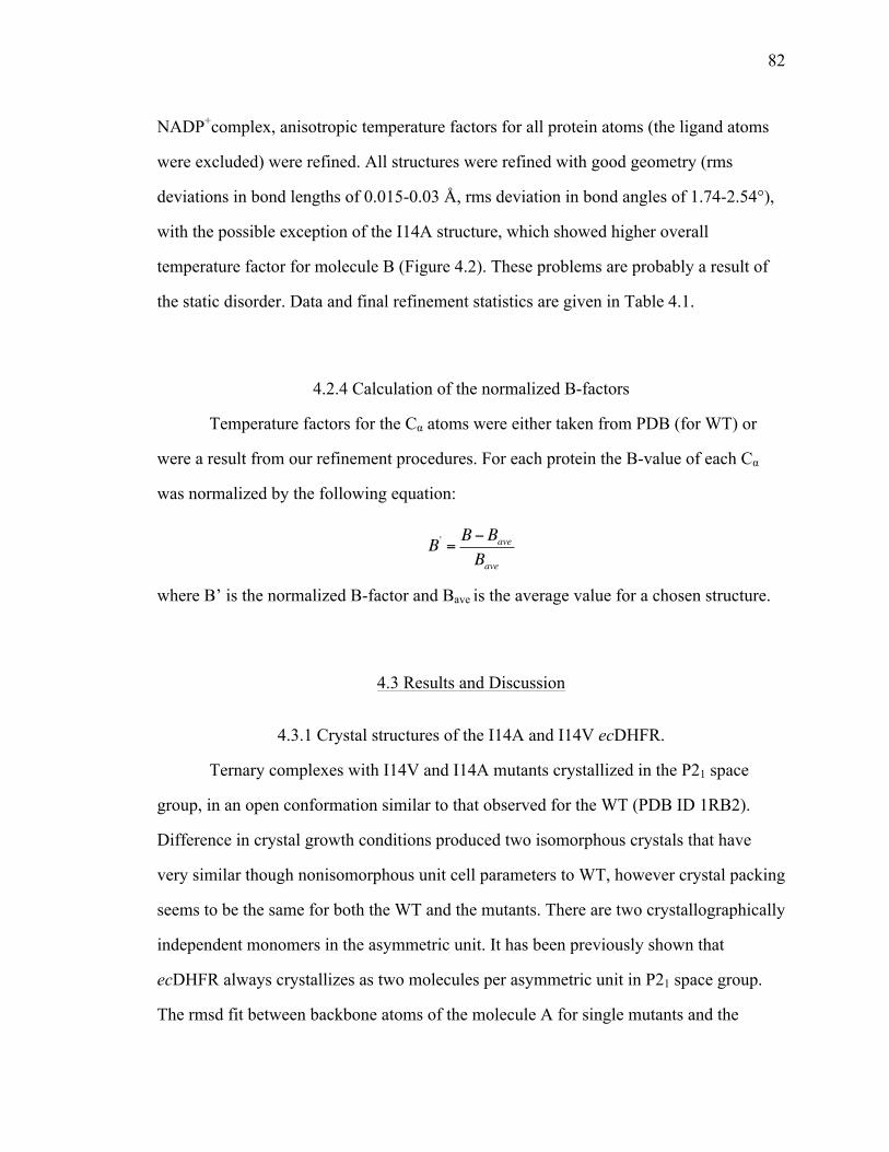
82
NADP+complex, anisotropic temperature factors for all protein atoms (the ligand atoms
were excluded) were refined. All structures were refined with good geometry (rms
deviations in bond lengths of 0.015-0.03 Å, rms deviation in bond angles of 1.74-2.54°),
with the possible exception of the I14A structure, which showed higher overall
temperature factor for molecule B (Figure 4.2). These problems are probably a result of
the static disorder. Data and final refinement statistics are given in Table 4.1.
4.2.4 Calculation of the normalized B-factors
Temperature factors for the Cα atoms were either taken from PDB (for WT) or
were a result from our refinement procedures. For each protein the B-value of each Cα
was normalized by the following equation:
B' = B−BaveBave
where B’ is the normalized B-factor and Bave is the average value for a chosen structure.
4.3 Results and Discussion
4.3.1 Crystal structures of the I14A and I14V ecDHFR.
Ternary complexes with I14V and I14A mutants crystallized in the P21 space
group, in an open conformation similar to that observed for the WT (PDB ID 1RB2).
Difference in crystal growth conditions produced two isomorphous crystals that have
very similar though nonisomorphous unit cell parameters to WT, however crystal packing
seems to be the same for both the WT and the mutants. There are two crystallographically
independent monomers in the asymmetric unit. It has been previously shown that
ecDHFR always crystallizes as two molecules per asymmetric unit in P21 space group.
The rmsd fit between backbone atoms of the molecule A for single mutants and the

83
molecule A for the wild type enzyme in its open conformation is 0.29 Å for I14V and
0.33 Å for I14A. Table 4.1 summarizes the X-ray data collection and refinement. The
difference in the number of fitted atoms between the two structures is a result of the
alternative conformations of few surface residues. The difference in the number of fitted
ligand atoms is a result of the disorder observed for the nicotinamide-ribose moiety for
NADP+ in the molecule B of I14A mutant.
4.3.1.1 NADP+cofactor binding and the architecture of the
active site
Both substrate mimic and the co-factor are present in each molecule. Kinetic and
structural studies have shown that during the kinetic cycle NADPH molecule binds to
occluded conformation of the protein, in which M20 loop protrudes into the
nicotinamide-ribose binding pocket. The initial binding of the NADPH molecule occurs
through 2’-phosphate-ADP moiety. The consequent opening of the M20 loop allows for
the nicotinamide-ribose moiety to bind in the active site. When this occurs the
nicotinamide-ribose moiety forms stabilizing van der Waals contacts and H-bonds with
residues that are part of the M20 loop. These interactions favor the closed form of the
M20 loop forming a potentially reactive conformation. It has been observed previously
that binding of the nicotinamide-ribose moiety is strongly influenced by the presence of
pterin ring. When M20 loop is in the open conformation the p-aminobenzoylglutamate
binding cleft is widened significantly creating a 8 Å opening to the nicotinamide binding
site. This opening allows for the nicotinamide-ribose moiety to be bound in the active-
site, as observed in the molecule A, however rotation about the PN-O3 bond can place
the nicotinamide-ribose moiety out into the solvent, as in molecule B. Comparison to
other ternary structures of WT ecDHFR, which crystallized in various crystalline forms
but in the open conformation, reveals the same observation.

84
Figure 4.3a shows the binding of the NADP+ and folate in the active site of I14A
ecDHFR. Several important active-site residues in direct contact with either the
nicotinamide ring of NADP+, or pterin ring of folate are also presented. As indicated by
the electron density maps the positions of all the atoms are well-defined. The overlap
with WT structure indicates no significant changes in the position of the active-site
residues or folate, however it indicates a slight displacement of the nicotinamide ring
towards the pterin ring in mutants from the original position in WT structure (Figure
4.3b). This is also evident from an increase in the hydrogen bond between the
nicotinamide O7N and I5(N) (Figure 4.4). This displacement in the nicotinamide ring
results in the slight difference in donor-acceptor distance (distance between the C4 of
nicotinamide ring to C6 of pterin ring) between the WT (3.6 Å) and I14V/I14A (3.2 Å)
enzymes.
4.3.1.2 M20 loop conformation
The open conformation is the most frequently occurring conformation
crystallographically and is the only conformation that was observed for the I14A and
I14V mutants. However, even though it has been shown that closing of the M20 loop is
an important step in the catalysis of ecDHFR, as the residues in the loop position
nicotinamide ring in the close proximity to the pterin ring, replacement of the part of the
M20 loop (residues 16-19) with a single glycine residue yields a slow, but nonetheless an
active enzyme. The hydride transfer rate is reduced by more than 500-fold, however
neither Km for H2F or NADPH are significantly affected.189 This could suggest that even
the transient population of the open conformational state may play a role in the catalytic
cycle. We attempted to crystallize these mutants in different space groups by varying
experimental conditions of crystal growth (temperature, pH, salt content, additives, etc.).
Moreover, since I14G mutant crystallized in P21212 space group, in which ternary
complexes of WT ecDHFR crystallize in closed conformation, we attempted both macro

85
and microseeding of I14A with I14G crystals. Even though we obtained crystals with
different cell dimensions and crystal morphologies, all of these crystals crystallized in the
P21 space group. These results could indicate that these mutants do not readily adopt the
closed conformational ensemble, as it was recently suggested for G121V mutant.190 This
could provide some explanation for the reduction in the hydride transfer rate constant for
these mutants.
Reduction in the DAD observed for the mutants in comparison to WT is
contradictory to the predictions based on our kinetic and MD results. There we observed
that the reduction in the size of this hydrophobic residue results in the formation of the
“steric” hole that allows for the nicotinamide ring to relax towards the residue 14, leading
to longer DADs for the mutants. However, MD simulations also indicated that
nicotinamide ring samples larger conformational space in the mutant structures, which
reflected itself in an increase in the H-bond distance between the nicotinamide O7N and
I5(N), which is consistent with the x-ray crystal structures (Figure 4.4). However, the
decrease in the DAD that we observe for the mutant structures could be a result of the
nicotinamide ring being able to sample larger conformational space, as it is not as tightly
constrained in the active site.
4.3.1.3 Analysis of the normalized B-factors
One way to extract dynamic information from static picture of an X-ray structure
is to calculate the temperature factor (B-factor) details that contain specifics regarding the
electron density distribution around a particular atom and is associated with thermal
motions present in the crystal before and after flash - freeze. However, B-factor
distribution needs to be analyzed with care, as errors in the model could also lead to high
B-factors. Rapid cooling of the crystals in the liquid nitrogen can trap different
conformational states of the protein that exist at physiological temperature. Since data
collection for the WT enzyme was performed at room temperature, WT structure shows
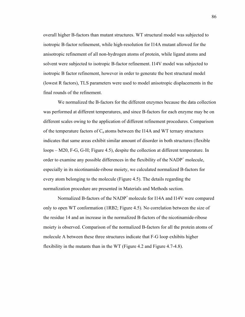
86
overall higher B-factors than mutant structures. WT structural model was subjected to
isotropic B-factor refinement, while high-resolution for I14A mutant allowed for the
anisotropic refinement of all non-hydrogen atoms of protein, while ligand atoms and
solvent were subjected to isotropic B-factor refinement. I14V model was subjected to
isotropic B factor refinement, however in order to generate the best structural model
(lowest R factors), TLS parameters were used to model anisotropic displacements in the
final rounds of the refinement.
We normalized the B-factors for the different enzymes because the data collection
was performed at different temperatures, and since B-factors for each enzyme may be on
different scales owing to the application of different refinement procedures. Comparison
of the temperature factors of Cα atoms between the I14A and WT ternary structures
indicates that same areas exhibit similar amount of disorder in both structures (flexible
loops – M20, F-G, G-H; Figure 4.5), despite the collection at different temperature. In
order to examine any possible differences in the flexibility of the NADP+ molecule,
especially in its nicotinamide-ribose moiety, we calculated normalized B-factors for
every atom belonging to the molecule (Figure 4.5). The details regarding the
normalization procedure are presented in Materials and Methods section.
Normalized B-factors of the NADP+ molecule for I14A and I14V were compared
only to open WT conformation (1RB2; Figure 4.5). No correlation between the size of
the residue 14 and an increase in the normalized B-factors of the nicotinamide-ribose
moiety is observed. Comparison of the normalized B-factors for all the protein atoms of
molecule A between these three structures indicate that F-G loop exhibits higher
flexibility in the mutants than in the WT (Figure 4.2 and Figure 4.7-4.8).

87
4.3.2 Crystal structure of the I14G ecDHFR.
Ternary complex with I14G ecDHFR crystallized in the P21212 space group with
a single monomer in the asymmetric unit. Comparable WT crystal structure (PDB ID
1RX2) crystallized in the same space group, P212121, with the nonisomorphous unit cell
parameters to I14G. In P212121 space group WT crystallizes in the closed conformation,
which has been proposed to be the representation of the reactive conformation. The rmsd
fit between backbone atoms of I14G mutant and the wild type enzyme in its closed
conformation is 0.28 Å. Table 4.1 summarizes the X-ray data collection and refinement.
The difference in the number of fitted atoms between the two structures is a result of the
alternative conformations observed for few surface residues. Moreover, parts of the F-G
loop for I14G mutant were not modeled in due to sporadic electron density in those
regions. OMIT maps were used when such structural features were in doubt.
4.3.2.1 NADP+ cofactor binding, the architecture of the
active site and the M20 loop conformation
Analysis of the Fo-Fc maps of the cofactor-binding region for the I14G mutant,
after molecular replacement (with ligands removed), indicated that NADP+ is bound.
However, poor electron density for the nicotinamide-ribose moiety could not indicate if it
is disordered in the active site, or if it is bound out of the active site and disordered in the
solvent, or both (we modeled NADP+ molecule in the active site; Figure 4.9). We
generated iterative composite OMIT map in order to resolve this structure feature. The
map suggests that there is no electron density either in the active site or outside of it that
could be assigned to the nicotinamide ribose moiety (Figure 4.10). We also observed
poor electron density for the M20 and F-G loops, two loops that are in direct contact with
each other. Parts of the loops have been modeled in as suggested by iterative composite
OMIT map (Figure 4.10). Previous structural studies on WT ecDHFR showed that the
mobility of the M20 loop depends on the freedom of movement of the F-G loop. Detail
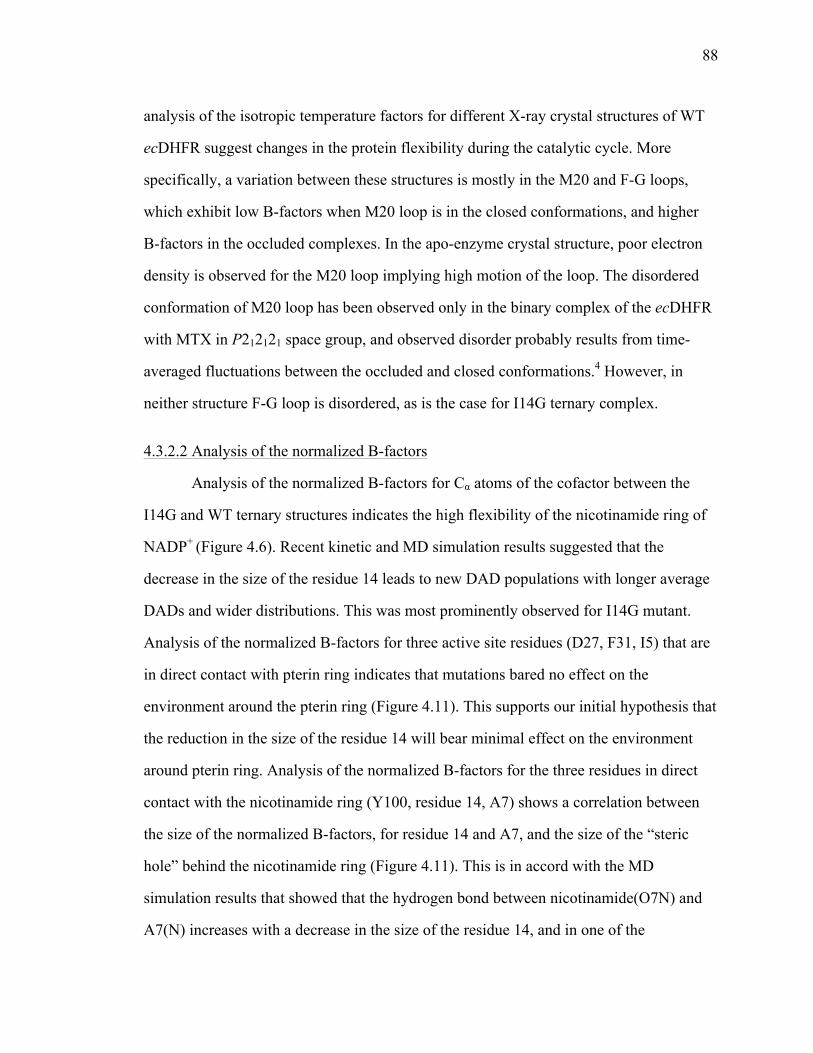
88
analysis of the isotropic temperature factors for different X-ray crystal structures of WT
ecDHFR suggest changes in the protein flexibility during the catalytic cycle. More
specifically, a variation between these structures is mostly in the M20 and F-G loops,
which exhibit low B-factors when M20 loop is in the closed conformations, and higher
B-factors in the occluded complexes. In the apo-enzyme crystal structure, poor electron
density is observed for the M20 loop implying high motion of the loop. The disordered
conformation of M20 loop has been observed only in the binary complex of the ecDHFR
with MTX in P212121 space group, and observed disorder probably results from time-
averaged fluctuations between the occluded and closed conformations.4 However, in
neither structure F-G loop is disordered, as is the case for I14G ternary complex.
4.3.2.2 Analysis of the normalized B-factors
Analysis of the normalized B-factors for Cα atoms of the cofactor between the
I14G and WT ternary structures indicates the high flexibility of the nicotinamide ring of
NADP+ (Figure 4.6). Recent kinetic and MD simulation results suggested that the
decrease in the size of the residue 14 leads to new DAD populations with longer average
DADs and wider distributions. This was most prominently observed for I14G mutant.
Analysis of the normalized B-factors for three active site residues (D27, F31, I5) that are
in direct contact with pterin ring indicates that mutations bared no effect on the
environment around the pterin ring (Figure 4.11). This supports our initial hypothesis that
the reduction in the size of the residue 14 will bear minimal effect on the environment
around pterin ring. Analysis of the normalized B-factors for the three residues in direct
contact with the nicotinamide ring (Y100, residue 14, A7) shows a correlation between
the size of the normalized B-factors, for residue 14 and A7, and the size of the “steric
hole” behind the nicotinamide ring (Figure 4.11). This is in accord with the MD
simulation results that showed that the hydrogen bond between nicotinamide(O7N) and
A7(N) increases with a decrease in the size of the residue 14, and in one of the

89
populations observed for I14G mutant it is completely broken. This loss in rigidity is
reflected in higher normalized B-factors for A7.
4.4 Conclusion
The kinetic analysis on the series of active site mutants of residue 14 (i.e., I14V,
A, G) shows a change in the DAD population distribution at TRS for mutants.85 MD
simulation data indicated that mutations affect the enzyme’s ability to organize the active
site for hydride transfer. High-resolution structures for series of active-site mutants
suggest some perturbations in ground state geometry and motions of certain active-site
residues as well as two flexible loops. Moreover, the removal of steric bulk at position
14 could also lead to an increase in the flexibility of the nicotinamide ring. This has been
previously observed for mutants of analogous residue in horse-liver alcohol
dehydrogenase.191 Inability to obtain closed conformation of the mutants could be a direct
result of the mutations, which led to a shift in the relative populations of open and closed
forms. This shift in the relative populations of open and closed forms may contribute to
the reduced activity seen for the mutants. The high-resolution ecDHFR structures
obtained for these mutants could be used for future simulation of hydride tunneling in
ecDHFR by QM/MM computational methods.
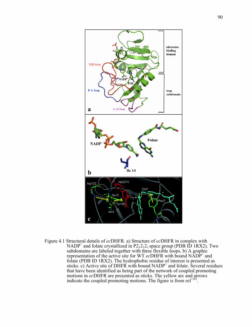
90
Figure 4.1 Structural details of ecDHFR. a) Structure of ecDHFR in complex with NADP+ and folate crystallized in P212121 space group (PDB ID 1RX2). Two subdomains are labeled together with three flexible loops. b) A graphic representation of the active site for WT ecDHFR with bound NADP+ and folate (PDB ID 1RX2). The hydrophobic residue of interest is presented as sticks. c) Active site of DHFR with bound NADP+ and folate. Several residues that have been identified as being part of the network of coupled promoting motions in ecDHFR are presented as sticks. The yellow arc and arrows indicate the coupled promoting motions. The figure is from ref 181.
b
c
a
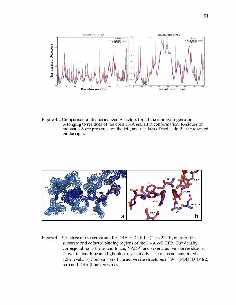
91
Figure 4.2 Comparison of the normalized B-factors for all the non-hydrogen atoms belonging to residues of the open I14A ecDHFR conformation. Residues of molecule A are presented on the left, and residues of molecule B are presented on the right.
Figure 4.3 Structure of the active site for I14A ecDHFR. a) The 2Fo-Fc maps of the substrate and cofactor binding regions of the I14A ecDHFR. The density corresponding to the bound folate, NADP+ and several active-site residues is shown in dark blue and light blue, respectively. The maps are contoured at 1.5σ levels. b) Comparison of the active site structures of WT (PDB ID 1RB2; red) and I14A (blue) enzymes.
!
a" b"
!
Residue!number! Residue!number!
Normalized!B
2factors!

92
Figure 4.4 Interactions of NADP+ with active site residues in I14A mutant (left) and WT (PDB ID 1RB2, right). Hydrogen bonds with their respective values are shown as dotted lines. The overlapping residues are highlighted. The figure was generated using LigPlus.
Figure 4.5 Putty cartoon of B-factor variation on the I14A (left) and WT (right) ternary structures, colored from low to high (blue to red).

93
Figure 4.6 Comparison of the normalized B-factors for all the non-hydrogen atoms in the NADP+ molecule starting from the adenine ring to the nicotinamide ring. Details about the normalization of the B-factors are presented in the materials and methods. Different colors are representation of normalized B-factors for different crystal structures in the following order: 1RX2 (WT ecDHFR, P212121; red), 1RB2 (WT ecDHFR, P21; molecule A – green; molecule B – blue), I14A (molecule A - light blue; molecule B - yellow), I14V (molecule A – black; molecule B – grey), and I14G (purple). Since I14V and I14A crystallize as two molecules per asymmetric unit, we present the normalized B-factors for molecule A in which the nicotinamide ring of NADP+ is bound in the active site.
I14G I14V
WI14
WT

94
Figure 4.7 Comparison of the normalized B-factors for all the non-hydrogen atoms belonging to residues of the open I14V ecDHFR conformation. Residues of molecule A are presented on the left, and residues of molecule B are presented on the right.
!
Normalized!B
-factors!!
Residue!number!Residue!number!
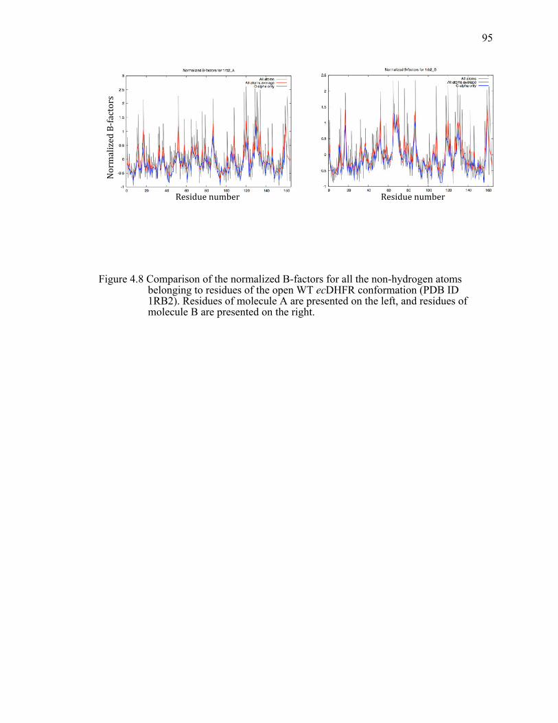
95
Figure 4.8 Comparison of the normalized B-factors for all the non-hydrogen atoms belonging to residues of the open WT ecDHFR conformation (PDB ID 1RB2). Residues of molecule A are presented on the left, and residues of molecule B are presented on the right.
!
Normalized!B
-factors!
Residue!number! Residue!number!

96
Figure 4.9 The 2Fo-Fc maps of the substrate and cofactor binding regions of the I14G ecDHFR complexed with NADP+ and folate. Maps are contoured in blue at 1.5σ levels. Difference maps are contoured in green and red at 3σ levels. Ligand models and protein backbone are shown in pink.
!
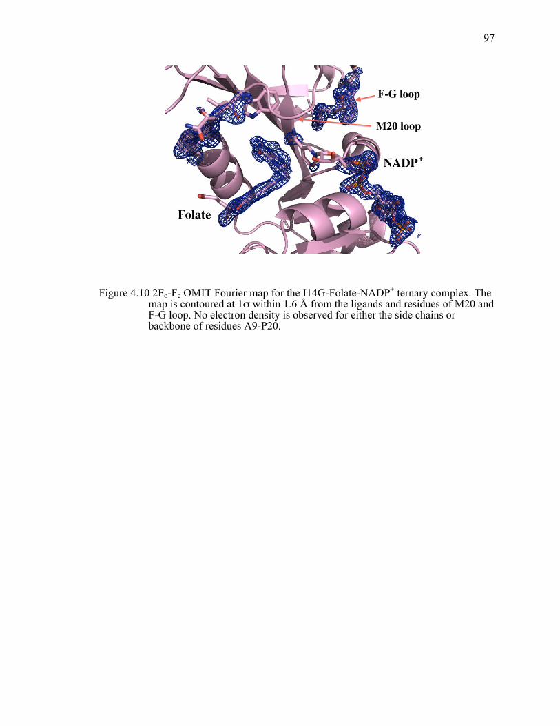
97
Figure 4.10 2Fo-Fc OMIT Fourier map for the I14G-Folate-NADP+ ternary complex. The map is contoured at 1σ within 1.6 Å from the ligands and residues of M20 and F-G loop. No electron density is observed for either the side chains or backbone of residues A9-P20.
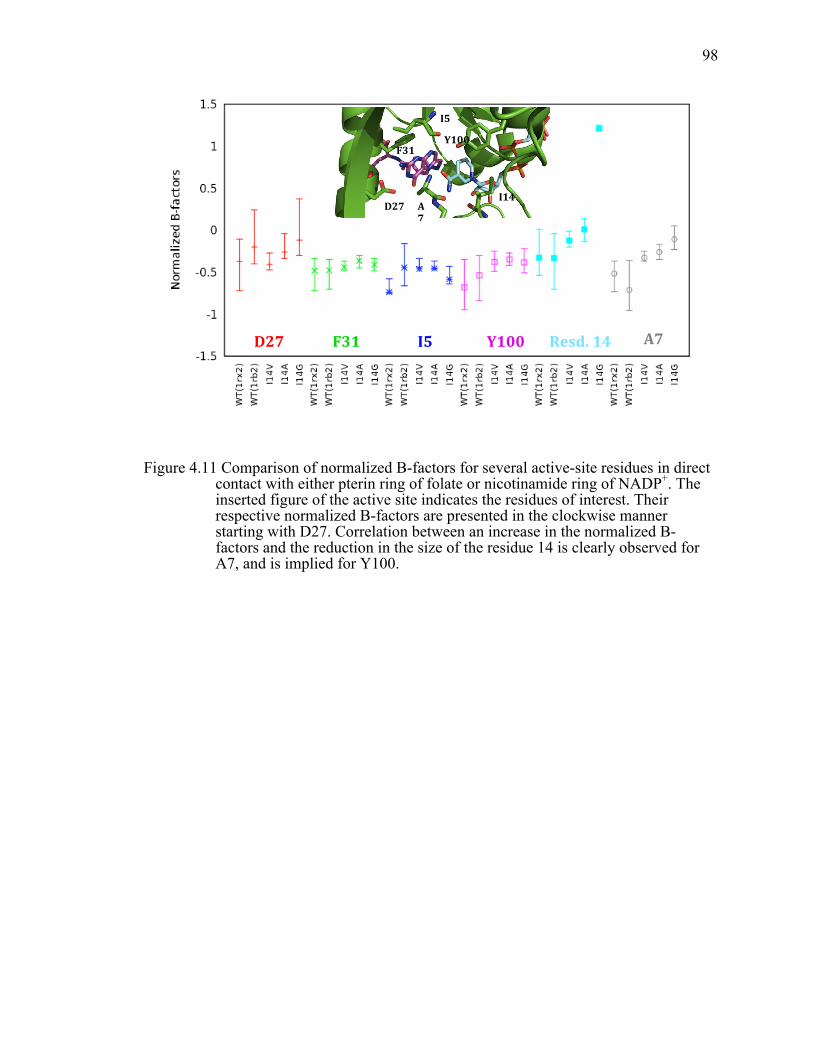
98
Figure 4.11 Comparison of normalized B-factors for several active-site residues in direct contact with either pterin ring of folate or nicotinamide ring of NADP+. The inserted figure of the active site indicates the residues of interest. Their respective normalized B-factors are presented in the clockwise manner starting with D27. Correlation between an increase in the normalized B-factors and the reduction in the size of the residue 14 is clearly observed for A7, and is implied for Y100.
!
D27$7$
F31$1$
I5$5$
Y100$0$
Resd.$14$14$
I14$
4$A7$
D27$
7$
F31$1$
I5$5$Y100$0$
A7$$$

99
Table 4.1 Data collection and refinement statistics for I14V, I14A and I14G ecDHFR mutants.
I14V DHFR I14A DHFR I14G DHFR
PDB ID XXX XXX XXX Data collection Source ALS (LBNL) BeamLine 4.2.2 Temperature (K) 100 100 100 Wavelenght (Å) 1.000 0.855 1.000 Space group P21 P21 P21212 Cell Dimensions a, b, c (Å) 35.08 60.25
74.18 35.12 59.51 74.23
33.49 34.44 134.25
a, b, g (˚) 90.00 95.97 90.00
90.00 95.53 90.00
90.00 90.00 90.00
Resolution (Å) 1.5 1.35 1.45 Rmerge 0.040 (0.166) 0.031 (0.565) 0.048 (0.372) <I/s(I)> 17.3 (3.6) 21.2 (2.3) 21.8 (3.6) Completeness (%) 95.6 (89.7) 99.7 (100) 97.3 (84.3) Multiplicity 3.86 (3.75) 3.7 (3.7) 6.5 (4.0) Mosaicity (˚) 0.57 0.178 0.191 B factor from Wilson plot (Å2)
17.9 13.8 12.2
Refinement Resolution Range (Å) 73.78-1.50 73.88-1.35 67.13-1.45 No. reflections 44649 63230 26211 No. unique reflections Rwork/Rfree 17.41/23.11 16.42/19.88 16.67/21.91 No. atoms Protein 2601 2630 1221 Ligand/ion 160 143 80 Water 496 352 162 B-factors Protein 22.50 20.6 18.5

100
Table 4.1 Continued Ligand/ion 27.6 20.3 28.9 Water 19.8 30.6 30.6 R.m.s. deviations Bond lengths (Å) 0.029 0.015 0.025 Bond angles (˚) 2.537 1.738 2.196 Ramachandran plot Favored (%) 98.76 100.00 98.03 Allowed (%) 1.24 0.00 1.97 Disallowed (%) 0.00 0.00 0.00

101
CHAPTER 5.
LOOKING ON THE OTHER SIDE: ROLE OF THE RESIDUE F31 IN
THE DHFR CATALYZED HYDRIDE-TRANSFERv
5.1 Introduction
The role of the dynamical effects in assisting enzymatic reactions is quite
complex and therefore still widely debated. It is now generally accepted that enzyme
motions can play a significant role in different parts of reaction cycle such as substrate
binding, product release, and large conformational changes necessary for the stabilization
of the intermediates. These dynamical changes occur over different timescales ranging
from the domain motions (seconds) to bond vibrations (femtoseconds). Due to
complexity of enzyme dynamics, it is quite challenging to examine the dynamic
contributions to the enzyme catalyzed reaction. Subpicosecond vibrational modes that
may cause transient compression along the reaction coordinate in H-transfer reactions
have been studied on different enzymatic systems such as: morphinone reductase,66,163,192
dihydrofolate reductase,1,84,85,193 yeast alcohol dehydrogenase,72,194 horse-liver alcohol
dehydrogenase,39,195 formate dehydrogenase,87 and more. Such motions are believed to
compress the reaction barrier and lead to degeneration of the donor and acceptor’s energy
levels, therefore enhancing both quantum mechanical (QM) tunneling probability of the
transferred hydrogen, and over-the-barrier transfer. Due to experimental limitations in
probing such motions, their role has been usually inferred indirectly through
measurement of pressure/temperature dependence of kinetic isotope effects (KIEs), by
molecular dynamic (MD) simulations, or by quantum-mechanical/molecular mechanics
(QM/MM) calculations. However, recent advances in the two-dimensional infrared
v This chapter is in preparation to be submitted to the journal of Biochemistry

102
spectroscopy (2D-IR) allowed for an experimental way to measure the femto- to
picosecond timescale motions;125 1H-15N heteronuclear NOE can also provide some
information about these fast timescale motions.1,5,179
The analysis of the temperature dependence motivated the development of various
phenomenological models denoted here as Marcus-like models. For more in-depth
discussion on these models please refer to following publications: 39,44,57,65,66,73,85,89,125,127,173-
177,196. In short, according to this model, after the formation of the reactive complex (a
process sometimes referred to as preorganization), heavy-atom motions are necessary to
bring the system to a point where donor and acceptor wells are degenerate and hydrogen
tunneling is plausible (i.e. tunneling ready state, TRS).39,58,62,72,74,116,118,120,127,174 This
process is often referred to as reorganization, as it assists the structural changes in the
substrate that occur as the reaction proceeds from ground state to TRS. At the TRS,
fluctuations in the donor-acceptor distance (DADe) vi affect the tunneling probability, and
therefore will be a determinant factor of the magnitude and the temperature dependence
of the KIEs. Certain QM/MM calculations support this view and suggest that temperature
dependence/independence of the intrinsic KIEs mainly reflects the temperature
dependence of DAD.51,59,197,198 According to Marcus-like models, temperature
independent KIEs observed for majority of the wild-type (WT) enzymes68,83,87,120,123,199-
201 indicate systems wherein the active-site at the TRS is optimal for H-tunneling with a
short average DADe and narrow DAD distribution. These models suggest that shorter
DADe decreases the KIE, and broader DADe distribution (lower DADe-fluctuations
frequency) leads to elevated ∆Ea (more temperature dependent KIEs). However, certain
experimental and computational studies have also shown that compression of DADc
vi DADe here refers to the distance from the H-reactant state to the H-product state, rather
than to the distance between two carbons - donor and acceptor distance (DADc). The first directly reflects the kinetic and energetic effects of the H-transfer, while the second reflects the heavy atoms conformation as observed in the x-ray crystal structures and MD simulations. For example, for a linear C-H-C transfer DADc = DADe + 2 x C-H distance.

103
alone might not increase the hydride transfer rate and/or decrease the barrier height, or
that a decrease in the DADc needs to necessarily lead to a decrease in tunneling.200,202-204
Here, we report kinetic, structural and MD simulation studies on the impact of the
active-site mutation on the hydride transfer reaction in dihydrofolate reductase from
Escherichia coli (ecDHFR). EcDHFR is a small, monomeric enzyme that catalyzes the
transfer of the pro-R-hydride from the C4 position of NADPH to the re-face of C6
position on 7,8-dihydrofolate (H2F) to produce S-5,6,7,8-tertrahydrofolate (H4F). The
hydride transfer in ecDHFR has been well characterized, with both experimental and
computational studies suggesting that thermally activated fluctuations of the DAD are
important in assisting hydride transfer.63,83-85,198,205The study on a series of active site
mutants of residue 14, situated behind the nicotinamide ring, showed an increase in the
temperature dependence of the KIEs (∆Ea) with a decrease in the size of this active site
residue.85 One of roles of this highly conserved residue was suggested to be in assisting
the hydride transfer by keeping the nicotinamide ring in the close proximity to the pterin
ring, i.e., restricting the DAD to short and narrow conformational space in the WT
enzyme. Another residue of similar interest is F31, a strictly conserved hydrophobic
residue,206 located behind the pterin ring of H2F (H-acceptor), opposite from residue 14
(Figure 5.1). It interacts with the pteroyl moiety of H2F through van der Waals contacts
with the edge of the phenyl ring toward faces of both the pterin ring and p-aminobenzoyl
group.207 As evident from the Figure 1, in contrast to I14, which only interacts with the
nicotinamide ring of NADP+, folate is “wrapped around” F31, which seems to anchor all
three moieties of the substrate (pteryl, p-aminobenzoyl, and glutamyl). Residue F31 has
been shown to be very important for both the binding of the H2F as well as catalyzed
reaction.208 The importance of hydrophobic interactions in binding of drugs, such as
methotrexate (MTX) and 2,4-diamino-6,7-dimethylpteridine (DAM), has been shown
previously, where F31V mutation lead to 140-fold decrease in the binding constant for

104
MTX,207 as well as 30-fold decrease in the binding constant of the H2F (Table 5.1 and
refs 207-209).
QM/MM calculations suggested that residue F31 belongs to a network of coupled
promoting motions.18,181 More specifically, it was shown that as reaction evolves from the
reactant to the transition state, residue 31 moves closer to the substrate, while the angle
between the acceptor carbon and the methylene amino linkage in H2F increases and the
DADc decreases. Based on these experimental and computational studies on ecDHFR, as
well as relevant studies of active site mutants in other enzymes,66,123,134,210 we decided to
extend our initial study on the residue I14 to F31V mutant. We examined the effect of
this mutation on the nature of the hydride transfer by measuring the temperature
dependence of the intrinsic KIEs, by obtaining X-ray crystal structures of the relevant
ternary complex, and by performing MD simulations.
5.2 Materials and methods
5.2.1 Construction of expression vector.
The sequence of the mutagenic, forward primer (F31V-Fwrd) is 5’- GCC-GAT-
CTC-GCC-TGG-GTT-AAA-CGC-AAC-ACC-3’, and the sequence of the mutagenic,
reverse primer (F31V-Rev) is 5’- GGT-GTT-GCG-TTT-AAC-CCA-GGC-GAG-ATC-
GGC-3’, where the site of the mutation is underlined. Site-directed mutagenesis was
completed using pET22b-WTecDHFR as a template and following the protocol provided
in the Stratagene instruction manual. The sequence of the final plasmid, containing the
desired mutation, was verified with automated DNA sequencing through the DNA
facility-University of Iowa. Primers were purchased from Integrated DNA Technologies.
Expression and Purification of F31V DHFR. The mutant was expressed in the
E.coli strain ER2566, which contains a deletion of the DHFR gene (folA) and
thymidylate synthase (thyA), and was a generous gift from S. J. Benkovic at the Penn

105
State University (University Park, PA). The enzyme was purified and stored as reported
for other DHFR mutants.106
5.2.2 Preparation of F31V DHFR ternary complexes for
crystallization.
Purified protein was either stored at -80°C until further use, or loaded onto a
Superdex-75 (GE) gel filtration column pre-equilibrated with the buffer containing 20
mM Tris-HCl, pH 7.5. DHFR eluted in a single peak, which was tested for the
monodispersity using dynamic light scattering on the NanoStar (Wyatt Technology).
Concentration of the pooled fractions was measured using UV absorbance
(ε280=74.6 mM-1cm-1) and Bradford assay. Due to the low solubility of folate and ~20-
fold higher Kd(folate) for F31V DHFR than for the WT ecDHFR, folate was added to a
dilute protein solution at ~10 fold molar excess relative to F31V DHFR mutant. The
sample was then incubated on ice for at least 1 hour, and afterwards filtered using
Milipore 0.22um filter. Lastly, the binary complex was concentrated to a final
concentration of ~15 mg/ml (as determined by Bradford assay). NADP+ was added as a
solid to the concentrated binary DHFR-Folate complex at a five-fold molar excess to
enzyme. Preparation of ternary complex with MTX and NADP+ was performed in the
same manner as for the enzyme-Folate-NADP+ complex, however no filtering was done
prior to concentrating the enzyme-MTX binary complex.
5.2.3 Crystallization conditions.
Both enzyme-Folate-NADP+ and enzyme-MTX-NADP+ ternary complexes were
crystallized via the hanging drop vapor diffusion method by mixing 400 nl well solution
and 400 nl of prepared ternary complex using TTP LabTech Mosquito. In more detail:
106
F31V-NADP+-Folate ternary complex crystallized in the P21 space group with
two molecules in the asymmetric unit, under the following conditions: 15 mg/ml F31V
DHFR, 0.1M Tris pH 8.5, 0.5M LiCl and 30.5% w/v PEG 6,000; at 18°C.
F31V-NADP+ binary complex crystallized in the P21 space group with two
molecules in the asymmetric unit, under the following conditions: 13.5 mg/ml F31V
DHFR, 0.1 M HEPES pH 7.5, 25% w/v PEG 6,000; at 18˚C. This crystal was a result of
the failed attempt to obtain the ternary F31V-NADP+-Folate complex.
F31V-NADP+-MTX ternary complex crystallized in the P212121 space group with
two molecules in the asymmetric unit, under the following conditions: 16 mg/ml F31V
DHFR, 0.1 M Bis-Tris propane pH 6.5, 20% w/v PEG 3,350, 0.2 M sodium acetate
anhydrous; at 18˚C. Crystal initially appeared after 10 days, but was harvested after 3
weeks.
5.2.4 Data collection and structure determination.
Crystals were flashed-cooled in liquid nitrogen, and data were collected at 100 K
at the 4.2.2 synchotron beamline at the Advance Light Source of the Lawrence Berkley
National Laboratory. The data were either processed using d*Trek (for F31V-MTX-
NADP+),183 mosflm (for F31V-NADP+),211 or XDS (for F31V-Folate-NADP+).184 We
faced initial difficulties in processing the data for the F31V-MTX-NADP+ complex due
to multiple lattices being present in the crystal (partially reflected in the mosaicity; Table
5.3), and the twinning of the crystal. Initial refinement indicated P43212 space group, with
single molecule present in the asymmetric unit; however, further rounds of refinement
did not yield a model with desirable statistics for a structure of this resolution. Analysis
of the data with Xtriage indicated significant twinning and Zanuda suggested P212121
space group, whose unit cell parameters are very similar and close to be considered as
P43212 space group.
107
The structures were determined by molecular replacement in CCP4185 (with
PHASER186), using the coordinates for the refined WT ecDHFR structure (PDB 1RX2)
with ligands removed to generate the initial model. Refinement and model building were
done using REFMAC187 and COOT188. Model bias was avoided during the initial
refinement by removing the substrate and cofactor from the model. Moreover, iterative
composite OMIT maps were used for the F31V-MTX-NADP+ complex where structure
features were in doubt. In all structures hydrogens were included in riding positions, and
isotopic temperature factors were refined. Alternative conformations of amino acids and
cofactor were assigned on the basis of the visual evidence such as divergent electron
densities at the +1σ level above the average in 2|Fo|-|Fc| maps, and from either simple
composite OMIT maps or iterative composite OMIT maps. The final models were well
described by the electron density maps. However, side chains of the certain residues
situated on the surface of the protein (in most cases lysine, arginine and glutamic acid
residues) were not well described by density. These side chains were not deleted, but
were left with their occupancies equal to one, with temperature factors reflecting their
disorder. Final structures were checked with MolProbity.212 All residues have favored or
allowed backbone conformational angles. Several residues occupy unusual rotameric
states, however, clear and strong electron densities are in support of such positions. Data
and final refinement statistics are given in Table 5.3.
5.2.5 Synthesis of Labeled Cofactors for 1˚KIEs.
[Ad-14C]-NAD+ (specific radioactivity of >220mCi/mmol) was purchased from
Amersham Pharmacia, glucose dehydrogenase from Bacillus megaterium (GluDH) was
obtained from Affymetrix/USB, while all the other compounds were purchased from
Sigma. Synthesis, purification and storage of the labeled cofactors have been presented in
detail before.95,111 Briefly: (R)-[4-3H]NADPH (680 mCi/mmol) was synthesized by

108
reduction of NADP+ with 2-Deoxy-D-Glucose-3H using glucose dehydrogenase from
Bacillus megaterium (GluDH), followed by oxidation of the resulting NADPH with
acetone using alcohol dehydrogenase from Thermoanaerobium brockii (tbADH), and
final reduction using GluDH and unlabeled glucose, as described previously.111 [Ad-14C]-
NADPH (50mCi/mmol) was prepared by phosphorylation of [Ad-14C]-NAD+ using
NAD+ kinase from chicken liver to produce [Ad-14C]-NADP+, followed by reduction
with glucose using GluDH as described elsewhere.95 (R)-[Ad-14C,4-2H]-NADPH (50
mCi/mmol) was prepared from [Ad-14C]-NADP+, which was synthesized as described
above, and subsequent reduction with deuterated glucose and GluDH.142 All synthesized
cofactors have been purified by semipreparative reverse-phase HPLC on a Supelco
column and stored at -80 ˚C prior to use as discussed elsewhere.109
5.2.6 Kinetic isotope effect measurements and
determination of the intrinsic KIEs.
(R)-[4-3H]NADPH and [Ad-14C]-NADPH, and, (R)-[4-3H]NADPH and (R)-[Ad-14C,4-2H]-NADPH were combined in the 5:1 radioactivity ratio in order to measure H/T
and D/T 1˚KIEs, respectively. Each mixture was co-purified on an analytical reverse-
phase HPLC column, divided into aliquots containing 300,000 dpm of 14C, and frozen in
liquid nitrogen for short-term storage at -80 ˚C. All experiments were performed in
MTEN buffer (50 mM MES, 25 mM Tris, 25 mM ethanolamine and 100 mM NaCl) at
pH=9.0 over the 5-45 ˚C temperature range. All reaction mixtures contained around 400-
fold excess of H2F (1.7 mM) over NADPH (4 µM). The reactions were quenched by
adding an excess of methotrexate (Kd=1 nM), and samples were stored on dry ice prior to
HPLC analysis103. Before the HPLC-LSC analysis, samples were thawed and bubbled
with oxygen to ensure complete oxidation of the product H4F. The samples were then

109
separated by reverse-phase HPLC using a method described elsewhere.109 The observed
KIEs were calculated according to ref. 29 using the following equation:
)](1ln[
)1ln(
∞
−
−=
RRf
fKIEt
where the fractional conversion (f) was determined from the ratio of 14C in the product
and reactant, and Rt and R∞ are the ratio 3H/14C at each time point and at infinite time,
respectively. The intrinsic KIEs were calculated using the modified Northrop
equation35,74
1)/(1)/(
1)(
1)(34.3/1
1
1
1
−
−=
−
−
−
−
−
−
TH
TH
DT
HT
kkkk
KVKV
obs
obs
where T(V/K)Hobs and T(V/K)Dobs are the observed H/T and D/T KIEs, respectively, and
kH/kT represents the intrinsic H/T KIE. The intrinsic KIEs were calculated numerically
from all possible combinations of observed H/T and D/T values. The isotope effects on
the activation parameters for the intrinsic KIEs were calculated by a nonlinear fit of all
intrinsic values to the Arrhenius equation for KIEs:
kl / kh = Al / Ah •eΔEah−l /RT
where kl and kh are the rate constants for light and heavy isotopes, respectively, Al/Ah is
the isotope effect on the Arrhenius preexponential factor, ∆Ea h-l is the difference in
energy of activation between the two isotopic reactions, R is the gas constant and T is the
absolute temperature.
5.2.7 Presteady-state kinetics.
All kinetic measurements were obtained on a stopped-flow apparatus (Applied
Photophysics Ltd., Leatherhead, U.K.), with dead time of 1.6 ms, a 2-mm path length,

110
and a thermally regulated sample cell. The enzyme reaction was monitored by taking
advantage of fluorescent resonance energy transfer (FRET) between DHFR and
NADPH.6 Enzyme tryptophans were excited at 290 nm, and FRET results in the strong
fluorescence by the reduced cofactor at 450 nm, which is observed through a 390 nm
output filter. All experiments were carried out at 25˚C and pH=7 in a 1x MTEN buffer.
The concentrations of substrates were determined spectrophotometrically by using the
extinction coefficients of 6200 M-1 cm-1 at 340 nm (for NADPH), and 28000 M-1cm-1 at
282 nm (for H2F). In the experiment, 20 µM enzyme (either wtDHFR or the F31V
mutant) was preincubated with NADPH (10 µM) in MTEN buffer at 25˚C. The reaction
was initiated by rapid mixing with 300 µM H2F. We used the average of at least five runs
from three independent experiments for data analysis. All data were analyzed by non-
linear least squares fits to single exponential function using KaleidaGraph 3.5.
5.2.8 Determination of temperature stability using dynamic
light scattering (DLS)
Melting temperatures (Tm) were determined for each of the enzyme-folate
complexes. WT and F31V ecDHFRs were purified using previously published
protocol,137 concentrated to ~ 10-15 mg/ml and stored either as an apoenzyme at -80 °C
or as a binary complex with folate at -20 °C. Prior to Tm measurements, ~2ml of either
sample was thawed on ice. Sample was purified on the Superdex-75 (GE) gel filtration
column pre-equilibrated with the buffer containing 20 mM Tris-HCl, pH 7.5. Fractions
containing pure enzyme were pooled together, concentrated to ~1-10 mg/ml, spun down
for 10 min at 10,000 rpm and 4 ˚C, and filtered using 0.02 µm Whatman filter. The final
concentration was determined spectrophotometrically (ε280nm = 74.6 mM-1cm-1) and was
at least 1.0 mg/ml, equivalent to 55 µM as a monomer. The measurements were
performed at temperatures from 35 to 80˚C at a scan rate of 1.0 ˚C/min. Size distributions

111
were measured with a Dynapro Nanostar (Wyatt Technology Malvern Instruments,
USA), based on DLS. The particle distribution from DLS measurements was derived by
deconvolution of the measured intensity autocorrelation function of the sample.
Generally, deconvolution was accomplished using a non-negatively constrained least
squares (NNLS) fitting algorithm included in the Dynamics software. The z-average
molecular “size” in terms of the hydrodynamic diameter (dH) in solution was determined.
The viscosity of the buffer used was calculated using a solvent builder interface and takes
the effects of buffer salts into account. For both F31V mutant and the WT ecDHFR the
hydrodynamic radii exhibited a sigmoid relationship as a function of temperature. We
used Dynamics program for fitting in order to obtain temperature and radius at the
inflection point of the sigmoidal curve.
5.2.9 Numerical modeling
The numerical modeling was based on the Kuznetsov and Ulstrup model for
adiabatic H-transfer reactions,57 but with significant modifications relative to other
attempts to implement this type of model. The model and its implementation have been
described previously,73 and a simple program that can fit and explain any level of
temperature dependence of KIE is available free of charge at
http://chemmath.chem.uiowa.edu/webMathematica/kohen/marcuslikemodel.html
5.2.10 MD simulations.
MD simulations on F31V DHFR were performed using the Amber9 package213,
as it was previously done for active site mutants of residue I14.85 Details regarding the
system setup, ligand parameterization, MD runs, data fitting and analysis are presented in
ref 85. Briefly, all ten 6 ns simulations were run starting from the crystal structure of
wtDHFR in complex with folate and NADP+ (PDB entry 1RX2).144,213 The folate and

112
NADP+ ligands were replaced by N5 protonated 7,8-dihydrofolate (H2F) and NADPH
respectively. The protonation state for all ionizable residues was set corresponding to pH
7, and the final protein structure was solvated with a previously equilibrated truncated
octahedron box of TIP3P water molecules. The size of the box ensures that all protein
atoms are at least 9 Å away from the edges of the box. Subsequent mutations were
performed in silico by changing the corresponding amino acid in the original structure.
During the simulations AMBER99 force field154 parameters were used for all residues.
The NADP+ and NADPH parameters were taken from the Amber parameter database.155
Gaff force field156 parameters together with RESP157 charges calculated at the HF/6-31G*
level were used to generate the parameter files for H2F. All simulations were run with the
PMEMD module of the AMBER9 package.144,213 An equilibration protocol was applied
that consists of performing an initial energy minimization, followed by a slow heating to
the desired temperature using a linear temperature ramp from 100 to 300 K during 80 ps
at constant volume and a subsequent pressure stabilization run at 300 K and 1 bar over
100 ps. Position frames, which were used for analyzing trajectories, were collected at 2
ps intervals. The ptraj module of Amber9 was used to analyze the data extracted from the
MD simulations. Data from the DADc distribution were fitted with a combination of
Gaussian and Split Gaussian functions using the open source software for non linear
curve fitting, Fityk.162 For the equations and procedures used for the fitting please refer to
the Chapter 3.
113
5.3 Results and Discussion
5.3.1 Temperature dependence of intrinsic KIEs for F31V
DHFR; isotope effects on activation Arrhenius parameters
and thermal stability of F31V vs. WT
We measured 1˚ H/T and D/T KIEs on the second order rate constant, kcat/KM,
with F31V ecDHFR in 5-45 ˚C temperature range as previously described for WT83 and
other ecDHFR’s mutants. 85,103,141 The intrinsic values were calculated using modified
Northrop equation,20,104 and fitted to the Arrhenius equation in order to obtain the isotope
effect on the activation energy and the Arrhenius pre-exponential factors (Figure 5.2A).
Table 5.1 summarizes kinetic and binding parameters for the WT ecDHFR and relevant
active site mutants, i.e., I14 and F31.
The KIE on the hydride transfer of F31V shows a biphasic behavior, which
suggests an intrinsic phase transition at 35 ˚C or a change in mechanism. A break in the
temperature dependence of the intrinsic KIEs has not been previously observed for
mutants of ecDHFR, however it has been observed for several thermophilic
enzymes,90,210 as well as for certain mutants of mesophilic enzymes such as thymidylate
synthase.201 Due to this break in the temperature dependence of intrinsic KIEs for F31V,
the data were fitted in temperature ranges 45-35 ˚C and 35-5 ˚C independently. Above
the 35 ˚C, KIEs are temperature-independent with a larger magnitude in comparison to
WT enzyme, resulting in also a larger isotope effect on the Arrhenius pre-exponential
parameter, suggesting that F31V-catalyzed hydride transfer occurs predominately via
quantum mechanical tunneling, as it is the case for WT (Figure 5.2A).83 According to
Marcus-like models, this trend would suggest that the average DAD (DADave) at the
TRS, above 35˚C, is longer than for the WT, reflecting itself in the size of the intrinsic
KIEs. However, reorganization of the active site still allows the system to reach a well-

114
defined TRS, which is characterized by a short and narrow DAD distribution, as evident
from the temperature independent KIEs.
In contrast, below 35 ˚C, intrinsic KIEs were temperature-dependent, and as a
consequence the isotope effect on the Arrhenius pre-exponential factor was inverse and
below the semi-classical limit.29 Such a steep temperature dependence of KIEs resemble
that of I14G ecDHFR,85 suggesting that the structural and dynamic properties of F31V
(i.e., the DAD distribution at the TRS) reflect those of I14G. In other words, the heavy
atom reorganization does not bring the system to an optimal TRS, and therefore thermally
activated fluctuations allow the system to sample shorter DADs at higher temperatures,
resulting in temperature dependent KIEs.
In order to quantitatively link the size and temperature dependence of the KIEs to
the population distribution along the DAD coordinate, we fitted the data to a simple
phenomenological model with two adjustable parameters, which is equivalent to number
of parameters one obtains from fitting to Arrhenius equation.73 These parameters are, in
our minds, more physically meaningful than Arrhenius parameters, since the data are
described in the terms of the distribution of the DADs. This simple fitting model has been
recently used to explain the temperature-dependence of KIEs for various enzymatic
systems, including DHFR.73 It is important to note that similar procedures for fitting
experimental data to Marcus-like models have been developed by others,66-68,123 however
those models were more complex, system specific and required larger number of
parameters and thus fitted data with lower statistical confidence. The model used here for
comparison of various ecDHFRs assumes adiabatic hydrogen tunneling.73 In this model,
1˚ KIEs are presented by the following ratio of integrals:
KIE = kLkH
=γ (mL,DADe )e
E (DADe )/kBT dDADe0
∞
∫γ (mH ,DADe )e
E (DADe )/kBT dDADe0
∞
∫ [1]

115
where the first factor in the integral represents the probability of tunneling as a function
of the DADe and the mass of the particle (mL and mH represent the mass of the light and
heavy isotope respectively). The second integrated term represents a Boltzmann
distribution of DADe. The whole expression is integrated over all possible DADs. For
more details please refer to ref 73. For systems that exhibit either little or moderate
temperature dependence of the KIEs, the data are fitted to a model with a single DAD
population, where the DADave and force constant of the corresponding harmonic DAD
potential are adjustable fitting parameters. For F31V, this type of distribution is suitable
to describe KIE data above 35 ˚C (Table 5.2). This simple distribution fails in the case of
the steep temperature dependence of KIEs, i.e. below 35 ˚C for F31V, as it was the case
for the I14G ecDHFR. Therefore, in the situation where donor and acceptor are not well
constrained at the TRS, a model with two distinct populations, describing a broader
DAD’s distribution, is implemented to fit the experimental data. For that model, the first
population is not isotopically sensitive since it is centered at some arbitrary short DAD
where the zero point energies for all the isotopes are above the barrier. Due to the short
distance, below van der Walls radius, that ensemble of conformations is of high energy
and quite small. A second, lower-energy population, centered at the longer DADc (first
fitting parameter), is more stable and populated and where H-tunneling depends on the
mass of the isotope. These two populations are in thermal equilibrium (Boltzmann
distribution), and the difference in free energy between the two populations is the second
parameter of the regression, ∆G. This model provides a good fit to the KIE data below
the 35 ˚C. Table 5.2 summarizes all the fitting parameters for F31V mutant as well as for
active site mutants of I14 and the WT. The results of the single population above the 35
˚C suggest that even though DADave for the F31V mutant is just slightly larger than for
the WT, the force constant for the DAD potential is smaller suggesting a larger DAD
distribution at TRS. However, the mutation from the large phenylalanine to valine could
create a “steric hole” opening behind the dihydropterin ring of the H2F allowing for the

116
substrate to adopt different conformations. The large temperature dependence of the KIEs
below 35 ˚C suggest that at lower temperatures F31V is trapped in the conformational
space where most of the DADc conformations are much longer that above 35 ˚C. The
fitting to the two-population model suggests that the average DADc is significantly longer
for the F31V in comparison to WT. For temperatures below 35 ˚C, a comparison between
F31V and I14G reveals smaller ∆G and shorter average DADc for F31V mutant, which is
in accordance with its smaller ∆Ea (as reflected in its lower temperature dependence of
KIEs).
Similar biphasic behavior of the intrinsic KIEs has been previously reported for
several enzymatic systems, which also exhibited faster rate constants at higher
temperatures.90,201 In contrast, for F31V we observed an estimated 4 fold reduction in
enzyme activity above 35 ˚C. It is quite possible that the rate of the hydride transfer
might be faster at 40 ˚C than at 35 ˚C, however temperature instability of the mutant
protein could result in smaller fraction of reactive enzyme. However, despite the
reduction in the total amount of the reactive enzyme that population appears to be well
oriented for H-tunneling. In order to test this hypothesis we measured the melting
temperature (Tm) using dynamic light scattering (DLS) technique.214 The Tm determined
for the enzyme-Folate binary complex was slightly lower for F31V than for the WT
(52.0±0.2 and 55.8±0.3, respectively). The Dynamics program was used for data fitting in
order to obtain temperature at the inflection point of the sigmoidal curve. These data are
in accordance with the hypothesis that the F31V mutation has destabilizing effect on the
enzyme, which may explain its behavior above 35 ˚C.

117
5.3.2 Commitment on the second order rate constant,
kcat/KM
In most organic and enzymatic reactions, observed KIEs are smaller than their
respective intrinsic values, since chemical step (bond cleavage) is not rate-limiting. As a
consequence the chemical step is masked by other isotopically insensitive steps (i.e.
substrates binding, product release, conformation changes, protonation changes, etc.), a
term noted as kinetic complexity. This kinetic complexity for the KIEs obtained by
competitive method, can be described by the following equation102
rf
rfobs CC
EIECCKIEKIE
++
∗++=
1int (2)
where KIEobs is the observed KIE, KIEint is the intrinsic KIE, EIE is the
equilibrium isotope effect, and Cf and Cr are the forward and reverse commitments,
respectively. Under the experimental conditions of the DHFR reaction in the KIE
experiment, Cr≈0 and EIE is close to unity so the equation 2 can be simplified to
following equation:
f
fobs C
CKIEKIE
+
+=
1int (3)
where Cf represent the sum of the ratios between the rate of the forward isotopically
sensitive hydride-transfer step and each of the rate constants of the preceding backward
isotopically insensitive steps.102 We calculated the Cf values from all the possible
combinations between the intrinsic and observed KIE values, and present their averages
and standard deviations in the Figure 5.2B. Interestingly, in contrast to the WT, the
forward commitment for F31V exhibits similar trend to that of its intrinsic KIEs, which
explains why the observed KIEs presented the same breaking point but opposite
temperature dependence under 35 ˚C (Table 5.4). Below the 35 ˚C, forward commitments
follow a linear trend suggesting that in contrast to the WT, a single kinetic step might be

118
responsible for much of the forward commitment. Since F31V mutation has a significant
effect on the binding of the H2F (Table 5.1), it is plausible to suggest that binding of H2F
(which binds after the labeled NADPH)6 is contributing significantly to the forward
commitment under kcat/KM conditions.
5.3.3 Structural effects of the F31V mutation.
In order to resolve the effect of the mutation on the protein structure we
determined the x-ray crystal structure of the F31V in a ternary complex with folate and
NADP+, as was previously done for WT ecDHFR.4 We chose that particular ternary
complex since it is considered to represent the Michaelis complex, as both compounds
mimic the substrate and cofactor, respectively. Another ternary complex of interest was
enzyme-MTX-NADP+ complex. It was suggested on several different occasions that
although MTX, the tightest inhibitor of ecDHFR, does not resemble the transition state
itself, its binding geometry in the E:MTX:NADP+ complex allows for the stabilization of
the sub van der Waals contacts between the hydride donor and acceptor, by allowing the
nicotinamide ring of the NADP+ to further protrude in the active site towards the acceptor
carbon.4,190
Extensive crystallographic and NMR studies showed that WT ecDHFR displays
significant conformational flexibility in different stages of the kinetic cycle. One of the
most prominent components of this flexibility is loop movements (of M20, F-G and G-H
loops) and subdomain rotation. M20 loop is also referred to as the active site loop, since
it forms direct contact with the nicotinamide ring of the NADP+. Extensive X-ray studies
on the WT ecDHFR showed that M20 loop assumes several different conformations in
the crystalline state, denoted as open, closed and occluded.4 The closed and the occluded
conformations have been also observed in solution NMR experiments,179 which
suggested that M20 loop exists in the closed conformation prior to hydride transfer.

119
However, transient population of the open conformational state may play a role in
catalytic cycle since: (i) the open conformation facilitates both substrate binding and
product release; and (ii) replacement of the part of the M20 loop (residues 16-19) with
glycine still yields an enzyme that exhibits some catalytic activity.189 The open M20 loop
conformation has been observed only in certain crystalline forms and it seems to be
stabilized by crystal lattice contacts, i.e. when ecDHFR:Fol:NADP+ ternary complex
crystallizes in P21 space group M20 loop is always in the open conformational state.4 Our
goal was to determine two ternary complexes’ structures within the same crystalline form
as for the WT. This way any possible structural changes could be attributed to the
mutation rather than to differences in crystal-packing forces. We also attempted to obtain
these ternary structures in what is considered a representation of the reactive complex
(where the M20 loop is in the closed conformation).
We were able to obtain and solve three x-ray crystal structures of F31V DHFR:
F31V:Fol:NADP+, F31V:MTX:NADP+, and F31V:NADP+ binary complex. All
structures were solved using molecular replacement (MR) modeling with
ecDHFR:NADP+:Fol as the MR model (PDB ID 1RX2). Table 5.3 summarizes all the
statistics pertaining the processing of the data and refinement. F31V:Fol:NADP+ and
F31V:NADP+ binary complex were solved in the P21 space group, while
F31V:MTX:NADP+ ternary complex was obtained in the P212121 space group. In all
three X-ray crystal structures M20 loop is in the open conformation. It has been shown
that regardless of the ligated state, when ecDHFR crystallized in P21 space group, M20
loop is always in the open conformation as this conformation seems to be stabilized by
the crystal lattice contacts. Therefore, F31V:Fol:NADP+ has been compared to equivalent
WT crystal structure with same ligands (PDB ID 1RB2). Another characteristic of the P21
crystalline form is that ecDHFR seems to always crystallize as two crystallographically
independent monomers in the asymmetric unit. For the WT4 and series of I14 mutants,215
we observed that in the molecule A both folate and NADP+ are bound, and the

120
nicotinamide-ribose moiety of NADP+ is rotated into the active site. On the other hand, in
the molecule B, even though both substrate and cofactor are bound, the nicotinamide-
ribose moiety is rotated out of the binding pocket and in most cases disordered in the
solvent. These two positions of the nicotinamide ring could represent the two significant
ecDHFR populations in the solution, since it is has been shown that the NADPH
molecule initially binds through the 2’-phosphate-ADP moiety, before nicotinamide-
ribose moiety “flips” into the active site, which is associated with closing of the M20
loop.
Comparison between the F31V:Fol:NADP+ complex and the WT shows that the
secondary structure of the F31V is the same as for the WT indicating that mutation bears
no effect on the overall structure of the mutant. In the molecule A of the F31V complex
both substrate and NADP+ are bound, however nicotinamide ring is rotated out of the
active site. On the other hand, in the molecule B only NADP+ is bound and lack of any
other unassigned electron density in the active site indicates that no folate is bound. Even
though folate was added in the excess of the enzyme, and left to equilibrate with enzyme
for long period of time, no folate bound in the molecule B is probably the result of an
increase in the Kd for folate by ~20 fold due to the mutation (Table 5.1). In the molecule
A, we noticed a slight rotation of the p-amino-benzoyl and pterin moieties since the size
of the residue 31 is significantly reduced and allows for such motion (Figure 5.3B).
We attempted to obtain F31V:Fol:NADP+ complex in a different space group (i.e.
P212121) where M20 loop is in a closed conformation by varying temperature of the
crystal growth, pH of the mother liquid, type of salt and participate. We observed new
crystals under completely different experimental conditions and pH (analysis of the all
available ecDHFR crystal structures in PDB showed that P212121 space group usually
occurs near physiological conditions). The pH of the mother liquid in which these
crystals appeared is 7.5 and the crystals contained only NADP+ bound to the enzyme.
This binary complex crystallized in the P21 space group, again with two molecules per
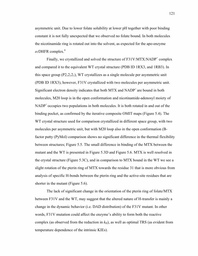
121
asymmetric unit. Due to lower folate solubility at lower pH together with poor binding
constant it is not fully unexpected that we observed no folate bound. In both molecules
the nicotinamide ring is rotated out into the solvent, as expected for the apo-enzyme
ecDHFR complex.4
Finally, we crystallized and solved the structure of F31V:MTX:NADP+ complex
and compared it to the equivalent WT crystal structure (PDB ID 1RX3, and 1RB3). In
this space group (P212121), WT crystallizes as a single molecule per asymmetric unit
(PDB ID 1RX3), however, F31V crystallized with two molecules per asymmetric unit.
Significant electron density indicates that both MTX and NADP+ are bound in both
molecules, M20 loop is in the open conformation and nicotinamide-adenosyl moiety of
NADP+ occupies two populations in both molecules. It is both rotated in and out of the
binding pocket, as confirmed by the iterative composite OMIT maps (Figure 5.4). The
WT crystal structure used for comparison crystallized in different space group, with two
molecules per asymmetric unit, but with M20 loop also in the open conformation (B-
factor putty (PyMol) comparison shows no significant difference in the thermal flexibility
between structures; Figure 5.5. The small difference in binding of the MTX between the
mutant and the WT is presented in Figure 5.3D and Figure 5.6. MTX is well resolved in
the crystal structure (Figure 5.3C), and in comparison to MTX bound in the WT we see a
slight rotation of the pterin ring of MTX towards the residue 31 that is more obvious from
analysis of specific H-bonds between the pterin ring and the active-site residues that are
shorter in the mutant (Figure 5.6).
The lack of significant change in the orientation of the pterin ring of folate/MTX
between F31V and the WT, may suggest that the altered nature of H-transfer is mainly a
change in the dynamic behavior (i.e. DAD distribution) of the F31V mutant. In other
words, F31V mutation could affect the enzyme’s ability to form both the reactive
complex (as observed from the reduction in kH), as well as optimal TRS (as evident from
temperature dependence of the intrinsic KIEs).

122
5.3.4 Molecular dynamic simulations
In order to obtain the molecular description of the active site caused by the
mutation of the residue F31, since we could not obtain the X-ray crystal structure in the
closed “reactive” conformation, we performed MD simulations at 25 ˚C (298 K) as
previously done for the WT and a series of I14 mutants.85 We started from the structure
of the WT ecDHFR (PDB ID 1RX2), and performed mutation in silico. Moreover, we
performed ten independent 6 ns runs, differing only in the initial velocities, in order to
adequately sample the conformational landscape. The comparison of resulting
conformation is presented in Figure 5.7, where two parameters of interest are described,
DADc and Φ. The relative donor acceptor orientation was measured as the angle (Φ)
between the planes that fit the nicotinamide moiety of NADPH and the pterin ring of
H2F. The DADcs’ and Φs’ distributions for F31V (Figure 5.7, 1D plots on y and x-axis)
were fitted as described in ref.85 (Table 5.5). The correlation is presented as a 2-D plot in
Figure 5.7.
As evident from the Table 5.5, and Figure 5.7, the average DADc is slightly larger
for the F31V mutant with wider distribution, however the relative orientation between the
nicotinamide and pterin rings deviate significantly from the values observed for WT-like
population (Table 5.5, population I). For this mutant we observe two distinct populations
for the Φ angle, where both populations (VI and VII) deviate significantly from the
optimal 30 degrees observed for WT. What is more interesting is that both pterin ring and
the p-amino benzoyl group of H2F rotate towards the residue 31 (Figure 5.7), which is in
accord with the X-ray crystal structures discussed above. What was initially surprising is
that nicotinamide ring moves towards the pterin ring, probably due to strong van der
Waals interactions between two rings, which results in the same average DADc that is
found in the WT ecDHFR, however with the slightly larger distribution. Even though
average DADcs for F31V and the WT are similar, the angle Φ is greatly affected and
deviates significantly from the optimal value found in the WT ecDHFR structure, as

123
noticeable in the Figure 5.7. The average DADc for both populations, VI and VII,
remains the same due to rotation of the nicotinamide ring towards the pterin ring. This
has not been observed for I14 mutants, where the mutations only affected the motion of
the nicotinamide ring with which residue 14 forms a direct contact. On the other hand, it
seems that the effects of F31V mutation are more comprehensive. Rotation of the pterin
ring of H2F away from the nicotinamide ring of NADPH affects the HOMO-LUMO
orbital overlap, a phenomenon that has been observed previously for the population II
(Chapter 3).85 This deviation of Φ from the optimal value has a detrimental effect on the
DADe at the TRS and the rate.166 Consequently, we can asses that population VII
contributes little to reactivity. A more quantitative assessment of the reactivity of either
populations would require high-level QM/MM calculations, which are beyond the scope
of this work. In the Figure 5.7 we framed the probable reactive conformation with a
“black box”. This approach is similar to the near-attack-conformation presented
previously,216 but is only presented as a simple visual tool to emphasize the decrease in
the F31V population that would have a favorable DADc and Φ angle.
Lastly, we have also monitored several hydrogen bonds formed between
pterin/nicotinamide rings and few active site residues. The one that showed significant
deviation from the values observed for the WT is the H-bond between the backbone of
Ala 7 and the amide of the nicotinamide ring. It seems that one of the bonds becomes
significantly shorter for the mutant locking nicotinamide ring in the unfavorable
orientation in comparison to pterin ring. Another interesting observation was the higher
atomic rmsd values for the residues belonging to M20 loop (residues 15-35) and residues
40-60 (Figure 5.8) of F31V mutant in comparison to WT.
The MD simulation provides molecular insight and therefore an assessment of the
effect of the mutation on the DADc (average distance, distribution of populations, and
their dynamics) and provides insight on effects on DADe (donor-acceptor orbitals overlap
deducted from the angel Φ). The MD simulation results obtain here support the

124
interpretation of the kinetic findings via the Marcus-like model. Like the previous study
on the residue 14, it seems that the smaller side chain “relaxes” the active-site leading to
broadening of configurational sampling within the reactive ternary complex.
5.4 Conclusion
The current study extends our understanding of the structure-dynamics-activity
relations in enzyme-catalyzed reactions. Since active site residues are in the direct contact
with either the substrate or the cofactor, studies of the active site mutants aid in
rationalizing the relationship between the active site structure, motions, and the catalyzed
chemical reaction. Temperature dependence of the intrinsic KIEs has been used to assess
the extent of the specific mutation on the chemical step (bond formation/dissociation).
Here, we extended our initial study on active-site residue I14,85,141 to the opposite side of
the active site, applying similar tools. The break in the temperature dependence of the
intrinsic KIEs for F31V is associated with the reduced protein stability, providing
possible rational to the phase transition or change in mechanism observed at 35 ̊C. Both
fitting of the KIE data to the Marcus-like model and MD simulation data suggest that
longer DADs with broader distributions contribute to the steep temperature dependence
of KIEs observed below 35 ̊C for F31V. Moreover, MD simulation data as well as X-ray
crystal structures indicate that mutation worsens the protein’s ability to organize the
active site for efficient hydride transfer. Even though these structures and simulations are
representation of the ground-state structure, it seems that deviations in the position of the
substrate and the cofactor in F31V affect the ability of the heavy atoms to reorganize and
bring the system to an “optimal” TRS. Together these findings suggest that in the WT
ecDHFR residue F31 participates in the restrictive active-site motions that modulate the
DAD by “pushing” the donor and acceptor closer to each other. We hope that these
results will entice further investigations such as high-level QM/MM calculations and

125
examination of the systems dynamics at millisecond to picosecond timescale motions
using NMR relaxation experiments121 and two-dimensional infrared spectroscopy.125

126
Figure 5.1 The active-site of ecDHFR emphasizing the role of F31 as a support of the pterin ring of H2F. The nicotinamide ring is highlighted in light blue, and the folate in magenta. Several other residues that form important hydrogen bonds with either nicotinamide ring or pterin ring are highlighted.
!I14$
4$
D27$
7$
F31$
1$
I5$
5$ Y100$0$
A7$
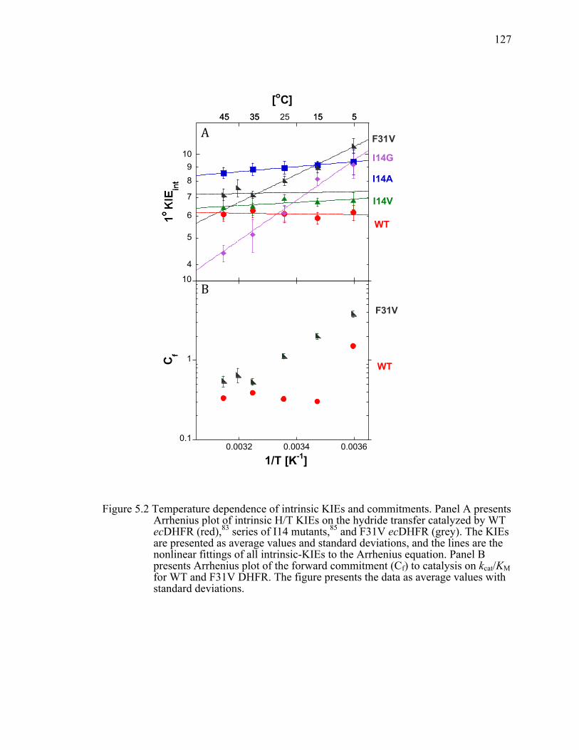
127
Figure 5.2 Temperature dependence of intrinsic KIEs and commitments. Panel A presents Arrhenius plot of intrinsic H/T KIEs on the hydride transfer catalyzed by WT ecDHFR (red),83 series of I14 mutants,85 and F31V ecDHFR (grey). The KIEs are presented as average values and standard deviations, and the lines are the nonlinear fittings of all intrinsic-KIEs to the Arrhenius equation. Panel B presents Arrhenius plot of the forward commitment (Cf) to catalysis on kcat/KM for WT and F31V DHFR. The figure presents the data as average values with standard deviations.
4
5
6
7
89
10
0.0032 0.0034 0.0036
H/Tint I14AH/Tint I14VH/Tint I14GH/Tint F31V45 H/Tint F31VH/Tint wtDHFRH/Tint I14A aveH/Tint I14V aveH/Tint I14G aveH/Tint F31V ave
I14A
I14V
WT
I14G
1/T [K-1]
525 153545 5153545
[oC]
F31V
!
0.1
1
10
0.0032 0.0034 0.0036
Cf
F31V
WT
1/T [K-1]
A
B

128
Figure 5.3 F31V mutant in comparison to WT ecDHFR. A) Active site structure of F31V-Folate-NADP+ ternary complex illustrating the binding of the folate in the relation to D27, I94, and T113. The folate is presented with electron density 2Fo-Fc map contoured at 1.5 σ within 1.6 Å of the ligand, and shown in blue. For better visibility of the folate we omitted showing residue I5 that also forms an important H-bond with pterin ring, and residue 31. B) Structural comparison of the active site of F31V-Folate-NADP+ (grey) and WT-Folate-NADP+ (pink; 1RB2). Several active site residues are shown as sticks together with both folate and NADP+. Structures have been aligned in PyMol (residues exhibiting low B-factors: 1-45, 90-100, 109-116; this alignment gave lowest RMS value). Slight rotation of the p-amino-benzoyl moiety towards the residue 31 is observed. C) Active site structure of F31V-MTX-NADP+ ternary complex illustrating the binding of the MTX and NADP+ in the relation to D27, I94, and T113. MTX and NADP+ are presented with electron density 2Fo-Fc map contoured at 1.2 σ within 1.6 Å of the ligand with several active site residues. NADP+ is bound in a dual conformation. D) Structural comparison of the active site of F31V-MTX-NADP+ (grey) and WT-MTX-NADP+ (pink-1RB3; purple-1RX3). Several active site residues are shown as sticks together with MTX and NADP+. Structures have been aligned in PyMol as described in panel B.

129
Figure 5.4 2Fo-Fc OMIT Fourier map for the F31V-MTX-NADP+ ternary complex contoured at 1σ within 1.6 Å from the ligands for the molecule A (left) and molecule B (right).
Figure 5.5 B-factor putty (PyMol) comparison between the molecule A of F31V-MTX-NADP+ (left panel), WT (PDB ID 1RX3; middle panel), and molecule A of WT (PDB ID 1RB3; right panel).

130
Figure 5.6 A 2D representation of the MTX bound in the active site of molecule A of F31V-NADP+ (left panel), and WT-NADP+ (PDB ID 1RB3; right panel) generated from the LigPlot+ analysis.217 Most interactions are preserved between the mutant and the WT; the major difference is an increase in the two H-bonds between MTX and residues I94 and I5 in F31V complex, due to rotation of the pterin ring. Several H-bonds are conserved such as between the pterin ring of MTX and residues Asp27, and glutamyl moiety and Arg57.

131
Figure 5.7 MD studies of the relative orientation of the H-donor and acceptor. Correlation plot between DADc and the relative orientation of donor and acceptor (φ) for WT (red) and F31V (black). Overlaid WT and F31V DADc and φ distributions are shown on the y and x-axis respectively. Inset represents the comparison of the average structures for the WT and F31V. For F31V mutant we observe two distinct populations that both have slightly longer DADc and where pterin ring is twisted towards the residue 31 (for detail definition of populations I-V and DADc and φ please refer to Figure 3.4). The black box (φ-value between 15°-45° and DADc below 3.5 Å) encapsulates the estimated 90% of the reactive conformers. That box is analogues to the “Near Attack Conformation” concept,216 and while it does not represent a meaningful energetic or kinetic space of states, it illustrates the geometrical bottleneck through which reactive conformation have to pass on their way to the TRS. Larger population of ground states in that box indicates larger fraction of reactive states.
!

132
Figure 5.8 RMS fluctuation for all residues of wtDHFR, presented in black, and average of ten 6 ns runs for F31V shown in red.
!

133
Table 5.1 Reaction rates, binding constants and isotope effects on Arrhenius activation parameters for several active site mutants of ecDHFR.
DHFR Parameters
WT
I14V
I14A
I14G
F31V
kcat [s-1] 12.3 ±0.7c ND 15 ±1.7g ND 26 ±10d
kHa[s-1] 228 ±8c 33.3 ±3.1f 5.7 ±0.3f 0.22 ±0.04f 17.7 ±0.1i
Kd (H2F) [µM]b 0.21 ±0.03d
ND 0.22 ±0.07g
ND 6.6 ±0.5d
Kd (NADPH) [µM]b 0.38±0.04d ND 1.4 ±0.1g ND 0.22 ±0.02d
AH/AT 7.0 ±1.5e 4.2 ±0.4f 4.7 ±0.5h 0.024±0.003f 6.42 ±1.28i
0.165±0.012i
∆Ea(T-H), [kcal/mol]
-0.1 ±0.2e 0.27 ±0.05f
0.39 ±0.06h
3.31 ±0.07f 0.08 ±0.10i
2.30 ±0.04i
a. Pre-steady state rate constants of hydride transfer at 25˚C and pH 7.0
b. Thermodynamic dissociation constants determined at 25˚C and pH 7.0.
c. Ref. 6
d. Ref. 207
e. Ref. 83
f. Ref. 85
g. Ref. 140
h. Ref. 141
i. This work.
ND = not determined

134
Table 5.2 Experimental ∆Ea (from Table 1) and regression parameters for WT ecDHFR and the active site mutants studied to date.
Enzyme ∆Ea (kcal/mol)
Fitting Parameters Reference
1 Population 2 Populations DADc f DADc ∆G WT -0.1 ±0.2 3.06 >250 3.06 >2.5 73,83
I14V 0.27 ±0.05 3.08 200 3.06 2.3 73,85
I14A 0.39 ±0.06 3.09 190 3.07 2.4 73,85
I14G 3.31 ±0.07 NA NA 3.31 3.8 73,85
F31V (>35˚)
0.08 ±0.10 3.07 144 3.06 3.5
F31V (<35˚)
2.30 ±0.04 NA NA 3.23 2.61
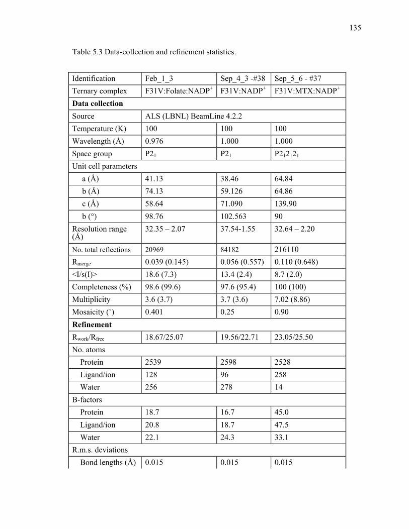
135
Table 5.3 Data-collection and refinement statistics.
Identification Feb_1_3 Sep_4_3 -#38 Sep_5_6 - #37 Ternary complex F31V:Folate:NADP+ F31V:NADP+ F31V:MTX:NADP+
Data collection Source ALS (LBNL) BeamLine 4.2.2 Temperature (K) 100 100 100 Wavelength (Å) 0.976 1.000 1.000 Space group P21 P21 P212121 Unit cell parameters a (Å) 41.13 38.46 64.84 b (Å) 74.13 59.126 64.86 c (Å) 58.64 71.090 139.90 b (°) 98.76 102.563 90 Resolution range (Å)
32.35 – 2.07 37.54-1.55 32.64 – 2.20
No. total reflections 20969 84182 216110 Rmerge 0.039 (0.145) 0.056 (0.557) 0.110 (0.648) <I/s(I)> 18.6 (7.3) 13.4 (2.4) 8.7 (2.0) Completeness (%) 98.6 (99.6) 97.6 (95.4) 100 (100) Multiplicity 3.6 (3.7) 3.7 (3.6) 7.02 (8.86) Mosaicity (˚) 0.401 0.25 0.90 Refinement Rwork/Rfree 18.67/25.07 19.56/22.71 23.05/25.50 No. atoms Protein 2539 2598 2528 Ligand/ion 128 96 258 Water 256 278 14 B-factors Protein 18.7 16.7 45.0 Ligand/ion 20.8 18.7 47.5 Water 22.1 24.3 33.1 R.m.s. deviations Bond lengths (Å) 0.015 0.015 0.015

136
Table 5.3 Continued Bond angles (˚) 1.547 2.011 1.888 Ramachandran plot, residues in Most favored regions (%)
97.78 99.69 94.90
Allowed regions (%)
2.22 0.31 5.10
Disallowed (%) 0.00 0.00 0.00 Note: Values in parentheses are for the highest resolution shell
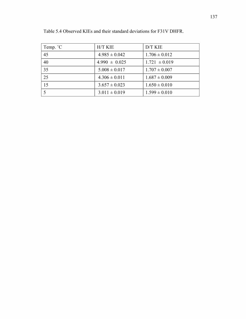
137
Table 5.4 Observed KIEs and their standard deviations for F31V DHFR.
Temp. ˚C H/T KIE D/T KIE 45 4.985 ± 0.042 1.706 ± 0.012 40 4.990 ± 0.025 1.721 ± 0.019 35 5.008 ± 0.017 1.707 ± 0.007 25 4.306 ± 0.011 1.687 ± 0.009 15 3.657 ± 0.023 1.650 ± 0.010 5 3.011 ± 0.019 1.599 ± 0.010
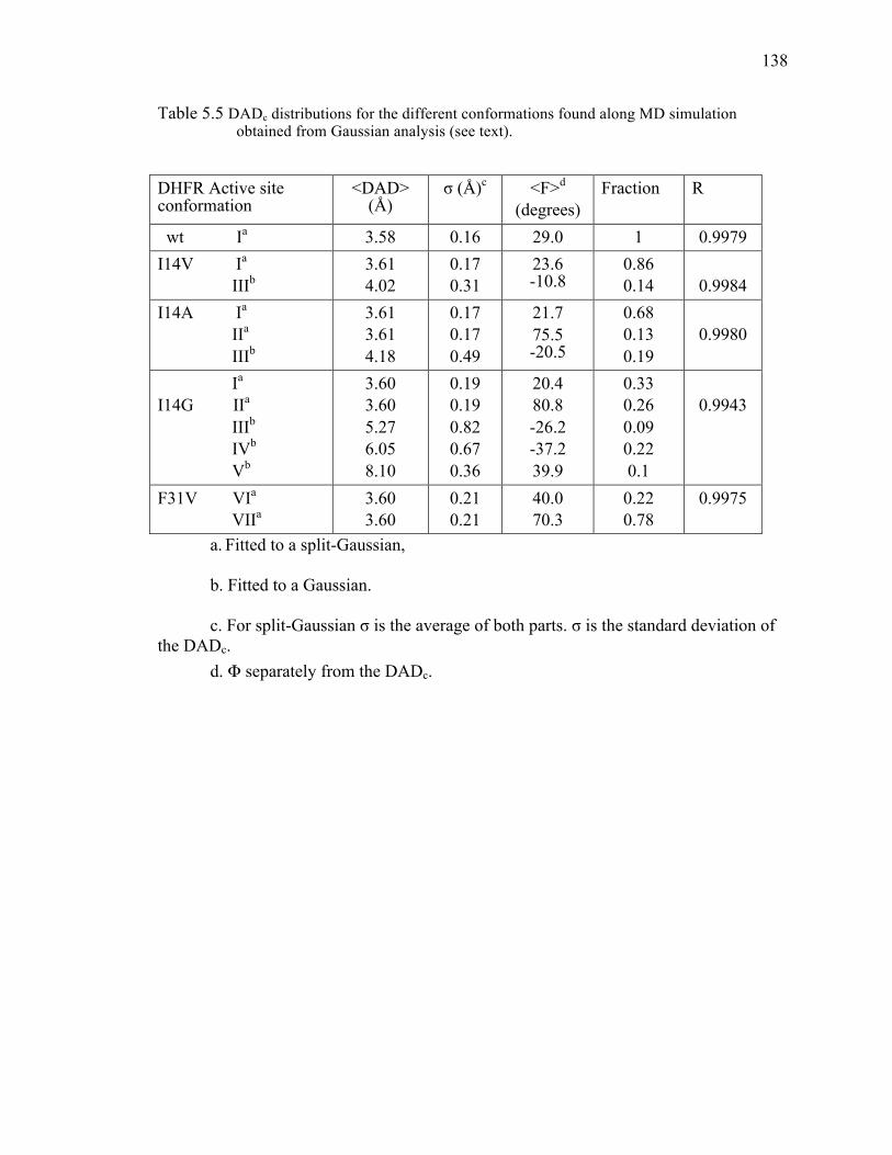
138
Table 5.5 DADc distributions for the different conformations found along MD simulation obtained from Gaussian analysis (see text).
DHFR Active site conformation
<DAD> (Å)
σ (Å)c <F>d (degrees)
Fraction R
wt Ia 3.58 0.16 29.0 1 0.9979 I14V Ia IIIb
3.61 4.02
0.17 0.31
23.6 -10.8
0.86 0.14
0.9984
I14A Ia
IIa IIIb
3.61 3.61 4.18
0.17 0.17 0.49
21.7 75.5 -20.5
0.68 0.13 0.19
0.9980
Ia
I14G IIa
IIIb IVb
Vb
3.60 3.60 5.27 6.05 8.10
0.19 0.19 0.82 0.67 0.36
20.4 80.8 -26.2 -37.2 39.9
0.33 0.26 0.09 0.22 0.1
0.9943
F31V VIa
VIIa 3.60 3.60
0.21 0.21
40.0 70.3
0.22 0.78
0.9975
a. Fitted to a split-Gaussian,
b. Fitted to a Gaussian.
c. For split-Gaussian σ is the average of both parts. σ is the standard deviation of the DADc.
d. Φ separately from the DADc.
139
CHAPTER 6.
EVIDENCE THAT A ‘DYNAMIC KNOCKOUT’ IN ESCHERICHIA
COLI DIHYDROFOLATE REDUCTASE DOES AFFECT THE
CHEMICAL STEP OF CATALYSISvii
6.1 Introduction
One of the contemporary questions in enzymology is the link between protein
dynamics and function. Significant advancements have been achieved toward
characterizing protein motions across wide-ranging timescales from seconds to
femtoseconds and investigating their functional relevance. For instance, the importance
of protein motions on the milli- to microsecond timescale has been recognized to be
crucial to both substrate binding and product release, often contributing to the rate-
limiting step in the catalytic turnover. However, the precise role of the protein motions in
assisting chemical transformation has remained unclear and is still source of a heated
debate. Elucidating the role of the protein motions is crucial to understanding enzyme
mechanisms, as this knowledge will greatly assist in widening their applicability in
industrial and medicinal settings. Previous kinetic and computational studies on the
model enzyme in question, dihydrofolate reductase from Escherichia coli (ecDHFR),
suggest that protein fluctuations responsible for the reorganization of the heavy atoms in
the active site and the conformational sampling of the donor - acceptor distance (DAD)
are crucial to facilitating H-transfer.8,18,84,85,139,197,198,218-221 While none of the
experimental data indicated involvement of non-statistical dynamicsviii in catalysis, and
vii This chapter has been submitted to the journal of Nature Chemistry.
viii Non-statistical dynamics: molecular motions that are not in thermal equilibrium with their environment. Their role in enzymology has been matter of on-going debate(Hay, S. & Scrutton, N.S. Good vibrations in enzyme-catalyzed reactions. Nat. Chem. 4, 161-168 (2012); Glowacki, D.R., Harvey, J.N. & Mulholland, A.J. Taking Ockham’s razor to enzyme dynamics

140
none of the calculations (besides one exception222) required non-statistical dynamics,
researchers presented different opinions regarding the role of dynamics in catalyzing the
chemical step.
The effect of protein dynamics on the chemical coordinate of the reaction has
been indirectly investigated through the examination of the temperature dependence of
KIEs and by computational methods. The advantage of this experimental method is that
the temperature dependence of KIEs is highly sensitive to the changes in DAD, which
can modulate the degree of the wavefunction overlap between the donor and acceptor
states. As suggested by several phenomenological models, denoted here as Marcus-like
models (presented later in more detail), if the conformational fluctuations assist the
chemical transfer through modulation of the DAD and the wave-function overlap, then
that will manifest in an increase in the temperature dependence of the KIEs.39,40,42,125 In
such analysis, however, extraction of the intrinsic KIEs is challenging, as the chemical
step in most enzymatic systems is not rate-limiting and is therefore masked by other,
isotopically insensitive steps when the reactive C-H bond is isotopically labeled. This
kinetic complexity masks the intrinsic KIEs and leads to the observed KIEs that are
smaller than their intrinsic value. In single-turnover experiments, as conducted in refs 1,193, the kinetic complexity is commonly associated with conformational changes and
changes in protonation that occur prior to the formation of the reactive ternary complex.
One of the most widely used enzymatic models for the structure/function/dynamic
relationship is ecDHFR. EcDHFR is a small, flexible protein that catalyzes the reduction
of 7,8-dihydrofolate (H2F) to 5,6,7,8-tetrahydrofolate (H4F), with the stereospecific
transfer of a pro-R hydride from the C4 position of the reduced nicotinamide adenine
dinucleotide phosphate (NADPH) to the C6 of the dihydropterin ring of H2F, which is
and catalysis. Nature Chem. 141, 169-176 (2012); Editorial. Of polemics and progress. Nature Chem. 4, 141 (2012))

141
already protonated at its N5 position prior to hydride transfer occurring (Figure
6.1).7,9,193,223 EcDHFR has an α/β structure consisting of a central eight-stranded β sheet
and four α-helices connected with several loop regions. One of the loops is a flexible
active-site loop, the M20 loop (residues 9-24), which undergoes large conformational
changes between the Michaelis complex and the product complex.4,224 Extensive X-ray
studies showed that the M20 loop can adopt four characteristic conformations and its
movement is coordinated with different stages of the catalytic cycle.4 In the solution it
exists in two dominant conformations: closed, where the loop packs closely against the
nicotinamide ring, and the occluded, where the loop protrudes into the active site,
sterically blocking the nicotinamide binding pocket.5 Alignment of human DHFR and
ecDHFR sequences suggested that the occluded conformation in the human DHFR is
destabilized due to a biproline sequence at the end of the M20 loop, leading to an
increase in the rigidity of the loop in comparison to the one in ecDHFR.1
In 2011, Bhaba et al1 studied the possible correlation between millisecond M20-
loop motions (examined via NMR relaxation experiments) and the apparent hydride-
transfer rate (examined via single-turnover measurement) using three mutants of
ecDHFR: N23PP, S148A and N23PP-S148A. The double mutant N23PP-S148A,ix and
N23PP (S148A mutation seems to have little or no effect) were named “dynamic
knockout” mutants, as their respective flexible M20 loops rigidified, resembling the
rigidity of the M20 loop in human DHFR. NMR results suggested that millisecond-
timescale fluctuations in the active site of WT ecDHFR are equally abrogated in the
ix Bhaba et al.(Bhabha, G. et al. A dynamic knockout reveals that conformational
fluctuations influence the chemical step of enzyme catalysis. Science 332, 234-238 (2011)) showed that addition of the bi-proline sequence in N23PP and N23PP-S148A mutants is responsible for the suppression of the mili- to microsecond timescale motions of the M20 loop in the Michaelis complex having also a significant effect on khyd measured by FRET-stopped-flow. On the other hand, while S148A mutation affects the mili- to microsecond timescale motions of M20 loop, it has no significant effect on khyd. Therefore, the kinetic study here is performed on the N23PP ecDHFR instead on the double mutant.
142
N23PP as in the double mutant, while the x-ray structure of the relevant ternary complex
of the double mutant suggested that mutation barred no effect on the structure of the
active site. Moreover, the addition of the bi-proline sequence significantly reduced the
single-turnover rate (khyd) relative to the WT. Although the observed H/D KIEs at 25 ˚C
were the same as for the WT, the authors suggested that the altered millisecond-timescale
motions modified the chemical step (as evident from reduced apparent khyd) and thus
might be directly coupled to the H-transfer. This dramatic conclusion was followed by
theoretical225 and experimental193 studies that challenged this interpretation. Importantly,
EVB calculations225 implied that the depressed catalytic effects in the mutants are due to
changes in reorganization energy. It is vital to note that while motions along the slow
conformational coordinate, which were examined by the NMR relaxation experiments,
have a substantial effect on the rate of the turnover and the rate constant for the apparent
hydride transfer, they might not be coupled to the much faster chemical step (dissociation
of the C-H bond at femto- picosecond time scale219). Experimental studies of the
temperature dependence of KIEs in a variety of enzymatic systems39,74,125,176,226 indirectly
indicate the existence of the fast motions, proposed to occur at the femto- to picosecond
timescale, and their possible coupling to the reaction coordinate. Therefore, Allemann
and coworkers, 193 using the same experimental method as ref 1 for examining the rate of
H-transfer and its KIEs, assessed the temperature dependence of KIEs for the N23PP-
S148A ecDHFR. Their observed single-turnover KIEs were the same as for the WT
ecDHFR throughout the temperature range, and the authors concluded that there is no
detectable dynamic coupling of protein motions to the hydride transfer.193
In the current paper, we report the temperature dependence of the intrinsic KIEs
and hence allow for new assessment of the effect of the N23PP mutation on the chemical
step in ecDHFR. Our findings indicate that the temperature dependence of the kinetic
complexity in the stopped-flow measurements1,193 masked the differences between the
WT and mutant, as the intrinsic KIEs and their temperature dependence are actually quite
143
different (vide infra). These findings invert the conclusions of ref 193, and indirectly imply
that motions faster than the millisecond motions examined by NMR1 are also altered and
affect the chemical step in question (C-H→C).
6.2 Materials and methods
Chemicals. [Ad-14C]-NAD+ (specific radioactivity of >220mCi/mmol) was
purchased from Perkin Elmer. H2F was prepared by dithionate reduction of folic acid as
described previously.107 Glucose dehydrogenase from Bacillus megaterium (bmGluDH)
was purchased from Affymetrix/USB. All other materials were purchased from Sigma.
(R)-[4-3H]-NADPH was synthesized through a 3-step synthesis as described before. 111
[Ad-14C]-NADPH was prepared from [Ad-14C]-NAD+ using NAD+ kinase from chicken
liver, followed by reduction with glucose using bmGluDH.95 (R)-[Ad-14C,4-2H]-NADPH
was synthesized through the reduction of [Ad-14C]-NADP+ with deuterated glucose and
bmGluDH.142 All synthesized materials were purified and stored as described
previously.109 N23PP ecDHFR was expressed and purified as described before.1
Kinetic isotope effect measurements. KIEs with N23PP ecDHFR were measured
as described before for the WT and numerous DHFR mutants.84,85,104 In short, we
combined (R)-[4-3H]-NADPH and [Ad-14C]-NADPH, and, (R)-[4-3H]-NADPH and (R)-
[Ad-14C,4-2H]-NADPH in the 5:1 radioactivity ratio in order to measure H/T and D/T
1˚KIEs, respectively. Each mixture was co-purified on an analytical reverse-phase HPLC
column, divided into aliquots containing 300,000 dpm of 14C, and frozen in liquid
nitrogen for short-term storage at -80 ˚C. All experiments were performed in MTEN
buffer (50 mM MES, 25 mM Tris, 25 mM EtOH-NH2 and 100 mM NaCl) at pH=9.0 over
the 5-45 ˚C temperature range. All reaction mixtures contained around 200-fold excess of
H2F (0.85 mM) over NADPH (4 µM). The reactions were quenched by adding an excess
of methotrexate (Kd=1 nM), and samples were stored on dry ice prior to HPLC
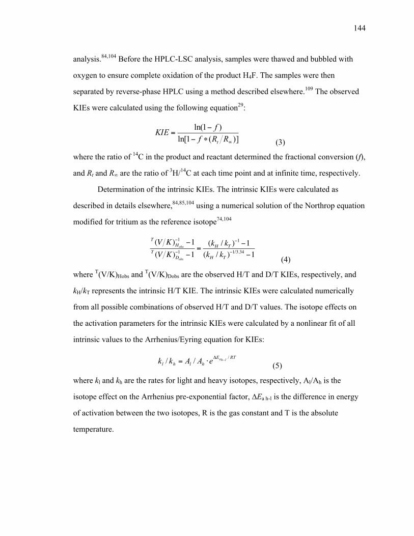
144
analysis.84,104 Before the HPLC-LSC analysis, samples were thawed and bubbled with
oxygen to ensure complete oxidation of the product H4F. The samples were then
separated by reverse-phase HPLC using a method described elsewhere.109 The observed
KIEs were calculated using the following equation29:
)](1ln[
)1ln(
∞∗−−
=RRf
fKIEt (3)
(3)
where the ratio of 14C in the product and reactant determined the fractional conversion (f),
and Rt and R∞ are the ratio of 3H/14C at each time point and at infinite time, respectively.
Determination of the intrinsic KIEs. The intrinsic KIEs were calculated as
described in details elsewhere,84,85,104 using a numerical solution of the Northrop equation
modified for tritium as the reference isotope74,104
T (V K )Hobs
−1 −1T (V K )Dobs
−1 −1=(kH / kT )
−1 −1(kH / kT )
−1/3.34 −1 (4) (4)
where T(V/K)Hobs and T(V/K)Dobs are the observed H/T and D/T KIEs, respectively, and
kH/kT represents the intrinsic H/T KIE. The intrinsic KIEs were calculated numerically
from all possible combinations of observed H/T and D/T values. The isotope effects on
the activation parameters for the intrinsic KIEs were calculated by a nonlinear fit of all
intrinsic values to the Arrhenius/Eyring equation for KIEs:
(5)
where kl and kh are the rates for light and heavy isotopes, respectively, Al/Ah is the
isotope effect on the Arrhenius pre-exponential factor, ∆Ea h-l is the difference in energy
of activation between the two isotopes, R is the gas constant and T is the absolute
temperature.
RTEhlhl
lhaeAAkk /// −Δ
⋅=

145
6.3 Results and Discussion
6.3.1 Temperature dependence of the KIEs
Pre-steady-state kinetic measurements (under single-turnover conditions)
indicated that both dynamically altered mutations (N23PP and N23PP/S148A) reduced
single-turnover rate to the same extent (~15 fold), 1 while the observed H/D KIEs and
their temperature dependence for the double mutant coincided with the values obtained
for WT ecDHFR (Table 1 in ref 193). However, under single-turnover conditions the
chemistry in ecDHFR is not rate-limiting,6,84 resulting in smaller observed KIEs. More
specifically, under the pre-steady-state conditions used in refs,1,193 the rate measured
includes at least three kinetic steps: the isotopically insensitive flip of the nicotinamide
ring in and out of the active site, the closing of the M20 loop, the protonation of H2F, and
the hydride transfer itself.64,84 This raised concern that both studies of N23PP DHFR
mutants1,193 might have captured not only the effects on the chemical step (C-H→C
transfer) but also a more complex process that involves the preparation of the system
prior to the tunneling-ready state (TRS; see below) at which chemistry (H-transfer)
occurs.
The current work measures H/T and D/T KIEs on the second-order rate constant
(kcat/KM or V/K) under competitive conditions. The combination of these two observed
KIEs affords assessment of their intrinsic values as described before.104 In short, we
measured competitive H/T and D/T KIEs with WT83 and N23PP ecDHFR under the same
conditions using mixtures of [4R-3H]-NADPH with [Ad-14C]-NADPH and mixtures of
[Ad-14C, 4R-2H]-NADPH and [4R-3H]-NADPH for H/T and D/T KIE measurements,
respectively. Intrinsic KIEs were extracted from the observed values using the modified
Northrop equation.104 and were fitted to the Arrhenius equation (Figure 6.2; also see
Table 6.1 and Table 6.2) as previously described.84,104 Table 1 compares the isotope
effects on the Arrhenius activation parameters for the WT and N23PP ecDHFRs. Using
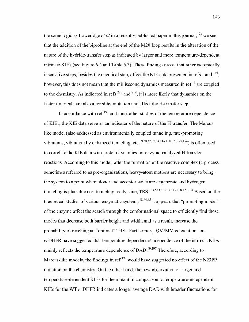
146
the same logic as Loweridge et al in a recently published paper in this journal,193 we see
that the addition of the biproline at the end of the M20 loop results in the alteration of the
nature of the hydride-transfer step as indicated by larger and more temperature-dependent
intrinsic KIEs (see Figure 6.2 and Table 6.3). These findings reveal that other isotopically
insensitive steps, besides the chemical step, affect the KIE data presented in refs 1 and 193;
however, this does not mean that the millisecond dynamics measured in ref 1 are coupled
to the chemistry. As indicated in refs 225 and 219, it is more likely that dynamics on the
faster timescale are also altered by mutation and affect the H-transfer step.
In accordance with ref 193 and most other studies of the temperature dependence
of KIEs, the KIE data serve as an indicator of the nature of the H-transfer. The Marcus-
like model (also addressed as environmentally coupled tunneling, rate-promoting
vibrations, vibrationally enhanced tunneling, etc.39,58,62,72,74,116,118,120,127,174) is often used
to correlate the KIE data with protein dynamics for enzyme-catalyzed H-transfer
reactions. According to this model, after the formation of the reactive complex (a process
sometimes referred to as pre-organization), heavy-atom motions are necessary to bring
the system to a point where donor and acceptor wells are degenerate and hydrogen
tunneling is plausible (i.e. tunneling ready state, TRS).39,58,62,72,74,116,118,127,174 Based on the
theoretical studies of various enzymatic systems,48,64,65 it appears that “promoting modes”
of the enzyme affect the search through the conformational space to efficiently find those
modes that decrease both barrier height and width, and as a result, increase the
probability of reaching an “optimal” TRS. Furthermore, QM/MM calculations on
ecDHFR have suggested that temperature dependence/independence of the intrinsic KIEs
mainly reflects the temperature dependence of DAD.49,197 Therefore, according to
Marcus-like models, the findings in ref 193 would have suggested no effect of the N23PP
mutation on the chemistry. On the other hand, the new observation of larger and
temperature-dependent KIEs for the mutant in comparison to temperature-independent
KIEs for the WT ecDHFR indicates a longer average DAD with broader fluctuations for

147
the mutant. Similar effects of mutations on the intrinsic KIEs have been analyzed by
QM/MM calculations,197 which indicated that the mutations perturbed the orientation and
relative dynamics of the DAD. These altered fluctuations probably occur at a faster
timescales (fs-ns) than the ones examined in ref 1 (on the ms timescale), suggesting that
the N23PP mutation probably alters a broad range of dynamics, from ms to fs timescales.
Overall, the x-ray crystal structure obtained for N23PP/S148A was very similar to
the WT ecDHFR structure (for the same ternary complex). One of the only differences
was a decrease in the average distance between the donor and acceptor carbons from the
3.3 to 2.9 Å (note that the DAD used in the Marcus-like models is not that of the carbons
but rather from H-reactant state to H-product state). Some studies deemed this change
insignificant;1 however, it could be quite significant if relevant to the reactive
conformation.225 It is important to note that crystal structures are representations of the
ground-state structures, and DAD at the TRS might differ significantly from the
population most likely to crystallize. While the ground state in the crystal structures1
indicated shorter distance for the mutant, its average DAD and its distribution at the TRS
are probably longer, as the difference in the temperature dependence of the KIEs
suggests.
6.3.2 Kinetic complexity.
The main goal of this paper is the same as that of refs 1,193: to examine possible
dynamic coupling of protein motions to the hydride transfer step in ecDHFR. However,
examinations of observed and intrinsic KIEs ultimately led to opposing conclusions.
Therefore, it is important to comprehend the difference in nature of the KIE experiment
performed here and done formerly1,193 in order to understand why no significant
differences in KIEs between WT and N23PP mutants of ecDHFR were previously

148
observed. The explanation lies in the kinetic complexity still masking the intrinsic KIEs
when measuring pre-steady state rates.
In most enzymatic and organic reactions, observed KIEs on bond cleavage are
generally smaller than their intrinsic values, due to kinetic complexity (i.e. substrates
binding, product release, conformation changes, protonation changes, etc.) that masks the
intrinsic KIE values. The following equation presents this kinetic complexity:
KIEobs =KIEint +Cf +Cr ∗EIE
Cf +Cr +1 (1)
where KIEobs is the observed KIE, KIEint is the intrinsic KIE, EIE is the equilibrium
isotope effect, and Cf and Cr are the forward and reverse commitments to catalysis,
respectively. For the hydride transfer reaction catalyzed by ecDHFR, the Cr≈0 and the
EIE is close to unity because the reaction is very exothermic and the H-isotope is bound
to sp3 carbon in both reactant and product states. Therefore, we can simplify equation (1)
to equation (2):
KIEobs =KIEint +C1+C
(2)
where the commitment C is the sum of the ratios between the rate of the forward
isotopically sensitive hydride-transfer step and each of the rates of the preceding
backward isotopically insensitive steps.102 Consequently, we can calculate C from the
observed and intrinsic KIEs at each temperature. Figure 6.3 presents the commitment
plotted on a logarithmic scale against the inverse absolute temperature. The C on either
V/K or single-turnover KIEs was calculated from their respective observed values and the
intrinsic values (obtained here) using equation (2).
The observed KIE on V/K involves microscopic rate constants for all the kinetic
steps from binding of the labeled substrate (NADPH) to the first irreversible step. The
observed KIEs measured under single-turnover conditions include steps from the binding
of H2F to the enzyme-NADPH complex to the hydride-transfer step (i.e. the flip of the
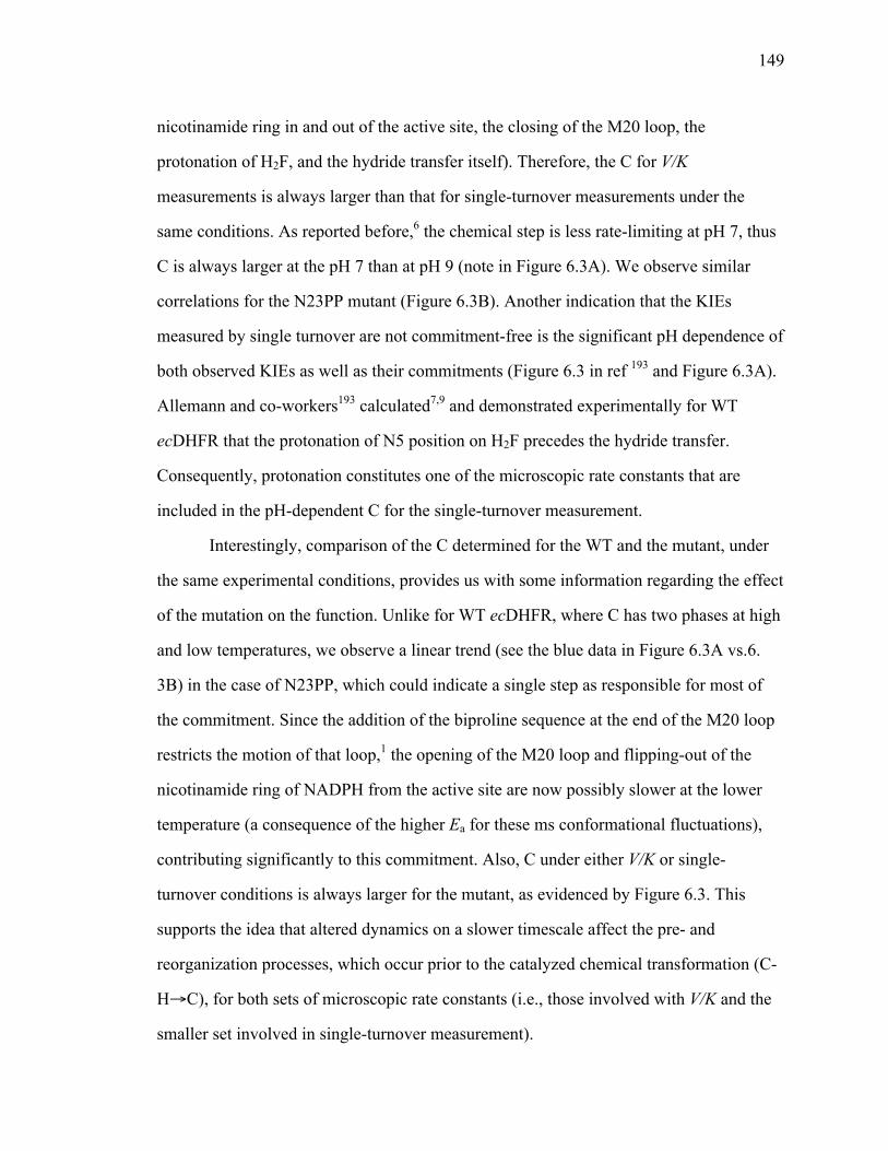
149
nicotinamide ring in and out of the active site, the closing of the M20 loop, the
protonation of H2F, and the hydride transfer itself). Therefore, the C for V/K
measurements is always larger than that for single-turnover measurements under the
same conditions. As reported before,6 the chemical step is less rate-limiting at pH 7, thus
C is always larger at the pH 7 than at pH 9 (note in Figure 6.3A). We observe similar
correlations for the N23PP mutant (Figure 6.3B). Another indication that the KIEs
measured by single turnover are not commitment-free is the significant pH dependence of
both observed KIEs as well as their commitments (Figure 6.3 in ref 193 and Figure 6.3A).
Allemann and co-workers193 calculated7,9 and demonstrated experimentally for WT
ecDHFR that the protonation of N5 position on H2F precedes the hydride transfer.
Consequently, protonation constitutes one of the microscopic rate constants that are
included in the pH-dependent C for the single-turnover measurement.
Interestingly, comparison of the C determined for the WT and the mutant, under
the same experimental conditions, provides us with some information regarding the effect
of the mutation on the function. Unlike for WT ecDHFR, where C has two phases at high
and low temperatures, we observe a linear trend (see the blue data in Figure 6.3A vs.6.
3B) in the case of N23PP, which could indicate a single step as responsible for most of
the commitment. Since the addition of the biproline sequence at the end of the M20 loop
restricts the motion of that loop,1 the opening of the M20 loop and flipping-out of the
nicotinamide ring of NADPH from the active site are now possibly slower at the lower
temperature (a consequence of the higher Ea for these ms conformational fluctuations),
contributing significantly to this commitment. Also, C under either V/K or single-
turnover conditions is always larger for the mutant, as evidenced by Figure 6.3. This
supports the idea that altered dynamics on a slower timescale affect the pre- and
reorganization processes, which occur prior to the catalyzed chemical transformation (C-
H→C), for both sets of microscopic rate constants (i.e., those involved with V/K and the
smaller set involved in single-turnover measurement).

150
Most importantly, the difference in the temperature dependences of the C for the
single-turnover measurements for the WT vs. the mutant explains the similarity in the
observed KIEs for these enzymes as reported in ref 193. The C values for the mutant are
larger across the temperature range and are more temperature dependent than for the WT.
Similar correlation in the temperature dependence of the intrinsic KIEs between the
mutant and the WT is observed (Figure 6.2) leading to compensation of the mutation
effect on C and on the intrinsic KIE.
6.4 Conclusions
Here we have shown that in contrast to previous reports in Science and this
journal,1,193 restriction in the flexibility of the M20 loop in the N23PP ecDHFR led in fact
to a change in the nature of the hydride-transfer step, as indicated by the change in the
temperature dependence of the intrinsic KIEs. Moreover, the new findings indicate that
the mutation altered a broad range of motions (from ms to fs timescales). These include
ms-µs motions examined in ref 1, which impaired the ability of the enzyme to achieve
optimal TRS, resulting in larger kinetic complexity. Additionally, alteration of much
faster dynamics affected the isotopically sensitive chemical step (C-H→C), resulting in
temperature-dependent intrinsic KIEs. To gain more insight into which motions
contribute to chemistry of ecDHFR, attempts to probe femto-picosecond dynamics of the
wild type and mutant are underway using vibrational spectroscopy (e.g., 2D-IR125).
Lastly, the dynamic-knockout mutant was created in order to resemble the dynamic of the
M20 loop in human DHFR. It would be of interest to undergo similar analysis presented
here in order to probe the nature of the hydride transfer in human DHFR.

151
Figure 6.1 The reaction catalyzed by DHFR. R = adenine dinucleotide 2’ phosphate and R’ = (p-aminobenzoyl) glutamate. It was shown previously that the protonation of the N5 position occurs prior to hydride transfer.7,9,193,223
Figure 6.2 Arrhenius plot of intrinsic H/T KIEs on the hydride transfer catalyzed by WT ecDHFR (red)83 and N23PP DHFR (blue). The figure shows the KIEs as average values and standard deviations, and the lines are the nonlinear fittings of all calculated intrinsic KIEs to the Arrhenius equation.
4
5
6
7
8
910
0.0032 0.0034 0.0036
525 153545 5153545
[oC]
1o K
IEin
t
1/T [K-1]
WT
N23PP

152
Figure 6.3 Comparison of the Arrhenius plot of C. a) C for WT ecDHFR determined for the observed V/K KIEs at pH 9 (blue circles),83 and observed KIEs obtained under single turnover conditions at pH 7 (red circles) and at pH 9 (purple circles).193 b) C for N23PP determined for the observed V/K KIEs at pH 9 (blue squares), and observed KIEs obtained under single turnover conditions at pH 7 (red squares) and at pH 9 (purple squares).193 The figure presents the data as average values with standard deviations. The lines are an interpolation of the data and do not represent any fitting.
1
10
0.0032 0.0034 0.0036
C fo
r N23
PP
ecD
HFR
1/T [K-1]
b.C
for W
T ec
DH
FR
515253545[oC]
a.
0.1
1

153
Table 6.1 Observed KIEs and their standard deviations for N23PP DHFR.
Temp. ˚C H/T KIE D/T KIE 45 4.402 ± 0.048 1.694 ± 0.007 35 3.995 ± 0.046 1.668 ± 0.016
25 2.984 ± 0.031 1.554 ± 0.012 15 2.181 ± 0.012 1.420 ± 0.005 5 1.765 ± 0.019 1.312 ± 0.008
Table 6.2 Intrinsic KIEs and their standard deviations for N23PP DHFR.
Temp. ˚C H/T KIE H/D KIE D/T KIE 45 8.025 ± 0.266 4.302 ± 0.099 1.865 ± 0.019 35 8.360 ± 0.602 4.427 ± 0.223 1.888 ± 0.041 25 8.479 ± 0.608 4.471 ± 0.223 1.896 ± 0.041 15 9.313 ± 0.426 4.775 ± 0.152 1.950 ± 0.027 5 9.571 ± 0.880 4.867 ± 0.337 1.966 ± 0.058
Note: These values are calculated from the observed H/T and D/T KIE data using Northrop’s method as described in the Materials and methods. The errors indicate the 95% confidence level.

154
Table 6.3 Comparative kinetic parameters of the WT and N23PP ecDHFRs.
Parameters WT N23PP kcat [s-1] 12a 2.5 ± 1c
kH [s-1] 220 ± 10a 14.2 ± 1c
AH/AT 7.0 ± 1.5b 2.31 ± 0.16d
∆EaT-H, [kcal/mol] -0.1 ± 0.2b 0.78 ± 0.04d
Note: The two rates kcat and kH are the observed values determined at pH 7 and 25 ˚C. The two isotopic activation parameters AH/AT and ∆Ea T-H are obtained from the fitting of the intrinsic KIEs to the Arrhenius equation.
a Ref. 6. b Ref. 83. c Ref. 1. d This work.

155
CHAPTER 7.
SUMMARY, IMPACT AND FUTURE DIRECTIONS
The central motivation for the research in this thesis is to better understand the
role of dynamics and the extent of hydrogen tunneling in enzymatic system.
Contemporary theories such as Marcus-like models propose that enzymes with
temperature independent KIEs (i.e., most wild type enzymes, like WT ecDHFR), have
optimally organized active-site structure. Perturbations—such as deviations from the
physiological temperature/pressure or certain mutations—can lead to temperature
dependent KIEs. According to Marcus-like models, temperature dependence of KIEs
mostly reflects the deviation of the donor-acceptor distance (DAD) from the optimal
value, which is modulated by certain motions in the active site. To further investigate this
hypothesis we studied the physical features of a C-H→C transfer by vigorously
perturbing the DAD. Experiments completed and discussed in Chapters 3 and 5 were
designed to alter the DAD and its dynamics via systematic reduction in the size of the
two hydrophobic active site residues. The major goal of the study was to examine some
basic assumptions in physical chemistry and enzymology and provide a benchmark for
the C-H→C transfer reactions, not just enzymatic reactions, but also give some
implications for solution reactions in general. The role of the dynamics in the enzymatic
reaction was examined indirectly via temperature dependence of KIEs, and not by
actually characterizing the active site fluctuations (i.e., spectroscopically or otherwise)
that might be on the same timescale as the reaction and therefore relevant to the
chemistry.
Prior to conducting the KIEs, it was necessary to obtain pure isotopically labeled
reagents. As those are not commercially available we had to develop appropriate
synthetic methodologies. Certain synthetic and analytical procedures were already
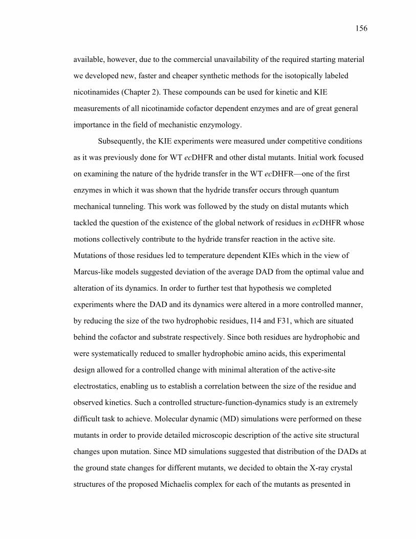
156
available, however, due to the commercial unavailability of the required starting material
we developed new, faster and cheaper synthetic methods for the isotopically labeled
nicotinamides (Chapter 2). These compounds can be used for kinetic and KIE
measurements of all nicotinamide cofactor dependent enzymes and are of great general
importance in the field of mechanistic enzymology.
Subsequently, the KIE experiments were measured under competitive conditions
as it was previously done for WT ecDHFR and other distal mutants. Initial work focused
on examining the nature of the hydride transfer in the WT ecDHFR—one of the first
enzymes in which it was shown that the hydride transfer occurs through quantum
mechanical tunneling. This work was followed by the study on distal mutants which
tackled the question of the existence of the global network of residues in ecDHFR whose
motions collectively contribute to the hydride transfer reaction in the active site.
Mutations of those residues led to temperature dependent KIEs which in the view of
Marcus-like models suggested deviation of the average DAD from the optimal value and
alteration of its dynamics. In order to further test that hypothesis we completed
experiments where the DAD and its dynamics were altered in a more controlled manner,
by reducing the size of the two hydrophobic residues, I14 and F31, which are situated
behind the cofactor and substrate respectively. Since both residues are hydrophobic and
were systematically reduced to smaller hydrophobic amino acids, this experimental
design allowed for a controlled change with minimal alteration of the active-site
electrostatics, enabling us to establish a correlation between the size of the residue and
observed kinetics. Such a controlled structure-function-dynamics study is an extremely
difficult task to achieve. Molecular dynamic (MD) simulations were performed on these
mutants in order to provide detailed microscopic description of the active site structural
changes upon mutation. Since MD simulations suggested that distribution of the DADs at
the ground state changes for different mutants, we decided to obtain the X-ray crystal
structures of the proposed Michaelis complex for each of the mutants as presented in

157
Chapter 4. The initial goal was to determine crystal structures for all mutants in the
ternary complex with NADP+ and folate within single crystal packing so that distortions
due to crystal-packing effects remained constant for the series of structures. Yet, that
proved to be quite challenging and currently unattainable for I14A and I14V mutants
(whose crystal structures were obtained in the “inactive” form of the ternary Michaelis
complex). However, it was attainable for the “most extreme” mutant, I14G, whose X-ray
crystal structure indicated that the mutation led to a significant disorder of one of the
crucial flexible loops as well as of the nicotinamide ring of the NADP+. Efforts are
currently underway to improve both the quality of the crystals as well as to obtain
crystals of different crystal morphologies. The long-term goal is to collect X-ray
diffraction data on high-quality crystals (<1.3Å) at 100 and 273 K in order to determine
the temperature dependence of the anisotropic B-factors for each of the mutants.
The last project focused on N23PP mutant, which was coined in the field as a
dynamic knockout mutant since certain mili-microsecond timescale motions in the active
site were abrogated due to mutation. This enzyme was examined by measuring the
temperature dependence of KIEs in order to determine if the mutation affects also the
nature of the hydride transfer. By showing that N23PP mutant exhibits temperature
dependent KIEs unlike WT ecDHFR, we were able to abate the controversy in the field
regarding the effect of this mutation on the chemical step (breaking and forming of the
chemical bond). According to the Marcus-like model, these results indirectly suggest that
mutation affects the dynamics occurring on the same timescale as the chemical step,
similarly as it was observed for the series of I14 mutants, but to a different extent.
Several independent studies examined the nature of the H-transfers in the
enzymatic systems and implemented Marcus-like models to rationalize the experimental
results, however the enzyme motions that were suggested to be coupled to H-transfer
were not measured. Future studies therefore will focus on measurements of relevant
dynamics to identify whether the dynamics predicted by the Marcus-like models, or MD
158
and QM/MM/MD simulations correlate with kinetic findings. A broad timescale (femto –
millisecond) of dynamics will be examined for all the mutants using both vibrational and
NMR spectroscopies, as well as QM/MM calculations. More specifically, fast dynamics
at the active site will be measured by two-dimensional infrared spectroscopy (2D-IR), as
it was previously done for the formate dehydrogenase.125 As these experiments require
development of necessary IR probes, current efforts are focused in that direction, with
two IR probes under testing.
On the other hand, NMR relaxation experiments will be implemented to obtain
detailed site-specific information about protein dynamics at timescales ranging from
nanoseconds to milliseconds (ns-ms), through examination of relaxation parameters such
as the general order parameter (S2), effective correlation time for internal motion (τε),
contribution to spin-spin relaxation through 15N exchange broadening (Rex), and other
methods which have been previously applied to ecDHFR. In addition to the more
common α-13C and 15N-amide labeling NMR relaxation studies will also use labeled side
chains, which could be even more relevant in elucidating dynamic effects that are
coupled to the chemical step. Different NMR studies will focus on DAD using NOEs
between ligands and spin relaxation of sites within the ligands. Such studies will yield
information regarding the average DAD and ns-ms dynamics of the donor and acceptor
environment. The MD studies of DHFR (Chapter 3), suggest that the differences in
average DADs for I, A, and G in position 14 should be well within NOE detection and
resolution limits. Lastly, we will also extend our efforts to other active site residues, such
as I94, that is in contact with pterin ring of H2F and was recently suggested to also
contribute in keeping the donor and acceptor in close proximity.17 Initially the I94A
mutant will be examined followed by the double mutant I14A/I94A using the same
methodologies. We believe that the dynamic picture that will emerge from these
combined computational, kinetic and spectroscopic experiments will indicate which

159
dynamics correlate to effects on the hydride transfer and, ultimately, will yield a more
comprehensive picture of enzyme reactivity.
160
BIBLIOGRAPHY
(1) Bhabha, G.; Lee, J.; Ekiert, D. C.; Gam, J.; Wilson, I. A.; Dyson, H. J.; Benkovic, S. J.; Wright, P. E. A dynamic knockout reveals that conformational fluctuations influence the chemical step of enzyme catalysis. Science 2011, 332, 234-238.
(2) Warlick, C. A.; Sweeney, C. L.; McIvor, R. S. Maintenance of differential methoxtrexate toxicity between cells expressing drug-resistant and wild-type dihydrofolate reductase activities in the presence of nucleosides through nucleoside transport inhibition. Biochem. Pharmacol. 2000, 59, 141-151.
(3) Lee, J.; Natarajan, M.; Nashine, V. C.; Socolich, M.; Vo, T.; Russ, W. P.; Benkovic, S. J.; Ranganathan, R. Surface Sites for Engineering Allosteric Control in Proteins. Scince 2008, 322, 438-442.
(4) Sawaya, M. R.; Kraut, J. Loop and subdomain movements in the mechanism of Escherichia coli dihydrofolate reductase: crystallographic evidence. Biochemistry 1997, 36, 586-603.
(5) Boehr, D. D.; McElheny, D.; Dyson, H. J.; Wright, P. E. The dynamic energy landscape of dihydrofolate reductase catalysis. Science 2006, 313, 1638-1642.
(6) Fierke, C. A.; Johnson, K. A.; Benkovic, S. J. Construction and evaluation of the kinetic scheme associated with dihydrofolate reductase from Escherichia coli. Biochemistry 1987, 26, 4085-4092.
(7) Miller, G. P.; Benkovic, S. J. Stretching exercises--flexibility in dihydrofolate reductase catalysis. Chem. Biol. 1998, 5, R105-R113.
(8) Castillo, R.; Andrés, J.; V., M. Catalytic mechanism of dihydrofolate reductase enzyme. A combined quantum-mechanical/molecular-mechanical characterization of transition state structure for the hydride transfer step. J. Am. Chem. Soc. 1999, 121, 12140–12147.
(9) Ferrer, S.; Silla, E.; Martı, S.; Moliner, V. Catalytic mechanism of dihydrofolate reductase enzyme. A combined quantum-mechanical/molecular-mechanical characterization of the N5 protonation step. J. Phys. Chem. B. 2003, 107, 14036–14041.
(10) Loveridge, E. J.; Allemann, R. K. Effect of pH on hydride transfer by Escherichia coli dihydrofolate reductase. ChemBioChem 2011, 12, 1258–1262.
(11) Rod, T. H.; Radkiewicz, J. L.; Brooks, C. L. Correlated motion and the effect of distal mutations in dihydrofolate reductase. Proc. Natl. Acad. Sci. U.S.A. 2003, 100, 6980-6985.
(12) Chen, Y. Q.; Kraut, J.; Blakley, R. L. C., R. Determination by Raman spectroscopy of the pKa of N5 of dihydrofolate bound to dihydrofolate reductase: mechanistic implications. Biochemistry 1994, 33, 7021-7026.

161
(13) Falzone, C. J.; Wright, P. E.; Benkovic, S. J. Dynamics of a flexible loop in dihydrofolate reductase from Escherichia coli and its implication for catalysis. Biochemistry 1994, 33, 439-442.
(14) Cannon, W. R.; Garrison, B. J.; Benkovic, S. J. Electrostatic characterization of enzyme complexes: evaluation of the mechanism of catalysis of dihydrofolate reductase. J. Am. Chem. Soc. 1997, 119, 2386-2395.
(15) Schnell, J. R.; Dyson, H. J.; Wright, P. E. Effect of cofactor binding and loop conformation on side chain methyl dynamics in Dihydrofolate Reductase. Biochemistry 2004, 43, 374–383.
(16) Radkiewicz, J. L.; Brooks III, C. L. Protein dynamics in enzymatic catalysis: exploration of dihydrofolate reductase. J. Am. Chem. Soc. 2000, 122, 225-231.
(17) Arora, K.; Brooks, C. L. I. Functionally important conformations of the Met20 loop in dihydrofolate reductase are populated by rapid thermal fluctuations. J. Am. Chem. Soc. 2009, 131, 5642-5647.
(18) Wong, K. F.; Selzer, T.; Benkovic, S. J.; Hammes-Schiffer, S. Chemical theory and computation special feature: impact of distal mutations on the network of coupled motions correlated to hydride transfer in dihydrofolate reductase. Proc. Natl. Acad. Sci. U.S.A. 2005, 102, 6807-6812.
(19) Agarwal, P. K.; Webb, S. P.; Hammes-Schiffer, S. Computational studies of the mechanism for proton and hydride transfer in liver alcohol dehydrogenase. J. Am. Chem. Soc. 2000, 122, 4803-4812.
(20) Cook, P. F. Enzyme Mechanism from Isotope Effects; CRC Press: Boca Raton, Fl., 1991.
(21) Cleland, W. W. Measurement of isotope effects by the equilibrium perturbation technique. Methods in Enzymol. 1980, 64, 104-125.
(22) Huskey, W. P.; Schowen, R. L. Reaction-coordinate tunneling in hydride transfer reactions. J. Am. Chem. Soc. 1983, 105, 5704-5706.
(23) Kohen, A.; Jensen, J. H. Boundary conditions for the Swain-Schaad relationship as a criterion for hydrogen tunneling. J. Am. Chem. Soc. 2002, 124, 3858-3864.
(24) Cha, Y.; Murray, C. J.; Klinman, J. P. Hydrogen tunneling in enzyme reactions. Science 1989, 243, 1325-1330.
(25) Kohen, A. Kinetic isotope effects as probes for hydrogen tunneling, coupled motion and dynamics contributions to enzyme catalysis. Prog. React. Kin. Mech. 2003, 28, 119-156.
(26) Klinman, J. P. Quantum mechanical effects in enzyme-catalyzed hydrogen transfer reaction. Trends Biochem. Sci. 1989, 14, 368-373.
(27) Kohen, A.; Klinman, J. P. Enzyme catalysis: beyond classical paradigms. Acc. Chem. Res. 1998, 31, 397-404.

162
(28) Kohen, A.; Klinman, J. P. Hydrogen tunneling in biology. Chem. Biol. 1999, 6, R191-198.
(29) Melander, L.; Saunders, W. H. Reaction rates of isotopic molecules; 4th ed.; Krieger, R.E.: Malabar, FL, 1987.
(30) Swain, C. G.; Stivers, E. C.; Reuwer, J. F.; Schaad, L. J. Swain Schaad relationship. J. Am. Chem. Soc. 1958, 80, 5885.
(31) Saunders, W. H. The Swain Schaad relationship in elimination reactions. J. Am. Chem. Soc. 1985, 107, 164-169.
(32) Huskey, W. P. Origin of apparent Swain-Schaad deviations in criteria for tunneling. J. Phys. Org. Chem. 1991, 4, 361-366.
(33) Bigeleisen, J.; Wolfsberg, M. Theoretical and experimental aspects of isotope effects in chemical kinetics. Adv. Chem. Phys. 1958, 1, 15-76.
(34) Cleland, W. W., Multiple isotope effects in enzyme-catalyzed reactions In Enzyme Mechanism from Isotope Effects; Cook, P. F., Ed.; CRC Press: Boca Raton, Fl., 1991, p 247-268.
(35) Northrop, D. B., Intrinsic isotope effects in enzyme catalyzed reactions In Enzyme mechanism from isotope effects; Cook, P. F., Ed.; CRC Press: Boca Raton, Fl., 1991, p 181-202.
(36) Cook, P. F., Kinetic and regulatory mechanisms of enzymes from isotope effects In Enzyme Mechanism from Isotope Effects; Cook, P. F., Ed.; CRC Press: Boca Raton, Fl., 1991, p 203-230.
(37) Cleland, W. W., Enzyme mechanisms from isotope effects In Isotope effects in chemistry and biology; Kohen, A., Limbach, H. H., Eds.; Taylor & Francis, CRC Press: Boca Raton, FL, 2006; Vol. Ch. 37, p 915-930.
(38) Cleland, W. W. Adv. Enzymol. 1977, 45, 273-303.
(39) Nagel, Z. D.; Klinman, J. P. Update 1 of: Tunneling and dynamics in enzymatic hydride transfer. Chem. Rev. 2010, 110, PR41-67.
(40) Kohen, A.; Roston, D.; Stojković, V.; Wang, Z., Kinetic Isotope Effects in Enzymes In Encyclopedia of Analytical Chemistry; Meyers, R. A., Ed.; John Wiley & Sons, Ltd: Chichester, UK, 2011; Vol. S1-S3, p 77-99.
(41) Stojković, V.; Kohen, A. Enzymatic H-transfers: Quantum Tunneling and Coupled Motion from Kinetic Isotope Effect Studies. Isr. J. Chem. 2009, 49, 163-173.
(42) Sen, A.; Kohen, A., Quantum effects in enzyme kinetics In Quantum tunneling in enzyme catalyzed reactions; Allemann, R., Scrutton, N., Eds.; Royal Society of Chemistry: London, UK, 2009; Vol. CH. 6, p 161-178.
(43) Bell, R. P. The tunnel effect in chemistry; Chapman & Hall: London & New York., 1980.

163
(44) Truhlar, D. G. Tunneling in enzymatic and nonenzymatic hydrogen transfer reactions. J. Phys. Org. Chem. 2010, 23, 660–676.
(45) Alhambra, C.; Corchado, J. C.; Sanchez, M. L.; Gao, J.; Truhlar, D. G. Quantum dynamics of hydride transfer in enzyme catalysis. J. Am. Chem. Soc. 2000, 122.
(46) Alhambra, C.; Corchado, J.; Sanchez, M. L.; Garcia-Viloca, M.; Gao, J.; Truhlar, D. G. Canonical variational theory for enzyme kinetics with the protein mean force and multidimensional quantum mechanical tunneling dynamics. Theory and application to liver alcohol dehydrogenase. J. Phys. Chem. B 2001, 105, 11326–11340.
(47) Cui, Q.; Elstner, M.; Karplus, M. A theoretical analysis of the proton and hydride transfer in liver alcohol dehydrogenase (LADH). J. Phys. Chem. B 2002, 106, 2721-2740.
(48) Antoniou, D.; Schwartz, S. D. Internal enzyme motions as a source of catalytic activity: rate-promoting vibrations and hydrogen tunneling. J. Phys. Chem. B 2001, 105, 5553-5558.
(49) Pu, J.; Ma, S.; Garcia-Viloca, M.; Gao, J.; Truhlar, D. J.; Kohen, A. Nonperfect synchronization of reaction center rehybridization in the transition state of the hydride transfer catalyzed by dihydrofolate reductase. J. Am. Chem. Soc. 2005, 127, 14879-14886.
(50) Kanaan, N.; Martí, M.; Moliner, V.; Kohen, A. A QM/MM study of the catalytic mechanism of the thymidylate synthase. Biochemistry 2006, 46, 3704-3713.
(51) Kanaan, N.; Marti, S.; Moliner, V.; Kohen, A. QM/MM Study of Thymidylate Synthase: Enzymatic Motions and the Temperature Dependence of the Rate Limiting Step. Journal of Physical Chemistry A 2009, 113, 2176-2182.
(52) Sutcliffe, M. J.; Scrutton, N. S. A new conceptual framework for enzyme catalysis: Hydrogen tunneling coupled to enzyme dynamics in flavoprotein and quinoprotein enzymes. . Eur. J. Biochem. 2002, 13, 3096-3102.
(53) Francisco, W. A.; Knapp, M. J.; Blackburn, N. J.; Klinman, J. P. Hydrogen tunneling in peptidylglycine-hydroxylating monooxygenase. J. Am. Chem. Soc. 2002, 124, 8194-8195.
(54) Caratzoulas, S.; Schwartz, S. C. A computational method to discover the existence of promoting vibrations for chemical reactions in condensed phases. J. Chem. Phys. 2001, 114, 2910-2918.
(55) Borgis, D.; Hynes, J. T. Molecular-Dynamics Simulation For a Model Nonadiabatic Proton Transfer Reaction in Solution. J. Chem. Phys. 1991, 94, 3619-3628.
(56) Borgis, D.; Hynes, J. T. Dynamical theory of proton tunneling transfer rates in solution - general formulation. Chem. Phys. 1993, 170, 315-346.
(57) Kuznetsov, A.; Ulstrup, J. Proton and hydrogen atom tunneling in hydrolytic and redox enzyme catalysis. Can. J. Chem. 1999, 77, 1085-1096.

164
(58) Warshel, A.; Olsson, M. H. M.; Villa-Freixa, J., Computer simulations of isotope effets in enzyme catalysis In Isotope effects in chemistry and biology; Kohen, A., Limbach, H. H., Eds.; Taylor & Francis, CRC Press: Boca Raton, FL, 2006, p 621-644.
(59) Hatcher, E.; Soudackov, A. V.; Hammes-Schiffer, S. Proton-coupled electron transfer in soybean lipoxygenase. J. Am. Chem. Soc. 2004, 126, 5763-5775.
(60) Hatcher, E.; Soudackov, A. V.; Hammes-Schiffer, S. Proton-coupled electron transfer in soybean lipoxygenase: dynamical behavior and temperature dependence of kinetic isotope effects. J. Am. Chem. Soc. 2007, 129, 187-196.
(61) Agarwal, P. K.; Webb, S. P.; Hammes-Schiffer, S. Computational studies of the mechanism for proton and hydride transfer in liver alcohol dehydrogenase. J. Am. Chem. Soc. 2000, 122, 4803–4812.
(62) Villa, J.; Warshel, A. Energetics and dynamics of enzymatic reactions. J. Phys. Chem B 2001, 105, 7887-7907.
(63) Liu, H.; Warshel, A. Origin of the temperature dependence of isotope effects in enzymatic reactions: the case of dihydrofolate reductase. J. Phys. Chem. B 2007, 111, 7852-7861.
(64) Agarwal, P. K.; Billeter, S. R.; Hammes-Schiffer, S. Nuclear quantum effects and enzyme dynamics in dihydrofolate reductase catalysis. J. Phys. Chem. B 2002, 106, 3283-3293.
(65) Kanaan, N.; Ferrer, S.; Marti, S.; Garcia-Viloca, M.; Kohen, A.; Moliner, V. Temperature Dependence of the Kinetic Isotope Effects in Thymidylate Synthase. A Theoretical Study. J. Am. Chem. Soc. 2011, 133, 6692-6702.
(66) Pudney, C. R.; Johannissen, L. O.; Sutcliffe, M. J.; Hay, S.; Scrutton, N. S. Direct Analysis of Donor-Acceptor Distance and Relationship to Isotope Effects and the Force Constant for Barrier Compression in Enzymatic H-Tunneling Reactions. J. Am. Chem. Soc. 2010, 132, 11329-11335.
(67) Knapp, M. J.; Rickert, K. W.; Klinman, J. P. Temperature-dependent isotope effects in soybean lipoxygenase-1: Correlating hydrogen tunneling with protein dynamics. J. Am. Chem. Soc. 2002, 124, 3865-3874.
(68) Meyer, M. J.; Tomchick, D. R.; Klinman, J. P. Enzyme structure and dynamics affect hydrogen tunneling: the impact of a remote side chain (I553) in soybean lipoxygenase-1. Proc. Natl. Acad. Sci. USA 2008, 105, 1146-1151.
(69) Hay, S.; Scrutton, N. S. Incorporation of hydrostatic pressure into models of hydrogen tunneling highlights a role for pressure-modulated promoting vibrations. Biochemistry 2008, 47, 9880-9887.
(70) Knapp, M. J.; Rickert, K.; Klinman, J. P. Temperature-dependent isotope effects in soybean lipoxygenase-1: Correlating hydrogen tunneling with protein dynamics. J. Am. Chem. Soc. 2002, 124, 3865-3874.

165
(71) Meyer, M. P.; Tomchick, D. R.; Klinman, J. P. Enzyme structure and dynamics affect hydrogen tunneling: The impact of a remote side chain (I553) in soybean lipoxygenase-1. Proc. Nat. Acad. Sci. U.S.A. 2007, 105, 1146-1151.
(72) Roston, D.; Kohen, A. Elusive transition state of alcohol dehydrogenase unveiled Proc. Natl. Acad. Sci. 2010, 107, 9572-9577.
(73) Roston, D.; Cheatum, C. M.; Kohen, A. Hydrogen donor-acceptor fluctuations from kinetic isotope effects: a phenomenological model. Biochemistry 2012, 51, 6860-6870.
(74) Kohen, A., Kinetic isotope effects as probes for hydrogen tunneling in enzyme catalysis In Isotope effects in chemistry and biology; Kohen, A., Limbach, H. H., Eds.; Taylor & Francis, CRC Press: Boca Raton, FL, 2006; Vol. Ch. 28, p 743-764.
(75) Roston, D.; Wang, Z.; Kohen, A., Experimental and theoretical studies of enzyme-catalyzed hydrogen transfer reactions. In Structural and mechanistic enzymology: Bringing together experiments and computing.; Christov, C. Z., Karabencheva, T., Eds.; Burlington, Academic Press, Elsevier Inc.: 2012; Vol. 87, p 155-180.
(76) Masgrau, L., Roujeinikova, A., Johannissen, L. O., Hothi, P., Basran, J., Ranaghan, K. E., Mulholland, A. J., Sutcliffe, M J., Scrutton, N. S., Leys, D. Atomic Description of an Enzyme Reaction Dominated by Proton Tunneling. Science 2006, 312, 237-241.
(77) Basran, J., Sutcliffe, M. J., Scrutton, N. S. Enzymic H-Transfer Requires Vibration-Driven Extreme Tunneling. Biochemistry 1999, 38, 3218-3222.
(78) Basran, J., Sutcliffe, M. J., Scrutton, N. S. Deuterium isotope effects during carbon-hydrogen bond cleavage by trimethylamine dehydrogenase: implications for mechanism and vibrationally assisted hydrogen tunneling in wild-type and mutant enzymes J. Biol. Chem. 2001, 276, 24581-24587.
(79) Harris, R. J., Meskys, R., Sutcliffe, M. J., Scrutton, N. S. Kinetic Studies of the Mechanism of Carbon-Hydrogen Bond Breakage by the Heterotetrameric Sarcosine Oxidase of Arthrobacter sp. 1-IN. Biochemistry 2000, 39, 1189-1198.
(80) Basran, J., Harris, R. J., Sutcliffe, M. J., Scrutton, N. S. H-tunneling in the Multiple H-transfers of the Catalytic Cycle of Morphinone Reductase and in the Reductive Half-reaction of the Homologous Pentaerythritol Tetranitrate Reductase J. Biol. Chem. 2003, 278, 43973-43982.
(81) Bandarian, V., Reed, G. H. Isotope Effects in the Transient Phases of the Reaction Catalyzed by Ethanolamine Ammonia-Lyase: Determination of the Number of Exchangeable Hydrogens in the Enzyme-Cofactor Complex. Biochemistry 2000, 39, 12069-12075.
(82) Cheng, M. C., Marsh, E. N. Isotope Effects for Deuterium Transfer between Substrate and Coenzyme in Adenosylcobalamin-Dependent Glutamate Mutase. Biochemistry 2005, 44, 2686-2691.
166
(83) Sikorski, R. S.; Wang, L.; Markham, K. A.; Rajagopalan, P. T. R.; Benkovic, S. J.; Kohen, A. Tunneling and coupled motion in the E. coli dihydrofolate reductase catalysis. J. Am. Chem. Soc. 2004, 126, 4778-4779.
(84) Wang, L.; Goodey, N. M.; Benkovic, S. J.; Kohen, A. Coordinated effects of distal mutations on environmentally coupled tunneling in dihydrofolate reductase. Proc. Nat. Acad. Sci. USA 2006, 103, 15753-15758.
(85) Stojković, V.; Perissinotti, L.; Willmer, D.; Benkovic, S.; Kohen, A. Effects of the donor acceptor distance and dynamics on hydride tunneling in the dihydrofolate reductase catalyzed reaction. J. Am. Chem. Soc. 2012, 134, 1738-1745.
(86) Yahashiri, A.; Howell, E. E.; Kohen, A. Tuning of the H-Transfer Coordinate in Primitive vs. Well-Evolved Enzymes. ChemPhysChem 2008, 9, 980 – 982.
(87) Bandaria, J. N.; Cheatum, C. M.; Kohen, A. Examination of enzymatic H-tunneling through kinetics and dynamics. J. Am. Chem. Soc. 2009, 131, 10151–10155.
(88) Agrawal, N.; Lesley, S. A.; Kuhn, P.; Kohen, A. Mechanistic studies of a flavin-dependent thymidylate synthase. Biochemistry 2004, 43, 10295-10301.
(89) Wang, Z.; Kohen, A. Thymidylate synthase catalyzed H-transfers: Two chapters in one tale. J. Am. Chem. Soc. 2010, 132, 9820-9825.
(90) Kohen, A.; Cannio, R.; Bartolucci, S.; Klinman, J. P. Enzyme dynamics and hydrogen tunneling in a thermophilic alcohol dehydrogenase. Nature 1999, 399, 496-499.
(91) Voet, D.; Voet, J. G. Biochemistry; 2nd ed.; Wiley, 1995.
(92) Hixson, S. S.; Hixson, S. H. Photochemical labeleing of yeast alcohol dehydrogenase with an azide analog of NAD+. Photochem. Photobiol. 1973, 18, 135-138.
(93) Cook, P. F.; Oppenheimer, N. J.; Cleland, W. W. Secondary deuterium and nitrogen- 15 isotope effects in enzyme-catalyzed reactions. Chemical mechanism of liver alcohol dehydrogenase? Biochemistry 1981, 20, 1817-1825.
(94) Stone, S. R.; Mark, A.; Morrison, J. F. Interaction of analogues of nicotinamide adenine dinucleotide phosphate with dihydrofolate reductase from Escherichia coli. Biochemistry 1984, 23, 4340-4346.
(95) Markham, K. A.; Sikorski, R. S.; Kohen, A. Synthesis and utility of 14C-labeled nicotinamide cofactors. Anal. Biochem. 2004, 325, 62–67.
(96) Woenckhaus, C.; Jeck, R., In Pyridine nucleotide coenzymes: chemical, biochemical, and medical aspects; Dolphin, D., Poulson, R., Avramovic, O., Eds.; John Wiley & Sons Inc.: New York, 1987; Vol. 2, p 449-568.
(97) Dutta, S.; Cook, R. J.; Houtman, J. C. D.; Kohen, A.; Cheatum, C. M. Characterization of azo-NAD to assess its potential as a two-dimensional infrared probe of enzyme dynamics. Anal. Biochem 2010, 407, 241-246.

167
(98) Markham, K. A.; Kohen, A. Analytical procedures for the preparation, isolation, analysis and preservation of reduced nicotinamides. Curr. Anal. Chem. 2006, 2, 379-388.
(99) Antikainen, N. M.; Smiley, R. D.; Benkovic, S. J.; Hammes, G. G. Conformation coupled enzyme catalysis: single-molecule and transient kinetics investigation of dihydrofolate reductase. Biochemistry 2005, 44, 16835-16843.
(100) Stojkovic, V.; Perissinotti, L.; Willmer, D.; Benkovic, S.; Kohen, A. Effects of the donor acceptor distance and dynamics on hydride tunneling in the dihydrofolate reductase catalyzed reaction. J. Am. Chem. Soc. 2012, 134, 1738-1745.
(101) Wang, L.; Goodey, N. M.; Benkovic, S. J.; Kohen, A. Coordinated effects of distal mutations on environmentally coupled tunneling in dihydrofolate reductase. Proc. Natl. Acad. Sci. USA 2006, 103, 15753-15758.
(102) Cook, P. F.; Cleland, W. W. Enzyme kinetics and mechanism; Taylor & Francis Group LLC: New York, NY, 2007.
(103) Wang, L.; Tharp, S.; Selzer, T.; Benkovic, S. J.; Kohen, A. Effects of a distal mutation on active site chemistry. Biochemistry 2006, 45, 1383-1392.
(104) Sen, A.; Yahashiri, A.; Kohen, A. Triple isotopic labeling and kinetic isotope effects: Exposing H-transfer steps in enzymatic systems. Biochemistry 2011, 50, 6462-6468.
(105) Schramm, V. L. Binding isotope effects: boon and bane. Curr. Opin. Chem. Biol. 2007, 11, 529-536.
(106) Cameron, C. E.; Benkovic, S. J. Evidence for a functional role of the dynamics of glycine-121 of Escherichia coli dihydrofolate reductase obtained from kinetic analysis of a site-directed mutant. Biochemistry 1997, 36, 15792-15800.
(107) Blakley, R. L. Crystalline dihydropteroylglutamic acid. Nature 1960, 188, 231-232.
(108) Kohen, A.; Jonsson, T.; Klinman, J. P. Effect of protein glycosylation on catalysis: changes in hydrogen tunneling and enthalpy of activation in the glucose oxidase reaction. Biochemistry 1997, 36, 2603-2611.
(109) Markham, K. A.; Sikorski, R. S.; Kohen, A. Purification, analysis, and preservation of reduced nicotinamide adenine dinucleotide 2'-phosphate. Anal. Biochem. 2003, 322, 26-32.
(110) Yahashiri, A.; Sen, A.; Kohen, A. Microscale synthesis and kinetic isotope effect analysis of (4R)-[Ad-14C, 4-2H] NADPH and (4R)-[Ad-3H,4-2H] NADPH. J. Labelled Compd. Radiopharm. 2009, 52, 463-466.
(111) McCracken, J. A.; Wang, L.; Kohen, A. Synthesis of R and S tritiated reduced β-nicotinamide adenine dinucleotide 2' phosphate. Anal. Biochem. 2003, 324, 131-136.

168
(112) Agrawal, N.; Kohen, A. Microscale synthesis of 2-tritiated isopropanol and 4R-tritiated reduced nicotinamide adenine dinucleotide phosphate. Anal. Biochem. 2003, 322, 179-184.
(113) Ben-Hayyim, G.; Hochman, A.; Avron, M. Phosphoadenosine diphosphate ribose, a specific inhibitor of nicotinamide adenine dinucleotide phosphate enzymes. J. Biol. Chem. 1967, 242, 2837-2839.
(114) Pauling, L. Nature of forces between large molecules of biological interest. Nature 1948, 161, 707-709.
(115) Here we define dynamics as any motion along the reaction coordinate and not as only non-equilibrium motions.
(116) Schwartz, S. D., Vibrationally enhanced tunneling from the temperature dependence of KIE In Isotope effects in chemistry and biology; Kohen, A., Limbach, H. H., Eds.; Taylor & Francis, CRC Press: Boca Raton, FL, 2006; Vol. Ch. 18, p 475-498.
(117) Truhlar, D. G., Variational transition state theory and multidimensional tunneling for simple and complex reacrions in the gas phase, solids, Liquids, and enzymes In Isotope effects in chemistry and biology; Kohen, A., Limbach, H. H., Eds.; Taylor & Francis, CRC Press: Boca Raton, FL, 2006; Vol. Ch. 22, p 579-620.
(118) Kiefer, P. M.; Hynes, J. T., Interpretation of primary kinetic isotope effects for adiabatic and nonadiabatic proton-transfer reactions in a polar environment In Isotope effects in chemistry and biology; Kohen, A., Limbach, H. H., Eds.; Taylor & Francis, CRC Press: Boca Raton, FL, 2006; Vol. Ch. 21, p 549-578.
(119) Sutcliffe, M. J.; Masgrau, L.; Roujeinikova, A.; Johannissen, L. O.; Hothi, P.; Basran, J.; Ranaghan, K. E.; Mulholland, A. J.; Leys, D.; Scrutton, N. S. Hydrogen tunnelling in enzyme-catalyzed H-transfer reactions: flavoprotein and quinoprotein systems. Philos. Trans. R. Soc. B, Biol. Sci. 2006, 361, 1375-1386.
(120) Fan, F.; Gadda, G. Oxygen- and Temperature-Dependent Kinetic Isotope Effects in Choline Oxidase: Correlating Reversible Hydride Transfer with Environmentally Enhanced Tunneling. J. Am. Chem. Soc. 2005, 127, 17954–17961.
(121) Boehr, D. D.; McElheny, D.; Dyson, H. J.; Wright, P. E. The dynamic energy landscape of dihydrofolate reductase catalysis. Science 2006, 313, 1638-1642.
(122) Hammes-Schiffer, S., Selected Theoretical Models and Computational Methods for Enzymatic Tunnelling In Quantum Tunnelling in Enzyme-Catalysed Reactions; Allemann, R. K., Scrutton, N. S., Eds.; Royal Society of Chemistry: Cambridge, 2009, p 79-104.
(123) Nagel, Z. D.; Dong, M.; Bahnson, B. J.; Klinman, J. P. Impared protein conformational landscapes as revealed in anomalous Arrhenius prefactors. Proc. Natl. Acad. Sci. U.S.A. 2011, 108, 10520-10525.
(124) Hay, S.; Sutcliffe, M. J.; Scrutton, N. S., Probing coupled motions in enzymatic hydrogen tunnelling reactions: beyond temperature-dependence studies of kinetic isotope effects. In Quantum Tunnelling in Enzyme-Catalysed Reactions Scrutton, N. S., Allemann, R. K., Eds.; RSC Publishing: 2009, p 199-218.
169
(125) Bandaria, J. N.; Dutta, S.; Nydegger, M. W.; Rock, W.; Kohen, A.; Cheatum, C. M. Characterizing the dynamics of functionally relevant complexes of formate dehydrogenase. Proc Natl Acad Sci USA 2010, 107, 17974-17979.
(126) Marcus, R. A.; Sutin, N. Electron transfer in chemistry and biology. Biochem. Biophys. Acta 1985, 811, 265-322.
(127) Marcus, R. A. H and other transfers in enzymes and in solution: theory and computations, a unified View. 2. Applications to experiment and computations. J. Phys. Chem. B. 2007, 111, 6643-6654.
(128) Francisco, W. A., Knapp, M. J., Blackburn, N. J., Klinman, J. P. Hydrogen Tunneling in Peptidylglycine .alpha.-Hydroxylating Monooxygenase. J Am Chem Soc 2002, 124, 8194-8195.
(129) Antoniou, D.; Caratzoulas, S.; Kalyanaraman, C.; Mincer, J. S.; Schwartz, S. D. Barrier passage and protein dynamics in enzymatically catalyzed reactions. Eur. J. Biochem. 2002, 269, 3103–3112.
(130) Hammes-Schiffer, S. Hydrogen tunneling and protein motion in enzyme reactions. Acc. Chem. Res. 2006, 39, 93-100.
(131) Two different, yet related DADs are used in this paper: The first, DADe, represents the donor-acceptor distance in the energy space, i.e., the distance between the hydrogen in the reactant state and in the product state (e.g., Figure 1, the double-well system along the DHA coordinate). DADe depends on the overlap between the electronic orbitals of the donor and acceptor, and is most relevant to the kinetic measurements presented here. The second, DADc, refers to the donor-acceptor distance between two carbons that donate, or accept the hydride. The DADc is the one addressed in the structures and MD calculations. The importance of the orbital steering in catalysis has been previously shown (e.g., Mesecar, A. D.; Stoddard, B. L.; Koshland, D. E. Science 1997, 1227, 1202-1206). DADc relates to DADe, through the DHA angle and the C-H distance at each stage of the reaction coordinate. E.g., when the DHA angle is 1180˚ and the C-H is in its ground state (~1991.1904 Å) for both reactant and product, than DADc = DADe + 1992 x 1991.1904 Å. Also, when the DHA angle is fixed (on the reaction’s time scale), then both DADs’ fluctuations are coherent.
(132) Wang, Z.; Kohen, A. Thymidylate synthase catalyzed H-transfers: Two chapters in one tale. J. Am. Chem. Soc. 2010, 132, 9820-9825.
(133) McCusker, K. P.; Klinman, J. P. An Active-Site Phenylalanine Directs Substrate Binding and C-H Cleavage in the .alpha.-Ketoglutarate-Dependent Dioxygenase TauD. J. Am. Chem. Soc. 2010, 132, 5114-5120.
(134) Bahnson, B. J.; Colby, T. D.; Chin, J. K.; Goldstein, B. M.; Klinman, J. P. A Link between protein structure and enzyme catalyzed hydrogen tunneling. Proc. Nat. Acad. Sci. Usa. 1997, 94, 12797-12802.
(135) Colby, T. D.; Bahnson, B. J.; Chin, J. K.; Klinman, J. P.; Goldstein, B. M. Active site modifications in a double mutant of liver alcohol dehydrogenase: structural studies of two enzyme-ligand complexes. Biochemistry 1998, 37, 9295-9304.

170
(136) Pudney, C. R.; McGrory, T.; Lafite, P.; Pang, J.; Hay, S.; Sutcliffe, M. J.; Scrutton, N. S. Parallel pathways and free-energy landscapes for enzymatic hydride transfer probed by hydrostatic pressure. ChemBioChem 2009, 10, 1379-1384.
(137) Fierke, C. A.; Benkovic, S. J. Probing the functional role of threonine-113 of Escherichia coli dihydrofolate reductase for its effect on turnover efficiency, catalysis, and binding. Biochemistry 1989, 28, 478-486.
(138) Wang, L.; Goodey, N. M.; Benkovic, S. J.; Kohen, A. The role of enzyme dynamics and tunneling in catalyzing hydride transfer: Studies of distal mutants of dihydrofolate reductase. Phil. Trans. R. Soc. B 2006, 361, 1307-1315.
(139) Radkiewicz, J.; Brooks, C. Protein dynamics in enzymatic catalysis: exploration of dihydrofolate reductase. J. Am. Chem. Soc. 2000, 122, 225-231.
(140) Adams, J. A.; Fierke, C. A.; Benkovic, S. J. The function of amino acid residues contacting the nicotinamide ring of NADPH in dihydrofolate reductase from Escherichia coli. Biochemistry 1991, 30, 11046-11054.
(141) Stojković, V.; Perissinotti, L. L.; Lee, J.; Benkovic, S. J.; Kohen, A. The effect of active-site isoleucine to alanine mutation on the DHFR catalyzed H-transfer. Chem. Comm. 2010, 46, 8974-8976.
(142) Yahashiri, A.; Sen, A.; Kohen, A. Microscale synthesis and kinetic isotope effect analysis of (4R)-[Ad-14C, 4-2H] NADPH and (4R)-[Ad-3H,4-2H] NADPH. J. Labelled Compd. Radiopharm. 2009, 52, 463-466
(143) Case, D. A.; Darden, T. A.; Cheatham, T. E.; Simmerling, C. L.; Wang, J.; Duke, R. E.; Luo, R.; Crowley, M.; Walker, R. C.; Zhang, W.; Merz, K. M.; Wang, B.; Hayik, S.; Roitberg, A.; Seabra, G.; Kolossva´ry, I.; Wong, K. F.; Paesani, F.; Vanicek, J.; Wu, X.; Brozell, S. R.; Steinbrecher, T.; Gohlke, H.; Yang, L.; Tan, C.; Mongan, J.; Hornak, V.; Cui, G.; Mathews, D. H.; Seetin, M. G.; Sagui, C.; Babin, V.; Kollman, P. A. University of California: San Francisco, 2006.
(144) Pearlman, D. A.; Case, D. A.; Caldwell, J. W.; Ross, W. S.; Cheatham III, T. E.; DeBolt, S.; Ferguson, D.; Seibel, G.; Kollman, P. A. "AMBER", a package of computer programs for applying molecular mechanics, normal mode analysis, molecular dynamics and free energy calculations to stimulate the structural and energetic properties of molecules. Comput. Phys. Commun. 1995, 91, 1-41.
(145) Poe, M.; Hoogsteen, K.; Mathews, D. A. Proton magnetic resonance studies on Escherichia coli dihydrofolate reductase. Assignment of histidine C-2 protons in binary complexes with folates on the basis of the crystal structure with methotrexate and on chemical modifications. J. Biol. Chem 1979, 254, 8143-8152.
(146) Bystroff, C.; Oatley, S. J.; Kraut, J. Crystal structures of Escherichia coli dihydrofolate reductase: the NADP+ holoenzyme and the folate.NADP+ ternary complex. Substrate binding and a model for the transition state. Biochemistry 1990, 29, 3263-3277.
(147) Cummins, P. L.; Rammarayan, K.; Singh, U. C. G., J. E. Molecular dynamics/free energy perturbation study on the relative affinities of the binding of reduced and oxidized NADP to dihydrofolate reductase. J. Am. Chem. Soc. 1991, 113, 8247-8256.

171
(148) Jensen, J. H.; Li, H.; Bas, D. C.; Rogers, D. M. 2007.
(149) Birdsall, B.; Roberts, G. C. K.; Feeney, J.; Burgen, A. S. V. Phosphorus-31 NMR studies of the binding of adenosine-2'-phosphate to Lactobacillus casei dihydrofolate reductase. FEBS Lett. 1977, 80, 313-316.
(150) Berendsen, H. J. C.; Postma, J. P. M.; Van Gunsteren, W. F.; DiNola, A.; Haak, J. R. Molecular dynamics with coupling to an external bath. J. Chem. Phys. 1984, 81, 3684-3690.
(151) Darden, T.; York, D.; Perdersen, L. Particle mesh Ewald: an Nlog(N) method for Ewald sums in large systems. J. Chem. Phys. 1993, 98, 10089-10092.
(152) Essmann, U.; Perera, L.; Berkowitz, M. L.; Darden, T.; Lee, H.; Pedersen, L. G. A smooth particle mesh Ewald method. J. Chem. Phys. 1995, 103, 8577-8593.
(153) Ryckaert, J. P.; Ciccotti, G.; Berendsen, H. J. C. Numerical integration of the Cartesian equations of motion of a system with constraints: molecular dynamics of n-alkanes. J. Comput. Phys. 1977, 23, 327-341.
(154) Cheatham, T. E.; Cieplak, P.; Kollman, P. A. A modified version of the Cornell et al. force field with improved sugar pucker phases and helical repeat. J. Biomol. Struct. Dyn. 1999, 16, 845-862.
(155) Walker, R. C.; De Souza, M. M.; Mercer, I. P.; Gould, I. R.; Klug, D. R. Large and Fast Relaxations inside a Protein: Calculation and Measurement of Reorganization Energies in Alcohol Dehydrogenase. J. Phys. Chem. B 2002, 106, 11658-11665.
(156) Wang, J.; Wolf, R. M.; Caldwell, J. W.; Kollamn, P. A.; Case, D. A. Development and testing of a general Amber force field. J. Comput. Chem. 2004, 25, 1157-1174.
(157) Wang, J.; Cieplak, P.; Kollman, P. A. How well does a restrained electrostatic potential (RESP) model perform in calculating conformational energies of organic and biological molecules? J. Comput. Chem. 2000, 21, 1049-1074.
(158) Humphrey, W.; Dalke, A.; Schulten, K. VMD - Visual Molecular Dynamics. J. Molec. Graphics. 1996, 14, 33-38.
(159) Turner, P. J.; Grace Development Team. http://plasma-gate.weizmann.ac.il/Grace/.
(160) Vasilief, I. http://soft.proindependent.com/qtiplot.html.
(161) Ltd., P. o. V. P. In Persistence of Vision Raytracer 3.6 ed.; Retrieved from http://www.povray.org/download/:2004.
(162) Wojdyr, M. Fityk: a general-purpose peak fitting program. J. Appl. Cryst. 2010, 43, 1126-1128.
(163) Hay, S.; Sutcliffe, M. J.; Scrutton, N. S. Promoting motions in enzyme catalysis probed by pressure studies of kinetic isotope effects. Proc. Natl. Acad. Sci. U.S.A. 2007, 104, 507-512.

172
(164) Kamerlin, S. C. L.; Mavri, J.; Warshel, A. Examining the case for the effect of barrier compression on tunneling, vibrationally enhanced catalysis, catalytic entropy and related issues. FEBS Letters 2010, 584, 2759-2766.
(165) Khavrutskii, I. V.; Price, D. J.; Lee, J.; Brooks, C. L. I. Conformational change of the methionine 20 loop of Escherichia coli dihydrofolate reductase modulates pKa of the bound dihydrofolate. Protein Science 2007, 16, 1087-1100.
(166) Mesecar, A. D.; Stoddard, B. L.; Koshland, D. E. Orbital steering in the ... structural changes with large catalytic consequences. Science 1997, 227, 202-206.
(167) Garcia-Viloca, M.; Truhlar, D. G.; Gao, J. Reaction-path energetics and kinetics of the hydride transfer reaction catalyzed by dihydrofolate reductase. Biochemistry 2003, 42, 13558-13575.
(168) Creighton, T. E. Proteins. Structure and Molecular Principles.; Freeman: New York, N. Y., 1984.
(169) Andrews, J.; Fierke, C. A.; Birdsall, B.; Ostler, G.; Feeney, J.; Roberts, G. C.; Benkovic, S. J. A kinetic study of wild-type and mutant dihydrofolate reductases from Lactobacillus casei. Biochemistry 1989, 28, 5743-5750.
(170) Benkovic, S. J.; Fierke, C. A.; Naylor, A. M. Insights into enzyme function from studies on mutants of dihydrofolate reductase. Science 1988, 239, 1105-1110.
(171) Schnell, J. R.; Dyson, H. J.; Wright, P. E. Structure, dynamics, and catalytic function of dihydrofolate reductase. Annu. Rev. Biophys. Biomol. Struct. 2003, 33, 119-140.
(172) Reyes, V. M.; Sawaya, M. R.; Brown, K. A.; Kraut, J. Isomorphous crystal structures of Escherichia coli dihydrofolate reductase complexed with folate, 5-deazafolate, and 5,10-dideazatetrahydrofolate: mechanistic implications. Biochemistry 1995, 34, 2710-2723.
(173) Borgis, D. C.; Lee, S. Y.; Hynes, J. T. A dynamical theory of nonadiabatic proton and hydrogen atom transfer reaction rates in solution. Chem. Phys. Lett. 1989, 162, 19-26.
(174) Hammes-Schiffer, S., Kinetic isotope effects for proton-coupled electron transfer reactions In Isotope effects in chemistry and biology; Kohen, A., Limbach, H. H., Eds.; Taylor & Francis, CRC Press: Boca Raton, FL, 2006; Vol. Ch. 19, p 499-520.
(175) Schwartz, S. D.; Schramm, V. L. Enzymatic transition states and dynamic motion in barrier crossing. Nat. Chem. Biol. 2009, 5, 551-558.
(176) Hay, S.; Scrutton, N. S. Good vibrations in enzyme-catalyzed reactions. Nat. Chem. 2012, 4, 161-168.
(177) Wang, Z.; Abeysinghe, T.; Finer-Moore, J. S.; Stroud, R. M.; Kohen, A. A remote mutation affects the hydride transfer by disrupting concerted protein motions in thymidylate synthase. J. Am. Chem. Soc. 2012, In press.
173
(178) Warshel, A.; Olsson, M. H. M.; Villa-Freixa, J., Computer simulations of isotope effects in enzyme catalysis. In Isotope effects in chemistry and biology; Kohen, A., Limbach, H. H., Eds.; Taylor & Francis, CRC Press: Boca Raton, FL
, 2006, p 621-644.
(179) Osborne, M. J.; Schnell, J.; Benkovic, S. J.; Dyson, H. J.; Wright, P. E. Backbone dynamics in dihydrofolate reductase complexes: role of loop flexibility in the catalytic mechanism. Biochemistry 2001, 40, 9846-9859.
(180) McElheny, D.; Schnell, J. R.; Lansing, J. C.; Dyson, H. J.; Wright, P. E. Defining the role of active-site loop fluctuations in dihydrofolate reductase catalysis. Proc. Nat. Acad. Sci. U.S.A. 2005, 102, 5032-5037.
(181) Agarwal, P. K.; Billeter, S. R.; Rajagopalan, P. T. R.; Benkovic, S. J.; Hammes-Schiffer, S. Network of coupled promoting motions in enzyme catalysis. Proc. Nat. Acad. Sci. U.S.A. 2002, 99, 2794–2799.
(182) Miller, G. P.; Benkovic, S. J. Deletion of a highly motional residue affects formation of the Michaelis complex for Escherichia coli dihydrofolate reductase. Biochemistry 1998, 37, 6327-6335.
(183) Pflugrath, J. W. The finer things in X-ray diffrcation data collection. Acta. Cryst. D 1999, 55, 1718-1725.
(184) Kabsch, W. XDS. Acta Cryst. D 2010, 66, 125-132.
(185) Collaborative Computational Project, N. The CCP4 suite: programs for protein crystallography. Acta. Cryst. D 1994, 50, 760-763.
(186) McCoy, A. J.; Grosse-Kunstleve, R. W.; Adama, P. D.; Winn, M. D.; Storoni, L. C.; Read, R. J. Phaser crystallographic software. J. Appl. Cryst. 2007, 40, 658-674.
(187) Murshundov, G. N.; Vagin, A. A.; Dodson, E. J. Refinement of macromolecular structures by the maximum-likelihood method. Acta. Cryst. D 1997, 53, 240-255.
(188) Emsley, P.; Cowtan, K. Coot: model-building tools for molecular graphics. Acta. Cryst. D 2004, 60, 2126-2132.
(189) Li, L.; Falzone, C. J.; Wright, P. E.; Benkovic, S. J. Functional role of a mobile loop of Escherichia coli dihydrofolate reductase in transition-state stabilization. Biochemistry 1992, 31, 7826-7833.
(190) Mauldin, R. V.; Sapienza, P. J.; Petit, C. M.; Lee, A. L. Structure and dynamics of the G121V dihydrofolate reductase mutant: Lessons from a transition-state inhibitor complex. PLoS ONE 2012, 7, e33252.
(191) Colby, T. D.; Bahnson, B. J.; Chin, J. K.; Klinman, J. P.; Goldstein, B. M. Active site modifications in a double mutant of liver alcohol dehydrogenase: structural studies of two enzyme-ligand complexes. Biochemistry 1998, 37, 9295-9034.
174
(192) Johannissen, L. O.; Scrutton, N. S.; Sutcliffe, M. J. How does pressure affect barrier compression and isotope effects in an enzymatic hydrogen tunneling reaction? Angew. Chem. Int. Ed. 2011, 50, 2129-2132.
(193) Loveridge, E. J.; Behiry, E. M.; Guo, J.; Allemann, R. K. Evidence that a 'dynamic knockout' in Escherichia coli dihydrofolate reductase does not affect the chemical step of catalysis. Nat. Chem. 2012, 4, 292-297.
(194) Plapp, B. V. Conformational changes and catalysis by alcohol dehydrogenase. Arch. Biochem. Biophys. 2010, 493, 3-12.
(195) Antoniou, D.; Basner, J.; Nunez, S.; Schwartz, S. D. Computational and theoretical methods to expolore the relation between enzyme dynamics and catalysis. Che. Rev. 2006, 106, 3170-3187.
(196) Truhlar, D. G. Tunneling in enzymatic and nonenzymatic hydrogen transfer reactions. J. Phys. Org. Chem. 2010, 23, 660-676.
(197) Liu, H. B.; Warshel, A. Origin of the temperature dependence of isotope effects in enzymatic reactions: The case of dihydrofolate reductase. J. Phys. Chem. B 2007, 111, 7852-7861.
(198) Pu, J.; Ma, S.; Gao, J.; Truhlar, D. G. Small temperature dependence of the kinetic isotope effect for the hydride transfer reaction catalyzed by Escherichia coli dihydrofolate reductase. J. Phys. Chem. B. 2005, 109, 8551-8556.
(199) Nagel, Z. D.; Klinman, J. P. Tunneling and dynamics in enzymatic hydride transfer. Chem. Rev. 2006, 106, 3095 - 3118.
(200) Pudney, C. R.; Johannissen, L. O.; Sutcliffe, M. J.; Hay, S.; Scrutton, N. S. Direct analysis of donor-acceptor distance and relationship to isotope effects and the force constant for barrier compression in enzymatic H-tunneling reactions. J. Am. Chem. Soc. 2010, 132, 11329–11335.
(201) Hong, B.; Maley, F.; Kohen, A. The role of Y94 in proton and hydride transfers catalyzed by thymidylate synthase. Biochemistry 2007, 46, 14188-14197.
(202) Liu, H.; Warshel, A. Origin of the temperature dependence of isotope effects in enzymatic reactions: the case of dihydrofolate reductase. J. Phys. Chem. B 2007, 111, 7852-7861.
(203) Pudney, C. R.; McGrory, T.; Lafite, P.; Pang, J.; Hay, S.; Leys, D.; Sutcliffe, M. J.; Scrutton, N. S. Parallel pathways and free-energy landscapes for enzymatic hydride transfer probed by hydrostatic pressure. ChemBioChem 2009, 10, 1379-1384.
(204) Mesecar, A. D.; Stoddard, B. L.; Koshland, D. E. Orbital Steering in the Catalytic Power of Enzymes: Small Structural Changes with Large Catalytic Consequences. Science 1997, 277, 202-206.
(205) Hammes, G. G.; Benkovic, S. J.; Hammes-Schiffer, S. Flexibility, diversity, and cooperativity: Pillars of enzyme catalysis. Biochemistry 2011, 50, 10422-10430.

175
(206) Chen, J.; Dima, R. I.; Thirumalai, D. Allosteric communication in dihydrofolate reductase: Signaling neetwork and pathways for closed to occluded transition and back. J. Mol. Biol. 2007, 374, 250-266.
(207) Taira, K.; Benkovic, S. J. Evaluation of the importance of hydrophobic interactions in drug binding to dihydrofolate reductase. Journal of Medicinal Chemistry 1988, 31, 129-137.
(208) Chen, J. T.; Taira, K.; Tu, C. P.; Benkovic, S. J. Probing the functional role of phenylalanine-31 of Escherichia coli dihydrofolate reductase by site-directed mutagenesis. Biochemistry 1987, 26, 4093-4100.
(209) Taira, K.; Chen, J. T.; Fierke, C. A.; Benkovic, S. J. Protein engineering of dihydrofolate reductase. pH dependency of of Phe-31 mutants. Bull. Chem. Soc. Jpn. 1987, 60, 3025-3030.
(210) Nagel, Z. D.; Meadows, C. W.; Dong, M.; Bahnson, B. J.; Klinman, J. P. Active site hydrophobic residues impact hydrogen tunneling differently in a thermophilic alcohol dehydrogenase at optimal versus nonoptimal temperatures. Biochemistry 2012, 51, 4147-4156.
(211) Battye, T. G.; Kontogiannis, L.; Johnson, O.; Powell, H. R.; Leslie, A. G. iMOSFLM: a new graphical interface for diffraction-image processing with MOSFLM. Acta Cryst. D. 2011, 67, 271-281.
(212) Chen, V. B.; Arendall, W. B.; Headd, J. J.; Keedy, D. A.; Immormino, R. M.; Kapral, G. J.; Murray, L. W.; Richardson, J. S.; Richardson, D. C. MolProbity: all-atom structure validation for macromolecular crystallography. Acta Crystallogr., Sect. D: Biol. Crystallogr. 2010, D66, 12-21.
(213) Case, D. A. AMBER 9. et. al University of California: San Francisco, 2006.
(214) Nobbmann, U.; Connah, M.; Fish, B.; Varley, P.; Gee, C.; Mulot, S.; Chen, J.; Zhou, L.; Lu, Y.; Sheng, F.; Yi, J.; Harding, S. E. Dynamic light scattering as a relative tool for assessing the molecular integrity and stability of monoclonal antibodies. Biotechnol Genet Eng 2007, 24, 117-128.
(215) Stojkovic, V.; Gahkar, L.; Kohen, A. unpublished results.
(216) Mazumder-Shivakumar, D.; Kahn, K.; Bruice, T. C. Computational study of the ground state of thermophilic indole glycerol phosphate synthase: Structural alterations at the active site with temperature. J. Am. Chem. Soc. 2004, 126, 5936-5937.
(217) Laskowski, R. A.; Swindells, M. B. LigPlot+: multiple ligand-protein interaction diagrams for drug discovery. J. Chem. Inf. Model. 2011, 51, 2778-2786.
(218) Maglia, G.; Allemann, R. K. Evidence for environmentally coupled hydrogen tunneling during dihydrofolate reductase catalysis. J. Am. Chem. Soc. 2003, 125, 13372-13373.
(219) Boekelheide, N.; Salomón-Ferrer, R.; Miller, T. F. Dynamics and dissipation in enzyme catalysis. Proc. Natl. Acad. Sci. USA 2011, 108, 16159–16163.

176
(220) Pisliakov, A. V.; Cao, J.; Kamerlin, S. C.; Warshel, A. Enzyme millisecond conformational dynamics do not catalyze the chemical step. Proc. Nat. Acad. Sci. USA 2009, 106, 17359-17364.
(221) Liu, H.; Warshel, A. The catalytic effect of dihydrofolate reductase and its mutants is determined by reorganization energies. Biochemistry 2007, 46, 6011–6025.
(222) Dametto, M.; Antoniou, D.; Schwartz, S. D. Barrier crossing in dihydrofolate reductase does not involve a rate-promoting vibration. Mol. Phys. 2012, 110, 531-536.
(223) Miller, G. P.; Benkovic, S. J. Strength of an interloop hydrogen bond determines the kinetic pathway in catalysis by Escherichia coli dihydrofolate reductase. Biochemistry 1998, 37, 6336-6342.
(224) Venkitakrishnan, R. P.; Zaborowski, E.; McElheny, D.; Benkovic, S. J.; Dyson, H. J.; Wright, P. E. Conformational changes in the active site loops of dihydrofolate reductase during the catalytic cycle. Biochemistry 2004, 43, 16046-16055.
(225) Adamczyk, A. J.; Cao, J.; Kamerlin, S. C. L.; Warshel, A. Catalysis by dihydrofolate reductase and other enzymes arises from electrostatic preorganization, not conformational motions. Proc. Nat. Acad. Sci. USA 2011, 108, 14115-14120.
(226) Benkovic, S. J.; Hammes-Schiffer, S., Dihydrofolate reductase: hydrogen tunneling and protein motion In Hydrogen-Transfer Reactions; Hynes, J. T., Ed.; Wiley-VCH Verlag GmbH & Co. KGaA: Weinheim, Germany, 2007; Vol. 4, p 1439-1454.