connective tissue holding it all together!. connective tissue is found everywhere! main...
TRANSCRIPT
Connective Tissue
Holding it all together!

Connective tissue is found everywhere! Main classes/types:1. Connective tissue proper2. Cartilage3. Bone4. Blood

Properties
1. common origin Mesenchyme 2. degrees of vascularity 3. great degree of “non-living” extracellular
matrix

Connective Tissue
Binds structures Provides support and protection Serve as frameworks, fill spaces, store fat,
produce blood cells, protect against infection, repair damaged tissue
Abundance of matrix btw cells Blood supply varies.

Structural elements:
Ground substance (matrix) FibersCells
Wide range of varying tissue!Different structuresdifferent functions!

Ground Substance Unstructured material Fills spaces/contains fibersComposed of: interstitial fluidcell adhesion proteins glue like functionProteoglycansprotein core (point of
attachment) “GAGs” (interlock trapping water)

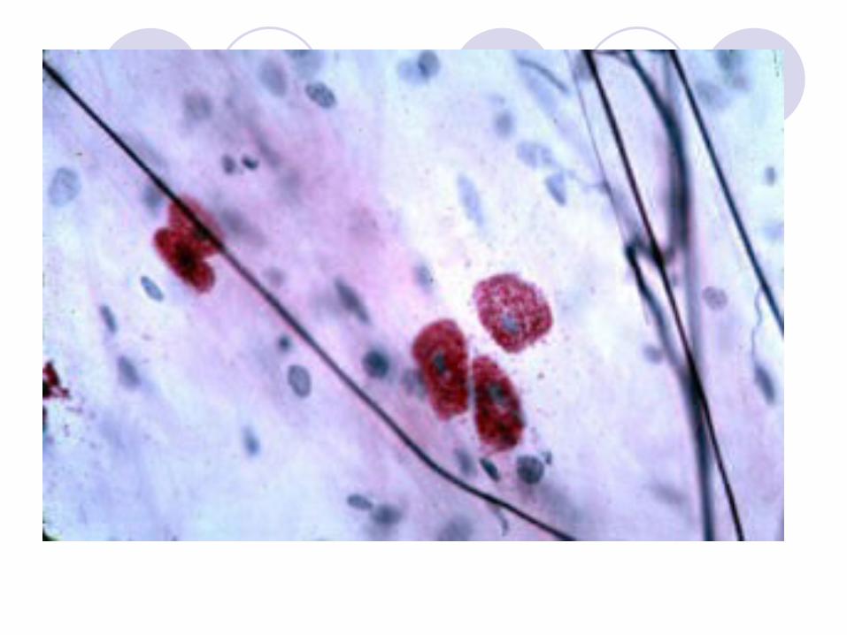
Major Cell Types: Blasts vs. Cytes
1. Blast CellsFibroblasts- large star shaped cells that produce
fibers by secreting proteins into the matrix of CT tissue.
Chondroblast – build cartilage tissueOsteoblast – build osseous (bone tissue)
Blasts build!!!!!

More Cells
Fat Cells/Adipocytes – store energy/nutrientsWhite Blood Cells – respond to injury and
infection to protect and heal the tissueMast Cells – stick around blood vessels,
detect foreign microorganisms and start the inflammatory response against them.
Macrophages – “big eaters” phagocytize lots of foreign material, from bacteria, to old cells, proteins, and dust.

Fibroblast—laying matrix


CONNECTIVE TISSUE-FIBER TYPESA. Collagenous fibers- Thick threads of
protein (collagen) Grouped in long parallel bundles, they are
flexible, but only slightly elastic.
Great tensile strength (ligaments and tendons)

B. Elastic fibers-Composed of protein elastinThese fibers branch, forming complex
networks.(weaker than collagenous fiber, however
they stretch and can return back to there shape.)

Elastic Fibers
C. Reticular Fibers Very thin collagenous fibers Highly branched, form supporting
networks (LIKE A NET)

Reticular Fibers
When it comes to CT you can have a variety of types….Loose CT Areolar tissueReticular tissue Adipose tissue
Dense CT regular and irregular Cartilage types: Hyaline, Elastic,
Fibrocartilage

I.Loose Connective Tissue Loose Connective Tissue Forms delicate thin membranes
throughout the body
Main cellFibroblasts separated by a gel-like matrix
Binds skin to underlying organs and fills spaces between muscles
Lies beneath epithelium

1. Areolar = “packaging material of the body” Prototype for CT Having all types of fibers, cells, etc.
Function1. support and binds other tissue2. holds body fluids; job of matrix/ground
substance3. Defend against infection4. storing nutrients as fat


2 Loose Connective: Reticular
Has a network of reticular fibers.Typically loose ground substanceFunction is to typically support other cell
types such as white blood cells and mast cells.
Thus it is found in lymphoid organs, such as lymph nodes, bone marrow, and spleen.

3
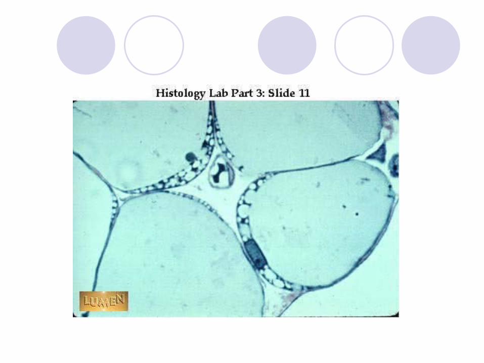
Adipose Tissue (considered loose connective)
FATspecialized form of CT, develop when certain cells store fat in droplets within their cytoplasm.
Beneath the skin, stored in many other locations as well.
Cushions, insulates, stores energy.
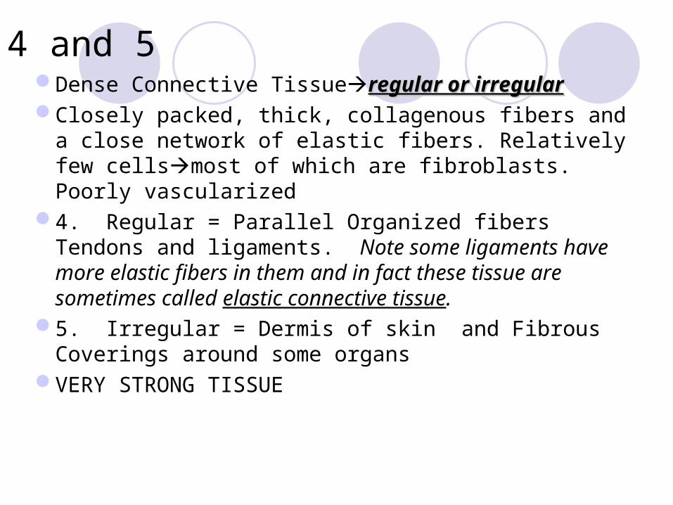
4 and 5Dense Connective Tissueregular or irregularregular or irregularClosely packed, thick, collagenous fibers and a close
network of elastic fibers. Relatively few cellsmost of which are fibroblasts. Poorly vascularized
4. Regular = Parallel Organized fibers Tendons and ligaments. Note some ligaments have more elastic fibers in them and in fact these tissue are sometimes called elastic connective tissue.
5. Irregular = Dermis of skin and Fibrous Coverings around some organs
VERY STRONG TISSUE


Cartilage
Rigid CTprovides support, frameworks, and attachments, protects underlying tissue and forms structural models for developing bone.
Cartilage matrixabundant, largely composed of colagenous fibers embedded in a gel-like ground substance . A large percentage of water up to 80%. Lots of GAGs (proteoglycans)
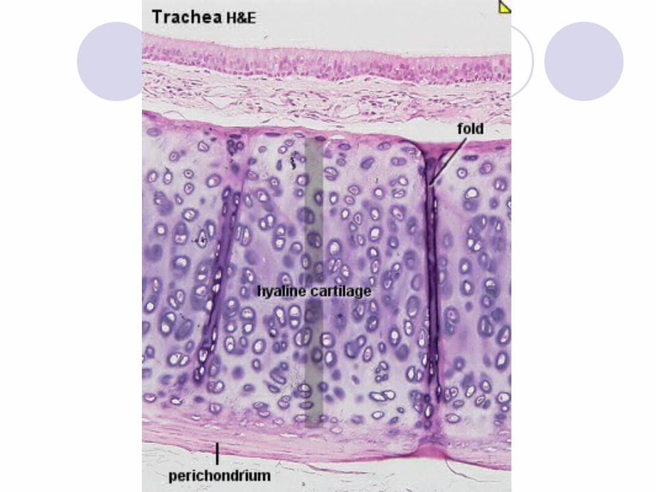
Avascular, receives nourishment from membranes surrounding it. (Perichondrium)
Cartilagea special case
SPECIAL CARTILAGE CELLSCHONDROBLASTS, form
developing/growing cartilageCHONDROCYTES, occupy small
chambers called lacunae
Cartilage structures are enclosed in a covering of CTperichondrium (contains the blood vessels that deliver nutrients)
THE INTERCELLULAR MATRIX DISTINGUISHES THE TYPE OF CARTILAGE!

6 Hyaline Cartilage
*most common type *very fine collagenous fibers (looks like white glass)Location: end of bones, joints, soft part of
nose, trachea

7 Elastic Cartilage
Dense network of elastic fibers Much more flexible than hyaline cartilage Location: framework of ear, parts of larynx

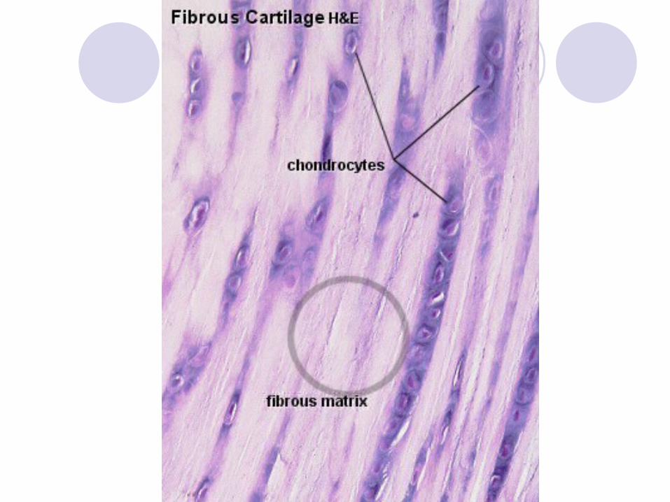
8 Fibrocartilage
Tough tissue, contains many collagenous fibers
-shock absorber for structures subjected to pressure.
Location: vertebrae (discs), knees, pelvic girdle.