connective tissue diseases - university of pretoria · two types of autoimmune diseases –...
TRANSCRIPT

Connective Tissue Diseases
Dr. C. C. Visser
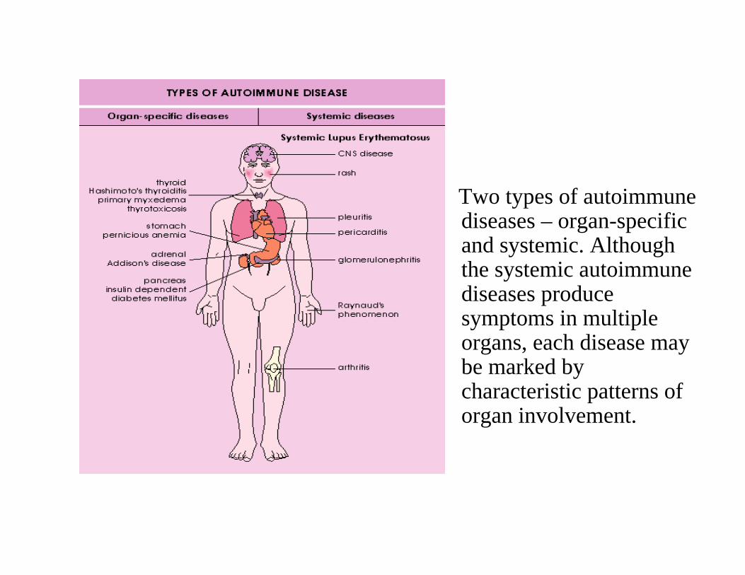
Two types of autoimmune diseases – organ-specific and systemic. Although the systemic autoimmune diseases produce symptoms in multiple organs, each disease may be marked by characteristic patterns of organ involvement.

Systemic lupus erythematosis


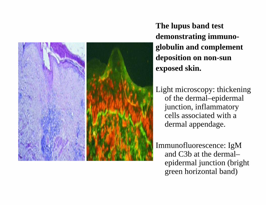
The lupus band testdemonstrating immuno-globulin and complementdeposition on non-sunexposed skin.
Light microscopy: thickening of the dermal–epidermal junction, inflammatory cells associated with a dermal appendage.
Immunofluorescence: IgMand C3b at the dermal–epidermal junction (bright green horizontal band)
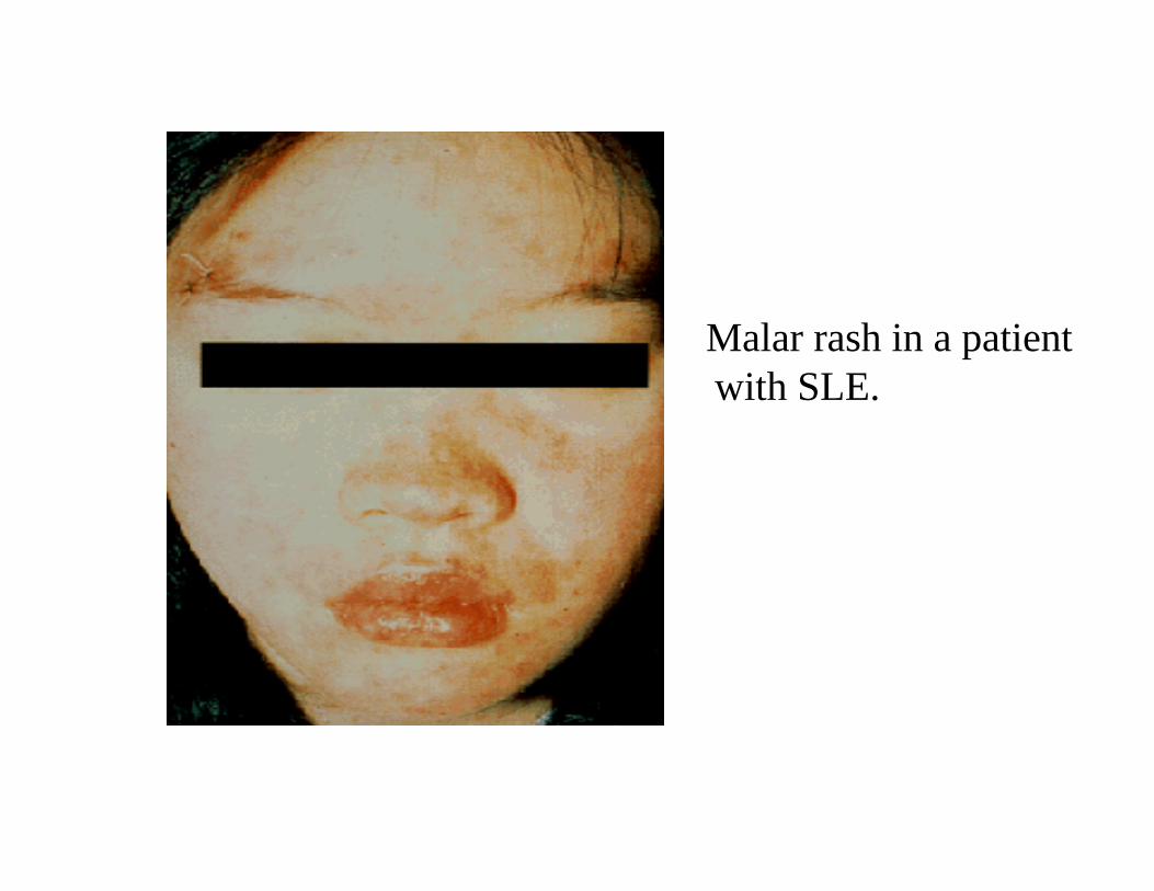
Malar rash in a patient with SLE.
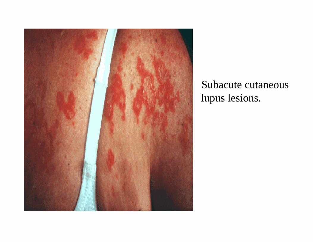
Subacute cutaneouslupus lesions.
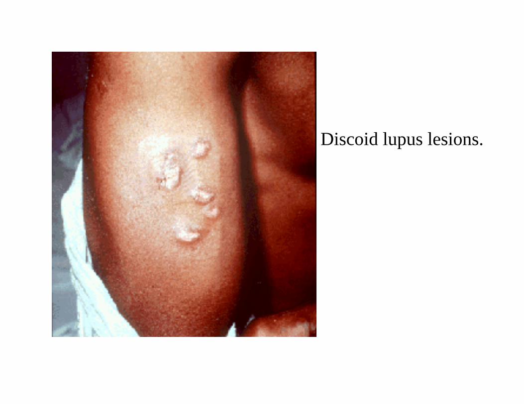
Discoid lupus lesions.

Alopecia in a patient with SLE.

Early vasculitic lesions over the tips of the toes in a patient with active SLE.

Mouth ulcers in a patient with SLE.

Nasal septalperforation in a patient with SLE.

Gangrene of the toe in a patient with SLE andvasculitis.

Swan neck deformities in a patient with SLE.

Fists of the previous patient showing the deformities reduced.

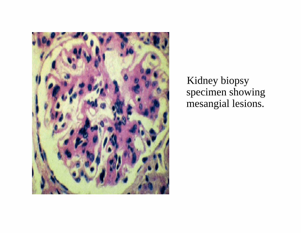
Kidney biopsy specimen showingmesangial lesions.
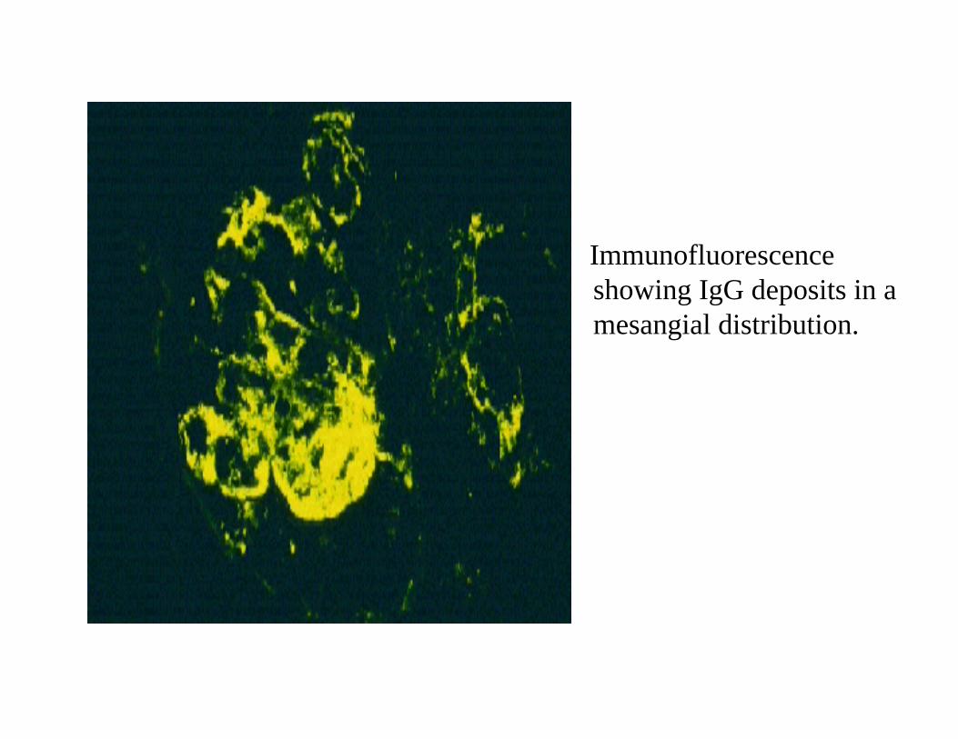
Immunofluorescenceshowing IgG deposits in amesangial distribution.
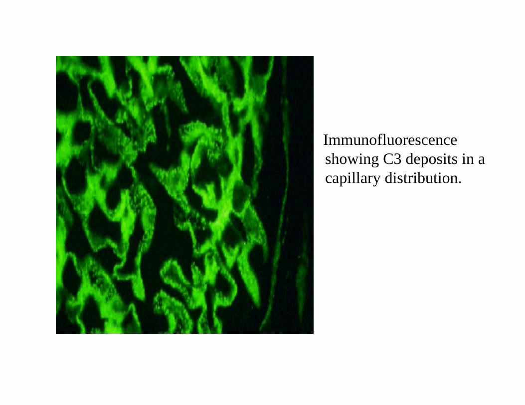
Immunofluorescenceshowing C3 deposits in a capillary distribution.
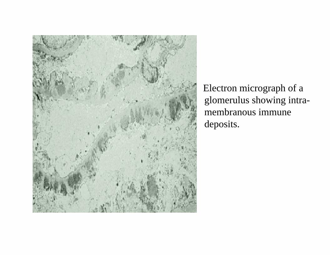
Electron micrograph of aglomerulus showing intra-membranous immune deposits.

Electron micrograph of aglomerulus showingsubendothelial immune deposits.


Funduscopic examination in a patient with SLE demonstrating cytoidbodies.
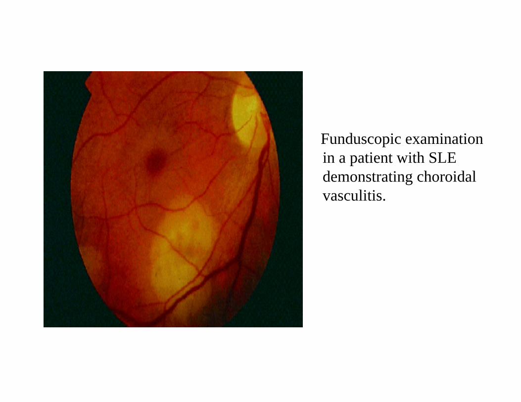
Funduscopic examination in a patient with SLE demonstrating choroidal vasculitis.
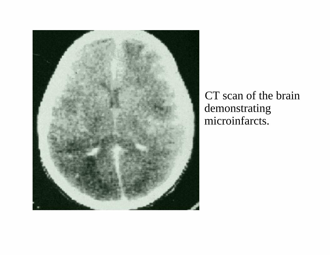
CT scan of the brain demonstratingmicroinfarcts.
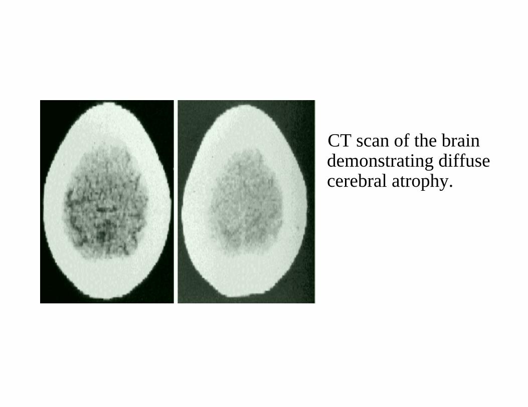
CT scan of the brain demonstrating diffuse cerebral atrophy.

Technetium brain scan demonstrating increased uptake in a ‘draped curtain’ pattern on the anterior view. A normal anterior view is shown on the left for comparison.

Chest radiographs demonstrating the development of a pericardial effusion in a patient with SLE.
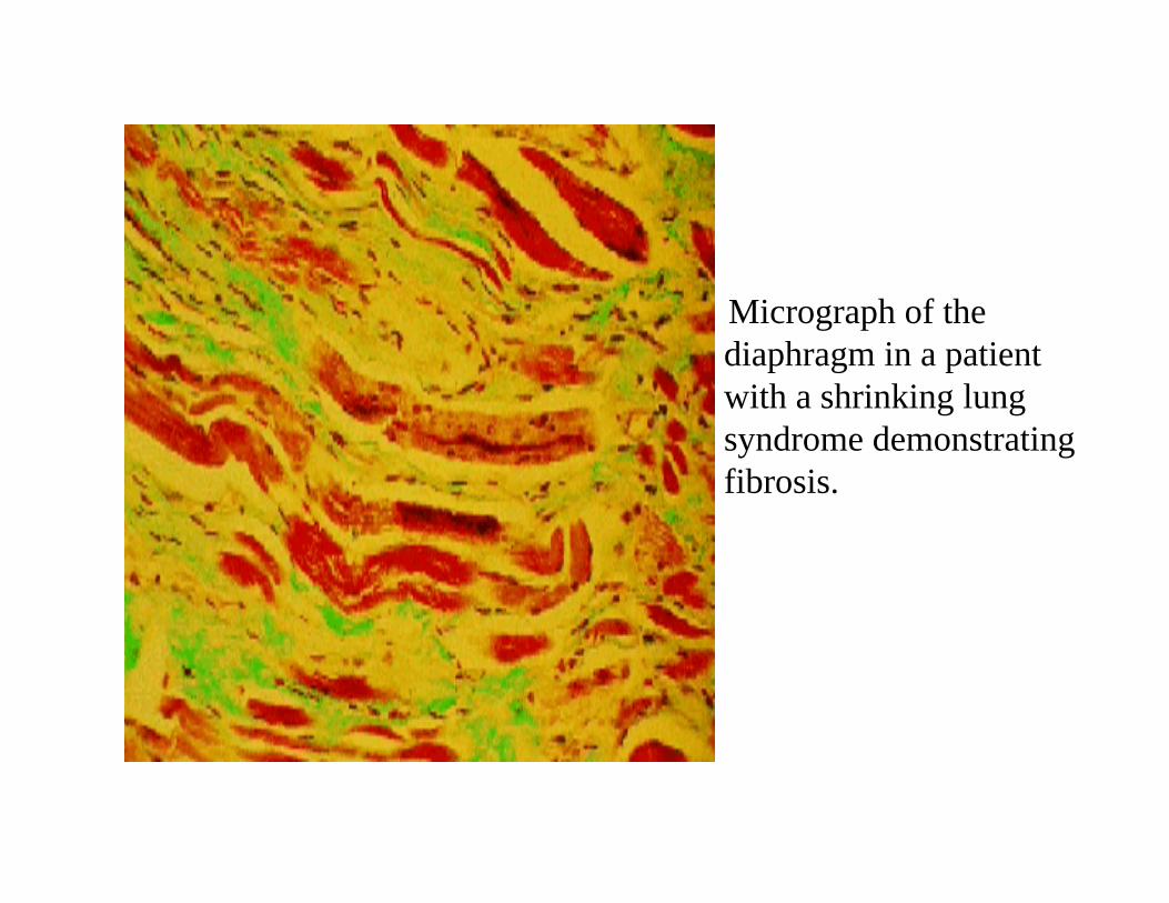
Micrograph of the diaphragm in a patient with a shrinking lung syndrome demonstrating fibrosis.
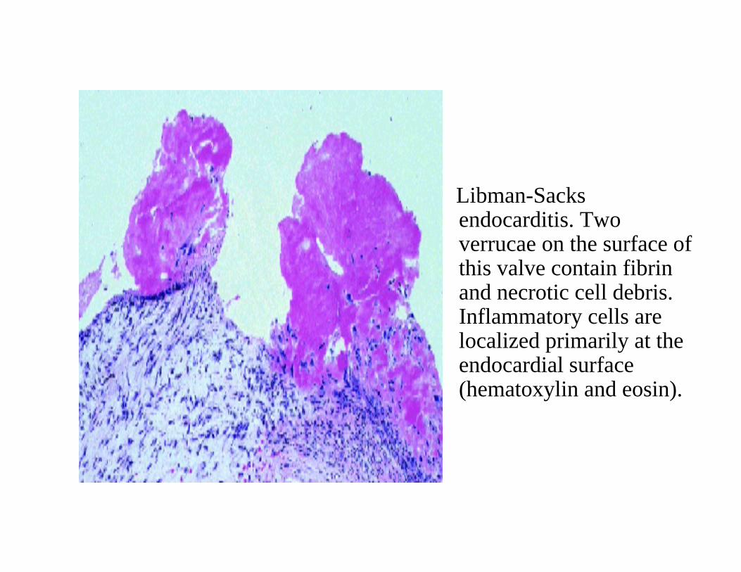
Libman-Sacksendocarditis. Twoverrucae on the surface of this valve contain fibrin and necrotic cell debris. Inflammatory cells are localized primarily at theendocardial surface (hematoxylin and eosin).

Diagnosis


Management


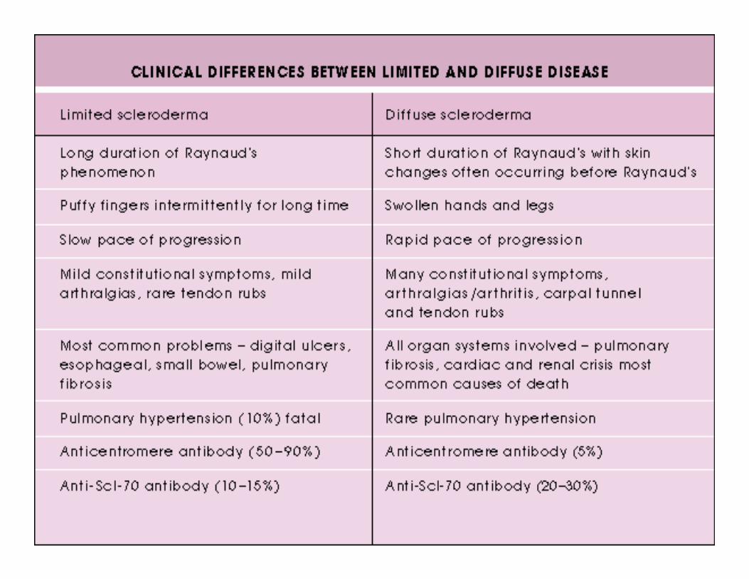
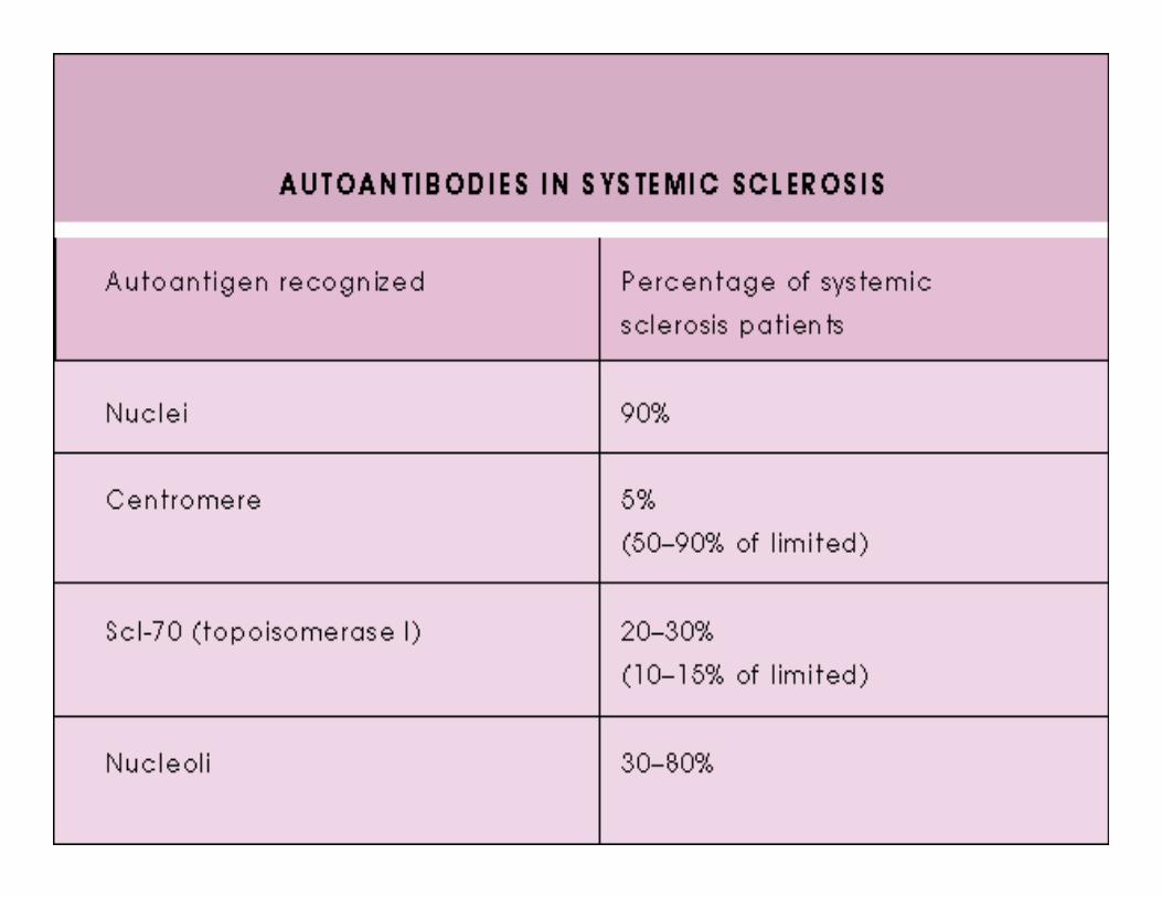
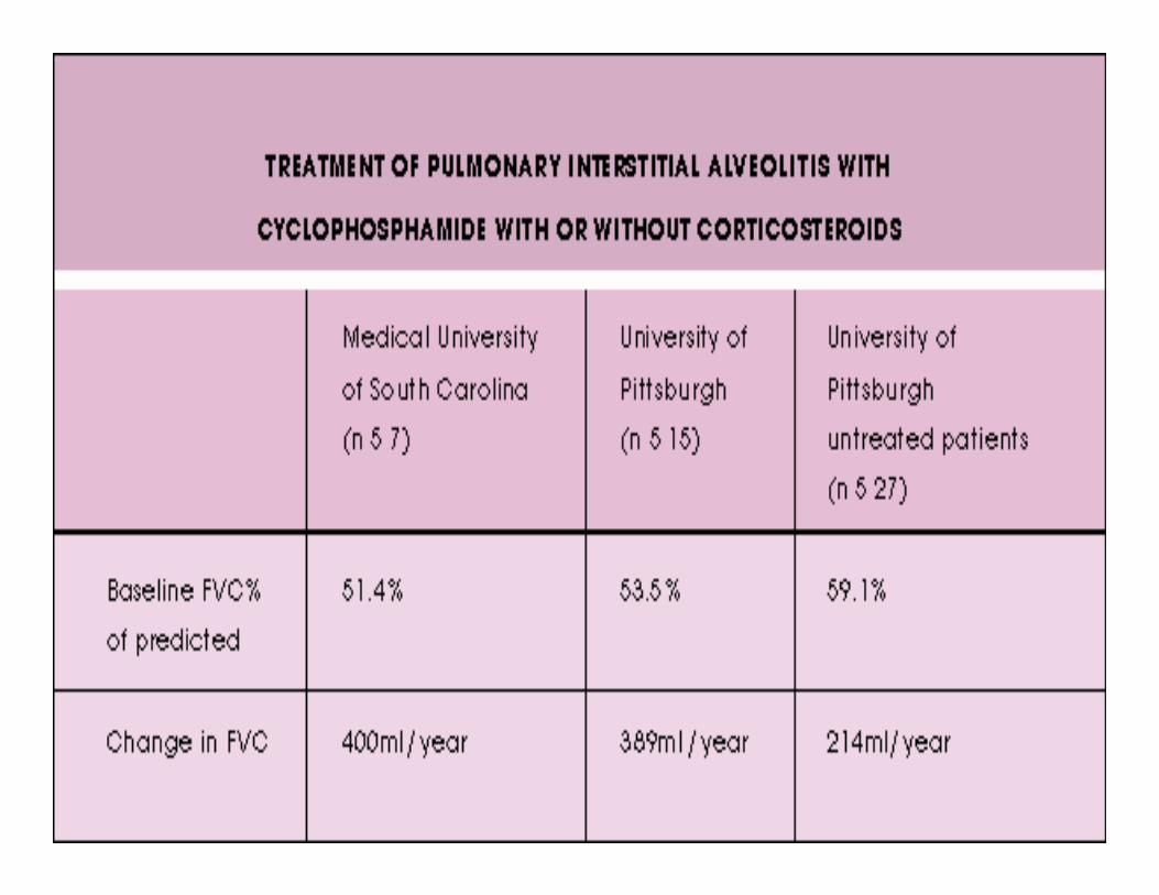
Systemic sclerosis


The hands of a young woman with Raynaud’sphenomenon. There is sharply demarcated cyanosis of the fingers with more proximallivedoid venularcongestion.

Multiple digital ischemiculcerations. Small areas of infarction at different stages of development and of variable severity of the fingertips of a young woman with several months of rapidly progressive scleroderma.
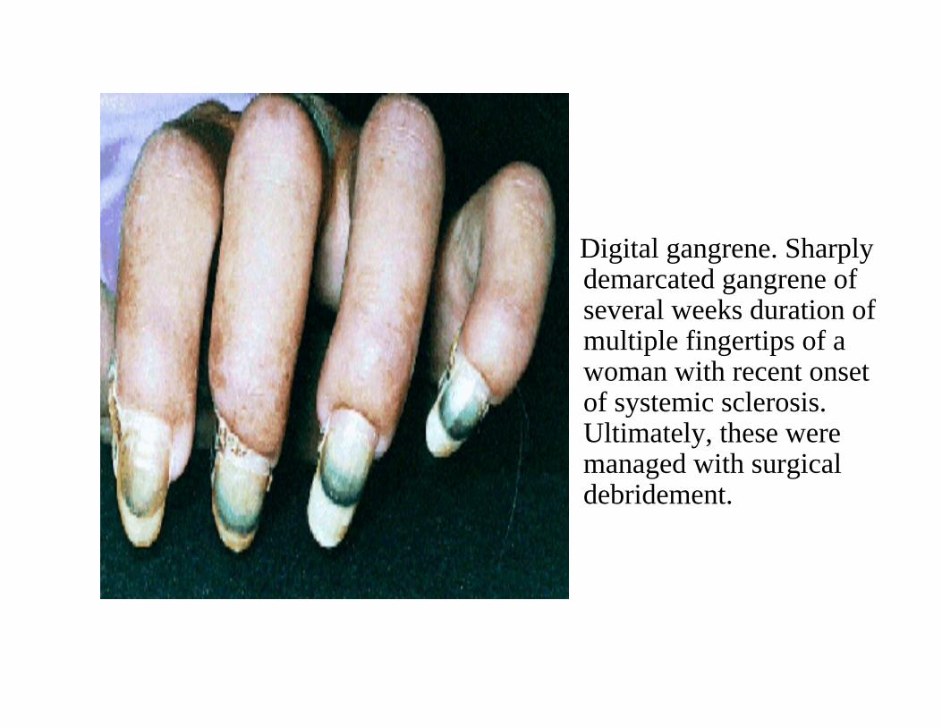
Digital gangrene. Sharply demarcated gangrene of several weeks duration of multiple fingertips of a woman with recent onset of systemic sclerosis. Ultimately, these were managed with surgicaldebridement.
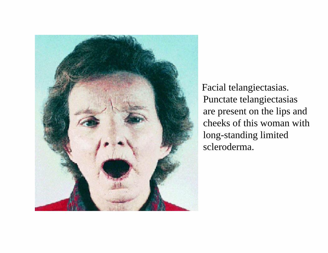
Facial telangiectasias.Punctate telangiectasiasare present on the lips and cheeks of this woman with long-standing limitedscleroderma.

Early, puffy scleroderma. Extensive edema of the fingers and hands in a man with several months of preceding Raynaud’sphenomenon. Skin was not clinically thickened but became so on follow up.
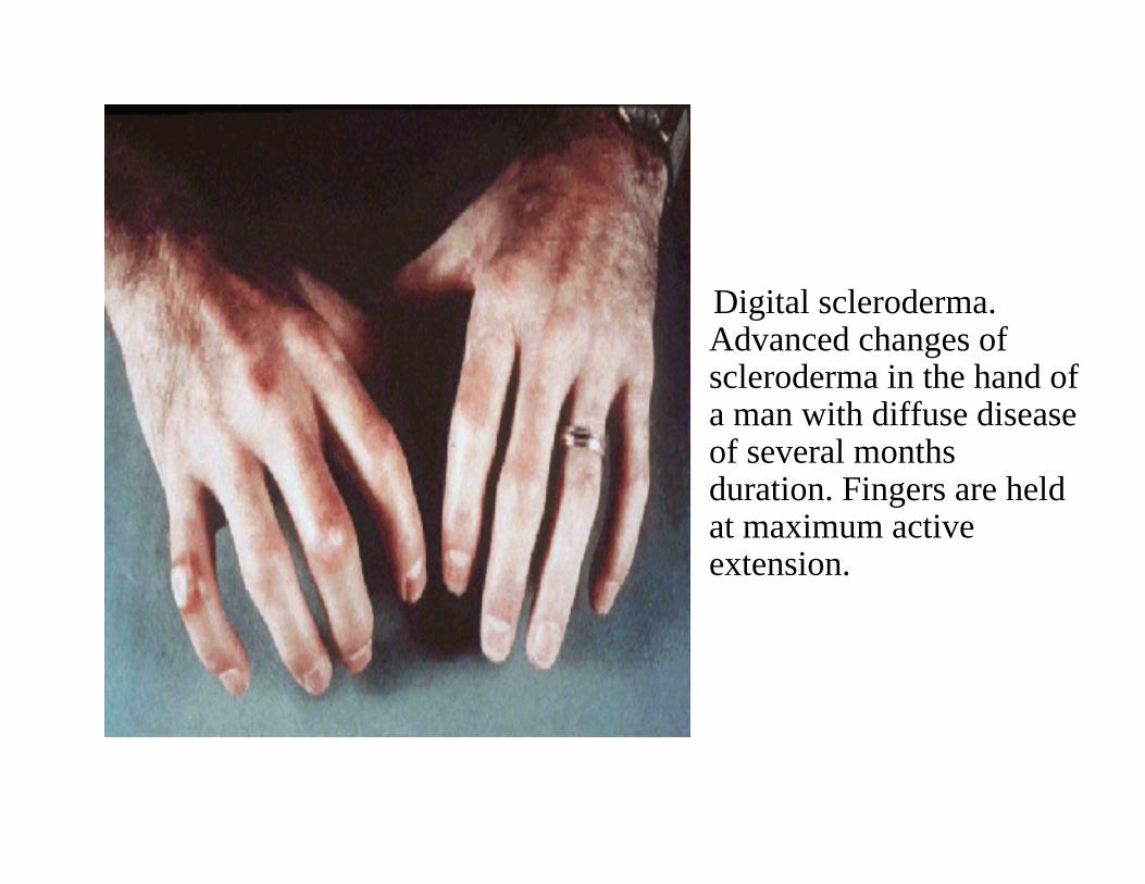
Digital scleroderma. Advanced changes ofscleroderma in the hand of a man with diffuse disease of several months duration. Fingers are held at maximum active extension.

Facial scleroderma. Taut smooth skin over the face of a woman with long-standing disease. Oral aperture is reduced and radial furrowing is present about the lips.
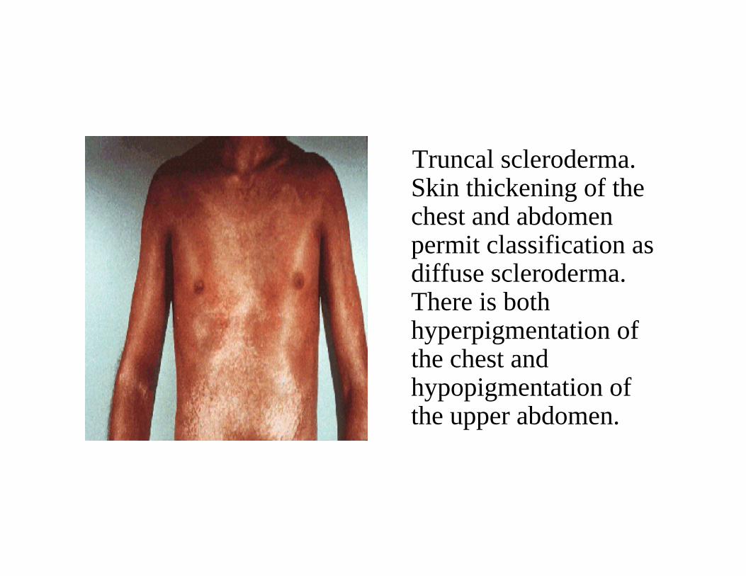
Truncal scleroderma. Skin thickening of the chest and abdomen permit classification as diffuse scleroderma. There is bothhyperpigmentation of the chest andhypopigmentation of the upper abdomen.
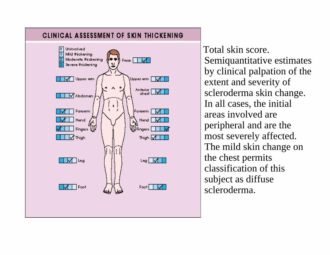
Total skin score.Semiquantitative estimates by clinical palpation of the extent and severity ofscleroderma skin change. In all cases, the initial areas involved are peripheral and are the most severely affected. The mild skin change on the chest permits classification of this subject as diffusescleroderma.
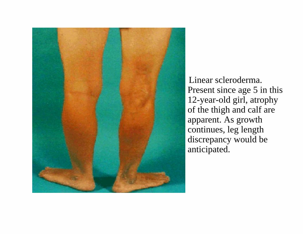
Linear scleroderma. Present since age 5 in this 12-year-old girl, atrophy of the thigh and calf are apparent. As growth continues, leg length discrepancy would be anticipated.
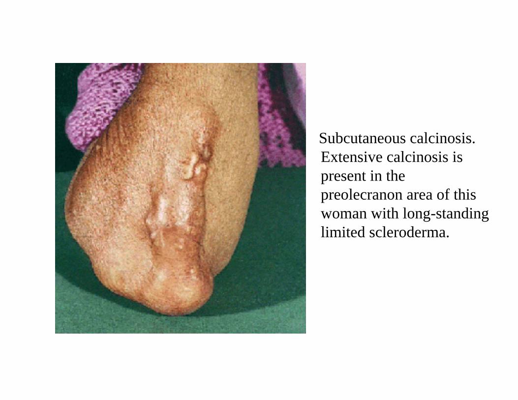
Subcutaneous calcinosis. Extensive calcinosis is present in thepreolecranon area of this woman with long-standing limited scleroderma.
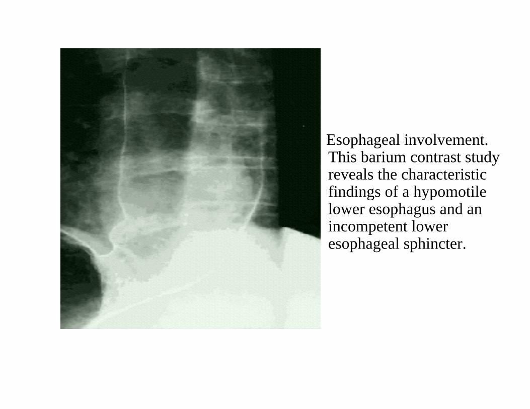
Esophageal involvement. This barium contrast study reveals the characteristic findings of a hypomotilelower esophagus and an incompetent loweresophageal sphincter.
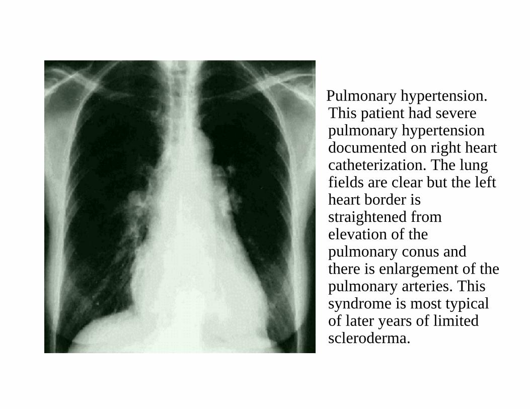
Pulmonary hypertension. This patient had severe pulmonary hypertension documented on right heartcatheterization. The lung fields are clear but the left heart border is straightened from elevation of the pulmonary conus and there is enlargement of the pulmonary arteries. This syndrome is most typical of later years of limitedscleroderma.
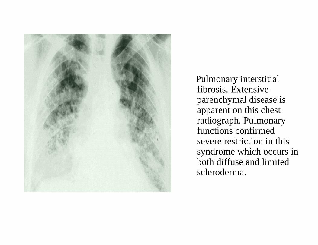
Pulmonary interstitial fibrosis. Extensiveparenchymal disease is apparent on this chest radiograph. Pulmonary functions confirmed severe restriction in this syndrome which occurs in both diffuse and limitedscleroderma.

Sicca syndrome inscleroderma. The tongue is parched andhypopapillated in this woman with siccasyndrome complicating long-standing diffusescleroderma.

Excess collagen deposition in the dermis of a patient with systemic sclerosis. There is marked thickening of the dermis with collagen and some inflammatory infiltrate surrounding vasculature. There is entrapment ofcutaneous glands. Although atrophy of the epidermal rete pegs is a frequent finding, it is not evident in this example. (Hematoxylin and eosin).


Dermatomyositis/Polymyositis

Diagnosis
• Muscle pain and weakness proximally• Raised creatine kinase• Abnormalities on EMG• Muscle biopsy
• Dermatomyositis– Grotton papules– Heliotrope rash


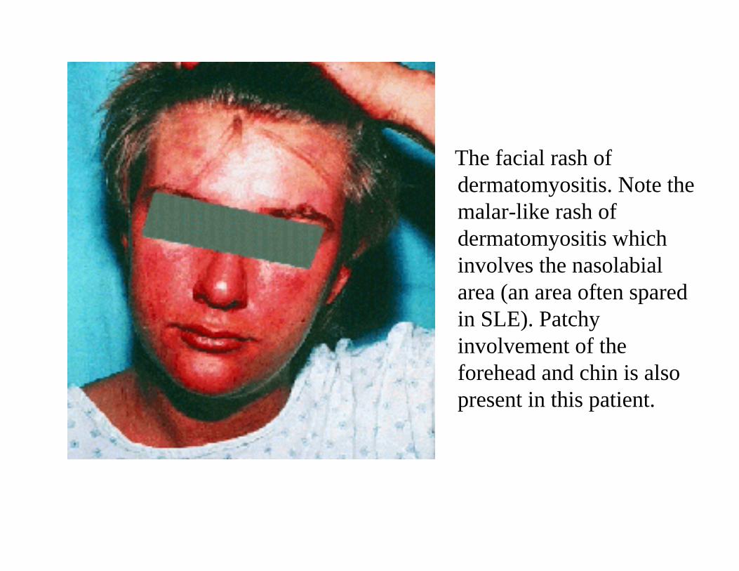
The facial rash ofdermatomyositis. Note themalar-like rash ofdermatomyositis which involves the nasolabialarea (an area often spared in SLE). Patchy involvement of the forehead and chin is also present in this patient.
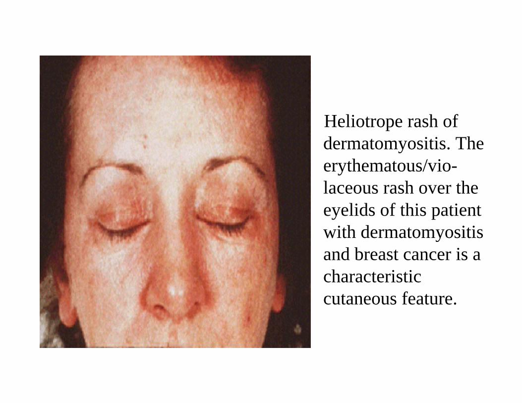
Heliotrope rash ofdermatomyositis. Theerythematous/vio-laceous rash over the eyelids of this patient with dermatomyositisand breast cancer is a characteristiccutaneous feature.

Gottron sign. Thiserythematous, scaling rash over the knuckles and dorsum of the hand is a common early sign indermatomyositis. It can be distinguished from the rash of SLE which usually affects the phalanges and spares the knuckles.
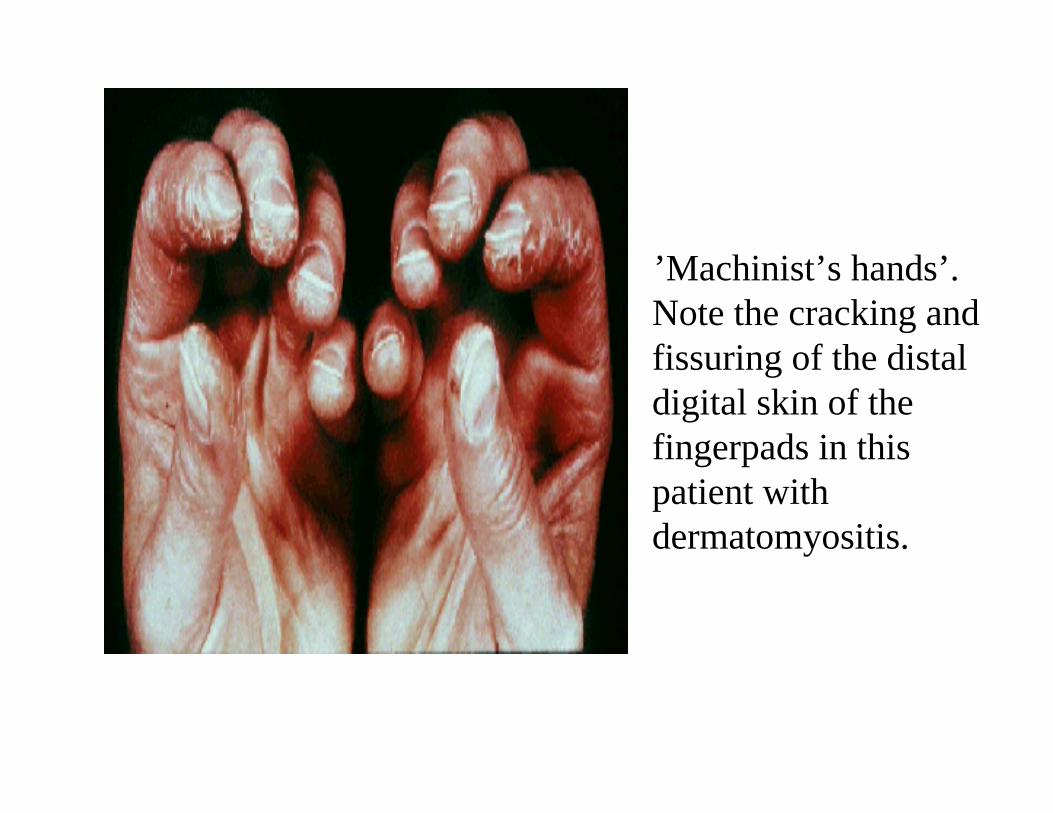
’Machinist’s hands’. Note the cracking and fissuring of the distal digital skin of thefingerpads in this patient withdermatomyositis.
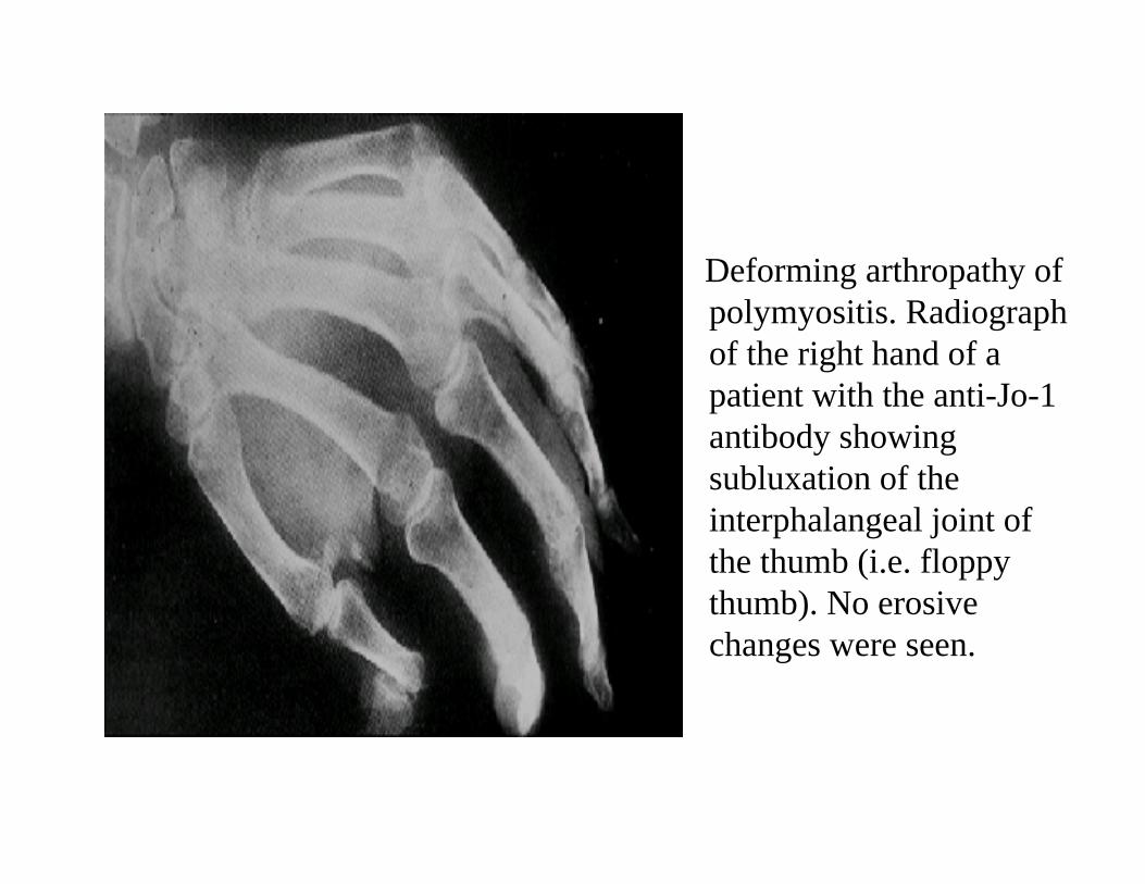
Deforming arthropathy ofpolymyositis. Radiograph of the right hand of a patient with the anti-Jo-1 antibody showing subluxation of theinterphalangeal joint of the thumb (i.e. floppy thumb). No erosive changes were seen.
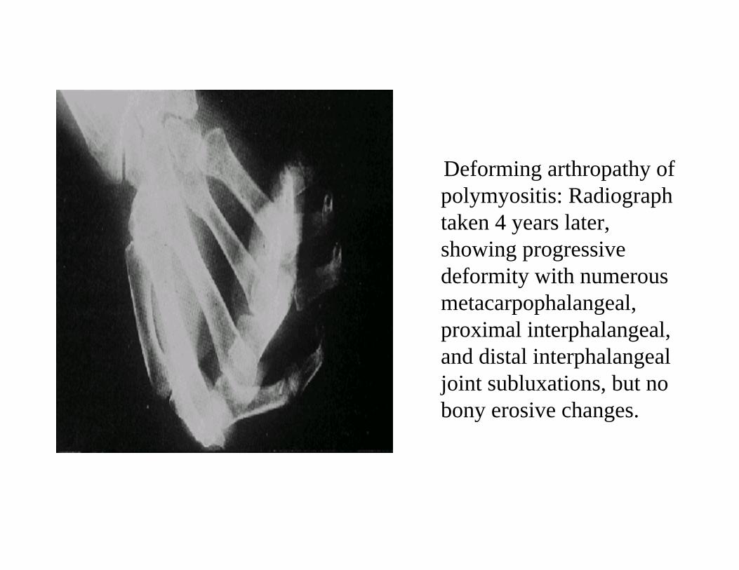
Deforming arthropathy of polymyositis: Radiograph taken 4 years later, showing progressive deformity with numerousmetacarpophalangeal, proximal interphalangeal, and distal interphalangealjoint subluxations, but no bony erosive changes.
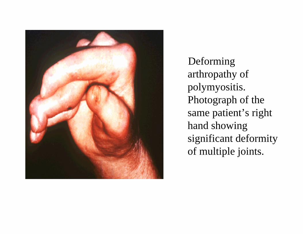
Deforming arthropathy ofpolymyositis. Photograph of the same patient’s right hand showing significant deformity of multiple joints.

Interstitial lung disease ofpolymyositis/dermatomyositis. Chest radiograph of a patient with interstitial lung disease anddermatomyositisdemonstrating severe basilar fibrosis and mid-lung interstitial changes as well.
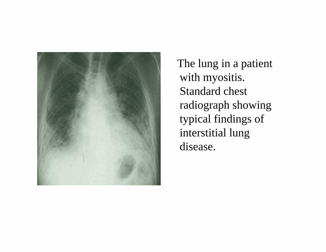
The lung in a patient with myositis. Standard chest radiograph showing typical findings of interstitial lung disease.

Gross autopsy specimen from the heart of a patient withmyositis who died from myocarditisshowing dilated left ventricle and fibrosis

Diagnosis
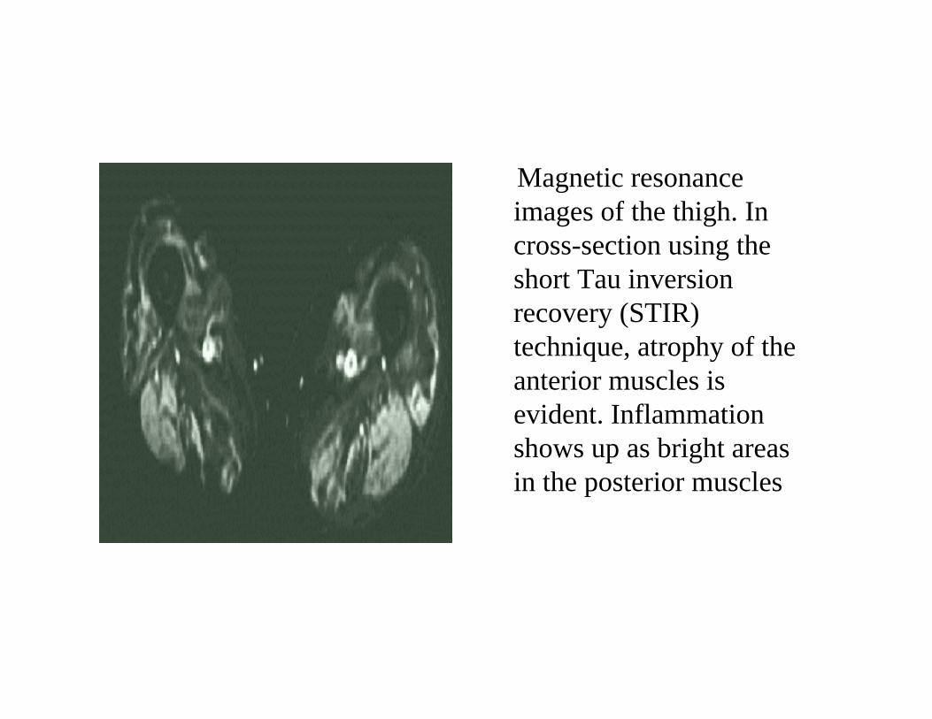
Magnetic resonance images of the thigh. In cross-section using the short Tau inversion recovery (STIR) technique, atrophy of the anterior muscles is evident. Inflammation shows up as bright areas in the posterior muscles
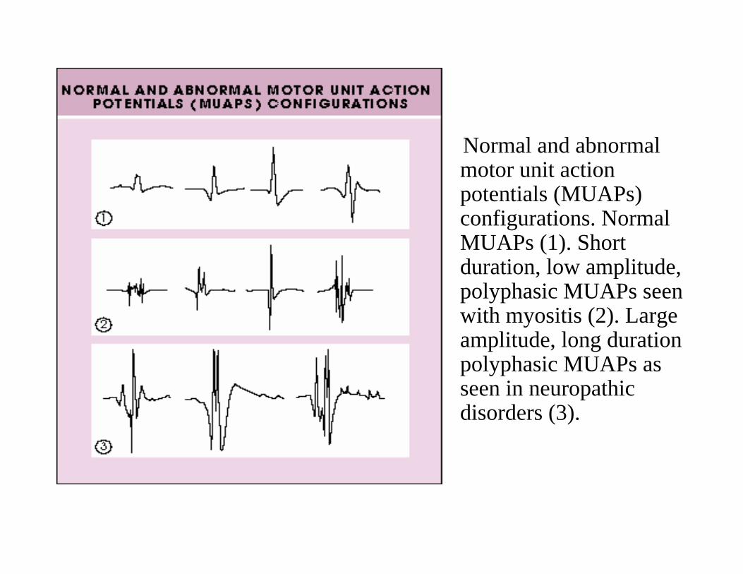
Normal and abnormal motor unit action potentials (MUAPs) configurations. NormalMUAPs (1). Short duration, low amplitude,polyphasic MUAPs seen with myositis (2). Large amplitude, long durationpolyphasic MUAPs as seen in neuropathicdisorders (3).

Distribution of lymphocytes in muscle biopsies from patients with myositis.
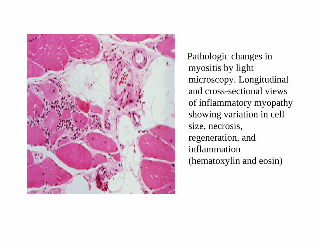
Pathologic changes inmyositis by light microscopy. Longitudinal and cross-sectional views of inflammatory myopathyshowing variation in cell size, necrosis, regeneration, and inflammation (hematoxylin and eosin)

Skin biopsy of a Gottron’slesion in a patient withdermatomyositis. The biopsy demonstrateshyperkeratosis, epidermal thinning, vacuolardegeneration of the basal layer, dilated superficial capillaries withperivascular lymphohistiocyticinfiltrates, and mild mucindeposition in the dermis.
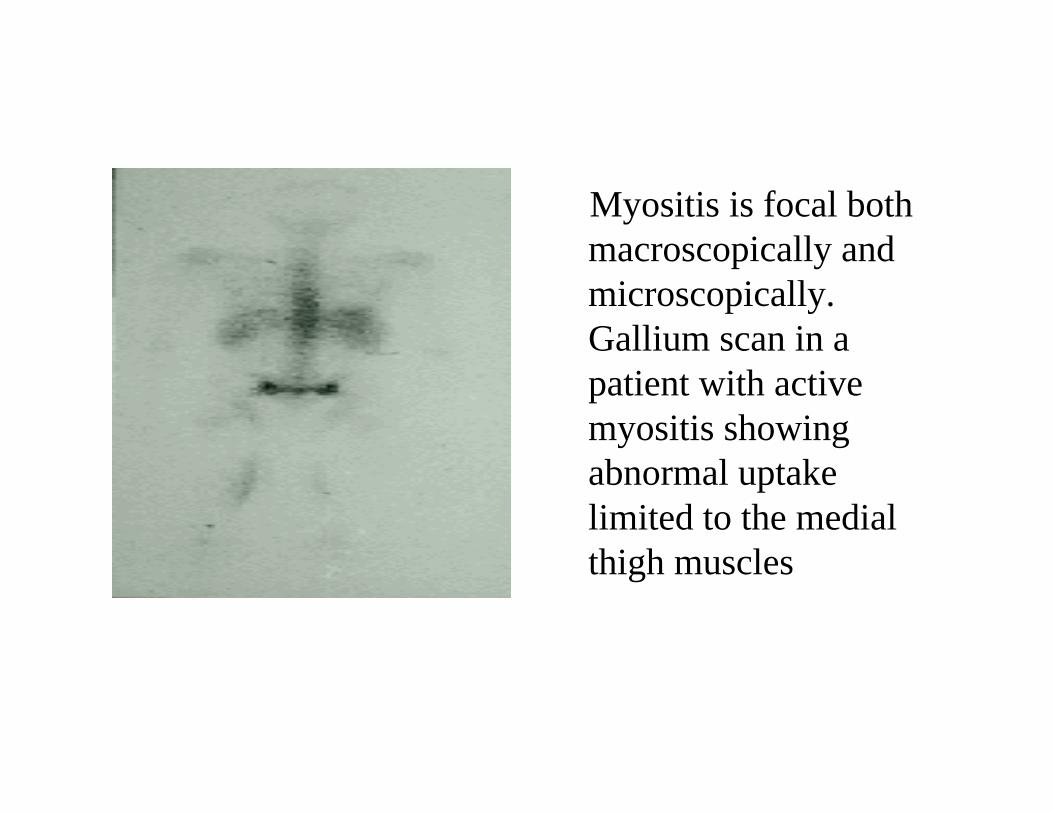
Myositis is focal both macroscopically and microscopically. Gallium scan in a patient with activemyositis showing abnormal uptake limited to the medial thigh muscles


Treatment
• Symptomatic• Steroids• Immunosuppressants (methotrexate)