confusing endodontic cases case series report...keywords: diagnosis, endodontics, skin lesion,...
TRANSCRIPT

| 26 | Smile Dental Journal | Volume 6, Issue 2 - 2011
AbstractHere, we introduce some confusing endodontic cases, which we had experienced in our clinic. The first case had been misdiagnosed as a skin lesion and received extended skin treatment. The second case had been misdiagnosed as a periodontal lesion and received periodontal treatment by two former dentists. The third case exhibited oral malodor and the patient worried about his breath odor for a long period. These three cases have received endodontic treatment in our clinic, and the patients were free from their long-lasting problems. We also discuss other confusing cases with literature mini-review.
Keywords: Diagnosis, Endodontics, Skin Lesion, Periodontics.
IntroductionMaking a diagnosis is one of the most important stages in dental treatment. We sometimes encounter endodontic cases in which making a straight forward diagnosis can be a real challenge. Furthermore, endodontic lesions sometimes manifest symptoms that are similar to those of other diseases. However, one of the difficult points in endodontic treatment is that the lesion cannot be seen from the outside, and unless we take a 3D radiograph, we need to speculate about what is happening in the bone. It is also dangerous for dentists to make a diagnosis with preconceptions. Here, we report endodontic cases that had been misdiagnosed as skin or periodontal diseases. Conversely, certain diseases, including malignant diseases, are sometimes misdiagnosed as endodontic lesions. We also present a literature review of these diseases that show similar manifestations to endodontic disease.
Case Reports on Endodontic Lesion-derived Symptoms that had been MisdiagnosedSkin problem caused by an endodontic lesionA 22-year-old male patient visited the Fukuoka Dental College Hospital complaining of a prolonged fistula in the right lower cheek skin.1 His former doctor treated the patient for 6 months. Repeated skin incisions and long-term antibiotic therapy were not effective and the doctor referred the patient to us. An external dental fistula with a diameter of about 10mm and continuous pus discharge was observed (Figure 1A). By X-ray findings and electric pulp examination, we diagnosed an external dental fistula caused by chronic purulent apical periodontitis of tooth 46 (Figure 1B). We performed an infected root canal treatment including anaerobic culture examination. The response to the root canal treatment was good. The size of the external dental fistula dramatically decreased and the skin lesion became dry at the third visit (Figure 2A). After confirming that intra-root canal bacteria were negative, the root canals were filled (Figure 2B).The external dental fistula completely closed after 2 months. Two and half years after the root canal filling, the patient came for tooth maintenance. The external dental fistula had not recurred (Figure 3A) and radiolucency in the periapical and furcation area had disappeared (Figure 3B).
Confusing Endodontic CasesCase Series Report
Masahiro Yoneda
Section of General Dentistry, Department of General Dentistry, Fukuoka Dental College, Fukuoka, Japan
Nao Suzuki
Section of General Dentistry, Department of General Dentistry, Fukuoka Dental College, Fukuoka, Japan
Sonia M. Macedo
Faculdade de Odontologia da Universidade de São PauloSão Paulo, Brazil
Hisashi Anan
Section of Operative Dentistry and Endodontology, Department of Odontology, Fukuoka Dental CollegeFukuoka, Japan
Takao Hirofuji
Section of General Dentistry, Department of General Dentistry, Fukuoka Dental College, Fukuoka, Japan

Smile Dental Journal | Volume 6, Issue 2 - 2011| 27 |
(Fig. 2A) External dental fistula after the initiation of endodontic treatment. The pus discharge has stopped and the external dental fistula is dry.
(Fig. 2B) Dental X-ray photograph at the root canal filling.
(Fig. 3A) Right facial view at 2.5 years after the root canal filling. The external dental fistula has not recurred.
(Fig. 3B) Dental X-ray photograph at 2.5 years after the root canal filling. The radiolucency at the apex and furcation area of tooth 46 has disappeared.
(Fig. 1B) Dental X-ray photograph at the first visit. Radiolucency at the apex and furcation area of tooth 46 is observed.
(Fig. 1A) Right facial view at the first visit. An external dental fistula with pus discharge is observed at the right cheek area.

| 28 | Smile Dental Journal | Volume 6, Issue 2 - 2011
pulp examination but a gutta-percha point inserted from the fistula reached the mesial root apex. Consequently, we diagnosed a type I endoperiodontal lesion of tooth 46. Soon after root canal treatment, the gingival condition at tooth 46 improved without periodontal treatment (Figure 5A). After confirming the absence of bacteria, the root canals were filled (Figure 5B). After 3 months, the furcation bone loss was on the way to recovery (data not shown). At 5 years after the root canal filling, the patient came for treatment of other caries. The gingival condition was good and the furcation bone loss had not recurred (Figure 6A, B).
Gingival swelling and furcation bone loss caused by an endodontic lesionA 59-year-old female attended our hospital with a chief complaint of mobility of tooth 46 and recurrent gingival swelling around the tooth (Figure 4A). She had previously received dental treatment from two dentists who performed gingival incisions, scaling and root planning, but her gingival condition did not improve.2 Her tooth 46 manifested the symptoms of typical periodontitis, such as gingival swelling, tooth mobility, pus discharge from the periodontal pocket and furcation bone loss (Figure 4B). The tooth responded to electric
(Fig. 6A) Lateral view of the right teeth and gingiva at 5 years after the root canal filling. Gingival condition is good.
(Fig. 6B) X-ray photograph of tooth 46 at 5 years after the root canal filling. There is no furcation bone loss.
(Fig. 4A) Lateral view of the right teeth and gingiva at the first visit. Gingival swelling is observed around the margin of tooth 46.
(Fig. 4B) X-ray photograph of tooth 46 at the first visit. Bone resorption is observed at the furcation area, and the periodontium around the mesial root of tooth 46 is enlarged.
(Fig. 5A) Lateral view of the right teeth and gingiva after initiation of endodontic treatment. Soon after initiation of the infected root canal treatment, the gingival swelling has disappeared.
(Fig. 5B) X-ray photograph of tooth 46 after the root canal filling. Furcation bone loss remains at the root canal filling.
Smile Dental Journal | Volume 6, Issue 2 - 2011| 29 |
third visit and restored with metal bridge (Figures 8A, B). We measured the breath odor and compared it with that at the initial visit. All of the malodor scores were found to have decreased (Figure 9) and the patient was satisfied with our treatment.
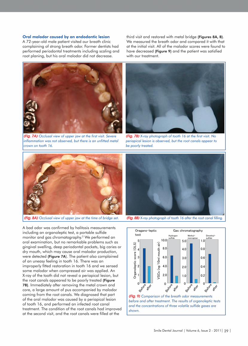
Oral malodor caused by an endodontic lesionA 72-year-old male patient visited our breath clinic complaining of strong breath odor. Former dentists had performed periodontal treatments including scaling and root planing, but his oral malodor did not decrease.
A bad odor was confirmed by halitosis measurements including an organoleptic test, a portable sulfide monitor and gas chromatography.3 We performed an oral examination, but no remarkable problems such as gingival swelling, deep periodontal pockets, big caries or dry mouth, which may cause oral malodor production, were detected (Figure 7A). The patient also complained of an uneasy feeling in tooth 16. There was an improperly fitted restoration in tooth 16 and we sensed some malodor when compressed air was applied. An X-ray of the tooth did not reveal a periapical lesion, but the root canals appeared to be poorly treated (Figure 7B). Immediately after removing the metal crown and core, a large amount of pus accompanied by malodor coming from the root canals. We diagnosed that part of the oral malodor was caused by a periapical lesion of tooth 16, and performed an infected root canal treatment. The condition of the root canals had improved at the second visit, and the root canals were filled at the
(Fig. 9) Comparison of the breath odor measurements before and after treatment. The results of organoleptic tests and the concentrations of three volatile sulfide gases are shown.
(Fig. 7A) Occlusal view of upper jaw at the first visit. Severe inflammation was not observed, but there is an unfitted metal crown on tooth 16.
(Fig. 7B) X-ray photograph of tooth 16 at the first visit. No periapical lesion is observed, but the root canals appear to be poorly treated.
(Fig. 8A) Occlusal view of upper jaw at the time of bridge set. (Fig. 8B) X-ray photograph of tooth 16 after the root canal filling.

| 30 | Smile Dental Journal | Volume 6, Issue 2 - 2011
There is also a possibility to misdiagnose malignant diseases as an endodontic lesion. In fact, a gingival squamous cell carcinoma mimicking a dentoalveolar abscess has been reported.24 Some metastatic carcinomas manifesting as periapical lesions have also been reported as well as an oral-derived carcinoma.25,26 An osteosarcoma27 and a mesenchymal chondrosarcoma28 resembling dental periapical lesions have also been reported. Some lymphomas were misdiagnosed as endodontic lesions and the proper treatments were delayed.29 Although some cases may be difficult to diagnose,30 we always need to consider the possibility of malignant diseases.
In this way, making diagnosis is important in the treatment of endodontic cases and cases, which manifest endodontic-like symptoms.
Acknowledgment This work was partly supported by a Grant-in-Aid for Scientific Research (No. 20592249) from the Japanese Ministry of Education, Science, Sports and Culture.
References1. Yoneda M, Anan H, Motooka N, Hirofuji T, Matsumoto A, Isobe
R, Kabashima H, Maeda K. Disappearance of an external dental fistula with an endodontic treatment of the causative tooth. Jap J Conserv Dent. 2003;46(2):143-8.
2. Yoneda M, Motooka N, Naito T, Maeda K, Hirofuji T. Resolution of furcation bone loss after non-surgical root canal treatment: application of a peptidase-detection kit for treatment of type I endoperiodontal lesion. J Oral Sci. 2005;47(3):143-7.
3. Yoneda M, Uchida H, Suzuki N, Mine M, Iwamoto T, Masuo Y, Naito T, Hatano Y, Hirofuji T. A case report of tooth wear associated with a patient’s inappropriate efforts to reduce oral malodor caused by endodontic lesion. Int J Dent. 2009, article ID 727481, 5a, doi:1155/2009/727481.
4. Tagami H, Yoshitake K. Chronic dental fistula on the nose. Acta Dermatol Venereol. 1977;57(4):365-71.
5. Chidyllo SA. Intraoral examination inpyogenic facial lesions. Am Fam Phys. 1992;46(2):461-60.
6. Marasco PV JR, Taylor RG, Marks RG, Argenta LC. Dentocutaneous fistula. Anals Plastic Surg. 1992;29(3):205-10.
7. Steiner DR. A lesion of endodontic origin misdiagnose as a globulomaxillary cyst. J Endod. 1999;25(4):277-81.
8. Cohen PR, Eliezri YD. Cutaneous odontogenic sinus simulating a basal cell carcinoma: case report and literature review. Plast Reconst Surg. 1990;86(1):123-7.
9. Cohenca N, Karni S. Rotstein I. Extraoral sinus tract misdiagnosed as an endodontic lesion. J Endod. 2003;29(12):841-3.
10. Held JL, Yunakov MJ, Barber RJ, Grossman ME. Cutaneous sinus of dental origin: a diagnosis requiring clinical and radoglogic correlation. Cutis. 1989;43(1):22-4.
11. Yasui H, Yamaguchi M, Ichimiya M, Yoshikawa Y, Hamamoto Y, Muto M. A case of cutaneous odontogenic sinus. J Dermatol. 2005;32(10):852-5.
12. Benatti BB, Carvalho MD, Gomes BP, de Toledo S, Nociti Jr FH, Nogueira-Fiho Gda R. Importance of differential diagnosis in endodontic-periodontal lesions: case reports. Gen Dent. 2003;51(3):246-8.
13. Yoneda M, Naito T, Suzuki N, Yoshikane T, Hifofuji T. Oral malodor associated with internal resorption. J Oral Sci. 2006:48(2):89-92.
14. Tonzetich J. Production and origin of oral malodor: a review of mechanisms and methods of analysis. J Periodotol. 1977;48(1):13-20.
Summary and DiscussionEndodontic lesions usually manifest typical symptoms such as percussion pain, gingival swelling or a dental fistula around the root apex. X-ray photographs also indicate the causative teeth by radiolucency around the root apex. Consequently, dentists can usually identify endodontic lesions. However, the lesions sometimes spread to the skin in distant areas and are misdiagnosed as skin lesions. Patients with an external fistula sometimes visit a dermatologist. If they are properly diagnosed, they will be referred to a dentist and the lesion will soon be resolved.4,5 However, diagnostic errors can result in multiple excisions, biopsies and ineffective long-term antibiotic therapy.6,7 A case of odontogenic sinus simulating a basal cell carcinoma has been reported.8 On the other hand, a case of extraoral sinus that was not caused by an endodontic lesion has also been reported.9 Therefore, careful examination and close contact between dentists and dermatologists are strongly recommended.10,11
Endoperiodontal lesions are sometimes difficult to diagnose. Endoperiodontal lesions are sometimes misdiagnosed, and the importance of a differential diagnosis is noted.12 There is a possibility that oral malodor can be caused by an endodontic lesion. These cases may be rare, but dentists need to consider such cases in the diagnosis of oral manifestations. We have also reported a case of oral malodor associated with internal root resorption.13 The main causes of oral pathologic halitosis are periodontal disease and tongue coating,14,15 and oral malodor caused by an endodontic lesion is rarely reported. However, careful examination is necessary if no apparent causes are found in the oral cavity of a patient with oral malodor.
The main theme of this case series report is to introduce several symptoms caused by endodontic lesions. However, the opposite cases are sometimes reported, in which the manifestations of other diseases are very similar to those of endodontic lesions. Periapical cemental dysplasia is known to be similar to periapical granuloma.16,17 To prevent misdiagnosis, application of electric pulp examination and confirmation of the dental history are recommended. Giant cementoblastoma can also be misdiagnosed as an endodontic lesion.18 Paradental cysts mimicking a radicular cyst19 and a simple bone cyst20 have been reported. Since there is a possibility that these cases can be misdiagnosed as endodontal lesions, detailed clinical and radiographic examinations are necessary. Other osseous diseases, such as ossifying fibroma,21 focal cemento-osseous dysplasia22 and Stafne’s bone cavity,23 have also been reported. We need to be careful in the diagnosis of these diseases, because they sometimes manifest similar symptoms to those of a periapical lesion.

Smile Dental Journal | Volume 6, Issue 2 - 2011| 31 |
15. Yaegaki K, Coil JM. Examination, classification, and treatment of halitosis; clinical perspectives. J Can Dent Assoc 2000;66(5):257-61.
16. Ward MR, Periapical cemental dysplasia: a case report. New Zeal Dent J. 1993;89(395):53-4.
17. Smith S, Patel K, Hoskinson AE. Periapical cemental dysplasia: a case of misdiagnosis. Brit Dent J. 1998;185(3)122–3.
18. Puterman M, Fliss DM, Sidi J, Zirkin H. Giant cementoblastoma simulating a peridental infection. J Laryngol Otol.1988;102(3):264-6.
19. Silva TA, Batista AC, Camarini ET, Lara VS, Consolaro A. Paradental cyst mimicking a raducular cyst on the adjacent tooth: case report and review of terminology. J Endod. 2003;29(1):73-6.
20. Fregnani ER, de Moraes Ramos FM, Nadalin MR, Silva-Sousa YT, Da Cruz Perez DE. Simple bone cyst: possible misdiagnosis in periapical pathology. Gen Dent. 2007;55(2):129-31.
21. De Moraes Ramos-Perez FM, Soares UN, ilva-Sousa YT, Da Cruz Perez DE. Ossifying fibroma misdiagnosed as chronic apical periodontitis. J Endod. 2010;36(3):546-8.
22. Galgano C, Samson J, Kuffer R, Lombardi T. Focal cemento-osseous dysplasia involving a mandibular lateral incisor. Int Endod J. 2003;46(12):907-11.
23. Bornstein MMM, Wiest R, Balsiger R, Reichart PA. Anterior Stafne’s bone cavity mimicking a periapical lesion of endodontic origin: report of two cases. J Endod. 2009;35(11):1598-602.
24. Lee JJ, Cheng SJ, Lin SK, Chiang CP, Yu CH, Kok SH. Gingival squamous cell carcinoma mimicking a dentoalveolar abscess: report of a case. J Endod. 2007;33(2):177-80.
25. Khalili M, Mahboobi N, Shams J. Metastatic breast carcinoma initially diagnosed as pulpal/periapical disease: a case report. J Endod. 2010;36(5):922-5.
26. Thompson IO, Phillips VM, Kalan M. Metastatic squamous carcinoma manifesting as a periapical lesion. J Dent Assoc South Africa. 1992;47(11):481-3.
27. Soares RC, Soares AF, Souza LB, Santos AL, Pinto LP. Osteosarcoma of mandible initially resembling lesion of dental periapex: a case report. Brit J Otorhinolaryngol. 2005;71(2):242-5.
28. Bueno MR, De Carvalhosa AA, Castro PH, Pereira KC, Borges FT, Estrela C. Mesenchymal chondrosarcoma mimicking apical periodontitis. J Endod. 2008;34(11):1415-9.
29. Graham RM, Thomson EF, Cousin GC, Kumar SN, Awasthi A. A case of faciel lymphoma mimicking dental infection. Dntal Update. 2009;36(4):244-6.
30. Saund D, Kotecha S, Rout J, Dietrich T. Non-resolving periapical inflammation: a malignant deception. Int Endod J. 2010;43(1):84-90.
• Crêt-du-Locle 4 • CH-2304 La Chaux-de-Fonds • Switzerland •• Tél.: +41 (0)32 924 22 44 • Fax: +41 (0)32 924 22 55 • [email protected] • www.fkg.ch •