conformational preferences of a chimeric peptide hiv-1 immunogen from the c4−v3 domains of gp120...
TRANSCRIPT

Conformational Preferences of a Chimeric Peptide HIV-1 Immunogen from theC4-V3 Domains of gp120 Envelope Protein of HIV-1 CAN0A Based on Solution
NMR: Comparison to a Related Immunogenic Peptide from HIV-1 RF†
Hai M. Vu,‡ Robert de Lorimier,‡ M. Anthony Moody,§ Barton F. Haynes,§ and Leonard D. Spicer*,‡,|
Departments of Biochemistry, Immunology, Medicine, and Radiology, Duke UniVersity Medical Center,Durham, North Carolina 27710
ReceiVed NoVember 8, 1995; ReVised Manuscript ReceiVed February 13, 1996X
ABSTRACT: A critical problem to overcome in HIV vaccine design is the variability among HIV strains.One strategy to solve this problem is the construction of multicomponent immunogens reflective of commonHIV motifs. Currently, it is not known if these motifs should be based primarily on amino acid sequenceor higher-order structure of the viral proteins or a combination of the two. In this paper, we report NMR-derived solution conformations for a synthetic peptide taken from the C4 and V3 domains of HIV-1CAN0A gp120 envelope protein. This peptide, designated T1-SP10CAN0(A), is compared to a recentlyreported C4-V3 peptide, T1-SP10RF(A) from the HIV-1 RF strain [de Lorimieret al. (1994)Biochemistry33, 2055-2062], in terms of conformational features and immune responses in mice [Hayneset al. (1995)AIDS Res. Hum. RetroViruses 11, 211-221]. The T1 segment of 16 amino acids from the gp120 C4domain is identical in both peptides and exhibits nascent helical character. The SP10 region, taken fromthe gp120 V3 loop, differs from that of T1-SP10RF(A) in both sequence and conformations. A reverseturn is observed at the conserved GPGX sequence. The rest of the SP10 domain is extended with theexception of the last three residues which show evidence for a helical arrangement. Modeling of the turnregion of the T1-SP10CAN0(A) peptide shows exposure of a continuous apolar stretch of side chainssimilar to that reported in the crystal structure of a V3 peptide from HIV-1 MN complexed with amonoclonal antibody [Riniet al. (1993)Proc. Natl. Acad. Sci. U.S.A. 90, 6325-6329]. This hydrophobicpatch is interrupted by a charged Lys residue in the T1-SP10RF(A) peptide. This observation suggeststhat the HIV-1 CAN0A and HIV-1 RF C4-V3 peptides can induce widely different anti-HIV antibodies,consistent with immunogenic results.
Recent work defining appropriate targets of anti-HIV1
antibodies which inactivate or neutralize the virus hascentered on the third variable (V3) and fourth constant (C4)domains of HIV-1 envelope glycoprotein gp120 (Palkeretal., 1988, 1989; Harouseet al., 1991; Girard & Sheare, 1993;Letvin, 1993; Mooreet al., 1993; Thaliet al., 1993; Hayneset al., 1995a) and specific regions of HIV-1 envelopeglycoprotein gp41 (Purtscheret al., 1994). Since peptide
segments corresponding to certain epitopes of pathogens caninduce antibodies that recognize original intact antigens andneutralize the virus (Lerner, 1984), Hayneset al. (1995a)have synthesized four chimeric peptides, each of whichincludes segments taken from the gp120 C4 domain and fromthe gp120 V3 loops of four HIV-1 variants. The twopeptides under consideration here are C4-V3 CAN0A,whose primary sequence is KQIINMWQEVGKAMYATR-PHNNTRKSIHMGPGKAFYTTG, and C4-V3 RF, whoseprimary sequence is KQIINMWQEVGKAMYATRPNNN-TRKSITKGPGRVIYATG.These peptides are of interest in that epitopes of the gp120
V3 region of the C4-V3 peptides induce potent anti-HIVneutralizing antibodies that inhibit the growth of HIV in Tcell linesin Vitro (Palkeret al., 1989; Hayneset al., 1995a)and the C4 T helper region of the peptides activates peptide-primed T cells that recognize native HIV gp120 (Palkeretal., 1989; Hartet al., 1990). The gp120 V3 segment of C4-V3 peptides also contains an HLA class I restricted cytotoxicT lymphocyte (CTL) epitope restricted by HLA-B7 (Safritet al., 1994). Moreover, these four C4-V3 peptides arecurrently being used as a multicomponent (polyvalent)immunogen to boost anti-HIV immune responses in HLA-B7-typed, HIV-infected patients (Haynes, 1995b). Recentstudies of this polyvalent HIV envelope C4-V3 peptidemixture in mice demonstrated that the V3 sequences of eachC4-V3 peptide had separate and distinct immunogenicproperties in that HIV RF and HIV CAN0A V3 peptides
† Supported in part by National Institutes of Health Grants GM 41829(L.D.S.) and AI35351 (B.F.H.) and the Department of Defense, Army(DAMD 17-94-J-4467). H.M.V. and R.d.L. were supported by NIHTraining Fellowships AI07392-06 and AI07217, respectively. B.F.H.is a Carter Wallace Fellow in Retroviral Research. The Duke NMRCenter was established with partial support from the NIH, NSF, andNorth Carolina Biotechnology Center.* Address correspondence to Leonard D. Spicer, Department of
Biochemistry, Box 3711, Duke University Medical Center, Durham,NC 27710.
‡ Department of Biochemistry.§ Departments of Immunology and Medicine.| Department of Radiology.X Abstract published inAdVance ACS Abstracts,April 1, 1996.1 Abbreviations: HIV, human immunodeficiency virus; gp120, 120
kDa surface glycoprotein; gp41, 41 kDa transmembrane glycoprotein;C4, fourth constant domain of gp120; V3, third variable domain ofgp120; C4-V3 peptide, synthetic 39-mer that begins with 16 residuesfrom the C4 domain and ends with 23 residues from the V3 domain ofHIV gp120; CTL, cytotoxic T lymphocytes; HLA, human leukocyte-associated antigen; CAN0A, T1-SP10CAN0(A); RF, T1-SP10RF(A);NOE, nuclear Overhauser effect; NOESY, nuclear Overhauser effectspectroscopy; TOCSY, total correlation spectroscopy; DQF-COSY,double-quantum-filtered correlation spectroscopy.
5158 Biochemistry1996,35, 5158-5165
0006-2960/96/0435-5158$12.00/0 © 1996 American Chemical Society
+ +
+ +

induce different anti-HIV antibody responses (Hayneset al.,1995a). These data prompted us to undertake the analysisof the solution conformations of these C4-V3 peptides, usinghigh-field NMR spectroscopy, in order to explore thecontribution of structural features to their differential anti-genicities.Nuclear magnetic resonance is an effective method of
studying conformational propensity of small immunogenicpeptides in solution (Dyson & Wright, 1991, 1995; deLorimier & Spicer, 1994). Thus, a nonapeptide derived frominfluenza virus haemagglutinin was shown to adopt a typeII â turn in water (Dysonet al., 1985), whereas preferencefor helical conformations in an aqueous solution wasdemonstrated for a 22-amino acid T cell-stimulating peptideof sperm whale myoglobin (Walthoet al., 1989). In recentyears, HIV-1 immunogenic peptides have become a subjectof interest in this field. A stableR helix was reported for asegment of 25 residues from HIV-1 Tat that constituted theRNA-binding domain (Mujeebet al., 1994). Dysonet al.(1992) observed helical turns in a 23-mer derived from atransmembrane protein of simian immunodeficiency virusof rhesus macaque origin.Solution conformations of synthetic peptides corresponding
to the major neutralizing determinants of various HIV-1strains have also been reported. In one case, a sequence of24 amino acids taken from the V3 region of the HIV-1 IIIBisolate was shown by two-dimensional NMR to exist in anascent helical conformation in aqueous solution and as amore stable helix in trifluoroethanol (Zviet al., 1992).Others have seen evidence for reverseâ turns at the tips ofthe V3 loop peptides from HIV-1 MN gp120 (Chandrasekharet al., 1991) and HIV-1 RF gp120 (de Lorimieret al., 1994).These turns were also observed by X-ray crystallography inHIV-1 MN V3 peptides bound to their antibodies (Rinietal., 1993; Ghiaraet al., 1994). In addition, initial efforts tomap a specific antibody binding epitope from an HIV-1 IIIBV3 peptide complex using NMR have recently been reported(Zvi et al., 1995). In order to identify conformational motifsthat may confer immunogenic specificity, we describe heredetailed studies of solution conformational preferences fora C4-V3 peptide, T1-SP10CAN0(A) (C4-V3 CAN0A), forcomparison with a previously reported C4-V3 peptide, T1-SP10RF(A) (C4-V3 RF) (de Lorimieret al., 1994).
MATERIALS AND METHODS
Peptide Synthesis and Purification.The T1 peptide is aT helper epitope from the C4 HIV gp120 region, and theSP10 segment contains CTL, T helper, and B cell neutral-izing antibody determinants from the gp120 V3 loop region(Ceaseet al., 1987; Palkeret al., 1989; Takahashiet al.,1992). The C4-V3 CAN0A peptide was synthesized bythe FMOC method on an ABI 431A peptide synthesizer,consistent with earlier reported methods (de Lorimieret al.,1994; Hayneset al., 1995a). The end product was N-terminal amine and C-terminal acid. High-performancereversed phase liquid chromatography was used to purifythe product by running a gradient of 0.1% trifluoroaceticacid (TFA) in H2O and 0.08% TFA in CH3CN through aVydac C18 column. The molecular weight of the purifiedpeptide was determined by electrospray mass spectroscopyto be 4463.51 (calculated MW is 4464.19).NMR Experiments.Peptide solutions of 4 mM concentra-
tion were prepared in 90% H2O and 10% D2O (pH 3.95).
All spectra were obtained on a Varian Unity 500 MHzspectrometer at 5°C with a 5 mm inverse probe optimizedfor proton detection. Two-dimensional proton double-quantum-filtered correlation spectra, DQF-COSY (Piantiniet al., 1982; Ranceet al., 1983), total correlation spectra,TOCSY (Bax & Davis, 1985), and nuclear Overhauser effectspectra, NOESY (Jeeneret al., 1979), were acquired withmixing times of 60 and 85 ms for the TOCSY and 300 msfor the NOESY experiments. The water peak was sup-pressed by presaturation during the relaxation delay and themixing period. For the TOCSY experiment, 256 freeinduction decays (64 scans each) of 1024 complex pointsper increment were recorded (512 FIDs for NOESY) in aspectral width of 10 ppm in each dimension. Temperature-shift coefficients of 35 amide protons were obtained fromseven TOCSY spectra (60 ms mixing time) over the rangeof 278-296 K using a linear least squares method to fit thetemperature dependence of the measured chemical shifts.Data Analysis.Felix 2.1 (Biosym Technologies Inc.) was
used to process data on an SGI Indigo workstation. Timedomain data in the directly acquired dimension were zero-filled to 2048 points, phase-shifted by 90° (75° for NOESY),and multiplied by a sinebell-squared window function beforeFourier transformation. Zero- and first-order phase adjust-ments and a polynomial baseline correction were applied tothe F2 dimension before the second transformation. Spectrawere referenced against the methyl protons of the sodiumsalt of 3-(trimethylsilyl)propionic-2,2,3,3-d4 acid which wereset at 0 ppm.Resonance assignments were analyzed using standard
methods (Wu¨thrich, 1986). Proton resonances were assignedto their spin systems using scalar-coupling data from theDQF-COSY and TOCSY spectra. The sequential arrange-ments of the amino acids were in turn sorted out by NOESYspectra. The patterns of short- and medium-range NOEconnectivities were used to determine conformations(Wuthrich, 1986; Dyson & Wright, 1991).Molecular Modeling. Insight II (Biosym Technologies
Inc.) was used to create CPK models for segments from thetip of the V3 loop: RKSIHMGPGKAFY for C4-V3CAN0A and RKSITKGPGRVIY for C4-V3 RF. Theconformational features and secondary structural elementsrepresented by the NOE correlation patterns for short- andmedium-range NOESY peaks were used to construct thebackbone configurations. For C4-V3 CAN0A, residuesRKSIHM and AFY were assigned with an extendedâconformation, whereas a type IIâ turn was defined for thesequence GPGK. Similarly, RKSITK and VIY in C4-V3RF were assigned with an extendedâ conformation, whileGPGR was given a type Iâ turn. The coordinates of theinitial conformers were exported to Discover 3.1 (BiosymTechnologies Inc.), and parameters from a consistent valenceforce field were assigned to all atoms. Potential energieswere calculated by a quadratic function and minimized firstby the steepest descent and then by the Newton-Raphsonalgorithm. After the first 100 iterations, cross terms andMorse potentials were included in the calculation. Nonbondenergy cutoffs for Coulombic and van der Waals interactionswere 9.50 Å, and the dielectric constant was set to 1. Theenergy-minimized structure was used as a starting point tosample other conformational space by molecular dynamics(Discover 3.1). The system was equilibrated at a targettemperature of 298 K in the initialization phase. An NVT
Conformations of HIV-1 gp120 C4-V3 Peptides by NMR Biochemistry, Vol. 35, No. 16, 19965159
+ +
+ +
ensemble (constant volume, constant temperature) with adirect velocity scaling method was used to control thetemperature. Dynamic trajectories were calculated by thevelocity-Verlet algorithm every 1 fs for 300 ps, duringwhich period energy minimization was performed at 5 psintervals. The reported models were those of the moleculardynamics lowest-energy conformers after they were subjectedto a final energy minimization.
RESULTS
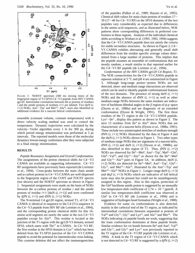
Peptide Resonance Assignment and OVerall Conformation.The assignments of the proton chemical shifts for C4-V3CAN0A are available as supporting information. C4-V3RF assignments have previously been reported (de Lorimieret al., 1994). Cross-peaks between the main chain amideandR-carbon protons in C4-V3 CAN0A are well-dispersedin the fingerprint region of the COSY and TOCSY spectra(not shown) and the NOESY spectrum as shown in Figure1. Sequential assignments were made on the basis of NOEsbetween theR-carbon protons of residuei and the amideprotons of residuei+1 [dRN (i, i+1)] and were confirmedby dâN (i, i+1) and dNN (i, i+1) NOEs.The N-terminal C4 gp120 region, termed T1, of C4-V3
CAN0A is identical in sequence to the C4 (T1) sequence inthe C4-V3 peptide from HIV RF (de Lorimieret al., 1994),and the corresponding proton chemical shifts for this 16-amino acid segment are nearly the same in the two C4-V3peptides except for Ala16. This residue is located at thejunction of the T1 region with the gp120 V3 domain, termedSP10, which starts with Thr17 in CAN0A. In C4-V3 RF,the first residue in the SP10 domain is Cys17 which has beendeleted from the T1-SP10 junction of the C4-V3 CAN0Apeptide to avoid the potential for intermolecular cross-linking.This cysteine deletion did not affect the immunogenicities
of the peptides (Palkeret al., 1989; Hayneset al., 1995).Chemical shift values for main chain protons of residues 17-39 (17-40 for C4-V3 RF) in the SP10 domains of the twopeptides vary considerably as expected due to differencesin the amino acid sequences, and as illustrated below, NOEpatterns show corresponding differences in preferred con-formers in these regions. Analysis of the individual chemicalshifts according to Wishartet al. (1991, 1992, 1994) suggeststhat the C4-V3 CAN0A peptide does not satisfy the criteriafor stable secondary structures. As shown in Figure 2, C4-V3 CAN0A exhibits alternating and generally small shiftdifferences from the residue specific average values deter-mined from a large number of proteins. This implies thatthe peptide assumes an ensemble of conformations that aremostly random, a result similar to that reported earlier forthe C4-V3 RF peptide (de Lorimieret al., 1994).Conformation of the HIV CAN0A gp120 C4 Region, T1.
The NOE connectivities for the C4-V3 CAN0A peptide inaqueous solution at 5°C and pH 4 are summarized in Figure3. Although long-range proton-proton NOEs are notdetected, short- and medium-range interactions are observedwhich can be used to identify peptide conformational featuresof the two domains. The presence of strong dRN (i, i+1)NOEs and the absence of dNN (i, i+1) as well as othermedium-range NOEs between the same residues are indica-tive of backbone dihedral angles in theâ region ofφ,ψ space(Dyson et al., 1988a) and are diagnostic of an extendedconformation (Dyson & Wright, 1991). The first threeresidues of the T1 region in the C4-V3 CAN0A peptide,Lys1-Ile3 , display this pattern as shown in Figure 3. Thenext 13 residues from Ile4 to Ala16 show NOEs that arecharacteristic of a nascent helix (Dysonet al., 1988a, 1991).These include two uninterrupted stretches of medium-strengthdNN (i, i+1) NOEs illustrated by the data in Figure 4 andthe dRN (i, i+1) NOEs shown in Figure 1. Two other typesof medium-range NOEs that are signatures of a nascent helix,dNN (i, i+2) and dRN (i, i+2) (Dysonet al., 1988b), arealso identified in this region of T1. Thus, dNN (i, i+2)NOEs are observed for Ile4-Met6, Asn5-Trp7, Met6-Gln8,Gln8-Val10, and Gly11-Ala13 as shown for the Ile4-Met6and Gly11-Ala13 pairs in Figure 5A. In addition, dRN (i,i+2) NOEs are detected for Ile4-Met6, Asn5-Trp7, Glu9-Gly11, and Met14-Ala16, illustrated by the Asn5-Trp7 andMet14-Ala16 NOEs in Figure 1. Longer-range dRN (i, i+3)and dRâ (i, i+3) NOEs which are indicative of full helicalturns may also be present but could not be unambiguouslyassigned in this region. Also in this region, protection ofthe Gln8 backbone amide proton is suggested by an unusuallylow temperature-shift coefficient of 2.74× 10⁻³ ppm/K. Asimilar low temperature-shift coefficient was observed atGln8 in C4-V3 RF (de Lorimier et al., 1994) and issuggestive of hydrogen bond formation (Wrightet al., 1988).Evidence for some cis conformations is also detected,
mainly in the carboxyl end of the T1 region, as indicated byinterresidual correlations between sequentialR protons ofVal10 and Gly11, Gly11 and Lys12, and Ala13 and Met14. TheNOEs indicating cis peptide bonds are weak, suggesting thatthe trans conformation dominates at these peptide bonds.Similar evidence for cis bonds between Ile4 and Asn5, Val10
and Gly11, and Gly11 and Lys12 was previously reported inthe T1 region of the C4-V3 RF peptide (de Lorimieret al.,1994). A kink in the T1 region of C4-V3 CAN0A whichis not detected in C4-V3 RF is suggested by a dâN (i, i+2)
FIGURE 1: NOESY spectrum (300 ms mixing time) of thefingerprint region of T1-SP10 C4-V3 peptide from HIV CAN0Agp120. Interresidue correlations between theR protons of residuesi and the amide protons of residuesi+1 are labeled. Two dRN (i,i+2) NOEs, Asn5-Trp7 and Met14-Ala16, were also identified asadditional evidence for a nascent helical conformation.
5160 Biochemistry, Vol. 35, No. 16, 1996 Vu et al.
+ +
+ +

NOE (Osterhoutet al., 1989) between aâ proton of Trp7
and the amide proton of Glu9.Conformation of the HIV CAN0A gp120 V3 Loop Region,
SP10. The N-terminal sequence of the SP10 region of C4-V3 CAN0A tends to exhibit an extendedâ conformation asshown by the absence of dNN (i, i+1) and presence of dRN(i, i+1) connectivities for residues Thr17-Pro19, Asn22, andLys25 (Figure 3). In addition, all measurable vicinal spin-spin coupling constants between the amide andR proton inthis region range from 9 to 10 Hz which further imply aâconformation (Wagneret al., 1986). However, the mostprominent conformational feature over the full SP10 domainis a reverseâ turn comprised of residues Gly30-Lys33. Thissequence is part of the V3 neutralizing determinant in gp120of HIV-1. Previous reports have both predicted reverse turnsin this region (La Rosaet al., 1990) and presented evidencefor turn structures within fragments of corresponding V3
loop sequences derived from the HIV MN (Chandrasekharet al., 1991; Riniet al., 1993; Ghiaraet al., 1994), HIV IIIB/LAI (Zvi et al., 1992), and HIV RF (de Lorimieret al., 1994)HIV-1 strains. Within this sequence of the CAN0A peptide,the following connectivities are detected: a weak dNN (i,
FIGURE 2: Deviation between the observedR proton chemical shifts of T1-SP10 C4-V3 peptide from HIV CAN0A gp120 and the averagechemical shifts of the corresponding amino acids whose values were taken from Wishartet al. (1991). The values reported for the RFpeptide are also included for comparison, but the chemical shift of Cys17 was omitted so that corresponding residues in the two SP10regions can be aligned. Solid bars denote T1-SP10 C4-V3 peptide from HIV RF gp120, and open bars are for T1-SP10 C4-V3 peptidefrom HIV CAN0A gp120.
FIGURE 3: NOE connectivity patterns of T1-SP10 C4-V3 peptidefrom HIV CAN0A gp120. Open boxes denote unambiguouslyabsent NOEs; black boxes and their heights show the presence ofNOEs and their relative intensities. An asterisk signifies ambiguityof NOE assignment due to peak overlap.
FIGURE 4: dNN (i, i+1) NOEs in the T1 region of T1-SP10 C4-V3 peptide from HIV CAN0A gp120. The sequential connectivitytrace begins with Ile4 and ends at Tyr15. Also shown is a dNN (i,i+1) peak between Gly32 and Lys33 in the SP10 portion of thepeptide. The spectrum was obtained with a 300 ms mixing time.
Conformations of HIV-1 gp120 C4-V3 Peptides by NMR Biochemistry, Vol. 35, No. 16, 19965161
+ +
+ +

i+1) NOE between the third (Gly32) and fourth (Lys33)residues of the turn shown in Figure 4, a medium to strongdRN (i, i+1) NOE between the second (Pro31) and third(Gly32) residues shown in Figure 1, and a weak dRN (i, i+2)NOE between the second and fourth residues (Pro31-Lys33)shown in Figure 5B. This specific combination of NOEs isdiagnostic of aâ turn (Dysonet al., 1988a). The nature ofthe (i, i+2) NOE between the second and fourth residues ofa â turn indicates its type. In this case, the fact that a dRN(i, i+2) NOE is observed together with the unambiguousabsence of a dδN (i, i+2) between Pro31 and Lys33 suggeststhat the sequence GPGK comprises a type IIâ turn.Immediately C-terminal to this turn is an extended regionfrom Phe35 to Thr 37 which is followed by a nascent helicalor turn conformation at the C-terminal end of C4-V3CAN0A as suggested by a weak dRN (i, i+2) NOE betweenThr37 and Gly39. The overall composite sequence of observedconformations for the T1-SP10CAN0(A) peptide is illustratedin Figure 6 along with the corresponding preferred confor-
mations reported earlier for the C4-V3 RF peptide (deLorimier et al., 1994).
DISCUSSION
Previous studies have shown that the chimeric C4-V3peptides designated T1-SP10 induce anti-HIV neutralizingantibodies for HIV grown in T cell lines and induce anti-HIV T helper lymphocytes and cytotoxic T lymphocytes(Palkeret al., 1989). These peptides have also recently beencharacterized for their effectiveness as a multicomponent,polyvalent immunogen (Hayneset al., 1995a). Resultsindicate differential antigenicities from each component withrespect to priming and boosting responses as well as cross-reactivity characteristics toward antibody production aimedat specific HIV-1 strains. In particular, the T1-SP10CAN0-(A) and T1-SP10MN(A) C4-V3 peptides appear to be morebroadly reactive than either T1-SP10RF(A) or T1-SP10EV91-(A) peptides. Our interest in studying conformationalcharacteristics for these peptides represents an initial effortto establish likely conformations for related immunogenicsequences from C4-V3 peptides and to compare confor-mational features between particular peptides in this seriesto gain insight into structural contributions to specificimmunogenic behavior. We have previously reported aconformational analysis of the T1-SP10RF(A) peptide (deLorimier et al., 1994), to which the results of this study ofT1-SP10CAN0(A) are compared and discussed below in thecontext of immunogenic differences between the two peptides(Hayneset al., 1995a).The T1 segments of the C4-V3 CAN0A and C4-V3 RF
peptides are taken from the fourth constant domain of HIV-1gp120 which is a component of the HIV gp120 CD4 bindingsite (Capon & Ward, 1991) and also is a potent T helperepitope (Ceaseet al., 1987; Palkeret al., 1989). The T1regions in the C4-V3 peptides from HIV CAN0A and RFare identical in primary amino acid sequence and are shownhere to be similar in their solution conformations. They bothexhibit evidence for nascent helix conformations as indicatedby the presence of dNN (i, i+2) and dRN (i, i+2) NOEs(Figure 3; de Lorimieret al., 1994). The observation of dRN(i, i+3) and dRâ (i, i+3) NOEs within this region of C4-V3 RF indicates a full helical turn conformation from Val10
to Met14. Such NOEs could not be unambiguously assignedin the case of C4-V3 CAN0A due to the degeneracy ofchemical shift values.The SP10 portions of the C4-V3 CAN0A and C4-V3
RF peptides are derived from the V3 loop regions of gp120in HIV-1 CAN0A and HIV-1 RF, respectively. This loop
FIGURE 5: NOESY spectrum (300 ms mixing time) showingmedium-range NOEs. (A) Two dNN (i, i+2) cross-peaks that arisefrom Ile4-Met6 and Gly11-Ala13 correlations which are indicativeof a nascent helical conformation. (B) The dRN (i, i+2) NOEbetween Pro31 and Lys33 is shown here as part of the evidence fora type IIâ turn at the tip of the V3 loop.
FIGURE6: Secondary structural conformer comparison between theT1-SP10 C4-V3 peptides from HIV CAN0A and RF gp120.Numbers indicate the first amino acid residues from a specifiedconformational region.
5162 Biochemistry, Vol. 35, No. 16, 1996 Vu et al.
+ +
+ +
is highly variable in sequence in various strains of HIV-1gp120 and consists of 35 amino acids flanked by two cysteineresidues that form a disulfide bridge (Leonardet al., 1990).It contains a potent neutralizing determinant of gp120(Javaherianet al., 1989; Zwartet al., 1991). Consequently,it has been a target for numerous attempts at designingvaccines against HIV-1 (Eminiet al., 1992; Hayneset al.,1995a). Parallel efforts have been made to elucidatestructural features of this region to better understand the basisfor its reactivity. The predicted structural elements of theV3 loop of HIV-1 gp120 based on a neural network approachis Cys-â strand-reverseâ turn-â strand-R helix-Cys in thatorder (La Rosaet al., 1990). According to this prediction,the reverseâ turn is located roughly at the center of the loopand includes a highly conserved Gly-Pro-Gly tripeptide.X-ray crystal structures of the corresponding GPGR sequencefrom the HIV-1 MN isolate when bound to Fab fragmentsof antibodies, 50.1 (Riniet al., 1993) and 59.1 (Ghiaraetal., 1994), indicated a type IIâ turn. Nuclear magneticresonance studies of free peptides derived from this regionof the HIV-1 MN, IIIB, and RF strains in aqueous solutionalso suggested a reverseâ turn within this sequence at thetip of the V3 loop (Chandrasekharet al., 1991; Zviet al.,1992; de Lorimieret al., 1994).We report here that conformational preference for a reverse
â turn also exists in the V3 loop of HIV-1 CAN0A gp120.The corresponding GPGK sequence in the SP10 portion ofthe C4-V3 CAN0A peptide tends to form a type IIâ turnfrom Gly30 to Lys33 as indicated by a combination of dRN(Pro31-Gly32), dRN (Pro31-Lys33), and dNN (Gly32-Lys33)NOEs. For HIV RF, due to a dδN (i, i+1) NOE betweenPro32 and Gly33 (second and third residues of the turn), deLorimier et al. (1994) suggested a type Iâ turn for thesequence GPGR. Thus, while the preferred type of reverseturn and therefore theφ andψ main chain bond angles mayvary in the solution conformers observed for this domain ofdifferent HIV-1 strains, it appears that both free peptidestend to form reverse turn conformations consistent with thegeneral topology found in the HIV MN V3 loop peptidecomplexed with Mab 50.1 (Riniet al., 1993). This turnconformation may be important for direct antibody and Bcell receptor recognition and also may provide an effectivemethod of presenting hydrophobic residues required forinteraction and binding (Riniet al., 1993).An extendedâ strand conformation N-terminal to the
reverse turn is observed for C4-V3 CAN0A from Thr17 toMet29 as illustrated in Figure 6. This segment of the V3loop of C4-V3 RF, which is also generally extended inconformation, exhibits evidence for an additional type Iâturn within residues Arg19-Pro20-Asn21-Asn22. The asparagineresidue at position 21, which is correlated with a highpropensity forâ turns (Dysonet al., 1988a), is replaced byhistidine in C4-V3 CAN0A (TRPNNN in C4-V3 RFversus TRPHNN in C4-V3 CAN0A). This substitution maycontribute to the abrogation of a reverseâ turn at this positionand will likely have influence on the immunogenicity ofthis B7-restricted CTL epitope of HIV gp120 (Safritet al.,1994). The C-termini of the SP10 regions of both C4-V3CAN0A and C4-V3 RF show NOEs characteristic of anextendedâ conformation immediately following the centralturn and end with a three-amino acid stretch of nascent helix.Taken together, the NMR-derived solution conformations
of the C4-V3 CAN0A and C4-V3 RF peptides show
similarities in the C4 regions but differ considerably in theV3, SP10 peptide segments. The solution conformation ofthe SP10 portion of C4-V3 CAN0A V3 loop includes anextendedâ segment from Thr17 to Met29, a type II â turnfrom Gly30 to Lys33, a second extended structure from Ala34
to Tyr36, and a nascent helix from Thr37 to Gly39. Bycomparison, the C4-V3 RF peptide exhibits two sequenceswhere type Iâ turns are preferred conformations, the secondof which is some nine residues N-terminal from the centralturn (de Lorimieret al., 1994). Furthermore, the V3 portionof the C4-V3 RF peptide shows preferred conformationsin the sequence SIT that are not detected in the correspondingregion of C4-V3 CAN0A.Initial studies of the immunological activities of C4-V3
RF and C4-V3 CAN0A peptides have determined the abilityof each peptide to prime and/or boost antibody responses toitself and other peptides in the polyvalent mixture in Balb/cmice (Hayneset al., 1995a). In addition, [3H]thymidineincorporation into splenocytes from immunized mice wasassayed to evaluate the relative proliferative responsesinduced by each peptidein Vitro. Results indicated that serafrom mice immunized with the C4-V3 CAN0A peptideinduced an anti-HIV CAN0A antibody titer that was 20 timesgreater than that induced by the C4-V3 RF peptide forcorresponding anti-HIV RF antibodies. Mice primed andboosted with C4-V3 CAN0A peptide also stimulatedsignificant antibody production directed toward C4-V3 RFand other C4-V3 HIV peptides, whereas the C4-V3 RFpeptide immunogen alone produced antibodies almost ex-clusively to HIV RF and not to HIV CAN0A (Haynesetal., 1995a). The fact that the V3 region of RF peptideexhibits conformations not detected in CAN0A or MN(unpublished data; Chandrasekharet al., 1991) suggests thatthe immunogenic specificity of RF arises from these con-formational features. Studies are underway to test thishypothesis.It seems unlikely that the T1 regions alone contribute
differently to the overall immunogenicities of the T1-SP10peptides since their primary amino acid sequences areidentical and the NMR-detected conformational features ofthis segment are very similar between the C4-V3 CAN0Aand C4-V3 RF peptides (de Lorimieret al., 1994). It ispossible however that both sequence and conformationalvariations of the two SP10 V3 segments of the C4-V3peptides determine their differential immunogenic behaviorand are responsible for different levels of antibody productionand T cell proliferation induced by HIV CAN0A and RFC4-V3 peptides. Indeed, it has been suggested that the typespecificities of the antibodies generated to various HIVstrains are defined by residues flanking the reverseâ turn inthe V3 loop that vary from one strain to another (Meloenetal., 1989).To better understand the interplay of primary and second-
ary structural elements of C4-V3 HIV peptides, we haveconstructed molecular models of the antibody-binding sitesfrom the SP10 domains of CAN0A and RF C4-V3 peptideson the basis of NMR results as outlined above. The modelswere subjected to molecular dynamics for a total of 300 psfollowed by energy minimization. The space-filling CPKmodels generated for the tips of the V3 loops of HIV CAN0Aand RF are depicted in Figure 7. By developing theconformational model for a single short domain of thepeptide, we avoid any implication that other regions of the
Conformations of HIV-1 gp120 C4-V3 Peptides by NMR Biochemistry, Vol. 35, No. 16, 19965163
+ +
+ +

molecule have time-correlated conformers. Indeed, forlargely unstructured biopolymers, the conformational ele-ments of independent domains experimentally observed insolution NMR studies may not be present at the same timein the same molecules. Crystallographic studies of thecocrystal of the comparable V3 segment (16 residues) ofgp120 from the HIV-1 MN strain complexed with itsmonoclonal antibody 50.1 show extensive contacts betweenthe antigen-binding pocket of Fab 50.1 and the side chainsof residues located immediately N-terminal to the tip of theV3 loop (Rini et al., 1993). For MN, these residues are Ile(P314), Ile (P316), Gly (P319), and Pro (320) and appear toform a continuous hydrophobic surface that not only interactswith the Fab fragment but also is required for high-affinitybinding in mutagenesis studies. In the C4-V3 CAN0A andC4-V3 RF peptides compared here, the amino acidscorresponding to these four hydrophobic residues are I, M,G, and P and I, K, G, and P, respectively, shown darkenedin Figure 7. Like the X-ray crystal structure of the antibody-bound MN sequence, C4-V3 CAN0A exhibits an apolarpatch that includes residues Ile27, Met29, Gly30, and Pro31.Our models show that the presence of methionine in placeof isoleucine does not alter the geometry of the apolar patchin that it appears to be flat, much like that in the crystalstructure of MN (Figure 7; Riniet al., 1993). For the C4-V3 RF peptide, however, this extended stretch of amino acidsis interrupted by a positively charged residue, a lysine atthe second position of the patch (gray in Figure 7). Thus,the V3 region of HIV CAN0A, but not of RF, may containa structural motif similar to that of HIV MN. As mentionedabove, Hayneset al. (1995a) demonstrated that, while HIVMN and CAN0A are immunologically similar to each otherin that they both induce cross-reactive antibodies, HIV RFis very type-specific in antibody responses induced. Thatis, whereas antibodies raised against C4-V3 RF peptide donot recognize antigens from other HIV strains, those raisedagainst C4-V3 CAN0A or C4-V3 MN peptides can cross-react to each other and to the HIV RF V3 loop as well. Fromthese models, we therefore suggest that the selectivity andrestriction to cross-reactivity of anti-HIV RF V3 loopantibodies may be due to the presence of the positivelycharged lysine residue, the side chain of which is confor-mationally presented in the center of the hydrophobic region.This feature likely induces antibodies specific for its recogni-tion. The presence and continuity of an apolar surface inproximity to the GPGX turn region of MN and CAN0A V3peptides may be important for antibody cross-reactivity.
ACKNOWLEDGMENT
We acknowledge Drs. Anthony Ribeiro and RonaldVenters for helpful assistance and Dr. David Myers forsharing his preliminary spectra and analysis of CAN0A.
SUPPORTING INFORMATION AVAILABLE
One table of the proton resonance assignments of the T1-SP10CAN0(A) C4-V3 peptide (1 page). Ordering informa-tion is given on any current masthead page.
REFERENCES
Bax, A., & Davis, D. (1985)J. Magn. Reson. 65, 355-360.Capon, D., & Ward, R. (1991)Annu. ReV. Immunol. 9, 649-678.Cease, K., Margalit, H., Cornette, J., Putney, S., Robey, W.,Ouyang, C., Streicher, H., Fischinger, P., Gallo, R., DeLisi, C.,& Berzofsky, J. (1987)Proc. Natl. Acad. Sci. U.S.A. 84, 4249-4253.
Chandrasekhar, K., Profy, A., & Dyson, H. (1991)Biochemistry30, 9187-9194.
Clapham, P., McKnight, A., Simmons, G., & Weiss, R. (1993)Philos. Trans. R. Soc. London, Ser. B 342, 67-73.
Clerici, M. (1993)AIDS 7, S135-S140.Cordonnier, A., Montagnier, L., & Emerman, M. (1989)Nature340, 571-574.
de Lorimier, R., & Spicer, L. (1994)Tech. Protein Chem. V, 423-430.
de Lorimier, R., Moody, M., Haynes, B., & Spicer, L. (1994)Biochemistry 33, 2055-2061.
Dyson, H., & Wright, P. (1991)Annu. ReV. Biophys. Biophys.Chem. 20, 519-538.
Dyson, H., & Wright, P. (1995)FASEB J. 9, 37-42.Dyson, H., Cross, K., Houghten, R., Wilson, I., Wright, P., &Lerner, R. (1985)Nature 318, 480-483.
Dyson, H., Rance, M., Houghten, R., Lerner, R., & Wright, P.(1988a)J. Mol. Biol. 201, 161-200.
Dyson, H., Rance, M., Houghten, R., Wright, P., & Lerner, R.(1988b)J. Mol. Biol. 201, 201-217.
Dyson, H., Norrby, E., Hoey, K., Parks, E., Lerner, R., & Wright,P. (1992)Biochemistry 31, 1458-1463.
Emini, E., Schleif, W., Numberg, J., Conley, A., Eda, Y., Tokiyoshi,S., Putney, S., Matsushita, S., Cobb, K., Jett, C., Eichberg, J., &Murthy, K. (1992)Nature 355, 728-730.
Feinberg, M., & Greene, W. (1992)Curr. Opin. Immunol. 4, 466-474.
Fenouillet, E., Gluckman, J., & Jones, I. (1994)Trends Biochem.Sci. 19, 65-70.
Ghiara, J., Stura, E., Stanfield, R., Profy, A., & Wilson, I. (1994)Science 264, 82-85.
Girard, M., & Shearer, G. (1993)AIDS 7, S115-S116.Goudsmit, J., Debouck, C., Meloen, R., Smit, L., Bakker, M., Asher,D., Wolff, A., Gibbs, C., & Gajdusek, D. (1988)Proc. Natl.Acad. Sci. U.S.A. 85, 4478-4482.
Guo, C., Jardetzky, T., Garrett, T., Lane, W., Strominger, J., &Wiley, D. (1992)Nature 360, 364-366.
Harouse, J., Bhat, S., Spitalnik, S., Laughlin, M., Stefano, K.,Silberberg, D., & Gonzalez-Searano, F. (1991)Science 253,320-323.
Harrison, S., Wang, J., Yan, Y., Garrett, T., Liu, J., Moebius, U.,& Reinherz, E. (1992)Cold Spring Harbor Symp. Quant. Biol.62, 541-548.
Hart, M., Palker, T., Matthews, T., Langlois, A., Lerche, N., Martin,M., Scearce, R., McDanal, C., Bolognesi, D., & Haynes, B.(1990)J. Immunol. 145, 2677-2685.
Haynes, B. (1995b)DiVision of AIDS Treatment Resource InitiatiVe(DATRI) Protocol 010, A Phase I Trial of C4-V3 PolyValentPeptide Vaccine in HIV-1 Infected Persons, Division of AIDS,NIAID, NIH, Bethesda, MD.
Haynes, B., Moody, A., Heinley, C., Korber, B., Millard, W., &Searce, R. (1995a)AIDS Res. Hum. RetroViruses 11, 211-221.
Holley, H., & Karplus, M. (1989)Proc. Natl. Acad. Sci. U.S.A.86, 152-156.
FIGURE 7: CPK models of the tips of the V3 loops of CAN0A andRF. Important basic residues are shown in gray, and the nonpolarresidues of the apolar patches are darkened. Note the protrusion ofa positively charged lysine residue in the middle of the hydophobicregion in RF which differentiates it from CAN0A.
5164 Biochemistry, Vol. 35, No. 16, 1996 Vu et al.
+ +
+ +
Javaherian, K., Langlois, A., McDanal, C., Ross, K., Eckler, L.,Jellis, C., Profy, A., Rusche, J., Bolognesi, D., Putney, S., &Matthews, T. (1989)Proc. Natl. Acad. Sci. U.S.A. 86, 6768-6772.
Jeener, J., Meier, B., Bachman, P., & Ernst, R. (1979)J. Chem.Phys. 71, 4546-4553.
Johnston, M., & Hoth, D. (1993)Science 260, 1286-1293.LaRosa, G., Davide, J., Weinhold, K., Waterbury, J., Profy, A.,Lewis, J., Langlois, A., Dressman, G., Boswell, R., Shadduck,P., Holley, L., Karplus, M., Bolognesi, D., Matthews, T., Emini,E., & Putney, S. (1990)Science 249, 932-935.
Leonard, C., Spellman, M., Riddle, L., Harris, R., Thomas, J., &Gregory, T. (1990)J. Biol. Chem. 265, 10373-10382.
Lerner, R. (1984)AdV. Immunol. 36, 1-44.Letvin, N. (1993)N. Engl. J. Med. 329, 1400-1405.Madrenas, J., Wange, R., Wang, J., Isakov, N., Samelson, L., &Germain, R. (1995)Science 267, 515-518.
Meloen, R., Liskamp, R., & Goudsmit, J. (1989)J. Gen. Virol. 70,1505-1512.
Moore, J., Thali, M., Jameson, B., Vignaux, F., Lewis, G., Poon,S., Charles, M., Fung, M., Sun, B., Durda, P., A° kerblom, L.,Wahren, B., Ho, D., Sattentau, Q., & Sodroski, J. (1993)J. Virol.67, 4785-4796.
Mujeeb, A., Bishop, K., Peterlin, M., Turck, C., Parslow, T., &James, T. (1994)Proc. Natl. Acad. Sci. U.S.A. 91, 8248-8252.
Osterhout, J., Baldwin, R., York, E., Stewart, J., Dyson, H., &Wright, P. (1989)Biochemistry 28, 7059-7064.
Palker, T., Clark, M., Langlois, A., Matthews, T., Weinhold, K.,Randall, R., Bolognesi, D., & Haynes, B. (1988)Proc. Natl.Acad. Sci. U.S.A. 85, 1932-1936.
Palker, T., Matthews, T., Langlois, A., Tanner, M., Martin, M.,Scearce, R., Kim, J., Berzofsky, J., Bolognesi, D., & Haynes,B. (1989)J. Immunol. 142, 3612-3619.
Piantini, U., Sorensen, O., Bodenhausen, G., & Ernst, R. (1982)J.Am. Chem. Soc. 104, 6800-6801.
Prutscher, M., Trkoln, A., Gruber, G., Buchacher, A., Predl, R.,Steindl, F., Tauer, C., Berger, R., Barrett, N., & Jungbauer, A.(1994) AIDS Res. Hum. RetroViruses 10, 1651-1658.
Putney, S. (1992)Trends Biochem. Sci. 19, 65-70.Rammensee, H., Falk, K., & Ro¨tzschke, O. (1993)Curr. Opin.Immunol. 5, 35-44.
Rance, M., Sorensen, O., Bodenhausen, G., Wagner, G., Ernst, R.,& Wuthrich, K. (1983)Biochem. Biophys. Res. Commun. 117,479-485.
Rini, J., Stanfield, E., Stura, E., Salinas, P., Profy, A., & Wilson,I. (1993)Proc. Natl. Acad. Sci. U.S.A. 90, 6325-6329.
Safrit, J., Lee, A., Andrews, C., & Koup, R. (1994)J. Immunol.153, 3822-3830.
Sattentau, Q., & Moore, J. (1993)Philos. Trans. R. Soc. London,Ser. B 342, 59-66.
Steinaa, L., Sorensen, A., Nielsen, J., & Hansen, J. (1994)Arch.Virol. 139, 263-271.
Takahashi, H., Nakagawa, Y., Pendleton, C., Houghten, R.,Yokomuro, K., Germain, R., & Berzofsky, J. (1992)Science 255,333-336.
Thali, M., Moore, J., Furman, C., Charles, M., Ho, D., Robinson,J., & Sodroski, J. (1993)J. Virol. 67, 3978-3988.
Venet, A., & Walker, B. (1993)AIDS 7, S135-S140.Wagner, G., Neuhaus, D., Wo¨rgotter, E., Vasa´k, M., Kagi, J., &Wuthrich, K. (1986)J. Mol. Biol. 187, 131-135.
Waltho, J., Feher, V., Lerner, R., & Wright, P. (1989)FEBS Lett.250, 400-404.
Wishart, D., & Sykes, B. (1994)J. Biomol. NMR 4, 171-180.Wishart, D., Sykes, B., & Richards, F. (1991)J. Mol. Biol. 222,311-333.
Wishart, D., Sykes, B., & Richards, F. (1992)Biochemistry 31,1647-1651.
Wright, P., Dyson, H., & Lerner, R. (1988)Biochemistry 27, 7167-7175.
Wuthrich, K. (1986)NMR of Proteins and Nucleic Acids, JohnWiley & Sons, New York.
Zvi, A., Hiller, R., & Anglister, J. (1992)Biochemistry 31, 6972-6979.
Zvi, A., Kustanovich, I., Feigelson, D., Levy, R., Eisenstein, M.,Matsushita, S., Richalet-Se´cordel, P., Regenmortel, M., &Anglister, J. (1995)Eur. J. Biochem. 229, 178-187.
Zwart, G., Langedijk, H., van der Hoek, L., de Jong, J., Wolfs, T.,Ramantarsing, C., Bakker, M., de Ronde, A., & Goudsmit, J.(1991)Virology 181, 481-489.
BI952665X
Conformations of HIV-1 gp120 C4-V3 Peptides by NMR Biochemistry, Vol. 35, No. 16, 19965165
+ +
+ +