conformational landscape of l-threonine in neutral, acid and basic...
TRANSCRIPT

Tetrahedron: Asymmetry 24 (2013) 1537–1547
Contents lists available at ScienceDirect
Tetrahedron: Asymmetry
journal homepage: www.elsevier .com/locate / tetasy
Conformational landscape of L-threonine in neutral, acid and basicsolutions from vibrational circular dichroism spectroscopyand quantum chemical calculations
0957-4166/$ - see front matter � 2013 Elsevier Ltd. All rights reserved.http://dx.doi.org/10.1016/j.tetasy.2013.09.025
⇑ Corresponding author at present address: Grupo de Química Física Teórica yExperimental FQM173, Edificio B3, Campus Las Lagunillas, Facultad de CienciasExperimentales, Universidad de Jaén, E-23071 Jaén, Spain. Tel.: +34 953 212754;fax: +34 953 212940.
E-mail address: [email protected] (J.J. López-González).
María Mar Quesada-Moreno a, Ana África Márquez-García a, Juan Ramón Avilés-Moreno b,Juan Jesús López-González a,⇑a University of Jaén, Department of Physical and Analytical Chemistry, Campus Las Lagunillas, E-23071 Jaén, Spainb Laboratoire de Physique des Lasers, Atomes et Molécules, UMR 8523 C.N.C.S. - Université de Lille 1, Bât. P5, 59655 Villeneuve d’Ascq Cedex, France
a r t i c l e i n f o a b s t r a c t
Article history:Received 26 June 2013Accepted 26 September 2013Available online 15 November 2013
The biological relevance of amino acids is well known. They can be used as zwitterionic, cationic or anio-nic forms according to the pH of the medium where they are. Thus, our aim herein was to study the con-formational preference of the polar amino acid L-threonine [C4H9NO3, (2S,3R)-2-amino-3-hydroxybutyricacid] under different pH conditions. A conformational study in an aqueous solution of the dissociationequilibrium of the amino acid L-threonine was carried out for this purpose. We recorded, at room tem-perature, the Mid-IR, Far-IR, Raman and VCD spectra of L-threonine from the aqueous solutions at pH val-ues 5.70 (zwitterionic species), 1.00 (protonated species) and 13.00 (deprotonated species). The numberof conformers found with the conformational search was 9 zwitterions, 27 anions and 52 cations. Boththe study of the conformational landscape and the theoretical analysis of the vibrational features wereaccomplished by using DFT and ab initio calculations, that is, B3LYP/6-311++G(d,p) level of theory forall the conformers obtained from the conformational search, M062X/6-311++G(d,p) and MP2/6-311++G(d,p) levels of theory for the most stable conformers. The presence of water was included withthe IEF-PCM implicit hydration model. With regard to the zwitterion, the importance of the analysis ofthe low frequency region (700–30 cm–1) in the Far-IR spectra should be noted, because it provides rele-vant information that can be used to determine the presence of the most stable structures.
� 2013 Elsevier Ltd. All rights reserved.
1. Introduction
Amino acids not only play a key role in protein folding but alsotake part in lypophilic interactions and influence secondary struc-ture properties. They are bifunctional compounds, which containan amine and a carboxylic acid group and have a side chain thatvaries among the different amino acids.1,2 Furthermore, under-standing the structures and conformational preferences of thezwitterionic forms is important to gain insight into protein folding,which is governed by hydrophilic and hydrophobic interactionswhich determine the H-bonding scheme.3–7 Studying their behav-iour in water is a major interest because it is the natural mediumfor biological molecules.7,8 Naturally occurring amino acids havea large conformational flexibility, and computational studiesshould correctly determine conformers and statistical populations
found from experimental data.9,10 The dissociation equilibrium de-pends on the pH of the medium in which the amino acid is placed.The experimental dissociation constants of threonine, pKa, corre-sponding to the dissociation of the carboxyl and protonated aminogroups are 9.10 and 2.09, respectively. Thus, we can deduce thatthe presence of anions will be found at pH 13.00, cations at pH1.00 and zwitterions at pH 5.70.11
Polar amino acids, such as L-serine (L-Ser or S) and L-threonine (L-Threo, L-Thr or T), with a hydroxyl group on their side chains, takepart in pivotal biological processes, such as biosynthesis and phos-phorylation.12–16 Threonine13 is a polar a-amino acid with thechemical formula HO2CCH(NH2)CH(OH)CH3. Together with serine,threonine is one of two proteinogenic amino acids that have an alco-hol group (tyrosine is not an alcohol but a phenol, since its hydroxylgroup is bonded directly to an aromatic ring, giving it different acid/base and oxidative properties). It is also one of two common aminoacids that bear a chiral side chain, along with isoleucine.
Moreover, the threonine residue is susceptible to numerousposttranslational modifications. The hydroxy side-chain can un-dergo O-linked glycosylation. In addition, threonine residues un-dergo phosphorylation through the action of a threonine kinase.

1538 M. M. Quesada-Moreno et al. / Tetrahedron: Asymmetry 24 (2013) 1537–1547
It can be obtained in its phosphorylated form as phosphothreo-nine.17 It is also one of two amino acids out of the 20 with two ste-reogenic centers. It can thus exist as four possible stereoisomerswith the following configurations: (2S,3R), (2R,3S), (2S,3S) and(2R,3R). However, the name L-threonine is used for one single dia-stereomer, (2S,3R)-2-amino-3-hydroxybutanoic acid. The secondstereoisomer (2S,3S), which is rarely present in Nature, is calledL-allo-threonine.
Hernández et al.9 reported an analysis of the IR–Raman spectraof L-Ser and L-Threo in H2O and D2O solutions in the 2000–800 cm�1 and 2000–500 cm�1 regions, respectively. AdditionallyXu and Lin carried out a detailed theoretical study with ab initiocalculations of L-Threo and L-allo-Threo in the gas phase.18
Herein, an experimental and theoretical study of Mid-IR, FarIR,Raman and VCD spectroscopic features of L-threonine in aqueoussolutions under three representative pH conditions is reported(see Fig. 1 for the atom numbering). The pH values were: 5.70(zwitterionic species, ThreoZW), 1.00 (protonated species, Threo-CAT) and 13.00 (deprotonated species, ThreoAN).
L-Threo
pH 1.00 5.70 13.00
Cation (protonated) Zwitterion Anion (deprotonated)
Figure 1. Molecular structure and atom numbering adopted herein for the L-Threo in the zwitterionic, protonated and deprotonated forms.
Based on the vibrational analysis of the zwitterions, determin-ing the number of possible conformers present in solution is acomplicated task, given the similar IR and Raman signature ofthe most stable conformers.19–27 Thus, in order to obtain additionalinformation, we also analysed the FarIR spectrum in the solidphase of L-threonine. Furthermore, VCD measures the differentialabsorption of left versus right circularly polarized IR incident lightin the molecular vibrational transition by a chiral molecule.28 Thesigns of the rotatory strength could be helpful in determining theabsolute configuration of the L-threonine. As vibrational tech-niques, IR and Raman, VCD technique is also sensitive to the pres-ence of different conformers.28–30 The vibrational studies of thecations, anions and zwitterions of this amino acid performed here-in are more complete than those in the literature,9 not only be-cause of the study by VCD, but also because the region between700 and 30 cm�1 is analysed by means of IR spectroscopy in thecase of the zwitterions.
In many works, biomolecules have been studied by using chir-optical techniques such as vibrational circular dichroism (VCD) orRaman optical activity (ROA); an explicit solvation model has beenconsidered to account for the solvent effects.31–43
Our strategy was to test the influence of two different DFT func-tionals, B3LYP and M062X, in the prediction of the most stable con-formers at different pH values. We concluded that a good election
in the functional is also important in order to get a reasonable setof the most stable conformers. In order to stabilize the zwitterionic,anionic and cationic structures of L-Threo, solvent (water) effectswere taken into account by using IEF-PCM formalism as imple-mented in GAUSSIAN09. The M062X functional is also a high nonlocal-ity functional with double the amount of nonlocal exchange (2�).It is parametrized for non-metals and contains a treatment for non-covalent interactions. The B3LYP functional does not contain anoncovalent interaction treatment. Due to the system under study,L-Threo is a polar flexible molecule which presents noncovalentinteractions, it would be reasonable to assume that the result ofthe calculation of the relative energies for it with the M062X func-tional was better than those obtained with the B3LYP functional.Similar results have been obtained in the study of the zwitterionicstructures of L-Phe and L-Tyr.27
In the literature,44 a thorough study on the molecular propertiesof H-bonded complexes using a large set of functionals has beenreported. In these complexes, the charge transfer interactions playan important role in successfully describing the structure and
energy of those H-bonded systems. The literature44 also suggeststhat the M062X functional is a good choice in terms of accuracy/cost for the calculation of interaction energy of flexible polar sys-tems as the amino acid studied herein. Additionally, the results ob-tained for L-Phe and L-Tyr,27 S-(�)-Perillyc acid26 and L-Ser45 pointout in the same direction, that is, the M062X better reproduces theexperimental spectra (solid or solution phases) than the B3LYP ap-proach, even with an implicit treatment of solvent.46 All of the con-formers obtained from the conformational search, that is, 88structures, were optimized at the B3LYP/6-311++G(d,p) level oftheory and the most stable ones at M062X/6-311++G(d,p) level.
Finally, a MP2 calculation was performed for the most stableconformers in order to compare the ab initio relative energies withthe DFT results. This is the first time that a comparison of the struc-tures and vibrational spectra of L-threonine calculated with thesetwo DFT functionals and the MP2 method has been performedand that an analysis of L-threonine at different pH in aqueous solu-tions has been carried out.
In addition, we would like to point out the reasonable resultsobtained herein with the implicit model, using both functionalsand MP2 calculations, in the prediction of the FarIR–IR–Raman–VCD spectra. The structures obtained herein were very close (interms of amino acid structure) to those obtained in the literature9
with the explicit model, which is an excellent result. This is the

M. M. Quesada-Moreno et al. / Tetrahedron: Asymmetry 24 (2013) 1537–1547 1539
reason, in our opinion, for the good agreement between our theo-retical and experimental data, that is, the most stable set of con-formers obtained with the M062X functional for the zwitterionicforms are similar to those obtained with the explicit model inthe literature.9
2. Experimental procedure
A commercial L-threonine sample (99%) was purchased fromSigma–Aldrich. Mid-IR, Raman and Far-IR spectra of L-threoninewere recorded for the solid samples, without performing anyprevious purification. Likewise, the IR and Raman spectra were re-corded in aqueous solutions at room temperature and different pHconditions: 5.70 (neutral media), 1.00 (acid media) and 13.00(basic media). Zwitterionic, protonated (cation) and deprotonated(anion) species are present at these three representative pH values.We also recorded the thin film spectra of these three solutions bythe vibrational circular dichroism (VCD) technique. Doubly dis-tilled water was used as the solvent. In order to obtain solutionsat different pH values, a 4 M solution of HCl or NaOH (Sigma–Aldrich) was added to fresh solutions of L-threonine (0.45 M,pH = 5.70).
A Crison GPL 21 pHmeter was used to measure the pH of thedifferent solutions. The accuracy of the device is 0.01 and its detec-tion range is 1–13. All data obtained were corrected to 25 �C(298 K).
An FT-IR 4100 JASCO spectrometer, equipped with a Globarsource, a DGTS detector and KBr optics, was used to record theMid-IR spectrum with the ATR accessory for the solid and liquidsamples at different pH conditions. The IR spectra were recorded
Table 1Calculated molecular populations and relative energies (with ZPE correction) for the zwioptimized at the B3LYP, M062X and MP2 levels of theory
B3LYP/6-311++G(d,p) M062X/6-311
Conf. DE0 (kJ/mol) %Pop.a Conf. DE0 (k
Zwitterion
ThreoZW1 0.0 37.4 ThreoZW1 0.0ThreoZW2 0.3 33.6 ThreoZW2 1.2ThreoZW4 3.0 11.1 ThreoZW3 2.7ThreoZW5 3.3 9.9 ThreoZW4 3.2ThreoZW3 3.8 8.0 ThreoZW5 4.2ThreoZW6 21.0 0.0 ThreoZW6 19.9ThreoZW7 21.4 0.0 ThreoZW7 21.6
Cation
ThreoCAT1 0.0 26.3 ThreoCAT1 0.0ThreoCAT3 0.1 25.5 ThreoCAT2 3.5ThreoCAT4 0.2 24.1 ThreoCAT3 4.7ThreoCAT2 1.4 14.8 ThreoCAT4 5.3ThreoCAT5 5.6 2.8 ThreoCAT5 7.0ThreoCAT6 6.9 1.6 ThreoCAT6 8.8
ThreoCAT7 9.1ThreoCAT8 10.4
Anionb
ThreoAN1 0.0 49.0 ThreoAN1 0.0ThreoAN3 2.7 16.6 ThreoAN2 0.8ThreoAN5 4.0 9.7 ThreoAN3 1.7ThreoAN4 4.2 8.9 ThreoAN4 6.2ThreoAN7 4.7 7.4 ThreoAN5 6.8ThreoAN6 5.0 6.5 ThreoAN6 7.0ThreoAN9 15.0 0.1 ThreoAN7 7.1ThreoAN8 15.4 0.1 ThreoAN8 16.2
ThreoAN9 16.2ThreoAN10 17.9ThreoAN11 20.4ThreoAN12 22.5
a Boltzmann population from DE0 taking T = 298.16 K. DE0: zero point corrected relatb ThreoAN2 conformer only found with the M062X functional.
in the 4000–600 cm–1 range, with 200 scans and a resolution of1 cm–1 and 4 cm�1 for the solid and liquid samples, respectively.For the baseline correction, the water spectrum at the same pHas the measured spectrum of L-Ser was subtracted.
The Raman spectrum of L-threonine was recorded using a Bru-ker RF100/S FT-Raman spectrometer, equipped with an Nd:YAG la-ser (excitation line at 1064 nm) and a Ge detector cooled at liquidnitrogen temperature. The solid spectrum was measured using astandard solid sample support in the 4000–100 cm–1 range, with200 scans and a resolution of 1 cm�1. The solution spectrum wasmeasured using standard liquid cells in the 4000–500 cm�1 usefulregion, with 200 scans and a resolution of 4 cm�1.
The FarIR spectrum of the solid L-threonine was recorded bymeans of a Bruker Vertex 70 in the 700–30 cm�1 range, with a res-olution of 4 cm�1 and 200 scans, using a platinum ATR (singlereflection diamond ATR accessory) and the silicon beamsplitterfor the Far-IR region.
The VCD spectra of L-threonine in thin film were recorded usinga JASCO FVS-4000 FTIR spectrometer, equipped with MCTV (2000–800 cm–1) detector. In order to obtain very thin films of the sample,we prepared very low concentration solutions of the sample at dif-ferent pH values and we evaporated the solvent on KCl ‘Real Crys-tal IR Sample Cards’. These films were measured in severalpositions by rotating the sample around both the beam propaga-tion axes (90� and 180�) and that perpendicular to it (180�) in orderto minimize the presence of artifacts in the VCD spectra.47–49 Allspectra were recorded using a KCl support, with a resolution of4–8 cm–1 and 4000–8000 scans by blocks of 2000 scans. With re-gard to the baseline correction, we subtracted the KCl support sig-nals for the film spectra.
tterionic (ZW), protonated (cation) and deprotonated (anion) conformers of L-Threo
++G(d,p) MP2/6-311++G(d,p)
J/mol) %Pop.a Conf. DEe (kJ/mol) %Pop.a
41.8 ThreoZW1 0.0 41.125.2 ThreoZW3 1.6 21.414.0 ThreoZW2 1.8 19.511.3 ThreoZW4 3.5 10.2
7.7 ThreoZW5 4.1 7.80.00.0
60.8 ThreoCAT1 0.0 40.415.0 ThreoCAT3 1.5 22.1
9.0 ThreoCAT4 2.1 17.27.3 ThreoCAT2 3.0 12.23.6 ThreoCAT7 6.4 3.01.8 ThreoCAT5 6.5 2.91.5 ThreoCAT8 7.2 2.20.9
39.8 ThreoAN1 0.0 44.629.2 ThreoAN3 1.2 27.720.3 ThreoAN5 3.6 10.2
3.2 ThreoAN6 4.5 7.12.6 ThreoAN7 4.6 7.02.4 ThreoAN4 6.5 3.22.3 ThreoAN9 15.5 0.10.1 ThreoAN8 15.8 0.10.1 ThreoAN10 19.8 0.00.00.00.0
ive energy; DEe: equilibrium relative energy.

Table 2Some relevant molecular structural parameters of L-Threo (zwitterionic, protonatedand deprotonated) performed at the B3LYP, M062X and MP2 levels of theory. SeeFig. 1 for for atom numbering
B3LYPa M062Xb MP2c
ThreoZW1s1(O4C3C2N1)/� �171 �154 �161s2(H5N1C2C3)/� �14 �30 �23s3(C3C2C9C11)/� 168 �176 �179s5(O10C9C2N)/� 48 62 59s6(H13O10C9C2)/� 79 70 71
ThreoZW2s1(O4C3C2N1)/� �174 �174 �174s2(H5N1C2C3)/� �12 �15 �12s3(C3C2C9C11)/� 166 171 172s5(O10C9C2N)/� 48 51 52s6(H13O10C9C2)/� �174 �175 �174
ThreoCat1s1(O4C3C2N1)/� 8 6 4s2(H5N1C2C3)/� �143 �160 �160s3(C3C2C9C11)/� 168 179 179s4(H18O6C3C2)/� �178 �179 �179s5(O10C9C2N)/� 50 58 58s6(H13O10C9C2)/� �174 �177 �173
ThreoCat2s1(O4C3C2N1)/� 7 9 7s2(H5N1C2C3)/� �143 �152 �157s3(C3C2C9C11)/� 163 174 176s4(H18O6C3C2)/� �178 �179 �179s5(O10C9C2N)/� 42 51 53s6(H13O10C9C2)/� 94 94 96
ThreoAn1s1(O4C3C2N1)/� �171 �171 180s2(H5N1C2C3)/� �17 �21 12s3(C3C2C9C11)/� �62 �58 �60s5(O10C9C2N)/� �171 �170 �170s6(H13O10C9C2)/� �43 �45 �39
ThreoAn2s1(O4C3C2N1)/� 175 174 173s2(H5N1C2C3)/� 151 152 152s3(C3C2C9C11)/� �61 �58 �58s5(O10C9C2N)/� �170 �169 �169s6(H13O10C9C2)/� �38 �40 �39
ThreoAn3s1(O4C3C2N1)/� �174 �173 �175s2(H5N1C2C3)/� �17 �21 �20s3(C3C2C9C11)/� 161 168 167s5(O10C9C2N)/� 47 52 51s6(H13O10C9C2)/� �37 �41 �40
a B3LYP: 6-311++G(d,p) basis set.b M062X: 6-311++G(d,p) basis set.c MP2: 6-311++G(d,p) basis set.
1540 M. M. Quesada-Moreno et al. / Tetrahedron: Asymmetry 24 (2013) 1537–1547
3. Theoretical methodology
The vibrational study of L-Threo required an extensive confor-mational search, which was performed for the ThreoZW, ThreoCATand ThreoAN forms by means of molecular mechanics with differentforce fields (MMFF and SYBYL)50,51 and using a SPARTAN program.52
When a fulfilled conformational energy was reached (20 kJ/mol)giving 9, 27 and 52 conformers for the zwitterionic, anionic and cat-ionic structures of L-Threo, all of these conformers were optimizedusing B3LYP53–55 functional and the 6-311++G(d,p) basis set. More-over, the lowest non-redundant conformers were selected for fur-ther optimization of the structures and the calculation of theirrelative energies (with ZPE corrections) using another density func-tional approach (M062X56,57) and the 6-311++G(d,p) basis set. TheM062X functional is a high nonlocality approach with double theamount of nonlocal exchange (2�); it is parametrized only fornon-metals and contains a treatment of noncovalent interactions.Given the context of our study, and because L-Threo is a polar flex-ible molecule which presents noncovalent interactions, one couldsurmise that the result of the calculation of their relative energieswith the M062X functional was better than those obtained withthe B3LYP functional.44 Finally, a second-order perturbation theory(MP2) calculation58 with the 6-311++G(d,p) basis set was accom-plished in order to compare the MP2 relative energies with theDFT results. These two different DFT methods were compared tomake certain of the internal consistency of the theoretical modelsand their predictive values. All of the energies presented hereinare zero point corrected relative energies. The harmonic IR, Ramanand VCD spectra of each conformer were obtained for both theB3LYP and M062X functionals and the 6-311++G(d,p) basis set.
Comparison of the results obtained with different functionals isa difficult task because the reproducibility of the results with othersystems is not always evident. In this way, we tested the results ob-tained with the B3LYP and M062X approaches with those obtainedwith the well known MP2 approach. In the case of ThreoZW, wewere able to conclude that: (i) the MP2 and M062X approaches givethe same results in terms of relative energy; (ii) even if the B3LYPfunctional calculates the same structures as the MP2 and M062Xapproaches, the calculated relative energies seem to be very differ-ent; (iii) the experimental data agreed better with the M062X andMP2 results than with B3LYP results. In the case of ThreoAN andThreoCAT, on the one hand, the B3LYP functional gives similarrelative energies to the MP2 method and not to the M062X one,while on the other hand, the experimental spectra seem to matchbetter with these two approaches than with M062X calculation.
All of the theoretical calculations and vibrational analysis werecarried out using three software packages: (1) Spartan08,52 for themolecular mechanics conformational search; (2) GAUSSIAN09,59 forthe structure re-optimization and spectra calculation and (3)VEDA62,63 programme for P.E.D (Potential Energy Distribution) ma-trix calculations.
Implicit solvent (water) effects were taken into account usingthe IEF-PCM formalism60 as implemented in GAUSSIAN09.
4. Results and discussion
4.1. Theoretical conformational stability
The conformational search with the MMFF and SYBYL forcefields found respective totals of 9, 27 and 52 conformers for thezwitterionic, anionic and cationic structures of L-Threo. All of thesestructures were optimized at the B3LYP/6-311++G(d,p) level oftheory. The number of non-redundant conformers was 7 zwitteri-ons, 19 anions and 27 cations. The most stable conformers werealso optimized at the M062X/6-311++G(d,p) level and the relativeenergies of some of these conformers were calculated with the
MP2/6-311++G(d,p) method. Table 1 gives the relative energies(zero point corrected, DE0 and equilibrium, DEe) and Boltzmannpopulations (%Pop) obtained for the L-Threo at three different the-oretical levels: B3LYP, M062X and MP2 with the same 6-311++G(d,p) basis set. Table 2 shows some relevant molecularstructural parameters, in particular the dihedral angles, of thisamino acid obtained at the same levels of theory specified above.Meanwhile, Fig. 2 shows the structures, relative energies and intra-molecular H-bond distances of the most stable zwitterions, proton-ated (cation) and deprotonated (anion) forms of L-Threo optimizedat these three theoretical levels.
The conformational landscape derived from the DFT and MP2 re-sults corroborates that the most populated structures are: (i) fourconformers for ThreoZW (92% of population); (ii) three conformersfor ThreoCAT (85% of population); and (iii) three conformers forThreoAN (89% of population).
The conformational distribution changes for ThreoZW, ThreoCATand ThreoAN when DFT and MP2 methods are used, with the same

Figure 2. Structures of the most stable zwitterions, protonated (cation) and deprotonated (anion) structures of L-Threo optimized at the M062X/6-311++G(d,p) level oftheory. Relative energies (zero point corrected) are given in kJ/mol in parenthesis at the M062X/6-311++G(d,p), MP2/6-311++G(d,p) and B3LYP/6-311++G(d,p) levels oftheory, respectively. H-bond values are shown in pm.
M. M. Quesada-Moreno et al. / Tetrahedron: Asymmetry 24 (2013) 1537–1547 1541
basis set, are other notable points that require comment. For in-stance, the B3LYP, M062X and MP2 methods predict thatThreoZW1 conformer is the most stable, with a population around40%. The ThreoZW2 conformer is predicted to be the second interms of energy with the use of B3LYP (34%) and M062X (25%) ap-proaches, but the third with the MP2 method (20%). Furthermore,the B3LYP/6-311++G(d,p) level of theory predicts that ThreoZW3,ThreoZW4 and ThreoZW5 conformers are fifth, third and the fourthin energy, with respective populations at approximately 8%, 11%and 10%. If we calculate the relative energies for these threeconformers with the M062X/6-311++G(d,p) or the MP2/6-311++G(d,p) levels, the results change drastically becauseThreoZW4 and ThreoZW5 are predicted to be the fourth and fifth
in energy, with populations at approximately 10 and 8%, respec-tively. ThreoZW3 is predicted to be the third (14%) with M062Xand the second (21%) with MP2 level.
In the cases of ThreoAN and ThreoCAT, the similarity among rel-ative energies obtained with B3LYP and MP2 levels of theoryshould be noted. ThreoAN2 is only found with the M062X func-tional and, as discussed below, it does not seem to be presentexperimentally. Table 1 offers further details on the conforma-tional landscapes of ThreoCAT and ThreoAN.
These results are important because the calculated IR, Ramanand VCD spectra are different depending on the predicted relativeenergies and, consequently, populations. The conformational pref-erence changes according to the theoretical model used. Since the

1542 M. M. Quesada-Moreno et al. / Tetrahedron: Asymmetry 24 (2013) 1537–1547
basis set is the same in all three cases, the relative energy differ-ences found using DFT methods are due to the use of differenttreatments for electron density, that is, B3LYP and M062X func-tionals. A good prediction of the most stable structures will con-tribute much more to our understanding of the experimentalfeatures.
Table 2 shows some relevant molecular structural parametersof L-Threo (ZW, CAT and AN), with particular attention paid tosome characteristic dihedral angles, calculated at the DFT (B3LYPand M062X functionals) and MP2 calculations (see Fig. 1 for atomnumbering). The variations in the compared dihedral angles aresmaller than 17�, when we compare the results obtained withB3LYP, M062X and MP2 calculations with the 6-311++G(d,p) basisset. This fact confirms that the optimized structures are the samein all three cases. The best results correspond to the MP2 calcula-tion, but this method is also the most demanding in terms of com-putational cost. It seems that the best choice in terms of accuracy(structure, energy, and harmonic spectra) and computational costcould be the M062X functional for ThreoZW conformers. This func-tional is more demanding in computational cost than the B3LYPone but much less than the second-order perturbative (MP2) calcu-lations. Nevertheless, B3LYP and M062X functionals seem to workwell for ThreoAN and ThreoCAT structures.
With regard to the ThreoZW1 and ThreoZW2 conformers of L-Threo, the torsion angle s6 (H13–O10–C9–C2) was found to haverespective values of 73� and �174�. In the ThreoZW1 conformer,a weak intramolecular hydrogen bond was formed between O10–H13 and O4@C3, with a length of 239 pm. However, another differ-ent intramolecular hydrogen bond is formed between H13–O10 andH14–N1 in the ThreoZW2 conformer, with a length of 216 pm. Theonly difference between these conformers is a turn of the HO- (hy-droxyl) group, which changes the torsion angle value from 73� to�174�, giving a different and stronger H-bond in ThreoZW2. ForThreoCAT1 and ThreoCAT2, all angles were seen to have similar val-ues except for the torsion angle s6 (H13–O10–C9–C2); with values of�175� and 95�, respectively. These values are different because thehydroxyl group turns again. There are two H-bonds that stabilizeboth conformers: one between H13–O10 and H7–N1 and the otherbetween N1–H17 and O4@C3.
For ThreoAN, dihedral angles of ThreoAN1, ThreoAN3 and Threo-AN4 have different values. In ThreoAN1, an intramolecular hydro-gen bond is formed between N1–H5 and O6@C3, with a length of215 pm. However, the amino group turns in ThreoAN3 giving a dif-ferent hydrogen bond between N1–H7 and O6@C3, with a length of225 pm. In ThreoAN4 it not only turns the amino but also the –CH(OH)–CH3 group, causing one different hydrogen bond betweenH13–O10 and H5–N1, with a length of 214 pm.
Finally, it should be noted that the structures (M062X and MP2)obtained herein are in good agreement with the most stable struc-tures obtained in the literature,9 using the explicit solvation model.
In the case of L-Threo zwitterions: our most stable structureThreoZW1 corresponds with the first and second conformers ob-tained in the literature9(g�g� and g+g+ conformers), that is, theorientation of the OH, COO� and NH3
+ groups of threonine moietyis similar. Additionally, our second conformer, ThreoZW2, is foundas the third structure in the literature,9 that is, tg�. This way, wecan say that our conformational research with an MM methodand the later optimization of the structures give reasonable results.The similarity between the most stable conformers obtained by usand the literature9 confirms the good agreement between ourexperimental data and theoretical data.
4.2. Experimental analysis and spectroscopic features
The experimental IR (including the FarIR region for theThreoZW), Raman and VCD (Figs. 3–6) spectra confirm the presence
of the most stable conformers of the ZW (Figs. 3 and 6), CAT (Figs. 4and 6) and AN (Figs. 5 and 6) of L-Threo in an aqueous solution andthe solid phase (only for the zwitterionic structures), and is in goodagreement with the theoretical calculations, especially M062X forthe ThreoZW form and B3LYP-M062X for the ThreoCAT and Threo-AN structures. The single scaling factors from the NIST database61
for the B3LYP/6-311++G(d,p) (0.967) and M062X/6-311++G(d,p)(0.950) levels were used to carry out the vibrational analysis ofthe species studied. All of the percentages of the normal modescontributions were taken from the P.E.D. matrix obtained withthe VEDA program.62,63
ThreoAN, ThreoZW and ThreoCAT forms have, respectively, 42,45 and 48 vibrational normal modes, belonging to the unique irre-ducible representation (A) of its common symmetry point group,C1. The recorded IR, Raman and VCD spectra of L-Threo at differentpH in aqueous solution, thin film and powder sample (only forzwitterions) are displayed in Figures 3–6.
For vibrational analysis, we took into account the IR–Raman–VCD data of L-Threo in the solid phase and in solution at differentpH conditions. As can be seen, the IR, Raman and VCD spectra (seeFigs. 3–6) are complex and in order to accomplish a reasonableinterpretation of them, some remarks about the most helpful andrelevant spectroscopic features are needed.
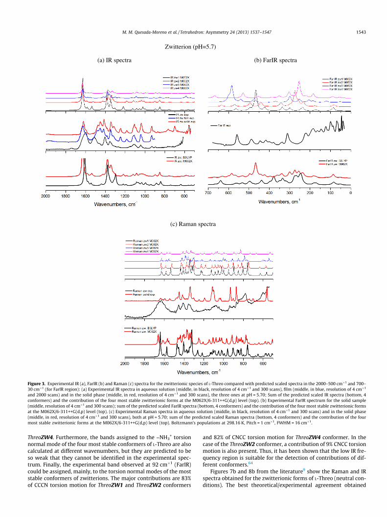
4.2.1. IR and Raman spectra4.2.1.1. Neutral pH. Figure 3 shows the recorded IR, Ramanand FarIR spectra of L-Threo in aqueous solution at pH = 5.70, aswell as in thin film and powder sample. Fig. 3 also shows the the-oretically calculated spectra of the four most stable zwitterionicforms at the B3LYP/6-311++G(d,p) and M062X/6-311++G(d,p) lev-els of theory. As it can be seen, this last level reasonably reproducesthe experimental spectra. With regard to the IR data (Fig. 3a), wecould assign some bands to the contribution of different conform-ers of ThreoZW, for instance: (a) The experimental band observedat 1600 cm�1 (IR, 1632 cm�1 in Raman) could be assigned to a nor-mal mode with a contribution from the P.E.D. matrix of 86% ofasym. COO� str. motion of the four most stable zwitterionic struc-tures; (b) The band at 1506 cm�1 (IR, 1493 cm�1 in Raman) couldbe assigned to a normal mode with contributions of 72% asym.NH3
+ bend. and 16% HNCC torsion motions of the four most stableThreoZW conformers. (c) The experimental band at 1403 cm�1 (IRand Raman) could be assigned to two normal modes of the fourmost stable zwitterions of L-Threo. The first one has a contributionof 70% sym. NH3
+ bend. and the second one has a contribution of56% asym. CH3 bend. and 14% HCCC torsion. A contribution of25% of HOC bend. is also found for ThreoZW3 in the last normalmode. For ThreoZW2, ThreoZW3 and ThreoZW4 conformers, thefirst one also has an additional contribution of 14% sym. COO-
str. (d) The band observed at 1041 cm�1 (IR and Raman) could bealso assigned to two normal modes mainly due to the presenceof ThreoZW1 and ThreoZW2. The first one with a contribution of12% asym. NH3
+ bend., 26% CH wagg. and 34% HNCC torsion mo-tions for both conformers, and the second one with a contributionof 40% CC str. and 22% HOC bend. motions for ThreoZW1 conformerand 14% CC str., 28% HOC bend., 11% HNCC torsion and 10% HCCCtorsion for ThreoZW2 conformer.
Figure 3b shows the FarIR spectrum of the solid L-Threo. This re-gion shows features that correspond to normal modes with contri-butions from waggings, rockings and torsions. We highlight one ofthe most strong and relevant bands, which is assigned to the OHCCtorsion normal mode. This appears at different wavenumbersdepending on the conformer: at 560 cm�1 (FarIR and Raman ofthe solid) for ThreoZW3 with a theoretical contribution of 73%,487 cm�1 (FarIR) for ThreoZW1 with a theoretical contribution of93%, 310 cm�1 (FarIR) for ThreoZW2 with a contribution of 81%and 9% of HCCC torsion and at 222 cm�1 with 90% (FarIR) for
Zwitterion (pH=5.7)
(a) IR spectra (b) FarIR spectra
(c) Raman spectra
Figure 3. Experimental IR (a), FarIR (b) and Raman (c) spectra for the zwitterionic species of L-Threo compared with predicted scaled spectra in the 2000–500 cm�1 and 700–30 cm�1 (for FarIR region): (a) Experimental IR spectra in aqueous solution (middle, in black, resolution of 4 cm�1 and 300 scans), film (middle, in blue, resolution of 4 cm�1
and 2000 scans) and in the solid phase (middle, in red, resolution of 4 cm�1 and 300 scans), the three ones at pH = 5.70; Sum of the predicted scaled IR spectra (bottom, 4conformers) and the contribution of the four most stable zwitterionic forms at the M062X/6-311++G(d,p) level (top). (b) Experimental FarIR spectrum for the solid sample(middle, resolution of 4 cm�1 and 300 scans); sum of the predicted scaled FarIR spectra (bottom, 4 conformers) and the contribution of the four most stable zwitterionic formsat the M062X/6-311++G(d,p) level (top). (c) Experimental Raman spectra in aqueous solution (middle, in black, resolution of 4 cm�1 and 300 scans) and in the solid phase(middle, in red, resolution of 4 cm�1 and 300 scans), both at pH = 5.70; sum of the predicted scaled Raman spectra (bottom, 4 conformers) and the contribution of the fourmost stable zwitterionic forms at the M062X/6-311++G(d,p) level (top). Boltzmann’s populations at 298.16 K, Pitch = 1 cm�1, FWHM = 16 cm�1.
M. M. Quesada-Moreno et al. / Tetrahedron: Asymmetry 24 (2013) 1537–1547 1543
ThreoZW4. Furthermore, the bands assigned to the –NH3+ torsion
normal mode of the four most stable conformers of L-Threo are alsocalculated at different wavenumbers, but they are predicted to beso weak that they cannot be identified in the experimental spec-trum. Finally, the experimental band observed at 92 cm�1 (FarIR)could be assigned, mainly, to the torsion normal modes of the moststable conformers of zwitterions. The major contributions are 83%of CCCN torsion motion for ThreoZW1 and ThreoZW2 conformers
and 82% of CNCC torsion motion for ThreoZW4 conformer. In thecase of the ThreoZW2 conformer, a contribution of 9% CNCC torsionmotion is also present. Thus, it has been shown that the low IR fre-quency region is suitable for the detection of contributions of dif-ferent conformers.64
Figures 7b and 8b from the literature9 show the Raman and IRspectra obtained for the zwitterionic forms of L-Threo (neutral con-ditions). The best theoretical/experimental agreement obtained

Anion (deprotonated L-Threo) (pH=13.00)
(b) IR spectra
(d) Raman spectra
Figure 5. Experimental IR (a) and Raman (b) spectra for the deprotonated species ofL-Threo compared with predicted scaled spectra in the 2000–500 cm�1 region: (a)Experimental IR spectra in aqueous solution (middle, in black, resolution of 4 cm�1
and 300 scans) and in film (middle, in blue, resolution of 4 cm�1 and 2000 scans),both at pH = 13.00; sum of the predicted scaled IR spectra (bottom, 3 conformers)and the contribution of the three most stable deprotonated forms at the M062X/6-311++G(d,p) level (top). (b) Experimental Raman spectrum in aqueous solution atpH = 13.00 (middle, resolution of 4 cm�1 and 300 scans); sum of the predictedscaled Raman spectra (bottom, 3 conformers) and the contribution of the threemost stable protonated forms at the M062X/6-311++G(d,p) level (top). Boltzmann’spopulations at 298.16 K, Pitch = 1 cm�1 and FWHM = 16 cm�1.
Cation (protonated L-Threo) (pH=1.00)
(a) IR spectra
(b) Raman spectra
Figure 4. Experimental IR (a) and Raman (b) spectra for the protonated species of L-Threo compared with predicted scaled spectra in the 2000–500 cm�1 region: (a)Experimental IR spectra in aqueous solution (middle, in black, resolution of 4 cm�1
and 300 scans) and in film (middle, in blue, resolution of 4 cm�1 and 2000 scans),both at pH = 1.00; sum of the predicted scaled IR spectra (bottom, 2 conformers)and the contribution of the two most stable protonated forms at the M062X/6-311++G(d,p) level (top). (b) Experimental Raman spectrum in aqueous solution atpH = 1.00 (top, resolution of 4 cm�1 and 300 scans); sum of the predicted scaledRaman spectra (bottom, 2 conformers) and the contribution of the two most stableprotonated forms at the M062X/6-311++G(d,p) level (top). Boltzmann’s populationsat 298.16 K, Pitch = 1 cm�1, FWHM = 16 cm�1.
1544 M. M. Quesada-Moreno et al. / Tetrahedron: Asymmetry 24 (2013) 1537–1547
was with the explicit solvation model, but it is evident that theirreproduction of the experimental IR–Raman data is not better thanour own theoretical/experimental agreement obtained with theimplicit solvation model and the M062X functional. This fact illus-trates the great difficulty of this topic. Moreover, our data includeboth IR and Raman spectra in aqueous solutions and a comparisonwith the solid ones (less interesting in terms of biological
importance). In Figure 3, our experimental/theoretical comparisonis shown, where the Raman data reveal a lot of information and wecan see how the bands at 1400 cm�1, 1300 cm�1 and 1100 cm�1
are well reproduced by the weighted Raman spectrum. In our opin-ion, this fact is due to the similar most stable conformational setobtained with the M062X and MP2 methods, which is comparableto that given in the literature.9

L-Threo VCD
(a) VCD zwitterion
(b) VCD cation
(c) VCD anion
Figure 6. Experimental VCD spectra (middle) for the zwitterion (a), cation (b) andanion (c) species of L-Threo (resolution of 4 cm�1 and 2000 scans) compared withpredicted scaled spectra in the 2000–900 cm�1 region. In all the cases: sum of thepredicted scaled VCD spectra (bottom, 4 conformers for zwitterion, 2 conformersfor cation and 3 conformers for anion) and the contribution of the most stableconformers (top, the same ones used in the sum) at the M062X/6-311++G(d,p) level(bottom). Boltzmann’s populations at 298.16 K, Pitch = 1 cm�1, FWHM = 16 cm�1.
M. M. Quesada-Moreno et al. / Tetrahedron: Asymmetry 24 (2013) 1537–1547 1545
4.2.1.2. Acid pH. The recorded IR (4a) and Raman (4b) spectraof L-Threo in acid solution are shown in Figure 4. Some bands inthese spectra could be assigned to the presence of the two moststable cationic structures of this species, for example: (a) The bandobserved at 1728 cm�1 (IR and Raman) could be assigned to a nor-mal mode with a contribution from the P.E.D. matrix of 87% of C@Ostr. motion of the two most stable cationic forms. This indicatesthat, as expected, the protonation of the carboxylate group takesplace at a highly acid pH. (b) The experimental band at1621 cm�1 (IR, broad band at 1637 cm�1 in Raman) could be as-signed to a normal mode with contributions of 72% asym. NH3
+
bend. and 21% HNCC torsion motions owing, mainly, to the pres-ence of ThreoCAT1 and ThreoCAT2 conformers. (c) The band at1527 cm�1 (IR) could be assigned to a normal mode, which is acombination of 81% of sym. NH3
+ bend. and 8% asym. CH3 bend.of the two most stable ThreoCAT conformers. (d) The band ob-served at 1455 cm�1 (IR and Raman) could be assigned to two nor-mal modes of the two most stable ThreoCAT conformers. The firstone has a contribution of 65% asym. CH3 bend. and 17% HCCC tor-sion. An additional contribution of 11% sym. NH3
+ bend. is onlypresent in the case of ThreoCAT2 conformer. The second one hasa contribution of 34% CC str. and 14% CH wagg. for ThreoCAT1and, likewise, a contribution of 7% CC str., 18% C@O str. and 13%CH wagg. for ThreoCAT2. (e) The experimental band at 1124 cm�1
(IR and Raman) could be assigned once again to the combinationof the two normal modes of the two most stable cationic forms.With regard to the ThreoCAT1 conformer, the first one has a contri-bution of 18% CC str. and 59% HOC bend. (COOH group). However,the contribution of 24% C@O str. and 47% HOC bend. (COOH group)was found for the ThreoCAT2 conformer. The second one has a con-tribution of 26% CC str. and 11% HCCC torsion for both conformers.However, there are additional contributions, 11% HNCC torsion inThreoCAT1 and 9% C@O str. in ThreoCAT2. (f) The band at1052 cm�1 (IR and Raman) could be assigned to two normal modeswith a contribution of 10% asym. NH3
+ bend., 19% CH wagg. and35% HNCC torsion for both conformers from the first one. In thesecond one, the main contributions are 24% HOC bend. (–CH2OHgroup), 22% CC str., 12% NC str., 12% HCCC torsion and 8% HNCCtorsion for ThreoCAT1 and 24% HOC bend. (–CH2OH group), 7%CC str, 9% C@O str. and 21% HCCC torsion for ThreoCAT2. (g) Theband observed at 935 cm�1 (IR, 934 cm�1 in Raman) could be alsoassigned to two normal modes. The first one has the main contri-butions of 41% NC str. and 26% HCCC torsion due to the presenceof the two most stable cationic forms. For ThreoCAT1, a small con-tribution of 11% asym. CH3 bend. is also present. The second onehas a contribution of 46% CC str. and 18% HCCC torsion for thetwo conformers. In this case the small contribution of asym. CH3
bend. is due to ThreoCAT2 conformer. (h) The experimental bandobserved at 748 cm�1 (Raman) could be assigned to different nor-mal modes depending on the conformer. For ThreoCAT1, the nor-mal mode has contributions of 23% CC str., 20% OCOC torsion,15% OCO bend. and 12% NC str. For ThreoCAT2, a 7% C@O str.,17% OCOC torsion, 16% OCO bend. and 30% NC str. contribute tothe corresponding normal mode.
4.2.1.3. Basic pH. Fig. 5 shows the recorded IR (5a) and Ra-man (5b) spectra of L-Threo in basic solution. Some bands in theIR and Raman spectra could be assigned to the presence of thethree most stable anionic structures if we carried out the analysisof the P.E.D. matrix obtained with VEDA program. For instance: (a)The band at 1655 cm�1 (IR, broad band at 1635 cm�1 in Raman)could be assigned to a normal mode with contributions of 71%asym. COO� str. and 10% NH2 scissor motions of the three most sta-ble conformers of ThreoAN; (b) However, the band at 1560 cm�1
(IR) could be assigned to a normal mode with contributions of

1546 M. M. Quesada-Moreno et al. / Tetrahedron: Asymmetry 24 (2013) 1537–1547
15% asym. COO� str., 62% NH2 scissor. and 18% HNCC torsion mo-tions of the ThreoAN1 and ThreoAN3 conformers. These bands arenot present in ThreoZW and ThreoCAT spectra, because the NH2
group is only found at basic pH. (c) The experimental band ob-served at 1265 cm�1 (IR, 1269 cm�1 in Raman) could be assignedto a normal mode with contributions of 28% sym. COO� str., 21%CH wagg., 15% HCCO torsion and 15% HNC bend. of the ThreoAN1and ThreoAN3 and to a combination of two normal modes in thecase of ThreoAN2, that is, 72% CH wagg. motion for the first oneand 46% CCCH torsion, 10% HOC bend. (–CH2OH group) and 9%CH wagg. motions for the second one; (d) The band at 1072 cm�1
(IR) could be assigned to a normal mode with contributions of47% CO str. (-CH2OH group), 10% CC str. and 10% HCCC torsion mo-tions of the three most stable conformers, but with a major contri-bution of ThreoAN1; (e) The band observed at 984 cm�1 (IR andRaman) could be assigned to a normal mode with contributionsof 26% HCCC torsion, 11% CC str. and 10% CCH bending motionsof the three most stable conformers of ThreoAN; (f) The experimen-tal band at 841 cm�1 (Raman) could be assigned to a normal modewith contributions of 21% CC str., 21% HNCC torsion and 13% NH2
wagg. motions of the three most stable conformers.
4.2.2. VCD spectrumWe examined the chiroptical properties of L-threonine in rela-
tion to its structure by means of the combined use of vibrationalcircular dichroism (VCD) and computational calculations. Theinterpretation of the VCD spectroscopic features of medium sizechiral compounds is a difficult task.20,29 VCD spectroscopy is a use-ful technique in determining the absolute configuration of thestructures at different pH values of L-threonine. A good agreementwas obtained between the experimental and theoretical VCD spec-tra, confirming our previous vibrational analysis. Figure 6 displaysthe recorded and theoretical VCD spectra of zwitterions (a), cations(b) and anions (c) in the 2000–900 cm–1 spectral region. In this fig-ure, the bottom and top panels show the predicted scaled VCDspectra for the most stable conformers and the middle panels showexperimental VCD spectra. The DFT and MP2 calculations are per-formed in the harmonic approximation. A few experimental VCDbands evidence the presence of the ThreoZW (Fig. 6a), ThreoCAT(Fig. 6b) or ThreoAN (Fig. 6c) chiral structures, taking into accountthe most stable conformers. We have commented on some of therelevant bands below, which were observed in IR and/or Ramanand previously mentioned in Section 4.2.1.:
(a) In the case of the zwitterion, the three (�, +, +) bands at1627 cm�1 (VCD and film IR), at 1560 cm�1 (VCD and filmIR) and at 1405 cm�1 (VCD and film IR) could be due to thepresence of the four most stable ThreoZW conformers. Inaddition, another interesting (�) band at 1038 cm�1 (VCD,1041 cm�1 in film IR) could be due to the presence ofThreoZW1 and ThreoZW2. These match well with those pre-dicted by theoretical spectra.
(b) With regard to the cation, once again we could assign somerelevant bands to the most stable conformers: the (�, +)bands observed at 1744 cm�1 and 1731 cm�1 (VCD,1738 cm�1 in film IR) and the (�) band at 1595 cm�1 (VCD,1600 cm�1 in film IR) could be due to the presence of thetwo most stable ThreoCAT conformers. The three (+, �, �)bands at 1414 cm�1 (VCD, 1422 cm�1 in film IR),1115 cm�1 (VCD, 1130 cm�1 in film IR) and at 1034 cm�1
(VCD and film IR) could be due to the presence of the twomost stable conformers of the cation. These match well withthe experimental and theoretical results.
(c) Concerning the anion, the following (�, �) band: at1583 cm�1 (VCD, 1580 cm�1 in film IR) and at 1290 cm�1
(VCD and film IR), could be due to the presence of the three
most stable ThreoAN conformers. These are also in goodagreement with theoretical data.
5. Conclusion
The conformational preference of L-Threo at different proton-ation states has been studied from theoretical and experimentalpoint of views. We have measured the Far-IR, IR, VCD and Ramanspectra of L-threonine in solution at different pH values as wellas in the solid-state, and all of the bands were assigned based onscaled M062X/6-311++G(d,p) calculations. The experimental spec-tra have been compared with a set of quantum chemical calcula-tions, that is, DFT (B3LYP and M062X functionals) and MP2methods, with the same 6-311++G(d,p) basis set. All of the exper-imental spectra were in good agreement with the theoretical spec-tra. It can be clearly seen that different structures of L-threonineare present according to the pH of the medium.
The analysis of the film VCD spectra threw light on this matter,especially with regard to the determination of the most stable con-formers depending on the pH, that is, four zwitterions at pH 5.70,two cations at pH 1.00 and three anions at pH 13.00.
The analysis of the low frequency region (700–30 cm–1) in theFar-IR spectra reveals important information in order to identifythe most stable conformers of ThreoZW in the solid phase, whichconfirms the presence of four zwitterion structures, which is inagreement with the aqueous solution data.
The theoretical implicit model IEF-PCM used for the simulationof the water seems to reproduce in a suitable way the experimentaldata when the M062X functional is used for the zwitterions of thismolecule and when the B3LYP functional is used for cations andanions, even without the explicit treatment of water.
Moreover, some conclusions can be addressed in the case ofzwitterions of L-threonine: (i) MP2 and M062X approaches givethe same results in terms of relative energy; (ii) even if theB3LYP functional calculates the same structures as the MP2 andM062X approaches, the calculated relative energy seems to be verydifferent; (iii) the experimental data agree better with the M062Xand MP2 results than with B3LYP results. Yet, in the case of cationsand anions, the B3LYP functional agrees better with the MP2 meth-od in relation to relative energies and similarity with experimentalspectra.
In conclusion we have shown that IR, Raman and, in particularFar-IR and VCD techniques, are complementary techniques in thestudy of biologically interesting species. The use of them combinedwith quantum chemical calculations may help clarify the confor-mational landscape of flexible biological species such as the onesdescribed herein.
Acknowledgments
This work has been supported by the Junta de Andalucía (pro-ject P08-FQM-04096). Authors thank the University of Jaén forcontinuing financial support and its CICT for instrumental facilities.Juan Ramón Avilés Moreno thanks Junta de Andalucía for Post-Docgrant. MMQM thanks the University of Jaén for a predoctoral fel-lowship. The authors also thank D. Francisco Hermoso Torres forhis help in the laboratory.
References
1. Albrecht, G.; Corey, R. B. J. Am. Chem. Soc. 1939, 61, 1087–1103.2. Tortonda, F. R.; Pascual-Ahuir, J. L.; Silla, E.; Tunonon, I.; Ramirez, F. J. J. Chem.
Phys. 1998, 109, 592–602.3. Biophysical Chemistry; Allen, J. P., Ed.; Wiley Blackwell: New York, 2008. ISBN
978-4051-2436-2.4. Jukes, T. H. Biochem. Biophys. Res. Commun. 1967, 27, 573–578.

M. M. Quesada-Moreno et al. / Tetrahedron: Asymmetry 24 (2013) 1537–1547 1547
5. Bada, J. L.; Galvin, D. P.; McDonald, G. D.; Becker, L. Science 1998, 279, 362–365.6. Kim, T. K.; Jhon, M. S. J. Mol. Liq. 1994, 59, 179–186.7. Stryer, L.; Berg, J. M.; Tymoczko, J. L. Bioquímica, 5th ed.; Ed. Reverté, 2003.
ISBN 9788429175844.8. Schulz, G. E.; Schrimer, R. H. Principles of Protein Structure; Springer-Verlag:
New York, 1990. ISBN 3-540-90386-0.9. Hernández, B.; Pflüger, F.; Adenier, A.; Nsangou, M.; Kruglik, S. G.; Ghomi, M. J.
Chem. Phys. 2011, 135(055101), 1–7.10. Zhu, P.; Yang, G.; Poopari, M. R.; Bie, Z.; Xu, Y. ChemPhysChem 2012, 13, 1272–
1281.11. Voet, D.; Voet, J. G. Bioquímica, 3rd ed.; Ed. Médica Panamericana, 2006. ISBN
950-06-2301-3.12. Krebs, E. G. Philos. Trans. R. Soc. B 1983, 302, 3–11.13. Hunter, T.; Cooper, J. A. Annu. Rev. Biochem. 1985, 1, 897–930.14. Blackshear, P. J.; Nairn, A. C.; Kuo, J. F. FASEB J. 1988, 2, 2957–2969.15. Hunter, T. Methods Enzymol. 1991, 200, 3–37.16. Johnson, L. N. Biochem. Soc. Trans. 2009, 37, 627–641.17. Lehninger, A. L.; Nelson, D. L.; Cox, M. M. Principles of Biochemistry, third ed.; W.
H. Freeman: New York, 2000. ISBN 1-57259-153-6.18. Xu, X.; Lin, Z. J. Mol. Struct. (Thoechem) 2010, 962, 23–32.19. Partal Ureña, F.; Avilés Moreno, J. R.; López González, J. J. J. Phys. Chem. A 2008,
112, 7887–7893.20. Avilés Moreno, J. R.; Partal Ureña, F.; López González, J. J. Phys. Chem. Chem.
Phys. 2009, 11, 2459–2467.21. Partal Ureña, F.; Avilés Moreno, J. R.; López González, J. J. Tetrahedron:
Asymmetry 2009, 20, 89–97.22. Avilés Moreno, J. R.; Partal Ureña, F.; López González, J. J. Vib. Spectrosc. 2009,
51, 318–325.23. Avilés Moreno, J. R.; Partal Ureña, F.; López González, J. J. Struct. Chem. 2011, 1,
67–76.24. Avilés Moreno, J. R.; Ureña Horno, E.; Partal Ureña, F.; López González, J. J.
Spectrochim. Acta A 2011, 79, 767–776.25. Partal Ureña, F.; Avilés Moreno, J. R.; López González, J. J. Tetrahedron:
Asymmetry 2012, 23, 515–525.26. Avilés Moreno, J. R.; Partal Ureña, F.; López González, J. J. Tetrahedron:
Asymmetry 2012, 23, 780–788.27. Aviles Moreno, J. R.; Quesada Moreno, M. M.; Partal Ureña, F.; López González,
J. J. Tetrahedron: Asymmetry 2012, 23, 1084–1092.28. Taniguchi, T.; Miura, N.; Nishimura, S.; Monde, K. Mol. Nutr. Food Res. 2004, 48,
246–254.29. López González, J. J.; Partal Ureña, F.; Avilés Moreno, J. R.; Mata, I.; Molins, E.;
Claramunt, R. M.; López, C.; Alkorta, I.; Elguero, J. New J. Chem. 2012, 36, 749–758.
30. Reza Poopari, M.; Dezhahang, Z.; Yang, G.; Xu, Y. ChemPhysChem 2012, 13,2310–2321.
31. Deplazes, E.; van Bronswijk, W.; Zhu, F.; Barron, L. D.; Ma, S.; Nafie, L. A.;Jalkanen, K. J. Theor. Chem. Acc. 2008, 119, 155–176.
32. Jalkanen, K. J.; Degtyarenko, I. M.; Nieminen, R. M.; Cao, X.; Nafie, L. A.; Zhu, F.;Barron, L. D. Theor. Chem. Acc. 2008, 119, 191–210.
33. Degtyarenko, I. M.; Jalkanen, K. J.; Gurtvenko, A. A.; Nieminen, R. M. J. Phys.Chem. B 2007, 111, 4227–4234.
34. Jürgensen, V. W.; Jalkanen, K. Phys. Biol. 2006, 3. S63- S79.35. Jalkanen, K. J.; Nieminen, R. N.; Knapp-Mohammady, M.; Suhai, S. Int. J.
Quantum Chem. 2003, 92, 239–259.36. Jalkanen, K. J.; Nieminen, R. M.; Frimand, K.; Bohr, J.; Bohr, H.; Wade, R. C.;
Tajkhorshid, E.; Suhai, S. Chem. Phys. 2001, 265, 125–151.
37. Frimand, K.; Bohr, H.; Jalkanen, K. J.; Suhai, S. Chem. Phys. 2000, 255, 165–194.38. Knapp-Mohammady, M.; Jalkanen, K. J.; Nardi, F.; Wade, R. C.; Suhai, S. Chem.
Phys. 1999, 240, 63–77.39. Tajkhorshid, E.; Jalkanen, K. J.; Suhai, S. J. Phys. Chem. B 1998, 102, 5899–5913.40. Han, W.-G.; Jalkanen, K. J.; Elstner, M.; Suhai, S. J. Phys. Chem. B 1998, 102,
2587–2602.41. Jalkanen, K. J.; Bohr, H. G.; Suhai, S. Density Functional and Neural Network
Analysis: Hydration Effects and Spectroscopic and Structural Correlations in SmallPeptides and Amino Acids. Proceedings of the International Symposium onTheoretical and Computational Genome Research, Heidelberg, Germany,Plenum Press, NY, NY USA, March 24–27, 1997, 255.
42. Jalkanen, K. J.; Suhai, S. Chem. Phys. 1996, 208, 81–116.43. Deng, Z.; Polavarapu, P. L.; Ford, S. J.; Hecht, L.; Barron, L. D.; Ewig, C. S.;
Jalkanen, K. J. J. Phys. Chem. 1996, 100, 2025–2034.44. Steinmann, S. N.; Piemontesi, C.; Delachat, A.; Corminboeuf, C. J. Chem. Theory
Comput. 2012, 8, 1629–1640.45. Quesada-Moreno, M. M.; Avilés-Moreno, J. R.; Márquez-García, A. A.; Partal-
Ureña, F.; López González, J. J. J. Mol. Struct. 2013, 1046, 136–146.46. Ho, J.; Klamt, A.; Coote, M. L. J. Phys. Chem. A 2010, 114, 13442–13444.47. Kuroda, R.; Harada, T.; Shindo, Y. Rev. Sci. Instrum. 2004, 72, 3802–3810.48. Buffeteau, T.; Lagugné-Labarthet, F.; Sourisseau, C. Appl. Spectrosc. 2005, 59,
732–745.49. Merten, C.; Kowalik, T.; Hartwig, A. Appl. Spectrosc. 2008, 62, 901–905.50. Halgren, T. A. J. Comput. Chem. 1996, 17, 490–519.51. Clark, M.; Cramer, R. D., III; Opdensch, N. V. J. Comput. Chem. 1989, 10, 982–
1012.52. Spartan’08 for Linux. Wavefunction, Inc.53. Becke, A. D. Phys. Rev. A 1988, 38, 3098–3100.54. Becke, A. D. J. Chem. Phys. 1993, 98, 5648–5652.55. Lee, C.; Yang, W.; Parr, R. G. Phys. Rev. B 1988, 37, 785–789.56. Zhao, Y.; Truhlar, D. G. Chem. Phys. Lett. 2011, 502, 1–13.57. Mardirossian, N.; Parkhill, J. A.; Head-Gordon, M. Phys. Chem. Chem. Phys. 2011,
13, 19325–19377.58. Møller, C.; Plesset, M. S. Phys. Rev. 1934, 46, 618–622.59. Frisch, M. J.; Trucks, G. W.; Schlegel, H. B.; Scuseria, G. E.; Robb, M. A.;
Cheeseman, J. R.; Scalmani, G.; Barone, V.; Mennucci, B.; Petersson, G. A.;Nakatsuji, H.; Caricato, M.; Li, X.; Hratchian, H. P.; Izmaylov, A. F.; Bloino, J.;Zheng, G.; Sonnenberg, J. L.; Hada, M.; Ehara, M.; Toyota, K.; Fukuda, R.;Hasegawa, J.; Ishida, M.; Nakajima, T.; Honda, Y.; Kitao, O; Nakai, H.; Vreven, T.;Montgomery, J. A., Jr.; Peralta, J. E.; Ogliaro, F.; Bearpark, M.; Heyd, J. J.;Brothers, E.; Kudin, K. N.; Staroverov, V. N.; Kobayashi, R.; Normand, J.;Raghavachari, K.; Rendell, A.; Burant, J. C.; Iyengar, S. S.; Tomasi, J.; Cossi, M.;Rega, N.; Millam, J. M.; Klene, M.; Knox, J. E.; Cross, J. B.; Bakken, V.; Adamo, C.;Jaramillo, J.; Gomperts, R.; Stratmann, R. E.; Yazyev, O.; Austin, A. J.; Cammi, R.;Pomelli, C.; Ochterski, J. W.; Martin, R. L.; Morokuma, K.; Zakrzewski, V.G.;Voth, G. A.; Salvador, P.; Dannenberg, J. J.; Dapprich, S.; Daniels, A. D.; Farkas,Ö.; Foresman, J. B.; Ortiz, J. V.; Cioslowski, J.; Fox, D. J. GAUSSIAN09; RevisionA.01, Gaussian Inc., Wallingford CT, 2009.
60. Tomasi, J.; Mennucci, B.; Cammi, R. Chem. Rev. 2005, 105, 2999–3093.61. NIST Standard Reference Database 101; Computational Chemistry Comparison
and Benchmark DataBase: http://cccbdb.nist.gov/vibscalejust.asp.62. Jamróz, M.H.; Vibrational Energy Distribution Analysis: VEDA 4, program,
Warsaw, 2004–2010.63. Jamróz, M. H. Spectrochim. Acta A 2013, 114, 220–230.64. Huet, T. R.; AvilesMoreno, J. R.; Pirali, O.; Tudorie, M.; Partal Ureña, F.; Lopez
Gonzalez, J. J. J. Quant. Spectrosc. Radiat. Transfer 2012, 113, 1261–1265.