conformational lability of lipases observed in the absence ... · from rhizopus delemq crystallized...
TRANSCRIPT
Conformational lability of lipases observed in the absence of an oil-water interface: crystallographic studies of enzymes from the fungi Humicola lanuginosa and Rhizopus delemar
Urszula Derewenda, b r a Swenson, Yunyi Wei, Ruth Green, Peter M. Kobos, Rolf Joerger; Michael J. Haas,” and Zygmunt S. Derewenda’ Medical Research Council of Canada Group in Protein Structure and Function, Department of Biochemistry, University of Alberta, Edmonton, Alberta, Canada; and United States Department of Agriculture, Agricultural Research Service,+ North Atlantic Area Eastern Regional Research Center, Philadelphia, PA
Abstract Considerable controversy exists regarding the exact nature of the molecular mechanism of interfacial activation, a process by which most lipases achieve maximum catalytic ac- tivity upon adsorption to an oil water interface. X-ray crystallo- graphic studies show that lipases contain buried active centers and that displacements of entire secondary structure elements, or “lids,’’ take place when the enzymes assume active conforma- tions [Derewenda, U., A. M. Brzozowski, D. M. Lawson, and Z. S. Derewenda. 1992. Biochemistry: 31: 1532-1541; van Til- beurgh, H., M-P. Egloff, C. Martinez, N. Rugani, R. Verger, and C. Cambillau. 1993. Nature: 362: 814-820; Grochulski, P., L. Yunge, J. D. Schrag, E Bouthillier, P. Smith, D. Harrison, B. Rubin, and M. Cygler. 1993. J Biol. Chem. 268: 12843-128471. A simple two-state model inferred from these results implies that the “closed” conformation is stable in an aqueous medium, ren- dering the active centers inaccessible to water soluble sub- strates. I We now report that in crystals of the Humicola lanuginosa lipase the “lid” is significantly disordered irrespective of the ionic strength of the medium, while in a related enzyme from Rhizopus delemq crystallized in the presence of a detergent, the two molecules that form the asymmetric unit show different “lid” conformations. These new results call into question the sim- plicity of the “enzyme theory” of interfacial activation. - Derewenda, U., L. Swenson, Y. Wei, R. Green, P. M. Kobos, R. Joerger, M. J. Haas, and Z. S. Derewenda. Conformational lability of lipases observed in the absence of an oil-water inter- face: crystallographic studies of enzymes from the fungi Humi- cola lanuginosa and Rhizopus delemar. J. Lipid Res. 1994. 35: 524-534.
Supplementary key words protein structure enzyme mechanism protein-lipid interactions
enzyme to an oil-water interface and subsequent lipolysis. The enzyme’s catalytic activity increases dramatically at the interface, a phenomenon known as interfacial activa- tion and recognized long ago (2). The hitherto unresolved question relates to the molecular basis of this process. Two theories have been proposed: the “substrate theory” (3, 4) which focuses on the quality of the lipid-water interface, and the “enzyme theory” (5) which postulates conforma- tional changes in the enzyme upon adsorption to an inter- face. Recently, considerable progress has been achieved in our understanding of structure-function relationships in lipases, owing largely to several X-ray crystallographic studies: a 1.9 A analysis of a fungal enzyme from Rhizomucor miehei (RmL) (6); studies of the human pan- creatic lipase (hPL) in its native form (7) as well as com- plexed with the boyine precursor of its accessory protein, colipase (8); a 1.8 A structure of fungal enzyme from Ge- otrichurn candidum (9); a 2.06 A study of its homologue from Candida rugosa (lo), and-most recently-a 3.0 A study of a lipase from Pseudomonas glumae (11). All of these lipases were found to contain catalytic centers with chymotrypsin-like triads [Ser ... His ... Asp(Glu)] which are shielded from the solvent by one or more loops or helices, for which the term “lids” has been coined. These observa- tions seemed to indicate that it is the inhibition of the en- zyme’s activity in the aqueous phase by the lid positioned over the active site, rather than a genuine activation
Lipases (E.C. 3.1.1.3) are ubiquitous enzymes of con- siderable physiological significance and industrial poten- tial (1). The interaction of lipases with insoluble substrates does not conform to the Michaelis-Menten kinetics, but instead involves two distinct steps: the adsorption of the
Abbreviations: RmL, Hhizomucor mzehei lipase; HIL, Humicola fanuginosa lipase; RdL, Rhizopus delemar lipase; hPL, human pancreatic li- pase; GcL, Geotrichum candidum lipase; CrL, Candida rugosa lipase.
‘To whom reprint requests should be addressed at: MRC of Canada Group in Protein Structure and Function, Department of Biochemistry, University of Alberta, 4-74 Medical Services Building, Edmonton, Al- berta. Canada T6G 2H7.
524 Journal of Lipid Research Volume 35, 1994
by guest, on April 2, 2019
ww
w.jlr.org
Dow
nloaded from
process at the interface, that is responsible for the effect of interfacial activation, a hypothesis consistent with the enzyme theory. Subsequent structural characterization of RmL complexed with two covalently bound inhibitors (12, 13) and of hPL in its active form complexed with bovine pro-colipase (14) revealed the stereochemical nature of the conformational changes that lipases undergo upon activa- tion. Additional insight was gained from the comparison of the GcL and CrL structures (lo), the latter representing the “open,” or active, conformation in this particular family of fungal enzymes.
The structural differences observed between the inac- tive and active molecules range from a relatively simple, rigid body hinge-type motion of a single helix in RmL (12, 13) to much more complex reorganizations of multi- ple lids, involving profound changes in their secondary structures (14). However, in general terms, the functional consequences of these changes are very similar: the sub- strate gains access to an otherwise buried active site; the oxy-anion hole, critical for the stabilization of tetrahedral intermediates during the catalytic event, is formed; and a significant nonpolar surface is created at the entrance to the active site. All these observations yield strong support for the view that conformational changes in lipases consti- tute the dominant factor in modulating the enzymes’ ac- tivities at the oil-water interface, thus weakening the plausibility of the substrate theory.
In this paper we report the crystal structures of two new lipases purified from filamentous Cungi: the Humicola lanuginosa lipase (HlL) solved at 1.85 A resolution and the Rhizopus delemar lipase (RdL) solved at 2.6 A resolution. The conformational lability of the “lids” observed in these structures in spite of the aqueous medium from which they were crystallized strongly suggests that the “two state” enzyme model of interfacial activation is an over- simplification, and that the lipid-water interface may have considerable influence on the behavior of lipolytic enzymes.
MATERIALS AND METHODS
The HlL enzyme was purified and characterized bi- ochemically and its cDNA has been cloned (15). High quality crystals were obtained using the hanging drop method under the following conditions: 3-p1 volumes of an aqueous protein solution (1.2% as indicated by the Bio-Rad protein assay) were added to equal volumes of the reservoir solution containing 4% polyethylene glycol 6000 in 100 mM sodium acetate buffer, pH 4.0, 1 mM K2PdC14 and 3 mM spermine. K2PdC14 could be replaced by NaAuClZ2. The 6-p1 drops were suspended over the
*It is not clear why the addition of Au or Pd salts should have produced such high quality crystals, which could not be grown from any other solutions despite years of efforts. In the final electron density map we do not find any indication of bound metal ions.
reservoir in the usual way. The crystals, which took a few weeks to grow, were orthorhombic, a = 103.2 A , b =
52.0 A, c = 47.7 A , space group P212121 with one molecule in the asymmetric unit.
The crystallization of the mature and precursor forms of RdL has been described elsewhere (16). Briefly, the ma- ture enzyme was isolated either from cultures of R. delemar ATCC 34612, and purified to homogeneity by a combina- tion of affinity chromatography on AFFI GEL-oleic acid and ion exchange chromatography on CM-Sephadex. Various crystallization conditions yielded several crystal forms of which only one was suitable for further crystallo- graphic studies. It was obtained by the hanging drop method using 2.5-p1 drops of aqueous protein solution (ca. 1% concentration) and equal volumes of the well so- lution containing 40% ammonium sulfate, 100 mM so- dium acetate (pH 6.0), 20 pl 2-methyl-2,4-pentanediol (MPD), and 0.05 mM N,N-dimethyl-octadecylamine-N- oxide. These crystals were found to be monoclinic, space group C2, a = 92.8 A, b = 128.9 A , c = 178.3 A , p = 135.8O, with two molecules per asymmetric unit. Other relevant crystallographic details and a brief outline of structure solution procedures are given in Table 1.
RESULTS AND DISCUSSION
General comparison of the two lipases with RmL Both the HlL and RdL are very similar in their molecu-
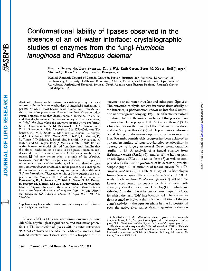
lar architecture to the previously described lipase from Rhizomucor miehei (Fig. l), in spite of the fact that the levels of amino acid identity between each of the two en- zymes and RmL are not high (-26% for HlL vs. RmL and - 50% for RdL vs. RmL). As was expected, the most significant differences occur in the regions of the surface loops, whereas the core of the fold remains largely con- served. RdL contains three disulfide bonds homologous to those found in RmL, whereas HlL contains two of these (Cys22-Cys268, and Cys36-Cys41) and an additional one between CyslO4 and CyslO7.
The relatively high resolution of the Humicola lanuginosa lipase structure ensures a generally high quality of the electron density map and resulting atomic coordinates (Table 1). The structure of the active center (Fig. 2A), which includes Ser146, His256, and Asp199, is virtually identical to that observed in RmL, and is equally reminiscent of the triads found in the subtilisin and chymotrypsin fami- lies of serine proteinases. Ser146 adopts a strained secon- dary conformation (r#1=57’, $= -125O), again similar to that observed in RmL (4=62O, $= -121’) and other alp hydrolases containing the so-called nucleophilic elbow (17). The oxy-anion hole is formed by Ser83, homologous to Ser82 in RmL (12, 13).
Derewenda et al. Conformational lability in lipases 525
by guest, on April 2, 2019
ww
w.jlr.org
Dow
nloaded from

TABLE 1. Crystallographic and refinement data
HIL
V a r i a b 1 e PEG a.s.’ p.b.’ RdL
X-ray data“ Resolution (A) Completeness (%) Reflections for which I > 2 0 (I) (%) Average redundancy < no~,b,Jno(,,,quc) >
Mean fractional change in intensity (%) R,,,,, (%)
Refinement statistics‘ Total number of reflections used for refinement Resolution limits R factor (within 7.5-1.85 A sphere) R factor on all reflections Number of non-hydrogen atoms Number of water molecules r.m.s. deviation from ideal bond distance ( A ) r.m.s. deviation from ideal bond ansles (planar) (deg.) r.m.s. deviation from ideal planes ( A )
1.85 2.05 2.07 2.6
84.5 80.2 78.2 79.9 4.72 3.85 3.99 2.37 8.08 10.45 11 .1 8.8
29.6 24.5
HIL (PEG) RdL
100 92 93 87
18756 7.5-1.85 A
16.7% 18% 2294 223
0.016 2.90 0.016
16071 7.5-2.6 A
17.8% 19.5% 4380 229
0.025 3.56 0.032
“X-ray data were collected with a Siemens area detector and processed using XENGEN (21); all subsequent cal- culations were done using the CCP4 software package (Daresbury Laboratory, U.K.) and X-PLOR (22). Electron density map fitting and structure analysis were done with the help of FRODO (23).
’a.s. signifies the HIL crystal soaked in 50% ammonium sulfate, while p.b. denotes the crystal soaked in the 70% phosphate buffer.
‘The structure of HIL was solved by the Molecular Replacement method using previously obtained low resolu- tion models of HIL and the RmL structure. (Historically, HIL was the first triglyceride lipase to have its structure determined by X-ray crystallogfaphy, using a hexagonal crystal form and multiple isomorphous replacement method, albeit at low resolution (3.25 A). Details of this early work will be published elsewhere. The structure was subse- quently refined at 2.6 A using triclinic crystals of the enzyme (Derewenda, U., Y . Wei, A. M. Brzozowski, L. Swenson, Z. S. Derewenda, unpublished results). The latter model, along with the highly refined homologous struc- ture of RmL, provided us with the guidelines for the interpretation of the results of theQMolecular Replacement solution.). The starting conventional crystallographic R factor was 0.504, within 7.5-1.8 A shell; rigid body refine- ment followed by simulated annealing (X-PLOR) reduced it to 0.294. At this stage electron density maps became readily interpretable, but the “lid” helix (residues 84-99) was ill defined. Further refinement was conducted using the restrained least-squares method (24), and ca. 150 ordered water molecules were added. There was no clearly interpretable electron density other than that a low-contour corresponding to the “lid” in its closed conformation; additionally, significant noise was observed in the region corresponding to the open conformation. Two models, one with the “lid” in the closed conformation, and the other with the “lid” in the open conformation as derived from the inhibited RmL structure were subjected to one round of simulated annealing and electron density maps were then calculated. In each case the electron density had similar appearance. Consequently, we used the interpretable, albeit very weak, density to model the “lid” in the closed conformation, although high levels of non-interpretable noise persisted in the region expected to be occupied by the “lid” in the open conformation. The structure of RdL was solved by the Molecular Replacement method using the molecular model of RmL and the AMORE program (25) in its CCP4 implementation. The details are described elsewhere (16). Restrained least-squares refinement of the partial model allowed calculation of difference electron density maps that gradually revealed the remaining de- tails of the structure. The two molecules in the asymmetric unit were treated independently to allow for possible differences induced by packing forces. The present model contains only 30 water molecules and some further refine- ment using higher resolution data will be necessary for a detailed description of the solvent structure.
The overall structure of the Rhizopus delemar lipase is even closer to RmL (Fig. IC). However, the lower resolu- tion of the study (2.6 A ) imposed by the quality of the crystals resulted in an electron density map of somewhat poorer quality and hence the accuracy of the coordinates is not as high as in HlL (Table 1). Again, the active center is made up of a triad of Ser145, His258, and Asp204, with the nucleophilic serine showing the strained conformation characteristic of its location within the nucleophilic elbow. The oxy-anion hole is presumably formed in the active
species by the hydroxyl and main-chain amide groups of Thr83.
The obvious homology of the three enzymes and resul- tant structural similarities do not obviate a need for a detailed examination of their three-dimensional struc- tures. However, the details of the overall protein architec- ture fall outside the scope of this report, and will be dis- cussed extensively elsewhere. Here we focus exclusively on the implications of these structures with regard to the molecular basis of interfacial activation.
526 Journal of Lipid Research Volume 35, 1994
by guest, on April 2, 2019
ww
w.jlr.org
Dow
nloaded from

R.miehei H. 1 anug. R.delem.
R. miehei H.lanug. R.delem.
R.miehei H.lanug. R.delem.
R.miehei H.lanug. R.delem.
A
10 20 30 40 so 60 1 SidggiraatsqeineltyyttlsAnsYCrtvi pgat wdCih Cdate Dlkiiktwstli yDtnam 1 evsqdlfnqfnlfaqysAaaYCgknndapagtnitCtgnaCpevekaDatflysfedsgvgDvtgf
rswpgnk wdcvq Cqkwvp Dgkiittftsll sDtngy
--c' 70 80 90 100 110 120 130
66 vargdseKtiyivFRGssSirnwIadltFvpvsy ppvsGtkvHkGFldsygeVqnelvatVldqfkqyPsYk 67 1aldntnKlivlsFRGsrSienwIQnlnrdlkein~csGcrgH~tsSwrsVa~lrqkVedavre~dYr 67 vlrsdkqKtiylvFRGtnSfrsaItdivFnfsdy kpvkGakvfiaWlsSyeqVvndyfpWqeultahPtYk
-1 140 150 160 170 180 190 200 V
u 138 VavTGElSUjGAtAllcalDLyqreeglsssnlfl~~~~aF~~ VsTGipyrRtvnerDIVPhlP 140 VvfTGHSLGGUAtvagaDLr gngy didvfsyGaPElVGnraFAefltv~ly~thtnDIVrlP
rlspknlsiftvGgPRVGnptFAyyv eSTGipfqRtvhkrDIVPhvP
210 220 230 240 250 V260 210 PaaFGflmqeEyWItd nspe tvqvctsdletsdcsnsivPf tsvldsLsYFgintGlCt 208 PreFGysBsspmksgtlvpvtrndivkiegi datggnn q PnipdipaXLwYF gliGtCl 211 PqsFGflRpgvEsWIksgt sn vqictseietkdcsnsivpf tsildELsYFdineGsC1
U U
65 66 66
137 139 138
209 207 210
269 269 269
Fig. 1. A: A structure-based sequence alignment of RmL, HIL, and RdL. The conserved amino acids are shown in upper case, while the secondary structure elements are indicated below the sequences; the arrows indicate the three active site amino acids. B: A stereo diagram showing the superposi- tion of the RmL (dashed line) and HIL structures; only the CCI atoms and the catalytic triad in HlL are shown. C: a comparison of RmL (dashed) and RdL; details as in B.
Derewenda et al. Conformational lability in lipases 527
by guest, on April 2, 2019
ww
w.jlr.org
Dow
nloaded from

A
B c <=
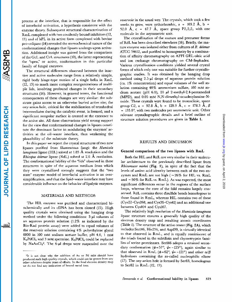
Fig. 2. Difference (Fnb\ -F,,,,) electron density map showing (A) the three residues that form the catalytic triad in HIL; and (B) the lid region. The figures were prepared using the same electron density map obtained after the triad residues and the 16 lid residues (including the hinge regions) were excluded from the model and the remainder of the structure was subjected to three cycles of restrained refinement to remove any bias. The catalytic triad residues (A) are contoured at 4 * the root-mean-square deviation from the mean, while in (B) a lower contour, 2.5 r.m.s., was used as there is no interpretable density at the higher level.
Structures of the "lids" strongly amphipatic helix that translocates as a rigid body during the activation process. In the RmL molecule the lid is clearly visible in the electron density map (6), sug- gesting intrinsic stability of this structure. In contrast, in
Humicolu lanuginosu lipase. ~~~~d on the detailed study of the stereochemistry of the conformational changes in R ~ L (13) as well as on the structure-based se., quence alignment (Fig. 1A) we assume that, in general terms, the lids in HlL and RdL each contain two hinge
terminal, residues 91-95) and a six-residue (85-90)-long
'To avoid confusion, the amino acid numbering in this section relates regions (the N-terminal One, residues 83-843 and the '- to the RmL sequence except in figures, For comparison of all three se- quences, see ~ i ~ . 1 ~ .
528 Journal of Lipid Research Volume 35 , 1994
by guest, on April 2, 2019
ww
w.jlr.org
Dow
nloaded from

50
40
30 A m “ 2 0
10
0 1 ’ 1 . 1 . 1 . ~ ‘ 1 . 1 . 1 ‘ 1 ‘ 1 . l ~ 1
75 77 79 81 83 85 87 89 91 93 95 97 99
I v l s F R G S r S I e N W I g n L n F d l k e - Y i v - - - - s - - r - - - a d - t - v p v s
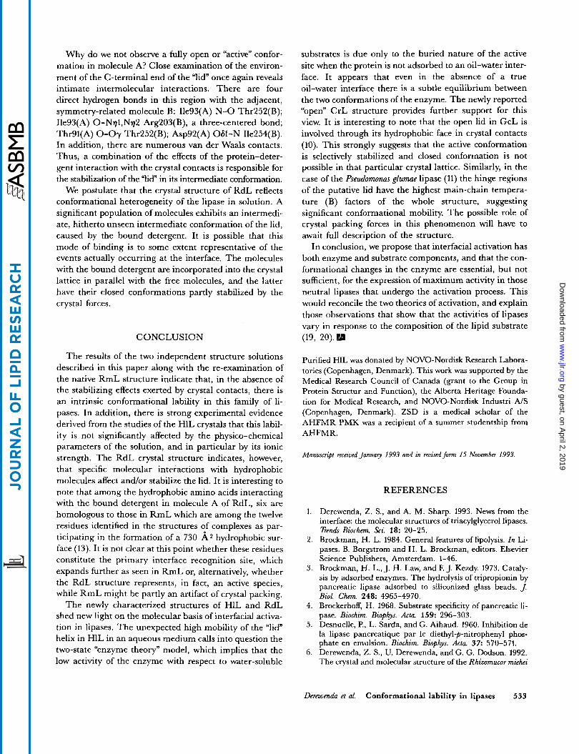
Fig. 3. A comparison of mean temperature factors (averaged for main chain atoms within each amino acid) in the “lid” fragment between RmL and HIL. To allow for unbiased comparison, in both cases prior to final comparison the temperature factors were reset to 15 A Z and subjected to 5 cycles of restrained least-squares refinement, followed by 10 cycles of unrestrained B factor refinement, and 1 final cycle of restrained refinement. The sequence of the “lid” in HIL is shown (top) and compared with that of RmL (bottom) where the dashes represent conserved residues (upper case in the HIL sequence).
the HlL molecule, well-defined electron density extends only up to residue 82 and then from 99 onwards (Fig. 2B). For the intervening sequence the density is poor and frag- mented, although it corresponds quite well with respect to its location to the closed conformation of the lid. It is noteworthy that the quality of the density deteriorates gradually towards the center of the helix, so that at the point of Trp88 there is virtually no interpretable density. Also, all the side chains homologous to those identified in the RmL lid as involved in the interaction with the main domain of the lipase molecule in its closed form have poorly defined densities, even at low contour levels. These features of the electron density are consistent with the high mean isotropic temperature (B)* factors observed for the lid in HlL, significantly higher than the corresponding ones in RmL (Fig. 3). It should be pointed out that, given the limited resolution of the X-ray data, it is not possible to differentiate between the genuine temperature (B) fac- tors, which stem from the thermal fluctuations of atoms, and the effects of low occupancy factors, which define the percentage of time a given site is occupied by an atom. We therefore view the high temperature (B) factors of the HlL lid as representing the combined effects of the two factors, a view supported by the high noise of the difference elec-
‘The temperature (B) factor is related to the observed mean square displacement of an atom from its average position (< LIZ>) by the for- mula B=(8 +/3)* < u z > .
tron density map in the region corresponding to the open conformation of the lid.
RmL exhibits very low activity towards water-soluble substrates. In the native molecule of this lipase (Le., in the closed, or inactive, conformation) the interface between the lid and the main domain is made up exclusively of in- terdigitating hydrophobic amino acids, without a single hydrogen bond or salt bridge involved (6). It has been postulated (6) that this architecture is sufficiently stable in aqueous solution to keep the active site permanently oc- cluded from the solvent, while in a nonpolar medium the conformation becomes destabilized causing the lid to swing into a new position. The observed disorder of the lid in the crystal structure of the homologous HlL is in conflict with this view. In order to resolve this apparent incompatibility we investigated two possibilities: u) that crystal packing forces may influence the stability of the RmL lid, and b) that low ionic strength of the crystalliza- tion medium plays a role in determining the conformation of the lid in HlL.
Upon careful re-examination of the native RmL struc- ture (6) we find that there are at least two direct hydrogen bonds between the “lid” and a symmetry-related molecule, additional interactions with this molecule mediated via water molecules, and also close van der Waals contacts. In contrast, in the presently studied HlL crystals, the lid is not only free of intermolecular contacts, but in addition its alternative (open) conformation is possible within the framework of the existing crystal lattice. Thus the postu-
Dereroada et al. Conformational lability in lipases 529
by guest, on April 2, 2019
ww
w.jlr.org
Dow
nloaded from

A
Fig. 4. (A) Difference electron density (Fob\ - F,,,,) observed for the lid in molecule A of RdL. As in the previous case the map was calculated with the lid residues omitted and three refinement cycles were carried out to remove bias. The contour level was set to 2.5 * r.m.s. (B) Same for molecule B.
lated stability of the RmL lid in an aqueous phase seems to be, at least in part, caused by crystal contacts.
The effect of the crystallization medium was also exa- mined. The crystals of HlL had been grown under low ionic strength conditions, whereas those of RmL where obtained from high ionic strength phosphate buffer (6). Two crystals of HlL were soaked in concentrated salt solu- tions: one in 50% ammonium sulfate and the other in 70% phosphate buffer. The latter medium is identical to that from which crystals of RmL were grown. Data were collected and processed as before (Table l), and each form was subjected to additional least-squares refinement prior
to the final analysis. Final difference electron density maps calculated with coefficients (Fsoak - F,,,) revealed many small changes in the dispositions of numerous sur- face side chains as well as in the details of the solvation of the molecular surface, consistent with an observed significant mean fractional change in the intensities. However, in both cases the transfer to a high ionic strength medium resulted only in marginal improvements of the appearance of the electron density maps in the envi- rons of the lid. There was no indication of any significant stabilization of the HlL lid in any single conformation. A comparison of the final temperature (B) factors in all the
530 Journal of Lipid Research Volume 35, 1994
by guest, on April 2, 2019
ww
w.jlr.org
Dow
nloaded from

Fig. 4. (C) A schematic overlap generated using the program MOLSCRIPT (26), showing the positions of the lids in the two independent molecules of RdL in comparison with the conformation found in the inhibited RmL complex. The structures have been superimposed on the central P-sheet atoms.
three crystals (data not shown) also indicated that in quantitative terms no significant stabilization of the “lid” was achieved during the experiment.
The asymmetric unit of the RdL crystals contains two molecules of the enzyme (denoted A and B) that have been independently refined. Under these conditions it is not uncommon to observe small differences in atomic structure between the two molecules, differences caused by the crystal’s packing forces and usually restricted to external loops which medi- ate intermolecular interactions. However, in cases of in- trinsic molecular mobility, particularly involving domain or sub-domain hinge type motion, two (or sometimes more) molecules with quite different conformations are occasionally observed within one asymmetric unit. This
Rhizopus delemar lipase.
conformational variability in the solid state seems to sam- ple a more or less continuous range of conformations in solution. A classic example is the Met6 + Ile thermola- bile mutant of the T 4 lysozyme, which displays five differ- ent crystal conformations (18). The RdL crystals are clearly an example of such phenomenon. The “lid” of one of the molecules (molecule B) is in the expected closed conformation blocking access to the active site, while in molecule A the lid assumes a unique conformation, inter- mediate between the closed one and the open one, as defined by the structures of inhibited RmL. In both cases the electron density is very clear (Fig. 4A and B).
In molecule B (closed conformation) the lid is involved in a crystal contact with an adjacent molecule A, although the interactions are limited to the N-terminal end of the
Derewenda et al. Conformational lability in lipases 531
by guest, on April 2, 2019
ww
w.jlr.org
Dow
nloaded from

lid. In particular there is one direct main chain-main chain hydrogen bond (Arg87(B) 0-N Leu59(A)), and electrostatic interactions between the side chain of Arg87(B) and two acidic residues (Asp92 and Asp62) in the A molecule. It is very interesting that the quality of electron density deteriorates beyond Arg87, where there are no further crystal contacts. The resolution of this study (2.6 A) is not sufficient to allow for reliable refine- ment of temperature factors, but the appearance of the electron density strongly suggests that the region not stabilized by crystal contacts is quite mobile. This obser- vation is fully consistent with our conclusions derived from the comparison of the RmL and HlL crystal struc- tures (see above).
Molecule B of RdL is unique for two reasons. First, as mentioned above, the lid assumes a hitherto unobserved intermediate conformation (Fig. 4C). The electron den- sity is fairly well defined and the “lid” appears to be rela- tively stable. Once again we searched for the source of the
stability of this new molecular species. We find that the final difference electron density map (which, in general terms, ought to reveal any significant discrepancy be- tween the model and the experimental data) contains significant continuous positive density within the cavity created by two adjacent B molecules (Fig. 5). More specifically, this cavity is located between two symmetry- related lids in the neighboring B molecules, and is lined by an impressive constellation of hydrophobic amino acids, including Phe86, Ile93, Phe95, Phell2, Leu146, Pro178, Va1206, Va1209, Pro210, Pro214 Phe214, and their equivalents in the adjacent molecule. In addition, en- trance to this cavity is blocked by two symmetry-related side chains of Phe251 from two close-by molecules A. AS a detergent (N,N-dimethyl-octadecylamine-N-oxide) was used in the crystallization medium and proved critical for the growth of X-ray quality crystals, we conclude that the positive density filling the hydrophobic cavity corresponds to the bound detergent.
Fig. 5. Diagrammatic representations of the packing of two B molecules of RdL, showing the hydrophobic ravity containing significant positive difference electron density indicative of nonspecific binding of the detergent. Only the three active site residues are shown in full; otherwise main chain atoms only are included. The lid and the cata- lytic triad are shown in bold lines.
532 Journal of Lipid Research Volume 3 5 , 1994
by guest, on April 2, 2019
ww
w.jlr.org
Dow
nloaded from
Why do we not observe a fully open or “active” confor- mation in molecule A? Close examination of the environ- ment of the C-terminal end of the ’lid” once again reveals intimate intermolecular interactions. There are four direct hydrogen bonds in this region with the adjacent, symmetry-related molecule B: Ile93(A) N - 0 Thr252(B); Ile93(A) O-Nql,Nq2 Arg203(B), a three-centered bond; Thr91(A) 0-Oy Thr252(B); Asp92(A) 061-N Ile254(B). In addition, there are numerous van der Waals contacts. Thus, a combination of the effects of the protein-deter- gent interaction with the crystal contacts is responsible for the stabilization of the ”lid” in its intermediate conformation.
We postulate that the crystal structure of RdL reflects conformational heterogeneity of the lipase in solution. A significant population of molecules exhibits an intermedi- ate, hitherto unseen intermediate conformation of the lid, caused by the bound detergent. It is possible that this mode of binding is to some extent representative of the events actually occurring at the interface. The molecules with the bound detergent are incorporated into the crystal lattice in parallel with the free molecules, and the latter have their closed conformations partly stabilized by the crystal forces.
CONCLUSION
The results of the two independent structure solutions described in this paper along with the re-examination of the native RmL structure indicate that, in the absence of the stabilizing effects exerted by crystal contacts, there is an intrinsic conformational lability in this family of li- pases. In addition, there is strong experimental evidence derived from the studies of the H1L crystals that this labil- ity is not significantly affected by the physico-chemical parameters of the solution, and in particular by its ionic strength. The RdL crystal structure indicates, however, that specific molecular interactions with hydrophobic molecules affect and/or stabilize the lid. It is interesting to note that among the hydrophobic amino acids interacting with the bound detergent in molecule A of RdL, six are homologous to those in RmL which are among the twelve residues identified in the structures of complexes as par- ticipating in the formation of a 730 A 2 hydrophobic sur- face (13). It is not clear at this point whether these residues constitute the primary interface recognition site, which expands further as seen in RmL or, alternatively, whether the RdL structure represents, in fact, an active species, while RmL might be partly an artifact of crystal packing.
The newly characterized structures of H1L and RdL shed new light on the molecular basis of interfacial activa- tion in lipases. The unexpected high mobility of the “lid” helix in HlL in an aqueous medium calls into question the two-state “enzyme theory” model, which implies that the low activity of the enzyme with respect to water-soluble
substrates is due only to the buried nature of the active site when the protein is not adsorbed to an oil-water inter- face. It appears that even in the absence of a true oil-water interface there is a subtle equilibrium between the two conformations of the enzyme. The newly reported “open” CrL structure provides further support for this view. It is interesting to note that the open lid in GcL is involved through its hydrophobic face in crystal contacts (10). This strongly suggests that the active conformation is selectively stabilized and closed conformation is not possible in that particular crystal lattice. Similarly, in the case of the Pseudomonas glumae lipase (11) the hinge regions of the putative lid have the highest main-chain tempera- ture (B) factors of the whole structure, suggesting significant conformational mobility. The possible role of crystal packing forces in this phenomenon will have to await full description of the structure.
In conclusion, we propose that interfacial activation has both enzyme and substrate components, and that the con- formational changes in the enzyme are essential, but not sufficient, for the expression of maximum activity in those neutral lipases that undergo the activation process. This would reconcile the two theories of activation, and explain those observations that show that the activities of lipases vary in response to the composition of the lipid substrate (19, 20).1
Purified HIL was donated by NOVO-Nordisk Research Labora- tories (Copenhagen, Denmark). This work was supported by the Medical Research Council of Canada (grant to the Group in Protein Structur and Function), the Alberta Heritage Founda- tion for Medical Research, and NOVO-Nordisk Industri A/S (Copenhagen, Denmark). ZSD is a medical scholar of the AHFMR PMK was a recipient of a summer studentship from AHFMR.
Manuscript received January 1993 and in reuisedfon 15 Nouember 1993.
REFERENCES
1.
2.
3.
4.
5.
6.
Derewenda, Z. S., and A. M. Sharp. 1993. News from the interface: the molecular structures of triacylglycerol lipases. Eends Biochem. Sci. 18: 20-25. Brockman, H. L. 1984. General features of lipolysis. In Li- pases. B. Borgstrom and H. L. Brockman, editors. Elsevier Science Publishers, Amsterdam. 1-46. Brockman, H. L., J. H. Law, and F. J. Kezdy. 1973. Cataly- sis by adsorbed enzymes. The hydrolysis of tnpropionin by pancreatic lipase adsorbed to siliconized glass beads. J. Biol. Chem 248: 4965-4970. Brockerhoff, H. 1968. Substrate specificity of pancreatic li- pase. Biochim. Biophys. Acta. 159: 296-303. Desnuelle, P., L. Sarda, and G. Aihaud. 1960. Inhibition de la lipase pancreatique par le diethyl-p-nitrophenyl phos- phate en emulsion. Biochim. Biophys. Acta. 37: 570-571. Derewenda, Z. S., U. Derewenda, andG. G. Dodson. 1992. The crystal and molecular structure of the Rhizomucor miehei
Derewenda et al. Conformational lability in lipases 533
by guest, on April 2, 2019
ww
w.jlr.org
Dow
nloaded from

triglyceride lipase at 1.9 A resolution. J. Mol. Biol. 227:
7. Winkler, F. K., A. DArcy, and W. Hunziker. 1990. Struc- ture of human pancreatic lipase. Nature (London). 343:
8. van Tilbeurgh, H., L. Sarda, R. Verger, and C. Cambillau. 1992. Structure of the pancreatic lipase-procolipase com- plex. Nature (London). 359: 159-162.
9. Schrag, J. D., and M. Cygler. 1993. 1.8 A refined structure of the lipase from Geotrichum candidum. J. Mol. Biol. 230:
10. Grochulski, P., L. Yunge, J. D. Schrag, E Bouthillier, P. Smith, D. Harrison, B. Rubin, and M. Cygler. 1993. In- sights into interfacial activation from an open structure of Candida rugosa lipase. J. Biol. Chem. 268: 12843-12847. Noble, M. E. M., A. Cleasby, L. N. Johnson, M. R. Eg- mond, and L. G. J. Frenken. 1993. The crystal structure of triacylglycerol lipase from Pseudomonas glumae reveals a par- tially redundant catalytic aspartate. FEBS Lett. 331:
12. Brzozowski, A. M., U. Derewenda, Z. S. Derewenda, G. G. Dodson, D. M. Lawson, J. P. Turkenburg, F. Bjorkling, B. Huge-Jensen, S. A. Patkar, and L. Thim. 1991. A model for interfacial activation in lipases from the structure of a fun- gal lipase-inhibitor complex. Nature (London). 351: 491-494.
13. Derewenda, U., A. M. Brzozowski, D. M. Lawson, and Z. S. Derewenda. 1992. Catalysis at the interface: the anat- omy of a conformational change in a triglyceride lipase. Bi- ochemistry. 31: 1532-1541.
14. van Tilbeurgh, H., M-P. Egloff, C. Martinez, N. Rugani, R. Verger, and C. Cambillau. 1993. Interfacial activation of the lipase-procolipase complex by mixed micelles revealed by X-ray crystallography. Nature (London). 362: 814-819.
15. Boel, E., B. Huge-Jensen, H. F. Woeldike, E. Gormsen, M. Christensen, F, Andreasen, and L. Thim. 1991. Cloning and expression of industrially important fungal lipases. In Lipases, Structure, Mechanism and Genetic Engineering. L. Alberghina, R. D. Schmid and R. Verger, editors. VCH Publishers, Weinheim. 207-219. Swenson, L., R. Green, R. Joerger, M. Haas, K. Scott, Y .
818-839.
771-774.
575-591.
11.
123-128.
16.
Wei, U. Derewenda, D. M. Lawson, and Z. S. Derewenda. 1994. Crystallization and preliminary crystallographic studies of the precursor and mature forms of a neutral li- pase from the fungus Rhizopus delemar. Proteins: Struct. Funct. Genet. In press.
17. Ollis, D. I., E. Cheah, M. Cygler, B. Dijkstra, E Frolow, S. M. Franken, M. Harel, S. J. Remington, I. Silman, J. Schrag, J. Sussman, K. H. G. Verschueren, and A. Gold- man. 1992. The alp hydrolase fold. Protein Eng. 5: 197-211. Faber, H. R., and B. W. Matthews. 1990. A mutant T4 lysozyme displays five different crystal conformations. Na- ture (London). 348: 263-266.
19. Muderhwa, J. M., and H. L. Brockman. 1992. Lateral lipid distribution is a major regulator of lipse activity. Implica- tions for lipid-mediated signal transduction. J Bzol. Chem.
20. Wilcox, R. W., T. Thuren, P. Scisson, J. D. Schmitt, M. Kennedy, and M. Waite. 1993. Regulation of rat hepatic li- pase by the composition of monomolecular films of lipid. Biochemistv. 32: 5752-5758. Howard, A. J., G. L. Gilliland, B. C. Finzel, T. L. Poulos, D. H. Ohlendorf, and F. Salemme. 1987. Use of an imaging proportional counter in macromolecular crystallographyJ Appl. Cystallogr. 20: 383-387.
22. Brunger, A. T. 1988. X-PLOR Manual. Yale University, New Haven, CT.
23. Jones, A. 1978. A graphics model building and refinement system for macromolecules. J. Appl. CryJtallogr. 11:
24. Hendrickson, W. A. 1985. Stereochemically restrained refinement of macromolecular structures. Methods Enzymol.
25. Navaza, J. 1992. AMoRe: a new package for molecular replacement. Zn Proceedings of the CCP4 Study Weekend. E. J. Dodson, S. Gower and W. Wolf, editors. SERC, UK.
26. Kraulis, P. J. 1991. MOLSCRIPT a program to produce both detailed and schematic plots of protein structures. J. Appl. Crystallogr. 24: 946-950.
18.
267: 24184-24192.
21.
268-272.
115: 252-270.
87-91.
534 Journal of Lipid Research Volume 35, 1994
by guest, on April 2, 2019
ww
w.jlr.org
Dow
nloaded from