composition of myelin from peripheral and central … of myelin from peripheral and central nervous...
TRANSCRIPT

Composition of myelin from peripheral and central nervous systems of the squirrel monkey
L. A. HORROCKS
Laboratory of Neurochemistry, Cleveland Psychiatric Institute, Cleveland, Ohio 44109
ABSTRACT Myelin was prepared from the brachial plexus and cervical spinal cord of adult squirrel monkeys (Saimiri sciurm). Brachial plexus myelin contained a larger amount of sphingomyelin and smaller amounts of cholesterol, lipid galactose, ethanolamine phosphoglyceride, choline phospho- glyceride, and alk-1-enyl ether than spinal cord myelin when compared as ratios to total lipid phosphorus. The peripheral nervous system myelin had a higher proportion of protein. All of these differences were statistically significant.
Thus peripheral nervous system myelin and central nervous system myelin differ in protein content and lipid composition in this subhuman primate.
KEY WORDS myelin . nervous system . peripheral central . squirrel monkey . electron microscopy . lipid composition proteolipid protein
A SUBSTANTIAL AMOUNT of information is now available on the composition of CNS myelin from a number of vertebrate species. This information has been used for the construction of models of the molecular ultrastructure of the CNS myelin membrane (1-3). The composition of CNS myelin from subhuman primates has not been de- scribed, and only conflicting information is available for mammalian PNS myelin lipids. The lipid composition of PNS myelin has been reported for the rat and guinea pig by Evans and Finean (4) and for the ox by O’Brien, Sampson, and Stern (5). Since these authors did not in- clude a direct comparison of CNS and PNS in the experi- mental design, they arrived at different conclusions re- garding possible differences in lipid composition between
Abbreviations: PNS, peripheral nervous system; CNS, central nervous system; TLC, thin-layer chromatography; Na&DTA, tetrasodium ethylenediamine tetraacetate ; EPG, ethanolamine phosphoglycerides ; CPG, choline phosphoglycerides; Sph, sphingo- myelin; SPG, serine phosphoglycerides; IPG, inositol phospho- glycerides (including mono-, di-, and triphosphoinositides).
CNS and PNS myelin. Differences between CNS and PNS myelin have been shown by physical studies on myelin ultrastructure using X-ray diffraction and elec- tron microscopy ( 6 ) and by chemical studies on proteo- lipid proteins (7). Since CNS myelin is formed by oligo- dendroglia cells and PNS myelin is formed by Schwann’s cells, the purpose of the present investigation was to de- termine if the oligodendroglia and the Schwann’s cells produce myelin membranes with significant differences in composition in a subhuman primate.
METHODS
Tissue Fractionation and Extraction The 18 squirrel monkeys (Suimiri sciureus) used as sub- jects for this experiment were maintained on Purina monkey chow for a t least 4 months before sacrifice. Weights of the animals ranged from 320 to 790 g. The brachial plexus and samples of cervical spinal cord were removed within 5 min after decapitation. The tissues were stored at 4OC in the dispersion media until pro- cessed. Dispersion was accomplished with a Potter- Elvehjem tissue grinder equipped with a Teflon pestle. Four or five strokes were used to disperse spinal cord, but minced brachial plexus required twelve strokes and a sub- stantial residue remained undispersed. A fraction with the properties of myelin was then isolated by one of the following three methods, in which a Spinco model L ul- tracentrifuge was used.
Method A The dispersion medium was 1 .O M sucrose-0.003 M
Na4EDTA. The dispersion was centrifuged for 2 hr at 40,OOOp in a SW 25.1 swinging bucket rotor.’
1.
Subsequent experiments have shown that flotation for 60 min in steps 1, 2, and 5 is sufficient. Also, sedimentation for 15 min at 13,500 g in steps 3,4, and 6 produces a firm myelin pellet with a clear supernatant fluid.
JOURNAL OF LIPID RESEARCH VOLUME 8, 1967 569
by guest, on June 24, 2018w
ww
.jlr.orgD
ownloaded from
2. After the tube had been sliced with a Spinco tube- slicer, the floating layer was removed by syringe, dis- persed in 0.8 M sucrose-0.003 M NadEDTA, and recentri- fuged as in step 1.
The floating layer was dispersed in distilled water and sedimented for 10 min at 100,000 g in a No. 40 r0tor.l
The supernatant fluid was discarded and the pellet was dispersed and sedimented as in step 3.
The supernatant fluid was discarded and the pellet was dispersed in 0.8 M sucrose-0.003 M Na4EDTA. The dispersion was again centrifuged for 2 hr at 40,000 g in the SW 25.1 swinging bucket rotor.
6 . The floating layer was dispersed in distilled water and sedimented three times as in step 3.
None of the myelin was lost if 1 .O M sucrose was used for dispersion. If 0.8 M sucrose was used for the first step, some of the myelin remained in the infranatant fluid and the floating layer was difficult to recover. For spinal cord material, steps 5 and 6 were probably not necessary, but step 5 for the PNS preparation gave a small pellet and a cloudy infranatant which were discarded.
3.
4.
5.
Method B
1. The dispersion medium was 0.32 M sucrose-0.001 M Na4EDTA-0.003 M Na2HP04. The dispersion was centrifuged for 15 min at 13,500 g in a No. 40 rotor.
The supernatant suspension was discarded and the pellet was suspended by syringe in 25 ml of 0.8 M sucrose- 0.001 M Na4EDTA-0.006 M Na2HP04. The latter suspen- sion was centrifuged for 60 min at 40,000 g in an SW 25.1 swinging bucket rotor.
After the tube had been sliced with a Spinco tube- slicer, the floating layer was removed by syringe, diluted with distilled water, and sedimented as in step 1.
The supernatant fluid was discarded and the pellet was resuspended in distilled water. At this point the preparation was usually stored overnight, then sedimented as in step 1.
A continuous density gradient was formed in SW 25.1 centrifuge tubes by the admixture of 10-ml portions of 1.0 M sucrose and 0.32 M sucrose in a Buchler appara- tus (Buchler Instruments, Inc., Fort Lee, N. J.). The pellet from step 4 was suspended in 5 ml of 0.32 M sucrose and layered over the gradient. The tubes were centrifuged as in step 2.
The clear supernatant fluid was removed and dis- carded. The tube was sliced and the single compact myelin layer (brachial plexus) or the light myelin layer (spinal cord) was removed. The tube slicing was repeated for removal of the heavy myelin layer (spinal cord). The layers were diluted with distilled water and the myelin was sedimented three times for 15 min at 13,500 g in a No. 40 rotor.
2.
3.
4.
5.
6.
Method C This method was a combination of steps 1-4 of Method
B and steps 5 and 6 of Method A. Steps 1-4 of Method B were adapted from the myelin isolation procedure of Cuzner, Davison, and Gregson (8).
Electron Microscopy Myelin suspensions were examined by both negative and positive staining procedures. For negative staining, equal volumes of the myelin suspension and 2% potassium phosphotungstate were mixed, a drop of the mixture was placed on a carbon-coated grid with a platinum loop, and the grid was dried on filter paper.
For positive staining, equal volumes of the myelin sus- pension and 8% glutaraldehyde buffered to pH 7.2 with 0.2 M cacodylate buffer were mixed and kept at 4*C for 3 hr before centrifugation in polyallomer tubes for 15 min at 13,500 g in a No. 40 rotor. The pellet was sus- pended in 1% osmium tetroxide in 0.2 M cacodylate buffer for 60 min, then centrifuged. Dehydration with 70%, 95%, and 100% ethanol was accomplished by sus- pending the pellet, centrifuging the mixture, and decant- ing the supernatant liquid. After dehydration, portions of the pellet were embedded in Maraglas (Polysciences, Inc., Rydal, Penn.) (9). Sections of 400-800 A thickness were obtained with an LKB (Stockholm, Sweden) ultra- tome, mounted on carbonized grids, and stained with lead citrate. All grids were examined with a Siemens (Karlsruhe, Germany) Elmiskop I electron microscope at 60 kv.
Extraction The final white pellet was dispersed in distilled water to a volume of 1.0 ml and then mixed with 35 ml of chloro- form-methanol 2 : 1. A clear or very slightly opalescent solution was obtained. 7 ml of 0.58% NaCl (10) was added to the tube, which was kept at 4OC overnight. The upper phase was removed and the lower phase was washed twice with an upper phase mixture (10). The washed lower phase was taken to dryness with additions of absolute ethanol and brought to a final volume of 10 ml in chloroform for analysis.
Analytical MethodP The protein content of portions of the aqueous myelin suspension was determined by a modification (1 1) of the method of Lowry, Rosebrough, Farr, and Randall (12) with bovine serum albumin as the standard. Phosphorus (13), galactose (14), cholesterol (15), alk-1-enyl ethers (16, 17), and the phospholipid class distribution were de- termined for portions of the lipid extract. The galactose values were corrected for charring by subtraction of a blank containing the same quantities of lipid but without anthrone. For cholesterol determinations, a portion of the
570 JOURNAL OF LIPID RESEARCH VOLUME 8, 1967
by guest, on June 24, 2018w
ww
.jlr.orgD
ownloaded from

TABLE 1 COMPOSITION OF SQUIRREL MONKEY SPINAL CORD MYELIN PREPARED BY DIFFERENT METHODS
Method of Preparation
A B C
Number of preparations 8 6 4 Protein (%) 19.8’ - 20.4 f 1.6 Lipid P (pmoleslg tissue) 31 .4 f 3.3 40.1 f 2.3 15.1 f 1.3
mole ratio, componcnf to lipid P
Cholesterol 1.30 f 0.03 1.32 f 0.02 1.40 f 0.004 Lipid galactose 0.73 f 0.04 0.56 f0.0lt 0.68 f0.04$ Alk-1-enyl ether 0.37 f 0.02 - 0.37 f 0.03 EPG 0.432 f 0.007 0.438 f 0.020 0.446 f 0.012 CPG 0.187 f 0.007 0.191 f 0.012 0.186 f 0.002
0.172 f 0.006 0.162 f 0.018 0.140 f 0.005 0.176 f 0.008 0.195 f 0.014 0.196 f 0.012 SPG + IPG
SPh
Values are the mean f SEM. * Mean of two samples. t P < 0.01 for difference between A and B. $ P < 0.05 for difference between B and C.
lipid extract was taken to dryness under Nz, then dis- solved in ethanol. For determinations of the phospho- lipid class distributions by TLC, a portion of the lipid ex- tract containing 0.3-0.6 pinole of the phosphorus was applied to a 4 cm lane on a 0.5 mm layer of Silica Gel G. The plate was developed for 10 cm in chloroform-metha- nol-15 N ammonium hydroxide 65 : 25 : 4 (1 8). Lipids were detected by ninhydrin spray and iodine vapor (17). The entire lane was marked as indicated in Table 1 and areas were scraped into separate boiling tubes with the aid of a 1 inch plastic blade. The phosphorus of these areas and of blanks was assayed according to Gottfried ( 1 8 ~ ) . The phospholipid composition was calculated from the corrected absorbance values. The lipid composition has been expressed in terms of mole ratios of compo- nents to total lipid phosphorus. Statistical comparisons of possible differences between CNS and PNS myelin lipid compositions were made by analysis of variance (1 3) *
RESULTS
All of the inyelin preparations were almost completely soluble in a mixture of 34 parts of chloroform-methanol 2: 1 and one part of water. Negligible contamination of myelin fractions could be found upon examination of numerous electron micrographs. Occasionally, a strand of collagen was found in negatively stained preparations. Typical micrographs, selected only for sharpness of focus, are shown in Figs. 1-8. The myelin shown in Figs. 1-3 shows numerous multilamellar structures in various states of disintegration, probably due to relatively long storage at low osmolality. A comparison of Figs. 4 and 5 demonstrates that contamination by microsomes can be
detected by the presence of small vesicles in negatively- stained preparations. No microsomes were present in myelin preparations (Figs. 3 and 4). Fig. 5 also shows that the “microsomal” fraction which was discarded con- tained numerous small myelin fragments. The PNS myelin illustrated in Figs. 6-8 is similar to that shown by O’Brien et al. (5).
All three methods were used to prepare myelin from samples of cervical spinal cord (Table 1). The amount of myelin lipid phosphorus obtained from 188-385 mg of spinal cord by Method A (six preparations) was 35.4 f 2.8 pmoles/g (mean f SEM). Preparations from 908-1713 mg of spinal cord gave myelin lipid phosphorus yields of 19, 40, and 1 5 pmoles/g for Methods A, B, and C respec- tively. Norton and Autilio (20) have reported a yield of 63 pmoleslg of bovine white matter. The results for myelin prepared by Method B are for the heavy and light fractions combined. The amounts of cholesterol and lipid galactose relative to lipid P in the heavy and light fractions were not significantly different as judged by a test for correlated samples. The lipid compositions by Methods A and C were the same, with the possible excep- tion of sphingomyelin and serine plus inositol phcspho- glycerides, but myelin prepared by Method B had a lower relative amount of lipid galactose. The differences were statistically significant.
Methods A and B were used to prepare myelin from brachial plexus (Table 2). The yield of myelin was much lower from this tissue, presumably because of difficulties in dispersing the tissue and because of the large amount of connective tissue. The relative amounts of cholesterol and lipid galactose were larger for Method B in this case but the differences were not significant. The alk-1-enyl ether values were not different.
HORROCKS P N S and C Y S Myelin Lipids 371
by guest, on June 24, 2018w
ww
.jlr.orgD
ownloaded from
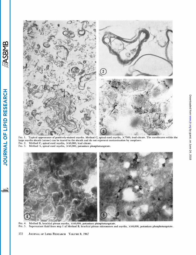
FIG. 1 . large myelin sheath (arrow) can be traced to the sheath and do not reprrsrnt contamination by axoplasm. FIG. 2. FIG. 3.
lypical appearancr ot positively-stained myelin. Method C , spinal cord myelin, X7500, lead citrate. 1 tic. inciiibrancs within the
Method C, spinal cord myelin, X60,000, lead citrate. Method A, spinal cord myelin, X 60,000, potassium phosphotungstatr.
t 1
r
d
rc
FIG. 4. FIG. 5 .
Si2
Method B, brachial plcxus myelin, X60,000, potassium phosphotungstate. Supernatant fluid from step 1 of Method R, brachial plexus inicrosomrs and myelin, X60,000, potassium phosphotungstate.
by guest, on June 24, 2018w
ww
.jlr.orgD
ownloaded from

FIG. ( I .
FIG. 7. FIG. 8.
Mcthod C:, brachial plcsus niycliii, X21,(100, 1 ~ ~ ~ 1 citi-atc.. Method C , brachial plrxus myrlin, X60,000, Ir;icl citrate. Mrtharl .\, brachial plexus tnvrlin. X60.000, Ir;id citratr.
The protein values were calculated by dividing the amount of protein by the sum of the protein and the lipids [using the molecular weights calculated by Aiitilio, Norton, and Terry (21) ] followed by inriltiplication by 100. The protein content of squirrel inonkey myelin was 20y0 and 30%, respectively, for spinal cords and brachial plextis. These values wcre significantly different by the Maim-[Vhitncy U test (LJ = 0; 11 = 6, 5 ; P < 0.002). Possible differences in the relative lipid coinpositions werr tested by an analysis of variance between means for corre- lated samples (Table 3) using valiies for the same coni- ponent from the saiiie aniinal.
The TLC procedure produced discrete spots of Sph, CPG, and EPG. The area above EPG was also taken for phosphorus determination. The amount found, which varied from 0 to of the total phosphorus, was in- cluded i n the calculations of phospholipid brit is not tabulated. Serine phosphoglycerides were the major com- ponents of the phospholipid fraction designated SPG + IPG. Xborit 3-474 of the total phosphorus, which may
represent polyphosphoinositides, were found at the origin on these TLC plates. Less than 1% of the total phos- phorus, probably inositol phosphoglyceride, was in a spotjrist ahovc the serine phosphoglyceridcs.
DISCUSSION
Isolatiorr c!f Myelin
Three procedures for the isolation of iiiyelin frciri the spinal cord (CNS) and two procedures for the isolation of myelin from the brachial plexus (PNS) of yorinq adult squirrel monkeys were rised in this investiqation. As in all other stridies that include the isolation of myelin frac- tions, myelin is defined as a lipid-protein complex which has a hydrated density lower than that of 0.8 M sucrose, occupies one or two narrow bands when at cqriilibririm in a sucrose density gradient, and has a niicroscopic ap- pearance similar to that of the myelin sheath i n tissues. Isolated myelin has been found to be rich i n ethanol- amine plasmaloqen, galactolipids, and cholesterol, to
by guest, on June 24, 2018w
ww
.jlr.orgD
ownloaded from
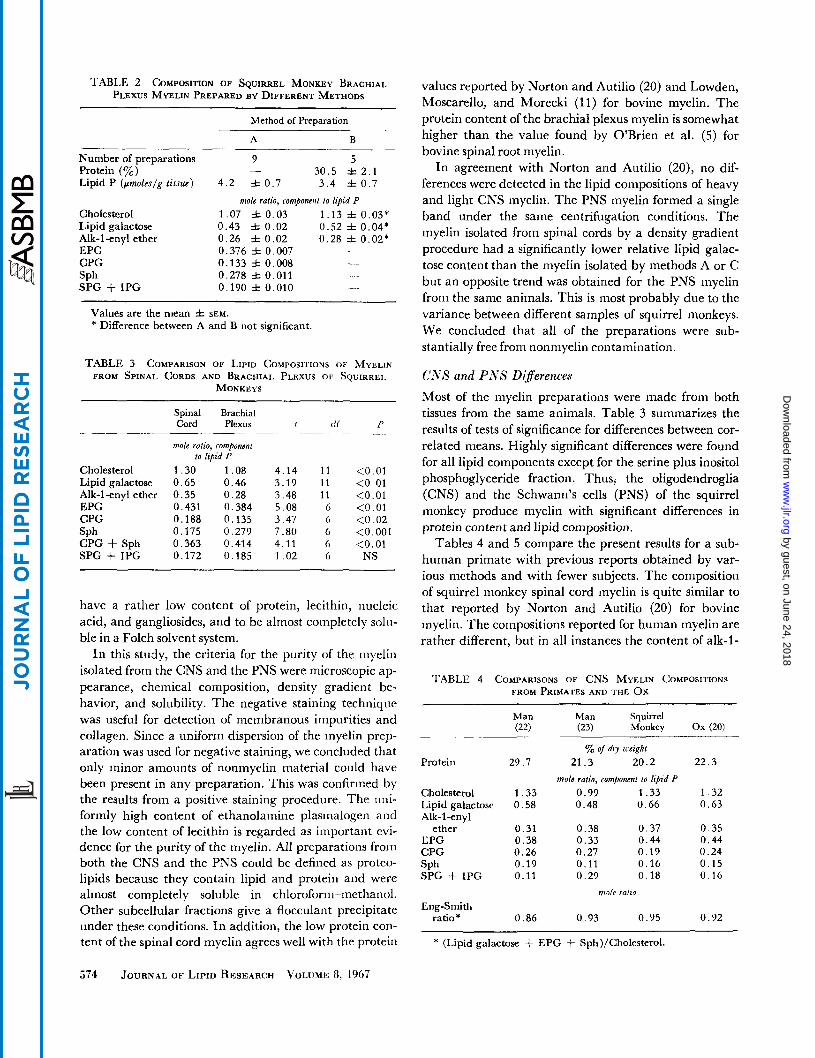
TABLE 2 COMPOSITION OF SQUIRREL MONKEY BRACHIAL PLEXUS MYELIN PREPARED BY DIFFERENT METHODS
Method of Preparation
A B
Number of preparations Protein (yo) Lipid P (pmoleslg tissue)
Cholesterol Lipid galactose Alk-1-enyl ether EPG CPG
SPG + IPG SPh
9 5 30 .5 f 2 . 1
4 . 2 f 0 . 7 3 . 4 & 0 . 7 mole ratio, componcnl lo lipid P
1.07 f 0 . 0 3 1 .13 f 0.03* 0 .43 f 0 .02 0 .52 f 0.04* 0 .26 =t 0 . 0 2 0 .28 f 0 . 0 2 * 0.376 f 0.007 0 .133 =t 0.008 _ _ 0.278 It 0.011 0.190 =t 0.010 - .
-
-
Values are the mean f SEM. * Difference between A and B not significant.
TABLE 3 COMPARISON OF LIPID COMPOSITIONS OF MYELIN FROM SPINAL CORDS AND BRACHIAL PLEXUS OF SQUIRREL
MONKEYS
Spinal Brachial Cord Plexus 1 tl f P
mole ratio, componcnt to lipid P
.- . - ~. -
Cholesterol 1 .30 1 .08 4 . 1 4 1 1 <0.01 Lipid galactose 0 .65 0 .46 3 . 1 9 1 1 <0.01 Alk-1-enyl ether 0 .35 0 .28 3 .48 1 1 <0.01 EPG 0.431 0 .384 5 . 0 8 6 <0.01 CPG 0.188 0.135 3 .47 6 <0 .02 SPh 0.175 0.279 7 .80 6 <0.001 CPG + Sph 0.363 0.414 4.11 6 <0.01 SPG + IPG 0.172 0 . 1 8 5 1 . 0 2 6 NS
have a rather low content of protein, lecithin, nucleic acid, and gangliosides, and to be almost conipletely solii- ble in a Folch solvent system.
In this study, the criteria for the purity of the myelin isolated from the CNS and the PNS were microscopic ap- pearance, chemical composition, density gradient be- havior, and solubility. The negative staining technique was useful for detection of membranous impurities and collagen. Since a uniform dispersion of the myelin prep- aration was used for negative staining, we concluded that only minor amounts of nonmyelin material could have been present in any preparation. This was confirmed by the results from a positive staining procedure. The uni- formly high content of ethanolamine plasmalogen and the low content of lecithin is regarded as important evi- dence for the purity of the myelin. All prcparations from both the CNS and the PNS could be defined as proteo- lipids because they contain lipid and protein and were almost completely soluble in chloroform-methanol. Other subcellular fractions give a flocculant precipitate under these conditions. In addition, the low protein con- tent of the spinal cord myelin agrees well with the protein
values reported by Norton and Autilio (20) and Lowden, Moscarello, and Morecki (11) for bovine myelin. The protein content of the brachial plexus myelin is somewhat higher than the value found by O’Brien et al. ( 5 ) for bovine spinal root myelin.
In agreement with Norton and Autilio (20), no dif- ferences were detected in the lipid compositions of heavy and light CNS myelin. The PNS myelin formed a single band under the same centrifugation conditions. The myelin isolated from spinal cords by a density gradient procedure had a significantly lower relative lipid galac- tose content than the myelin isolated by methods A or C but an opposite trend was obtained for the PNS myelin from the same animals. This is most probably due to the variance between different samples of squirrel monkeys. We concluded that all of the preparations were sub- stantially free from nonmyelin contamination.
CIVS and PlVS DI:fferences
Most of the myelin preparations were made from both tissues from the same animals. Table 3 summarizes the results of tests of significance for differences between cor- related means. Highly significant differences were found for all lipid components except for the serine plus inositol phosphoglyceride fraction. Thus, the oligodendroglia (CNS) and the Schwann’s cells (PNS) of the squirrel monkey produce myelin with significant differences in protein content and lipid composition.
Tables 4 and 5 compare the present results for a sub- human primate with previous reports obtained by var- ious methods and with fewer subjects. The composition of squirrel monkey spinal cord myelin is quite similar to that reported by Norton and Autilio (20) for bovine myelin. The compositions reported for human myelin are rather different, but in all instances the content of alk-l-
‘I’ABLE 4 COMPARISONS OF CNS MYELIN COMPOSITIONS FROM PRIMATES AND THE Ox
Man Man S q u i r r e 1 (22) (23) Monkey Ox (20)
yo o/ dry might Protein 29 .7 21 .3 20 .2 2 2 . 3
Cholesterol Lipid galactose Alk-1-enyl
ether EPG CPG SPh SPG + IPG
mole ratio, component IO @id P 1 . 3 3 0.99 1.33 1 . 3 2 0 .58 0 .48 0 .66 0 . 6 3
0 .31 0 .38 0 .37 0 .35 0 . 3 8 0 .33 0 .44 0 .44 0 .26 0 .27 0 .19 0 .24 0 . 1 9 0.11 0 . 1 6 0 .15 0.11 0 . 2 9 0 . 1 8 0 .16
mole ra/io
Eng-Smith ratio * 0.86 0 . 9 3 0 .95 0 .92
* (Lipid galactose + EPG + Sph)/Cholesterol.
574 JOURNAL OF LIPID RESEARCH VOLUME 8, 1967
by guest, on June 24, 2018w
ww
.jlr.orgD
ownloaded from
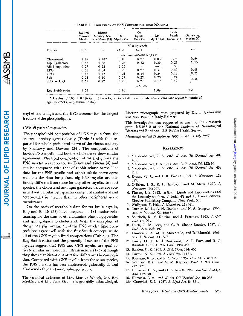
TABLE 5 COMPARISON OF PNS COMPOSITIONS FROM MAMMALS
Squirrel Rhesus ox Rabbit Monkey Monkey Sci- Ox Spinal Rat Sciatic Guinea pig Myelin atic Nerve (24) Myelin (5) Root (5) Myelin (4) Nerve (24) Myelin (4)
yo of dry weight - - - Protein 30.5 - 24.2 33.3
Cholesterol 1.09 1.48* 0.86 0.77 0.83 0.78 0.64 Lipid galactose 0.46 0.38 0.24 0.22 0.33 0.25 1.15 Alk-1-enyl ether 0.27 0.28 0.25 - - 0.30 EPG 0.38 0.34 0.26 0.27 0.37 0.40 0.45 CPG 0.13 0 . 1 3 0.21 0.24 0.24 0.16 0.21
0.28 0.30 0.27 0.22 0.20 SPh
Eng-Smith ratio 1.03 0.90 1.08 >2
mole ratio, compomnt 10 lipid P
-
0.24 {0.34 SPG + IPG 0.19 0.22 0.26 0.27 0.19 0.19
mole rafio
* A value of 0.83 z!= 0.016 (n = 8 ) was found for sciatic nerve lipids from rhesus monkeys at 9 months of age (Horrocks, unpublished data).
enyl ethers is high and the EPG account for the largest fraction of the phospholipids.
P N S Myelin Composition The phospholipid composition of PNS myelin from the squirrel monkey agrees closely (Table 5) with that re- ported for whole peripheral nerve of the rhesus monkey by Sheltawy and Dawson (24). The compositions of bovine PNS myelin and bovine whole nerve show similar agreement. The lipid composition of rat and guinea pig PNS myelin was reported by Evans and Finean (4) and can be compared with that of rabbit sciatic nerve. The data for rat PNS myelin and rabbit sciatic nerve agree well but the data for guinea pig PNS myelin are dis- tinctly different from those for any other myelin. In most species, the cholesterol and lipid galactose values are con- sistent with a relatively greater content of cholesterol and cerebrosides in myelin than in other peripheral nerve membranes.
On the basis of metabolic data for rat brain myelin, Eng and Smith (25) have proposed a 1 : l molar rela- tionship for the sum of ethanolamine phosphoglycerides and sphingolipids to cholesterol. With the exception of the guinea pig myelin, all of the PNS myelin lipid com- positions agree well with the Eng-Smith concept, as do all of the CNS myelin lipid compositions (Table 4). The Eng-Smith ratios and the proteolipid nature of the PNS myelin suggest that PNS and CNS myelin are qualita- tively similar in molecular ultrastructure (1-3) although they show significant quantitative differences in composi- tion. Compared with CNS myelin from the same species, the PNS myelin has less cholesterol, galactolipid, and alk-1 -enyl ether and more sphingomyelin.
The technical assistance of Mrs. Marilyn Waugh, Mr. Roy Meckler, and Mr. John Ozolins is gratefully acknowledged.
Electron micrographs were prepared by Dr. T. Samorajski and Mrs. Paulette Rady-Reimer.
This investigation was supported in part by PHS research grant NB-05510 of the National Institute of Neurological Diseases and Blindness, US. Public Health Service.
Manuscript received 28 September 1966; accepted 5 J u ~ 1967.
1.
2. 3.
4.
5.
6.
7. 8.
9.
10.
11.
12.
13. 14. 15. 16.
17.
18.
REFERENCES
Vandenheuvel, F. A. 1963. J . Am. Oil Chemists’ Soc. 40: 455. Vandenheuvel, F. A. 1965. Ann. N . Y. Acad. Sci. 122: 57. Vandenheuvel, F. A. 1966. J . Am. Oil Chemists’ SOC. 43: 258. Evans, M. J., and J. B. Finean. 1965. J . Neurochem. 12: 729. O’Brien, J. S., E. L. Sampson, and M. Stern. 1967. J. Neurochem . 14: 357. Finean, J. B. 1963. In Brain Lipids and Lipoproteins and the Leucodystrophies. J. Folch-Pi and H. Bauer, editors. Elsevier Publishing Company, New York. 57. Wolfgram, F. 1966. J . Neurochem. 13: 461. Cuzner, M. L., A. N. Davison, and N. A. Gregson. 1965. Ann. N . Y. Acad. Sci. 122: 86. Spurlock, B., V. Kattine, and J. Freeman. 1963. J . Cell Biol. 17: 203. Folch, J., M. Lees, and G. H. Sloane Stanley. 1957. J . Biol. Chem. 226: 497. Lowden, J. A., M. A. Moscarello, and R. Morecki. 1966. Can. J . Biochem. 44: 567. Lowry, 0. H., N. J. Rosebrough, A. L. Farr, and R. J. Randall. 1951. J . Biol. Chem. 193: 265. Bartlett, G. R. 1959. J . Biol. C h m . 234: 466. Carroll, K. K. 1960. J . Lipid Res. 1: 171. Bowman, R. E., and R. C. Wolf. 1962. Clin. C h m . 8: 302. Gottfried, E. L., and M. M. Rapport. 1962. 3. Biol. Chem. 237: 329. Horrocks, L. A., and G. B. Ansell. 1967. Biochim. Biophys. Acta. 137: 90. Horrocks, L. A. 1963. J . Am. Oil Chemists’ SOC. 40: 235.
18a. Gottfried, E. L. 1967. J . Lipid Res. 8 : 321.
HORROCKS P N S and CNS Myelin Lipids 575
by guest, on June 24, 2018w
ww
.jlr.orgD
ownloaded from

19. Ferguson, G. A. 1966. Statistical Analysis in Psychology and Education. McGraw-Hill Book Company, New York. 2nd edition.
20. Norton, W. T., and L. A. Autilio. 1966. J . Neurochem. 13: 213.
Neurochem. 11: 17.
22. Norton, W. T., S. E. Poduslo, and K. Suzuki. 1966. J.
23. O’Brien, J. S., and E. L. Sampson. 1965. J. Lipid Res.
24. Sheltawy, A., and R. M. C. Dawson. 1966. Biochem. J.
25. Eng, L. F., and M. E. Smith. 1966. Lipids. 1: 296.
Neuropathol. Exptl. Neurol. 25: 582.
6: 537.
loo: 12. 21. Autilio, L. A., W. T. Norton, and R. D. Terry. 1964. J .
576 JOURNAL OF LIPID RESEARCH VOLUME 8, 1967
by guest, on June 24, 2018w
ww
.jlr.orgD
ownloaded from