composition of bacterial communities associated … of bacterial communities associated with aurelia...
TRANSCRIPT

Composition of Bacterial Communities Associated with Aurelia auritaChanges with Compartment, Life Stage, and Population
Nancy Weiland-Bräuer,a Sven C. Neulinger,a Nicole Pinnow,a Sven Künzel,b John F. Baines,b,c Ruth A. Schmitza
Institute for General Microbiology, Christian Albrecht University Kiel, Kiel, Germanya; Max Planck Institute for Evolutionary Biology, Plön, Germanyb; Institute forExperimental Medicine, Christian Albrecht University Kiel, Kiel, Germanyc
The scyphozoan Aurelia aurita is recognized as a key player in marine ecosystems and a driver of ecosystem change. It is thusintensely studied to address ecological questions, although its associations with microorganisms remain so far undescribed. Inthe present study, the microbiota associated with A. aurita was visualized with fluorescence in situ hybridization (FISH) analy-sis, and community structure was analyzed with respect to different life stages, compartments, and populations of A. aurita by16S rRNA gene amplicon sequencing. We demonstrate that the composition of the A. aurita microbiota is generally highly dis-tinct from the composition of communities present in ambient water. Comparison of microbial communities from differentdevelopmental stages reveals evidence for life stage-specific community patterns. Significant restructuring of the microbiotaduring strobilation from benthic polyp to planktonic life stages is present, arguing for a restructuring during the course of meta-morphosis. Furthermore, the microbiota present in different compartments of the adult medusa (exumbrella mucus and gastriccavity) display significant differences, indicating body part-specific colonization. A novel Mycoplasma strain was identified inboth compartment-specific microbiota and is most likely present inside the epithelium as indicated by FISH analysis of polyps,indicating potential endosymbiosis. Finally, comparison of polyps of different populations kept under the same controlled labo-ratory conditions in the same ambient water showed population-specific community patterns, most likely due the genetic back-ground of the host. In conclusion, the presented data indicate that the associated microbiota of A. aurita may play importantfunctional roles, e.g., during the life cycle.
Multicellular eukaryotes in marine systems provide diversesurfaces for attachment of and colonization by microbes,
e.g., mucosa-covered epithelia. Bacteria, often present in highnumbers in the surrounding marine environment, tend to colo-nize these surfaces or even invade the surface epithelium. Multi-cellular organisms nowadays are largely recognized as “metaor-ganisms” comprising the macroscopic host and its synergisticinterdependence with bacteria, archaea, and fungi specifically as-sociated with the host (1, 2). Depending on the variety of differentniches provided by the host, which can change according to de-velopmental stage, diet, or other environmental factors, diverseand specific microbial communities can establish themselves withhosts (3). Previous investigations demonstrated that marine in-vertebrates harbor bacteria as stable associates recruited from theocean’s pool of potential colonizers (4–6). A well-studied exampleis the symbiosis between the squid Euprymna scolopes and Vibriofischeri, which is crucial for certain developmental steps of thesquid (7, 8). Additionally, endosymbionts living inside host cellsor in the intracellular space of the host’s tissue are well docu-mented for marine invertebrates, e.g., in hydrothermal vent sys-tems, including the giant tubeworm Riftia pachyptila andassociated chemolithoautotrophic bacteria capable of oxidizinghydrogen sulfide (9). Furthermore, members of the class Molli-cutes are known to be (endo)symbionts of various invertebrates(10–12).
Scyphozoa (jellyfish) represent a class of Cnidaria, which is atthe base of the metazoan tree. They play an important role in themarine ecosystem by substantially affecting the structure of theplanktonic food web (13–15). They release large amounts of nu-trients and dissolved organic matter (16, 17) resulting from theirmetabolic activities, presumably directly stimulating bacterialgrowth and potentially influencing bacterial community compo-
sition (18–20). The cosmopolitan moon jelly Aurelia aurita, likelyrepresenting a collection of species as recently shown by molecularmarkers (21), is one of the most widely distributed scyphozoansand is found in almost all warm and temperate waters of coastalzones worldwide (22–24). Due to its ability to tolerate a wide rangeof environmental conditions (14, 25), especially temperature andsalinity, and its highly diverse food spectrum, it successfully colo-nizes different environments (26) and often causes jellyfishblooms around the world. Such blooms not only impact food webstructures by predation but also play an important role in thedynamics of nutrients and oxygen in planktonic food webs (14).The diphasic life cycle of A. aurita alternates between free-livingpelagic sexual (medusa) and sessile benthic asexual (polyp)phases. After release from sexual reproduction, larvae settle onsuitable seafloor substrata, followed by metamorphosis into thesessile benthic stage, the polyp. Transition from the benthic phase
Received 15 May 2015 Accepted 19 June 2015
Accepted manuscript posted online 26 June 2015
Citation Weiland-Bräuer N, Neulinger SC, Pinnow N, Künzel S, Baines JF, SchmitzRA. 2015. Composition of bacterial communities associated with Aurelia auritachanges with compartment, life stage, and population. Appl Environ Microbiol81:6038 – 6052. doi:10.1128/AEM.01601-15.
Editor: P. D. Schloss
Address correspondence to Ruth A. Schmitz, [email protected].
N.W.-B. and S.C.N. contributed equally to this work.
Supplemental material for this article may be found at http://dx.doi.org/10.1128/AEM.01601-15.
Copyright © 2015, American Society for Microbiology. All Rights Reserved.
doi:10.1128/AEM.01601-15
6038 aem.asm.org September 2015 Volume 81 Number 17Applied and Environmental Microbiology
on June 5, 2019 by guesthttp://aem
.asm.org/
Dow
nloaded from

to the pelagic phase occurs when the juvenile pelagic stage, theephyra, is formed and released by the polyp through the process ofstrobilation (see Fig. S1 in the supplemental material). The me-dusa has a very simple morphology, with two tissue layers: theepidermis, covering the outer body surface, and the gastrodermis,forming the boundary of the gastric cavity. The mesoglea betweenthese tissue layers, consisting mainly of collagen, provides stabilityfor the animal. The prominent oral arms of the medusa harborbrood sacs, where the developing planula larvae are sheltered untilrelease. The polyp exhibits an even simpler anatomy, lacking tis-sues for sexual reproduction. Based on strobilation frequency andthe temperatures required to induce strobilation, different popu-lations of A. aurita can be distinguished, e.g., the arctic boreallineage (Baltic and North Seas) and the temperate Mediterraneanlineage (e.g., Eastern Atlantic-Roscoff). In addition, Schroth et al.showed that limited gene flow among populations around theworld causes cryptic speciation and proposed at least seven differ-ent species of the genus Aurelia based on mitochondrial and nu-clear DNA analyses (16S and internal transcribed spacer 1 [ITS-1]/5.8S rRNA gene) (27). In summary, different A. auritasubpopulations can be correlated with their geographic origin; forexample, the BOR lineage includes Baltic Sea and North Sea spec-imens, whereas the UBI lineage, among others, includes EasternAtlantic-Roscoff specimens. To date, most studies have addressedecological questions (22, 27, 28) as well as the physiology (29–31)and development (32–34) of A. aurita; however, no studies onassociated microbes are available. Consequently, due to its simpleanatomy, evolutionary age, alteration of different life stages, widedistribution, and important role in the marine ecosystem, A. au-rita is an attractive model organism to study the metaorganismconcept (2).
The establishment of next-generation sequencing techniqueshas revolutionized the study of microorganisms in natural envi-ronments (35) and profoundly altered our awareness of microbialecosystems (36, 37). Thus, the present study aimed to analyze themicrobiota associated with different life stages, compartments,and populations of A. aurita by using 16S rRNA gene-based am-plicon sequencing to gain insight into transiently and specificallyassociated bacteria of A. aurita and, ultimately, into interactionsbetween the host and its microbiota. Moreover, in the long term,detailed knowledge of the microbial consortia of scyphozoa suchas A. aurita may provide critical insights for understanding a mi-crobe-dependent life history and its evolutionary consequences.
MATERIALS AND METHODSMedusa sampling. Individual A. aurita (Linnaeus) medusae (mean um-brella diameter, 26 cm) were sampled from one location in the Kiel Bight,Baltic Sea (54°19.4=N, 10°08.5=E) in June 2013 by using a dip net. Theanimals were transported immediately to the laboratory, washed thor-oughly with sterile filtered artificial seawater (ASW) to remove nonasso-ciated microbes, and processed for further analysis. Processing of animalsstarted with sampling of mucus from ex- and subumbrellas by scrapingand sampling the slime. Furthermore, samples of the gastric cavity weretaken with a pipette by penetrating the mouth. Water from the same sitewas collected by bucket sampling, from which reference water sampleswere taken. Water samples were filtered through 0.22-�m polyethersul-fone membrane filters (Millipore Corporation, Bedford, MA), and DNAwas extracted from the respective filters as described below.
Aurelia aurita husbandry. Polyp populations originating from theSouthern English Channel (Roscoff, France) and the North Sea (Helgo-land, Germany) were provided by Thomas Bosch (CAU Kiel) and were
kept in the laboratory under artificial conditions for 10 years. Baltic Seaspecimens were raised from planula larvae originating from medusaecaught in the Kiel Bight in September 2009 and transferred to the labora-tory for rearing under artificial conditions. The polyps were kept in 2-litervessels at 20°C in aerated artificial seawater (Reef Crystals; AquariumSystems, Sarrebourg, France) with a salinity of 30 practical salinity units(PSU). Twice a week, animals were fed freshly hatched Artemia salina, andthe water was changed completely. Feeding of animals was ceased at least4 days prior to the analysis of microbial consortia to exclude contamina-tion by food. Strobilation of Roscoff polyps for the production of ephyraewas induced in the laboratory after reducing the temperature to 8°C for atleast 1 month. A subset of obtained ephyrae continued to develop intojuvenile medusae.
Generation of sterile A. aurita polyps and ephyrae. In order to ob-tain sterile/germfree polyps, polyps were treated with an antibiotic mix-ture present in the water (Provasoli’s antibiotic [AB] mixture with finalconcentrations of 360,000 U/liter penicillin G, 1.5 mg/liter chloramphen-icol, 1.8 mg/liter neomycin, and 9,000 U/liter polymyxin B [all compo-nents from Carl Roth, Karlsruhe, Germany]), kept under the above-men-tioned conditions in sterilized (by filtration through 0.22-�m filters)artificial seawater, and fed antibiotic-treated A. salina. Antibiotic-treatedephyrae originated from AB-treated polyps. The absence of bacteria wasconfirmed by plating of homogenized polyps/ephyrae onto R2A agarplates (Roth, Karlsruhe, Germany). After incubation at 19°C for 5 days,the CFU were determined. An absence of CFU indicated successful anti-biotic treatment. For culture-independent analysis, total DNA was ex-tracted, and the 16S rRNA genes were amplified as described below. Therespective impacts of AB treatment are depicted in Fig. 3 and 4D.
Phylogenetic classification of A. aurita polyps. DNA was extractedfrom 10 single polyps from each respective Roscoff, Baltic Sea, and NorthSea population kept in the laboratory under the same artificial conditionsby using the Wizard genomic purification kit (Promega, Madison, WI,USA). Polyps were homogenized in 480 �l 50 mM EDTA with a pestle andincubated at 37°C for 30 min after the addition of proteinase K (LifeTechnologies, Darmstadt, Germany). The remaining preparation stepswere performed according to the manufacturer’s protocol. DNA tem-plates (10 ng) were subjected to PCR with a 25-�l total volume by using aGoTaq DNA polymerase kit (Promega, Madison, WI, USA). Cycling con-ditions were 30 cycles of 30 s at 95°C, 30 s at 50°C, and 60 s at 72°C.Aurelia-specific mitochondrial 16S rRNA primers (L5=-CTCTTGTAAGGTGAAGCC and H5=-CATAATTCAACATCGAGG) and specific primersencompassing the ITS-1/5.8S rRNA region of nuclear 18S rRNA (F5=-TAACAAGGTTTCCGTAGG and R5=-CTC AGA CAG ACA TGC TCC)were used as described previously by Schroth et al. (27). PCRs were used todetermine the DNA sequences, performed by the sequencing facility at theInstitute of Clinical Molecular Biology (IKMB), University of Kiel, Kiel,Germany. Tree reconstruction was performed with both mitochondrial16S rRNA and nuclear 18S rRNA sequences by using Phylogeny.fr (http://www.phylogeny.fr/) (38, 39) with ClustalW for multiple alignment,Gblocks for alignment curation, and the maximum likelihood/PhyMLmethod for construction of the phylogenetic tree. Previously reportedsequence data from Aurelia populations deposited in GenBank (accessionnumbers AF461398 and AF461399 for 16S rRNA and AF461405 andAF461406 for ITS-1/5.8S rRNA) were used for classification of polyps intoknown subpopulations.
Fluorescence in situ hybridization and microscopic analysis. Polyps(Roscoff population, 6 to 8 replicates) were washed three times with filter-sterilized artificial seawater and relaxed by incubation with 2% urethane(Sigma-Aldrich, Schnelldorf, Germany) for 5 min. Polyps were incubatedin Carnoy fixative (60% [vol/vol] ethanol, 30% [vol/vol] chloroform,10% [vol/vol] glacial acetic acid) for 2 h at 4°C. Fixed samples were dehy-drated three times with 100% ethanol for 5 min each. Semithin sections(0.5 �m) were prepared after several dehydration steps (5 min each, with50%, 75%, and 100% ethanol), embedding in LR White (Sigma-Aldrich),and polymerization of the samples in LR White at high temperature.
Aurelia aurita Microbiota
September 2015 Volume 81 Number 17 aem.asm.org 6039Applied and Environmental Microbiology
on June 5, 2019 by guesthttp://aem
.asm.org/
Dow
nloaded from

Whole polyps and semithin sections were further incubated with 10�g/ml proteinase K (Fermentas, St. Leon-Rot, Germany) and 10 mg/mllysozyme (Roth, Karlsruhe, Germany) in PBT (phosphate-buffered saline[PBS], 1% Tween 80 [pH 7.4]) for 1 h at 37°C. This was followed by twowashing steps with Milli-Q water and further dehydration. Hybridizationwas performed in hybridization buffer (900 mM NaCl, 20 mM Tris-HCl[pH 7.4], various concentrations of formamide [see Table S2 in the sup-plemental material], 0.01% SDS) with the respective probes (workingsolution of 5 ng/�l; Firmicutes probes LGC0354a, -b, and -c were mixed inequimolar proportions) at the specified temperature for 3 h (see Table S2in the supplemental material). Finally, samples were incubated in washingbuffer (80 mM NaCl with 35% formamide or 225 mM NaCl with 20%formamide in hybridization buffer, 20 mM Tris-HCl [pH 7.4], 20 mMEDTA, and 0.01% SDS) for 15 min at a temperature 2°C below the spec-ified temperature and washed with Milli-Q water. Probes with the corre-sponding formamide concentrations in the buffers and incubation tem-peratures are listed in Table S2 in the supplemental material. Forcounterstaining, samples were incubated with SYTO9 (Invitrogen, Darm-stadt, Germany) or 4=,-6-diamidino-2-phenylindole (DAPI) (Roth,Karlsruhe, Germany) at room temperature for 20 min and washed withMilli-Q water. Light microscopy and fluorescence microscopy were per-formed with an Axio Scope microscope and Axio Vision software (Zeiss,Jena, Germany). The entire three-dimensional structure of the polyp andits associated microbiota was recorded by scanning along the depth byusing a TCS SP confocal laser scanning microscope (Leica, Wetzlar, Ger-many) and recording the stacks of cross sections simultaneously at thecorresponding excitation wavelengths. For each field of view, an appro-priate number of optical slices were acquired with a Z-step of 1 �m.Digital image acquisition, postprocessing, analysis of the confocal laserscanning microscopy (CLSM) optical thin sections, three-dimensionalreconstructions, and calculation of bacterial coverage were performedwith the corresponding Leica software (provided for the TCS SP confocallaser scanning microscope). For detection of each bacterial phylum, threeindependent fluorescence in situ hybridization (FISH) experiments wereperformed, and 10 confocal stacks were compared for each independentexperiment. Six polyps for each treatment were used for calculation ofbacterial coverage of polyps.
Nucleic acid isolation and 16S rRNA gene amplicon sequencing.Bacterial genomic DNA of the A. aurita microbiota was isolated with theAquaPure genomic DNA kit (Bio-Rad, Munich, Germany) from 10 mgmucus; 5 mg of the gastric cavity sample; pools of 10 polyps, 20 ephyrae, 1strobila, and 1 juvenile medusa per sample type (all samples representingapproximately the same wet weight); and 1 liter of reference water filteredthrough a 0.22-�m-pore-size membrane filters. DNA isolation was per-formed with 6 replicates for each sample type. PCR amplification of the
V1-V2 hypervariable region of the 16S rRNA gene was performed withprimers listed in Table S3 in the supplemental material. Amplicons weresize checked and purified by using the MinElute gel extraction kit (Qiagen,Hilden, Germany). Purified amplicons were quantified by using theQuant-iT PicoGreen kit (Invitrogen, Darmstadt, Germany), and equalamounts thereof were pooled for subsequent pyrosequencing. Pyrose-quencing was carried out according to the manufacturer’s instructions,using the GS FLX Titanium series kit (sequencing kit XLR70, Pico Titerplate kit 70x75, SV emulsion PCR [emPCR] kit/Lib-A, and MaintenanceWash kit; Roche, Mannheim, Germany). The libraries were sequenced byusing the 454 GS-FLX Titanium sequencer (Roche) at the Max PlanckInstitute for Evolutionary Biology (Plön, Germany).
Study design. In order to reduce practical and computational work-loads, an incomplete block design was conceived, corresponding to fourexperimental setups, which in part made use of the same samples. Thesesetups comprised examination of differences in epibacterial communitycompositions according to the following factors (Table 1): (i) four lifestages (polyp, strobila, ephyra, and juvenile medusa) with individualsfrom the Roscoff population, (ii) compartment types of medusae from theBaltic Sea with microbial samples from the mucus and gastric cavity, (iii)individuals from different populations (Roscoff, Baltic Sea, and NorthSea), and (iv) AB-treated versus untreated control polyps from theRoscoff population. Each factor level encompassed six replicates, four tosix of which yielded read libraries sufficient for analysis.
Sequence processing. Sequence processing was conducted withmothur v1.33.3 (40), as recently described (41). The mothur greengenesreference taxonomy (http://www.mothur.org/w/images/9/9d/Gg_13_5_99.taxonomy.tgz) built from the May 2013 release of the greengenes da-tabase (42) was used for sequence and operational taxonomic unit (OTU)classification. This database was complemented by selected sequences ofLabrenzia spp. (National Center for Biotechnology Information [NCBI]GenBank accession numbers AJ582083, AY628423, and AJ878875). Ref-erence sequences were trimmed to the V1-V2 primer region to improvethe accuracy of the classification (43). A random subset of 3,034 sequencesper sample (corresponding to the smallest number of reads across sam-ples) was generated to eliminate bias due to unequal sampling effort. Atotal of 1,865 OTUs were identified at the 97% similarity level.
Sequence abundance plots. Downstream statistical analysis was per-formed in R v3.1.1 (44) with custom scripts (available upon request).Initial characterization of bacterial community composition was per-formed by summarizing OTU abundances at the genus level, normalizedby the total number of reads per sample. Normalized genus abundanceswere visualized as stacked bar plots.
Alpha diversity analysis. Effective OTU richness (Shannon numberequivalent [1D]) (45, 46) was calculated from the sample-by-OTU table
TABLE 1 Study design
Study type Sample Population Keeping condition(s) Origin
Life stage Polyp Roscoff 10 yr in the laboratory, artificial seawater, temp of 20°C Laboratory stockStrobila Roscoff Artificial seawater, temp of 10°C PolypEphyra Roscoff Artificial seawater, temp of 10°C StrobilaJuvenile medusa Roscoff Artificial seawater, temp of 20°C Ephyra
Compartment Medusa mucus Baltic Sea Freshly hatched Baltic SeaMedusa gastric cavity Baltic Sea Freshly hatched Baltic Sea
Population Polyp Roscoff 10 yr in the laboratory, artificial seawater, temp of 20°C Laboratory stockPolyp Baltic Sea 10 yr in the laboratory, artificial seawater, temp of 20°C Laboratory stockPolyp North Sea 10 yr in the laboratory, artificial seawater, temp of 20°C Laboratory stock
AB treatment Polyp Roscoff 10 yr in the laboratory, artificial seawater, temp of 20°C Laboratory stockEphyra Roscoff Artificial seawater, temp of 10°C PolypAB-treated polyp Roscoff 10 yr in the laboratory, artificial seawater, temp of 20°C Laboratory stockAB-treated ephyra Roscoff Artificial seawater, temp of 10°C AB-treated polyp
Weiland-Bräuer et al.
6040 aem.asm.org September 2015 Volume 81 Number 17Applied and Environmental Microbiology
on June 5, 2019 by guesthttp://aem
.asm.org/
Dow
nloaded from
with the R package vegan (47). Effects of A. aurita life stages, provenance,compartments, and AB treatment in connection with sample type on 1Dwere assessed. For this purpose, 1D was fitted to these experimental factorsin generalized linear models by using the glm function of the R statspackage. In the case of the factor “compartment,” samples of A. auritawere paired (i.e., for each mucus sample, there was a corresponding gas-tric cavity sample from the same medusa), while reference water sampleswere unpaired. In order to analyze both sample types in the same statisti-cal model, all samples had to be treated as unpaired. The stochastic part ofall models was described either by a gamma or an inverse Gaussian distri-bution. Models were compared by means of the Akaike information cri-terion (AIC) and validated according to recommendations described pre-viously (48). As a measure of model performance, the ratio betweenexplained and residual deviances (so-called pseudo-R2) was calculatedand adjusted by sample size and the number of explanatory variablesusing Ezekiel’s formula (49). Since the different experimental setups wereused for some of the same samples, model significances were corrected formultiple testing with the Bonferroni-Holm method (“q values”) (50).Upon significance of the overall model corresponding to a certain exper-iment, pairwise factor-level combinations of the respective descriptor(s)were tested for significance where applicable. Pairwise tests were correctedfor multiple testing by the Benjamini-Hochberg procedure (false discov-ery rate [FDR]) (51).
Beta diversity analysis. The extent of change in OTU compositionexplained by the above-mentioned experimental factors was explored byredundancy analysis (RDA) with Hellinger-transformed OTU abun-dances (41, 52, 53). In general, the RDA model was formulated as “OTUabundances � F,” with factor F being either “life stage,” “provenance,” or“compartment.” The factors “antibiotic” and “type” were tested for inter-action as “OTU abundances � antibiotic � type.”
Models were evaluated with the vegan function rda, followed by ano-va.cca, using 10,000 random permutations. Homoscedasticity waschecked by using the vegan betadisper function, followed by anova.betadis-per to assess significance. Upon determination of the significance of theoverall model, pairwise factor-level combinations were tested for signifi-cance. Corrections for multiple testing across and within models wereconducted as described above for alpha diversity analysis. OTUs signifi-cantly correlated with any axis in a significant RDA model were deter-mined by using the envfit function with 105 permutations, followed byBenjamini-Hochberg correction. In order to reduce the number of tests inthis procedure, OTUs were prefiltered according to their vector lengthscalculated from the corresponding RDA scores (scaling, 1) by profile like-lihood selection (54). OTUs that were significant at an FDR of 10% werefurther subjected to indicator analysis with the multipatt function of the Rpackage indicspecies v1.7.1 (55) with 105 permutations. Indicator OTUs,in analogy to indicator species in the sense described in reference 55, areOTUs that prevail in a certain sample type while being found only infre-quently and irregularly in other sample types compared to the former.Thus, an indicator OTU can be considered “characteristic” for its sampletype.
Phylogenetic tree construction for Mycoplasma associated with A.aurita. DNA sequences were taxonomically classified via a BLAST searchusing the 16S rRNA database of the NCBI. Tree reconstruction was per-formed with full-length 16S rRNA sequences by using Phylogeny.fr (http://www.phylogeny.fr/) (38, 39) with the maximum likelihood method.
Nucleotide sequence accession numbers. Sequences were submittedto the NCBI GenBank database under accession number KP299289. Se-quence data were submitted to the NCBI Sequence Read Archive underaccession number SRP049658.
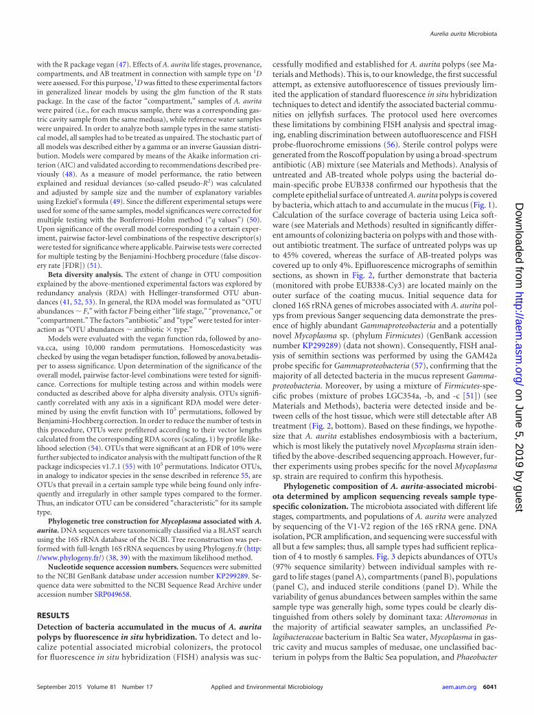
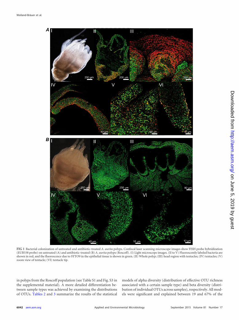
RESULTSDetection of bacteria accumulated in the mucus of A. auritapolyps by fluorescence in situ hybridization. To detect and lo-calize potential associated microbial colonizers, the protocolfor fluorescence in situ hybridization (FISH) analysis was suc-
cessfully modified and established for A. aurita polyps (see Ma-terials and Methods). This is, to our knowledge, the first successfulattempt, as extensive autofluorescence of tissues previously lim-ited the application of standard fluorescence in situ hybridizationtechniques to detect and identify the associated bacterial commu-nities on jellyfish surfaces. The protocol used here overcomesthese limitations by combining FISH analysis and spectral imag-ing, enabling discrimination between autofluorescence and FISHprobe-fluorochrome emissions (56). Sterile control polyps weregenerated from the Roscoff population by using a broad-spectrumantibiotic (AB) mixture (see Materials and Methods). Analysis ofuntreated and AB-treated whole polyps using the bacterial do-main-specific probe EUB338 confirmed our hypothesis that thecomplete epithelial surface of untreated A. aurita polyps is coveredby bacteria, which attach to and accumulate in the mucus (Fig. 1).Calculation of the surface coverage of bacteria using Leica soft-ware (see Materials and Methods) resulted in significantly differ-ent amounts of colonizing bacteria on polyps with and those with-out antibiotic treatment. The surface of untreated polyps was upto 45% covered, whereas the surface of AB-treated polyps wascovered up to only 4%. Epifluorescence micrographs of semithinsections, as shown in Fig. 2, further demonstrate that bacteria(monitored with probe EUB338-Cy3) are located mainly on theouter surface of the coating mucus. Initial sequence data forcloned 16S rRNA genes of microbes associated with A. aurita pol-yps from previous Sanger sequencing data demonstrate the pres-ence of highly abundant Gammaproteobacteria and a potentiallynovel Mycoplasma sp. (phylum Firmicutes) (GenBank accessionnumber KP299289) (data not shown). Consequently, FISH anal-ysis of semithin sections was performed by using the GAM42aprobe specific for Gammaproteobacteria (57), confirming that themajority of all detected bacteria in the mucus represent Gamma-proteobacteria. Moreover, by using a mixture of Firmicutes-spe-cific probes (mixture of probes LGC354a, -b, and -c [51]) (seeMaterials and Methods), bacteria were detected inside and be-tween cells of the host tissue, which were still detectable after ABtreatment (Fig. 2, bottom). Based on these findings, we hypothe-size that A. aurita establishes endosymbiosis with a bacterium,which is most likely the putatively novel Mycoplasma strain iden-tified by the above-described sequencing approach. However, fur-ther experiments using probes specific for the novel Mycoplasmasp. strain are required to confirm this hypothesis.
Phylogenetic composition of A. aurita-associated microbi-ota determined by amplicon sequencing reveals sample type-specific colonization. The microbiota associated with different lifestages, compartments, and populations of A. aurita were analyzedby sequencing of the V1-V2 region of the 16S rRNA gene. DNAisolation, PCR amplification, and sequencing were successful withall but a few samples; thus, all sample types had sufficient replica-tion of 4 to mostly 6 samples. Fig. 3 depicts abundances of OTUs(97% sequence similarity) between individual samples with re-gard to life stages (panel A), compartments (panel B), populations(panel C), and induced sterile conditions (panel D). While thevariability of genus abundances between samples within the samesample type was generally high, some types could be clearly dis-tinguished from others solely by dominant taxa: Alteromonas inthe majority of artificial seawater samples, an unclassified Pe-lagibacteraceae bacterium in Baltic Sea water, Mycoplasma in gas-tric cavity and mucus samples of medusae, one unclassified bac-terium in polyps from the Baltic Sea population, and Phaeobacter
Aurelia aurita Microbiota
September 2015 Volume 81 Number 17 aem.asm.org 6041Applied and Environmental Microbiology
on June 5, 2019 by guesthttp://aem
.asm.org/
Dow
nloaded from
in polyps from the Roscoff population (see Table S1 and Fig. S3 inthe supplemental material). A more detailed differentiation be-tween sample types was achieved by examining the distributionsof OTUs. Tables 2 and 3 summarize the results of the statistical
models of alpha diversity (distribution of effective OTU richnessassociated with a certain sample type) and beta diversity (distri-bution of individual OTUs across samples), respectively. All mod-els were significant and explained between 19 and 67% of the
FIG 1 Bacterial colonization of untreated and antibiotic-treated A. aurita polyps. Confocal laser scanning microscope images show FISH probe hybridization(EUB338 probe) on untreated (A) and antibiotic-treated (B) A. aurita polyps (Roscoff). (I) Light microscope images. (II to V) Fluorescently labeled bacteria areshown in red, and the fluorescence due to SYTO9 in the epithelial tissue is shown in green. (II) Whole polyp; (III) head region with tentacles; (IV) tentacles; (V)zoom view of tentacle; (VI) tentacle tip.
Weiland-Bräuer et al.
6042 aem.asm.org September 2015 Volume 81 Number 17Applied and Environmental Microbiology
on June 5, 2019 by guesthttp://aem
.asm.org/
Dow
nloaded from

variance in the data. Table 4 lists the results for the correspondingpairwise factor-level combinations. The main results of theseanalyses are presented below.
Life stages: significant restructuring of the microbiota dur-ing transition from benthic polyp to planktonic life stages. First,
microbial community patterns of different A. aurita life stages(polyp, strobila, ephyra, and juvenile medusa) of the Roscoff pop-ulation were analyzed. Polyps were kept in artificial seawater asdescribed in Materials and Methods. Strobilation of polyps for theproduction of ephyrae was induced by lowering the temperature,
FIG 2 Bacterial communities associated with A. aurita polyps identified by FISH analysis. Shown are epifluorescence micrographs of bacteria on semithin sections (0.5�m) of untreated (left) and antibiotic-treated (right) A. aurita polyps (Roscoff). Images are overlays of DAPI signals of polyp cell nuclei (blue) and hybridization signalsof the probes NONEUB338 (Cy3) (yellow), EUB338 (Cy3) (yellow), GAM42a (ATTO550) (orange), and LGC345a/b/c (ATTO647N) (red). Bars, 10 �m.
Aurelia aurita Microbiota
September 2015 Volume 81 Number 17 aem.asm.org 6043Applied and Environmental Microbiology
on June 5, 2019 by guesthttp://aem
.asm.org/
Dow
nloaded from
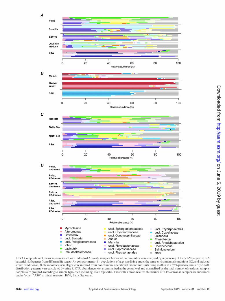
FIG 3 Composition of microbiota associated with individual A. aurita samples. Microbial communities were analyzed by sequencing of the V1-V2 region of 16Sbacterial rRNA genes from different life stages (A), compartments (B), populations of A. aurita living under the same environmental conditions (C), and inducedsterile conditions (D). Taxonomic assemblages were inferred from nonchimeric operational taxonomic units using mothur at a 97% pairwise similarity cutoff;distribution patterns were calculated by using R. OTU abundances were summarized at the genus level and normalized by the total number of reads per sample.Bar plots are grouped according to sample type, each including 4 to 6 replicates. Taxa with a mean relative abundance of �1% across all samples are subsumedunder “other.” ASW, artificial seawater; BSW, Baltic Sea water.
6044 aem.asm.org September 2015 Volume 81 Number 17Applied and Environmental Microbiology
on June 5, 2019 by guesthttp://aem
.asm.org/
Dow
nloaded from

and a subset of the resulting ephyrae continued to develop intojuvenile medusae (see Materials and Methods). Considering themain representatives of associated bacteria, as shown on the genuslevel in Fig. 3A, the dominating colonizers on polyps were Lacinu-trix, Phaeobacter, and Crenothrix; those on strobilating polypswere Crenothrix, Vibrio, and Rhodococcus; those on released ephy-rae were Crenothrix, Pseudoalteromonas, Loktanella, and Salini-bacterium; and those on juvenile medusae were Lacinutrix, La-brenzia, Loktanella, and Vibrio, whereas ambient water (artificialseawater) harbored mainly Alteromonas and Marivita. The effec-tive numbers of OTUs (OTU richness [1D]) were approximatelythe same in all examined life stages, as depicted in Fig. 4A (left),with the only notable difference in 1D being that between juvenilemedusae and ambient water, which, however, is not significantafter correcting for multiple testing (Table 4). In contrast, betadiversity shows a marked difference in community compositionbetween benthic polyps and planktonic life stages (Table 4 andFig. 4A, right), despite A. aurita jellyfish at all life stages being keptunder the same controlled laboratory conditions with the samepool of potential bacterial colonizers in ambient water. Pairwisecomparisons show that the group effect (expressed as explainedvariance [R2
adj] in Table 4) between the polyp group and the otherplanktonic life stages (strobila, ephyra, and juvenile medusa) iscomparable to that between these life stages and ambient water.Differences among the three planktonic life stages, while also sig-nificant, are less pronounced (Table 4 and Fig. 4A). In conclusion,the different life stages are characterized by a specific microbiotadistinct from that of ambient water. Moreover, the stages of thestrobilation event—strobila, ephyra, and juvenile medusa— har-bor a similar microbiota, which significantly differs from themicrobiota of the polyp. In addition, untreated and AB-treatedpolyps and ephyrae were analyzed, illustrating that the polyp-as-sociated microbiota is strongly affected by AB treatment, in agree-ment with data from the FISH analysis (Fig. 1, panels II to VI).Figure 4D illustrates a significant trend of higher effective OTU
richness in untreated samples (polyps and ephyrae) than in AB-treated samples.
Compartment-specific colonization of medusae. Amplifiedbacterial 16S rRNA gene sequences were retrieved from six adultmedusae freshly hatched from the Kiel Bight in the Baltic Sea andstudied with respect to potential differences in microbial compo-sition between the gastric cavity and exumbrellar mucus. As de-picted in Fig. 3B, the microbial composition of ambient water(Baltic Sea) is distinct from the microbiota detected in the gastriccavity and mucus. The main representative species in ambientwater is an unclassified Pelagibacteraceae species. The microbialconsortia associated with the medusa compartments are bothdominated by one unclassified Mycoplasma sequence. This is inagreement with the above-mentioned detection of a Mycoplasmasp. by Sanger sequencing and FISH analysis, most likely indicatingthe presence of a possible Mycoplasma endosymbiont (Fig. 2). Asmall number of sequences in the gastric cavity were identified asthe alphaproteobacterium Labrenzia, and a small number of se-quences in the mucus were identified as the alphaproteobacteriumRoseovarius. Alpha diversity analysis showed significant differ-ences in 1D values, both between the two compartments and be-tween both compartments and Baltic Sea water, with mucus re-vealing a substantially larger variability in OTU richness than theother two samples (Table 4 and Fig. 4B, left). Beta diversity anal-ysis revealed conspicuous compositional differences among thethree groups at the OTU level, with the largest difference beingbetween Baltic Sea water and the gastric cavity, followed by thatbetween Baltic Sea water and mucus (Table 4 and Fig. 4B, right).In contrast, bacterial communities of the gastric cavity and mucusdiffered considerably less from each other. Mucus-associated mi-crobes were more variable in composition than were those fromthe gastric cavity and Baltic Sea water. In summary, adult medusaefrom the Kiel Bight harbor specific bacterial consortia that aredistinct from those in seawater communities, especially in theirgastric cavity.
Population-specific community patterns most likely due thegenetic background of the host. Next, microbial community pat-terns of A. aurita polyps from the Roscoff, Baltic Sea, and NorthSea populations were analyzed. In order to use comparable culti-vation conditions, polyps were kept under identical artificial con-ditions (temperature, salinity, and bacterial colonizer pool inASW) in the laboratory for �10 years. Figure 3C illustrates thatthe major microbes are Lacinutrix, Phaeobacter, and Crenothrixfor the Roscoff population; unclassified Bacteria, unclassified Cox-iellaceae, and unclassified Sphingomonadaceae for the Baltic Seapopulation; and Lacinutrix, unclassified Sphingomonadaceae, andCrenothrix for the North Sea population. The Baltic Sea A. auritapolyps exhibited a markedly lower alpha diversity (�3 effective
TABLE 3 Overview of statistical models in beta diversity analysisa
Factor df R2adj Pmhv value P value q value
Life stage 4 0.39 0.96 0.000 0.000Compartment 2 0.23 0.00 0.000Provenance (population) 3 0.46 0.18 0.000 0.000Antibiotic � type 5 0.58 0.98 0.000 0.000a Hellinger-transformed OTU abundances were fitted to the respective experimentalfactor(s) by RDA. df, degree of freedom; R2
adj, measure of model performance(variance explained by the RDA model, adjusted to sample size and degree of freedom);Pmhv, type I error probability of the test for multivariate homogeneity of variance(homoscedasticity); P value, type I error probability of the respective model; q value,Bonferroni-Holm-corrected P value. Significant results are in boldface type.
TABLE 2 Overview of statistical models in alpha diversity analysisa
Factor Model df Pseudo-R2adj P value q value
Life stage GLM, gamma; link, log 4 0.19 0.010 0.010Compartment GLM, inverse Gaussian; link, ��2 2 0.50 0.000Provenance (population) GLM, gamma; link, log 3 0.60 0.000 0.000Antibiotic GLM, gamma; link, log 1 0.27 0.000 0.001a 1D was fitted to the respective experimental factor(s) given in by a generalized linear model (GLM). Model indicates the generalized linear model based on the stated distribution(gamma/inverse Gaussian) of the response variable with the stated link function between the stochastic (response variable) and systematic model parts. df, degree of freedom;pseudo-R2
adj, measure of model performance (ratio between explained and residual deviances in the generalized linear model), adjusted by using Ezekiel’s formula (49) (seeMaterials and Methods); P value, type I error probability of the respective model; q value, Bonferroni-Holm-corrected P value. Significant results are in boldface type.
Aurelia aurita Microbiota
September 2015 Volume 81 Number 17 aem.asm.org 6045Applied and Environmental Microbiology
on June 5, 2019 by guesthttp://aem
.asm.org/
Dow
nloaded from

OTUs) than that of their North Sea and Roscoff counterparts(�20 and �11 effective OTUs, respectively), while the latter twopopulations did not differ measurably in this respect (Table 4 andFig. 4C, left). This constellation was mirrored in beta diversity,with the strongest group effects being observed with samplesfrom the Baltic Sea population (R2
adj Baltic-North Sea � 61%;R2
adj Baltic-Roscoff � 61%) (Table 4 and Fig. 4C, right). With theexception of North Sea polyps, the alpha diversity was not signif-icantly higher in polyps than in ASW, whereas the difference inOTU composition between polyps and ASW was significant in allcases (Table 4). While OTU composition also differed signifi-cantly between polyps from the North Sea and Roscoff popula-tions, the respective group effect was much lower (R2
adj � 8%)(Table 4). The discrepancy between the Baltic Sea population andother polyp populations could be traced back to two indicatorOTUs specific to the former: OTU 3 (unclassified Bacteria), show-ing 96% similarity to an uncultured marine bacterial clone,“B1G5” (GenBank accession number GU317735), from the meso-pelagic Northeast Pacific Ocean (58), and OTU 19 (unclassifiedCoxiellaceae). The latter belongs to the order Legionellales, whichis commonly known for its many pathogenic species. Given thatthe three populations were kept under identical conditions in thesame artificial seawater for several years, the observed discrepan-cies are remarkable. The findings indicate that specific microbiotaare most likely due to the genetic background (subpopulation) ofthe host population, irrespective of the ambient water (and therespective microbial colonizer pool).
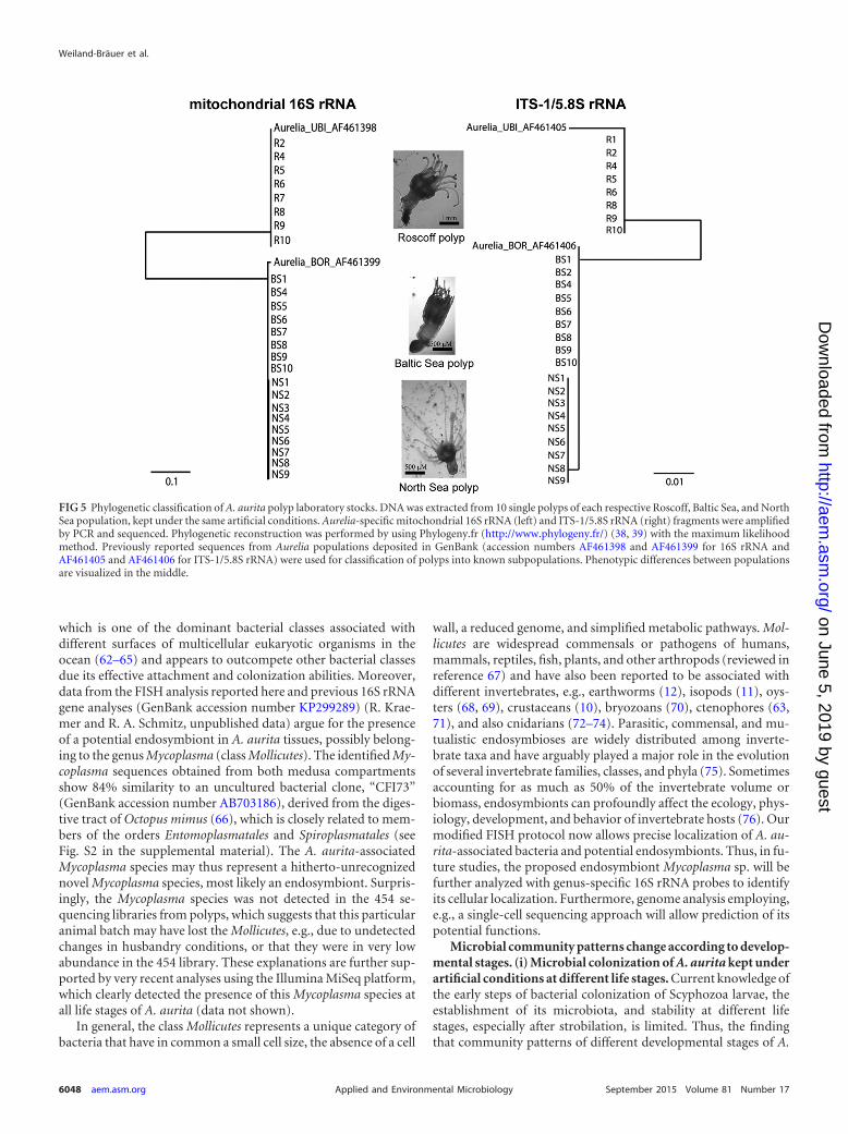
In order to characterize the genetic relationship between hostpopulations, we sequenced mitochondrial and nuclear markers(16S and ITS-1/5.8S rRNA) and classified the samples as describedpreviously by Schroth et al. (27). This analysis revealed that the 10replicate Roscoff polyps belong to the UBI lineage. Baltic Sea andNorth Sea polyps, on the other hand, were classified as belongingto the BOR lineage (Fig. 5). The latter clustered into two differentclades (populations) within the BOR lineage. Based on these data,we conclude that the three different polyp laboratory stocks rep-resent three different subpopulations of A. aurita, all harboring adistinct microbiota according to their genetic background andindependent of the ambient water.
DISCUSSION
Our results clearly demonstrate that A. aurita is colonized by spe-cific bacterial communities that significantly differ in diversity andcomposition from those of ambient water. Below, we discuss themain conclusions, association of microbiota, restructuring of mi-crobiota during the course of transformation from polyp toplanktonic life stages, and host population-specific colonization.
Association of microbes with A. aurita tissues. FISH analysisconfirms that the whole epithelial surface of A. aurita polyps iscovered by bacteria (Fig. 1, panel II). As reported previously forother animals, most interactions between the potential colonizersand A. aurita appear to occur at the mucosal surface, forming abarrier against ambient seawater (59–61). The majority of bacteriaidentified on polyps by FISH analysis are Gammaproteobacteria,
TABLE 4 Results of pairwise tests in alpha and beta diversity analysesa
Factor Comparison
Alpha diversity Beta diversity
Pseudo-R2adj P value FDR R2
adj P value FDR
Life stage Ephyra-juvenile medusa 0.16 0.089 0.177 0.17 0.002 0.003Ephyra-polyp �0.06 0.514 0.570 0.34 0.003 0.004Ephyra-strobila �0.06 0.570 0.570 0.20 0.002 0.003Ephyra-ASW 0.20 0.074 0.177 0.37 0.002 0.003Juvenile medusa-polyp 0.03 0.214 0.356 0.29 0.003 0.004Juvenile medusa-strobila 0.14 0.085 0.177 0.12 0.024 0.024Juvenile medusa-ASW 0.41 0.012 0.120 0.34 0.001 0.003Polyp-strobila 0.02 0.305 0.418 0.35 0.002 0.003Polyp-ASW 0.27 0.035 0.173 0.43 0.002 0.003Strobila-ASW �0.03 0.334 0.418 0.41 0.000 0.001
Compartment Baltic Sea water-gastric cavity 0.69 0.001 0.003 0.88 0.000 0.000Baltic Sea water-mucus 0.17 0.045 0.045 0.46 0.004 0.005Gastric cavity-mucus 0.52 0.020 0.030 0.19 0.029 0.029
Provenance Baltic Sea-North Sea 0.79 0.000 0.000 0.61 0.000 0.000Baltic Sea-Roscoff 0.61 0.001 0.002 0.61 0.002 0.003Baltic Sea-ASW 0.11 0.182 0.182 0.60 0.002 0.003North Sea-Roscoff 0.20 0.082 0.099 0.08 0.031 0.031North Sea-ASW 0.66 0.001 0.002 0.44 0.000 0.000Roscoff-ASW 0.27 0.035 0.052 0.43 0.002 0.003
Antibiotic � type Ephyra (AB)-ephyra (no AB) � � � 0.35 0.002 0.004Polyp (AB)-polyp (no AB) � � � 0.46 0.000 0.000ASW (AB)-ASW (no AB) � � � 0.24 0.018 0.018Ephyra (AB)-polyp (no AB) � � � 0.45 0.000 0.000Ephyra (AB)-ASW (no AB) � � � 0.42 0.004 0.006Polyp (AB)-ASW (no AB) � � � 0.40 0.005 0.006
a Tests were conducted for the pairs of factor levels or factor-level combinations stated in the “Comparison” column, as shown in Tables 1 and 2 for alpha and beta diversityanalyses, respectively. FDR, Benjamini-Hochberg-corrected P value (false discovery rate). “�” denotes values that were not determined. Results significant at the 5% level (both Pvalues and FDRs) are in boldface type.
Weiland-Bräuer et al.
6046 aem.asm.org September 2015 Volume 81 Number 17Applied and Environmental Microbiology
on June 5, 2019 by guesthttp://aem
.asm.org/
Dow
nloaded from

FIG 4 Distribution of effective OTU richnesses of bacterial communities associated with A. aurita and RDA models of Hellinger-transformed OTU abundances.(Left) Distribution of effective OTU richnesses of bacterial communities associated with A. aurita life stages (A), A. aurita medusa compartments (B), popula-tions (C), and induced sterile conditions (D). Box plots mark the medians (bold lines), interquartile ranges (colored boxes), outliers �1.5 times the interquartilerange from the box (whiskers), and outliers beyond (circles). Square brackets denote pairwise comparisons with P values of between 5% and 1% (*), between 1%and 0.1% (**), and between 0.1% and 0.01% (***). (Right) Graphical representation (distance plots) of the RDA model of Hellinger-transformed OTUabundances. Each point represents the whole microbial community of an individual sample, as mentioned above for panels A to C. Groups of related samplepoints are framed by polygons filled with the corresponding color to elucidate the distribution and variability of sample groups in the ordination space.
September 2015 Volume 81 Number 17 aem.asm.org 6047Applied and Environmental Microbiology
on June 5, 2019 by guesthttp://aem
.asm.org/
Dow
nloaded from
which is one of the dominant bacterial classes associated withdifferent surfaces of multicellular eukaryotic organisms in theocean (62–65) and appears to outcompete other bacterial classesdue its effective attachment and colonization abilities. Moreover,data from the FISH analysis reported here and previous 16S rRNAgene analyses (GenBank accession number KP299289) (R. Krae-mer and R. A. Schmitz, unpublished data) argue for the presenceof a potential endosymbiont in A. aurita tissues, possibly belong-ing to the genus Mycoplasma (class Mollicutes). The identified My-coplasma sequences obtained from both medusa compartmentsshow 84% similarity to an uncultured bacterial clone, “CFI73”(GenBank accession number AB703186), derived from the diges-tive tract of Octopus mimus (66), which is closely related to mem-bers of the orders Entomoplasmatales and Spiroplasmatales (seeFig. S2 in the supplemental material). The A. aurita-associatedMycoplasma species may thus represent a hitherto-unrecognizednovel Mycoplasma species, most likely an endosymbiont. Surpris-ingly, the Mycoplasma species was not detected in the 454 se-quencing libraries from polyps, which suggests that this particularanimal batch may have lost the Mollicutes, e.g., due to undetectedchanges in husbandry conditions, or that they were in very lowabundance in the 454 library. These explanations are further sup-ported by very recent analyses using the Illumina MiSeq platform,which clearly detected the presence of this Mycoplasma species atall life stages of A. aurita (data not shown).
In general, the class Mollicutes represents a unique category ofbacteria that have in common a small cell size, the absence of a cell
wall, a reduced genome, and simplified metabolic pathways. Mol-licutes are widespread commensals or pathogens of humans,mammals, reptiles, fish, plants, and other arthropods (reviewed inreference 67) and have also been reported to be associated withdifferent invertebrates, e.g., earthworms (12), isopods (11), oys-ters (68, 69), crustaceans (10), bryozoans (70), ctenophores (63,71), and also cnidarians (72–74). Parasitic, commensal, and mu-tualistic endosymbioses are widely distributed among inverte-brate taxa and have arguably played a major role in the evolutionof several invertebrate families, classes, and phyla (75). Sometimesaccounting for as much as 50% of the invertebrate volume orbiomass, endosymbionts can profoundly affect the ecology, phys-iology, development, and behavior of invertebrate hosts (76). Ourmodified FISH protocol now allows precise localization of A. au-rita-associated bacteria and potential endosymbionts. Thus, in fu-ture studies, the proposed endosymbiont Mycoplasma sp. will befurther analyzed with genus-specific 16S rRNA probes to identifyits cellular localization. Furthermore, genome analysis employing,e.g., a single-cell sequencing approach will allow prediction of itspotential functions.
Microbial community patterns change according to develop-mental stages. (i) Microbial colonization of A. aurita kept underartificial conditions at different life stages. Current knowledge ofthe early steps of bacterial colonization of Scyphozoa larvae, theestablishment of its microbiota, and stability at different lifestages, especially after strobilation, is limited. Thus, the findingthat community patterns of different developmental stages of A.
FIG 5 Phylogenetic classification of A. aurita polyp laboratory stocks. DNA was extracted from 10 single polyps of each respective Roscoff, Baltic Sea, and NorthSea population, kept under the same artificial conditions. Aurelia-specific mitochondrial 16S rRNA (left) and ITS-1/5.8S rRNA (right) fragments were amplifiedby PCR and sequenced. Phylogenetic reconstruction was performed by using Phylogeny.fr (http://www.phylogeny.fr/) (38, 39) with the maximum likelihoodmethod. Previously reported sequences from Aurelia populations deposited in GenBank (accession numbers AF461398 and AF461399 for 16S rRNA andAF461405 and AF461406 for ITS-1/5.8S rRNA) were used for classification of polyps into known subpopulations. Phenotypic differences between populationsare visualized in the middle.
Weiland-Bräuer et al.
6048 aem.asm.org September 2015 Volume 81 Number 17Applied and Environmental Microbiology
on June 5, 2019 by guesthttp://aem
.asm.org/
Dow
nloaded from

aurita differ raises the question of the functional role of associatedmicrobiota in general and concerning development. The fact thatA. aurita jellyfish at stages of strobilation harbor similar microbi-ota but differ significantly in their community patterns comparedto those of polyps argues that the A. aurita-associated microbiotaundergoes significant restructuring during the transition from thebenthic and immortal to the planktonic mortal life phases of theirhost. Moreover, polyps also show the highest number of indicatorOTUs compared to those at other life stages. This further supportsthe idea that polyps harbor a specific bacterial community of highdiversity, potentially essential for their sessile lifestyle (e.g., pro-viding vitamins, amino acids, or secondary metabolites) and pos-sibly important for the initiation of later developmental stages(e.g., strobilation initiation) but less important after the transitionto planktonic stages. Thus, restructuring of microbial communi-ties during strobilation may represent a life cycle strategy of A.aurita, where certain bacterial communities include memberswith specific (metabolic) functions important for specific devel-opmental stages of the host. On the other hand, it is also possiblethat animal development shapes a new microbiota due to changesin the surface architecture or the production of host compounds(e.g., differential expression patterns of antimicrobial peptidesduring different life stages). In the long term, detailed knowledgeon the composition of bacteria associated with different life stagesof A. aurita might provide insight into a microbiota-dependentlife history and its evolutionary consequences. Moreover, the roleof the associated microbiota might also be important to under-stand and control reproductive mechanisms to restrict jellyfishblooms. In this respect, it was recently demonstrated that a water-soluble membrane-diffusible molecule (an indole derivative) issignificantly involved in the induction of A. aurita proliferation(77). Since it is currently not clear whether the host exclusivelyproduces the effector molecule, it is attractive to speculate thatsimilar or antagonistic molecules might be synthesized by the as-sociated microbiota. Identification of such potential strobilationantagonists might facilitate the control of jellyfish blooms.
In regard to proposed functions of the microbiota during a lifecycle, it was previously reported that bacteria are important for thelarval settlement processes of most marine invertebrates (78),such as sponges (79), cnidarians (80), ascidians (81), and bryozo-ans (82). In agreement with these findings, no settlement of plan-ulae of the scyphozoans A. aurita (80) and Cassiopea andromeda(83) or the hydrozoan Hydractinia echinata (84) occurred in theabsence of microbes. However, the presence of bacteria inducedthe settlement and metamorphosis of each of these animals (85).
(ii) Microbial colonization of compartments of medusaecaught in the Baltic Sea. Living medusae were caught in the BalticSea to analyze the community patterns of two compartments, theexumbrella mucus and gastric cavity. Significant differencesamong the two studied compartments and Baltic Sea water wereidentified. Furthermore, the composition of the mucus-associatedmicrobiota was more variable than those of the gastric cavity andBaltic Sea water. Overall, adult medusae from the Kiel Bight ap-parently harbor bacterial consortia that are distinct from seawatercommunities, especially in their gastric cavity. Bacterial consortiaassociated with the animal’s outer surface are most likely moreinfluenced by the surrounding microbial community of ambientwater and external disturbance. This is likely the reason for thehigh similarity of the exumbrella mucus samples to the seawater
consortia and their high compositional variability compared tothat of gastric cavity-associated bacteria.
Provenance matters: population-specific community pat-terns. In this study, polyps of the Roscoff, North Sea, and BalticSea populations originating from geographically distinct regionswere kept under the same controlled artificial conditions in thelaboratory for �10 years (common garden experiment). Phyloge-netic analyses revealed a specific microbiota for each populationdistinct from and irrespective of that of the ambient water (Fig. 3Cand 4C), arguing for a significant influence of genetic background.The molecular phylogeny of A. aurita has been studied exten-sively, based on nuclear internal transcribed spacer 1 (ITS-1) andmitochondrial cytochrome c subunit I (COI), which led to taxo-nomic revisions. For example, Schroth et al. (27) suggested histor-ical speciation events and reconstructed the taxonomic classifica-tion of the genus Aurelia to at least seven different species. Dawsonand colleagues (21, 86) proposed a taxonomic system combiningmacromorphology and ITS-1, in addition to partial COI gene se-quences. Altogether, the global phylogeny of the genus Aureliareveals at least 16 phylogenetic branches, including seven popula-tions of A. aurita, and most of them appear to be regionally re-stricted.
The largest differences in microbial composition among thepolyp populations studied in this report were found between theNorth Sea/Roscoff and Baltic Sea populations. Phylogenetic clas-sification based on mitochondrial and nuclear DNA data revealedthree different subpopulations corresponding to the geographicoccurrence of natural A. aurita populations. North Sea and BalticSea polyps were more similar and thus clustered. This cluster(BOR lineage) deeply branched from the Roscoff polyps (UBIlineage) (Fig. 5). Of note, the differential subpopulations do notnecessarily match the phenotypic grouping of the populations.The two populations of the North Sea and Baltic Sea are geneti-cally unified by one lineage (BOR), but the phenotypic differencesare significant (Fig. 5). Baltic Sea specimens have much largerpolyps. North Sea and Roscoff specimens are substantially smallerthan those from the Baltic Sea and phenotypically more similar,although they differ genetically. Overall, our findings indicate thata correlation between the composition of the microbiota, geneticbackground, and phenotype of the host should be taken into ac-count. In nature, both host genetics as well as environmental fac-tors are likely important in controlling the acquisition and stabil-ity of a healthy microbiota. Indeed, several animal-bacteriumstudies show that the three components environment, host genet-ics, and microbiome are important to maintain homeostasis in thehost (87–89).
In conclusion, it appears that A. aurita-associated microbialcommunities are indeed unique and sample type specific (com-partment, life stage, and population). Taking the metaorganismconcept into account, we hypothesize that these specific microbi-ota serve numerous important functions, including contributingto the development of different life stages.
ACKNOWLEDGMENTS
This work was financially supported by the Federal Ministry of Educationand Research (BMBF) (ChemBiofilm Cluster of the GenoMik-Transfernetwork).
Polyp populations were provided by the work group of T. C. G. Bosch(Institute of Zoology, Christian Albrecht University Kiel). We thank the
Aurelia aurita Microbiota
September 2015 Volume 81 Number 17 aem.asm.org 6049Applied and Environmental Microbiology
on June 5, 2019 by guesthttp://aem
.asm.org/
Dow
nloaded from

staff of the Centre of Microscopy at Christian Albrecht University Kiel fortheir assistance.
REFERENCES1. Bosch TC, McFall-Ngai MJ. 2011. Metaorganisms as the new frontier.
Zoology (Jena) 114:185–190. http://dx.doi.org/10.1016/j.zool.2011.04.001.
2. McFall-Ngai M, Hadfield MG, Bosch TCG, Carey HV, Domazet-LosoT, Douglas AE, Dubilier N, Eberl G, Fukami T, Gilbert SF. 2013.Animals in a bacterial world, a new imperative for the life sciences. ProcNatl Acad Sci U S A 110:3229 –3236. http://dx.doi.org/10.1073/pnas.1218525110.
3. Zilber-Rosenberg I, Rosenberg E. 2008. Role of microorganisms in theevolution of animals and plants: the hologenome theory of evolution.FEMS Microbiol Rev 32:723–735. http://dx.doi.org/10.1111/j.1574-6976.2008.00123.x.
4. Dinasquet J, Granhag L, Riemann L. 2012. Stimulated bacterioplanktongrowth and selection for certain bacterial taxa in the vicinity of the cteno-phore Mnemiopsis leidyi. Front Microbiol 3:302. http://dx.doi.org/10.3389/fmicb.2012.00302.
5. Perez-Matos AE, Rosado W, Govind NS. 2007. Bacterial diversity asso-ciated with the Caribbean tunicate Ecteinascidia turbinata. Antonie VanLeeuwenhoek 92:155–164. http://dx.doi.org/10.1007/s10482-007-9143-9.
6. Piskorska M, Smith G, Weil E. 2007. Bacteria associated with the coralEchinopora lamellosa (Esper 1795) in the Indian Ocean-Zanzibar region.Afr J Environ Sci Technol 1:93–98.
7. Koch EJ, Miyashiro T, McFall-Ngai MJ, Ruby EG. 2014. Features gov-erning symbiont persistence in the squid-vibrio association. Mol Ecol 23:1624 –1634. http://dx.doi.org/10.1111/mec.12474.
8. McFall-Ngai MJ. 2014. The importance of microbes in animal develop-ment: lessons from the squid-vibrio symbiosis. Annu Rev Microbiol 68:177–194. http://dx.doi.org/10.1146/annurev-micro-091313-103654.
9. Cary SC, Cottrell MT, Stein JL, Camacho F, Desbruyeres D. 1997.Molecular identification and localization of filamentous symbiotic bacte-ria associated with the hydrothermal vent annelid Alvinella pompejana.Appl Environ Microbiol 63:1124 –1130.
10. Bi K, Huang H, Gu W, Wang J, Wang W. 2008. Phylogenetic analysis ofspiroplasmas from three freshwater crustaceans (Eriocheir sinensis, Pro-cambarus clarkia and Panaeus vannamei) in China. J Invertebr Pathol 99:57– 65. http://dx.doi.org/10.1016/j.jip.2008.06.008.
11. Fraune S, Zimmer M. 2008. Host-specificity of environmentally trans-mitted Mycoplasma-like isopod symbionts. Environ Microbiol 10:2497–2504. http://dx.doi.org/10.1111/j.1462-2920.2008.01672.x.
12. Nechitaylo TY, Timmis KN, Golyshin PN. 2009. ‘Candidatus Lumbri-cincola’, a novel lineage of uncultured Mollicutes from earthworms offamily Lumbricidae. Environ Microbiol 11:1016 –1026. http://dx.doi.org/10.1111/j.1462-2920.2008.01837.x.
13. Brodeur RD, Sugisaki H, Hunt GL, Jr. 2002 Increases in jellyfish biomassin the Bering Sea: implications for the ecosystem. Mar Ecol Prog Ser 233:89 –103. http://dx.doi.org/10.3354/meps233089.
14. Purcell JE. 2005. Climate effects on formation of jellyfish and ctenophoreblooms: a review. J Mar Biol Assoc UK 85:461– 476. http://dx.doi.org/10.1017/S0025315405011409.
15. Sommer U, Lengfellner K. 2008. Climate change and the timing, magni-tude, and composition of the phytoplankton spring bloom. Glob ChangBiol 14:1199 –1208. http://dx.doi.org/10.1111/j.1365-2486.2008.01571.x.
16. Nemazie DA, Purcell JE, Glibert PM. 1993. Ammonium excretion bygelationous zooplankton and their contribution to the ammonium re-quirements of microplankton in Chesapeake Bay. Mar Biol 116:451– 458.http://dx.doi.org/10.1007/BF00350062.
17. Schneider G, Behrends G. 1998. Top-down control in a neritic planktonsystem by Aurelia aurita medusae—a summary. Ophelia 48:71– 82. http://dx.doi.org/10.1080/00785236.1998.10428677.
18. Blanchet M, Pringault O, Bouvy M, Catala P, Oriol L, Caparros J,Ortega-Retuerta E, Intertaglia L, West N, Agis M. 20 November 2014.Changes in bacterial community metabolism and composition during thedegradation of dissolved organic matter from the jellyfish Aurelia aurita ina Mediterranean coastal lagoon. Environ Sci Pollut Res Int http://dx.doi.org/10.1007/s11356-014-3848-x.
19. Tinta T, Kogovsek T, Malej A, Turk V, Kirchman DL. 2012. Jellyfishmodulate bacterial dynamic and community structure. PLoS One7:e39274. http://dx.doi.org/10.1371/journal.pone.0039274.
20. Tinta T, Malej A, Kos M, Turk V. 2010. Degradation of the Adriaticmedusa Aurelia sp. by ambient bacteria. Hydrobiologia 645:179 –191.http://dx.doi.org/10.1007/s10750-010-0223-x.
21. Dawson MN, Sen Gupta A, England MH. 2005. Coupled biophysicalglobal ocean model and molecular genetic analyses identify multiple in-troductions of cryptogenic species. Proc Natl Acad Sci U S A 102:11968 –11973. http://dx.doi.org/10.1073/pnas.0503811102.
22. Dawson MN, Jacobs DK. 2001. Molecular evidence for cryptic species ofAurelia aurita (Cnidaria, Scyphozoa). Biol Bull 200:92–96. http://dx.doi.org/10.2307/1543089.
23. Gröndahl F. 1989. Evidence of gregarious settlement of planula larvae ofthe scyphozoan Aurelia aurita: an experimental study. Mar Ecol Prog Ser56:119 –125. http://dx.doi.org/10.3354/meps056119.
24. Mutlu E. 2001. Distribution and abundance of moon jellyfish (Aureliaaurita) and its zooplankton food in the Black Sea. Mar Biol 138:329 –339.http://dx.doi.org/10.1007/s002270000459.
25. Lucas CH. 2001. Reproduction and life history strategies of the commonjellyfish, Aurelia aurita, in relation to its ambient environment. Hydrobio-logia 451:229 –246. http://dx.doi.org/10.1023/A:1011836326717.
26. Greenberg N, Garthwaite RL, Potts DC. 1996. Allozyme and mor-phological evidence for a newly introduced species of Aurelia in SanFrancisco Bay, California. Mar Biol 125:401– 410. http://dx.doi.org/10.1007/BF00346320.
27. Schroth W, Jarms G, Streit B, Schierwater B. 2002. Speciation andphylogeography in the cosmopolitan marine moon jelly, Aurelia sp. BMCEvol Biol 2:1. http://dx.doi.org/10.1186/1471-2148-2-1.
28. Watanabe T, Ishii H. 2001. In situ estimation of ephyrae liberated frompolyps of Aurelia aurita using settling plates in Tokyo Bay, Japan. Hydro-biologia 451:247–258. http://dx.doi.org/10.1023/A:1011856929443.
29. Berking S, Czech N, Gerharz M, Herrmann K, Hoffmann U, Raifer H,Sekul G, Siefker B, Sommerei A, Vedder F. 2005. A newly discoveredoxidant defence system and its involvement in the development of Aureliaaurita (Scyphozoa, Cnidaria): reactive oxygen species and elemental io-dine control medusa formation. Int J Dev Biol 49:969 –976. http://dx.doi.org/10.1387/ijdb.052024sb.
30. Hansson LJ. 1997. Effect of temperature on growth rate of Aurelia aurita(Cnidaria, Scyphozoa) from Gullmarsfjorden, Sweden. Mar Ecol Prog Ser161:145–153. http://dx.doi.org/10.3354/meps161145.
31. Lucas CH. 1994. Biochemical composition of Aurelia aurita in relation toage and sexual maturity. J Exp Mar Biol Ecol 183:179 –192. http://dx.doi.org/10.1016/0022-0981(94)90086-8.
32. Berking S, Herrmann K. 2007. Compartments in Scyphozoa. Int J DevBiol 51:221–228. http://dx.doi.org/10.1387/ijdb.062215sb.
33. Ishii H, Takagi AI. 2003. Development time of planula larvae on the oralarms of the scyphomedusa Aurelia aurita. J Plankton Res 25:1447–1450.http://dx.doi.org/10.1093/plankt/fbg094.
34. Yuan D, Nakanishi N, Jacobs DK, Hartenstein V. 2008. Embryonicdevelopment and metamorphosis of the scyphozoan Aurelia. Dev GenesEvol 218:525–539. http://dx.doi.org/10.1007/s00427-008-0254-8.
35. Metzker ML. 2010. Sequencing technologies—the next generation. NatRev Genet 11:31– 46. http://dx.doi.org/10.1038/nrg2626.
36. Lee OO, Wang Y, Yang J, Lafi FF, Al-Suwailem A, Qian PY. 2011.Pyrosequencing reveals highly diverse and species-specific microbial com-munities in sponges from the Red Sea. ISME J 5:650 – 664. http://dx.doi.org/10.1038/ismej.2010.165.
37. Webster NS, Taylor MW, Behnam F, Lucker S, Rattei T, Whalan S,Horn M, Wagner M. 2010. Deep sequencing reveals exceptional di-versity and modes of transmission for bacterial sponge symbionts. En-viron Microbiol 12:2070 –2082. http://dx.doi.org/10.1111/j.1462-2920.2009.02065.x.
38. Dereeper A, Audic S, Claverie J-M, Blanc G. 2010. BLAST-EXPLORERhelps you building datasets for phylogenetic analysis. BMC Evol Biol 10:8.http://dx.doi.org/10.1186/1471-2148-10-8.
39. Dereeper A, Guignon V, Blanc G, Audic S, Buffet S, Chevenet F,Dufayard JF, Guindon S, Lefort V, Lescot M, Claverie JM, GascuelO. 2008. Phylogeny.fr: robust phylogenetic analysis for the non-specialist. Nucleic Acids Res 36:W465–W469. http://dx.doi.org/10.1093/nar/gkn180.
40. Schloss PD, Westcott SL, Ryabin T, Hall JR, Hartmann M, Hollister EB,Lesniewski RA, Oakley BB, Parks DH, Robinson CJ, Sahl JW, Stres B,Thallinger GG, Van Horn DJ, Weber CF. 2009. Introducing mothur:open-source, platform-independent, community-supported software for
Weiland-Bräuer et al.
6050 aem.asm.org September 2015 Volume 81 Number 17Applied and Environmental Microbiology
on June 5, 2019 by guesthttp://aem
.asm.org/
Dow
nloaded from
describing and comparing microbial communities. Appl Environ Micro-biol 75:7537–7541. http://dx.doi.org/10.1128/AEM.01541-09.
41. Langfeldt D, Neulinger SC, Heuer W, Staufenbiel I, Kunzel S, Baines JF,Eberhard J, Schmitz RA. 2014. Composition of microbial oral biofilmsduring maturation in young healthy adults. PLoS One 9:e87449. http://dx.doi.org/10.1371/journal.pone.0087449.
42. DeSantis TZ, Hugenholtz P, Larsen N, Rojas M, Brodie EL, Keller K,Huber T, Dalevi D, Hu P, Andersen GL. 2006. Greengenes, a chimera-checked 16S rRNA gene database and workbench compatible with ARB.Appl Environ Microbiol 72:5069 –5072. http://dx.doi.org/10.1128/AEM.03006-05.
43. Werner JJ, Koren O, Hugenholtz P, DeSantis TZ, Walters WA, Capo-raso JG, Angenent LT, Knight R, Ley RE. 2012. Impact of training sets onclassification of high-throughput bacterial 16s rRNA gene surveys. ISME J6:94 –103. http://dx.doi.org/10.1038/ismej.2011.82.
44. R Core Team. 2014. R package v3.1.1. R Foundation for Statistical Com-puting, Vienna, Austria.
45. Jost L. 2006. Entropy and diversity. Oikos 113:363–375. http://dx.doi.org/10.1111/j.2006.0030-1299.14714.x.
46. Jost L. 2007. Partitioning diversity into independent alpha and beta com-ponents. Ecology 88:2427–2439. http://dx.doi.org/10.1890/06-1736.1.
47. Oksanen J, Blanchet FG, Kindt R, Legendre P, Minchin PR, O=Hara RB,Simpson GL, Solymos P, Stevens MHH, Wagner H. 2013. Package‘vegan’. R Package version 254, p 20 –28. R Foundation for StatisticalComputing, Vienna, Austria.
48. Zuur AF, Ieno EN, Walker NJ, Saveliev AA, Smith GM. 2009. Mixedeffects models and extensions in ecology with R. Springer, New York, NY.
49. Ezekiel M. 1930. Methods of correlational analysis. Wiley, New York, NY.50. Holm S. 1979. A simple sequentially rejective multiple test procedure.
Scand J Stat 6:65–70.51. Benjamini Y, Hochberg Y. 1995. Controlling the false discovery rate: a
practical and powerful approach to multiple testing. J R Stat Soc Series BStat Methodol 57:289 –300.
52. Stratil SB, Neulinger SC, Knecht H, Friedrichs AK, Wahl M. 2014.Salinity affects compositional traits of epibacterial communities on thebrown macroalga Fucus vesiculosus. FEMS Microbiol Ecol 88:272–279.http://dx.doi.org/10.1111/1574-6941.12292.
53. Stratil SB, Neulinger SC, Knecht H, Friedrichs AK, Wahl M. 2013.Temperature-driven shifts in the epibiotic bacterial community compo-sition of the brown macroalga Fucus vesiculosus. Microbiologyopen2:338 –349. http://dx.doi.org/10.1002/mbo3.79.
54. Zhu M, Ghodsi A. 2006. Automatic dimensionality selection from thescree plot via the use of profile likelihood. Comput Stat Data Anal 51:918 –930. http://dx.doi.org/10.1016/j.csda.2005.09.010.
55. De Cáceres M, Legendre P. 2009. Associations between species andgroups of sites: indices and statistical inference. Ecology 90:3566 –3574.http://dx.doi.org/10.1890/08-1823.1.
56. Ainsworth TD, Fine M, Blackall LL, Hoegh-Guldberg O. 2006. Fluores-cence in situ hybridization and spectral imaging of coral-associated bac-terial communities. Appl Environ Microbiol 72:3016 –3020. http://dx.doi.org/10.1128/AEM.72.4.3016-3020.2006.
57. Wagner M, Amann R, Lemmer H, Schleifer KH. 1993. Probing activatedsludge with oligonucleotides specific for proteobacteria: inadequacy ofculture-dependent methods for describing microbial community struc-ture. Appl Environ Microbiol 59:1520 –1525.
58. Gao W, Shi X, Wu J, Jin Y, Zhang W, Meldrum DR. 2011. Phylogeneticand gene expression analysis of cyanobacteria and diatoms in the twilightwaters of the temperate northeast Pacific Ocean. Microb Ecol 62:765–775.http://dx.doi.org/10.1007/s00248-011-9891-y.
59. Daniels CA, Zeifman A, Heym K, Ritchie KB, Watson CA, Berzins I,Breitbart M. 2011. Spatial heterogeneity of bacterial communities in themucus of Montastraea annularis. Mar Ecol Prog Ser 426:29 – 40. http://dx.doi.org/10.3354/meps09024.
60. Mullen KM, Peters EC, Harvell CD. 2004. Coral resistance to disease, p377–399. In Rosenberg E, Loya Y (ed), Coral health and disease. Springer,New York, NY.
61. Ritchie KB. 2006. Regulation of microbial populations by coral surfacemucus and mucus-associated bacteria. Mar Ecol Prog Ser 322:1–14. http://dx.doi.org/10.3354/meps322001.
62. Franzenburg S, Fraune S, Künzel S, Baines JF, Domazet-Loso T, BoschTCG. 2012. MyD88-deficient Hydra reveal an ancient function of TLRsignaling in sensing bacterial colonizers. Proc Natl Acad Sci U S A 109:19374 –19379. http://dx.doi.org/10.1073/pnas.1213110109.
63. Hao W, Gerdts G, Peplies J, Wichels A. 2015. Bacterial communities associatedwith four ctenophore genera from the German Bight (North Sea). FEMS Micro-biol Ecol 91:1–11. http://dx.doi.org/10.1093/femsec/fiu006.
64. Schmitt S, Tsai P, Bell J, Fromont J, Ilan M, Lindquist N, Perez T,Rodrigo A, Schupp PJ, Vacelet J, Webster N, Hentschel U, Taylor MW.2012. Assessing the complex sponge microbiota: core, variable and spe-cies-specific bacterial communities in marine sponges. ISME J 6:564 –576.http://dx.doi.org/10.1038/ismej.2011.116.
65. Sfanos K, Harmody D, Dang P, Ledger A, Pomponi S, McCarthy P,Lopez J. 2005. A molecular systematic survey of cultured microbial asso-ciates of deep-water marine invertebrates. Syst Appl Microbiol 28:242–264. http://dx.doi.org/10.1016/j.syapm.2004.12.002.
66. Iehata S, Fernando V, Esteban M, Carlos R. 2012. Phylogenetic analysisof the bacterial community associated with digestive tract of wild Chileanoctopus (Octopus mimus Gould, 1852). University of Antofagasta, Bioin-novation Center, Faculty of Marine Resources, Antofagasta, Chile.
67. Razin S. 1985. Molecular biology and genetics of mycoplasmas (Molli-cutes). Microbiol Rev 49:419 – 455.
68. Harshbarger JC, Chang SC, Otto SV. 1977. Chlamydiae (with phages),mycoplasmas, and rickettsiae in Chesapeake Bay bivalves. Science 196:666 – 668. http://dx.doi.org/10.1126/science.193184.
69. McCoy RE. 1984. Mycoplasma-like organisms of plants and inverte-brates, p 792–793. In Bergey DH, Krieg NR, Holt JG (ed), Bergey’s manualof systematic bacteriology, vol 1. Williams & Wilkins, Baltimore, MD.
70. Zimmer RL, Woollacott RM. 1983. Mycoplasma-like organisms: occur-rence with the larvae and adults of a marine bryozoan. Science 220:208 –210. http://dx.doi.org/10.1126/science.220.4593.208.
71. Daniels C, Breitbart M. 2012. Bacterial communities associated with thectenophores Mnemiopsis leidyi and Beroe ovata. FEMS Microbiol Ecol 82:90 –101. http://dx.doi.org/10.1111/j.1574-6941.2012.01409.x.
72. Gray MW, Burger G, Lang BF. 1999. Mitochondrial evolution. Science283:1476 –1481. http://dx.doi.org/10.1126/science.283.5407.1476.
73. Kellogg CA, Lisle JT, Galkiewicz JP. 2009. Culture-independent char-acterization of bacterial communities associated with the cold-watercoral Lophelia pertusa in the northeastern Gulf of Mexico. Appl EnvironMicrobiol 75:2294 –2303. http://dx.doi.org/10.1128/AEM.02357-08.
74. Neulinger SC, Gärtner A, Järnegren J, Ludvigsen M, Lochte K, DulloW-C. 2009. Tissue-associated “Candidatus Mycoplasma corallicola” andfilamentous bacteria on the cold-water coral Lophelia pertusa (Sclerac-tinia). Appl Environ Microbiol 75:1437–1444. http://dx.doi.org/10.1128/AEM.01781-08.
75. Wernegreen JJ. 2012. Endosymbiosis. Curr Biol 22:R555–R561. http://dx.doi.org/10.1016/j.cub.2012.06.010.
76. Saffo MB. 1992. Invertebrates in endosymbiotic associations. Am Zool32:557–565.
77. Fuchs B, Wang W, Graspeuntner S, Li Y, Insua S, Herbst E-M, DirksenP, Böhm A-M, Hemmrich G, Sommer F. 2014. Regulation of polyp-to-jellyfish transition in Aurelia aurita. Curr Biol 24:263–273. http://dx.doi.org/10.1016/j.cub.2013.12.003.
78. Wieczorek SK, Murray AW, Todd CD. 1996. Seasonal variation in theeffects of hard substratum biofilming on settlement of marine inverte-brate larvae. Biofouling 10:309 –330. http://dx.doi.org/10.1080/08927019609386289.
79. Woollacott RM, Hadfield MG. 1996. Induction of metamorphosis inlarvae of a sponge. Invertebr Biol 115:257–262.
80. Schmahl G. 1985. Induction of stolon settlement in the scyphopolyps ofAurelia aurita (Cnidaria, Scyphozoa, Semaeostomeae) by glycolipids ofmarine bacteria. Helgoländer Meeresuntersuchungen 39:117–127.
81. Schuett C, Doepke H, Groepler W, Wichels A. 2005. Diversity of intra-tunical bacteria in the tunic matrix of the colonial ascidian Diplosomamigrans. Helgoland Mar Res 59:136 –140. http://dx.doi.org/10.1007/s10152-004-0212-4.
82. Kittelmann S, Harder T. 2005. Species- and site-specific bacterial com-munities associated with four encrusting bryozoans from the North Sea,Germany. J Exp Mar Biol Ecol 327:201–209. http://dx.doi.org/10.1016/j.jembe.2005.06.020.
83. Hofmann DK, Fitt WK, Fleck J. 1996. Checkpoints in the life-cycle ofCassiopea spp.: control of metagenesis and metamorphosis in a tropicaljellyfish. Int J Dev Biol 40:331–338.
84. Leitz T, Wagner T. 1993. The marine bacterium Alteromonas espejianainduces metamorphosis of the hydroid Hydractinia echinata. Mar Biol115:173–178. http://dx.doi.org/10.1007/BF00346332.
Aurelia aurita Microbiota
September 2015 Volume 81 Number 17 aem.asm.org 6051Applied and Environmental Microbiology
on June 5, 2019 by guesthttp://aem
.asm.org/
Dow
nloaded from

85. Müller WA, Leitz T. 2002. Metamorphosis in the Cnidaria. Can J Zool80:1755–1771. http://dx.doi.org/10.1139/z02-130.
86. Dawson MN, Martin LE. 2001. Geographic variation and ecological ad-aptation in Aurelia (Scyphozoa, Semaeostomeae): some implicationsfrom molecular phylogenetics. Hydrobiologia 451:259 –273. http://dx.doi.org/10.1023/A:1011869215330.
87. Campbell JH, Foster CM, Vishnivetskaya T, Campbell AG, Yang ZK,Wymore A, Palumbo AV, Chesler EJ, Podar M. 2012. Host genetic and
environmental effects on mouse intestinal microbiota. ISME J 6:2033–2044. http://dx.doi.org/10.1038/ismej.2012.54.
88. Koch H, Schmid-Hempel P. 2012. Gut microbiota instead of host genotypedrive the specificity in the interaction of a natural host-parasite system. EcolLett 15:1095–1103. http://dx.doi.org/10.1111/j.1461-0248.2012.01831.x.
89. Kostic AD, Howitt MR, Garrett WS. 2013. Exploring host-microbiotainteractions in animal models and humans. Genes Dev 27:701–718. http://dx.doi.org/10.1101/gad.212522.112.
Weiland-Bräuer et al.
6052 aem.asm.org September 2015 Volume 81 Number 17Applied and Environmental Microbiology
on June 5, 2019 by guesthttp://aem
.asm.org/
Dow
nloaded from