complications of pediatric elbow dislocations and ... · ter detail the anatomy. this chapter’s...
TRANSCRIPT

© 2015 AAOS Instructional Course Lectures, Volume 64 493
43
Pediatric injuries about the elbow are common. Treatment requires accurate recognition and prompt management. In children, the cartilaginous and un-ossifi ed distal humerus can confound the injury diagnosis. In equivocal cases,
additional imaging is warranted to bet-ter detail the anatomy. This chapter’s authors prefer intraoperative arthrog-raphy to delineate pathology and direct treatment. Early treatment of pediatric elbow injuries is more reliable than late
management. This chapter discusses the acute and chronic complications of pediatric elbow dislocations and Mon-teggia fracture-dislocations.
Pediatric Elbow DislocationsPediatric elbow dislocations are rel-atively uncommon; however, the ad-vent of extreme sports has resulted in an increasing number of high-velocity injuries in children. Similar to adults, posterior dislocations are the most common injury pattern.1,2 Children are prone to apophyseal injuries instead of ligamentous tears. After an elbow dislocation, the medial epicondyle with its attached collateral ligament is fre-quently displaced.
Acute ComplicationsWhen assessing radiographs of an el-bow dislocation, identifi cation of the medial epicondyle is crucial. Specifi -cally, a diligent search is necessary to ensure that the medial epicondyle is not residing within the ulnohumeral
Complications of Pediatric Elbow Dislocations and Monteggia Fracture-Dislocations
Scott H. Kozin, MDJoshua M. Abzug, MDShannon Safi er, MD
Martin J. Herman, MD
Dr. Kozin or an immediate family member serves as a paid consultant to or is an employee of Checkpoint Surgical and serves as a board member, owner, offi cer, or committee member of the American Society for Surgery of the Hand. Dr. Abzug or an immediate family member serves as a paid consultant to or is an employee of Axogen. Dr. Safi er or an immediate family member is a member of a speakers’ bureau or has made paid presentations on behalf of Orthopediatrics and serves as a paid consultant to or is an employee of Orthopediatrics and Medicrea. Dr. Herman or an immediate family member serves as a board member, owner, offi cer, or committee member of the American Academy of Orthopaedic Surgeons and the Pediatric Orthopaedic Society of North America.
AbstractPediatric elbow dislocations and Monteggia lesions are prone to acute and chronic compli-cations. A pediatric patient’s cartilaginous and unossifi ed distal humerus contributes to the risks of inaccurate diagnoses resulting from the misinterpretation of fi ndings on plain radiographs. The debate continues regarding the amount of acceptable displacement for medial epicondyle fractures. In contrast, the radial head should always point directly to the capitellum. Chronic complications include instability and arthritis. Instability, which can be subtle and diffi cult to diagnose, can occur in the medial or the posterolateral direc-tion, depending on the injured stabilizer. Restoration of stability remains the mainstay of treatment. Pediatric traumatic arthritis is extremely diffi cult to manage with surgery because of the limited number of reliable treatment options.
Instr Course Lect 2015;64:493–498.

Pediatrics
494 © 2015 AAOS Instructional Course Lectures, Volume 64
joint1 (Figure 1, A). If there is any question about the location of the medial epicondyle, advanced imaging studies can accurately assess its position
(Figure 1, B). Prompt recognition of the position of the epicondyle allows urgent surgical removal and fracture fi xation. Failure to recognize the in-carcerated medial epicondyle results in pain and stiffness (Figure 2).
When the elbow joint is not reduced, secondary calcifi cations with resultant stiffness in the elbow capsule are com-mon. These calcifi cations may be mis-interpreted as an additional injury. After the incarcerated medial epicondyle is recognized, urgent removal is indi cated. Delayed extrication of the medial epi-condyle is diffi cult and often results in suboptimal fi xation. Intraoperative fl uoroscopy is benefi cial in terms of identifying the fragment and guiding relocation of the epicondyle into the optimal position. Fixation can be ac-complished using a cannulated screw system or with tension wire fi xation. The choice depends on the size and the consistency of the fragment. The goal is simply to obtain a stable ulnohumeral joint with adequate motion for activities of daily living.
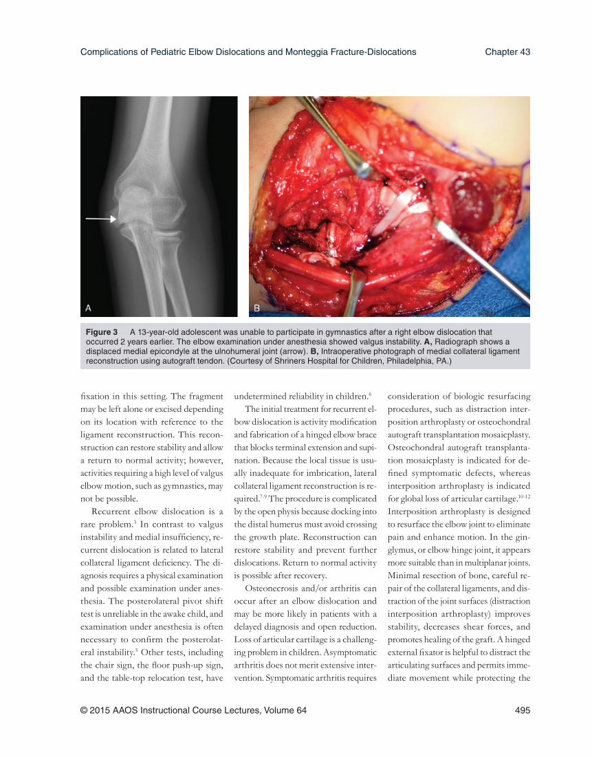
Chronic ComplicationsChronic complications after elbow dislocations are typically related to in-stability. Instability can occur in two different scenarios: valgus instability or recurrent elbow dislocations.3,4 Val-gus instability is related to nonunion or malunion of the medial epicondyle fracture and associated medial collateral ligament insuffi ciency. The instability often presents years after an injury as the child increases his or her activity level and valgus stress. The diagnosis requires a physical examination and imaging studies, such as intraoperative fl uoroscopy. Examination under anes-thesia may be necessary to confi rm the diagnosis. The initial treatments are ac-tivity modifi cation and fl exor-pronator strengthening. Failure to curtail the in-stability requires medial collateral liga-ment reconstruction4 (Figure 3). The procedure is complicated by malposi-tion of the epicondyle and distortion of the anatomy. Therefore, medial col-lateral ligament reconstruction requires ingenuity with regard to distal humeral
A, Lateral radiograph of the elbow of a 15-year-old, right hand–dominant adolescent boy with a posterior elbow dislocation and a displaced medial epicondyle fracture. B, A sagittal CT scan confi rms that the medial epicondyle is within the ulnohumeral joint. (Courtesy of Shriners Hospital for Children, Philadelphia, PA.)
Figure 1
A 12-year-old boy had persistent stiffness after an elbow dislocation that occurred 3 months earlier. CT scan shows the medial epicondyle within the ulnohumeral joint and periarticular calcifi cations. (Courtesy of Shriners Hospital for Children, Philadelphia, PA.)
Figure 2
Complications of Pediatric Elbow Dislocations and Monteggia Fracture-Dislocations Chapter 43
© 2015 AAOS Instructional Course Lectures, Volume 64 495
fi xation in this setting. The fragment may be left alone or excised depending on its location with reference to the ligament reconstruction. This recon-struction can restore stability and allow a return to normal activity; however, activities requiring a high level of valgus elbow motion, such as gymnastics, may not be possible.
Recurrent elbow dislocation is a rare problem.3 In contrast to valgus instability and medial insuffi ciency, re-current dislocation is related to lateral collateral ligament defi ciency. The di-agnosis requires a physical examination and possible examination under anes-thesia. The posterolateral pivot shift test is unreliable in the awake child, and examination under anesthesia is often necessary to confi rm the posterolat-eral instability.5 Other tests, including the chair sign, the fl oor push-up sign, and the table-top relocation test, have
undetermined reliability in children.6 The initial treatment for recurrent el-
bow dislocation is activity modifi cation and fabrication of a hinged elbow brace that blocks terminal extension and supi-nation. Because the local tissue is usu-ally inadequate for imbrication, lateral collateral ligament reconstruction is re-quired.7-9 The procedure is complicated by the open physis because docking into the distal humerus must avoid crossing the growth plate. Reconstruction can restore stability and prevent further dislocations. Return to normal activity is possible after recovery.
Osteonecrosis and/or arthritis can occur after an elbow dislocation and may be more likely in patients with a delayed diagnosis and open reduction. Loss of articular cartilage is a challeng-ing problem in children. Asymptomatic arthritis does not merit extensive inter-vention. Symptomatic arthritis requires
consideration of biologic resurfacing procedures, such as distraction inter-position arthroplasty or osteochondral autograft transplantation mosaicplasty. Osteochondral autograft transplanta-tion mosaicplasty is indicated for de-fi ned symptomatic defects, whereas interposition arthroplasty is indicated for global loss of articular cartilage.10-12 Interposition arthroplasty is designed to resurface the elbow joint to eliminate pain and enhance motion. In the gin-glymus, or elbow hinge joint, it appears more suitable than in multiplanar joints. Minimal resection of bone, careful re-pair of the collateral ligaments, and dis-traction of the joint surfaces (distraction interposition arthroplasty) improves stability, decreases shear forces, and promotes healing of the graft. A hinged external fi xator is helpful to distract the articulating surfaces and permits imme-diate movement while protecting the
A 13-year-old adolescent was unable to participate in gymnastics after a right elbow dislocation that occurred 2 years earlier. The elbow examination under anesthesia showed valgus instability. A, Radiograph shows a displaced medial epicondyle at the ulnohumeral joint (arrow). B, Intraoperative photograph of medial collateral ligament reconstruction using autograft tendon. (Courtesy of Shriners Hospital for Children, Philadelphia, PA.)
Figure 3

Pediatrics
496 © 2015 AAOS Instructional Course Lectures, Volume 64
interposition and soft-tissue repairs. Other treatment options include total elbow arthroplasty (contraindicated in a young patient), resection arthroplasty, and elbow arthrodesis. Elbow arthro-desis provides stability and eliminates pain; however; there is no optimum position that allows performance of all the activities of daily living.13
When performing interposition ar-throplasty, a long posterior longitudinal incision centered over the olecranon is preferred.11 The common extensor ori-gin and lateral collateral ligament origin are elevated from the lateral epicondyle and distal humerus. The joint surface is exposed by supinating the forearm and applying varus stress to the elbow. This maneuver rotates the ulna and radial head from the distal humerus. After dislocating the joint, the anterior and posterior capsules are elevated to increase motion. The deformed joint surfaces are refashioned to resemble an innate humerus. The goal is to re-create congruency between the trochlea and the olecranon.
The joint is reduced and assessed for satisfactory passive motion. The inter-position material of choice is then pro-cured. Allograft or autograft are both viable options. A two-ply interposition is fashioned that cloaks the distal hu-merus. The graft is secured to the distal humerus via transosseous mattress su-tures (Figure 4). An external fi xation distraction device is applied, with care taken to avoid injury to the radial nerve, and the joint is distracted 3 to 4 mm. The crucial step is to ensure the external fi xator is aligned at the joint axis to al-low the normal arc of motion. Accurate axis wire placement is the critical step because the fi xator is constructed about this wire. Closure is then accomplished, with a focus on repair of the collateral
ligament(s) and the common extensor and/or fl exor origins with transosseous sutures or bone anchors. Radiographs should be checked before leaving the operating room to confi rm joint reduc-tion and correct positioning of external fi xation. The olecranon should align with the trochlea, and the radial head should point to the capitellum. Poste-rior subluxation of the radial head infers posterolateral instability and must be corrected.
Monteggia Fracture-DislocationsAcute ComplicationsFailure to make the diagnosis is the most common, acute complication of a Monteggia fracture-dislocation.14 The radial head should align with the cap-itellum regardless of the radiographic view. The diagnosis may be missed be-cause the ulna fracture may be in the form of plastic deformation, which is diffi cult to appreciate without a full-length forearm radiograph, or the elbow radiograph may reveal subtle ra-dial head subluxation instead of frank dislocation.15 Radial head subluxation will progress to dislocation during the ensuing weeks, secondary to the unre-stricted biceps tendon pull on the radial tuberosity.
A patient with a delayed diagnosis requires prompt surgical management. Closed reduction is usually unsuccess-ful. Treatment includes correction of the ulna fracture and radiocapitellar joint reduction16-18 (Figure 5). An ulnar osteotomy is recommended to relocate the radial head and gain ulnar length. The apex of the osteotomy should be designed to redirect the radial head to-ward the capitellum. An extended lat-eral Kocher incision is performed. The ulna is cut with a saw at the point of
maximum angulation. The radiocapitel-lar joint is opened, and the radial head is reduced. Débridement of the radio-capitellar joint may be necessary to re-move any debris preventing reduction. The ulna is then fi xed with a plate and screw construct, and the stability of the radiocapitellar joint is assessed clinically and under fl uoroscopy. Frequently, the radial head is stable, and the remnants of the ligament are repaired. Persistent instability requires a reassessment of the ulnar osteotomy and/or ligament repair.
Chronic ComplicationsChronic complications are related to persistent dislocation of the radial head. The radial head is usually dislocated in an anterior direction and can block elbow fl exion. In addition, the loss of lateral column bony stability can lead to valgus instability. Treatment is directed at restoring radiocapitellar alignment and reestablishing lateral column sta-bility. The procedure follows the same tenets as those for subacute manage-ment, although the level of diffi culty is much greater. Instead of an acute ulnar
A 16-year-old adoles-cent had painful, posttraumatic left elbow arthritis after delayed open reduction for an elbow dislocation. Two-ply fascia lata was draped and secured over the distal humerus before application of an external fi x-ator. (Courtesy of Shriners Hospital for Children, Philadelphia, PA.)
Figure 4

Complications of Pediatric Elbow Dislocations and Monteggia Fracture-Dislocations Chapter 43
© 2015 AAOS Instructional Course Lectures, Volume 64 497
osteotomy, slow distraction osteogene-sis may be necessary to gain adequate length.19,20 During the consolidation phase, open joint reduction is per-formed with adjustment of the fi xator to optimize radiocapitellar alignment. The surgeon should be prepared to perform an annular ligament reconstruction to enhance stability of the radial head. Options for reconstruction include the Bell-Tawse procedure using a strip of triceps tendon16 or the technique using tendon graft described by Seel and Peterson.21 This chapter’s authors prefer the latter technique because the
reconstruction is more anatomic and results in better centering of the radial head.
SummaryElbow dislocations and Monteggia fracture-dislocations can result in both acute and chronic complications. Acute complications center on inaccurate di-agnoses and a subsequent delay in treat-ment. Chronic complications involve instability at the ulnohumeral or radio-capitellar joints. The management of chronic complications is more diffi cult, and outcomes are less predictable.
References 1. Rasool MN: Dislocations of the
elbow in children. J Bone Joint Surg Br 2004;86(7):1050-1058.
2. Carlioz H, Abols Y: Posterior disloca-tion of the elbow in children. J Pediatr Orthop 1984;4(1):8-12.
3. Osborne G, Cotterill P: Recurrent dislocation of the elbow. J Bone Joint Surg Br 1966;48(2):340-346.
4. Rohrbough JT, Altchek DW, Hyman J, Williams RJ III, Botts JD: Medial col-lateral ligament reconstruction of the elbow using the docking technique. Am J Sports Med 2002;30(4):541-548.
5. O’Driscoll SW, Bell DF, Morrey BF: Posterolateral rotatory instability of the elbow. J Bone Joint Surg Am 1991;73(3):440-446.
6. Charalambous CP, Stanley JK: Posterolateral rotatory instability of the elbow. J Bone Joint Surg Br 2008;90(3):272-279.
7. Cohen MS, Hastings H II: Rotatory instability of the elbow: The anatomy and role of the lateral stabilizers. J Bone Joint Surg Am 1997;79(2):225-233.
8. Yadao MA, Savoie FH III, Field LD: Posterolateral rotatory insta-bility of the elbow. Instr Course Lect 2004;53:607-614.
9. Savoie FH, Field LD, Ramsey JR: Pos-terolateral rotatory instability of the elbow: Diagnosis and management. Oper Tech Sports Med 2006;14:81-85.
10. Kokkalis ZT, Schmidt CC, So-tereanos DG: Elbow arthritis: Current concepts. J Hand Surg Am 2009;34(4):761-768.
11. Kozin SH, Zlotolow DA: Distraction interposition arthroplasty: Pediatric, in Glickel SZ, Bernstein RA, eds: Ar-thritis of the Hand and Upper Extremity: A Master Skills Publication. Rosemont, IL, American Society for Surgery of the Hand, 2011, pp 415-428.
12. Larson AN, Morrey BF: Interposition arthroplasty with an Achilles tendon allograft as a salvage procedure for the elbow. J Bone Joint Surg Am 2008;90(12):2714-2723.
13. Morrey BF, Askew LJ, Chao EY: A biomechanical study of normal
A 6-year-old boy had a missed Monteggia fracture-dislocation that occurred when he fell from a slide 3 months earlier. A, Lateral radiograph with anterior dislocation of the radial head and healed ulna fracture. B, Intraopera-tive photograph showing radiocapitellar joint reduction, temporary Steinmann pin fi xation, and ulnar osteotomy. C, Postoperative radiograph shows radio-capitellar joint reduction. (Courtesy of Shriners Hospital for Children, Philadel-phia, PA.)
Figure 5

Pediatrics
498 © 2015 AAOS Instructional Course Lectures, Volume 64
functional elbow motion. J Bone Joint Surg Am 1981;63(6):872-877.
14. Goh SH: Monteggia ‘fracture’-disloca-tion with bowing of the ulna: A pitfall for the unwary emergency physician. Eur J Emerg Med 2008;15(5):281-282.
15. Stoll TM, Willis RB, Paterson DC: Treatment of the missed Monteggia fracture in the child. J Bone Joint Surg Br 1992;74(3):436-440.
16. Gyr BM, Stevens PM, Smith JT: Chronic Monteggia fractures in chil-dren: Outcome after treatment with the Bell-Tawse procedure. J Pediatr Orthop B 2004;13(6):402-406.
17. Hasler CC, Von Laer L, Hell AK: Open reduction, ulnar osteotomy and external fi xation for chronic anterior dislocation of the head of the radius. J Bone Joint Surg Br 2005;87(1):88-94.
18. Rodgers WB, Waters PM, Hall JE: Chronic Monteggia lesions in chil-dren: Complications and results of reconstruction. J Bone Joint Surg Am 1996;78(9):1322-1329.
19. Bhaskar A: Missed Monteggia fracture in children: Is annular ligament reconstruction always required? Indian J Orthop 2009;43(4):389-395.
20. Exner GU: Missed chronic anterior Monteggia lesion: Closed reduction by gradual lengthening and angula-tion of the ulna. J Bone Joint Surg Br 2001;83(4):547-550.
21. Seel MJ, Peterson HA: Management of chronic posttraumatic radial head dislocation in children. J Pediatr Orthop 1999;19(3):306-312.
Video ReferenceKozin SH: Video. Posterolateral Pivot Shift Test. Philadelphia, PA, 2014.