complications of hyperthyroidism - intech - opencdn.intechopen.com/pdfs/46631.pdf · than patients...
TRANSCRIPT
Chapter 4
Complications of Hyperthyroidism
Irmak Sayin, Sibel Ertek and Mustafa Cesur
Additional information is available at the end of the chapter
http://dx.doi.org/10.5772/58196
1. Introduction
Some clinical views which appear during hyperthyroidism can be called complications ofhyperthyroidism. These clinical events are as follows.
1. Thyrotoxic heart disease
2. Progressive infiltrative ophtalmopathy in hyperthyroidism
3. Hyperthyroidism and bone
4. Thyroid chrisis
5. Thyrotoxic periodic paralysis
6. Thyrotoxicosis related psychosis and convulsion
7. Thyrotoxicosis related diabetes mellitus
2. Thyrotoxic heart disease
2.1. Effects of hyperthyroidism on heart-from pathophysiology to clinical complications
Cardiovascular signs and complications are generally the first alarming signs of hyperthyr‐oidism for any physician. Effects of thyroid hormones on the heart and cardiovascular systemcould be direct or indirect. Palpitations and exercise intolerance are the most frequent signs [1,2]. Although effects of iodization and world-wide use of radiocontrast agents may change theincidence, overt hyperthyroidism is common and affects 2-5% of the population [3,4]. Inhyperthyroid patients mortality is increased 20% and the major causes of death are cardiacproblems [5]. Atrial fibrillation is the most common and fearful arrythmic complication of
© 2014 The Author(s). Licensee InTech. This chapter is distributed under the terms of the Creative CommonsAttribution License (http://creativecommons.org/licenses/by/3.0), which permits unrestricted use,distribution, and reproduction in any medium, provided the original work is properly cited.
hyperthyroidism which occurs in an estimated 10-25% of all overtly hyperthyroid patients [6].Susceptibility to arrhythmic effects of thyroid hormones may have a genetic basis and recentlythe studies on molecular details of cardiac actions of thyroid hormones revealed someimportant knowledge [7]. Meanwhile, the cause of hyperthyroidism may also change thecardiovascular risk; patients with toxic multinoduler goitre have higher cardiovascular riskthan patients with Graves’ disease, probably because of older age, and patients with Gravesdisease may have autoimmune complications, such as valvular involvement, cardiomyopathyand pulmonary arterial hypertension [8]. Effects of thyroid hormones on the heart may begrouped as molecular or cellular mechanisms and hemodynamic effects. On the other hand,thyroid hormones have 2 types of effects on every tissue; genomic effects which occur moreslowly, and non-genomic effects. Severity of hyperthyroidism may also cause differences inclinical presentation of the patient, overt hyperthyroidism and subclinical hyperthyroidismbring differing degrees of risks to patients.
2.2. Molecular and cellular mechanisms of thyroid hormone effects on heart
Both T3 and T4 are lipophylic and they pass through the cellular membranes and theconversion of T4 to T3 occurs in many cells. Triiodothyronin (T3) is the active thyroidhormone and it has genetic and cellular effects on cardiac muscle and blood vessels. It actson THRs (thyroid hormone receptors) in the nucleus, creating dimers of 9-cis-retinoic acidreceptor (RXR) (9): the formed complexes recognize some specific DNA consensus sequen‐ces, the thyroid response elements (TRE), located in the enhanced region of the genes initiatethe transcription [9].
In myocytes, thyroid hormones act on many TREs, such as alpha myosin heavy chain fusion(MHC-α), sarcoplasmic reticulum calcium-activated ATPase (SERCA), the cellular membraneNa-K pump (Na-K ATPase), β1 adrenergic receptor, cardiac troponin I, atrial natriureticpeptide (ANP) [10-12] and some genes are also suppressed such as β-myosin heavy chainfusion (MHC-β), adenylic cyclase (IV and V) and the Na-Ca antiporter [13].
Thyroid hormone upregulates α, but downregulates β-chain in myocytes [14]. The final effectof thyroid hormones in animal studies is an increased rate of V1 isoform of MHC (MHCα/α)synthesis that is characteristically faster in myocardial fibre shortening [13,15]. A similar effecthas been also observed in preliminary human studies [16,17].
Thyroid hormones also have effects on SERCA, which is responsible for the rate of calciumuptake during diastole, by actions on calcium activated ATPase and its inhibitory cofactorphospholamban [18,19]. Thyroid hormones enhance myocardial relaxation by upregulatingexpression of SERCA, and downregulating expression of phospholamban. The greaterreduction in cytoplasmic calcium concentration at the end of the diastole increases themagnitude of systolic transient of calcium and augments its ability for activation of actin-myosin subunits. As a confirmation, phospholamban deficient mice showed no increase inheart rate after thyroid hormone treatment [20].
On the plasma membranes, T3 exerts direct extragenic actions on the functions other ionchannels such as Na/K ATPase, Na/Ca++exchanger, and some voltage gated K channels (Kv
Thyroid Disorders - Focus on Hyperthyroidism66
1.5, Kv 4.2, Kv 4.3) affecting myocardial and vascular functions [21,22] coordinating electro‐chemical and mechanical responses of myocardium [23,24]. It prolongs the activation of Nachannels in myocardial cells [25] and induces intracellular Na uptake and secondary activationof the Na-Ca antiporter, which can partly explain the positive inotropic effect. T3 exerts directeffect on L-type calcium channels resulting in abbreviation of action potential duration andpossibly L-type calcium channel mRNA expression [26,27].
The strong inotropic activity of thyroid hormones is probably due to an increased number ofβ-adrenergic receptors [28]. Circulating cathecholamine levels are in fact the same, but Gprotein and β-receptors increase [29]. The sensitivity of cardiovascular system to adrenergicstimulation does not change by thyroid hormones [30,31]. The changes in the heart rate resultfrom both an increase in sympathetic tone and decrease in parasympathetic tone [32,33].
These genomic effects fail to explain the fast actions of thyroid hormones on the cardiovascularsystem. Non-genomic effects promote rapid changes, such as increased cardiac output [34-36].The hemodynamic consequences of hyperthyroidism and nongenomic changes on plasmamembranes occur acutely and contribute to these rapid changes. Studies indicate that thyroidhormone activates acute phosphorylation of phospholamban, and that also partly explains thehomology between thyroid hormone and adrenergic system on the heart [32].
In an experimental study on rats, thyroid hormones upregulate connexin-40, a gap junctionprotein of myocardium important for the transport of electrical activity, and this may be oneof the pathogenetic mechanisms of atrial fibrillation in hyperthyroidism [37]. In another animalstudy the authors suggested that the connexin-43 phosphorylation was downregulated by T3in diabetic rats and decrease adaptation of heart to hyperglycemia and this may render theheart prone to ventricular arrythmias [38]. In fact thyroid hormone receptor alpha 1 ispredominantly expressed in cardiac myocardium and may have an important role in cardiacmyoblast differentiation by ERK kinase dependent process, but its clinical relevance is notknown [39]. Also ERK (extracellular signal-regulated kinase) pathway may have a role innegative cardiac remodelling and decreased cardiac contractile function in hyperthyroidism,by inhibition of the Raf-1/ERK pathway by T3 [40,41].
Meanwhile, the thyroid hormone may have direct (without autonomous nervous system)effects on the sinoatrial node [42,43] and oxidative stress in animal studies [44]. The heart rateincreases due to increased sinoatrial activity, a lower threshold for atrial activity, and ashortened atrial repolarisation [45,46]. Together with hemodynamic changes, i.e. the volumepreload increases due to activation of the renin-angiotensin system [47], contractility increasesdue to increased metabolic demand and the direct effect of the thyroid hormone on heartmuscle [48] and systemic vascular resistance decreases because of triiodotyronine-inducedperipheral vasodilatation [49]. The result is a dramatic increase in cardiac output [50].
Local type 2 iodothyronine deiodinase up-regulation may also be involved in cardiac remod‐elling via activation of thyroid hormone signalling pathways involving Akt and p38 MAPK(mitogen-activated protein kinase) in thyrotoxic-dilated cardiomyopathy [51]. Animal studiesshow the effects of thyroid hormones on soluble fractions of 5’-nucleosidase activity, viaprotein kinase C-related pathway [52]. Preclinical studies blocking Akt by angiotensin-II type
Complications of Hyperthyroidismhttp://dx.doi.org/10.5772/58196
67
2 receptor blocker showed that this blockage might prevent thyroxine-mediated cardiachypertrophy [53]. Meanwhile, hypertrophied myocytes may be susceptible to apoptoticstimulation by angiotensin II in hyperthyroidism [54].
Studies on rats show that mitochondrial ultrastructure was damaged in T3 treated hyperthy‐roid rat heart, leading to energy depletion and cardiac dysfunction [55]. Another study onhamsters revealed that long term hyperthyroidism cause increased left ventricular interstitialfibrosis, significant cardiac hypertrophy and deleterious cardiac remodeling characterized bymyocyte lengthening, chamber dilatation, decreased relative wall thickness, increased wallstress, and impaired global cardiac function whereas cardiomyocyte functions may beenhanced [56]. Some animal studies Show beneficial effects of angiotensin receptor blockerson myocytes as improved left ventricular longitudinal strain, heart rate and reduced cardio‐myocyte width, affecting structural remodelling beyond the anti-tachycardic effect of beta-blockers [57].
2.3. Hemodynamic effects of thyroid hormones
Hemodynamic effects of thyroid hormones are generally nongenomic and faster, by directeffects on the heart and blood vessels. In the peripheral vascular system, the rapid use ofoxygen, increased production of metabolic end products and relaxation of arterial smoothmuscle fibres by thyroid hormone cause peripheral vasodilatation [21]. This fall in peripheralvascular resistance (PVR) plays the central role in all hemodynamic changes caused by thyroidhormones [58]. Decreased PVR causes an increase in heart rate and selective increase in bloodflow of some organs (skin, skeletal muscles, heart), and a fall in diastolic pressure withconsequent widening of pulse pressure. Vasodilatation without an increase in renal blood flowcauses reduction in renal perfusion and activation of the renin-angiotensin system whichcauses sodium retention and increased blood volume [59]. In addition, thyroid hormonesregulate erythropoietin secretion and increased red cell mass may also contribute to bloodvolume increase [60]. Improved diastolic relaxation and increased blood volume increased leftventricular end-diastolic volume (LVEDV). Reduced PVR and increased LVEDV meansincreased preload and decreased afterload, thus the stroke volume increases. Increased strokevolume and increased heart rate leads to a doubling or tripling of cardiac output which cannotbe solely explained by the increased metabolic rate of the body [61].
The importance of the contribution of decreased systemic vascular resistance to the increasein systemic blood flow in patients with hyperthyroidism is evidenced by studies in which theadministration of arterial vasoconstrictors, atropine and phenylephrine, decreased peripheralblood flow and cardiac output by 34% in patients with hyperthyroidism but not in normalsubjects [62,63].
2.4. Clinical aspects of hyperthyroidism and subclinical hyperthyroidism on heart
Increased rate and strength of the heart contractility, together with an exaggerated responseof heart rate to exercise is present in hyperthyroid patients who describe this as palpitations.Diurnal heart rate variations are generally preserved. The most common ECG abnormality is
Thyroid Disorders - Focus on Hyperthyroidism68
sinusal tachycardia and shortened PR interval, and frequently intra-atrial conduction isprolonged, which is seen as increase in P wave duration. Intraventricular conduction delay inthe form of right bundle branch block is present in around 15% of patients, and atrioventricularblock may also occur due to unknown reasons. The most common rhythm disturbance inhyperthyroid patients is sinus tachycardia [32]. Its clinical impact however is overshadowedby that of patients with atrial fibrillation. The prevalence of atrial fibrillation (AF) and lesscommon forms of supraventricular tachycardia in this disease ranges between 2 to 20 percent[64,65]. AF is generally accompanied by rapid ventricular response. It is more common in menand its significance increases by age, after 40 years [65]. As in the case of angina or heart failure,the development of AF should not be attributed only to hyperthyroidism, and the underlyingorganic heart diseases should be investigated.
Atrial fibrillation usually reverses to a sinus rhythm by achievement of a euthyroid state, if thepatient is younger and the duration of hyperthyroidism is not long. The beta-adrenergicblockade may be effective in controlling the ventricular rate. Increased plasma clearance ofbeta-blockers may necessitate higher doses [66]. Among them propranolol has the advantageof blocking the conversion of T4 to T3 in peripheral tissues, however other cardioselective beta-blockers have a longer half-life and can be equally effective on the heart. In cardiac arrhythmiasintravascular infusion of calcium blockers should be avoided due to the risk of a further fallin PVR [67]. It is still controversial whether the patients with AF should have anticoagulanttherapy to prevent systemic embolization. It is advised to evaluate each patient on a case-by-case basis, and determine the risk of bleeding over embolization [68,69]. In younger patientswith hyperthyroidism and AF, who do not have other heart disease, hypertension, or inde‐pendent risk factors for embolization, the risk of anticoagulant therapy may suppress itsbenefits. But it would be appropriate to administer anticoagulant agents to older patients withknown or suspected heart diseases, or AF with longer duration. When oral anticoagulants willbe used, it should be considered that hyperthyroid patients will need smaller doses thaneuthyroid ones, due to faster elimination of vitamin-K dependent clotting factors [70].
In the patients with AF the maintenance of sinus rhythm is not possible until the euthyroidstate is restored, so electrical cardioversion is not recommended without confirming theeuthyroid status.
Many hyperthyroid patients experience exercise intolerance and exertional dyspnea, in partbecause of weakness in the skeletal and respiratory muscle [71] and also due to inability toincrease heart rate or lower vascular resistance further, as normally occurs in exercise [72]. Thetem “high output heart failure” has not been used in recent decades, because it is clear that theheart is still able to increase cardiac output at rest and with exercise. In the setting of lowvascular resistance and decreased preload, the cardiac functional reserve is compromised andcannot rise further to accommodate the demands of submaximal or maximal exercise [73].About 6% of thyrotoxic patients develop heart failure and less than 1% develop dilatedcardiomyopathy with impaired left ventricular systolic dysfunction, due to the tachycardia-mediated mechanism leading to an increased level of cytosolic calcium during diastole, withreduced contractility of the ventricle and diastolic dysfunction, often with tricuspid regurgi‐tation [74]. In the recent study of Yue et al, diastolic dysfunction was more prominent in
Complications of Hyperthyroidismhttp://dx.doi.org/10.5772/58196
69
thyrotoxic patients older than 40 years of age, whereas in younger ones marked reduction inperipheral vascular resistance and increased cardiac output were prominent [75].
Hyperthyroidism may complicate or cause pre-existing cardiac disease because of increasedmyocardial oxygen demand, increased contractility and heart rate, and may cause silentcoronary artery disease, anginas or compensated heart failure and even endothelial dysfunc‐tion [76]. Treatment of heart failure with tachycardia should include a beta-blocker, byconsidering its contraindications in each patient. Furosemide may help to reverse the volumeoverload, but digoxin is less beneficial when compared with euthyroid heart failure patientsbecause there may be relative resistance to its action, due to greater blood volume (distribution)and the need to block more Na-K-ATPase in myocardium [70].
Subclinical hyperthyroidism is a state characterised by low serum thyrotropin levels andnormal serum thyroid hormone concentrations. Over recent decades this state has also beenfound to be associated with some abnormalities in cardiac function. Enhanced systolic functionand impaired diastolic function due to slowed myocardial relaxation may cause increased leftventricular mass in these subjects, together with increased heart rate and arrhythmias bysimilar mechanisms as overt hyperthyroidism [77,78]. In people over 60 years of age subclinicalhyperthyroidism was associated with a tripled risk of atrial fibrillation during a 10-year follow-up period [79]. In a recent cross-sectional study with 29 patients, subclinical hyperthyroidismwas found to be related with impaired functional response to exercise with low oxygenconsumption and exercise threshold, together with slower heart rate recovery [80].
Besides antithyroid treatment strategies, beta-blocker therapy reduces heart rate andimproves left ventricular mass, but positive inotropic response persists. This subclinicalhyperthyroidism is associated with increased cardiovascular mortality [81]. In the study ofHeeringa et al with 1,426 patients, it was shown that even the high-normal thyroid functionmay increase the risk of AF [82]. Besides increased AF and thromboembolic events,increased left ventricular mass and left ventricular function may be the reason. Thus, it isadvised to measure serum thyrotropin in all elderly patients with systolic hypertension, awidened blood pressure, recent-onset angina, atrial fibrillation and any exacerbation ofischemic heart disease and treat it [72,83].
3. Progressive infiltrative ophtalmopathy in hyperthyroidism
3.1. Hyperthyroidism and eye disease
Thyroid eye disease (TED), also called thyroid-associated ophthalmopathy (TAO) and Graves'ophthalmopathy (GO), affects 25–50% of patients with Graves' disease [84]. Regardless ofwhether hyperthyroidism occurs first, the signs and symptoms of GO become manifest in 85%of patients within 18 months [85]. The clinical features are usually mild. Most commonsymptoms are; ocular irritation with redness and tearing,, sensitivity to light, double vision,blurring of vision, and feeling a pressure sensation behind the eyes. On physical examination,extraocular muscle dysfunction, proptosis, periorbital and eyelid edema, conjunctival chemos,
Thyroid Disorders - Focus on Hyperthyroidism70
lid lag and retraction (or stare), or exposure keratitis may be detected [86]. Most patientsexperience only the minor congestive signs of TAO (chemosis, injection, lid edema), withimprovement in several months without treatment. Approximately 28% of TAO cases aresevere and symptomatic for several years. Symptoms and signs include, restricted motilityleading to diplopia, exposure keratopathy, optic neuropathy and loss of vision [87,88].
TAO can be divided into two clinical stages; congestive and fibrotic. In the congestive orinflammatory stage, auto-immunity leads to inflammatory cellular infiltration of the musclesand transformation of fibroblasts into adipose tissue. The fibrotic stage is characterized byfibrosis which causes proptosis and strabismus [89,90].
3.2. Pathophysiology
In TAO, the most important pathological changes are enlarged extraocular muscles andincreased orbital fat. These changes result from a complex interplay among orbital fibroblasts,immune cells, cytokines, auto-antibodies, genetics, and environmental factors [84].
Interactions between orbital fibroblasts, immune cells, cytokines and autoantibodies
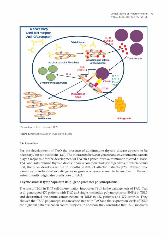
Patients with TAO, orbital tissue is infiltrated by inflammatory cells (T helper type 1 (Th1)and T helper type 2 (Th2) lymphocytes, B lymphocytes, mast cells, and macrophages) [91,92].These cells release cytokines which participate in tissue reactivity and remodeling.Normally, the antigens to which lymphocytes respond are foreign, and several tolerancemechanisms act to prevent the development of reactivity to self-antigens or autoimmuni‐ty, but these tolerance mechanisms sometimes fail and autoimmunity develops [93,94].Fibroblasts are a highly interactive cell type, described as “sentinel cells” [95]. They, respondto immune stimulation and actively participate in the inflammatory pathway [96,97]. Inpatients with TAO, orbital fibroblasts synthesize excess glycosaminoglycans (GAGs),including hyaluronan. These can differentiate into adipocytes, leading to the accumula‐tion of fat [98]. Orbital fibroblasts do not express the IL-1 receptor antagonist at levels foundin other fibroblasts, but also display lymphocyte costimulatory molecules such as CD40.These differentations result in excessively high levels of Cox-2 and PGE2 in response toproinflammatory cytokines [99,100]. T lymphocytes in the orbital tissue interact withfibroblasts. This interaction results with activation and proliferation of fibroblasts, synthe‐sis of extracellular macromolecules, and differentiation to adipocytes [101]. A summary ofthis model for the pathogenesis of TED is depicted in Figure 1 [102].
Autoantigen expression by orbital fibroblasts results in T lymphocyte accumulation to the orbit[103]. The autoantigen may be a TSH receptor (TSH-R) or an insulin-like growth factor-1receptor (IGF-1R) [103,104]. T lymphocytes in the orbital tissue induce fibroblast proliferationand hyaluronan synthesis. This result in orbital tissue remodeling [101]. Stimulation of orbitalfibroblasts by T lymphocytes results in production of chemokines (e.g. IL-16, RANTES) andcytokines (e.g. IL-6). These molecules initiate migration of T and B lymphocytes to the orbitaltissue and increase fibroblast presentation of autoantigens [97,101,105). Costimulatorymolecules, adhesion molecules, and cytokines like IFNγ, IL-1β, and TNFα play an importantrole in the interaction between T lymphocytes and fibroblasts. One of the communication
Complications of Hyperthyroidismhttp://dx.doi.org/10.5772/58196
71
pathways of T lymphocytes and orbital fibroblasts is the CD40-CD40 ligand pathway[97,100,101]. In TAO orbital fibroblasts express high levels of CD40 [97,106]. Activation byCD40L induces hyaluronan synthesis, IL-6 and IL-8, Cox-2 and PGE2 [97,101,107].
T lymphocyte-mediated activated fibroblasts release factors which promote and activate theproliferation of T lymphocytes. In this way fibroblasts perpetuate inflammation [101,108].Antonelli et al. found that orbital fibroblasts from TAO patients may modulate the activity ofT lymphocytes through the production of CXCL10. Serum CXCL10 levels were higher in activeTAO patients than in those with inactive disease. CXCL10 release enhances the migration ofT lymphocytes into the orbital tissue. These lymphocytes secrete IFNγ and TNFα. There is apositive feedback between CXCL10 and IFNγ – TNFα. Peroxisome proliferator-activatedreceptor-gamma (PPAR-γ) activation has an inhibitory role in this process [108]. Feldon et al.found that activated human T lymphocytes drive the differentiation of human fibroblasts toadipocytes. They showed that human T cells, when activated, strongly express Cox-2 andproduce PGs, possibly 15d-PGJ2, that are PPAR-γ ligands and human T cells also producePGD2. These findings showed that PGD2 converts to the PGJ series of PGs with the final productbeing 15d-PGJ2, a notable potent PPAR-γ ligand [109-111]. Feldon et al. also showed thathuman orbital fibroblasts express PPAR-γ and that 15d-PGJ2, PGD2, and 15d-PGD2 stronglyinduce adipogenesis [109]. Natural and synthetic activators of PPAR-γ stimulate lipidaccumulation and the expression and secretion of adiponectin [112,113]. PPAR-γ levels arehigher in orbital tissue from patients with active TAO [108,114].
Chen et al. showed higher mRNA levels of a macrophage chemoattractant called C-C motifchemokine ligand-2 (CCL2)/monocyte chemoattractant protein-1 (MCP-1) and dense infiltra‐tion with macrophages in the orbital fat compared with normal controls [115].
3.3. Smoking
There is a strong and consistent association between smoking and TAO [116]. But the exactmechanism by which smoking affects TAO is not known. Formation of superoxide radicalsand tissue hypoxia could be responsible. Cigarette smoke either contains or can generate avariety of oxidants and free radicals [117]. Orbital fibroblasts can be induced by tissue hypoxia(5% CO2 and 95% N2) and superoxide radicals, thus they proliferate and synthesize GAGs[118,119]. Mack et al. cultured orbital fibroblasts, obtained from patients undergoing orbitaldecompression for severe GO. They showed that, inreased human leukocyte antigen (HLA-DR) expression of orbital fibroblasts occurred in response to nicotine and tar only in thepresence of interferon-γ. These findings suggest that there is an interaction between smokingand orbital immune responses [120]. Many studies demonstrated a dose-response relationshipbetween the numbers of cigarettes smoked per day and TAO [121]. Smokers suffer more severeTAO than non-smokers. Smoking increases the progression of TAO after radioiodine therapyfor hyperthyroidism [116]. Eckstein et al. demonstrated that smoking influences the course ofTAO during treatment in a dose dependent manner. The response to treatment is delayed andconsiderably poorer in smokers [122]. Pfeilschifter et al. showed that former smokers had asignificantly lower risk for the occurrence of proptosis and diplopia than active smokers witha comparable lifetime cigarette consump
Thyroid Disorders - Focus on Hyperthyroidism72
Autoantibody(Anti-TSH receptor,Anti-IGF1 receptor)
Ab binds to orbital fibroblastsActivation and release
of chemokines
IL-16RANTESCXCL10
T lymphocytes
MHC IITCRIL-6
CD 40 L
IL-8CXCL10
IFN-γTNF-αPGD2PGJ2
Orbital tissue
CD 40
B lymphocytes accumulate
Proliferation
Production ofGAGs
Adipogenesis
(Data adapted from reference 102)
Figure 1. Pathophysiology of thyroid eye disease
3.4. Genetics
For the development of TAO the presence of autoimmune thyroid disease appears to benecessary, but not sufficient [124]. The interaction between genetic and environmental factorsplays a major role for the development of TAO in a patient with autoimmune thyroid disease.TAO and autoimmune thyroid disease share a common etiology; regardless of which occursfirst, the other develops within 18 months in 80% of affected patients [125]. Polymorphicvariations in individual somatic genes or groups of genes known to be involved in thyroidautoimmunity might also predispose to TAO.
Thymic stromal lymphopoietin (tslp) gene promoter polymorphisms
The role of TSLP in Th17 cell differentiation implicates TSLP in the pathogenesis of TAO. Tsaiet al. genotyped 470 patients with TAO at 3 single nucleotide polymorphisms (SNPs) in TSLPand determined the serum concentrations of TSLP in 432 patients and 272 controls. Theyshowed that TSLP polymorphisms are associated with TAO and that expression levels of TSLPare higher in patients than in control subjects. In addition, they concluded that TSLP mediates
Complications of Hyperthyroidismhttp://dx.doi.org/10.5772/58196
73
the differentiation of CD4+T cells into Th17 cells. According to this study the TSLP gene maybe a relevant candidate gene for susceptibility to TAO. TSLP genotypes may be used as geneticmarkers for the diagnosis and prognosis of TAO [126].
Toll-like receptor gene polymorphisms
Toll-like receptors (TLRs) are a family of pattern-recognition receptors, which play a role ineliciting innate/adaptive immune responses and developing chronic inflammation. Liao et alevaluated 6 TLR-4 and 2 TLR-9 gene polymorphisms in 471 GD patients (200 patients withTAO and 271 patients without TAO) from a Taiwan Chinese population. There was nostatistically significant difference in the genotypic and allelic frequencies of TLR-4 and TLR-9gene polymorphisms between the GD patients with and without TAO. In the sex-stratifiedanalyses they showed that the association between TLR-9 gene polymorphism and the TAOphenotype was more pronounced in the male patients. Their data suggest that TLR-9 genepolymorphisms are significantly associated with increased susceptibility of ophthalmopathyin male GD patients [127].
Polymorphisms of B7 molecules (CD80 and CD86)
Liao et al. evaluated genotypes of CD80 and CD86 polymorphism in GD patients. They foundthat the frequency of C allele at position rs_9831894 of the CD86 gene is different in patientswith GD (with and without TAO). They showed that the G-A haplotype has a protective effectin the development of TAO among patients with GD. Their data suggest that the polymor‐phisms of the CD86 gene may be used as genetic markers for making the diagnosis andprognosis of TAO [128].
Interleukin-1beta (IL1β) polymorphisms
Recent studies have demonstrated that IL1β plays a role in the development of TAO byinducing adipogenesis and accumulation of GAGs and prostaglandin E2 (PGE2) [129,130]. Liuet al. found that the SNPs rs3917368 and rs1143643 in the 3′ UTR and intron regions of IL1βand patients with the genotypes containing both rs3917368 A/A and rs1143643 A/A may beara higher risk of developing TAO. Thus we can speculate that IL1β polymorphisms can berelated with the development of TAO in GD [131].
Cytotoxic T lymphocyte antigen-4Vaidya et al. firstly reported an association between CTLA-4A/G polymorphism at codon 17and TAO [132], but this data was not confirmed by Allahabadia et al. in a larger cohort ofpatients [133]. Daroszewski et al. evaluated the relation between soluable CTLA-4 level andclinical manifestation of TAO and CTLA-4 gene polymorphisms. They found higher levelsof Serum sCTLA-4 in the TAO group than in controls. The level of sCTLA-4 was higher insevere TAO patients than in non-severe cases. They showed for the first time that the pres‐ence of the CTLA-4 gene polymorphisms Jo31 and CT60 were related with elevated sCTLA-4levels [134]. These data suggest that the polymorphisms of the CTLA-4 gene may be used asa genetic marker for making the diagnosis and prognosis of TAO. The role of sCTLA-4 andits full-length cell-bound analogue in autoimmunity remains uncertain.
Thyroid Disorders - Focus on Hyperthyroidism74
PPAR-γ gene polymorphism
The PPAR-γ transcription factor is involved in both adipogenesis and inflammation whichhave been implicated in the pathogenesis of TAO. Alevizaki et al. found no difference in thedistribution of the Pro(12)Ala PPAR-γ gene polymorphism between GD patients with andwithout TAO. But they showed that PPAR-γ polymorphism carriers had lower TSH-Rab levelsand lower clinical activity scores (CAS). According to this study patients with TAO who havethis polymorphism are associated with less-severe and less-active disease [135].
IL-23R polymorphisms
Huber et al. demonstrated that two IL-23R polymorphisms (rs10889677 and rs2201841) wereassociated with TAO. According to this study we can speculate that these variants may induceTAO by changing the expression and/or function of IL-23R, by promoting a pro-inflammatorysignaling cascade [136].
Protein tyrosine phosphatase-22
Lymphoid protein tyrosine phosphatase (LYP, aka PTPN-22) represents another negativeregulator of T cell activation. Syed et al. found an association between certain single nucleotidepolymorphisms (SNPs) in the protein tyrosine phosphatase (PTP) called PTPN12 and in‐creased risk of mild/moderate ophthalmopathy [137]. But this data should be confirmed inlarger studies.
Nuclear factor (NF)-[kappa]B1
Kurylowicz et al. showed a correlation between a polymorphism in the NF-[kappa]B1 genepromoter (-94ins/del adenine, thymine, thymine, guanine) and the development of TAO [138].
Human leukocyte antigen (HLA)
HLA class I-II genes are related for the development of TAO [139]. Akaishi et al. found TAOpatients with major extraocular muscle involvement have had a higher frequency of the HLA-DRB1*16 allele, but patients with minor extraocular muscle involvement have had a higherfrequency of the HLA-DRB1*03 allele [140]. In another study an association between HLA-A11,-B5,-DW12 and-DR14 and TAO has been found [141,142].
3.5. Cytokines
Autoimmune thyroid disease involves the activation of multiple cytokine networks. SerumIL-6 levels were greater in the patients with TAO than in those without eye disease [143] andboth Th1-and Th2-derived cytokines were elevated in TAO patients compared with controlsamples [144]. Chen et al. found that the exaggerated capacity of orbital fibroblasts to expresshigh levels of both IL-6 and its receptor in an anatomic site-selective manner could representan important basis for immune responses in TAO [145]. Higher serum levels of IL-17 wereobtained in TAO patients than in controls. Serum IL-17 concentration had significant correla‐tion with CAS [146]. Significantly higher PAI-1 [147] and IgE levels [148] were found in TAOpatients than in the control groups.
Complications of Hyperthyroidismhttp://dx.doi.org/10.5772/58196
75
3.6. Imaging procedures in TAO
Ultrasound (US)
US is of primary importance in orbital pathology because of its safety, non-invasiveness, shorttime of investigation, low cost, lack of radiation and application without need to prepare thepatient. In endocrine orbitopathy; the A scan demonstrates echographical widening of theperipheral orbital space and a widening of the muscle echo, while the B scan shows a highinternal echo of the connective tissue septa, increased reflection of the muscle belly anddistension of the retrobulbar optic nerve sheaths, enlargement of lacrimal gland and dilatationof the superior ophthalmic vein [149]. Although it has many advantages, US cannot effectivelydisplay the muscles at the apex of the orbit. US is not as effective as other diagnostic proceduresin delineating the relationship of orbital pathology to contiguous structures, nor is it reliablein imaging lesions of either the posterior orbit or those involving the bone walls [150].
Computed tomography (CT)
According to differential X-ray absorption CT can differentiate normal and abnormal struc‐tures of different tissue density. In CT orbital fat images as a black, low-density area, while incontrast to this extraocular muscles and the optic nerve image as higher-density areas [151].The primary areas of orbital involvement are extraocular muscles, and CT findings correlatewith clinical impressions of the severity of extraocular muscle enlargement [152]. Muscleinvolvement on CT of TAO is usually limited to the non-tendonous portion of the muscle. Dueto compression of the optic nerve by enlarged extraocular muscles near the orbital apex, opticnerve dysfunction can be seen in TAO [153]. Other findings that may be noted on CT arelachrymal gland enlargement, a dilated superior opthalmic vein, muscle belly enlargementand increase in orbital fat volume [154,55]. CT imaging is a non-invasive, simple, fast, and costeffective imaging procedure. Furthermore, having high sensitivity and correlation with clinicalfindings, CT imaging should be considered first during diagnostic evaluation of TAO.
Magnetic resonance imaging (MRI)
Pulse sequences that examine T2 in MRI can estimate the water content of tissues. In TAOlymphocytes infiltrate the orbital tissue that causes fibroblast stimulation. Fibroblasts producelarge amounts of GAGs. By binding large amounts of water, GAGs cause edema. Increasedwater content of thickened extra-ocular muscles cause elevated T2 [156]. In TAO there are twodifferent phases with disease activity. Medical treatment can be effective in the active stage.Therefore, for predicting the outcome of medical management the evaluation of diseaseactivity is important. Yokoyama et al. investigated whether MRI could assess the diseaseactivity in TAO. They found that MRI is not only a useful tool for detection of extraocularmuscle enlargement, but also for assessment of disease activity in TAO [157]. In conclusion,MRI is useful to assess TAO. But cost and availability are current limitations of this modality.Therefore, MRI should be used for the management of TAO in specialized patients and clinics.
Octreoscan (OCT)
It is thought that radionuclide accumulation is probably due to binding to somatostatinreceptors on lymphocytes, myoblasts, fibroblasts and endothelial cells. Another explanation
Thyroid Disorders - Focus on Hyperthyroidism76
is local blood pooling due to venous stasis by the orbital inflammation [158,159]. Krassas et al.found that OCT positivity is higher in Graves' patients with than in those without ophthalm‐opathy, and higher in patients with active TAO than in those with inactive TAO [160]. Postemaet al. demonstrated that OCT positivity correlates with activity of the TAO such as high CAS[161]. Thus, a positive orbital OCT means clinically active TAO, in which immunosuppressivetreatment might be of therapeutic benefit [162]. However, limitations such as cost, non-negligible radiation burden, non-specific examination for TAO, and finally, lack of evaluationof eye muscle swelling restrict the widespread use of this technique [163]. Orbital OCT ismainly indicated to select patients with TAO who will benefit from immunosuppression [162].
3.7. Other diagnostic techniques
Kuo et al. were first described in a positron emission tomography (PET/CT) study in a patientwith TAO. The detection of inflammation by 18F-fluorodeoxyglucose (FDG) uptake wassensitive and objectively demonstrable by this semi-quantitative imaging method [164].García-Rojas et al. carried out the first prospective study. FDG uptake of extra-ocular muscleswas statistically different between patients with and without TAO. In cases with TAO whereclinical doubt exists, PET/CT provides valuable and useful information for the diagnosis,characterization, and therapeutic decision [165]. Further research is required to define the roleof FDG-PET/CT in the management of TAO.
Another tool to measure orbital inflammation is the uptake of radioactively labeled substances,such as 67Gallium (Ga) citrate [166] and 99mTechnetium (Tc)-labeled agents [167,168]. In medicalliterature there are few studies about these nuclear imaging techniques.
3.8. Clinical classification systems
The treatment for TAO varies according to the level of disease activity. Thus, methods havebeen proposed to determine whether TAO is active (33). There are two main scoring systems[169,170]:
1. NOSPECS (No signs or symptoms; Only signs; Soft tissue involvement with symptomsand signs; Proptosis; Eye muscle involvement; Corneal involvement; Sight loss)
2. CAS (Clinical Activity Score) systems.
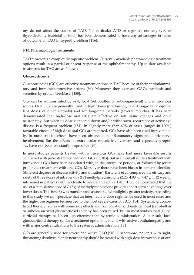
Neither NOSPECS nor CAS are both specific and completely reliable, but CAS is a simpleoffice-based tool. The modified NOSPECS criteria include lid retraction, soft tissue inflamma‐tion, proptosis, size difference, extra-ocular muscle involvement, corneal defects, and opticnerve compression [169]. CAS demonstrates the presence or absence of seven symptoms orsigns that indicate inflammation (Table-1) [171].
The score ranges from 0 to 7, with 0 to 2 characteristics indicating inactive TAO and 3 to 7characteristics indicating active TAO. The CAS has a high predictive value for the outcome ofimmunosuppressive treatment in TAO. The main determinant of therapeutic outcome isdisease activity, not disease duration [172]. In addition, the severity of disease should be
Complications of Hyperthyroidismhttp://dx.doi.org/10.5772/58196
77
assessed (Table-2). Dysthyroid optic neuropathy, corneal breakdown or both indicate that theTAO is sight-threatening and requires immediate treatment [116].
∙ Spontaneous retrobulbar pain
∙ Pain with eye movement
∙ Redness of the eyelids
∙ Redness of the conjunctiva
∙ Swelling of the eyelids
∙ Swelling of the caruncle
∙ Conjunctival edema (chemosis)
Table 1. Components of the Clinical Activity Score
∙ Lid aperture
∙ Swelling of the eyelids
∙ Redness of the eyelids
∙ Redness of the conjunctivae
∙ Conjunctival edema
∙ Inflammation of the caruncle or plica
∙ Exophthalmos
∙ Subjective diplopia score
∙ Eye muscle involvement
∙ Corneal involvement
∙ Optic nerve involvement
Table 2. Assesment of Disease Severity
3.9. Management
Smoking and TAO
Patients should be encouraged to quit smoking. In an observational study smoking cessationhas been associated with a decreased risk of the development of exophthalmos and diplopiain patients with Graves' disease [171].
Management of hyperthyroidism in patients with TAO
Uncontrolled thyroid functions (both hyper-and hypothyroidism) are related to severe TAO.Patients with hyperthyroidism can be treated with anti-thyroid drugs (ATDs), radioactiveiodine (RAI) therapy and surgical therapy. Some different studies have shown improve‐ment in TAO [173] by indirect beneficial effects [174] and a gradual decrease in TSH-receptor antibody (TRAb) levels during ATDs treatment [175]. There are confliciting reportsin medical literature about RAI therapy and severe TAO [176,177]. But this associationcannot be excluded. This risk can be eliminated by therapy with oral glucocorticoids (GCs)after RAI therapy and avoiding post-treatment hypothyroidism [116]. Thyroidectomy is aneffective treatment choice for the definitive cure of hyperthyroidism. Wilhelm et al. showedthat near-total/total thyroidectomy is safe and superior to subtotal thyroidectomy formanagement of hyperthyroidism in Graves' disease [178]. According to the European Groupon Graves' Orbitopathy (EUGOGO) Consensus Statement; ATD therapy and thyroidecto‐
Thyroid Disorders - Focus on Hyperthyroidism78
my do not affect the course of TAO. No particular ATD or regimen, nor any type ofthyroidectomy (subtotal or total) has been demonstrated to have any advantages in termsof outcome of TAO in hyperthyroidism [116].
3.10. Pharmacologic treatments
TAO represents a complex therapeutic problem. Currently available pharmacologic treatmentoptions result in a partial or absent response of the ophthalmopathy. Up to date availabletreatments for TAO are as follows:
Glucocorticoids
Glucocorticoids (GCs) are effective treatment options in TAO because of their antiinflamma‐tory and immunosuppressive actions (96). Moreover they decrease GAGs synthesis andsecretion by orbital fibroblasts [180].
GCs can be administrated by oral, local (retrobulbar or subconjunctival) and intravenousroutes. Oral GCs are generally used in high doses (prednisone, 60–100 mg/day or equiva‐lent doses of other steroids) and for long-time periods (several months). It has beendemonstrated that high-dose oral GCs are effective on soft tissue changes and opticneuropathy. But when its dose is tapered down and/or withdrawn, recurrence of active eyedisease is a frequent problem [181]. In slightly more than 60% of cases (range: 40–100%)favorable effects of high-dose oral GCs are reported. GCs have also been used intravenous‐ly. In most studies effects have been observed on inflammatory signs and optic nerveinvolvement. But the effects on extra-ocular muscle involvement, and especially propto‐sis, have not been constantly impressive [90].
In most studies patients treated with intravenous GCs have had more favorable resultscompared with patients treated with oral GCs [18,183]. But in almost all studies treatment withintravenous GCs have been associated with, in the interpulse periods, or followed by (oftenprolonged) treatment with oral GCs. Moreover there have been biases in patient selections(different degrees of disease activity and duration). Bartalena et al. compared the efficacy andsafety of three doses of intravenous (IV) methylprednisolone (2.25, 4.98, or 7.47 g in 12 weeklyinfusions) in patients with moderate to severe and active TAO. They demonstrated that theuse of a cumulative dose of 7.47 g of methylprednisolone provides short-term advantage overlower doses. This benefit was transient and associated with slightly greater toxicity. Accordingto this study we can speculate that an intermediate-dose regimen be used in most cases andthe high-dose regimen be reserved to the most severe cases of TAO [184]. Systemic glucocor‐ticoid therapy relates with some side effects and complications. Therefore, local (retrobulbaror subconjunctival) glucocorticoid therapy has been raised. But in most studies local gluco‐corticoid therapy had been less effective than systemic administration. As a result, localglucocorticoid therapy can be a treatment option in patients with active ophthalmopathy andwith major contraindications to the systemic administration [181].
GCs are generally used for severe and active TAO [90]. Furthermore, patients with sight-threatening dysthyroid optic neuropathy should be treated with high-dose intravenous or oral
Complications of Hyperthyroidismhttp://dx.doi.org/10.5772/58196
79
glucocorticoid agents [116]. Additionally, there are some case reports in medical literature,who had severe complicatons related with high-dose glucocorticoid pulse therapy [185].
Somatostatin (SST) analogues
SST receptors (SSTR) are expressed in many tissues, including activated lymphocytes. OCTwas proposed as a method to evaluate orbital inflammation in TAO. The use of the SST analog,octreotide, was first reported in TAO patients by Chang et al [186]. Uysal et al. evaluated theeffect of octreotide treatment in nine patients with TAO. Seven of the patients showedimprovement in CAS. Proptosis improved either slightly or significantly in seven patients.None of the patients has showed deterioration in the eye according to their findings [187]. Amajor disadvantage of octreotide is its short half-life, which requires multiple injections. Toovercome this limitation, new long-acting SST analogs (lanreotide / octreotide-LAR) have beendeveloped. Stan et al. carried out a randomized, double-blind, placebo-controlled trial ofoctreotide-LAR for treatment of TAO. In octreotide-LAR-treated patients, the CAS improvedwith a greater degree than in the placebo group. They noted improvement in eyelid fissurewidth, which suggest that octreotide LAR treatment may be effective in TAO patients withsignificant lid retraction [188]. In another study Krassas et al. aimed to investigate the orbitalIndium-111-pentetreotide activity after treatment with octreotide and lanreotide in patientswith TAO. All patients treated with SST analogs had a negative follow-up OCT, whereascontrols had a positive OCT. Both NOSPECS score and CAS had improved in the treatmentgroup, but there have been no changes in control subjects [189]. Most common side effects ofSST analog therapy in TAO patients were mild gastrointestinal symptoms occurring duringthe first week of treatment. Because of minimal side effects and proven efficacy, SST analogscould be a treatment option in selected patients. But we need further large, multi-center,prospective and randomized clinical trials and a comparable series of patients with otherestablished treatment options, to achieve more accurate outcomes. In addition, the high costof this treatment must also be taken into account.
Intravenous immunoglobulins (IVIG)
Antonelli et al. was published as the first non-randomized study on the use of IVIGs for TAO.They treated 7 patients with high-dose IVIGs alone (400 mg/kg/day for 5 consecutive days; thecycle was repeated five times at 3-week intervals) and 7 patients with IVIGs associated withorbital radiotherapy. The results were compared with a historical group of patients previouslytreated with high-dose GCs and orbital radiotherapy. They found that IVIGs, either alone orcombined with orbital radiotherapy, had improved the ocular conditions. This result did notdiffer from those obtained in the historical group [190]. In a randomized trial by Kahaly et al.19 patients with active TAO were treated with a 20-week course of oral prednisolone (P,starting dose 100 mg/day), and 21 received 1g immunoglobulin/kg body weight for 2 consec‐utive days every 3 weeks. The immunoglobulin course was repeated six times. The degree ofclinical improvement between two groups was not significant. There was a marked reductionof thyroid antibody titres in the immunoglobulin group. Side effects were more frequent andsevere during P than during immunoglobulin therapy [191]. But another study by Seppel etal. failed to show any beneficial effects of IVIGs in TAO [192]. In summary, treatment studieswith IVIGs include small numbers of patients and all of them are not randomized. In addition
Thyroid Disorders - Focus on Hyperthyroidism80
to this, treatment is quite expensive and is related with disease transmission using plasma-derived products. More prospective, randomized and controlled trials, which includes largeseries of patients are required to obtain more accurate results.
Immunosuppressive drugs
a. Cyclosporine A (CyA): The first report by Weetman et al. showed that CyA had positiveeffects on ocular-muscle function, visual acuity, exophthalmos and orbital muscle swel‐ling [193]. Prummel et al. compared two groups of 18 patients each of which were treatedwith either cyclosporine or prednisone. At the end of the study, treatment response wasobserved in 11 patients treated with prednisone, but only in 4 patients treated withcyclosporine [194]. Weissel et al. treated 8 patients with TAO, all of whom had compres‐sive optic nerve disease (CON), by a combined treatment with CyA and cortisone. CONdisappeared completely in all patients [195]. In summary, the use of CyA has been reportedin several studies. Given the side effects of CyA, some of which can be severe, it shouldnot be considered as a first-line treatment in TAO. The use of CyA might be maintainedin patients who are resistant to GCs alone, and in whom the persistent activity of the diseasewarrants a continuing medical intervention. According to the recent European ThyroidAssociation survey CyA or azathioprine were indicated as suitable therapeutic options byonly 6% and 2% of respondents respectively [196].
b. Methotrexate (MTX): MTX is not a novel drug, but it has not systematically been evaluatedin TAO management. Smith et al. carried out the unique study on the use of MTX for non-infectious orbital inflammatory disorders like TAO. They included 14 patients, and three ofthem had TAO. All three patients with TAO had an improvement in their ocular conditionsafter MTX treatment [196] According to the recent European Thyroid Association survey MTXwas indicated as a therapeutic option by only 1% of respondents [197]. MTX has many dose-dependent and reversible side effects and should not be used as a first-line treatment. Inpatients with GC dependency, it can be used at low doses with the aim of reducing the doseof GCs. However, to clarify the effectiveness of MTX in TAO management we need morecontrolled and randomized prospective studies.
Antioxidants
Bouzas et al. carried out a prospective, nonrandomized, comparative study on the effects ofthe antioxidant agents allopurinol and nicotinamide in TAO patients. Ocular conditionssignificantly improved in antioxidant-treated patients, more than placebo-treated patients[198]. Yoon et al. investigated the inhibitory effect of quercetin on inflammation in culturedwhole orbital tissue. Quercetin had a significant suppression of tissue IL-6,IL-8, IL-1β andTNFα mRNA expression in cultured orbital tissues from three TAO samples relative tountreated control tissue [199]. Marcocci et al. demonstrated that selenium administration (100μg twice daily) significantly improved quality of life, reduced ocular involvement, and slowedprogression of the disease in patients with mild TAO [200].
There is limited data about treatment TAO with antioxidants. We need larger, prospective andrandomized trials to clarify the role of antioxidant agents in the treatment of TAO.
Complications of Hyperthyroidismhttp://dx.doi.org/10.5772/58196
81
Cytokine antagonists
Chang et al. aimed to determine the effects of pentoxifylline (Ptx); a cytokine antagonist, onfibroblasts derived from patients with Graves' ophthalmopathy. Ptx treatment caused a dose-dependent inhibition of serum-driven fibroblast proliferation and glycosaminoglycan synthe‐sis [201]. Balazs et al. showed that Ptx has had beneficial effects on inflammatory symptomsof TAO and associated laboratory parameters [202]. In another study Ptx has improved thequality of life (QOL) of patients in the inactive phase of TAO [203]. According to these studiesPtx may be an effective and promising drug in the treatment of TAO.
TNF-α antagonists
Paridaens et al. assessed the effect of etanercept on clinical signs in TAO. After treatment themean CAS and ophthalmopathy index (OI) had decreased significantly. No adverse effectswere noted [204]. İnfliximab administration resulted with a significant reduction of inflam‐mation and improvement of visual function without noticeable short-term side effects in twopatients with active TAO [205,206]. But randomized prospective clinical trials are needed toobtain whether TNF-α antagonists are effective in reducing the inflammatory symptoms ofTAO, and can be administered safely for a long-term period without serious side effects.
Rituximab (RTX)
A patient with TAO, who was unresponsive to steroids, was treated with RTX. The CASdeclined from 5 to 2 in 3 months and the patient had peripheral B-cell depletion [207]. Whenthe effect of RTX therapy was compared with IV GCs, RTX had positively affected the clinicalcourse of TAO, independently of either thyroid function or circulating antithyroid antibodies,including TSH receptor antibody [208]. Silkiss et al. demonstrated a statistically significantdecrease in CAS from the baseline value. B-cell depletion had been observed and was welltolerated, and there were no adverse effects from the RTX infusions [206]. There is currentlyinsufficient evidence to support the use of RTX in patients with TAO. We need large rando‐mised conrolled trials (RCTs) for investigating the efficacy and safety of RTX versus placeboor corticosteroids in patients with active TAO to make adequate judgement of this noveltherapy for this condition
Rapamycin
A case of TAO, with dysthyroid optic neuropathy, who was refractory to steroids and orbitaldecompression surgery reported. Symptoms, visual acuity, color plate testing, and visual fieldsof the patient had been improved; despite the prednisone tapering [210]. On the basis thepathogenesis of TAO, rapamycin can be considered as a therapeutic option. But we need moreRCTs to assess the efficacy and safety of this drug.
Colchicine
A randomized clinical study showed that colchicine had a beneficial effect on the inflammatoryphase of TAO. Therefore, it was equally effective when compared to the classic treatment withcorticosteroids, but safer and better tolerated [211]. Due to the lack of controlled trials, it is notclear that these effects were related to the natural history of the ophthalmopathy or to theeffects of the drug.
Thyroid Disorders - Focus on Hyperthyroidism82
Thalidomide
Thalidomide plays a role in inhibiting adipogenesis of orbital fibroblasts in TAO [212]. Han etal. demonstrated the immunoregulatory effect of thalidomide on peripheral blood mononu‐clear cells in patients with TAO [213].
Peroxisome proliferator-activated receptor (PPAR) agonists / antagonists
Orbital fibroblasts from patients with TAO have treated with rosiglitazone, and the resultssuggested that TSHR expression in TAO orbital preadipocyte fibroblasts is linked to adipo‐genesis [214]. Several case reports of TAO exacerbation following the initiation of PPARagonists have been reported in the literature [215,216]. These findings suggest that novel drugswhich antagonize the PPAR signalling system can also be considered as a treatment option inTAO. The effects of PPAR-γ activation on CXCL10, CXCL9 and CXCL11 secretion in orbitalfibroblasts and preadipocytes were evaluated. The inhibitory role of PPAR-γ activation in theprocess demonstrated [217,218].These studies suggest that PPAR agonists can also be consid‐ered as a treatment option in TAO. There are conflicting reports in the medical literature,regarding the use of PPAR agonists and antagonists in the treatment of TAO.
3.11. Radiotherapy
Radiotherapy (RT) is a treatment option in TAO because of its non-specific anti-inflammatoryand specific immunosuppressive effects (lymphocytes infiltrating the orbital space are highlyradiosensitive) [219]. Moreover RT reduces GAG production by orbital fibroblasts [220]. RThas especially benefical effects on soft tissue changes and optic neuropathy. Unfortunately, inlongstanding TAO, the benefical effects for reduction in proptosis and the improvement inocular motility are not satisfactory [90]. A systematic review and meta-analysis of eightrandomized controlled trials showed that, in patients with moderate to severe TAO, RT 20 Gyis a valid therapeutic option which improves lots of ocular symptoms. According to medicalliterature the dose of 20 Gy can be considered the optimal dose for orbital RT of TAO. Thecumulative dose is usually fractionated in 10 daily doses over a 2-week period to reduce thecataractogenic effect [221]. Higher cumulative doses of RT does not improve the effectivenessof treatment [222]. Combined use of GCs and orbital RT was found to be more effective thanusing either one alone (140). Orbital RT is usually well tolerated. It may be associated with atransient exacerbation of inflammatory eye signs and symptoms, but this is unlikely to occurif GCs are concomitantly administered. Cataract is a possible complication of irradiation to thelens. Radiation retinopathy is an extremely rare complication of RT. Systemic microvasculardisease due to diabetes mellitus (DM) or to previous chemotherapy may increase the risk forradiation retinopathy [221].
A major concern about orbital RT is carcinogenecity. In a small cohort of patients treated withRT for TAO, there was no significant evidence of radiation-induced cancer death [224].Wakelkamp et al. evaluated the frequency of long-term complications of orbital RT for TAO(radiation-induced tumors, cataract, and retinopathy) in comparison with GCs. Mortality hasobtained similarly in the irradiated and nonirradiated patients [225]. Haenssle et al. reportedpigmented basal cell carcinomas 15 years after orbital RT therapy for TAO [226]. The long-
Complications of Hyperthyroidismhttp://dx.doi.org/10.5772/58196
83
term treatment results seem to be satisfactory. But long-term follow up studies with greaternumbers of patients are necessary to examine the risks and benefits more precisely. Orbitalradiotherapy, when properly performed, appears to be a safe procedure with limited side effects.
3.12. Plasmapheresis
In the first report by Dandona et al., a patient with Graves's disease with acute progressiveexophthalmos was treated with plasmapheresis. Their results has suggested that plasmaphe‐resis could be a useful treatment option in acute and rapidly progressive ophthalmopathy[227]. Glinoer et al. observed significant clinical improvement immediately after plasmaphe‐resis. The most significant effects were on soft tissue involvement, proptosis, intraocularpressure, and visual acuity [228]. In contrast to these, unfavorable effects of plasmapheresishave been reported [229,230]. Trials involving plasmapheresis provided conflicting results.We need RCTs to assess the efficacy and safety of plasmapharesis.
3.13. Total thyroid ablation
According to "shared" antigen(s) theory hypothesis; autoreactive T-lymphocytes which canrecognize and interact with one or more antigens shared by the thyroid and the orbital tissue,trigger the event [231]. If this hypothesis is correct, in patients with appropriate geneticbackground and exposed to relevant environmental risk factors, the presence of thyroid tissuecould be related with the development and progression of the ophthalmopathy [232,233]. Aprogressive decrease and disappearance of circulating auto-antibodies in initially antibody-positive thyroid cancer patients was demonstrated. This observation supports the theory thattotal thyroid ablation reduces thyroid autoimmunity [234]. Spinelli et al. reported majorefficacy in the ophthalmopathy by total thyroidectomy(TT) [235]. De Bellis et al. evaluated theeffect of TT alone or followed by post-surgical 131Iodine with respect to methimazole treatmenton the activity and severity of TAO. Patients in TT and 131Iodine showed an early significantdecrease and a further progressive reduction of the activity and severity of TAO during thefollow-up, without statistically significant differences. These studies suggest that TT alonecould be an appropriate alternative to improve TAO with a reduction of the cost/benefit ratio[236]. In conclusion, Total Thyroid Ablation (TTA) could be a possible treatment strategy forTAO. Its advantages are; better outcomes in the short term and a shorter period for theimprovement of TAO. Because of its costs and risks TTA can not be recommended as a first-line treatment option in TAO.
3.14. Surgical therapy
Orbital decompression
The goal of orbital decompression is to provide increased space for the increased orbital tissue,by removing the bony or fatty components of the orbit. In this way it is effective on proptosisand on the other ocular manifestations. This treatment could not act on the pathogenesis ofthe diesease. Several techniques have been used to remove portions of one to four walls of theorbit. The decision for which surgical techniques could be used, depends on the experience ofthe orbit surgeon and the clinical situation of the patient [237].
Thyroid Disorders - Focus on Hyperthyroidism84
The studies in the medical literature could not make any meaningful comparisons betweenthe surgical techniques. In the previous studies because of the risks of surgery, orbitaldecompression has been used in patients with marked proptosis and optic nerve compression,especially if no beneficial effect was obtained with other treatments. But in recent years theindications of orbital decompression has expanded. Garrity et al. reviewed the records of 428consecutive eye surgery patients at the Mayo Clinic. These were; optic neuropathy, severeorbital inflammation, proptosis, and glucocorticoid side effects [238]. According to theEUGOGO consensus statement on TAO; orbital decompression for exophthalmos (rehabilita‐tive surgery) could be delayed for at least 6 months, until the orbitopathy has been inactivefor a period, because surgery yields the best results when TAO is inactive. But in patients withTAO who are intolerant or non-responsive to GCs, orbital decompression can be consideredin the active phase. In conclusion, orbital decompression seems to be an effective and safetreatment for patients with TAO [116].
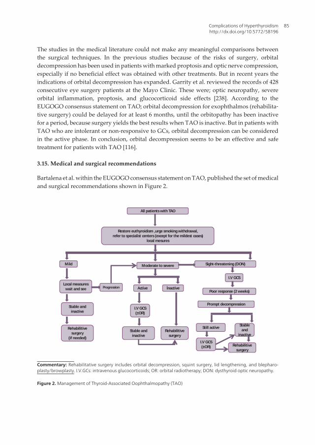
3.15. Medical and surgical recommendations
Bartalena et al. within the EUGOGO consensus statement on TAO, published the set of medicaland surgical recommendations shown in Figure 2.
All patients with TAO
Restore euthyroidism ,urge smoking withdrawal,refer to specialist centers (except for the mildest cases)
local mesures
Mild Moderate to severe Sight-threatening (DON)
Local measureswait and see
Stable andinactive
Rehabilitive surgery
(if needed)
Progression Active İnactive
I.V GCS(±OR)
Stable andinactive
Rehabilitive surgery
I.V GCS
Poor response (2 weeks)
Prompt decompression
Stable and
inactive
Still active
I.V GCS(±OR) Rehabilitive
surgery
Commentary: Rehabilitative surgery includes orbital decompression, squint surgery, lid lengthening, and blepharo‐plasty/browplasty. I.V.GCs: intravenous glucocorticoids; OR: orbital radiotherapy; DON: dysthyroid optic neuropathy.
Figure 2. Management of Thyroid-Associated Oophthalmopathy (TAO)
Complications of Hyperthyroidismhttp://dx.doi.org/10.5772/58196
85
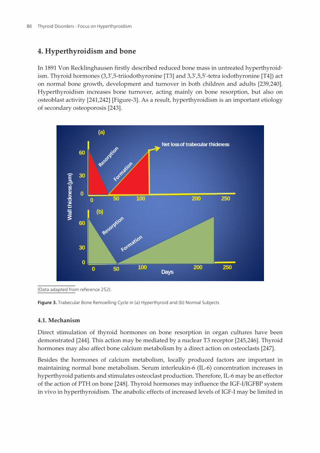
4. Hyperthyroidism and bone
In 1891 Von Recklinghausen firstly described reduced bone mass in untreated hyperthyroid‐ism. Thyroid hormones (3,3',5-triiodothyronine [T3] and 3,3',5,5'-tetra iodothyronine [T4]) acton normal bone growth, development and turnover in both children and adults [239,240].Hyperthyroidism increases bone turnover, acting mainly on bone resorption, but also onosteoblast activity [241,242] [Figure-3]. As a result, hyperthyroidism is an important etiologyof secondary osteoporosis [243].
00
00 200
60
30
60
30
50
50 100 250
100 200 250
(a)
(b)
Wal
l thi
ckne
ss (µ
m)
Days
Net loss of trabecular thickness
(Data adapted from reference 252).
Figure 3. Trabecular Bone Remoelling Cycle in (a) Hyperthyroid and (b) Normal Subjects
4.1. Mechanism
Direct stimulation of thyroid hormones on bone resorption in organ cultures have beendemonstrated [244]. This action may be mediated by a nuclear T3 receptor [245,246]. Thyroidhormones may also affect bone calcium metabolism by a direct action on osteoclasts [247].
Besides the hormones of calcium metabolism, locally produced factors are important inmaintaining normal bone metabolism. Serum interleukin-6 (IL-6) concentration increases inhyperthyroid patients and stimulates osteoclast production. Therefore, IL-6 may be an effectorof the action of PTH on bone [248]. Thyroid hormones may influence the IGF-I/IGFBP systemin vivo in hyperthyroidism. The anabolic effects of increased levels of IGF-I may be limited in
Thyroid Disorders - Focus on Hyperthyroidism86
hyperthyroidism due to the increases of inhibitory IGFBPs that can counteract the anaboliceffects and contribute to the observed net bone loss [249]. T3 activates fibroblast growth factorreceptor-1 (FGFR1) in bone and this can be related with the pathogenesis of skeletal disordersresulting from thyroid disease [250].
4.2. Changes in hyperthyroidism
Histological changes in hyperthyroidism
Thyroid hormones increase the activation of new remodeling cycles. These effects includepredominantly increased osteoclastic activity. Although; excess osteoblastic activity, osteoiddeposition (osteomalacia) and rarefaction (osteoporosis) were also described. These changeswere observed both in trabecular and cortical bone ([51]. In 1978 Mosekilde & Melsen dem‐onstrated a unique histomorphometric pattern; with evidence of both increased osteoblast andosteoclast activity in hyperthyroidism. These changes have given rise to a net loss of bonevolume and were evident in both cortical and trabecular bone. But cortical bone has been rathermore influenced (Figure-4) [252]. An in-vitro organ culture of fetal rat bone, demonstrated adirect stimulation of bone resorption by prolonged treatment with T4 or T3 [244].
Biochemical changes in hyperthyroidism
a. Calcium homeostasis in hyperthyroidism
The majority of patients with hyperthyroidism have normal or near normal total serumcalcium levels. However, ionized serum calcium levels are elevated in most of the patients.Changes in serum calcium levels correlate with serum T3 levels [253,254]. Generally hyper‐calcemia in hyperthyroidism tends to be mild or asymptomatic. Severe (>15 mg/dl) andsymptomatic hypercalcemia is rare [255]. Hypercalcemia usually resolves after attainment ofeuthyroid state by all therapeutic modalities, i.e. subtotal thyroid resection, antithyroid drugsand radioiodine therapy. Symptomatic hypercalcemia can be treated by rehydration, use ofcorticosteroids, calcitonin and phosphate therapy. Reduced renal tubular and intestinalcalcium reabsorption leads to increased urinary and fecal calcium [241]. The hyperadrenergicstate of hyperthyroidism contributes hypercalcemia [256]. These changes correlate positivelywith thyroid hormone levels and cortical osteoclastic activity. Hypercalcemia in hyperthyr‐oidism is unrelated to the parathyroid hormone (PTH) levels [253,254].
b. Phosphorous homeostasis in hyperthyroidism
Most of the patients have increased serum phosphorous levels. But some studies have shownnormal or low levels of serum phosphorous. Increased bone and tissue catabolisms which leadto excess input of phosphorous to the plasma, lower clearance and increased renal tubularreabsorption of phosphorous may lead to hyperphosphatemia in hyperthyroidism. Thesechanges are related to suppressed PTH levels and direct effects of thyroid hormones. Antithy‐roid treatment normalizes serum phosphorous concentration [253].
c. Parathyroid hormone secretion in hyperthyroidism
The reduction of serum PTH levels has been reported for the first time by Bouillon and DeMoor[257]. This observation confirmed by other clinical trials. Increased serum calcium levels inhibit
Complications of Hyperthyroidismhttp://dx.doi.org/10.5772/58196
87
PTH secretion, so there is an inverse relationship between serum PTH and calcium levels.Additionally, serum parathyroid hormone-related peptide (PTH-rP) levels increase inhyperthyroidism. In hyperthyroid patients, a significant elevation in PTH-rP levels wasobtained when compared with healthy controls. After treatment, levels of PTH-rP declined.PTH-rP could be a factor in the pathogenesis of hypercalcemia in hyperthyroid patients [258].
d. Vitamin D in hyperthyroidism
Serum concentrations of 25-hydroxyvitamin D3 (25(OH)D3)and 1,25-dihydroxyvitamin D3
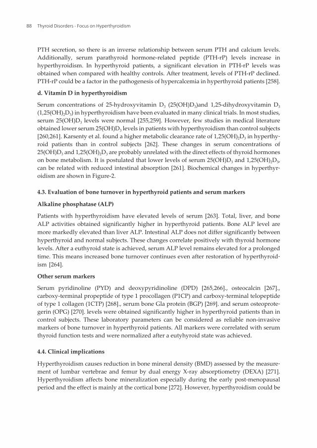
(1,25(OH)2D3) in hyperthyroidism have been evaluated in many clinical trials. In most studies,serum 25(OH)D3 levels were normal [255,259]. However, few studies in medical literatureobtained lower serum 25(OH)D3 levels in patients with hyperthyroidism than control subjects[260,261]. Karsenty et al. found a higher metabolic clearance rate of 1,25(OH)2D3 in hyperthy‐roid patients than in control subjects [262]. These changes in serum concentrations of25(OH)D3 and 1,25(OH)2D3 are probably unrelated with the direct effects of thyroid hormoneson bone metabolism. It is postulated that lower levels of serum 25(OH)D3 and 1,25(OH)2D3,can be related with reduced intestinal absorption [261]. Biochemical changes in hyperthyr‐oidism are shown in Figure-2.
4.3. Evaluation of bone turnover in hyperthyroid patients and serum markers
Alkaline phosphatase (ALP)
Patients with hyperthyroidism have elevated levels of serum [263]. Total, liver, and boneALP activities obtained significantly higher in hyperthyroid patients. Bone ALP level aremore markedly elevated than liver ALP. Intestinal ALP does not differ significantly betweenhyperthyroid and normal subjects. These changes correlate positively with thyroid hormonelevels. After a euthyroid state is achieved, serum ALP level remains elevated for a prolongedtime. This means increased bone turnover continues even after restoration of hyperthyroid‐ism [264].
Other serum markers
Serum pyridinoline (PYD) and deoxypyridinoline (DPD) [265,266]., osteocalcin [267].,carboxy-terminal propeptide of type 1 procollagen (P1CP) and carboxy-terminal telopeptideof type 1 collagen (1CTP) [268]., serum bone Gla protein (BGP) [269]. and serum osteoprote‐gerin (OPG) [270]. levels were obtained significantly higher in hyperthyroid patients than incontrol subjects. These laboratory parameters can be considered as reliable non-invasivemarkers of bone turnover in hyperthyroid patients. All markers were correlated with serumthyroid function tests and were normalized after a eutyhyroid state was achieved.
4.4. Clinical implications
Hyperthyroidism causes reduction in bone mineral density (BMD) assessed by the measure‐ment of lumbar vertebrae and femur by dual energy X-ray absorptiometry (DEXA) [271].Hyperthyroidism affects bone mineralization especially during the early post-menopausalperiod and the effect is mainly at the cortical bone [272]. However, hyperthyroidism could be
Thyroid Disorders - Focus on Hyperthyroidism88
associated with bone loss and may be a risk factor for the development of osteoporosis in pre-menopausal women [273]. It has been well established that hyperthyroidism leads to reducedBMD in female patients, but there is lack of acceptable data in male patients. Majima et al.evaluated BMD and bone metabolism in male patients with hyperthyroidism. The studydemonstrated a high prevalence of cortical bone loss in male patients with hyperthyroidism,especially in elderly patients [274].
Also, endogenous subclinical hyperthyroidism can be a risk factor for osteoporosis. In severalcross-sectional studies, BMD was decreased at multiple sites in pre-and post-menopausalwomen with endogenous subclinical hyperthyroidism [275-277]. This finding, however, wasnot confirmed in other cross-sectional observations in endogenous conditions [278]. Földes etal. demonstrated that numbers of pre-menopausal patients with endogenous sub-clinicalhyperthyroidism BMD of the lumbar spine, femoral neck and the midshaft of the radius werenot significantly decreased. But in post-menopausal women with long-lasting endogenoussub-clinical hyperthyroidism mean densitometric values were slightly, but significantly, lower[279]. Whether endogenous sub-clinical hyperthyroidism significantly affects bone metabo‐lism and increases the risk of fractures remains controversial.
Prolonged hyperthyroidism due to L-thyroxine(L-T4) treatment has been associated withreduced bone mass and thus with the potential risk of premature development of osteoporosis.The effects of L-T4 treatment have been evaluated in many clinical trials. In several studies,bone mineral density was decreased at multiple sites in pre-and post-menopausal womentreated with L-T4 [280,281]. In a study by Giannini et al. post-menopausal thyroidectomizedpatients showed significantly lower bone mass than pre-menopausal patients. A negativecorrelation between time since menopause and bone mass were obtained in post-menopausalL-T4 treated patients [282]. There is lack of acceptable data about the effects of L-T4 treatmenton bone metabolism in male patients. A mild deleterious effect of thyroid hormone excess inthe axial bone mass from male subjects obtained. Male patients with chronic TSH suppressionby L-T4 or history of hyperthyroidism should be assessed by BMD [283]. However, severalrecent studies have failed to show such a harmful effect of L-T4 treatment on BMD [284-286].
It should be noted that other risk factors may affect BMD in hyperthyroid patients. Theseinclude a relative deficiency of insulin-like growth factor type I, dehydroepiandrosteronesulfate [287], vitamin D receptor (VDR) polymorphisms [288] and estrogen [289]. The questionas to whether prolonged hyperthyroidism due to L-T4 treatment increases the risk of fracturesalso remains controversial.
4.5. Diagnosis
BMD measuring by DEXA scan is one of the best techniques to obtain the extent of bonechanges in hyperthyroidism. DEXA scan offers monitoring for response to therapy and withlittle radiation it gives very reliable measurements [290]. Quantitative ultrasound (QUS) mightgive information about bone mass and also on bone elasticity and structure. Advantages arethe lower expense, portability, and lack of radiation exposure [291]. Acotto et al. reportedsignificantly lower QUS parameters in hyperthyroid patients in comparison with controls
Complications of Hyperthyroidismhttp://dx.doi.org/10.5772/58196
89
[292]. For practical use, the development of quality standards and criteria for diagnosingosteoporosis are necessary.
Prevention and treatment of reduced bone density
Patients with a history of hyperthyroidism and who are treated with L-T4 replacement/supressive therapy should be considered at higher risk of reducing BMD and developingclinically significant osteoporosis. These patients should be encouraged to modify other riskfactors for osteoporosis such as smoking, drinking excessive alcohol, low Ca+² intake and lackof exercise [293]. In these patients adequate daily calcium intake should be provided. Kung etal. found that 1,000 mg. daily calcium supplementation prevents bone loss in post-menopausalwomen taking suppressive doses of L-T4 compared with a placebo [294]. Estrogen replacementtherapy prevents a decrease of BMD in postmenopausal women with previous hyperthyroid‐ism and subsequent L-T4 therapy [295,296]. These potentially beneficial effects of estrogenreplacement on BMD in postmenopausal women with a history of thyroid disease suggeststhat estrogen administration should be encouraged in this group.
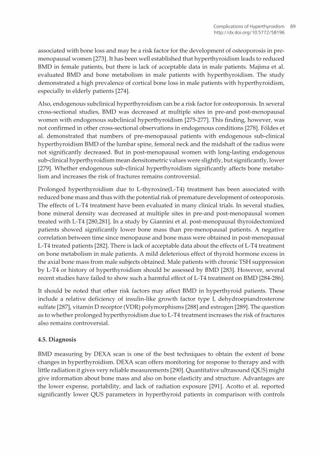
U-Ca+²U-PO4
PTH
Thyroid hormonesU-OHP
U-PYD / DPDALP
Osteocalcin
Ca+²
FaecalCa+²
PO4 Reducedintestinal
absorption
25(OH)D31,25(OH)2D3
N /
Abbreviations are as follows: calcium (Ca), alkaline phosphatase (ALP), 25 hydroxy vitamin D3 (25 OH D), 1,25 dihy‐droxy vitamin D3 (1,25 (OH)2D3), 24,25 dihydroxy vitamin D3 (S-24,25 (OH)2D), parathyroid hormone (PTH), phosphate(PO4), urinary calcium (U-Ca), urinary hydroxyproline (U-OHP), urinary phosphate (U-PO4), urinary pyridinium and de‐oxypyrodinoline (U-PYD/DPD). (Data adapted from reference 292).
Figure 4. Biochemical Changes in Hyperthyroidism.
Thyroid Disorders - Focus on Hyperthyroidism90
Titration of suppressive therapy to maintain serum TSH concentration between a slightly low(e.g. between 0.1-0.5 mU/l) range may prevent bone loss [297]. Guo et al demonstrated areduction of L-T4 dose in post-menopausal women with suppressed serum TSH levels relatedwith deceleration of bone turn over (serum osteocalcin and urinary excretion of bone collagen-derived pyridinium cross-links decreased) and increased BMD (lumbar/femoral) [298]. Carefultitration of L-T4 dosage to maintain biochemical euthyroidism is the most important way toprevent the adverse effect of T4 on bone.
The major question is whether the decrease in BMD caused by hyperthyroidism can bereversible. Many clinical trials have obtained that there is some reversibilty of bone mass aftertreatment of hyperthyroidism [293,299-301]. Another question is whether antiresorptivetreatment is effective on hyperthyroidism related osteoporosis. Two clinical trials evaluatedthe increase of BMD in osteoporotic/osteopenic hyperthyroid patients treated with only anti-thyroid drugs versus patients treated with anti-thyroid drugs and alendronate. The combina‐tion of anti-thyroid drugs and alendronate was found to be more efficacious than anti-thyroidtherapy alone [302,303]. Majima et al. evaluated the efficacy of risedronate for the treatmentof osteoporosis/osteopenia in patients with Graves' disease. The percentage increases in BMDat the lumbar spine and distal radius were significantly greater in patients treated with an anti-thyroid drug and risedronate than treated with an anti-thyroid drug only [304]. These findingsconclude that bisphosphonates can be considered as a treatment option in decreased bone masscases associated with hyperthyroidism. Kung et al. demonstrated intranasal calcitonin withcalcium supplements was not more effective than calcium supplements alone in preventingbone loss induced by thyroxine suppressive therapy in post-menopausal women [305]. Jódaret al. designed a prospective study to evaluate the effects of antiresorptive therapy with nasalsalmon calcitonin (CT) in hyperthyroid patients receiving standard medical treatment. Theyshowed that treatment with nasal CT has no additional beneficial effect compared with theattainment of the euthyroid state [306]. But in another study by Akçay et al. the addition ofintranasal calcitonin to the anti-thyroid drugs was found to be effective in preventing thedegradation of bone [307]. More prospective, randomized and controlled trials, which includelarge series of patients, are required to obtain more accurate results about antiresorptivetreatment in hyperthyroidism.
5. Thyroid storm
Thyroid storm (TS) [thyrotoxic crisis] is an uncommon, life-threatening condition reflectingthe augmentation of the manifestations of thyrotoxicosis. Mortality rates are high (10-20%) andoccur in less than 10% of patients hospitalized for thyrotoxicosis. TS occurs nine to ten timesmore commonly in women than in men, and the most common aetiology is Graves’ hyper‐thyroidism. However, any aetiology of thyrotoxicosis can cause TS. This situation can beprecipitated by a number of factors shown in Table-3 [308-312].
The pathophysiology of thyroid storm is not fully understood. The current theory is that;increased numbers of catecholaminergic receptors being exposed to increased catecholamine
Complications of Hyperthyroidismhttp://dx.doi.org/10.5772/58196
91
levels in states of stress. Decreased binding to thyroxine-binding globulin (TBG), markedlyincrease in free thyroid hormone levels may also play an important role [313].
∙ Severe infections (pulmonary) ∙ Direct trauma or surgical manipulation of the thyroid gland
∙ Diabetic ketoacidosis (DKA) ∙ Biological agents such as interleukin-2 and α-interferon
∙ Surgery ∙ Discontinuation of anti-thyroid medications or poor patient
adherence to the treatment
∙ Trauma ∙ Cerebrovascular disease
∙ Pulmonary thrombo-embolism ∙ Myocardial infarction (MI)
∙ Salicylates * ∙ Exogenous thyroid hormone intake
∙ Administration of large quantities of exogenous
iodine **
∙ Pregnancy
∙ Radioactive iodine therapy ∙ Unknown factors
*They increase the concentration of circulating free thyroid hormones to critical levels
**Such as iodinated contrast agents or amiodarone
Table 3. Precipitating Factors for Thyroid Storm
Clinical presentation of thyroid storm
Symptoms of thyroid storm are similar to those of hyperthyroidism, but they are more sudden,severe and extreme. The main findings are high fever (38-41° C) accompanied by excessivesweating and flushing. Other common symptoms include; altered mental status (confusion,agitation, overt psychosis, and in extreme cases, even coma), cardiovascular complications[tachycardia, cardiac arrhythmias (including atrial fibrillation), high systolic bood pressure,low diastolic bood pressure and congestive heart failure], diffuse muscle weakness, tremor orfasciculations and neuropsychiatric syndromes. Gastrointestinal involvement presents withnausea, vomiting, diarrhoea and abdominal pain. Liver dysfunction (cardiac failure withhepatic congestion or hypoperfusion, direct effect of the excess thyroid hormones) could bepresent with jaundice [309,314]. Elderly patients often present with apathy, stupor, cardiacfailure, coma and minimal signs of thyrotoxicosis so-called apathetic thyroid storm [313].
The scoring system suggested by Burch and Wartofsky (Table 4) illustrates the typical featuresof thyroid storm [315]. The clinical symptomatology is sometimes difficult to distinguish fromother medical emergencies [308]. Differential diagnosis of TS is shown in Table-5.
5.1. Diagnosis
Diagnosis depends on primarily clinical findings. There are no specific laboratory testsavailable. Serum thyroid hormone levels (i.e. free T3 [FT3] and free T4 [FT4]) are elevated withsuppressed TSH levels (with the rare exceptions being states of thyroid hormone resistance orTSH secreting pituitary adenomas). An immediate search should begin for precipitatingfactors. Pregnancy should be excluded urgently in any woman of childbearing age withurinary or plasma assessment of human chorionic gonadotropin (HCG) levels. Routine
Thyroid Disorders - Focus on Hyperthyroidism92
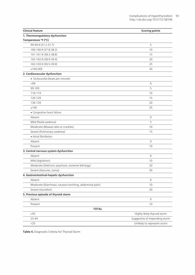
Clinical feature Scoring points
1. Thermoregulatory dysfunctionTemperature °F (°C)
99-99.9 (37.2-37.7) 5
100-100.9 (37.8-38.2) 10
101-101.9 (38.3-38.8) 15
102-102.9 (38.9-39.4) 20
103-103.9 (39.5-39.9) 25
≥104 (40) 30
2. Cardiovascular dysfunction
∙ Tachycardia (beats per minute)
<99 0
99-109 5
110-119 10
120-129 15
130-139 20
≥140 25
∙ Congestive heart failure
Absent 0
Mild (Pedal oedema) 5
Moderate (Bibasal rales or crackles) 10
Severe (Pulmonary oedema) 15
∙ Atrial fibrillation
Absent 0
Present 10
3. Central nervous system dysfunction
Absent 0
Mild (Agitation) 10
Moderate (Delirium, psychosis, extreme lethargy) 20
Severe (Seizures, coma) 30
4. Gastrointestinal-hepatic dysfunction
Absent 0
Moderate (Diarrhoea, nausea/vomiting, abdominal pain) 10
Severe (Jaundice) 20
5. Previous episode of thyroid storm
Absent 0
Present 10
TOTAL
>45 Highly likely thyroid storm
25-44 Suggestive of impending storm
<25 Unlikely to represent storm
Table 4. Diagnostic Criteria for Thyroid Storm
Complications of Hyperthyroidismhttp://dx.doi.org/10.5772/58196
93
biochemical tests, urinalysis, urine sediment, complete blood count, chest X-ray and relatedcultures should be made, to exclude infectious diseases. Electrocardiography (ECG) andcardiac enzymes should be procured, to exclude arrhythmia and myocardial infarction (MI)[313]. Leukocytosis, increased transaminase-bilirubin levels, hypoglycemia and lactic acidosiscan be seen in laboratory evaluation [316,317].
Neurolept malignant syndrome Hypertensive encephalopathy
Malignant hyperthermia Alcohol withdrawal
Phaeochromocytoma Benzodiazepine / barbiturate withdrawal
Hypoglycemia Opioid withdrawal
Hypoxia Heat stroke
Sepsis Encephalitis / meningitis
Table 5. Differential Diagnosis of Thyroid Storm
5.2. Management
The patient should be managed in an intensive care unit. ABCDEs (i.e. airway; breathing;circulation; disability, i.e. conscious level; and examination) should be provided as soon aspossible. Precipitant factors such as infection, trauma, MI, DKA, and other underlyingprocesses should be managed as per standard care [313].
General supportive care
First of all haemodynamic stability should be provided. Supportive care includes coolingmeasures, intravenous (IV) fluid-electrolyte replacement and nutritional support. In order tocontrol profound pyrexia cooling blankets and acetaminophen can be used. Salicylates shouldbe avoided (increase the concentration of circulating free thyroid hormones). Tachyarrhyth‐mias, which cause haemodynamic instability should be managed with cardioversion bydefibrillation. Otherwise, tachyarrhythmias can be managed with appropriate anti-arrhythmictherapy. Diuretics, ACE inhibitors and digitalis can be used carefully for heart failure. Basedon arterial blood gas (ABG) analysis and other assessments; ventilatory support, either withnon-invasive positive pressure ventilation (NIPPV) or intubated ventilation, could be per‐formed. In patients with severe agitation, medical intervention could be difficult. Sedativessuch as haloperidol or a benzodiazepine can be used in this situation. Phenobarbital increasesperipheral metabolism and inactivation of FT3-FT4. Chlorpromazine has the additional benefitof reducing body temperature through effects on central thermoregulation. However, theseagents should be monitored for respiratory side effects. Nutritional support is important. Thisincludes vitamin (e.g. thiamine) and glucose replacement (as liver glycogen stores are depletedduring a thyroid storm) [313].
Thyroid-specific therapy
Thyriod-specific therapy includes; decreasing thyroid hormone synthesis, prevention ofthyroid hormone release and decreasing peripheral action of circulating thyroid hormones[318]. In the treatment of thyroid storm, the five B’s should be kept in mind. This includes ‘Bs’:
Thyroid Disorders - Focus on Hyperthyroidism94
Block synthesis (i.e. antithyroid drugs); Block release (i.e. iodine); Block T4 into T3 conversion(i.e. highdose propylthiouracil [PTU], propranolol, corticosteroid and, rarely, amiodarone);Betablocker; and Block enterohepatic circulation (i.e. cholestyramine) [313].
Anti-thyroid drugs include PTU or carbimazole (or methimazole). PTU was preferred becauseof its more rapid onset of action, short half-life and inhibition of peripheral deiodinase enzyme-mediated conversion of T4 into T3. PTU could be administered orally or via a nasogastric (NG)tube, if the patient is not suitable for oral administration, both PTU and methimazole can begiven as a rectal suppository or enema. PTU is administered with a loading dose of 600mgfollowed by a dose of 200-250 mg every 4-6 hours. Carbimazole (or methimazole) is adminis‐tered at a dose of 20-30mg every 4-6 hours [318]. The major concern about PTU is hepatoxicity.In the thyroid storm management, there are not any comparative trials showing superiorityof PTU over either carbimazole or methimazole. It’s now recommended that using eithercarbimazole or methimazole in thyroid storm (unless there is a contra-indication such aspregnancy), and achieving T4 into T3 conversion inhibition just with beta-blockers andcorticosteroids [319].
Iodine should be administered at least 1 hour after PTU or carbimazole (or methimazole)administration. In this way undesired tyrosine residue iodination and enrichment of thyroidhormone stores could be prevented. Iodine could be administered in various formulations(Table-6) [313].
In order to block the adrenergic effects of thyroid hormones betablockers should be adminis‐tered immediately. A non-cardiac specific betablocker; propranolol can be used for thispurpose. Propranolol has the advantages of being suitable for IV administration and ofinhibition of peripheral T4-T3 conversion at high doses. Decompensated cardiac function,triggered with tachycardia, can be treated with beta-blockers. However, beta-blockers shouldbe used with caution in patients with a history of heart failure. Cardioselective beta-blockerssuch as metoprolol or atenolol could be used in patients with asthma and chronic obstructivepulmonary disease, which are contra-indications for non-cardiac specific betablockers. Ifunfavorable cardiac effects are expected, a short-acting beta-blocker; esmolol, can be used asan alternative drug. Diltiazem, a calcium-channel blocker, can be used when there is absolutecontra-indication for beta-blockers (Table-6) [313].
Thyroid hormones accelerate cortisol metabolism and may trigger an adrenal crisis. Thyroidcrisis give rise to low plasma levels of ACTH and cortisol. Therefore, against a possible adrenalinsufficiency, exogenous corticosteroid therapy should be administered [320]. Furthermore,corticosteroids inhibit peripheral conversion of T4 into T3 and have been shown to improveoutcomes in patients with thyroid storm [318]. Hydrocortisone or an equivalent corticosteroidcan be used for this purpose (Table-6). Then, the treatment should be withdrawn gradually,based on the required duration of steroid therapy.
Other possible treatment options are lithium carbonate, potassium perchlorate and cholestyr‐amine. Lithium inhibits thyroid hormone release from the gland and reduces iodination oftyrosine residues. Lithium carbonate can be an alternative treatment option when there is acontraindication or previous toxicity history to thionamide therapy. The major concern about
Complications of Hyperthyroidismhttp://dx.doi.org/10.5772/58196
95
lithium carbonate is it’s toxicity. Potassium perchlorat inhibits iodide transport into thethyrocyte [313]. Erdogan et al. demonstrated that the combination of thionamides, corticoste‐roids and potassium perchlorat for a short period could be effective in the treatment ofamiodarone-induced thyrotoxicosis [321]. The most important side effects of potassiumperchlorat are aplastic anaemia and nephritic syndrome. To inhibit the enterohepatic circula‐tion of thyroid hormones, a T4 and T3 binding resin, cholestyramine can be used [322].
Emerging treatments
In spite of all treatment approaches, clinical improvement could not be obtained. In thissituation, different treatment approaches can be tried in order to remove thyroid hormonesfrom the circulation.
Plasmapheresis should be considered as a treatment option, especially when patients havefailed or cannot tolerate conventional therapy. Plasmapheresis leads to amelioration ofsymptoms and a significant decline in thyroid hormone levels, providing a window to treatdefinitively with thyroidectomy [323,324]. In each session only small amounts of thyroidhormones can be removed from the circulation, therefore plasmapharesis can be repeated.Koball et al. reported a case of thyrotoxic crisis treated with plasmapheresis and single passalbumin dialysis. Thyroxine can be bound by albumin and removed by extracorporeal single-pass albumin dialysis (SPAD). The patient underwent two sessions of plasmapheresis withoutclinical response. After four SPAD treatments clinical status of the patient improved. Accord‐ing to this report SPAD represents a safe and efficient alternative to plasmapheresis [325].
Charcoal haemoperfusion has also been demonstrated to be effective in thyrotoxic states [326].
6. Thyrotoxic hypokalaemic periodic paralysis
Hyperthyroidism can be related with muscular disorders such as acute and chronic thyrotoxicmyopathies, exophthalmic ophthalmoplegia, myasthenia gravis, and periodic paralysis [327].The association between thyrotoxicosis and periodic paralysis was first described by Rosenfeldin 1902. Thyrotoxic hypokalaemic periodic paralysis (THPP) is a rare condition which occursin 2% of patients with thyrotoxicosis. THPP is generally sporadic, but autosomal recessive orautosomal dominant cases have been reported and may be associated with certain HLAhaplotypes [HLA-Bw46 / B5 / DRw8 / CW7] [328-330]. THPP has been reported in many ethnicgroups such as Asian populations and Caucasians [331]. THPP appears almost exclusively(85%) in young men between the ages of 20-39 [327].
Many aetiological factors that can lead to hyperthyroidism may be associated with THPP. Ithas been reported that the most important aetiologic factor is Graves’ disease according tolarge case series [332,333]. Other possible aetiological factors are subacute thyroiditis, silentthyroiditis [334], autonomously functioning thyroid nodules [335], thyrotropin-secretingpituitary adenomas [336,337], ingestion of excessive thyroxine [338], thyroxine-containingherbal and dietary supplements [339], iodine induced thyrotoxicosis with inadvertent use ofiodine or with drugs containing iodine such as iodinate contrast agents or amiodarone
Thyroid Disorders - Focus on Hyperthyroidism96
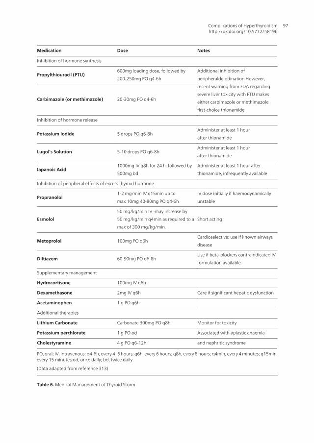
Medication Dose Notes
Inhibition of hormone synthesis
Propylthiouracil (PTU)600mg loading dose, followed by
200-250mg PO q4-6h
Additional inhibition of
peripheraldeiodination However,
recent warning from FDA regarding
severe liver toxicity with PTU makes
either carbimazole or methimazole
first-choice thionamide
Carbimazole (or methimazole) 20-30mg PO q4-6h
Inhibition of hormone release
Potassium Iodide 5 drops PO q6-8hAdminister at least 1 hour
after thionamide
Lugol’s Solution 5-10 drops PO q6-8hAdminister at least 1 hour
after thionamide
Iapanoic Acid1000mg IV q8h for 24 h, followed by
500mg bd
Administer at least 1 hour after
thionamide, infrequently available
Inhibition of peripheral effects of excess thyroid hormone
Propranolol1-2 mg/min IV q15min up to
max 10mg 40-80mg PO q4-6h
IV dose initially if haemodynamically
unstable
Esmolol
50 mg/kg/min IV -may increase by
50 mg/kg/min q4min as required to a
max of 300 mg/kg/min.
Short acting
Metoprolol 100mg PO q6hCardioselective; use if known airways
disease
Diltiazem 60-90mg PO q6-8hUse if beta-blockers contraindicated IV
formulation available
Supplementary management
Hydrocortisone 100mg IV q6h
Dexamethasone 2mg IV q6h Care if significant hepatic dysfunction
Acetaminophen 1 g PO q6h
Additional therapies
Lithium Carbonate Carbonate 300mg PO q8h Monitor for toxicity
Potassium perchlorate 1 g PO od Associated with aplastic anaemia
Cholestyramine 4 g PO q6-12h and nephritic syndrome
PO, oral; IV, intravenous; q4-6h, every 4_6 hours; q6h, every 6 hours; q8h, every 8 hours; q4min, every 4 minutes; q15min,every 15 minutes;od, once daily; bd, twice daily.
(Data adapted from reference 313)
Table 6. Medical Management of Thyroid Storm
Complications of Hyperthyroidismhttp://dx.doi.org/10.5772/58196
97
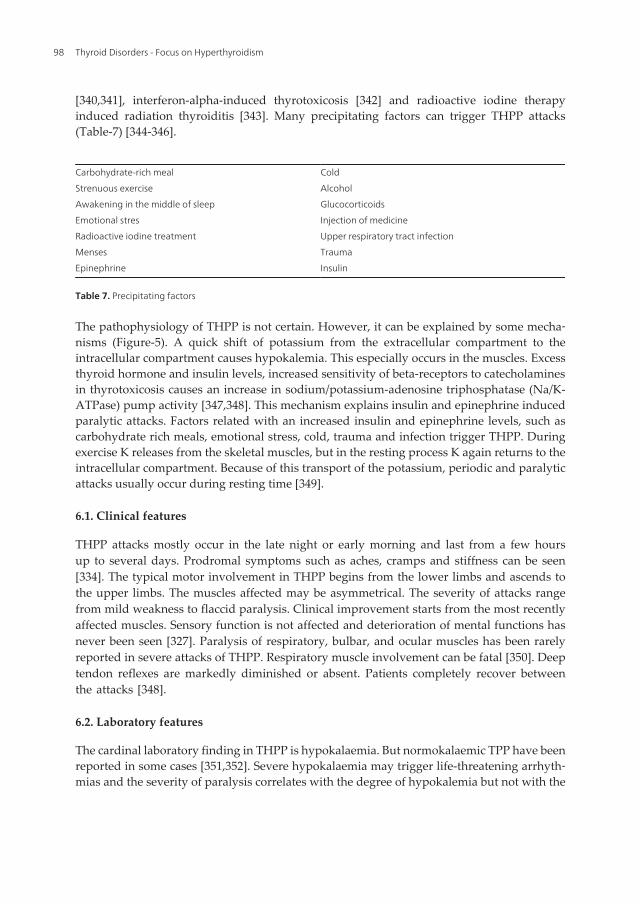
[340,341], interferon-alpha-induced thyrotoxicosis [342] and radioactive iodine therapyinduced radiation thyroiditis [343]. Many precipitating factors can trigger THPP attacks(Table-7) [344-346].
Carbohydrate-rich meal
Strenuous exercise
Awakening in the middle of sleep
Emotional stres
Radioactive iodine treatment
Menses
Epinephrine
Cold
Alcohol
Glucocorticoids
Injection of medicine
Upper respiratory tract infection
Trauma
Insulin
Table 7. Precipitating factors
The pathophysiology of THPP is not certain. However, it can be explained by some mecha‐nisms (Figure-5). A quick shift of potassium from the extracellular compartment to theintracellular compartment causes hypokalemia. This especially occurs in the muscles. Excessthyroid hormone and insulin levels, increased sensitivity of beta-receptors to catecholaminesin thyrotoxicosis causes an increase in sodium/potassium-adenosine triphosphatase (Na/K-ATPase) pump activity [347,348]. This mechanism explains insulin and epinephrine inducedparalytic attacks. Factors related with an increased insulin and epinephrine levels, such ascarbohydrate rich meals, emotional stress, cold, trauma and infection trigger THPP. Duringexercise K releases from the skeletal muscles, but in the resting process K again returns to theintracellular compartment. Because of this transport of the potassium, periodic and paralyticattacks usually occur during resting time [349].
6.1. Clinical features
THPP attacks mostly occur in the late night or early morning and last from a few hoursup to several days. Prodromal symptoms such as aches, cramps and stiffness can be seen[334]. The typical motor involvement in THPP begins from the lower limbs and ascends tothe upper limbs. The muscles affected may be asymmetrical. The severity of attacks rangefrom mild weakness to flaccid paralysis. Clinical improvement starts from the most recentlyaffected muscles. Sensory function is not affected and deterioration of mental functions hasnever been seen [327]. Paralysis of respiratory, bulbar, and ocular muscles has been rarelyreported in severe attacks of THPP. Respiratory muscle involvement can be fatal [350]. Deeptendon reflexes are markedly diminished or absent. Patients completely recover betweenthe attacks [348].
6.2. Laboratory features
The cardinal laboratory finding in THPP is hypokalaemia. But normokalaemic TPP have beenreported in some cases [351,352]. Severe hypokalaemia may trigger life-threatening arrhyth‐mias and the severity of paralysis correlates with the degree of hypokalemia but not with the
Thyroid Disorders - Focus on Hyperthyroidism98
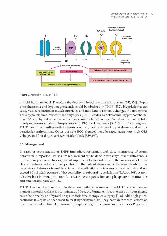
thyroid hormone level. Therefore the degree of hypokalaemia is important [353,354]. Hypo‐phosphataemia and hypomagnesaemia could be obtained in THPP [332]. Hypokalemia cancause vasoconstriction in muscle arterioles and may lead to ischemic changes in sarcolemma.Thus hypokalaemia causes rhabdomyolysis [355]. Besides hypokalaemia, hypophosphatae‐mia [356] and hyperthyroidism alone may cause rhabdomyolysis [357]. As a result of rhabdo‐myolysis, serum creatine phosphokinase (CPK) level increases [332,358]. ECG changes inTHPP vary from nondiagnostic to those showing typical features of hypokalaemia and seriousventricular arrhythmias. Other possible ECG changes include rapid heart rate, high QRSvoltage, and first-degree atrioventricular block [359,360].
6.3. Management
In cases of acute attacks of THPP immediate restoration and close monitoring of serumpotassium is important. Potassium replacement can be done in two ways; oral or intravenous.Intravenous potassium has significant superiority to the oral route in the improvement of theclinical findings and it is the major choice if the patient shows signs of cardiac dysrhythmia,respiratory distress or is unable to take oral medications. Potassium replacement should notexceed 90 mEq/24h because of the possibility of rebound hyperkalemia [327,344,361]. A non-selective beta-blocker, propranolol, increases serum potassium and phosphate concentrationsand ameliorates paralysis [362].
THPP does not disappear completely unless patients become euthyroid. Thus, the manage‐ment of hyperthyroidism is the mainstay of therapy. Permanent treatment is so important andcould be done by antithyroid drugs, radioiodine therapy or surgery [348]. Although gluco‐corticoids (GCs) have been used to treat hyperthyroidism, they have detrimental effects oninsulin sensitivity. Thus GCs can mimic this physiologic process and induce attacks. Physicians
ATP ATP
Intracellular compartment
Extracellular compartment
Musclemembrane
Ca++ pump
Abnormal Ca+ channel(voltage operated)
Thyrotoxicosis
Na+/ K+ ATPase activitiy
Catecholamine-induced hyperinsulinemia
Hypersensitivity to glucose load
Postexercise reuptake of K+ into muscle cell
Rebound response after intense exercise
K+ Ca++ Ca++
Ca++
Na+
Na+
3Na+
2K+
Figure-5: Pathophysiology of THPP
Figure 5. Pathophysiology of THPP
Complications of Hyperthyroidismhttp://dx.doi.org/10.5772/58196
99
should be aware of the risk of triggering THPP when using high-dose GCs in the thyrotoxicphase [363]. To prevent recurrence of attacks, precipitating factors should be avoided.
The use of propranolol is important during early treatment with anti-thyroid drugs or afterradioactive iodine when the euthyroid status is not yet achieved. Propranolol also reduces thefrequency and severity of attacks. If the patient's serum potassium level is between normalranges, prophylactic potassium supplementation is not effective to prevent the attacks.Acetazolamide may worsen the attacks in THPP and should be avoided [364,365].
7. Thyrotoxicosis related psychosis and convulsion
7.1. Hyperthyroidism and central nervous system
The brain, particularly the limbic system (amygdala and hippocampus), has a large numberof Tri-iodothyronine (T3) receptors. These receptors affect a variety of functions such asemotion, behavior, and long term memory [366]. Excess thyroid hormones affect neurotrans‐mitters such as serotonin, dopamine, or second messengers. This could be a possible explan‐ation for the relation between neuropsychiatric symptoms and hyperthyroidism [367].Evidence suggests that the modulation of the beta-adrenergic receptor response to catechola‐mines in the central nervous system by thyroid hormones in thyrotoxic patients, may contrib‐ute to psychotic behavior [368].
As a result, people with thyroid dysfunction could be presented with a wide variety ofneuropsychiatric symptoms such as anxiety, irritability, unstable mood, fatigue, insomnia,dementia, confusion state, depression, thyrotoxic crisis, seizures, pyramidal signs, chorea,attention deficit-hyperactivity disorder, sleepwalking (somnambulism)and Hashimoto'sencephalopathy. “Apathetic thyrotoxicosis” is an unusual presentation of hyperthyroidism,characterised by depression, psychomotor slowing and apathy and occurring mostly in theelderly [369-371].
7.2. Psychosis
More than 150 years ago, von Basedow first described a psychotic illness, probably mania, ina patient with exophthalmic goitre [372]. More recent case reports have emphasised depressive[373], manic [374], paranoid [375] and schizophreniform features [372]. But psychotic reactionsas the presenting feature of thyrotoxicosis is extremely rare, it was reported in 1% of cases andmost patients who develop psychosis have been previously diagnosed with mania and/ordelirium [376]. The occurrence of psychosis depends on the severity and duration of thyroiddisease and the underlying predisposition of the individual to psychiatric instability [377].
Most of the cases caused by thyrotoxicosis or hyperthyroidism have been described in patientswith Graves’ disease or toxic multinodular goiter. Therefore, transient thyroiditis or factitiousthyrotoxicosis can be related with neuropsychiatric symptoms, including organic psychosis[378]. Only one case each of postpartum thyroiditis [379] and subacute thyroiditis [377] have
Thyroid Disorders - Focus on Hyperthyroidism100
been reported related with psychosis. The relations between thyroid autoimmunity andanxiety disorders have been reported [380].
But it should be noted that psychiatric disorders can trigger hyperthyroxinemia [381].Differential diagnosis between the two conditions is important. In such cases the TSH level isusually in the upper “normal” range and free thyroxine elevation is modest and transient andis usually seen within 1 week. This entity has not been established clearly, but may beconsidered a form of non-thyroidal illness [382].
7.3. Management
Treatment of hyperthyroidism by standard anti-thyroid drugs (propylthiouracil, methima‐zole, carbimazole) results with improvement in mental and cognitive symptoms. Psychiatricimprovements parallel with improvements in endocrine symptoms. Trzepacz et a.l alsoshowed that a similar improvement both in psychiatric and endocrine symptoms by two weeksof propranolol treatment could be obtained [383,384].
Because of their slow onset of action and potential toxicity, psychotropic drugs (lithium,benzodiazepines, antidepressants and antipsychotics) are not recommended as the primarytreatment option for neuropsychiatric symptoms caused by hyperthyroidism. Dopaminereceptor blockade with an antipsychotic such as haloperidol can be used in patients with severeneuropsychiatric symptoms. If there is an exhibited psychiatric disease before hyperthyroid‐ism, β-adrenoceptor antagonists may not be effective and neuropsychiatric symptoms mayremain after euthyroidism has been achieved, in which case psychotropic drug treatmentshould be given. Selective serotonin reuptake inhibitors, lithium and benzodiazepines can beused for the treatment of neuropsychiatric symptoms [366].
If therapy fails with the above-mentioned options, more radical approaches such as radioactiveiodine ablation and thyroidectomy should be considered [377,385].
7.4. Convulsion
Seizures in hyperthyroidism have rarely been reported in medical literature. Most of the caseswere related with Graves’ hyperthyroidism [386-388]. Other aetiologic factors were massivelevothyroxine ingestion [389], L-thyroxine treatment for hypothyroidism [390,391], subclinicalhyperthyroidism [392] and Hashimoto’s encephalopathy (HE) [393,394]. The pathophysiologyof HE is not clear but increased antithyroid antibodies in all affected patients supports anautoimmune etiology. Generally, patients in medical literaure were euthyroid or hypothyroid.But Sakurai et al reported a case of HE associated with Basedow's disease presented withhyperthyroidism. Another possible aetiologic factor for seizures in hyperthyroidism may besuperior sagittal sinus (SSS) thrombosis. Hyperthyroidism may cause hypercoagulabilty,venous stasis and could be related with thrombosis. All cases of seizures in SSS thrombosis inmedical literature were known cases of hyperthyroidism [395-397].
The certain incidence of seizures in hyperthyroidism is still unknown. Song et al. recentlyinformed a 0.2% prevelance in a retrospective study [398]. Seizures were seen more frequent
Complications of Hyperthyroidismhttp://dx.doi.org/10.5772/58196
101
in women than in men, but no differences in the pattern of clinical seizures or electroence‐phalography (EEG) were demonstrated between the two gender [399]. Most of the reports arefor generations of childhood and young adults.
Hyperthroidism can exacerbate seizures and paroxysmal EEG abnormalities in patients witha diagnosis of epilepsy [390,400] or focal/generalized seizures only during thyrotoxicosis inpatients without established epilepsy [401]. The mechanisms of seizure induction by thyroidhormones are not clear, but there are some possible explanations. Thyroid hormones mayinfluence the activity of sodium potassium adenosine–triphosphatase, leading to severelyaltered concentrations of sodium in cerebral cells. In this way thyroid hormones triggerseizures by lowering the seizure threshold [402-405].
To confirm the diagnosis patients should be evaluated for the other causes of seizures. Anyepileptic focus or organic lesion should not be obtained with EEG, CT or MRI. Hypoglycemia,hypoxemia, serum electrolyte imbalance, acid-base imbalance or possible central nervoussystem infections should be excluded [406]. Hyperthyroidism should be confirmed by lowserum thyroid-stimulating hormone (TSH) and high concentrations of T4, T3, or both. EEGcan be used as another diagnostic tool. Severity of hyperthyroidism correlates with EEGparameters [399]. Generally EEG abnormalities regress after a euthyroid state achieved. ThusEEG can be used to assess response to treatment [406]. The reversibility of EEG findingsdemontrates that seizures in hyperthyroidism are functional, not organic [407]. But EEG maynot be positive in all cases of hyperthyroidism with seizures, and also positive EEGs in all casesof hyperthyroidism may not manifest clinical seizures [399].
7.5. Management
Treatment of hyperthyroidism with standard medical approaches (propylthiouracil, lugolsiodine, propanolol and dexamethasone with additional vitamin B complex) have been shownto be effective in patients with seizures. Anti-epileptic drugs may have been useful whenstandard medical approaches were not adequate to control seizures. Thus, various anti-epileptic drugs (carbamazepine, phenytoin, sodium valproate, diazepam, or clonazepam)were used to treat seizures in the acute phase [399,408]. HE responds to glucocorticoids,immunosuppressive therapy and plasmapheresis which supports the autoimmune etiology.Seizure exacerbations usually remit when patients become euthyroid with treatment [409,410].
8. Thyrotoxicosis related diabetes mellitus
Glucose metabolism disorders can be seen in thyroid disorders, especially in hyperthyroidism.Hyperthyroid patients have a higher risk of developing diabetes mellitus. The pathogenesisis complex and there is lack of data about prevalence and severity in the literature [411].
8.1. Epidemiology
In young individuals with an autoimmune thyroid disease (as Graves’ disease) it is commonto develop type 1 diabetes. It is due to the alteration of the immune system which leads to a
Thyroid Disorders - Focus on Hyperthyroidism102
pathological reaction against self-antigens. In fact other autoimmune diseases like celiacdisease can also occur. Thyroid auto-immunity is common in type 1 diabetic patients, with upto 27% of them having detectable titers of anti-thyroid peroxidase antibodies [412]. A preva‐lence study by Greco et al. confirmed the frequent association between Graves' disease andtype 1 diabetes. Graves' disease often preceded diagnosis of type 1 diabetes, particularly infemale subjects with a high age at diabetes onset [413].
In older patients, hyperthyroidism can be associated with type 2 diabetes. The pathogeneticmechanism can be explained by insulin resistance and metabolic derangement [414]. A studyshowed that patients on treatment for hyperthyroid Graves’ disease were almost twice as likelyto develop type 2 diabetes than the general population, although they were less likely to beoverweight and were less likely to have family history. This suggests an association betweenGraves' disease and type 2 diabetes [415].
8.2. Genetic predisposition
Studies have been performed for HLA-DR3, HLA-DR4 CTLA-4, and PTPN22. These studiessuggest that abnormalities in antigen presentation and T cell activation might play a significantrole in the shared genetic etiology of type 1 diabetes and auto-immune thyroid disease [416].
8.3. Hyperthyroidism and glucose homeostasis
The effect of hyperthyroidism on glucose homeostasis is complex [417]. Hyperthyroidismstimulates increased metabolism in many tissues, leading to an increased demand for glucose[418]. Altough having insulin resistance, as long as pancreatic beta cell functions remainnormal, overt diabetes does not develop in a hyperthyroidism.
Gastric emptying and intestinal absorption
It has been suggested that rapid gastric emptying and increased rates of intestinal absorptionof glucose could be responsible for altered glucose tolerance in hyperthyroidism [419]. Incontrast to this, recent studies found that gastric emptying has been decreased [420,421] orunchanged [422,423] in hyperthyroidism.
Liver
Hyperthyroidism can cause insulin resistance with direct and indirect effects on the liver. Thedirect effect is to increase basal hepatic glucose output by promoting gluconeogenesis andglycogenolysis [424]. A sympathetic pathway from the hypothalamic paraventricular nucleusto the liver has been proposed as a central pathway for modulation of hepatic glucosemetabolism by thyroid hormone, which forms the indirect effect [425].
Peripheral tissues
Glucose uptake in peripheral tissues especially in the skeletal muscle have been found to beincreased in hyperthyroisdism [426-429]. This increased uptake is mainly related with anincrease in insulin-stimulated glucose oxidation rates [430-433]. Furthermore, reducedglycogenogenesis and insulin-stimulated nonoxidative glucose elimination results with
Complications of Hyperthyroidismhttp://dx.doi.org/10.5772/58196
103
intracellular glucose being redirected towards glycolysis and lactate formation [428,429,434].The release of lactate from peripheral tissues back to the liver contributes to the Cori cyclewhere more hepatic glucose is being produced [434-436]. In the adipose tissue, lipolysisincreases in the fasting state. This results with an increased production of glycerol andnonesterified fatty acids (NEFA). Increased glycerol generated by lipolysis and increasedamino acids generated by proteolysis are used for gluconeogenesis. NEFA stimulate gluco‐neogenesis and provide substrate to other tissues such as muscle, for oxidation [437].
Cytokines
There is an interaction between thyroid hormones and adipose tissue derived cytokines. Effectsof thyroid hormones on production rates and plasma levels of these cytokines could be usedto explain mechanisms of insulin resistance in hyperthyroidism. Adipose tissue derivedcytokines are adiponectin, leptin, interleukin-6, tumor necrosis factor-α, resistin and visfatin.There are conflicting reports about the role of these cytokines to insulin resistance in hyper‐thyroidism [418].
Insulin and glucagon secretion
Decreased [438-442], normal, or even increased [437,443] levels of plasma insulin have beenreported in hyperthyroidism. There are conflicting reports about insulin secretion in hyper‐thyroidism. But it is more consistent that insulin degradation increases. Hyperthyroidismaugments renal clearance of insulin [428,444,445]. Another important pathologic changeinduced by long-term hyperthyroidism has been shown to be irreversible pancreatic damage[446-448]. Secretion and metabolic clearance rates of glucagon have been found increased inhyperthyroidism [449]. Increased levels of growth hormone and catecholamines that accom‐pany hyperthyroidism may further contribute to insulin resistance. Interorgan communicationin hyperthyroidism are shown in Figure-6 [418].
8.4. Subclinical hyperthyroidism and glucose homeostasis
Subclinical hyperthyroidism could be endogenous and exogenous. Both of them have alsobeen associated with insulin resistance in some studies [450,451]. But there are conflictingreports in the literaure about insulin resistance in exogenous subclinical hyperthyroidism.Yavuz et al. have reported reduced insulin sentivity in these patients [452,453]. In con‐trast to this Heemstra et al. have reported that insulin sensitivty was not altered [454].According to its cronicity and higher T3 levels endogenous subclinical hyperthyroidismmay have a larger impact on glucose homeostasis when compared to exogenous subclini‐cal hyperthyroidism [451].
8.5. Impact of hyperthyroidism on diabetes
In some young women with Type 1 diabetes, glucose control may fluctuate following child‐birth due to post-partum thyroiditis, when a state of hyperthyroidism is followed by hypo‐thyroidism. Routine screening of TSH is recommended in such patients 6-8 weeks followingdelivery [455]. Thyrotoxicosis and diabetic ketoacidosis can occur simultaneously. In this
Thyroid Disorders - Focus on Hyperthyroidism104
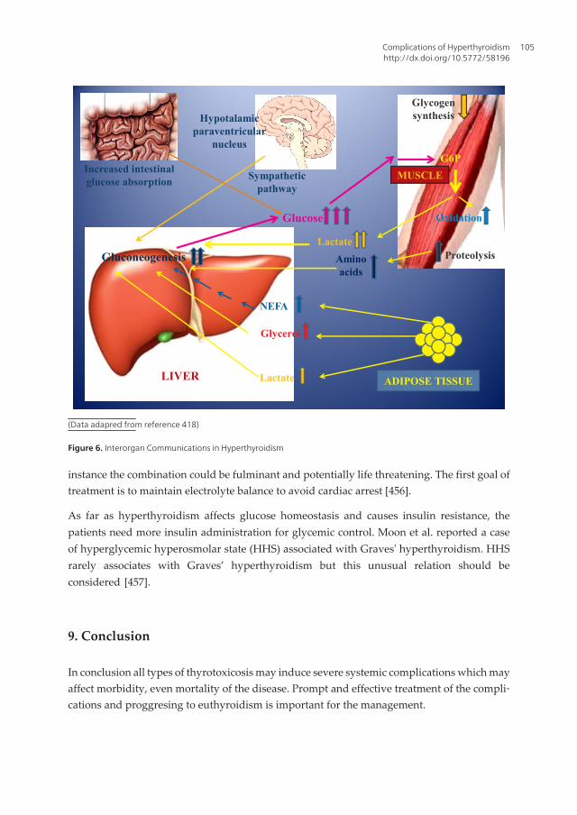
instance the combination could be fulminant and potentially life threatening. The first goal oftreatment is to maintain electrolyte balance to avoid cardiac arrest [456].
As far as hyperthyroidism affects glucose homeostasis and causes insulin resistance, thepatients need more insulin administration for glycemic control. Moon et al. reported a caseof hyperglycemic hyperosmolar state (HHS) associated with Graves' hyperthyroidism. HHSrarely associates with Graves’ hyperthyroidism but this unusual relation should beconsidered [457].
9. Conclusion
In conclusion all types of thyrotoxicosis may induce severe systemic complications which mayaffect morbidity, even mortality of the disease. Prompt and effective treatment of the compli‐cations and proggresing to euthyroidism is important for the management.
LIVER
MUSCLE
ADIPOSE TISSUELactate
Hypotalamicparaventricular
nucleus
Glycogensynthesis
NEFA
Glycerol
Gluconeogenesis
Increased intestinalglucose absorption
Proteolysis
G6P
Glucose Oxidation
LactateAmino acids
Sympatheticpathway
(Data adapred from reference 418)
Figure 6. Interorgan Communications in Hyperthyroidism
Complications of Hyperthyroidismhttp://dx.doi.org/10.5772/58196
105
Author details
Irmak Sayin1, Sibel Ertek2 and Mustafa Cesur2
1 Ufuk University, Medical Faculty, Department of Internal Medicine, Turkey
2 Ufuk University, Medical Faculty, Department of Endocrinology and Metabolic Disease,Turkey
References
[1] Ghandour A, Reust C. Hyperthyroidism: a stepwise approach to treatment. J FamPract 2011;60(7) 388-395.
[2] Palitzsch KD. Prevention and multimodal therapy of hyperthyroidism. Internist(Berl) 2008;49(12) 1428-1436.
[3] Wang C, Crapo LM. The epidemiology of thyroid disease and implications forscreening. Endocrinology and Metabolism Clinics of North America 1997;26(1)189-218.
[4] Bulow PI, Laurberg P, Knudsen N, Jorgensen T, Perrild H, Ovesen L, Rasmussen LB.Increase in incidence of hyprthyroidism predominantly occurs in young people afteriodine fortification of salt in Denmark. J Clin Endocrinol Metab 2006;91(10)3830-3834.
[5] Brandt F, Green A, Hegedüs L, Brix TH. A critical review and meta-analysis of theassociation between overt hyperthyroidism and mortality. Eur J Endocrinol2011;165(4) 491-497.
[6] Petersen P, Hansen JM. Stroke in thyrotoxicosis with atrial fibrillation. 1988;19(1)15-18.
[7] Bielecka-Dabrowa A, Mikhailidis DP, Rysz J, Banach M. The mechanisms of atrial fi‐brillation in hyperthyroidism. Thyroid Res. 2009;2(1) 4.
[8] Biondi B, Kahaly GJ. Cardiovascular involvement in patients with different causes ofhyperthyroidism. Nat Rev Endocrinol 2010;6(8) 431-443.
[9] Leng X, Bianco J, Tsai SY, Ozato K, O’Malley BW, Tsai MJ. Mechanisms for synergicactivation of thyroid hormone receptor and retinoid X receptor on different responseelements.J Biol Chem 1994;269(50) 31436-1442.
[10] Tsika RW, Bahl JJ, Leinwand LA, Morkin E. Thyroid hormone regulates expressionof a transfected human alpha-myosin heavy chain fusion gene in fetal rat heart cells.Proc Natl Acad Sci USA 1990;87(1) 379-383.
Thyroid Disorders - Focus on Hyperthyroidism106
[11] Zarain-Herzberg A, Marques J, Sukovich D, Periasamy M. Thyroid hormone receptormodulates the expression of the rabbit cardiac sarco(endo)plasmic reticulum Ca++-ATPase gene. J Biol Chem 1994;269(2) 1460-1467.
[12] Averyheart-Fullard V, Fraker LD, Murphy AM, Solaro RJ. Differential regulation ofslow skeletal and cardiac troponon I mRNA during development and by thyroidhormone in rat heart. J Mol Cell Cardiol 1994;26(5) 609-616.
[13] Morkin E. Regulation of myosin heavy chain genes in the heart. Circulation1993;87(5) 1451-1460.
[14] Ojamaa K, Klemperer JD, MacGlivray SS. Thyroid hormone and hemodynamic regu‐lation of beta-myosin heavy chain promoter in the heart. Endocrinology 1996;137(3)802-808.
[15] Izumo S, Nadal-Ginard B, Mahdavi V. All members of the MHC multigene family re‐spond to thyroid hormone in a highly tissue-specific manner. Science 1986;231(4738)597-600.
[16] Ladenson PW, Sherman SI, Boughman RL. Reversible alterations in myocardial geneexpression in a young man with dilated cardiomyopathy and hypothyroidism. Procnatl Acad Sci USA 1992; 89(12) 5251-5255.
[17] Magner J, Clarck W, Allenby P. Congestive heart failure and sudden death in ayoung woman with thyrotoxicosis. West J Med 1988;149(1) 86-91
[18] Dillman WH. Biochemical basis of thyroid hormone action in the heart. Am J Med1990;88(6) 626-630.
[19] Kiss E, Jakab G, Kranias EG, et al. Thyroid hormone-induced alteration in phospho‐lamban protein expression:regulatory effects on sarcoplasmic reticulum Ca2+trans‐port and myocardial relaxation.Circ Res 1994;75(2) 245-251.
[20] Kiss E, Brittsan AG, Elds I, et al. Thyroid hormone-induced alterations in phospho‐lamban deficient mouse hearts. Circ Res 1998;83(6) 608-613.
[21] Panagoulis C, Halapas A, Chariatis E, Driva P, Matsakas E. Hyperthyroidism andheart. Hellenic J Cardiol 2008;49(3) 169-175.
[22] Kasturi S, Ismail-Beigi F. Effect of thyroid hormone on the distribution and activity ofNa,K ATPase in ventricular myocardium. Arch Biochem Biophys 2008;475(2)121-127.
[23] Glick GG, Melikian J, Ismail-Beigi F. Thyoid enhancement of rat myocardial Na/KATPase preferential expression of alpha 2 activity on mRNA abundance. J Membr Bi‐ol 1990;115(3) 273-282.
[24] Ojamaa K, Sabet A, Kenessey A, Shenoy R, et al. Regulation of rat cardiac Kv 1.5gene expression by thyroid hormone is rapid and chamber specific. Endocrinology1999;140(7) 3170-3176.
Complications of Hyperthyroidismhttp://dx.doi.org/10.5772/58196
107
[25] Dudley SC Jr, Baumgarten CM. Bursting of cardiac sodium channels after acute ex‐posure to 3,5,3’– triiodo-L-thyronine. Circ Res 1993;73(2) 301-313.
[26] Walker JD, Crawford FA Jr, Mukherjee R, Spinale FG. The direct effects of 3,5,3’ –triiodo-L-thyronine (T3) on myocyte contractile processes. Insights into mechanismsof action. J Thorac Cardiovasc Surg 1995;110(5) 1369-1379.
[27] Chen WJ, Yeh YH, Lin KH, Chang GJ, Kuo CT. Molecular characterisation of thyroidhormone-inhibited atrial L-type calcium channel expression : implication for atrial fi‐brillation in hyperthyroidism. Basic res Cardiol 2011;106(2) 163-174.
[28] Hammond HK, White FC, Buxton IL, Salzstein P, Brunton LL, Longhurst JC. In‐creased myocardial beta-recepors and adrenergic responses in hyperthyroid pigs.Am J Physiol 1987;252(2 Pt 2) H283-H290.
[29] Levey GS, Klein I. Cathecholamine thyroid hormone interactions and the cardiovas‐cular manifestations of hyperthyroidism. Am J Med 1990;88(6) 642-646.
[30] Hoit BD, Khoury SF, Shao Y, Gabel M, Liggett SB, Walsh RA. Effects of thyroid hor‐mone on cardiac beta-adrenergic responsiveness in conscious baboons. Circulation1997;96(2) 592-598.
[31] Ojamaa K, Klein I, Sabet A, Steinberg SF. Chnages in adenylyl cyclase isoforms as amechanism for thyroid hormone modulation of cardiac beta adrenergic receptor re‐sponsiveness. Metabolism 2000;49(2) 275-279.
[32] Cacciatori V, Bellavere F, Pessarosa A, et al. Power spectral analysis of heart rate inhyperthyroidism. J Clin End Metab 1996;81(8) 2828-2835
[33] Klein I, Ojamaa K.Thyrotoxicosis and the heart. Endocrinol Metab Clin North Am1998;27(1) 51-62.
[34] Davis PJ, Davis FB. Acute cellular actions of thyroid hormone and myocardial func‐tion. Ann Thorac Surg 1993; 56 (1 Suppl) S16-S23.
[35] Klemperer JD, Klein I, Gomez M, et al. Thyroid hormone treatment after coronary ar‐tery by-pass surgery, N Eng J Med. 1995;333(23) 1522-1527
[36] Walker JD, Crawford FA, Kato S, et al. The novel effects of 3,5,3’ –triiodo-L-thyronineon myocyte contractile function and beta adrenergic responsiveness in dilated cardi‐omyopathy. J Thorac Cardiovasc Surg 1994;108(4) 672-679.
[37] Almeida NA, Corderio A, Machado DS, et al. Connexin 40 messenger ribonucleic ac‐tivity is posotovely regulated by thyroid hormone (TH)acting in cardiac atria via theTH receptor. Endocrinology 2009;150(1) 546-554.
[38] Lin H, Mitasikova M, Dlugosova K, et al. Thyroid hormones suppress epsilon PKC-signalling, downregulate connexin-43 and increase lethal arrhythmia susceptibility innondiabetic and diabetic rats.J Phsiol Pharm 2008;59(2) 271-285.
Thyroid Disorders - Focus on Hyperthyroidism108
[39] Pantos C, Xinaris C, Mourouzis I, Perimenis P, Politi E, Spanau D, Cokkinos DV.Thyroid hormone receptor alpha 1 : a switch to cardiac cell “metamorphosis”? JPhysiol Pharmacol 2008;59(2) 253-269.
[40] Suarez J, Scott BT, Suarez-Ramirez JA, Chavira CV, DillmannWH. Thyroid hormoneinhibits ERK phosphorylation in pressure overload induced hypertrophied mousehearts through a receptor-mediated mechanism. Am J Physiol Cll Physiol 2010;299(6)C1524-1529.
[41] Haddad F, Jiang W, Bodell PW, Qin AX, Baldwin KM. Cardiac myosin heavy chaingene regulation by thyroid hormone involves altered histone modifications. Am JPhysiol Heart Circ Physiol 2010;299(6) H1968-1980.
[42] Valcavi R, Menozzi C, Roti Elio et al. Sinus node function in hyperthyroid patients.JCEM 1992;75(1) 239-242
[43] Sun ZQ, Ojamaa K,Nakamura TY et al. Thyroid hormone increases pacemaker activi‐ty in rat neonatal atrial myocytes. J Moll Cell Cardiol 2001;33(4) 811-824.
[44] Messarah M, Saoudi M, Boulakaud MS, Feki AE. Oxidative stress induced by thyroiddysfunction in rat erythrocytes and heart. Environ Toxicol Pharmacol 2011;31(1)33-41.
[45] Cacciatori V, Bellavere F, Pezzarossa A, Dellera A, et al. Power spectral analysis ofheart rate in hyperthyroidism. J Clin Endocrinol Metab 1996;81(8) 2828-2835.
[46] Bielecka-Dabrowa A, Mikhailidis DP, Rysz J, Banach M. The mechanisms of atrial fi‐brillation in hyperthyroidism. Thyroid Res 2009;2(1) 4.
[47] Resnick LM, Laragh JH. Plasma rennin activity in syndromes of hyroid hormone ex‐cess and deficiency. Life Sci 1982;30(7-8) 58558-6.
[48] Polikar R, Burger AG, Scherrer U, Nicod P. The thyroid and the heart.Circulation1993;87(5) 1435-1441.
[49] Ojamaa K, Klemperer JD, Klein I. Acute effects of thyroid hormone on vascularsmooth muscle. Thyroid 1996;6(5) 505-512.
[50] Klein I, Ojamaa K, Thyroid hormone and the cardiovascular system. N Eng J Med2001;344(7) 501-509.
[51] Wang YY, Morimoto S, Du CK, et al. up-regulation of type-2 iodothyronine deiodi‐nase in dilated cardiomyopathy. Cardiovasc Res 2010;87(4) 636-646.
[52] Cotomacci G, Sarkis JJ, Fürstenau CR, Barreto-Chaves ML. Thyroid hormones are in‐volved in 5'-nucleotidase modulation in soluble fraction of cardiac tissue. Life Sci.2012;91(3-4) 137-142.
Complications of Hyperthyroidismhttp://dx.doi.org/10.5772/58196
109
[53] Carneiro-Ramos MS, Dinis GP, Nadu AP, et al. Blockage of angiotensin-II type 2 re‐ceptor prevents thyroxine-mediated cardiac hypertrophy by blocking Akt action. Ba‐sic Res cardiol 2010;105(3) 325-335.
[54] Wang YY, Jiao B, Guo WG, Che HL, Zu ZB. Excessive thyroxine enhances suscepti‐bility to apoptosis and decreased contractility of cardiomyocytes. Moll Cell Endocri‐nol 2010;320(1-2) 65-75.
[55] Maity S, Kar D, De K, Chander V, Bandyopadhyay A. Hyperthyroidism causes car‐diac dysfunction by mitochondrial impairment and energy depletion. J Endocrinol.2013;217(2) 215-228.
[56] Weltman NY, Wang D, Redetzke RA, Gerdes AM. Longstanding hyperthyroidism isassociated with normal or enhanced intrinsic cardiomyocyte function despite declinein global cardiac function. PLoS One. 2012;7(10) e46655. www.plosone.org/article/info/Adoi: 10.1371/journal.pone.0046655
[57] Kim BH, Cho KI, Kim SM, Kim JY, Choi BG, Kang JH, Jeon YK, Kim SS, Kim SJ, KimYK, Kim IJ. Irbesartan prevents myocardial remodeling in experimental thyrotoxiccardiomyopathy. Endocr J. 2012;59(10) 919-929.
[58] Park KW, Dai HB, Ojamaa K, Lowenstein E, Klein I, Sellke FW. The direct vasomotoreffect of thyroid hormones on rat skeleteal muscle resistance arteries. Anesth Analog1997;85(4) 734-738.
[59] Resnick LM, Laragh JH. Plasma renin activity in syndromes of thyroid hormone ex‐cess and deficiency. Life Sci 1982;30(7-8) 585-586.
[60] Klein I, Levey GS. Unusual manifestations of hypothyroidism. Arch Intern Med1984;144(1) 123-128.
[61] Fadel BM, Ellahham S, Ringel MD, Lindsay J, Wartofsky L, Burman KD. Hyperthy‐roid heart disease. Clin Cardiol 2000;23(6) 402-408.
[62] Kontos HA, Shapiro W, Mauck HP Jr, Richardson DW, Patterson JL Jr, Sharpe AR Jr,Mechanism of certain abnormalities of the circulation to the limbs in thyrotoxicosis. JClin Invest 1965;44 947-956.
[63] Theilen EO, Wilson WR. Hemodynamic effects of peripheral vasoconstriction in nor‐mal and thyrotoxic subjects. J Appl Physiol 1967;22(2) 207-210.
[64] Auer J, Scheibner P, Mische T, et al. Subclinical hyperthyroidism as a risk factor foratrial fibrillation. Am Heart J, 2001;142(5) 838-842.
[65] Vardas PE, Mavrakis HE. Atrial fibrillation : a symptom treated as a disease? Hellen‐ic J Cardiol 2006;47(4) 191-193.
[66] Shenfield GM. Influence of thyroid dysfunction on drug pharmacokinetics. ClinPharmacokinet 1981;6(4) 275-297.
Thyroid Disorders - Focus on Hyperthyroidism110
[67] Nakazawa HK, Sakurai K, Hamada N, Momotani N, Ito K. Management of atrial fi‐brillation in the post-thyrotoxic state. Am J Med 1982;72(6) 903-906.
[68] Petersen P, Hansen JM. Stroke in thyrotoxicosis with atrial fibrillation. Stroke1988;19(1) 15-18
[69] Gilligan DM, Ellenbogen KA, Epstein AE. The management of atrial fibrillation. AmJ Med 1996;101(4) 413-421.
[70] Chaudhury S, Ismail-Beigi F, Gick GG, Levenson E, Edelman IS. Effect of thyroidhormone on the abundance of Na,K,ATPase alpha subunit messenger ribonucleicacid. Mol Endocrinol 1987;1(1) 83-89.
[71] Klein I, Levey GS: The cardiovascular system in thyrotoxicosis. In Braverman LE,Utiger RD (eds): Werner and Ingbar’s the Thyroid : A Fundamental and ClinicalText.8th edition Philadelphia. Lippincott and Williams & Wilkins;2000
[72] Klein I, Ojamaa K. Thyroid hormone and the cardiovascular system. N Eng J Med2001; 344(7) 501-509.
[73] Kahaly GJ, Kampmann C, Mohr-Kahaly S. Cardiovascular hemodynamics and exer‐cise tolerance in thyroid disease. Thyroid 2002;12(6) 473-481
[74] Dahl P, Danzi S, Klein I. Thyrotoxic cardiac disease. Curr Heart Fail Re 2008;5(3)170-176.
[75] Yue WS, Chong BH, Zhang XH, et al. Hyperthyroidism-induced left ventricular dia‐stolic dysfunction: implication in hyperthyroidism-related heart failure. Clin Endo‐crinol (oxf) 2011;74(5) 636-643.
[76] Gu LQ, Zhao L, Zhu W, et al. Relationships between serum levels of thyroid hor‐mones and serum concentrations of symmetric dimethylarginine (ADMA) and N-ter‐minal-pro-B-type natriuretic peptide (NT-proBNP) in patients with Graves’ Disease.Endocrine 2011;39(3) 266-271
[77] Mercuro G, Panzuto MG, Bina A, et al. Cardiac function, physical exercise capacity,and quality of life during long-term thyrotropin suppressive therapy with levothyr‐oxine :effect of individual dose tailoring. J Clin Endocrinol Metab 2000;85(1) 159-164.
[78] Patane S, Marte F. Changing axis deviation and paroxysmal atrial flutter associatedwith subclinical hyperthyroidism. Int J Cardiol 2010;144(2) e31-e33.
[79] Sawin CT, Geller A, Wolf PA, et al. Low serum thyrotropin concentrations as a riskfactor for atrial fibrillation in older patients. N Eng j Med 1994;331(19) 1249-1252.
[80] Vigario Pdos S, Chachamovitz DS, Teixeira Pde F, Santos MA, Oliveira FP, VaismanM. Impaied functional and hemodynamic response to graded exercise testing and itsrecovery in patients with subclinical hyperthyroidism. Arq Bras Endocrinol Metabol2011;55(3) 203-212.
Complications of Hyperthyroidismhttp://dx.doi.org/10.5772/58196
111
[81] Biondi B, Fazio S, Carella C et al. Control of adrenergic overactivity by beta-blockadeimproves the quality of life in patients receiving long-term suppressive therapy withlevothyroxine. J Clin end Metab 1994;78(5) 1028-1033.
[82] Heeringa J, Hoogendoorn EH, van der Deure WM, et al. High-normal thyroid func‐tion and risk of atrial fibrillation: the Rotterdam Study. Arch Intern Med 2008;168(20)2219-2224.
[83] Fazio S, Palmieri EA, Lombardi G, Biondi B. Effects of thyroid hormone on cardio‐vascular system. Recent Prog Horm Res. 2004;59 31-50.
[84] Garrity JA, Bahn RS. Pathogenesis of Graves Ophthalmopathy: Implications for Pre‐diction, Prevention, and Treatment. American Journal of Ophthalmology. 2006;142(1)147–153.
[85] Marcocci C, Bartelena L, Bogazzi FM, Panicucci M, Pinchera A. Studies on the occur‐rence of ophthalmopathy in Graves’ disease. Acta Endocrinol (Copenh) 1989;120(4)473–478.
[86] Bahn RS. Assessment and management of the patient with Graves’ ophthalmopathy.Endocr Pract 1995;1(3) 172–178.
[87] Prummel M, Bakker A, Wiersinga W, et al. Multi-center study on the characteristicsand treatment strategies of patients with Graves' orbitopathy: the first EuropeanGroup on Graves' Orbitopathy experience. Eur J Endocrinol. 2003;148(5) 491–495.
[88] Schotthoefer EO, Wallace DK. Strabismus associated with thyroid eye disease. CurrOpin Ophthalmol. 2007;18(5) 361–365.
[89] Mourits MP, Koornneef L, Wiersinga WM, Prummel MF, Berghout A, van der GaagR. Clinical criteria for the assessment of disease activity in Graves' ophthalmopathy:a novel approach. Br J Ophthalmol. 1989;73(8) 639–644.
[90] Bartalena L, Pinchera A, Marcocci C. Management of Graves' ophthalmopathy: reali‐ty and perspectives. Endocr Rev. 2000;21(2) 168–199.
[91] Han R, Smith TJ. T helper type 1 and type 2 cytokines exert divergent influence onthe induction of prostaglandin E2 and hyaluronan synthesis by interleukin-1β in orbi‐tal fibroblasts: implications for the pathogenesis of thyroid-associated ophthalmop‐athy. Endocrinology. 2006;147(1) 13–19.
[92] Douglas RS, Gianoukakis AG, Goldberg RA, Kamat S, Smith TJ. Circulating mono‐nuclear cells from euthyroid patients with thyroid-associated ophthalmopathy exhib‐it characteristic phenotypes. Clinical & Experimental Immunology. 2007;148(1) 64–71.
[93] Powell JD. The induction and maintenance of T cell anergy. Clinical Immunology.2006;120(3) 239–246.
[94] Melchers F. Anergic B cells caught in the act. Immunity. 2006;25(6) 864–867.
Thyroid Disorders - Focus on Hyperthyroidism112
[95] Smith TJ. Insights into the role of fibroblasts in human autoimmune diseases. Clinical& Experimental Immunology. 2005;141(3) 388–397.
[96] Koumas L, Smith TJ, Phipps RP. Fibroblast subsets in the human orbit: Thy-1+andThy-1-subpopulations exhibit distinct phenotypes. European Journal of Immunology.2002;32(2) 477–485.
[97] Sempowski GD, Rozenblit J, Smith TJ, Phipps RP. Human orbital fibroblasts are acti‐vated through CD40 to induce proinflammatory cytokine production. American Jour‐nal of Physiology. 1998;274(3) C707–C714.
[98] Valyasevi RW, Erickson DZ, Harteneck DA, et al. Differentiation of human orbitalpreadipocyte fibroblasts induces expression of functional thyrotropin receptor. Jour‐nal of Clinical Endocrinology & Metabolism. 1999;84(7) 2257–2562.
[99] Cao HJ, Han R, Smith TJ. Robust induction of PGHS-2 by IL-1 in orbital fibroblastsresults from low levels of IL-1 receptor antagonist expression. American Journal ofPhysiology. 2003;284(6) C1429–C1437.
[100] Smith TJ, Sciaky D, Phipps RP, Jennings TA. CD40 expression in human thyroid tis‐sue: evidence for involvement of multiple cell types in autoimmune and neoplasticdiseases. Thyroid. 1999;9(8) 749–755.
[101] Feldon SE, John Park DJ, O'Loughlin CW, et al. Autologous T-lymphocytes stimulateproliferation of orbital fibroblasts derived from patients with Graves' ophthalmop‐athy. Investigative Ophthalmology & Visual Science. 2005;46(11) 3913–3921.
[102] Lehmann GM, Garcia-Bates TM, Smith TJ, et al. Regulation of Lymphocyte Functionby PPARgamma: Relevance to Thyroid Eye Disease-Related Inflammation. PPARRes. 2008;2008:895901.
[103] Pritchard J, Han R, Horst N, Cruikshank WW, Smith TJ. Immunoglobulin activationof T cell chemoattractant expression in fibroblasts from patients with Graves' diseaseis mediated through the insulin-like growth factor I receptor pathway. Journal of Im‐munology. 2003;170(12) 6348–6354.
[104] Smith TJ, Hoa N. Immunoglobulins from patients with Graves' disease induce hya‐luronan synthesis in their orbital fibroblasts through the self-antigen, insulin-likegrowth factor-I receptor. Journal of Clinical Endocrinology & Metabolism. 2004;89(10)5076–5080.
[105] de Carli M, D'Elios MM, Mariotti S, et al. Cytolytic T cells with Th1-like cytokine pro‐file predominate in retroorbital lymphocytic infiltrates of Graves' ophthalmopathy.Journal of Clinical Endocrinology & Metabolism. 1993;77(5) 1120–1124
[106] Smith TJ, Sempowski GD, Berenson CS, Cao HJ, Wang H-S, Phipps RP. Human thy‐roid fibroblasts exhibit a distinctive phenotype in culture: characteristic gangliosideprofile and functional CD40 expression. Endocrinology. 1997;138(12) 5576–5588.
Complications of Hyperthyroidismhttp://dx.doi.org/10.5772/58196
113
[107] Zhang Y, Cao HJ, Graf B, Meekins H, Smith TJ, Phipps RP. Cutting edge: CD40 en‐gagement up-regulates cyclooxygenase-2 expression and prostaglandin E2 produc‐tion in human lung fibroblasts. Journal of Immunology. 1998;160(3) 1053–1057.
[108] Antonelli A, Rotondi M, Ferrari SM, et al. Interferon-γ-inducible α-chemokineCXCL10 involvement in Graves' ophthalmopathy: modulation by peroxisome prolif‐erator-activated receptor-γ agonists. Journal of Clinical Endocrinology & Metabolism.2006;91(2) 614–620.
[109] Feldon SE, O'Loughlin CW, Ray DM, Landskroner-Eiger S, Seweryniak KE, PhippsRP. Activated human T lymphocytes express cyclooxygenase-2 and produce proadi‐pogenic prostaglandins that drive human orbital fibroblast differentiation to adipo‐cytes. The American Journal of Pathology. 2006;169(4) 1183–1193.
[110] Forman BM, Tontonoz P, Chen J, Brun RP, Spiegelman BM, Evans RM. 15-Deoxy-Δ12,14-prostaglandin J2 is a ligand for the adipocyte determination factor PPAR-γ. Cell.1995;83(5) 803–812.
[111] Kliewer SA, Lenhard JM, Willson TM, Patel I, Morris DC, Lehmann JM. A prosta‐glandin J2 metabolite binds peroxisome proliferator-activated receptor γ and pro‐motes adipocyte differentiation. Cell. 1995;83:813–819.
[112] Zhang J, Fu M, Cui T, et al. Selective disruption of PPARγ2 impairs the developmentof adipose tissue and insulin sensitivity. Proceedings of the National Academy of Sciencesof the United States of America. 2004;101(29) 10703–10708.
[113] Kumar S, Coenen MJ, Scherer PE, Bahn RS. Evidence for enhanced adipogenesis inthe orbits of patients with Graves' ophthalmopathy. Journal of Clinical Endocrinology &Metabolism. 2004;89(2) 930–935.
[114] Mimura LY, Villares SMF, Monteiro MLR, Guazzelli IC, Bloise W. Peroxisome prolif‐erator-activated receptor-γ gene expression in orbital adipose/connective tissues isincreased during the active stage of Graves’ ophthalmopathy. Thyroid. 2003;13(9)845–850.
[115] Chen M, Liao S, et al. Role of macrophage infiltration in the orbital fat of patientswith Graves' ophthalmopathy. Clin Endocrinol 2008;69(2) 332-337.
[116] Bartalena L, Baldeschi L, Dickinson A, et al. Consensus statement of the EuropeanGroup on Graves' Orbitopathy (EUGOGO) on management of GO. Eur J Endocrinol2008;158(3) 273-285.
[117] Yang SR, Chida AS, Bauter MR, Shafiq N, Seweryniak K, Maggirwar SB, Kilty I, Rah‐man I 2006 Cigarette smoke induces proinflammatory cytokine release by activation of NF-κB and posttranslational modifications of histone deacetylase in macrophages. Am J PhysiolLung Cell Mol Physiol 2006;291(1) L46–L57
Thyroid Disorders - Focus on Hyperthyroidism114
[118] Metcalfe RA, Weetman AP . Stimulation of extraocular muscle fibroblasts by cytokines andhypoxia: possible role in thyroid-associated ophthalmopathy. Clin Endocrinol (Oxf) 1994;40(1) 67–72.
[119] Burch HB, Lahiri S, Bahn RS, Barnes S . Superoxide radical production stimulates retrooc‐ular fibroblast proliferation in Graves’ ophthalmopathy. Exp Eye Res 1997;65(2) 311–316.
[120] Mack WP, Stasior GO, Cao HJ, Stasior OG, Smith TJ 1999 The effect of cigarettesmoke constituents on the expression of HLA-DR in orbital fibroblasts derived frompatients with Graves ophthalmopathy. Ophthal Plast Reconstr Surg 199;15(4) 260–271.
[121] Pfeilschifter J & Ziegler R. Smoking and endocrine ophthalmopathy: impact of smok‐ing severity and current vs lifetime cigarette consumption. Clinical Endocrinology1996;45(4) 477–481.
[122] Eckstein A, Quadbeck B, Mueller G, Rettenmeier AW, Hoermann R, Mann K, SteuhlP, Esser J. Impact of smoking on the response to treatment of thyroid associatedoph‐thalmopathy.Br J Ophthalmol. 2003 Jun;87(6) 773-776.
[123] Wiersinga WM. Smoking and thyroid. Clin Endocrinol (Oxf). 2013;79(2) 145-151.
[124] Salvi M. Zhang ZG. Haegert D. Woo M. Liberman A. Cadarso L. Wall JR. Patientswith endocrine ophthalmopathy not associated with overt thyroid disease have mul‐tiple thyroid immunological abnormalities. J Clin Endocrinol Metab. 1990;70 89–94.
[125] Wiersinga WM. Smit T. van der Gaag R. Koornneef L. Temporal relationship be‐tween onset of Graves' ophthalmopathy and onset of thyroidal Graves' disease. J En‐docrinol Invest. 1988;11(8) 615–619.
[126] Tsai KH, Tsai FJ, Lin HJ, Lin HJ, Liu YH, Liao WL, Wan L. Thymic stromal lympho‐poietin gene promoter polymorphisms and expression levels in Graves' disease andGraves' ophthalmopathy. BMC Med Genet. 2012;13 116.
[127] Liao WL, Chen RH, Lin HJ, Liu YH, Chen WC, Tsai Y, Wan L, Tsai FJ. Toll-like recep‐tor gene polymorphisms are associated with susceptibility to Graves' ophthalmop‐athy in Taiwan males.BMC Med Genet. 2010;11 154.
[128] Liao WL, Chen RH, Lin HJ, Liu YH, Chen WC, Tsai Y, Wan L, Tsai FJ. The associa‐tion between polymorphisms of B7 molecules (CD80 and CD86) and Graves' oph‐thalmopathy in a Taiwanese population. Ophthalmology. 2011;118(3) 553-557.
[129] Gianoukakis AG, Jennings TA, King CS, et al. Hyaluronan accumulation in thyroidtissue: evidence for contributions from epithelial cells and fibroblasts. Endocrinolo‐gy. 2007;148(1) 54–62.
[130] Cawood TJ, Moriarty P, O'Farrelly C, O'Shea D. The effects of tumour necrosis factor-alpha and interleukin1 on an in vitro model of thyroid-associated ophthalmopathy:contrasting effects on adipogenesis. Eur J Endocrinol. 2006;155 395–403.
Complications of Hyperthyroidismhttp://dx.doi.org/10.5772/58196
115
[131] Liu YH, Chen RH, Wu HH, Liao WL, Chen WC, Tsai Y, Tsai CH, Wan L, Tsai FJ. As‐sociation of interleukin-1beta (IL1B) polymorphisms with Graves' ophthalmopathyin Taiwan Chinese patients. Invest Ophthalmol Vis Sci. 2010;51(12) 6238-6246.
[132] Vaidya B. Imrie H. Perros P. Dickinson J. McCarthy MI. Kendall-Taylor P. Pearce SH.Cytotoxic T lymphocyte antigen-4 (CTLA-4) gene polymorphism confers susceptibili‐ty to thyroid associated orbitopathy. Lancet. 1999;354(9180) 743–744.
[133] Allahabadia A. Heward JM. Nithiyananthan R. Gibson SM. Reuser TT. Dodson PM.Franklyn JA. Gough SC. MHC class II region, CTLA4 gene, and ophthalmopathy inpatients with Graves' disease. Lancet. 2001;358(9286) 984–985.
[134] Daroszewski J. Pawlak E. Karabon L. Frydecka I. Jonkisz A. Slowik M. BolanowskiM. Soluble CTLA-4 receptor an immunological marker of Graves' disease and severi‐ty of ophthalmopathy is associated with CTLA-4 Jo31 and CT60 gene polymor‐phisms. Eur J Endocrinol. 2009;161(5) 787–793.
[135] Alevizaki M, Mantzou E, Cimponeriu A, Saltiki K, Philippou G, Wiersinga W. ThePro 12 Ala PPAR gamma gene polymorphism: possible modifier of the activity andseverity of thyroid-associated orbitopathy (TAO).Clin Endocrinol (Oxf). 2009;70(3)464-468
[136] Huber AK, Jacobson EM, Jazdzewski K, Concepcion ES, Tomer Y. Interleukin (IL)-23receptor is a major susceptibility gene for Graves' ophthalmopathy: the IL-23/T-help‐er 17 axis extends to thyroid autoimmunity. J Clin Endocrinol Metab. 2008;93(3)1077-1081.
[137] Syed AA, Simmonds MJ, Brand OJ, et al. Preliminary evidence for interaction ofPTPN12 polymorphism with TSHR genotype and association with Graves' oph‐thalmopathy. Clin Endocrinol (Oxf) 2007; 67(5) 663–667
[138] Kurylowicz A, Hiromatsu Y, Jurecka-Lubieniecka B, et al. Association ofNFKB1-94ins/del ATTG promoter polymorphism with susceptibility to and pheno‐type of Graves' disease. Genes Immun 2007; 8(7) 532–538.
[139] Weetman A P, Zhang L, Webb S, Shine B. Analysis of HLA-DQB and HLA-DPB al‐leles in Graves' disease by oligonucleotide probing of enzymatically amplified DNA.Clin. Endocrinol (Oxf) 1990;33(1) 65–71.
[140] Akaishi PM, Cruz AA, Silva FL, Rodrigues Mde L, Maciel LM, Donadi EA. The roleof major histocompatibility complex alleles in the susceptibility of Brazilian patientsto develop the myogenic type of Graves’ orbitopathy. Thyroid. 2008;18(4) 443–7.
[141] Inoue D, Sato K, Enomoto T et al.Correlation of HLA types and clinical findings inJapanese patients with hyperthyroid Graves' disease: evidence indicating the exis‐tence of four subpopulations. Clin. Endocrinol (Oxf) 1992;36(1) 75–82.
[142] Inoue D, Sato K, Maeda M et al. Genetic differences shown by HLA typing amongJapanese patients with euthyroid Graves' ophthalmopathy, Graves' disease and Ha‐
Thyroid Disorders - Focus on Hyperthyroidism116
shimoto's thyroiditis: genetic characteristics of euthyroid Graves' ophthalmopathy.Clin. Endocrinol (Oxf) 1991;34(1) 57–62.
[143] Molnar I., Balazs C. High circulating IL-6 level in Graves’ ophthalmopathy. Autoim‐munity 1997; 25(2) 91-96.
[144] Wakelkamp IM., Gerding MN.,Van Der Meer JW., Prummel MF., Wiersinga WM.Both Th1-and Th2-derived cytokines in serum are elevated in Graves’ ophthalmop‐athy. Clin. Exp. Immunol. 2000; 121(3) 453-457.
[145] Chen B, Tsui S, Smith TJ. IL-1 beta induces IL-6 expression in human orbital fibro‐blasts: identification of an anatomic-site specific phenotypic attribute relevant to thy‐roid-associated ophthalmopathy. J Immunol 2005; 175(2) 1310–1319.
[146] Kim SE, Yoon JS, Kim KH, Lee SY. Increased serum interleukin-17 in Graves' oph‐thalmopathy.Graefes Arch Clin Exp Ophthalmol. 2012;250(10) 1521-1526.
[147] Ujhelyi B, Gogolak P, Erdei A, Nagy V, Balazs E, Rajnavolgyi E, Berta A, Nagy EV.Graves' orbitopathy results in profound changes in tear composition: a study of plas‐minogen activator inhibitor-1 and seven cytokines. Thyroid. 2012;22(4) 407-414.
[148] Raikow RB, Tyutyunikov A, Kennerdell JS, Kazim M, Dalbow MH, Scalise D. Corre‐lation of serum immunoglobulin E elevations with clinical stages of dysthyroid orbit‐opathy. Ophthalmology. 1992; 99(3) 361–365.
[149] Cekić S, Stanković-Babić G. Application of ultrasound in diagnosing and follow-upof endocrine orbitopathy. Med Pregl. 2010;63(3-4) 241-248.
[150] Demer JL, Kerman BM. Comparison of standardized echography with magnetic res‐onance imaging to measure extraocular muscle size. Am J Ophthalmol. 1994;118(3)351-361.
[151] Casper DS, Chi TL & Trokel SL. Orbital Disease, Imaging and Analysis. Edn 1. Stutt‐gart: Thieme. 1993. / Pester RG & Hoover ED. Graves’ orbiopathy. In ComputerizedTomography in Orbital Diseases and Neuroopthalmology. Edn 1. ch 3. pp 97-114.Chicago: Yera Book Medical Publisher. 1984
[152] Nugent RA, Belkin RI, Neigel JM, Rootman J, Robertson WD, Spinelli J, Graeb DA.Graves orbitopathy: correlation of CT and clinical findings. Radiology. 1990;177(3)675-682.
[153] Barrett L, Glatt HJ, Burde RM, Gado MH. Optic nerve dysfunction in thyroid eye dis‐ease: CT. Radiology. 1988;167(2) 503-507.
[154] Feldon SE, Lee CP, Muramatsu SK, Weiner JM. Quantitative computed tomographyof Graves' ophthalmopathy. Extraocular muscle and orbital fat in development of op‐tic neuropathy. Arch Ophthalmol. 1985;103(2) 213-215.
[155] Hallin ES, Feldon SE. Graves' ophthalmopathy: II. Correlation of clinical signs withmeasures derived from computed tomography. Br J Ophthalmol. 1988;72(9) 678-682.
Complications of Hyperthyroidismhttp://dx.doi.org/10.5772/58196
117
[156] Nishikawa M, Yoshimura M, Toyoda N, Masaki H, Yonemoto T, Gondou A, Kato T,Kurokawa H, Furumura T, Inada M. Correlation of orbital muscle changes evaluatedby magnetic resonance imaging and thyroid-stimulating antibody in patients withGraves' ophthalmopathy. Acta Endocrinol (Copenh). 1993;129(3) 213-219.
[157] Yokoyama N, Nagataki S, Uetani M, Ashizawa K, Eguchi K. Role of magnetic reso‐nance imaging in the assessment of disease activity in thyroid-associated ophthalm‐opathy. Thyroid. 2002;12(3) 223-227.
[158] Wiersinga WM, Gerding MN, Prummel MF, Krenning EP. Octreotide scintigraphy inthyroidal and orbital Graves' disease. Thyroid. 1998;8(5) 433-436.
[159] Krassas G, Dumas A. Octreoscan in Graves' ophthalmopathy. Thyroid. 1997;7(5)805-806.
[160] Krassas GE, Dumas A, Pontikides N, Kaltsas T. Somatostatin receptor scintigraphyand octreotide treatment in patients with thyroid eye disease. Clin Endocrinol (Oxf)1995;42(6) 571-580.
[161] Postema PTE, Krenning EP, Wijngaarde R et al. (111In-DTPA-D-Phe1) octreotidescintigraphy in thyroidal and orbital Graves' disease: a parameter for disease activi‐ty? J Clin Endocrinol Metab 1994;79(6) 1845-1851.
[162] Krassas GE. Octreoscan in thyroid-associated ophthalmopathy. Thyroid. 2002;12(3)229-231.
[163] Krassas GE, Kahaly GJ. The role of octreoscan in thyroid eye disease. Eur J Endocri‐nol. 1999;140(5) 373-375.
[164] Kuo PH, Monchamp T, Deol P. Imaging of inflammation in Graves’ ophthalmopathyby positron emission tomography/computed tomography. Thyroid. 2006;16(4) 419–420.
[165] Konuk O, Atasever T, Unal M, Ayvaz G, Yetkin I, Cakir N, Arslan M, HasanreisogluB. Orbital gallium-67 scintigraphy in Graves' ophthalmopathy. Thyroid. 2002 ;12(7)603-608.
[166] Durak H, Söylev M, Durak I, Değirmenci B, Capa Kaya G, Uysal B. Tc-99m polyclo‐nal human immunoglobulin G imaging in Graves' ophthalmopathy. Clin Nucl Med.2000;25(9) 704-707.
[167] Lopes FP, de Souza SA, Dos Santos Teixeira Pde F, Rebelo Pinho Edos S, da FonsecaLM, Vaisman M, Gutfilen B. 99mTc-Anti-TNF-α scintigraphy: a new perspectivewithin different methods in the diagnostic approach of active Graves ophthalmop‐athy. Clin Nucl Med. 2012;37(11) 1097-1101.
[168] García-Rojas L, Adame-Ocampo G, Alexánderson E, Tovilla-Canales JL. 18-Fluoro‐deoxyglucose Uptake by Positron Emission Tomography in Extraocular Muscles ofPatients with and without Graves' Ophthalmology. J Ophthalmol. 2013;2013:529187.
Thyroid Disorders - Focus on Hyperthyroidism118
[169] Wiersinga WM, Prummel MF, Mourits MP, et al. Classification of the eye changes ofGraves' disease. Thyroid 1991;1(4) 357–360.
[170] Dolman PJ, Rootman J. VISA classification for Graves orbitopathy. Ophthal Plast Re‐constr Surg 2006;22(5) 319–324.
[171] Bartalena L, Tanda ML. Clinical practice. Graves' ophthalmopathy. N Engl J Med.2009;360(10) 994-1001
[172] Mourits MP, Prummel MF, Wiersinga WM, Koornneef L. Clinical activity score as aguide in the management of patients with Graves' ophthalmopathy. Clin Endocrinol(Oxf) 1997;47(1) 9-14.
[173] Bartalena L, Marcocci C, Bogazzi F, Manetti L, Tanda ML, Dell'Unto E,Bruno-BossioG, Nardi M, Bartolomei MP, Lepri A, Rossi G, Martino E, Pinchera A. Relation be‐tween therapy for hyperthyroidism and the course of Graves' ophthalmopathy. NEngl J Med. 1998;338(2) 73-78.
[174] Prummel MF, Wiersinga WM, Mourits MP, Koornneef L, Berghout A, van der GaagR Amelioration of eye changes of Graves' ophthalmopathy by achieving euthyroid‐ism. Acta Endocrinologica 1989; 121(Suppl 2):185–189
[175] Laurberg P, Wallin G, Tallstedt L, Abraham-Nordling M, Lundell G, Tørring O. TSH-receptor autoimmunity in Graves' disease after therapy with anti-thyroid drugs, sur‐gery, or radioiodine: a 5-year prospective randomized study. Eur J Endocrinol.2008;158(1) 69-75.
[176] Bałdys-Waligórska A, Gołkowski F, Kusnierz-Cabala B, Buziak-Bereza M,Hubalew‐ska-Dydejczyk A. Graves' ophthalmopathy in patients treated with radioiodine 131-I.Endokrynol Pol. 2011;62(3) 214-219.
[177] El-Kaissi S, Bowden J, Henry MJ, Yeo M, Champion BL, Brotchie P, Nicholson GC,Wall JR. Association between radioiodine therapy for Graves' hyperthyroidism andthyroid-associated ophthalmopathy. Int Ophthalmol. 2010;30(4) 397-405.
[178] Wilhelm SM, McHenryCR2010 Total thyroidectomy is superior to subtotal thyroidec‐tomy for management of Graves’ disease in the United States. World J Surg2010;34(6) 1261–1264.
[179] Bartalena L, Marcocci C, Bogazzi F, Bruno-Bossio G, Pinchera A. Glucocorticoid ther‐apy of Graves’ ophthalmopathy. Exp Clin Endocrinol 1991;97(2-3) 320–328.
[180] Smith TJ. Dexamethasone regulation of glycosaminoglycan synthesis in cultured hu‐man fibroblasts: similar effects of glucocorticoid and thyroid hormone therapy. J ClinInvest 1984;74(6) 2157–2163.
[181] Bartalena L, Marcocci C, Pinchera A. Treating severe Graves’ ophthalmopathy. Bail‐liere’s Clin Endocrinol Metab 1997;11(3) 521–536.
[182] Marcocci C, Bartalena L, Tanda ML, et al. Comparison of the effectiveness and tolera‐bility of intravenous or oral glucocorticoids associated with orbital radiotherapy in
Complications of Hyperthyroidismhttp://dx.doi.org/10.5772/58196
119
the management of severe Graves' ophthalmopathy: results of a prospective, single-blind, randomized study. J Clin Endocrinol Metab 2001;86(8) 3562-3567.
[183] Kahaly GJ, Pitz S, Hommel G, Dittmar M. Randomized, single blind trial of intrave‐nous versus oral steroid monotherapy in Graves' orbitopathy. J Clin Endocrinol Met‐ab 2005;90(9) 5234-5240.
[184] Bartalena L, Krassas GE, Wiersinga W et al; European Group on Graves' Orbitop‐athy. Efficacy and safety of three different cumulative doses of intravenous methyl‐prednisolone for moderate to severe and active Graves' orbitopathy. J ClinEndocrinol Metab. 2012;97(12) 4454-63.
[185] Gursoy A, Cesur M, Erdogan MF, et al. New-onset acute heart failure after intrave‐nous glucocorticoid pulse therapy in a patient with Graves' ophthalmopathy. Endo‐crine 2006;29(3) 513–516.
[186] Chang TC, Kao SCS, Huang KM 1992 Octreotide and Graves’ ophthalmopathy andpretibial myxoedema. Br Med J 1992;304(6820) 158.
[187] Uysal AR, Corapçioğlu D, Tonyukuk VC, Güllü S, Sav H, Kamel N, Erdoğan G. Ef‐fect of octreotide treatment on Graves' ophthalmopathy. Endocr J. 1999;46(4) 573-577.
[188] Stan MN, Garrity JA, Bradley EA, Woog JJ, Bahn MM, Brennan MD, Bryant SC,Achenbach SJ, Bahn RS 2006 Randomized, double-blind, placebo-controlled trial oflong-acting release octreotide for treatment of Graves’ ophthalmopathy. J Clin Endo‐crinol Metab 2006;91(12) 4817–4824.
[189] Krassas GE, Doumas A, Kaltsas T, Halkias A, Pontikides N. Somatostatin receptorscintigraphy before and after treatment with somatostatin analogues in patients withthyroid eye disease. Thyroid 1999;9(1) 47-52.
[190] Antonelli A, Saracino A, Alberti B, Canapicchi R, Cartei F, Lepri A, Laddaga M, Ba‐schieri L. High-dose intravenous immunoglobulin treatment in Graves' ophthalmop‐athy. Acta Endocrinol (Copenh) 1992;126(1) 13-23.
[191] Kahaly G, Pitz S, Muller-Forell W, Hommel G. Randomized trial of intravenous im‐munoglobulins versus prednisolone in Graves' ophthalmopathy. Clin Exp Immunol1996;106(2) 197-202.
[192] Seppel T, Schlaghecke R, Becker A, Engelbrecht V, Feldkamp J, Kornely E. High-doseintravenous therapy with 7S immunoglobulins in autoimmune endocrine ophthalm‐opathy. Clin Exp Immunol 1996;14(Suppl 15) S109-S114.
[193] Weetman AP, McGregor AM, Ludgate M, Beck L, Mills PV, Lazarus JH, Hall R. Cy‐closporin improves Graves' ophthalmopathy. Lancet. 1983;2(8348) 486-489.
[194] Prummel MF, Mourits MP, Berghout A, Krenning EP, van der Gaag R, Koornneef L,Wiersinga WM. Prednisone and cyclosporine in the treatment of severe Graves’ oph‐thalmopathy. N Engl J Med 1989;321(20) 1353–1359.
Thyroid Disorders - Focus on Hyperthyroidism120
[195] Weissel M, Zielinski CC, Hauff W, Till P. Combined therapy with cyclosporin A andcortisone in endocrine Basedow endocrine orbitopathy: successful use in compres‐sive optic neuropathy. Acta Med Austriaca. 1993;20(1-2) 9-13.
[196] Weetman AP, Wiersinga WM. Current management of thyroid-associated ophthalm‐opathy in Europe. Results of an international survey. Clin Endocrinol (Oxf)1998;49(1) 21-8.
[197] Smith JR, Rosenbaum JT. A role of methotrexate in the management of non-infec‐tious orbital inflammatory disease. Br J Ophthalmol 2001;85(10) 1220-1224.
[198] Bouzas EA, Karadimas P, Mastorakos G, Koutras DA 2000 Antioxidant agents in thetreatment of Graves' ophthalmopathy. Am J Ophthalmol 2000;129(5) 618-622.
[199] Yoon JS, Chae MK, Lee SY, Lee EJ. Anti-inflammatory effect of quercetin in a wholeorbital tissue culture of Graves' orbitopathy. Br J Ophthalmol. 2012;96(8) 1117-1121.
[200] Marcocci C, Kahaly GJ, Krassas GE, Bartalena L, Prummel M, Stahl M, Altea MA,Nardi M, Pitz S, Boboridis K, Sivelli P, von Arx G, Mourits MP, Baldeschi L, Benci‐velli W, Wiersinga W; European Group on Graves' Orbitopathy. Selenium and thecourse of mild Graves' orbitopathy. N Engl J Med. 2011;364(20) 1920-1931.
[201] Chang CC, Chang TC, Kao SC, Kuo YF, Chien LF. Pentoxifylline inhibits the prolifer‐ation and glycosaminoglycan synthesis of cultured fibroblasts derived from patientswith Graves' ophthalmopathy and pretibial myxoedema. Acta Endocrinol (Copenh).1993;129(4) 322-327.
[202] Balazs C, Kiss E, Vamos A, Molnar I, Farid NR. Beneficial effect of pentoxifylline onthyroid associated ophthalmopathy (TAO)*: a pilot study. J Clin Endocrinol Metab.1997;82(6) 1999-2002.
[203] Finamor FE, Martins JR, Nakanami D, Paiva ER, Manso PG, Furlanetto RP. Pentoxi‐fylline (PTX)--an alternative treatment in Graves' ophthalmopathy (inactive phase):assessment by a disease specific quality of life questionnaire and by exophthalmome‐try in a prospective randomized trial. Eur J Ophthalmol. 2004;14(4) 277-283.
[204] Paridaens D, van den Bosch WA, van der Loos TL, et al. The effect of etanercept onGraves' ophthalmopathy: a pilot study. Eye 2005;19(12) 1286–1289.
[205] Durrani OM, Reuser TQ, Murray PI. Infliximab: a novel treatment for sight-threaten‐ing thyroid associated ophthalmopathy. Orbit. 2005;24(2) 117-119.
[206] Komorowski J, Jankiewicz-Wika J, Siejka A, Lawnicka H, Kłysik A, Goś R, Majos A,Stefańczyk L, Stepień H. Monoclonal anti-TNFalpha antibody (infliximab) in thetreatment of patient with thyroid associated ophthalmopathy. Klin Oczna.2007;109(10-12) 457-460.
Complications of Hyperthyroidismhttp://dx.doi.org/10.5772/58196
121
[207] Salvi M, Vannucchi G, Campi I, et al. Efficacy of rituximab treatment for thyroid-as‐sociated ophthalmopathy as a result of intraorbital B-cell depletion in one patient un‐responsive to steroid immunosuppression. Eur J Endocrinol 2006; 154:511–517.
[208] Salvi M, Vannucchi G, Campi I et al. Treatment of Graves' disease and associatedophthalmopathy with the anti-CD20 monoclonal antibody rituximab: an open study.Eur J Endocrinol. 2007;156(1) 33-40.
[209] Silkiss RZ, Reier A, Coleman M, Lauer SA. Rituximab for thyroid eye disease. Oph‐thal Plast Reconstr Surg. 2010;26(5) 310-314.
[210] Chang S, Perry JD, Kosmorsky GS, et al. Rapamycin for treatment of refractory dys‐thyroid compressive optic neuropathy. Ophthal Plast Reconstr Surg 2007;23(3) 225–226.
[211] Stamato FJ, Maciel RM, Manso PG, Wolosker AM, Paiva ER, Lopes AC, FurlanettoRP. Colchicine in the treatment of the inflammatory phase of Graves' ophthalmop‐athy: a prospective and randomized trial with prednisone. Arq Bras Oftalmol.2006;69(6) 811-816.
[212] Zhang C, Zhang X, Ma L, Peng F, Huang J, Han H. Thalidomide inhibits adipogene‐sis of orbital fibroblasts in Graves' ophthalmopathy. Endocrine. 2012;41(2) 248-255.
[213] Han Hui, Huang Jiao, Zhang Xian-Feng et al. The immunoregulatory effect of thali‐domide on peripheral blood mononuclear cells in patients with thyroid-associatedophthalmopathy. Chin J End Met. 2013;29(7) 584-585.
[214] Valyasevi RW, Harteneck DA, Dutton CM, Bahn RS. Stimulation of adipogenesis,peroxisome proliferator-activated receptor-gamma (PPARgamma), and thyrotropinreceptor by PPARgamma agonist in human orbital preadipocyte fibroblasts. J ClinEndocrinol Metab. 2002;87(5) 2352-2358.
[215] Starkey K, Heufelder A, Baker G, Joba W, Evans M, Davies S, Ludgate M. Peroxi‐some proliferator-activated receptor-gamma in thyroid eye disease: contraindicationfor thiazolidinedione use? J Clin Endocrinol Metab. 2003;88(1) 55–59.
[216] Thiazolidinedione induced thyroid associated orbitopathy.BMC Ophthalmol. 2007;78.
[217] Antonelli A, Rotondi M, Ferrari SM, Fallahi P, Romagnani P, Franceschini SS, SerioM, Ferrannini E. Interferon-gamma-inducible alpha-chemokine CXCL10 involvementin Graves' ophthalmopathy: modulation by peroxisome proliferator-activated recep‐tor-gamma agonists. J Clin Endocrinol Metab. 2006;91(2) 614-620.
[218] Antonelli A, Ferrari SM, Fallahi P, Frascerra S, Santini E, Franceschini SS, FerranniniE. Monokine induced by interferon gamma (IFNgamma) (CXCL9) and IFNgammainducible T-cell alpha-chemoattractant (CXCL11) involvement in Graves' disease andophthalmopathy: modulation by peroxisome proliferator-activated receptor-gammaagonists. J Clin Endocrinol Metab. 2009;94(5) 1803-1809.
Thyroid Disorders - Focus on Hyperthyroidism122
[219] Bartalena L, Marcocci C, Manetti L, Tanda ML, Dell’Unto E, Rocchi R, Cartei F, Pin‐chera A. Orbital radiotherapy for Graves’ ophthalmopathy. Thyroid 1998;8(5) 439–441.
[220] Kahaly G, Beyer J. Immunosuppressant therapy of thyroid eye disease. Klin Wo‐chenschr 1988;66(21) 1049–1059.
[221] Viani GA, Boin AC, De Fendi LI, Fonseca EC, Stefano EJ, de Paula JS. Radiation ther‐apy for Graves' ophthalmopathy: a systematic review and meta-analysis of random‐ized controlled trials. Arq Bras Oftalmol. 2012;75(5) 324-332.
[222] Petersen IA, Kriss JP, McDougall IR, Donaldson SS 1990 Prognostic factors in the ra‐diotherapy if Graves’ ophthalmopathy. Int J Radiat Oncol Biol Phys 1190;19(2) 259–264.
[223] Wei RL, Cheng JW, Cai JP. The use of orbital radiotherapy for Graves' ophthalmop‐athy: quantitative review of the evidence. Ophthalmologica 2008; 222(1) 27–31.
[224] Schaefer U, Hesselmann S, Micke O, Schueller P, Bruns F, Palma C, Willich N. Along-term follow-up study after retro-orbital irradiation for Graves' ophthalmopathy.Int J Radiat Oncol Biol Phys. 2002;52(1) 192-197.
[225] Wakelkamp IM, Tan H, Saeed P, Schlingemann RO, Verbraak FD, Blank LE, Prum‐mel MF,Wiersinga WM. Orbital irradiation for Graves' ophthalmopathy: Is it safe? Along-term follow-up study. Ophthalmology. 2004;111(8) 1557-1562.
[226] Haenssle HA, Richter A, Buhl T, Haas E, Holzkamp R, Emmert S, Schön MP. Pig‐mented basal cell carcinomas 15 years after orbital radiation therapy for Graves oph‐thalmopathy. Arch Dermatol. 201;147(4) 511-522.
[227] Dandona P, Marshall NJ, Bidey SP, Nathan A, Havard CW. Successful treatment ofexophthalmos and pretibial myxoedema with plasmapheresis. Br Med J. 1979;1(6160)374-376.
[228] Glinoer D, Etienne-Decerf J, Schrooyen M, Sand G, Hoyoux P, Mahieu P, Winand R.Beneficial effects of intensive plasma exchange followed by immunosuppressivetherapy in severe Graves' ophthalmopathy. Acta Endocrinol (Copenh). 1986;111(1)30-38.
[229] Sawers JSA, Irvine WJ, Toft AD, Urbaniak SJ, Donaldson AA. Plasma exchange inconjunction with immunosuppressive drug therapy in the treatment of endocrineexophthalmos. J Clin Lab Immunol 1981;6(3) 245–250.
[230] Kelly W, Longson D, Smithard D, Fawcitt R, Wensley R, Noble J, Keeley J. An evalu‐ation of plasma exchange for Graves’ ophthalmopathy. Clin Endocrinol (Oxf)1983;18(5) 485–493.
[231] Burch HB, Wartofsky L. Graves' ophthalmopathy: current concepts regarding patho‐genesis and management. Endocr Rev 1993;14(6) 747-793.
Complications of Hyperthyroidismhttp://dx.doi.org/10.5772/58196
123
[232] Bartalena L, Marcocci C, Pinchera A. Graves' ophthalmopathy: a preventable dis‐ease? Eur J Endocrinol. 2002;146(4) 457-461.
[233] Wiersinga WM, Bartalena L. Epidemiology and prevention of Graves' ophthalmop‐athy. Thyroid. 2002;12(10) 855-860.
[234] Pacini F, Mariotti S, Formica N, Elisei R, Anelli S, Capotorti E, Pinchera A. Thyroidautoantibodies in thyroid cancer: incidence and relation with tumour outcome. ActaEndocrinol (Copenh) 1988;119(3) 373-380.
[235] Spinelli C, Bertocchini A, Lima M, Miccoli P. Graves-Basedow's disease in childrenand adolescents: total vs subtotal thyroidectomy. Pediatr Med Chir. 2002;24(5)383-386.
[236] De Bellis A, Conzo G, Cennamo G, Pane E, Bellastella G, Colella C, Iacovo AD, Pa‐glionico VA, Sinisi AA, Wall JR, Bizzarro A, Bellastella A. Time course of Graves'ophthalmopathy after total thyroidectomy alone or followed by radioiodine therapy:a 2-year longitudinal study. Endocrine. 2012;41(2) 320-326.
[237] Burch HB, Wartofsky L. Graves’ ophthalmopathy: current concepts regarding patho‐genesis and management. Endocr Rev 1993;14(6) 747–793.
[238] Garrity JA, Fatourechi V, Bergstralh EJ, Bartley GB, Beatty CW, DeSanto LW, Gor‐man CA. Results of transantral orbital decompression in 428 patients with severeGraves’ ophthalmopathy. Am J Ophthalmol 1993;116(5) 533–547
[239] Wojcicka A, Bassett JH, Williams GR. Mechanisms of action of thyroid hormones inthe skeleton. Biochim Biophys Acta. 2013;1830(7) 3979-3986.
[240] Manicourt D, Demeester-Mirkine N, Brauman H, Corvilain J. Disturbed mineral me‐tabolism in hyperthyroidism: good correlation with tri-iodothyronine. Clin Endocri‐nol (Oxf). 1979;10(4) 407-412.
[241] Mosekilde L, Eriksen EF, Charles P. Effects of thyroid hormones on bone and mineralmetabolism. Endocrinol Metab Clin North Am. 1990;19(1) 35-63.
[242] Lee MS, Kim SY, Lee MC, Cho BY, Lee HK, Koh CS, Min HK. Negative correlationbetween the change in bone mineral density and serum osteocalcin in patients withhyperthyroidism. J Clin Endocrinol Metab. 1990;70(3) 766-770.
[243] Franklyn, J. A. & Sheppard, M. C. The thyroid and osteoporosis. Trends Endocrinol.Metab. 1992;3: 113-116.
[244] Mundy GR, Shapiro JL, Bandelin JG. Direct stimulation of bone resorption by thyroidhormones. J Clin Invest. 1976;58(3) 529–534.
[245] Rizzoli R, Poser J, Bürgi U. Nuclear thyroid hormone receptors in cultured bone cells.Metabolism. 1986;35(1) 71–74.
Thyroid Disorders - Focus on Hyperthyroidism124
[246] Abu EO, Bord S, Horner A. The expression of thyroid hormone receptors in humanbone. Bone. 1997;21(2) 137–142.
[247] Britto JM, Fenton AJ, Holloway WR, Nicholson GC. Osteoblasts mediate thyroid hor‐mone stimulation of osteoclastic bone resorption. Endocrinology. 1994;134(1) 169–176.
[248] Lakatos P, Foldes J, Horvath C. Serum interleukin-6 and bone metabolism in patientswith thyroid function disorders. J Clin Endocrinol Metab. 1997;82(1) 78–81.
[249] Lakatos P, Foldes J, Nagy Z, Takacs I, Speer G, Horvath C, Mohan S, Baylink DJ,Stern PH. Serum insulin-like growth factor-I, insulin-like growth factor binding pro‐teins, and bone mineral content in hyperthyroidism. Thyroid. 2000;10(5) 417-423.
[250] Stevens DA, Harvey CB, Scott AJ, O'Shea PJ, Barnard JC, Williams AJ, Brady G, Sa‐marut J, Chassande O, Williams GR. Thyroid hormone activates fibroblast growthfactor receptor-1 in bone. Mol Endocrinol. 2003;17(9) 1751-1766.
[251] FOLLIS RH Jr. Skeletal changes associated with hyperthyroidism.Bull Johns HopkinsHosp. 1953;92(6) 405-421.
[252] Mosekilde L, Melsen F. A tetracycline-based histomorphometric evaluation of boneresorption and bone turnover in hyperthyroidism and hyperparathyroidism.ActaMed Scand. 1978;204(1-2) 97-102.
[253] Mosekilde L, Christensen MS. Decreased parathyroid function in hyperthyroidism:inter relationship between serum parathyroid hormone, calcium phosphorus metab‐olism and thyroid function. Acta Endocrinol (Copenh) 1977;84: 566–75.
[254] Baxter JD, Bondy PK. Hypercalcemia of thyrotoxicosis. Ann Intern Med. 1966;65(3)429–442.
[255] Bouillon, R., Muls, E. & De Moor, P. Influence of thyroid function on the serum con‐centration of 1,25-dihydroxyvitamin D3. J. Clin. Endocrinol. Metab.1980;51(4)793-797.
[256] Skrabanek P. Catecholamines cause the hypercalciuria and hypercalcaemia in pheo‐chromocytoma and in hyperthyroidism. Med Hypotheses. 1977;3(2) 59–62.
[257] Bouillon R, DeMoor P. Parathyroid function in patients with hyper or hypothyroid‐ism. J Clin Endocrinol Metab. 1974;38(6) 999–1004.
[258] Giovanella L, Suriano S, Keller F, Borretta G, Ceriani L. Evaluation of serum parathy‐roid hormone-related peptide in hyperthyroid patients. Eur J Clin Invest. 201;41(1)93-97.
[259] Macfarlane IA, Mawer EB, Berry J, Hann J. Vitamin D metabolism in hyperthyroid‐ism. Clin Endocrinol (Oxf) 1982;17(1) 51–59.
Complications of Hyperthyroidismhttp://dx.doi.org/10.5772/58196
125
[260] Velentzas C, Orepoulos DB, From G, Porret B, Rapopart A. Vitamin D levels in thyro‐toxcosis. Lancet. 1977;1(8007) 370–371.
[261] Yamashita H, Nognchi S, Takatsu K, Koike E, Murakami T, Watanabe S, et al. Highprevalence of vitamin D deficiency in Japanese female patients with Graves’ disease.Endocr J. 2001;48(1) 63–69.
[262] Karsenty G, Bouchard P, Ulmann A, Schaison G. Elevated metabolic clearance rate of1 alpha,25-dihydroxyvitamin D3 in hyperthyroidism. Acta Endocrinol (Copenh).1985;110(1) 70-74.
[263] Mosekilde L, Christensen MS, Melsen F, Sorensen NS. Effect of anti-thyroid treat‐ment on calcium phosphorus metabolism in hyperthyroidism.1: Chemical quantitiesin serum and urine. Acta Endocrinol (Copenh) 1978;87(4) 743–50.
[264] Tibi L, Patrick AW, Leslie P, Toft AD, Smith AF. Alkaline phosphatase isoenzymes inplasma in hyperthyroidism.Clin Chem. 1989;35(7) 1427-1430.
[265] Harvey RD, McHardy KC, Reid IW, Paterson F, Bewsher PD, Duncan A, Robins SP.Measurement of bone collagen degradation in hyperthyroidism and during thyro‐xine replacement therapy using pyridinium cross-links as specific urinary markers. JClin Endocrinol Metab. 1991;72(6) 1189-1194.
[266] Ohishi T, Takahashi M, Kushida K, Horiuchi K, Ishigaki S, Inoue T. Quantitativeanalyses of urinary pyridinoline and deoxypyridinoline excretion in patients withhyperthyroidism. Endocr Res. 1992;18(4) 281-290.
[267] Pandolfi C, Montanari G, Mercantini F, Sbalzarini G. Osteocalcin and hyperthyroid‐ism. Minerva Endocrinol. 1992;17(2) 75-78.
[268] Miyakawa M, Tsushima T, Demura H. Carboxy-terminal propeptide of type 1 pro‐collagen (P1CP) and carboxy-terminal telopeptide of type 1 collagen (1CTP) as sensi‐tive markers of bone metabolism in thyroid disease. Endocr J. 1996;43(6) 701-708.
[269] Garrel DR, Delmas PD, Malaval L, Tourniaire J. Serum bone Gla protein: a marker ofbone turnover in hyperthyroidism. J Clin Endocrinol Metab. 1986;62(5) 1052-1055.
[270] Amato G, Mazziotti G, Sorvillo F, Piscopo M, Lalli E, Biondi B, Iorio S, Molinari A,Giustina A, Carella C. High serum osteoprotegerin levels in patients with hyperthyr‐oidism: effect of medical treatment. Bone. 2004;35(3) 785-791.
[271] Wakasugi M, Wakao R, Tawata M, Gan N, Koizumi K, Onaya T. Bone mineral densi‐ty in patients with hyperthyroidism measured by dual energy X-ray absorptiometry.Clin Endocrinol (Oxf). 1993;38(3) 283-286.
[272] Ben-Shlomo A, Hagag P, Evans S, Weiss M. Maturitas. Early postmenopausal boneloss in hyperthyroidism. 2001;39(1) 19-27.
Thyroid Disorders - Focus on Hyperthyroidism126
[273] Boonya-Ussadorn T, Punkaew B, Sriassawaamorn N. A comparative study of bonemineral density between premenopausal women with hyperthyroidism and healthypremenopausal women. J Med Assoc Thai. 2010;93 (Suppl 6) S1-5.
[274] Majima T, Komatsu Y, Doi K, Takagi C, Shigemoto M, Fukao A, Morimoto T, Cor‐ners J, Nakao K. Negative correlation between bone mineral density and TSH recep‐tor antibodies in male patients with untreated Graves' disease. Osteoporos Int.2006;17(7) 1103-1110.
[275] Foldes J, Tarjan G, Szathmary M, Varga F, Krasznai I & Horvath C. Bone mineraldensity in patients with endogenous subclinical hyperthyroidism: is the thyroid sta‐tus a risk factor for osteoporosis? Clinical Endocrinology 1993;39(5) 521–527.
[276] Muddle AH, Houben AJ & Nieuwenhuijzen Kruseman AC. Bone metabolism duringanti-thyroid drug treatment of endogenous subclinical hyperthyroidism. Clinical En‐docrinology 1994;41(4) 421–424.
[277] Lemke J, Bogner U, Felsenber D, Peters H & Schleusener H. Determination of bonemineral density by quantitative computed tomography and single photon absorpti‐ometry in subclinical hyperthyroidism: a risk of early osteopenia in post-menopausalwomen. Clinical Endocrinology 1992;36(5) 511–517.
[278] Gurlek A & Gedik O. Effect of endogenous subclinical hyperthyroidism on bone me‐tabolism and bone mineral density in premenopausal women. Thyroid 1999;9(6) 539–543.
[279] Földes J, Tarján G, Szathmari M, Varga F, Krasznai I, Horvath C. Bone mineral densi‐ty in patients with endogenous subclinical hyperthyroidism: is this thyroid status arisk factor for osteoporosis? Clin Endocrinol (Oxf). 1993;39(5) 521-527.
[280] Adlin EV, Maurer AH, Marks AD, Channick BJ. Bone mineral density in postmeno‐pausal women treated with L-thyroxine. Am J Med. 1991;90(3) 360-366.
[281] De Rosa G, Testa A, Giacomini D, Carrozza C, Astazi P, Caradonna P. Prospectivestudy of bone loss in pre-and post-menopausal women on L-thyroxine therapy fornon-toxic goitre. Clin Endocrinol (Oxf). 1997;47(5) 529-535.
[282] Giannini S, Nobile M, Sartori L, Binotto P, Ciuffreda M, Gemo G, Pelizzo MR, D'An‐gelo A, Crepaldi G. Bone density and mineral metabolism in thyroidectomized pa‐tients treated with long-term L-thyroxine. Clin Sci (Lond). 1994;87(5) 593-597.
[283] Jódar E, Martínez-Díaz-Guerra G, Azriel S, Hawkins F. Bone mineral density in malepatients with L-thyroxine suppressive therapy and Graves disease. Calcif Tissue Int.2001;69(2) 84-87.
[284] Hanna FW, Pettit RJ, Ammari F, Evans WD, Sandeman D, Lazarus JH. Effect of re‐placement doses of thyroxine on bone mineral density.Clin Endocrinol (Oxf).1998;48(2) 229-234.
Complications of Hyperthyroidismhttp://dx.doi.org/10.5772/58196
127
[285] Nuzzo V, Lupoli G, Esposito Del Puente A, Rampone E, Carpinelli A, Del Puente AE,Oriente P. Bone mineral density in premenopausal women receiving levothyroxinesuppressive therapy. Gynecol Endocrinol. 1998;12(5) 333-337.
[286] Heijckmann AC, Huijberts MS, Geusens P, de Vries J, Menheere PP, WolffenbuttelBH. Hip bone mineral density, bone turnover and risk of fracture in patients on long-term suppressive L-thyroxine therapy for differentiated thyroid carcinoma. Eur J En‐docrinol. 2005;153(1) 23-29.
[287] Földes J, Lakatos P, Zsadányi J, Horváth C. Decreased serum IGF-I and dehydroe‐piandrosterone sulphate may be risk factors for the development of reduced bonemass in postmenopausal women with endogenous subclinical hyperthyroidism. EurJ Endocrinol. 1997;136(3) 277-281.
[288] Obermayer-Pietsch BM, Frühauf GE, Chararas K, Mikhail-Reinisch S, Renner W,Berghold A, Kenner L, Lackner C. Association of the vitamin D receptor genotype BBwith low bone density in hyperthyroidism. J Bone Miner Res. 2000;15(10) 1950-1955.
[289] Mysliwiec J, Zbucki R, Nikolajuk A, Mysliwiec P, Kaminski K, Bondyra Z, Dadan J,Gorska M, Winnicka MM. Estrogens modulate RANKL-RANK/osteoprotegerinmediated interleukin-6 effect on thyrotoxicosis-related bone turnover in mice. HormMetab Res. 2011;43(4) 236-240.
[290] Udayakumar N, Chandrasekaran M, Rasheed MH, Suresh RV, Sivaprakash S. Evalu‐ation of bone mineral density in thyrotoxicosis. Singapore Med J. 2006;47(11) 947-950.
[291] Njeh CF, Fuerst T, Diessel E, Genant HK. Is quantitative ultrasound dependent onbone structure? A reflection. Osteoporos Int. 2001;12(1) 1-15.
[292] Gómez Acotto C, Schott AM, Hans D, Niepomniszcze H, Mautalen CA, Meunier PJ.Hyperthyroidism influences ultrasound bone measurement on the Os calcis. Osteo‐poros Int. 1998;8(5) 455-459.
[293] Allain TJ, McGregor AM. Thyroid hormones and bone. J Endocrinol. 1993;139(1)9-18.
[294] Kung AWC, Yeung SSC. Prevention of bone loss induced by thyroxine suppressivetherapy in postmenopausal women: The effect of calcium and calcitonin. J Clin Endo‐crinol Metab. 1996;81(3) 1232–1236.
[295] Franklyn JA, Betteridge J, Holder R, Sheppard MC. Effect of estrogen replacementtherapy upon bone mineral density in thyroxine-treated postmenopausal womenwith a past history of thyrotoxicosis. Thyroid. 1995;5(5) 359-363.
[296] Schneider DL, Barrett-Connor EL, Morton DJ. Thyroid hormone use and bone miner‐al density in elderly women. Effect of estrogen. JAMA. 1994;271(16) 1245–1249.
[297] Rossouw JE, Anderson GL, Prentice RL et al; Writing Group for the Women's HealthInitiative Investigators.Risks and benefits of estrogen plus progestin in healthy post‐
Thyroid Disorders - Focus on Hyperthyroidism128
menopausal women. Principal results from the Women's Health Initiative random‐ized controlled trial. JAMA. 2002;288(3) 321–333.
[298] Guo CY, Weetman AP, Eastell R. Longitudinal changes of bone mineral density andbone turnover in postmenopausal women on thyroxine. Clin Endocrinol. 1997;46(3)301–307.
[299] Bayley TA, Harrison JE, McNeill KG, Mernagh JR. Effect of thyrotoxicosis and itstreatment on bone mineral and muscle mass. J Clin Endocrinol Metab. 1980;50(5)916-922.
[300] Toh SH, Claunch BC, Brown PH. Effect of hyperthyroidism and its treatment onbone mineral content. Arch Intern Med. 1985;145(5) 883-886.
[301] Rosen CJ, Adler RA. Longitudinal changes in lumbar bone density among thyrotoxicpatients after attainment of euthyroidism (Abstract). In: Program of the 74th AnnualMeeting of the Endocrine Society, San Antonio, Texas, 1992:436.
[302] Lupoli GA, Fittipaldi MR, Fonderico F, Panico A, Colarusso S, Di Micco L, Cavallo A,Costa L, Paglione A, Lupoli G. Methimazole versus methimazole and diphospho‐nates in hyperthyroid and osteoporotic patients. Minerva Endocrinol. 2005;30(2)89-94
[303] Yang LJ, Shen FX, Zheng JC, Zhang HL. Clinical application of alendronate for osteo‐porosis/osteopenia secondary to hyperthyroidism. Zhongguo Gu Shang. 2012;25(2)133-137.
[304] Majima T, Komatsu Y, Doi K, Takagi C, Shigemoto M, Fukao A, Morimoto T, Cor‐ners J, Nakao K. Clinical significance of risedronate for osteoporosis in the initialtreatment of male patients with Graves' disease. J Bone Miner Metab. 2006;24(2)105-113.
[305] Kung AWC, Yeung SSC. Prevention of bone loss induced by thyroxine suppressivetherapy in postmenopausal women: The effect of calcium and calcitonin. J Clin Endo‐crinol Metab. 1996;81(3) 1232–1236.
[306] Jódar E, Muñoz-Torres M, Escobar-Jiménez F, Quesada M, Luna JD, Olea N. Antire‐sorptive therapy in hyperthyroid patients: longitudinal changes in bone and mineralmetabolism. J Clin Endocrinol Metab. 1997;82(6) 1989-1994.
[307] Akçay MN, Akçay G, Bilen H. The effects of calcitonin on bone resorption in hyper‐thyroidism: a placebo-controlled clinical study. J Bone Miner Metab. 2004;22(2) 90-93.
[308] Chong HW, See KC, Phua J. Thyroid storm with multiorgan failure. Thyroid.2010;20(3) 333–336.
[309] Bhasin S, Ballani P, Mac RP. Endocrine problems in critically ill. In: Bongard FS, SueDY, Vintch JR, editors. Current diagnosis and treatment critical care. New York:McGraw-Hill; 2008. pp. 566–80.
Complications of Hyperthyroidismhttp://dx.doi.org/10.5772/58196
129
[310] Pimentel L, Hansen K. Thyroid disease in the emergency department: A clinical andlaboratory review. J Emerg Med. 2005;28(2) 201–209.
[311] Preziati D, La Rosa L, Covini G, Marcelli R, Rescalli S, Persani L, Del Ninno E, Mero‐ni PL, Colombo M, Beck-Peccoz P. Autoimmunity and thyroid function in patientswith chronic active hepatitis treated with recombinant interferon alpha-2a. Eur J En‐docrinol. 1995;132(5) 587-593.
[312] Mammen JS, Ghazarian SR, Rosen A, Ladenson PW. Patterns of Interferon-Alpha-In‐duced Thyroid Dysfunction Vary with Ethnicity, Sex, Smoking Status, and Pretreat‐ment Thyrotropin in an International Cohort of Patients Treated for Hepatitis C.Thyroid. 2013;23(9) 1151-1158
[313] Carroll R, Matfin G. Endocrine and metabolic emergencies: thyroid storm. Ther AdvEndocrinol Metab. 2010;1(3) 139-145.
[314] Goldberg PA, Inzucchi SE. Critical issues in endocrinology. Clin Chest Med.2003;24(4) 583–606.
[315] Burch, H.B. and Wartofsky, L.Life-threatening thyrotoxicosis. Thyroid storm. Endo‐crinol Metab Clin North Am 1993;22(2) 263-277.
[316] Izumi K, Kondo S, Okada T. A case of atypical thyroid storm with hypoglycemia andlactic acidosis. Endocr J. 2009;56(6) 747-752.
[317] Deng Y, Zheng W, Zhu J. Successful treatment of thyroid crisis accompanied by hy‐poglycemia, lactic acidosis, and multiple organ failure. Am J Emerg Med. 2012;30(9)2094.e5-6.
[318] Nayak, B. and Burman, K.Thyrotoxicosis and thyroid storm. Endocrinol Metab ClinN Am 2006;35(4) 663-686.
[319] Malozowski, S. and Chiesa, A. Propylthiouracil-Induced Hepatotoxicity and Death.Hopefully, Never More. J. Clin. Endocrinol. Metab 2010;95(7) 3161–3163.
[320] Andrioli M, Pecori Giraldi F, Cavagnini F. Isolated corticotrophin deficiency. Pituita‐ry. 2006;9(4) 289-295.
[321] Erdogan MF, Güleç S, Tutar E, Başkal N, Erdogan G. A stepwise approach to thetreatment of amiodarone-induced thyrotoxicosis. Thyroid. 2003;13(2) 205-209.
[322] Tsai, W.C., Pei, D., Wang, T., Wu, D.A., Li, J.C., Wei, C.L. et al. The effect of combina‐tion therapy with propylthiouracil and cholestyramine in the treatment of Graves’hyperthyroidism. Clin Endocrinol (Oxf) 2005;62(5) 521-524.
[323] Carhill A, Gutierrez A, Lakhia R, Nalini R. Surviving the storm: two cases of thyroidstorm successfully treated with plasmapheresis. BMJ Case Rep. 2012;2012.
Thyroid Disorders - Focus on Hyperthyroidism130
[324] Jha S, Waghdhare S, Reddi R, Bhattacharya P. Thyroid storm due to inappropriateadministration of a compounded thyroid hormone preparation successfully treatedwith plasmapheresis. Thyroid. 2012;22(12) 1283-1286.
[325] Koball S, Hickstein H, Gloger M, Hinz M, Henschel J, Stange J, Mitzner S. Treatmentof thyrotoxic crisis with plasmapheresis and single pass albumin dialysis: a case re‐port. Artif Organs. 2010;34(2) E55-8.
[326] Kreisner E, Lutzky M, Gross JL. Charcoal hemoperfusion in the treatment of levo‐thyroxine intoxication. Thyroid. 2010;20(2) 209-212.
[327] Tinker TD, Vannatta JB. Thyrotoxic hypokalemic periodic paralysis: report of fourcases and review of the literature (1). J Okla State Med Assoc. 1987;80(1) 11-15.
[328] Hawkins BR, Ma JT, Lam KS, Wang CC, Yeung RT. Association of HLA antigenswith thyrotoxic Graves' disease and periodic paralysis in Hong Kong Chinese. ClinEndocrinol (Oxf). 1985;23(3) 245-252.
[329] Tamai H, Tanaka K, Komaki G, Matsubayashi S, Hirota Y, Mori K, Kuma K, Kuma‐gai LF, Nagataki S. HLA and thyrotoxic periodic paralysis in Japanese patients. J ClinEndocrinol Metab. 1987;64(5) 1075-1078.
[330] Pandolfi C, Pellegrini L, Vitelli E, Sbalzarini G. Thyrotoxic periodic paralysis andGraves' disease. Description of an HLA-CW7-positive case. Minerva Endocrinol.1991;16(3) 153-155.
[331] Ahlawat SK, Sachdev A. Hypokalaemic paralysis. Postgrad Med J. 1999;75(882)193-197.
[332] Manoukian, M.A., Foote, J.A. & Crapo, L.M. Clinical and metabolic features of thyro‐toxic periodic paralysis in 24 episodes. Archives of Internal Medicine 199;159(6) 601–606.
[333] Silva, M.R., Chiamolera, M.I., Kasamatsu, T.S., Cerutti, J.M. & Maciel, R.M. Thyrotox‐ic hypokalemic periodic paralysis, an endocrine emergency: clinical and genetic fea‐tures in 25 patients. Arquivos Brasileiros de Endocrinologia e Metabologia 2004;48(1)196–215.
[334] Sanyal D, Bhattacharjee S. Thyrotoxic hypokalemic periodic paralysis as the present‐ing symptom of silent thyroiditis. Ann Indian Acad Neurol. 2013;16(2) 218-220.
[335] Ozaki H, Mori K, Nakagawa Y, Hoshikawa S, Ito S, Yoshida K. Autonomously func‐tioning thyroid nodule associated with thyrotoxic periodic paralysis. Endocr J.2008;55(1) 113-119.
[336] Ghosh S, Sahoo R, Rao S, Rath D. Pituitary macroadenoma: a rare cause of thyrotoxichypokalaemic periodic paralysis. BMJ Case Rep. 2013;2013.
Complications of Hyperthyroidismhttp://dx.doi.org/10.5772/58196
131
[337] Pappa T, Papanastasiou L, Markou A, Androulakis I, Kontogeorgos G, Seretis A, Pia‐ditis G. Thyrotoxic periodic paralysis as the first manifestation of a thyrotropin-se‐creting pituitary adenoma. Hormones (Athens). 2010;9(1) 82-86.
[338] Hannon MJ, Behan LA, Agha A. Thyrotoxic periodic paralysis due to excessive L-thyroxine replacement in a Caucasian man. Ann Clin Biochem. 2009;46(Pt 5) 423-5.
[339] Akinyemi E, Bercovici S, Niranjan S, Paul N, Hemavathy B. Thyrotoxic hypokalemicperiodic paralysis due to dietary weight-loss supplement. Am J Ther. 201;18(3) e81-3.
[340] Tran, H.A. Inadvertent iodine excess causing thyrotoxic hypokalemic periodic para‐lysis. Archives of Internal Medicine 2005;165(21) 2536.
[341] Laroia, S.T., Zaw, K.M., Ganti, A.K., Newman, W. & Akinwande, A.O. Amiodarone-induced thyrotoxicosis presenting as hypokalemic periodic paralysis. Southern Med‐ical Journal 202;95(11) 1326–1328.
[342] Cesur, M., Gursoy, A., Avcioglu, U., Erdogan, M.F., Corapcioglu, D. & Kamel, N.Thyrotoxic hypokalemic periodic paralysis as the first manifestation of interferon-al‐pha-induced Graves’ disease. Journal of Clinical Gastroenterology 2006;40(9) 864–865.
[343] Akar, S., Comlekci, A., Birlik, M., Onen, F., Sari, I., Gurler, O., Bekis, R. & Akkoc, N.Thyrotoxic periodic paralysis in a Turkish male; the recurrence of the attack after ra‐dioiodine treatment. Endocrine Journal 2005;52(1) 149–151.
[344] Cesur M, Bayram F, Temel MA, Ozkaya M, Kocer A, Ertorer ME, Koc F, Kaya A,Gullu S. Thyrotoxic hypokalaemic periodic paralysis in a Turkish population: threenew case reports and analysis of the case series. Clin Endocrinol (Oxf). 2008;68(1)143-152.
[345] Lin, S.H. Thyrotoxic periodic paralysis. Mayo Clinic Proceedings, 2005;80: 99–105.
[346] Mellgren, G., Bleskestad, I.H., Aanderud, S. & Bindoff, L. Thyrotoxicosis and parapa‐resis in a young woman. case report and review of the literature. Thyroid 2002;12(1)77–80.
[347] Cesur M, Ilgin SD, Baskal N, Gullu S. Hypokalemic paralysis is not just a hypokale‐mic paralysis. Eur J Emerg Med. 2008;15(3) 150-153.
[348] Kung, A.W. Clinical review: Thyrotoxic periodic paralysis: a diagnostic challenge.Journal of Clinical Endocrinology and Metabolism 2006;91(7) 2490–2495.
[349] Ober, K.P. Thyrotoxic periodic paralysis in the United States. Report of 7 cases andreview of the literature. Medicine 1992;71(3) 109–120.
[350] Liu YC, Tsai WS, Chau T, Lin SH. Acute hypercapnic respiratory failure due to thy‐rotoxic periodic paralysis. Am J Med Sci 2004;327(5) 264–267.
Thyroid Disorders - Focus on Hyperthyroidism132
[351] Gonzalez-Trevino, O. & Rosas-Guzman, J. Normokalemic thyrotoxic periodic paraly‐sis: a new therapeutic strategy. Thyroid 1999;9(1) 61–63.
[352] Wu, C.C., Chau, T., Chang, C.J. & Lin, S.H. An unrecognized cause of paralysis inED: thyrotoxic normokalemic periodic paralysis. American Journal of EmergencyMedicine 2003;21(1) 71–73.
[353] Randall, B.B. Fatal hypokalemic thyrotoxic periodic paralysis presenting as the sud‐den, unexplained death of a Cambodian refugee. American Journal of Forensic Medi‐cine and Pathology 1992;13(3) 204–206.
[354] Loh KC, Pinheiro L, Ng KS. Thyrotoxic periodic paralysis complicated by near-fatalventricular arrhythmias. Singapore Med J. 2005;46(2) 88-89.
[355] Singhal, P.C., Abramovici, M., Venkatesan, J. & Mattana, J. Hypokalemia and rhab‐domyolysis. Mineral and Electrolyte Metabolism 1991;17(5) 335–339.
[356] Amanzadeh, J. & Reilly, R.F. Jr. Hypophosphatemia: an evidencebased approach toits clinical consequences and management. Nature Clinic Practice Nephrology2006;2(3) 136–148.
[357] Lichtstein, D.M., Arteaga, R.B. Rhabdomyolysis associated with hyperthyroidism.American Journal of Medical Science 2006;332(2) 103–105.
[358] Sabau, I., Canonica, A. Hypokalaemic periodic paralysis associated with controlledthyrotoxicosis. Schweizerische Medizinische Wochenschrift 2000;130(44) 1689–1691.
[359] Ee, B., Cheah, J.S. Electrocardiographic changes in thyrotoxic periodic paralysis. Jour‐nal of Electrocardiology 1979;12(3) 263–279.
[360] Hsu, Y.J., Lin, Y.F., Chau, T., Liou, J.T., Kuo, S.W., Lin, S.H. Electrocardiographicmanifestations in patients with thyrotoxic periodic paralysis. American Journal ofMedical Science 2003;326(3) 128–132.
[361] Lu KC, Hsu YJ, Chiu JS, Hsu YD, Lin SH. Effects of potassium supplementation onthe recovery of thyrotoxic periodic paralysis. Am J Emerg Med 2004;22(7) 544–547.
[362] Lin SH, Lin YF. Propranolol rapidly reverses paralysis, hypokalemia, and hypophos‐phatemia in thyrotoxic periodic paralysis. Am J Kidney Dis 2001;37(3) 620–623.
[363] Tigas S, Papachilleos P, Ligkros N, Andrikoula M, Tsatsoulis A. Hypokalemic paraly‐sis following administration of intravenous methylprednisolone in a patient withGraves' thyrotoxicosis and ophthalmopathy. Hormones (Athens). 2011;10(4) 313-316.
[364] Shulkin D, Olson BR, Levey GS. Thyrotoxic periodic paralysis in a Latin-Americantaking acetazolamide. Am J Med Sci 1989;297(5) 337–338.
[365] Chen YC, Fang JT, Chang CT, Chou HH. Thyrotoxic periodic paralysis in a patientabusing thyroxine for weight reduction. Ren Fail 2001;23(1) 139–142.
Complications of Hyperthyroidismhttp://dx.doi.org/10.5772/58196
133
[366] Bunevicius R, Prange AJ Jr. Psychiatric manifestations of Graves’ hyperthyroidism:pathophysiology and treatment options. CNS Drugs 2006;20(11) 897-909.
[367] Stern RA, Prange AJ. Neuropsychiatric aspects of endocrine disorders. In: Kaplan HI,Sadock BJ, eds. Comprehensive Textbook of Psychiatry. 6th ed. Baltimore: Wiliams &Wilkins, 1995:241-251.
[368] Whybrow PC, Prange AJ Jr. A hypothesis of thyroid-catecholamine-receptor interac‐tion: its relevance to affective illness. Arch Gen Psychiatry 1981;38(1) 106-113.
[369] Hall RC. Psychiatric effects of thyroid hormone disturbance. Psychosomatics 1983;24(1) 7–11, 15–18.
[370] Aszalós Z. Some neurologic and psychiatric complications in endocrine disorders:the thyroid gland. Orv Hetil 2007;148(7) 303-310.
[371] Hughes JR. A review of sleepwalking (somnambulism): the enigma of neurophysiol‐ogy and polysomnography with differential diagnosis of complex partial seizures.Epilepsy Behav 2007;11(4) 483-491.
[372] Greer S & Parsons V. Schizophrenia-like psychosis in thyroid crisis. British Journal ofPsychiatry 1968;114(516) 1357–1362.
[373] Taylor JW. Depression in thyrotoxicosis. Americal Journal of Psychiatry 1975;132(5)552–553.
[374] Lee S, Chow CC, Wing YK, Leung CM, Chiu H, Chen CN. Mania secondary to thyro‐toxicosis. British Journal of Psychiatry 1991;159 712–713.
[375] Khuan TC. Psychoses associated with thyrotoxicosis: a retrospective study of twentycases. Medical Journal of Malaysia 1985;40(3) 247–251.
[376] Jane P. Gagliardi, Greg L. Clary. Treatment of Thyrotoxicosis – Induced Psychosis.Psychopharmacology Bulletin 2002;36(4) 7-13.
[377] Rizvi AA. "Thyrotoxic psychosis" associated with subacute thyroiditis. South Med J.2007;100(8) 837-840.
[378] Lee KA, Park KT, Yu HM, Jin HY, Baek HS, Park TS. Subacute thyroiditis presentingas acute psychosis: a case report and literature review. Korean J Intern Med.2013;28(2) 242-246.
[379] Bokhari R, Bhatara VS, Bandettini F, McMillin JM. Postpartum psychosis and post‐partum thyroiditis. Psychoneuroendocrinology 1998;23(6) 643-650.
[380] Carta MG, Loviselli A, Hardoy MC, Massa S, Cadeddu M, Sardu C, Carpiniello B,Dell’Osso L, Mariotti S. The link between thyroid autoimmunity (antithyroid peroxi‐dase autoantibodies) with anxiety and mood disorders in the community: a field ofinterest for public health in the future. BMC Psychiatry 2004;4:25.
Thyroid Disorders - Focus on Hyperthyroidism134
[381] Roca RP, Blackman MR, Ackerley MB, et al. Thyroid hormone elevations duringacute psychiatric illness: relationship to severity and distinction from hyperthyroid‐ism. Endocr Res 1990;16(4) 415–447.
[382] Spratt DI, Pont A, Miller MB, McDougall IR, Bayer MF, McLaughlin WT. Hyperthyr‐oxinemia in patients with acute psychiatric disorders. Am J Med 1982;73(1) 41-48.
[383] Trzepacz PT, McCue M, Klein I, et al. Psychiatric and neuropsychological response topropranolol in Graves’ disease. Biol Psychiatry 1988; 23(7) 678-88.
[384] Kathol RG, Turner R, Delahunt J. Depression and anxiety associated with hyperthyr‐oidism: response to antithyroid therapy. Psychosomatics 1986; 27(7) 501-5.
[385] Lazarus A, Jaffe R. Resolution of thyroid-induced schizophreniform disorder follow‐ing subtotal thyroidectomy: case report.Gen Hosp Psychiatry. 1986;8(1) 29-31.
[386] De Leu N, Unuane D, Poppe K, Velkeniers B. Seizures and postictal stupor in a pa‐tient with uncontrolled Graves' hyperthyroidism. BMJ Case Rep. 2012;2012.
[387] Hecht T, Brand J, Vlaho S. Encephalopathy and sinustachycardia in childhood--apossible differential diagnosis. J Pediatr Endocrinol Metab. 2012;25(1-2) 149-151.
[388] Vergely N, Garnier P, Guy C, Khalfallah Y, Estour B. Seizure during Graves' disease.Epileptic Disord. 2009;11(2) 136-137.
[389] Ho J, Jackson R, Johnson D. Massive levothyroxine ingestion in a pediatric patient:case report and discussion. CJEM. 2011;13(3) 165-168.
[390] Sundaram MB, Hill A, Lowry N. Thyroxine-induced petit mal status epilepticus.Neurology 1985;35(12) 1792-1793.
[391] Obeid T, Awada A, al Rajeh S, Chaballout A. Thyroxine exacerbates absence seizuresin juvenile myoclonic epilepsy. Neurology 1996;47(2) 605-606.
[392] Scorza FA, Arida RM, Cysneiros RM, Terra VC, de Albuquerque M, Machado HR,Cavalheiro EA. Subclinical hyperthyroidism and sudden unexpected death in epilep‐sy. Med Hypotheses. 2010;74(4) 692-694.
[393] Sakurai T, Tanaka Y, Koumura A, Hayashi Y, Kimura A, Hozumi I, Yoneda M, Inu‐zuka T Case report of a patient with Hashimoto's encephalopathy associated withBasedow's disease mimicking Creutzfeldt-Jakob disease. Brain Nerve. 2008;60(5)559-565.
[394] Li Voon Chong JS, Lecky BR, Macfarlane IA. Recurrent encephalopathy and general‐ised seizures associated with relapses of thyrotoxicosis. Int J Clin Pract. 2000;54(9)621-622.
[395] Dai A, Wasay M, Dubey N, Giglio P, Bakshi R. Superior sagittal sinus thrombosissecondary to hyperthyroidism. J Stroke Cerebrovasc Dis. 2000;9(2) 89-90.
Complications of Hyperthyroidismhttp://dx.doi.org/10.5772/58196
135
[396] Siegert CE, Smelt AH, de Bruin TW. Superior sagittal sinus thrombosis and thyrotox‐icosis. Possible association in two cases. Stroke. 1995;26(3) 496-497.
[397] Ra CS, Lui CC, Liang CL, Chen HJ, Kuo YL, Chen WF. Superior sagittal sinus throm‐bosis induced by thyrotoxicosis. Case report. J Neurosurg. 2001;94(1) 130-132.
[398] Song TJ, Kim SJ, Kim GS, Choi YC, Kim WJ. The prevalence of thyrotoxicosis-relatedseizures. Thyroid. 2010;20(9) 955-958.
[399] Philips S, Kuan C C, Hsu M C, Lue H C. Thyroid hormone-induced seizures a casereport and review of literature. Pediat Therapeut 2012;2(5) 1000129
[400] Su YH, Izumi T, Kitsu M, Fukuyama Y. Seizure threshold in juvenile myoclonic epi‐lepsy with Graves' disease. Epilepsia 1993;34(3) 488-492.
[401] Jabbari B, Huott AD. Seizures in thyrotoxicosis. Epilepsia 1980;21:91-96.
[402] Atterwill CK, Kingsbury AE, Balázs R. Effect of thyroid hormone on neural develop‐ment in vitro. Monogr Neural Sci 1983;9:50-61.
[403] Timiras PS, Woodbury DM, Ag SL. Effect of thyroxine and triiodothyronine on brainfunction and electrolyte distribution in intact and adrenalectomized rats. J PharmacolExp Ther 1955;115(2) 154-171.
[404] Woodburry DM, Hurley RE, Lewis NG, Mcarthur MW, Coprland WW, et al. Effect ofthyroxin, thyroidectomy and 6-n-propyl-2-thiouracil on brain function. J PharmacolExp Ther 1952;106(3) 331-340.
[405] Lin CS, Yiin KT, Lin WH, Huang SY. Thyrotoxicosis accompanied with periodic seiz‐ure attacks a case report and review of literature. Zhonghua Yi Xue Za Zhi (Taipei).1992;50(4) 335-337.
[406] Zander Olsen P, Stoier M, Siersbaek-Nielsen K, Molholm Hansen J, Scholer M, et al.Electroencephalographic findings in hyperthyroidism. Electroencephalogr Clin Neu‐rophysiol 1972;32(2) 171-177.
[407] Wohl MG, Shuman CR. Atypical syndromes in hyperthyroidism. Ann Intern Med1957;46(5) 857-867.
[408] Isojärvi JI, Turkka J, Pakarinen AJ, Kotila M, Rättyä J, et al. Thyroid function in mentaking carbamazepine, oxcarbazepine, or valproate for epilepsy. Epilepsia 2001;42(7)930-934.
[409] Gul Mert G, Horoz OO, Herguner MO, Incecik F, Yildizdas RD, Onenli Mungan N,Yuksel B, Altunbasak S. Hashimoto's Encephalopathy: Four Cases and Review of Lit‐erature. Int J Neurosci. 2013 Oct 2. [Epub ahead of print]
[410] Hernán Martínez J, Torres O, Mangual MM, Palermo C, Figueroa C, Santiago M,Miranda Mde L, González E. Hashimoto's encephalopathy: an underdiagnosed clini‐cal entity. Bol Asoc Med P R. 2013;105(1) 57-61.
Thyroid Disorders - Focus on Hyperthyroidism136
[411] Resmini E, Minuto F, Colao A, Ferone D. Secondary diabetes associated with princi‐pal endocrinopathies: the impact of new treatment modalities. Acta Diabetol.2009;46(2) 85-95.
[412] Vondra K, Vrbíková J, Sterzl L, Bílek R, Vondrova M, Zamrazil V: Thyroid autoanti‐bodies and their clinical relevance in young adults with type 1 diabetes during thefirst 12 years after diabetes onset. J Endocrinol Invest 2004, 27(8) 728-732.
[413] Greco D, Pisciotta M, Gambina F, Maggio F. Graves' disease in subjects with type 1diabetes mellitus: a prevalence study in western Sicily (Italy). Prim Care Diabetes.2011;5(4) 241-244.
[414] Lambadiari V, Mitrou P, Maratou E, Raptis AE, Tountas N, Raptis SA, Dimitriadis G.Thyroid hormones are positively associated with insulin resistance early in the devel‐opment of type 2 diabetes. Endocrine. 2011;39(1) 28-32.
[415] Komiya I, et al : Studies on the association of NIDDM in Japanese patients with hy‐perthyroid Graves' disease. Horm Res. 1992;38(5-6) 264-268.
[416] Huber A, Menconi F, Corathers S, Jacobson EM, Tomer Y. Joint genetic susceptibilityto type 1 diabetes and autoimmune thyroiditis: from epidemiology to mechanisms.Endocr Rev. 2008;29(6) 697-725.
[417] Chidakel A, Mentuccia D, Celi FS. Peripheral metabolism of thyroid hormone andglucose homeostasis. Thyroid 2005;15(8) 899-903.
[418] Mitrou P, Raptis SA, Dimitriadis G: Insulin action in hyperthyroidism: a focus onmuscle and adipose tissue. Endocr Rev 2010;31(5) 663-679.
[419] Ikeda T, Fujiyama K, Hoshino T, Takeuchi T, Tominaga M, Mashiba H. Glucose tol‐erance and gastric emptying in thyrotoxic rats. Metabolism. 1989;38(9) 874-877.
[420] Pfaffenbach B, Adamek RJ, Hagelmann D, Schaffstein J, WegenerM. Effect of hyper‐thyroidism on antral myoelectrical activity, gastric emptying and dyspepsia in man.Hepatogastroenterology 1997;44(17) 1500–1508
[421] Barczynski M, Thor P. Reversible autonomic dysfunction in hyperthyroid patients af‐fects gastric myoelectrical activity and emptying. Clin Auton Res 2001;11(4) 243–249
[422] Wegener M, Wedmann B, Langhoff T, Schaffstein J, AdamekR. Effect of hyperthyr‐oidism on the transit of a caloric solid-liquid meal through the stomach, the small in‐testine, and the colon in man. J Clin Endocrinol Metab 192;75(3) 745–749
[423] Jonderko K, Jonderko G, Marcisz C, GołabT. Gastric emptying in hyperthyroidism.AmJ Gastroenterol 1997;92(5) 835–838
[424] Brenta G. Why can insulin resistance be a natural consequence of thyroid dysfunc‐tion? J Thyroid Res. 2011;2011:152850.
[425] Klieverik LP, Janssen SF, Van Riel A, et al. Thyroid hormone modulates glucose pro‐duction via a sympathetic pathway from the hypothalamic paraventricular nucleus
Complications of Hyperthyroidismhttp://dx.doi.org/10.5772/58196
137
to the liver. Proceedings of the National Academy of Sciences of the United States of Ameri‐ca. 2009;106(14) 5966–5971.
[426] Laville M, Rio JP, Bougneres PF, Mornex R. Glucose metabolism in experimental hy‐perthyroidism: intact in vivo sensitivity to insulin with abnormal binding and in‐creased glucose turnover. Journal of Clinical Endocrinology & Metabolism. 1984;58(6)960–965.
[427] Bratusch-Marrain PR, Gasić S, Waldhäusl WK. Triiodothyronine increases splanchnicrelease and peripheral uptake of glucose in healthy humans. The American Journal ofPhysiology. 1984;247(5, part 1) E681–E687.
[428] Dimitriadis GD, Baker B, Marsh H, et al. Effect of thyroid hormone excess on action,secretion, and metabolism of insulin in humans. The American Journal of physiology.1985;248(5) E593–E601.
[429] Randin J, Scarriga B, Jequier F, Felber J. Studies of glucose and lipid metabolism andcontinues indirect calorimetry in Graves’ disease: effect of an oral glucose load. Jour‐nal of Clinical Endocrinology & Metabolism. 1985;61(6) 1165–1171.
[430] McCulloch AJ, Nosadini R, Pernet A, et al. Glucose turnover and indices of recyclingin thyrotoxicosis and primary thyroid failure. Clinical Science. 1983;64(1) 41–47.
[431] Sandler MP, Robinson RP, Rabin D, Lacy WW, Abumrad NN. The effect of thyroidhormones on gluconeogenesis and forearm metabolism in man. Clinical Endocrinology& Metabolism. 1983;56(3) 479–485.
[432] Celsing F, Blomstrand E, Melichna J, et al. Effect of hyperthyroidism on fibre-typecomposition, fibre area, glycogen content and enzyme activity in human skeletalmuscle. Clinical Physiology. 1986;6(2) 171–181.
[433] Foss MC, Paccola GMGF, Saad MJA, Pimenta WP, Piccinato CE, Iazigi N. Peripheralglucose metabolism in human hyperthyroidism. Journal of Clinical Endocrinology &Metabolism. 1990;70(4) 1167–1172.
[434] Dimitriadis GD, Leighton B, Vlachonikolis IG, et al. Effects of hyperthyroidism onthe sensitivity of glycolysis and glycogen synthesis to insulin in the soleus muscle ofthe rat. Biochemical Journal. 1988;253(1) 87–92.
[435] Parry-Billings M, Dimitriadis GD, Leighton B, et al. Effects of hyperthyroidism andhypothyroidism on glutamine metabolism by skeletal muscle of the rat. BiochemicalJournal. 1990;272(2) 319–322.
[436] Leighton B, Dimitriadis GD, Oarry-Billings M, Bond J, Kemp P, Newsholme EA. Thy‐roid hormone analogue SKF L-94901: effects on amino acid and carbohydrate metab‐olism in rat skeletal muscle in vitro. Biochemical Pharmacology. 1990;40(5) 1161–1164.
Thyroid Disorders - Focus on Hyperthyroidism138
[437] Dimitriadis GD, Mitrou P, Lambadiari V, Boutati E, Maratou E, Koukkou E, TzanelaM, Thalassinos N, Raptis SA. Glucose and lipid fluxes in the adipose tissue after mealingestion in hyperthyroidism. J Clin Endocrinol Metab 2006;91(3) 1112–1118.
[438] Taylor R, McCulloch AJ, Zeuzem S, Gray P, Clark F, Alberti KG. Insulin secretion,adipocyte insulin binding and insulin sensitivity in thyrotoxicosis. Acta Endocrinol1985;109(1) 96–103.
[439] Hales CN, Hyams DE. Plasma concentrations of glucose non-esterified fatty acid,and insulin during oral glucose tolerance tests in thyrotoxicosis. Lancet 1964;2(7350)69–71.
[440] Cavagnini F, Peracchi M, Raggi U, Bana R, Pontiroli AE, Malinverni A, Pinto M. Im‐pairment of growth hormone and insulin secretion in hyperthyroidism. Eur J Clin In‐vest 1974;4(1) 71–77.
[441] Roubsanthisuk W, Watanakejorn P, Tunlakit M, Sriussadaporn S. Hyperthyroidisminduces glucose intolerance by lowering both insulin secretion and peripheral insulinsensitivity. J Med Assoc Thai 2006;89(Suppl 5) S133–S140.
[442] Holness MJ, GreenwoodGK, SmithND,SugdenMC. PPAR-α activation and increaseddietary lipid oppose thyroid hormone signaling and rescue impaired glucose-stimu‐lated insulin secretion in hyperthyroidism. Am J Physiol Endocrinol Metab2008;295(6) E1380–E1389.
[443] Kabadi U, Eisenstein A. Impaired pancreatic a-cell response in hyperthyroidism. JClin Endocrinol Metab 1980;51(3) 478–482
[444] J. P. Randin, L. Tappy, and B. Scazziga. Insulin sensitivity and exogenous insulinclearance in Graves’ disease. Measurement by the glucose clamp technique and con‐tinuous indirect calorimetry. Diabetes 1986;35(2) 178-181
[445] Nijs HG, Radder JK, Foolich M, Krans HM. Increased insulin action and clearance inhyperthyroid newly diagnosed IDDM patient. Restoration to normal with antithy‐roid treatment. Diabetes Care 1989;12(5) 319-324.
[446] Malaisse WJ, Malaisse-Lagae F, McCraw EF. Effects of thyroid function upon insulinsecretion. Diabetes 1967;16(9) 643–646.
[447] S. Lenzen and H. Kucking. Inhibition of insulin secretion by L-thyroxine and thyro‐xine treatment in rats under the influence of drugs affecting the adrenergic nervoussystem. Acta Endocrinologica 1982;100(4) 527–533.
[448] H. M. Ximenes, S. Lortz, A. J°orns, and S. Lenzen. Triiodothyronine (T3)-mediatedtoxicity and induction of apoptosis in insulin-producing INS-1 cells. Life Sciences2007;80(22) 2045–2050
Complications of Hyperthyroidismhttp://dx.doi.org/10.5772/58196
139
[449] Dimitriadis G, Hatziagelaki E, Mitrou P, Lambadiari V, Maratou E, Raptis AE, GerichJE, Raptis SA. Effect of hyperthyroidism on clearance and secretion of glucagon inman. Exp Clin Endocrinol Diabetes. 2011;119(4) 214-217.
[450] Maratou E, Hadjidakis DJ, Peppa M, Alevizaki M, Tsegka K, Lambadiari V, Mitrou P,Boutati E, Kollias A, Economopoulos T, Raptis SA, Dimitriadis G. Studies of insulinresistance in patients with clinical and subclinical hyperthyroidism. Eur J Endocrinol.2010;163(4) 625-630.
[451] Rezzonico J, Niepomniszcze H, Rezzonico M, Pusiol E, Alberto M, Brenta G. The as‐sociation of insulin resistance with subclinical thyrotoxicosis. Thyroid. 2011;21(9)945-949.
[452] Yavuz DG, Yüksel M, Deyneli O, Ozen Y, Aydin H, Akalin S. Association of serumparaoxonase activity with insulin sensitivity and oxidative stress in hyperthyroidand TSH-suppressed nodular goitre patients. Clinical Endocrinology 2004;61(4) 515–521.
[453] Yavuz DG, Yazici D, Toprak A, Deyneli O, Aydin H, Yuksel M, Akalin S. Exogenoussubclinical hyperthyroidism impairs endothelial function in nodular goiter patients.Thyroid 2008;18(4) 395–400.
[454] Heemstra KA, Smit JW, Eustatia-Rutten CF, Heijboer AC, Fro°lich M, Romijn JA,Corssmit EP. Glucose tolerance and lipid profile in longterm exogenous subclinicalhyperthyroidism and the effects of restoration of euthyroidism, a randomised con‐trolled trial. Clinical Endocrinology 2006;65(6) 737–744.
[455] Wu, P. Thyroid Disease and Diabetes. Clinical Diabetes 2000; 18(1).
[456] Yeo KF, et Simultaneous presentation of thyrotoxicosis and diabetic ketoacidosis re‐sulted in sudden cardiac arrest. Endocr J. 2007;54(6) 991-993.
[457] Moon SW et al. A case of hyperglycemic hyperosmolar state associated with Graves'a case report. J Korean Med Sci. 2006;21(4) 765-767.
Thyroid Disorders - Focus on Hyperthyroidism140