complete genome sequence of rickettsia typhi and ... · plasm, and then exit the host cell by burst...
TRANSCRIPT

JOURNAL OF BACTERIOLOGY, Sept. 2004, p. 5842–5855 Vol. 186, No. 170021-9193/04/$08.00�0 DOI: 10.1128/JB.186.17.5842–5855.2004Copyright © 2004, American Society for Microbiology. All Rights Reserved.
Complete Genome Sequence of Rickettsia typhi and Comparison withSequences of Other Rickettsiae
Michael P. McLeod,1,2† Xiang Qin,1† Sandor E. Karpathy,1,2 Jason Gioia,3 Sarah K. Highlander,3George E. Fox,4 Thomas Z. McNeill,1,4 Huaiyang Jiang,1 Donna Muzny,1 Leni S. Jacob,1
Alicia C. Hawes,1 Erica Sodergren,1 Rachel Gill,1 Jennifer Hume,1 Maggie Morgan,1Guangwei Fan,1 Anita G. Amin,1 Richard A. Gibbs,1 Chao Hong,5 Xue-jie Yu,5
David H. Walker,5 and George M. Weinstock1,2,3*Human Genome Sequencing Center1 and Department of Molecular Virology and Microbiology,3 Baylor College of Medicine,Department of Biology and Biochemistry, University of Houston,4 and Department of Microbiology and Molecular Genetics,
University of Texas Health Science Center atHouston,2 Houston, and Department of Pathology, University of Texas Medical Branch, Galveston,5 Texas
Received 17 February 2004/Accepted 17 May 2004
Rickettsia typhi, the causative agent of murine typhus, is an obligate intracellular bacterium with a life cycleinvolving both vertebrate and invertebrate hosts. Here we present the complete genome sequence of R. typhi(1,111,496 bp) and compare it to the two published rickettsial genome sequences: R. prowazekii and R. conorii.We identified 877 genes in R. typhi encoding 3 rRNAs, 33 tRNAs, 3 noncoding RNAs, and 838 proteins, 3 ofwhich are frameshifts. In addition, we discovered more than 40 pseudogenes, including the entire cytochromec oxidase system. The three rickettsial genomes share 775 genes: 23 are found only in R. prowazekii and R. typhi,15 are found only in R. conorii and R. typhi, and 24 are unique to R. typhi. Although most of the genes arecolinear, there is a 35-kb inversion in gene order, which is close to the replication terminus, in R. typhi,compared to R. prowazekii and R. conorii. In addition, we found a 124-kb R. typhi-specific inversion, starting 19kb from the origin of replication, compared to R. prowazekii and R. conorii. Inversions in this region are alsoseen in the unpublished genome sequences of R. sibirica and R. rickettsii, indicating that this region is a hot spotfor rearrangements. Genome comparisons also revealed a 12-kb insertion in the R. prowazekii genome, relativeto R. typhi and R. conorii, which appears to have occurred after the typhus (R. prowazekii and R. typhi) andspotted fever (R. conorii) groups diverged. The three-way comparison allowed further in silico analysis of theSpoT split genes, leading us to propose that the stringent response system is still functional in these rickettsiae.
The rickettsiae are small (ca. 0.4 by 0.9 �m), gram-negative,aerobic, coccobacillary, �-proteobacteria. They are obligateintracellular parasites with a life cycle that involves both ver-tebrate and invertebrate hosts. Rickettsiae depend on hema-tophagous arthropods as vectors and primary reservoir, al-though small mammals, such as rats and opossums, also serveas amplifying hosts (8, 10). Rickettsia are classified into twogroups; the spotted fever group (SFG), which includes R. cono-rii, R. sibirica, and R. rickettsii, and the typhus group (TG),which includes R. prowazekii and R. typhi. Two rickettsiae (R.prowazekii and R. rickettsii) are on the list of agents of theAntiterrorism and Effective Death Penalty Act of 1996 becausethey are stable, highly infectious agents that cause severe dis-ease (7). Both Japan, during World War II, and the SovietUnion, during the Cold War, investigated the use of rickettsiaeas biological weapons (9, 35, 65).
Rickettsia typhi is the causative agent of murine typhus (en-demic typhus). Infection with R. typhi causes fever, headache,and myalgia and leads to disseminated, multisystem disease,including infection of the brain, lung, liver, kidney, and heartendothelia, lymphohistiocytic vasculitis of the central nervous
system, diffuse alveolar damage and hemorrhage, interstitialpneumonia, pulmonary edema, interstitial myocarditis andnephritis, portal triaditis, and cutaneous, mucosal, and se-rosal hemorrhages (63, 64). The nonspecificity and nonuni-formity of symptoms and the lack of specific diagnostic teststhat are effective during the acute stage of the illness oftenlead to misdiagnosis, delaying appropriate treatment. Al-though the mortality rate is low (1% of reported cases), insevere cases R. typhi can cause meningoencephalitis, inter-stitial pneumonia, and disseminated vascular lesions (12).Without specific treatment, 99% of those infected will clearthe disease within weeks, making proper accounting of R.typhi infections difficult (28).
Although R. typhi can be transmitted to the mammalian hostby the bite of an infected flea or louse (the rat flea Xenopsyllacheopis, the cat flea Ctenocephalides felis, or the rat lousePolyplax spinulos), the more important mechanism of inocula-tion is through the feces of the vector. R. typhi multiplies in theepithelium of the flea midgut, is shed in the feces, and is thendeposited during feeding. Organisms in the feces enter the hostthrough irritated, abraded skin. The bacterium is then hemat-ogenously spread and ultimately invades endothelial cells (12).Transmission can also occur via inhalation of aerosolized fecalparticles. To enter the host cell, R. typhi induces phagocytosisby an unknown mechanism. Once within the cell, the organ-isms rapidly escape the phagosome, multiply within the cyto-
* Corresponding author. Mailing address: Human Genome Se-quencing Center, Baylor College of Medicine, One Baylor Plaza,Alkek N1519, Houston, TX 77030-7783. Phone: (713) 798-4357. Fax:(713) 798-4373. E-mail: [email protected].
† M.P.M. and X.Q. contributed equally to this study.
5842
on January 5, 2020 by guesthttp://jb.asm
.org/D
ownloaded from

plasm, and then exit the host cell by burst lysis, allowing sub-sequent spread to other endothelial cells (65).
R. typhi is one of the leading causes of rickettsioses in theworld. Although distributed worldwide, it is most common inwarm coastal areas with large rat populations (12). Prior toWorld War II, 2,000 to 5,000 cases were reported annually inthe United States. Due to intensive efforts to control rat fleas,as well as rat populations, the number of incidences hasdropped to 9 to 72 cases per year from 1980 to 1998 (12, 28).Outbreaks continue to occur both around the world and in theUnited States. Murine typhus is considered endemic to theHawaiian Islands with five to six cases per year reported. How-ever, 47 cases were reported in 2002 leading the Hawaii De-partment of Health to institute active surveillance measuresand mandatory reporting of R. typhi test requests. Due to thenonspecific clinical signs associated with murine typhus, it isexpected that the actual number of cases was greater thanthose reported (40).
Rickettsial organisms have comparatively small genomes(1.1 to 1.3 Mb) that have arisen through reductive evolution asthey developed dependence on the host cell for necessary func-tions (6). As a result, their genomes are littered with pseudo-genes. The rickettsiae have a close evolutionary relationshipwith the progenitor of the mitochondria (5). The genomes ofR. prowazekii (1.11 Mb) Madrid E, an attenuated strain of thecausative agent of epidemic typhus, and R. conorii (1.27 Mb),the causative agent of boutonneuse fever and a member of theSFG of rickettsiae, have been sequenced previously (5, 52). Adraft genome of R. sibirica, the causative agent of North Asiantick typhus and a member of the SFG, has recently been de-posited in GenBank (accession no. NZ�AABW01000001) ashas the unpublished complete sequence of R. rickettsii (acces-sion number AADJ01000001).
We present here the complete genome of R. typhi and com-pare it to previously completed rickettsial genomes. We antic-ipate that the complete genome sequence of R. typhi will en-hance the opportunities for investigation of virulence factors,pathogenesis, attenuation, and novel targets for antimicrobialtherapy or blocking of pathogenic pathways. Of the five rick-ettsial genomes now available, this is only the second in theTG, the other three being SFG organisms. This second TGsequence will allow insight into phenotypic differences betweenR. prowazekii and R. typhi by genome comparison. This is likelyto yield important leads regarding the study of pathogenicitysince the case-fatality rate of louse-borne typhus fever is anorder of magnitude greater than that of murine typhus.
MATERIALS AND METHODS
Bacterial growth and DNA isolation. R. typhi strain Wilmington (ATCC VR-144) was cultivated in the yolk sacs of chicken embryos. The rickettsial inoculumwas adjusted so that most embryos died ca. 4 to 5 days after inoculation. Infectedeggs were incubated at 37°C until the embryos died. The yolk sacs were thenharvested and frozen at �80°C for later isolation of bacteria. The yolk sacs werehomogenized and diluted in 10 volumes of sucrose phosphate glutamate (SPG)buffer (0.22 M sucrose, 0.01 M potassium phosphate [pH 7.0], 0.005 M potassiumglutamate) (16). The suspension was centrifuged at 200 � g for 10 min to removedebris, and then the supernatant was centrifuged at 10,000 � g for 20 min topellet the rickettsial cells. The aqueous phase and fat layer were discarded, andthe pellet was resuspended in SPG buffer. The suspension was sonicated threetimes at 40 W for 10 s on ice. The bacteria were further purified by two roundsof discontinuous renografin gradient centrifugation by using 32, 36, and 42%steps (66) at 87,275 � g for 60 min. Light bands containing intact organisms were
collected and mixed with 2 volumes of SPG. The suspension was centrifuged at11,400 � g for 20 min, and the pellet containing the purified rickettsiae wassuspended in SPG buffer for storage. The rickettsial DNA was extracted from thepurified bacteria by using an IsoQuick nucleic acid extraction kit (ORCA Re-search, Inc., Bothell, Wash.) according to the manufacturer’s instructions.
Library construction and sequencing. Genomic DNA was sheared to a size of�2 kb by nebulization (CIS-US, Inc., Bedford, Mass.) and cloned into a deriv-ative of pUC18 by the double adapter method (2). These clones were used forwhole genome shotgun DNA sequencing by using dye terminator chemistry, anddata were collected on ABI 3700 sequencers (Applied Biosystems, Foster City,Calif.).
Sequence assembly. The quality for each base sequenced was determined byPHRED (26, 27). Initial assembly of 21,831 whole-genome shotgun reads byusing PHRAP (P. Green, unpublished data) gave 78 contigs. Linkages amongcontigs were built based on the read-pair information, and gaps between contigswere filled by template walking with the existing clone templates. Templatewalking reduced the assembly to a total of 17 contigs. Further ordering of thecontigs was accomplished by comparison of the contigs to the finished genomesof R. prowazekii and R. conorii. Gaps between contigs were then closed bysequencing products from PCRs across these regions. Low-quality regions on thecontigs were sequenced by template walking or by sequencing of PCR products.In the final assembly, the quality of each base was greater than or equal to aPHRAP value of 30. The average PHRAP value for the entire genome is 87.6.
Identical repeats in the R. typhi genome were detected by REPFIND (14), andBLASTN (1) was used for the identification of nonidentical repeats. A 124-kbinversion of the R. typhi sequence was observed compared to the finished ge-nomes of R. prowazekii and R. conorii. The inversion was examined by PCR withtwo pairs of primers across each junction region and confirmed to be authentic.In addition, the frameshift within dnaX (RT0856) and a premature stop codonwithin the tRNA nucleotidyltransferase gene (RT0619) were verified by rese-quencing the regions.
Gene identification and annotation. Glimmer (23) and GeneMark (48) wereused independently to predict open reading frames (ORFs). For GeneMark, weused the complete genomes of R. conorii and R. prowazekii as training models.We also used BLASTX to search for proteins that the hidden Markov modelprograms missed. This proved advantageous since it allowed us to discoverpseudogenes not predicted by the other programs. The annotation process wasperformed in four stages. The first was an automated step, in which we comparedthe entire sequence to many different databases including GenBank nr (67),SwissProt (15), COGs (58), EXProt (61), and Pfam (11). These results were thenloaded into a database for use by annotators. The next stage was a double-blindannotation in which regions of the genome were assigned to different annotatorsfor analysis. After each ORF had been annotated by two different persons, wereconciled the two annotations. This was followed by a final review and cleanupof the annotation. Visualization of gene predictions and sequence markers wasperformed by using the Genboree system (www.genboree.org).
We created a set of rules to keep the annotation as consistent as possible since10 individuals were involved. First, each identified region was assigned a class:rRNA, tRNA, ncRNA, protein, frameshift, pseudogene, other, sequencing error,or error. An ORF was considered a frameshift if a single base change wouldrestore the frame to encode a complete a protein that matched a known protein.Pseudogenes were defined as regions with one or more stops, typically in-frame,that interrupted the reading frame. These were typically detected by BLASTX,and the ends were identified by alignment of the DNA with the analogousregions in R. conorii and R. prowazekii. The “other” class was used as a flag forthe annotator to reinspect the ORF and seek assistance in determining geneorganization or function. The sequencing error class was used when the sequencequality was low, leading to ambiguity, or when additional sequencing of a regionwas required. The class “error” (not used in this study) was to be used where ourdata conflicted with known biological data. For proteins with an identified bio-chemical function and an Enzyme Commission (EC) number, we used the officialEC name as provided by ExPasy (http://us.expasy.org/). An example were thetRNA amino acid ligases, which are commonly known as amino acyl-tRNAsynthetases. We also included known alternative gene names to allow for textsearching on previously used names. Additional descriptive information wasadded when useful (e.g., a subunit designation). We used the term subunitinstead of chain in all cases of multiprotein functional units. For proteins wherethere was not enough evidence to be certain of the designation, we chose to usethe terms possible and probable (as opposed to putative, predicted, etc.) todesignate those that we believed were likely to be correct (probable) and thosefor which we were less confident (possible). Gene names followed the Demerecstandard (24), except for duplicated proteins where the gene name was followedby a number to indicate its order in the genome (e.g., iscA1 and iscA2) or in cases
VOL. 186, 2004 COMPLETE GENOME SEQUENCE OF RICKETTSIA TYPHI 5843
on January 5, 2020 by guesthttp://jb.asm
.org/D
ownloaded from

where meaning would be lost by removing the number that had been added byprevious groups (e.g., virBN and scaN). Genes whose closest nonrickettsialmatches were eukaryotic genes were designated “XYZ-like.”
Predicted proteins with unknown functions were placed in one of three cate-gories. A “hypothetical protein” is an ORF that has no database matches in anyother organism. A “conserved hypothetical protein” is defined as an ORF thathas matches to other proteins of unknown function in organisms outside of therickettsiae. Proteins that have significant matches only to other rickettsial se-quences are labeled “rickettsial conserved hypothetical proteins.”
Database submission. The annotated R. typhi genome sequence has beendeposited in GenBank and assigned accession number AE017197. The anno-tated genome and supplemental data are also available at http://www.hgsc.bcm.tmc.edu/microbial/Rtyphi.
RESULTS
Genome anatomy. The genome of R. typhi is a single circularchromosome consisting of 1,111,496 bp with an average G�Ccontent of 28.9% (Table 1). The origin of replication (nearbase pair number 1) was predicted based on this region beingone of the switching points of G�C skew (Fig. 1) and bysearching for potential DnaA-binding sequences. In addition,this region contains sequences found at replication origins ofother bacteria (see below). A 124-kb inversion with respect tothe R. prowazekii and R. conorii genomes (see below), shadedin Fig. 1, disrupts the continuity of the appearance of the G�Cskew, making this criterion less clear than in other bacterialgenomes. As in the other sequenced rickettsiae, the putativeorigin of replication in R. typhi is not located at the dnaAregion (coordinate 762,664) but lies ca. 350 kb from dnaAbetween uroporphyrinogen decarboxylase (hemE) and a con-served hypothetical protein gene.
The region from 19 to 142 kb in the R. typhi genome isinverted with respect to R. prowazekii and R. conorii (Fig. 2) (5,52) and to R. sibirica and R. rickettsii (results not shown). Thelatter three strains are all in the SFG, whereas R. prowazekii,like R. typhi, is in the TG. Thus, it is likely this is a recentinversion, occurring after the divergence of R. typhi and R.prowazekii. No repetitive sequences were found near the junc-tions of the inversion, and no gene was deleted in the invertedregion or in the flanking junction regions.
A second region of multiple rearrangements was locatedfrom 612 to 644 kb (Fig. 2). This region is similar in the two TGbacteria but differs from the SFG bacteria. Thus, the inversionlikely occurred after the divergence of TG and SFG rickettsiaebut before the divergence of R. typhi and R. prowazekii. Anal-ysis of the inversion junction regions in the TG bacteria
showed one short repeat in R. typhi (33 bp [three copies]) andtwo short repeats in R. prowazekii (46 and 28 bp [two and threecopies, respectively]). Although the TG had a similar structurein this region, the SFG bacteria were all different, indicatingcontinued rearrangements occurring after their divergence(Fig. 2). The R. rickettsii region could have been derived by anadditional inversion from the R. sibirica region, suggesting thatit was the most recent derivative. Comparison of R. sibirica andR. conorii shows an 81-kb inversion in the 660- to 743-kb regionof R. conorii which contains 47 ORFs. This region is next towhere the TG bacteria have inversions (corresponding to 618to 657 kb on R. conorii), indicating that this region is a hot spotfor rearrangements. Examination of the inversion junction re-gions in R. conorii revealed three repeats (114, 73, and 30 bp[two, three, and two copies, respectively]) in the junction re-gion at around 618 kb and seven repeats (114, 96, 92, 74, 54, 42,and 33 bp [55, 62, 44, 22, 3, 11, and 4 copies, respectively]) inthe junction region at around 657 kb. The sequences of theserepeats do not show sequence similarity to any known trans-poson or IS sequence. It is possible that repeats at the inver-sion junction regions contributed to the inversions that oc-curred in R. typhi, R. prowazekii, and R. sibirica.
Further comparison of the TG bacteria shows R. typhi ismissing a 12-kb segment of DNA (at 898 kb) compared to R.prowazekii (Fig. 2). As a result, nine R. prowazekii ORFs(RP709 to RP717) are not present in the R. typhi chromosome.This region is also absent from R. conorii (Fig. 2), as well as R.sibirica and R. rickettsii (not shown). This unique R. prowazekiiDNA may have been acquired by horizontal transfer, sincethree of the R. prowazekii pseudogenes within this region(RP710, RP711, and RP717) share similarity with transposonproteins.
Like R. prowazekii, the R. typhi genome has very few repet-itive sequences (5). Five major classes of repeats were found inthe R. typhi genome. The repeated sequences in R. typhi and R.prowazekii have similarities in sequence and locations withintheir genomes, although they are not similar to other knownrepeat sequences.
Proteome comparisons. Most predicted proteins in the R.typhi genome have matches to ORFs in R. prowazekii and R.conorii (Fig. 3), so the exceptions (Table 2) deserve attentionin determining the distinguishing characteristics of these threeorganisms. Most of the unique ORFs encode proteins of un-known function. A total of 24 R. typhi ORFs do not match any
TABLE 1. Comparison of genome statistics between R. typhi, R. prowazekii, R. conorii, R. sibirica, and R. rickettsii
Organism Genomesize (bp)
No. ofgenes
Coding region(% of
genome)a
% G�Cb for the:
Genome Codingregions
Noncodingregions
First codonposition
Second codonposition
Third codonposition
R. typhi 1,111,496 877 76.27 28.92 30.56 23.55 41.11 31.69 18.39R. prowazekii 1,111,523 872 76.24 29.00 30.59 23.61 41.07 31.75 18.44R. conorii 1,268,755 1,412 81.45 32.44 32.98 30.05 42.53 32.42 23.58R. sibiricac 1,250,021 1,234 77.76 32.47 32.90 30.94 42.85 32.50 23.35R. rickettsiid 1,257,710 NA NA 32.47 NA NA NA NA NA
a The coding regions include ORFs for proteins, rRNAs, tRNAs and ncRNAs except for R. sibirica.b The G�C content for the first, second, and third codon positions was calculated from protein-encoding ORFs.c The numbers calculated for R. sibirica are based on available protein-encoding ORFs (accession no. NZ_AABW01000001) only. They do not include ORFs for
rRNA, tRNA, or snRNA, which are not available.d The annotations for R. rickettsii (accession no. AADJ01000001) are not available (NA) in GenBank.
5844 MCLEOD ET AL. J. BACTERIOL.
on January 5, 2020 by guesthttp://jb.asm
.org/D
ownloaded from

ORFs in either R. conorii or R. prowazekii. All but two of theseare hypothetical proteins with no database matches. One is arickettsial conserved hypothetical protein (RT0301) that hadbeen previously annotated in R. typhi (accession number
CAC33749) and matches hypothetical proteins in R. rickettsiiand R. montanensis. The other is a conserved hypotheticalprotein found within the 23S rRNA gene that matches only acell-wall-associated hydrolase of Vibrio vulnificus.
1111496
R. tyt pyphi
0
200000
3000
00
40000
0
500000600000
700000
800000
9000
00
1000000
ypy
110000000000
Cell structure
Cell processes
Central Dogma
Energy metabolism
General metabolism
Pseudogene
Transport
Virulence
Regulation
RNA
Unknown
ncRNA
tRNA
rRNA
No match
19473
142288
898336
FIG. 1. Circular genome map of R. typhi. The outer circle shows the predicted coding regions on the plus strand, color-coded by functionalcategory. The second circle shows the predicted coding regions on the minus strand. The third circle shows the location of pseudogenes. The fourthcircle shows tRNAs (red), rRNAs (blue), and ncRNAs (green). The fifth circle shows the %G�C content. The innermost circle shows the G�Cskew. The shaded region illustrates the inversion with respect to R. prowazekii, while the arrow denotes the 12-kb insertion in R. prowazekii. Basepair 1 was set at the first base pair of the first ORF after the origin of replication.
VOL. 186, 2004 COMPLETE GENOME SEQUENCE OF RICKETTSIA TYPHI 5845
on January 5, 2020 by guesthttp://jb.asm
.org/D
ownloaded from

Fifteen ORFs common to R. typhi and R. conorii are absentin R. prowazekii. Thirteen are conserved hypothetical proteins,one is a thermostable carboxypeptidase, and one is the type IVsecretion system outer membrane component VirB3.
Twenty-three ORFs common to R. typhi and R. prowazekiiare absent in R. conorii. Seven are rickettsial conserved hypo-thetical proteins, and three are conserved hypothetical pro-teins. The others include an acetate kinase, a GTP-bindingprotein EngB and two proteins likely involved in type IV se-cretion. Only one type IV secretion substrate-binding protein(VirB4) is found in R. conorii, but two were annotated in R.typhi, suggesting that R. typhi and R. prowazekii may transporta substrate that R. conorii does not. The Sec7 domain-contain-ing protein, a possible substrate of the type IV secretion sys-tem, also has no R. conorii homolog. A group of three sugartransferases found within 5.4 kb of each other and possibly
functioning in lipopolysaccharide synthesis were among theproteins with no R. conorii homolog. In addition, the SpoTband SpoTc proteins, which were annotated as pseudogenes inR. prowazekii (see below), are absent in R. conorii. Finally, R.typhi and R. prowazekii share an Na�/H� antiporter, a methi-onine adenosyltransferase, and a poly-�-hydroxybutyrate poly-merase that are absent in R. conorii.
Seventeen hypothetical proteins that were present in theR. prowazekii genome but missing in R. typhi were identified.One contained an ankyrin domain (41), suggesting that itcould provide a unique adhesive or structural feature to R.prowazekii. Sixteen additional R. prowazekii genes arepresent in R. typhi as pseudogenes. Eight of these are pseu-dogenes of hypothetical, conserved hypothetical, or rickett-sial conserved hypothetical proteins. Six are pseudogenes ofcytochrome c oxidase proteins (discussed below). The R.
FIG. 2. Nucleotide alignment among R. typhi, R. prowazekii, R. conorii, R. sibirica, and R. rickettsii. Each line represents an alignment obtainedby BLASTN (E � 1e�10). (A) Nucleotide alignment between R. typhi and R. prowazekii genomes; (B) nucleotide alignment between R. typhi andR. conorii genomes; (C) nucleotide alignment between R. prowazekii and R. conorii genomes; (D) nucleotide alignment of the region from 600 to740 kb of R. conorii with the corresponding regions of R. prowazekii, R. typhi, R. sibirica, and R. rickettsii. Inversions are indicated by arrows.
5846 MCLEOD ET AL. J. BACTERIOL.
on January 5, 2020 by guesthttp://jb.asm
.org/D
ownloaded from
prowazekii Surf1 and Sco2 proteins were also found as pseu-dogenes in R. typhi.
The recently released whole-genome shotgun sequence of R.sibirica (accession number NZ�AABW01000001) was alsocompared to R. typhi, R. prowazekii, and R. conorii. R. sibiricawas most similar to R. conorii in genome size, ORF sequences,codon G�C composition (Table 1), and ORF order. All pre-dicted R. sibirica ORFs had hits to R. conorii ORFs. However,131 R. conorii ORFs did not have a match in R. sibirica.
Noncoding RNA genes. A total of 39 noncoding RNA geneshave been identified, and a single set of rRNA genes waspresent. As in the other rickettsiae, the 16S rRNA gene wasseparated from the 23S and 5S rRNA genes. The coordinateswere initially identified by locating highly conserved sequenceand secondary structural features that are proximal to theterminus of each rRNA. Final extension to predicted start andstop sites relied on distances from these conserved featuresseen in Escherichia coli, where the ends have been experimen-tally verified. Although an exhaustive search was not under-taken, three additional noncoding RNA genes were identified.These were genes for tmRNA, which is associated with trans-translation, the RNA component of RNase P, and the 4.5SRNA, which is associated with the signal recognition particle.All of these genes are present in the other rickettsial genomes,but not all were annotated. Thirty-three tRNA genes wereinitially identified with tRNAscan-SE (47) and subsequentlyverified for compliance with known tRNA structural features.The R. typhi tRNA genes have annotated equivalents in theother rickettsiae, although the assigned amino acid acceptor isdifferent in two cases (RT510 [Val-GAC versus RP523, Phe-GAA] and RT0757 [Met-CAT versus RP770, Ile-CAT]). Allamino acids are represented, but no tRNA genes were found
that would be expected to read the proline codons CCC andCCU. These codons are present in R. typhi, so it is possible thatthe rickettsiae have a proline tRNA (gene) of unusual struc-ture. The three arginine tRNA genes have the anticodonsACG, CCG, and UCU. It is possible the A of the ACG anti-codon may be converted to inosine to allow wobble reading ofthree codons (CGU, CGC, and CGA). Finally, there are threemethionine tRNA genes. One of these was identified as theinitiator based on the presence of the characteristic mispair inthe CCA stem. Since no tRNA was found that could obviouslydecode AUA, it is possible that one of the methionine tRNAsmay be an isoleucine tRNA.
Transcriptional control. Like R. prowazekii and R. conorii,R. typhi encodes four subunits of RNA polymerase core en-zyme (�, �, ��, and ) and two predicted sigma factors, themajor housekeeping sigma factor, 70, and a heat shock sigmafactor, 32, encoded by rpoH. Madan Babu (49) suggested thatloss of expressed sigma factor genes can initiate the accumu-lation of pseudogenes by eliminating entire sets of coregulatedgenes. No additional sigma factor-like sequences were de-tected in R. typhi, suggesting that most expressed genes arecoregulated at this level.
No repressor-like proteins were found to be encoded by theR. typhi genome and a limited repertoire of other transcriptionfactors that can respond to changes in the environment werepresent. The genome potentially contains four two-componentsignal transduction systems. An EnvZ-OmpR system (RT0412,RT0413), like those that function in osmoregulation in otherbacteria, was found. An NtrY-NtrX-like two-component sys-tem (RT0603 and RT0550) involved in nitrogen metabolism insome organisms was also observed. The envZ-ompR genes areadjacent, as is common for such sensor kinase and responseregulator systems, but the ntrY and ntrX genes are separated bymore than 90 kb. Two additional, nonadjacent, response reg-ulator genes and two sensor histidine kinase genes were alsoannotated in the R. typhi genome. One of the response regu-lators, CzcR (RT0061), is similar to the Caulobacter crescentusCtrA protein, which binds the Caulobacter origin of replicationat TTAAN7TTAA sequences and represses DNA replicationin swarmer cells (53). CtrA binds to similar sites in the R.prowazekii origin of replication and CzcR can complementCtrA function in C. crescentus (17). Presumably, the R. typhiCzcR protein has a similar DNA-binding activity since at leastfour TTAAN7TTAA-like sequences map within the putativeorigin of replication region. The histidine kinase partner forCzcR is uncharacterized but may be encoded by one of the two“unpaired” sensor kinases annotated in the R. typhi genome(RT0221 and RT0452). The last response regulator (RT0229)is similar to putative response regulators in the �-proteobac-teria; its histidine kinase partner is also unknown. Beyondtranscription initiation, R. typhi encodes proteins involved intranscript elongation (NusA, NusB, NusG, and GreA) andtermination (Rho), as do R. prowazekii and R. conorii.
Pathogenesis. Little is known about the genetic determi-nants required for the rickettsiae to invade a host and causedisease. Analysis of the finished genome has suggested 42genes that may be involved in virulence (Table 3). These in-clude hemolysins, enzymes that allow intracellular survival, andphospholipases that permit escape of the organism from thephagolysosome. Hemolysis is thought to play a role in the
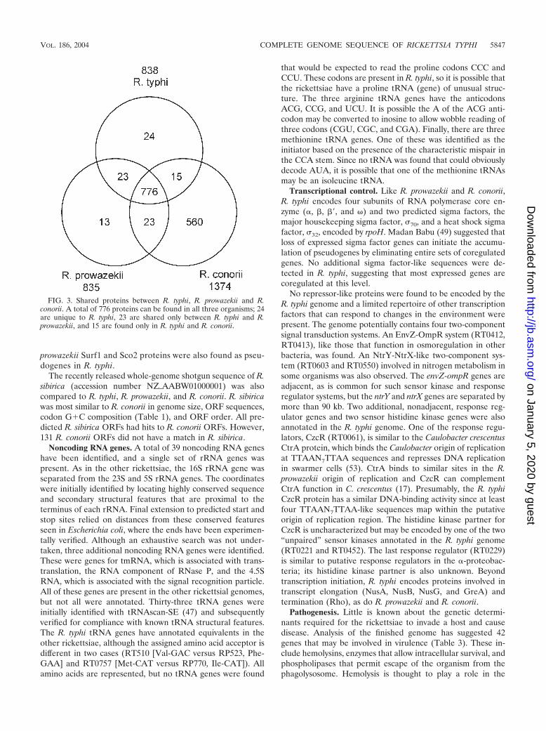
FIG. 3. Shared proteins between R. typhi, R. prowazekii and R.conorii. A total of 776 proteins can be found in all three organisms; 24are unique to R. typhi, 23 are shared only between R. typhi and R.prowazekii, and 15 are found only in R. typhi and R. conorii.
VOL. 186, 2004 COMPLETE GENOME SEQUENCE OF RICKETTSIA TYPHI 5847
on January 5, 2020 by guesthttp://jb.asm
.org/D
ownloaded from

TABLE 2. Genes with restricted distribution in rickettsial genomes
Category and RT no. FunctionPosition
Gene Description RP no. RC no.Start Stop
R. typhi ORFs not found in R. conoriior R. prowazekiiRT0148 Unknown 189321 189461 None Hypothetical protein None NoneRT0154 Unknown 195647 195811 None Hypothetical protein None NoneRT0155 Unknown 196020 196154 None Hypothetical protein None NoneRT0184 Unknown 233126 233230 None Hypothetical protein None NoneRT0199 Unknown 255711 255848 None Hypothetical protein None NoneRT0201 Unknown 258437 258853 None Conserved hypothetical
proteinNone None
RT0301 Unknown 382095 383057 None Rickettsial conservedhypothetical protein
None None
RT0364 Unknown 461966 462193 None Hypothetical protein None NoneRT0418 Unknown 529419 529619 None Hypothetical protein None NoneRT0480 Unknown 614027 614167 None Hypothetical protein None NoneRT0499 Unknown 640260 640412 None Hypothetical protein None NoneRT0505 Unknown 645384 645518 None Hypothetical protein None NoneRT0525 Unknown 668721 668858 None Hypothetical protein None NoneRT0538 Unknown 686867 687124 None Hypothetical protein None NoneRT0542 Unknown 694039 694113 None Hypothetical protein None NoneRT0601 Unknown 778825 778974 None Hypothetical protein None NoneRT0607 Unknown 790366 790560 None Hypothetical protein None NoneRT0664 Unknown 841537 841782 None Hypothetical protein None NoneRT0685 Unknown 868875 868988 None Hypothetical protein None NoneRT0708 Unknown 906635 906880 None Hypothetical protein None NoneRT0751 Unknown 955938 956084 None Hypothetical protein None NoneRT0772 Unknown 984575 984706 None Hypothetical protein None NoneRT0796 Unknown 1018722 1018862 None Hypothetical protein None NoneRT0835 Unknown 1066262 1066381 None Hypothetical protein None None
R. typhi ORFs found in R. conorii butnot in R. prowazekii
RT0034 Virulence 47136 47423 virB3 Probable type IVsecretory systemprotein VirB3
None RC0140
RT0172 General metabolism 217326 218498 None Carboxypeptidase None RC0228RT0213 Unknown 275036 275173 None Rickettsial conserved
hypothetical proteinNone RC0298
RT0270 Unknown 341040 341273 None Rickettsial conservedhypothetical protein
None RC1041
RT0314 Unknown 397194 397355 None Rickettsial conservedhypothetical protein
None RC0444
RT0373 Unknown 474114 474401 None Rickettsial conservedhypothetical protein
None RC0530
RT0520 Unknown 660968 661153 None Rickettsial conservedhypothetical protein
None RC0767
RT0563 Unknown 728779 728994 None Rickettsial conservedhypothetical protein
None RC0876
RT0610 Unknown 794494 794634 None Rickettsial conservedhypothetical protein
None RC0961
RT0623 Unknown 813926 814159 None Rickettsial conservedhypothetical protein
None RC0453
RT0665 Unknown 842115 842354 None Rickettsial conservedhypothetical protein
None RC1041
RT0670 Unknown 848273 848619 None Rickettsial conservedhypothetical protein(frameshift 848487–848490)
None RC1027
RT0672 Unknown 850822 851025 None Rickettsial conservedhypothetical protein
None RC1029
RT0742 Unknown 941930 942133 None Rickettsial conservedhypothetical protein
None RC1172
RT0872 Unknown 1104639 1104791 None Rickettsial conservedhypothetical protein
None RC1362
R. typhi ORFs found in R. prowazekiibut not in R. conorii
RT0026 Energy metabolism 27080 28237 ackA Acetate kinase RP110 NoneRT0033 Virulence 44546 46963 virB4a VirB4 protein
precursorRP103 None
Continued on following page
5848 MCLEOD ET AL. J. BACTERIOL.
on January 5, 2020 by guesthttp://jb.asm
.org/D
ownloaded from

pathogenesis (53, 68) of TG but not SFG of rickettsiae. R.prowazekii has two potential hemolysin genes, tlyA and tlyC (5),and the cloned tlyC from R. typhi can complement hemolyticactivity in a nonhemolytic Proteus mirabilis (54). Both tlyA(RT0543) and tlyC (RT0725) were found in R. typhi. Anotherprotein implicated in the virulence of R. prowazekii is thedinucleoside polyphosphate hydrodolase, InvA. This protein isthought to function by hydrolyzing toxic dinucleoside oligo-phosphates within the host cell to produce ATP, thus providingan environment more conducive to growth (30, 31). ORFRT0228 is 98% identical to the R. prowazekii InvA. Oxidativestress due to reactive oxygen species has been implicated inboth host cell injury caused by Rickettsia and in host defense toinfection (65). ORF RT0523, sodB, is an ortholog of an iron-
associated superoxide dismutase that is predicted to neutralizereactive oxygen species, allowing survival in the host cell. It hasbeen suggested that the rickettsiae use phospholipase A2 toexit the phagosome and the cell (69). A putative phospho-lipase-like protein (RT0590) with significant similarity to thepatatin family of proteins was found in the genome. Patatin isthe main storage protein found in potatoes but has also beenfound to have phospholipase A activity (39, 43). It is possiblethat this protein acts as a phospholipase in R. typhi. In R.conorii, the pldA gene has been shown to encode a protein withphospholipase D activity (55). An ortholog of pldA is present inR. typhi (RT0807). Outer membrane proteins, which are incontact with the host milieu and could function in virulence,have also been identified. These include omp1 (RT0150), a
TABLE 2—Continued
Category and RT no. FunctionPosition
Gene Description RP no. RC no.Start Stop
RT0035 Unknown 48098 48742 engB Probable GTP-bindingprotein EngB
RP102 None
RT0156 Unknown 196185 196379 None Rickettsial conservedhypothetical protein
RP164 None
RT0160 Unknown 199110 199382 None Rickettsial conservedhypothetical protein
RP169 None
RT0268 Unknown 337676 338362 None Rickettsial conservedhypothetical protein
RP277 None
RT0269 Unknown 338385 339527 None Rickettsial conservedhypothetical protein
RP278 None
RT0325 Unknown 412632 413753 None Rickettsial conservedhypothetical protein
RP335 None
RT0326 General metabolism 413782 415005 None Probableglycosyltransferase
RP337 None
RT0327 Unknown 415002 416213 None Possibleglycosyltransferase
RP337 None
RT0328 Unknown 416200 417237 None Conserved hypotheticalprotein
RP338 None
RT0330 Virulence 418166 419182 None WaaG-like sugartransferase
RP340 None
RT0341 Unknown 434960 435208 None Conserved hypotheticalprotein
RP351a None
RT0362 Virulence 458514 459878 None Sec7-domaincontaining proteintransport protein
RP374 None
RT0426 Unknown 535490 536665 None Rickettsial conservedhypothetical protein
RP439 None
RT0463 Virulence 593138 594691 None Possiblelipopolysaccharide1,2-glucosyltransferaseWaaJ
RP476 None
RT0549 Transport 710632 710895 None Possible Na�/H�
antiporterRP561a None
RT0615 Regulation 804351 805103 spoTb Guanosine-3,5-bis(diphosphate) 3-pyrophosphohydrolaseSpoTb
PG624 None
RT0616 Regulation 805109 805450 spoTc Guanosine-3,5-bis(diphosphate) 3-pyrophosphohydrolaseSpoTc
PG625 None
RT0710 Unknown 907724 908002 None Rickettsial conservedhypothetical protein
RP723 None
RT0764 Central dogma 968047 969195 metK Methionineadenosyltransferase
RP777 None
RT0793 Unknown 1012772 1013206 None Conserved hypotheticalprotein
RP806 None
RT0808 General metabolism 1027687 1029441 phbC Poly-�-hydroxybutyratepolymerase
RP820 None
VOL. 186, 2004 COMPLETE GENOME SEQUENCE OF RICKETTSIA TYPHI 5849
on January 5, 2020 by guesthttp://jb.asm
.org/D
ownloaded from
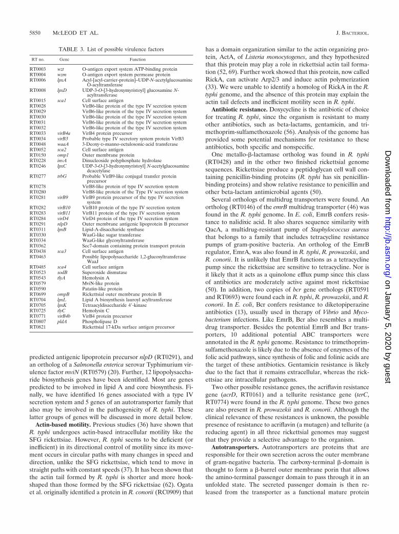
predicted antigenic lipoprotein precursor nlpD (RT0291), andan ortholog of a Salmonella enterica serovar Typhimurium vir-ulence factor mviN (RT0579) (20). Further, 12 lipopolysaccha-ride biosynthesis genes have been identified. Most are genespredicted to be involved in lipid A and core biosynthesis. Fi-nally, we have identified 16 genes associated with a type IVsecretion system and 5 genes of an autotransporter family thatalso may be involved in the pathogenicity of R. typhi. Theselatter groups of genes will be discussed in more detail below.
Actin-based motility. Previous studies (36) have shown thatR. typhi undergoes actin-based intracellular motility like theSFG rickettsiae. However, R. typhi seems to be deficient (orinefficient) in its directional control of motility since its move-ment occurs in circular paths with many changes in speed anddirection, unlike the SFG rickettsiae, which tend to move instraight paths with constant speeds (37). It has been shown thatthe actin tail formed by R. typhi is shorter and more hook-shaped than those formed by the SFG rickettsiae (62). Ogataet al. originally identified a protein in R. conorii (RC0909) that
has a domain organization similar to the actin organizing pro-tein, ActA, of Listeria monocytogenes, and they hypothesizedthat this protein may play a role in rickettsial actin tail forma-tion (52, 69). Further work showed that this protein, now calledRickA, can activate Arp2/3 and induce actin polymerization(33). We were unable to identify a homolog of RickA in the R.typhi genome, and the absence of this protein may explain theactin tail defects and inefficient motility seen in R. typhi.
Antibiotic resistance. Doxycycline is the antibiotic of choicefor treating R. typhi, since the organism is resistant to manyother antibiotics, such as beta-lactams, gentamicin, and tri-methoprim-sulfamethoxazole (56). Analysis of the genome hasprovided some potential mechanisms for resistance to theseantibiotics, both specific and nonspecific.
One metallo-�-lactamase ortholog was found in R. typhi(RT0428) and in the other two finished rickettsial genomesequences. Rickettsiae produce a peptidoglycan cell wall con-taining penicillin-binding proteins (R. typhi has six penicillin-binding proteins) and show relative resistance to penicillin andother beta-lactam antimicrobial agents (50).
Several orthologs of multidrug transporters were found. Anortholog (RT0146) of the emrB multidrug transporter (46) wasfound in the R. typhi genome. In E. coli, EmrB confers resis-tance to nalidixic acid. It also shares sequence similarity withQacA, a multidrug-resistant pump of Staphylococcus aureusthat belongs to a family that includes tetracycline resistancepumps of gram-positive bacteria. An ortholog of the EmrBregulator, EmrA, was also found in R. typhi, R. prowazekii, andR. conorii. It is unlikely that EmrB functions as a tetracyclinepump since the rickettsiae are sensitive to tetracycline. Nor isit likely that it acts as a quinolone efflux pump since this classof antibiotics are moderately active against most rickettsiae(50). In addition, two copies of bcr gene orthologs (RT0591and RT0693) were found each in R. typhi, R. prowazekii, and R.conorii. In E. coli, Bcr confers resistance to diketopiperazineantibiotics (13), usually used in therapy of Vibrio and Myco-bacterium infections. Like EmrB, Bcr also resembles a multi-drug transporter. Besides the potential EmrB and Bcr trans-porters, 10 additional potential ABC transporters wereannotated in the R. typhi genome. Resistance to trimethoprim-sulfamethoxazole is likely due to the absence of enzymes of thefolic acid pathways, since synthesis of folic and folinic acids arethe target of these antibiotics. Gentamicin resistance is likelydue to the fact that it remains extracellular, whereas the rick-ettsiae are intracellular pathogens.
Two other possible resistance genes, the acriflavin resistancegene (acrD, RT0161) and a tellurite resistance gene (terC,RT0774) were found in the R. typhi genome. These two genesare also present in R. prowazekii and R. conorii. Although theclinical relevance of these resistances is unknown, the possiblepresence of resistance to acriflavin (a mutagen) and tellurite (areducing agent) in all three rickettsial genomes may suggestthat they provide a selective advantage to the organism.
Autotransporters. Autotransporters are proteins that areresponsible for their own secretion across the outer membraneof gram-negative bacteria. The carboxy-terminal �-domain isthought to form a �-barrel outer membrane porin that allowsthe amino-terminal passenger domain to pass through it in anunfolded state. The secreted passenger domain is then re-leased from the transporter as a functional mature protein
TABLE 3. List of possible virulence factors
RT no. Gene Function
RT0003 wzt O-antigen export system ATP-binding proteinRT0004 wzm O-antigen export system permease proteinRT0006 lpxA Acyl-[acyl-carrier-protein]–UDP-N-acetylglucosamine
O-acyltransferaseRT0008 lpxD UDP-3-O-[3-hydroxymyristoyl] glucosamine N-
acyltransferaseRT0015 sca1 Cell surface antigenRT0028 VirB6-like protein of the type IV secretion systemRT0029 VirB6-like protein of the type IV secretion systemRT0030 VirB6-like protein of the type IV secretion systemRT0031 VirB6-like protein of the type IV secretion systemRT0032 VirB6-like protein of the type IV secretion systemRT0033 virB4a VirB4 protein precursorRT0034 virB3 Probable type IV secretory system protein VirB3RT0048 waaA 3-Deoxy-D-manno-octulosonic-acid transferaseRT0052 sca2 Cell surface antigenRT0150 omp1 Outer membrane proteinRT0228 invA Dinucleoside polyphosphate hydrolaseRT0246 lpxC UDP-3-O-[3-hydroxymyristoyl] N-acetylglucosamine
deacetylaseRT0277 trbG Probable VirB9-like conjugal transfer protein
precursorRT0278 VirB8-like protein of type IV secretion systemRT0280 VirB8-like protein of the Type IV secretion systemRT0281 virB9 VirB9 protein precursor of the type IV secretion
systemRT0282 virB10 VirB10 protein of the type IV secretion systemRT0283 virB11 VirB11 protein of the type IV secretion systemRT0284 virD4 VirD4 protein of the type IV secretion systemRT0291 nlpD Outer membrane antigenic lipoprotein B precursorRT0311 lpxB Lipid-A-disaccharide synthaseRT0330 WaaG-like sugar transferaseRT0334 WaaG-like glycosyltransferaseRT0362 Sec7-domain containing protein transport proteinRT0438 sca3 Cell surface antigenRT0463 Possible lipopolysaccharide 1,2-glucosyltransferase
WaaJRT0485 sca4 Cell surface antigenRT0523 sodB Superoxide dismutaseRT0543 tlyA Hemolysin ART0579 MviN-like proteinRT0590 Patatin-like proteinRT0699 ompB Rickettsial outer membrane protein BRT0704 lpxL Lipid A biosynthesis lauroyl acyltransferaseRT0705 lpxK Tetraacyldisaccharide 4�-kinaseRT0725 tlyC Hemolysin CRT0771 virB4b VirB4 protein precursorRT0807 pldA Phospholipase DRT0821 Rickettsial 17-kDa surface antigen precursor
5850 MCLEOD ET AL. J. BACTERIOL.
on January 5, 2020 by guesthttp://jb.asm
.org/D
ownloaded from

(38). Analysis of the R. typhi genome revealed the presence offive previously described autotransporter proteins: Sca1(RT0015), Sca2 (RT0052), Sca3 (RT0438), Sca4 (RT0485),and OmpB (RT0699) (5, 52). Work in the SFG rickettsiaeshowed that both OmpB and a related protein, OmpA (notfound in the TG), play a role in adhesion to host cells (45, 60).Although no function of the Sca proteins was demonstrated, itwas suggested that some autotransporters have adhesion orprotease activities and could play a role in rickettsial virulence(38, 69). A characteristic of bacterial adhesins that bind toeukaryotic extracellular matrix proteins is the presence ofamino acid repeats (57). A 50-residue repeat occurs six timeswithin the carboxy-terminal portion of the Sca2 passenger do-main, suggesting that this protein may be involved in binding ofR. typhi to host cells.
In R. typhi, each of the four Sca proteins is found within asingle predicted ORF, while at least one Sca protein has de-graded into multiple ORFs in both R. conorii and R.prowazekii. Sca1, Sca2, and Sca4 are intact in R. conorii, butportions of the Sca3 sequence are found in two different ORFs(RC0630 and RC0631). The R. typhi Sca1 sequence corre-sponds to three different ORFs in R. prowazekii (RP016,RP017, and RP018), whereas the Sca2-like sequence is foundin four different ORFs (RP081, RP082, RP083, and RP084).Sca1 and Sca2 contain only the �-domain and may function totransport other proteins across the outer membrane (69). Sca4is found in two large ORFs in R. prowazekii (RP498 andRP499).
Type IV secretion components. Several components of atype IV secretion system (19) found in R. conorii (52), R.prowazekii (5), and other �-proteobacteria, were present in R.typhi. Type IV secretion systems use a complex of transmem-brane proteins and a pilus to export protein or DNA substratesacross bacterial and eukaryotic cell membranes and into targetcells (18, 19).
Fifteen ORFs related to the Agrobacterium tumefaciens VirBtype IV secretion system were found in R. typhi. These includeorthologs to the inner membrane components VirB4 (RT0033and RT0771) and VirB11 (RT0283). In A. tumefaciens, VirB4binds to the DNA substrate since it is translocated across themembrane (19); the presence of two VirB4 proteins in R. typhimay indicate versatility in substrate selection. The substrate-binding protein VirD4 (19) was also present (RT0283). Theperiplasmic components VirB6, VirB8, and VirB10 (RT0282)were identified. These included two ORFs resembling VirB8(RT0278 and RT0280), in addition to five different ORFs withsimilarity to VirB6 (RT0028, RT0029, RT0030, RT0031, andRT0032). VirB6 functions in the stabilization of VirB5 andVirB7 (36), neither of which was found in R. typhi. VirB6 alsostabilizes VirB3 (RT0034) (36), which was found in R. typhi.Multiple VirB6-like ORFs may be the result of inactivation ofone or more genes or the development of new functions not yetdescribed. Outer membrane components VirB3 and VirB9(RT0277 and RT0281) were also found, but an ortholog ofVirB7, which bridges the pilus to the transmembrane complex,was not (36). Neither VirB2 nor VirB5, the major and minorpilus components, were identified (19). Because R. typhi livewithin the host cell cytoplasm, there may be no need to secretesubstrates across a eukaryotic host cell membrane, hence thelack of pilus components.
A potential protein substrate for a type IV secretion systemwas also found. R. typhi has a Sec7 domain-containing protein(RT0362) that is similar to RalF in Legionella pneumophila.RalF is secreted by the L. pneumophila dot/icm type IV secre-tion system (51). Along with a R. prowazekii ortholog, these arethe only three known examples of Sec7 family proteins inbacteria (51). Sec7 proteins, named for Saccharomyces cerevi-siae Sec7p, are guanine-nucleotide exchange factors and cofac-tors for ADP-ribosylation factors (ARFs), which function inthe regulation of membrane traffic and organelle structure ineukaryotic cells (25, 42). The similarity to the L. pneumophilaRalF protein suggests that RT0362 may be secreted by the R.typhi type IV system. In Legionella, the function of RalF isrelated to recruiting host ARFs to bacterium-containingphagosomes (51) but, since rickettsiae do not live in phago-somes, the role of the RalF-like protein in R. typhi is unknown.
Other transporters. In addition to the autotransporters,drug efflux pumps, and VirB secretion system, R. typhi hasproteins that function in transporting substances across the cellmembrane. There were five copies of an ADP/ATP translo-case, a transporter that allows R. typhi to acquire host cell ATP(5). As in other rickettsiae, there are multiple copies of theosmosensory proline-betaine transporter, ProP. We found 7copies of ProP in R. typhi compared to 11 copies in R. conoriiand 7 copies in R. prowazekii (52). Transport proteins for cellmembrane and cell wall components were found, includingthree copies of the murein peptide permease AmpG, two hemeexporters CcmAB (RT0781 and RT0259), and two O-antigenexporter proteins (RT0002 and RT0003). Also found was ahomolog of the SAM transporter of R. prowazekii (RT0056)that has recently been shown to transport S-adenosylmethi-onine into the bacterial cell (59). A variety of other transportproteins, including seven possible ABC transport systems, werefound. These transporters include a potential zinc/manganesetransporter (RT0013 and RT0612), a magnesium transporter(RT0572), potassium transporter (RT0798), a glutamine trans-porter (RT0118, RT0139, and RT0859), a putrescine/ornithinetransporter (RT0470), a ribonucleotide transporter (RT0040),and a sodium/proton symporter (RT0549). Four members ofthe Tol family—TolC (RT0216), TolB (RT0293), TolQ(RT0299), and TolR (RT0300)—were identified. Transporterswith less clear possible functions were given generic descrip-tions such as metal transporter and cation transporter. Stillothers with no known or postulated substrate were also found.In addition, a PSORT-B (29) analysis of coding regions iden-tified 39 hypothetical, conserved hypothetical, or rickettsialconserved hypothetical proteins that could be membrane lo-calized. Some of these may also function in transport.
Stringent response. In response to starvation and otherstress signals, bacteria increase their intracellular concentra-tion of GDP 3�-diphosphate and GTP 3�-diphosphate, or(p)ppGpp, in a process called the stringent response (21, 22).This leads to a downregulation of transcription and translation.In E. coli, the RelA and SpoT proteins modulate (p)ppGpplevels in the cell. No intact R. typhi ORF with significant sim-ilarity to either RelA or SpoT was found, although four smallORFs with significant similarity to SpoT were found. TheseORFs align to two different regions of SpoT (Fig. 4) and havebeen previously described and classified as pseudogenes or“split genes”, named spoTa, spoTb, spoTc, and spoTd (4, 5, 52).
VOL. 186, 2004 COMPLETE GENOME SEQUENCE OF RICKETTSIA TYPHI 5851
on January 5, 2020 by guesthttp://jb.asm
.org/D
ownloaded from
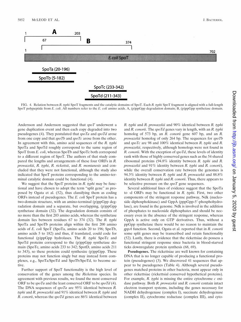
Andersson and Andersson suggested that spoT underwent agene duplication event and then each copy degraded into twopseudogenes (4). They postulated that spoTa and spoTd arosefrom one copy and that spoTb and spoTc arose from the other.In agreement with this, amino acid sequences of the R. typhiSpoTa and SpoTd roughly correspond to the same region ofSpoT from E. coli, whereas SpoTb and SpoTc both correspondto a different region of SpoT. The authors of that study com-pared the lengths and arrangements of these four ORFs in R.prowazekii, R. typhi, R. rickettsii, and R. montanesis and con-cluded that they were not functional, although the study alsoindicated that SpoT proteins corresponding to the amino-ter-minal catalytic domain could be functional (4).
We suggest that the SpoT proteins in R. typhi may be func-tional and have chosen to adopt the term “split gene” as pro-posed by Ogata et al. (52), thus classifying them as codingORFs instead of pseudogenes. The E. coli SpoT protein has atwo-domain structure, with an amino-terminal (p)ppGpp deg-radation domain and a separate, but overlapping, (p)ppGppsynthetase domain (32). The degradation domain consists ofno more than the first 203 amino acids, whereas the synthetasedomain lies between residues 67 to 374 (32). The R. typhiSpoTa and SpoTb proteins align within the first 200 aminoacids of E. coli SpoT (SpoTa, amino acids 20 to 196; SpoTb,amino acids 5 to 182) and thus, if translated, could code forfunctional (p)ppGpp hydrolases. The R. typhi SpoTc andSpoTd proteins correspond to the (p)ppGpp synthetase do-main (SpoTc, amino acids 233 to 342; SpotD, amino acids 211to 343), so these proteins could synthesize (p)ppGpp. Theseproteins may not function singly but may instead form com-plexes, e.g., SpoTa/SpoTd and SpoTb/SpoTd, to become ac-tive.
Further support of SpoT functionality is the high level ofconservation of the genes among the Rickettsia species. Inagreement with previous results, we found the most conservedORF to be spoTa and the least conserved ORF to be spoTd (4).The DNA sequences of spoTa are 95% identical between R.typhi and R. prowazekii and 91% identical between R. typhi andR. conorii, whereas the spoTd genes are 86% identical between
R. typhi and R. prowazekii and 90% identical between R. typhiand R. conorii. The spoTd genes vary in length, with an R. typhihomolog of 573 bp, an R. conorii gene 607 bp, and an R.prowazekii homolog of only 264 bp. The sequences for spoTband spoTc are 98 and 100% identical between R. typhi and R.prowazekii, respectively, although homologs were not found inR. conorii. With the exception of spoTd, these levels of identityrank with those of highly conserved genes such as the 54 sharedribosomal proteins (94.8% identity between R. typhi and R.prowazekii and 91% identity between R. typhi and R. conorii),while the overall conservation rate between the genomes is94.3% identity between R. typhi and R. prowazekii and 88.8%identity between R. typhi and R. conorii. Thus, there appears tobe selective pressure on the spoT gene sequences.
Several additional lines of evidence suggest that the SpoTato -d ORFs may be functional in R. typhi. First, two othercomponents of the stringent response pathway, Ndk (nucleo-side diphosphokinase) and GppA (pppGpp-5�-phosphohydro-lase), are found in the genome. Ndk is involved in the additionof phosphates to nucleoside diphosphates and should be nec-essary even in the absence of the stringent response, whereasGppA is active only on GTP derivatives. Thus, without appGpp synthetase there would be no reason to maintain thegppA function. Second, Ogata et al. reported that in R. conoriisome split genes may be transcribed and retain functionality(52). Lastly, there is evidence that the rickettsiae do possess afunctional stringent response since bacteria in blood-starvedticks downregulate protein synthesis (68, 69).
Pseudogenes. The rickettsiae are well known for containingDNA that is no longer capable of producing a functional pro-tein (pseudogenes) (3). We discovered 41 sequences that ap-pear to be pseudogenes (Table 4). Although several pseudo-genes matched proteins in other bacteria, most appear only inother rickettsiae (rickettsial conserved hypothetical proteins).For example, R. typhi is missing the entire cytochrome c oxi-dase pathway. Both R. prowazekii and R. conorii contain intactelectron transport systems, including the genes necessary forNADH dehydrogenase (complex I), succinate dehydrogenase(complex II), cytochrome reductase (complex III), and cyto-
FIG. 4. Relation between R. typhi SpoT fragments and the catalytic domains of SpoT. Each R. typhi SpoT fragment is aligned with a full-lengthSpoT polypeptide from E. coli. All numbers refer to the E. coli amino acids. A, (p)ppGpp degradation domain; B, (p)ppGpp synthetase domain.
5852 MCLEOD ET AL. J. BACTERIOL.
on January 5, 2020 by guesthttp://jb.asm
.org/D
ownloaded from

chrome oxidase (complex IV) (5, 52). Many bacteria containmore than one terminal oxidase for aerobic respiration underdiffering oxygen concentrations (44). Both R. prowazekii and R.conorii contain the genes coxABC, which encode a putativecytochrome c oxidase, and cydAB, which encode a putativecytochrome d ubiquinol oxidase. This situation is similar toMycobacterium smegmatis in which an aa3-type cytochrome coxidase is expressed during normal aerobic growth and acydAB-encoded bd-type oxidase is expressed during low oxygenconditions (44). In R. typhi, all three genes encoding cyto-chrome c oxidase (coxCAB; RTPG10, RTPG22, and RTPG23)have decayed and are likely nonfunctional. In addition, thegenes encoding the three cytochrome c oxidase assembly pro-teins, CoxW, CtaG, and CtaB (RTPG14, RTPG15, andRTPG18), have also decayed. As a result, it appears that R.typhi retains only one terminal oxidase for aerobic respiration.The remaining oxidase, CydAB (RT0207 and RT0208), is pre-dicted to be of the d-type that is commonly expressed during
low-oxygen conditions. Such cytochrome d oxidases generallyhave a higher affinity for oxygen than do cytochrome c oxidasesand thus mediate the transfer of electrons to water at a slowerrate (34). This may lead to physiological effects that play a rolein the attenuated disease manifestations of R. typhi comparedto R. prowazekii.
DISCUSSION
The genome of R. typhi is nearly identical to its close relativeR. prowazekii and highly similar to R. conorii and other SFGbacteria. The few differences between the two TG rickettsiaeinclude a 12-kb insertion in the genome of R. prowazekii, alarge inversion close to the origin of replication with no loss ofgenes in the region, and the fact that R. typhi has lost thecomplete cytochrome c oxidase system. In addition, R. typhihas several pseudogenes for which functional homologs arefound in R. prowazekii. The similarity of the genome sequence
TABLE 4. Pseudogenes found in R. typhi and their functional counterparts in R. prowazekii and R. conorii
RT no.Position
Description of functional counterpart RP no. RC no.Start Stop
RTPG01 19015 19631 tRNA/rRNA methyltransferase None RC0160RTPG02 47435 48207 Acetylglutamate kinase None RC0139RTPG03 61590 61776 RP088 RP088 NoneRTPG04 103201 103580 RP052 RP052 NoneRTPG05 105249 105804 RP050/RC0076 RP050 RC0076RTPG06 126004 126468 RC0049 None RC0049RTPG07 129317 129872 Sco2 protein precursor RP031 RC0042RTPG08 134057 134355 Rickettsial conserved protein with coenzyme PQQ synthesis
protein C domainNone RC0033
RTPG09 198021 198258 Conserved hypothetical protein RP167 RC0209RTPG10 229960 230742 Cytochrome c oxidase subunit III RP191 RC0240RTPG11 253368 253540 Rickettsial conserved hypothetical protein None RC0276RTPG12 258881 259212 Agrobacterium protein None NoneRTPG13 271206 272364 Rickettsial conserved hypothetical protein None RC0295RTPG14 314921 315914 Cytochrome c oxidase assembly protein CoxW RP257 RC0343RTPG15 373523 374000 Cytochrome c oxidase assembly protein CtaG RP304 RC0408RTPG16 383166 383538 Rickettsial conserved hypothetical protein None RC0425RTPG17 408613 408790 Rickettsial conserved hypothetical protein RP331 NoneRTPG18 425149 425995 Protoheme IX farnesyltransferase (cytochrome c oxidase
assembly protein CtaB)RP346 RC0469
RTPG19 462673 463315 Rickettsial conserved hypothetical protein None RC0519RTPG20 470138 471491 Rickettsial conserved hypothetical protein RP382 RC0526RTPG21 473514 474113 Cell filamentation protein None RC0529RTPG22 496379 497698 Cytochrome c oxidase subunit I RP405 RC0553RTPG23 497853 498935 Cytochrome c oxidase subunit II RP406 RC0555RTPG24 605974 606376 Rickettsial conserved hypothetical protein RP488 RC0732RTPG25 617447 617602 Hypothetical protein RC0669 None RC0669RTPG26 650083 650663 Hypothetical protein RC0754 None RC0754RTPG27 680777 680989 Hypothetical protein RP543 RP543 NoneRTPG28 687125 688408 Hypothetical protein RC0815 None RC0815RTPG29 693501 693801 Hypothetical protein RC0819 None RC0819RTPG30 753627 754173 3-Oxoacid coenzyme A-transferase subunit B None RC0906RTPG31 777824 778728 Mg/Co transport protein RC0943 None RC0943RTPG32 831141 831549 Hypothetical protein RC1011 None RC1011RTPG33 846656 846872 Hypothetical protein RC1025 None RC1025RTPG34 872509 872815 Hypothetical protein RC1065 None RC1065RTPG35 897902 898363 Bacterioferritin comigratory protein None RC1090RTPG36 900174 900814 Rickettsial conserved hypothetical protein None RC1093RTPG37 900807 901141 Rickettsial conserved hypothetical protein None RC1094RTPG38 913123 913788 Surf1-like protein RP733 RC1113RTPG39 920761 921226 Rickettsial conserved hypothetical protein None RC1129RTPG40 932587 932807 Hypothetical protein RC1157 None RC1157RTPG41 1105415 1105657 Hypothetical protein RC1365 None RC1365
VOL. 186, 2004 COMPLETE GENOME SEQUENCE OF RICKETTSIA TYPHI 5853
on January 5, 2020 by guesthttp://jb.asm
.org/D
ownloaded from

of R. typhi to the TG rickettsiae further illustrates the delin-eation between the TG and SFG rickettsiae. Based on com-parisons of these three genomes, it appears that rickettsiaemay have a functional stringent response system that utilizessplit genes of the SpoT protein as previously described. Fur-ther comparisons that include R. sibirica and R. rickettsii, asbriefly mentioned here, should provide additional insight intothe evolutionary relationship between these important patho-gens.
The rickettsiae appear to maintain nonfunctional DNA for amuch longer period than do other bacteria. It has been pro-posed that slow-growth times can account for this. However,there are many slow-growing bacteria that do not accumulatepseudogenes. It is possible that the rickettsiae retain this DNAfor a reason or that their pathway for removing nonfunctionalDNA is different. By comparing this genome to others, whichlose nonfunctional genes faster, we may discover the mecha-nisms by which organisms lose DNA. It is interesting that mostof the pseudogenes in R. typhi were found in regions withdeletions in R. prowazekii and R. typhi with respect to R. cono-rii, and in fact many appear to be remnants of the deletionevent. The pseudogenes may be functional in other rickettsiaeor indicative of regions that are not functional in any of therickettsiae.
The high degree of similarity between R. typhi and R.prowazekii illustrates the small differences that can affect viru-lence in different hosts. Thus, despite their close genetic relat-edness, R. prowazekii is highly pathogenic in humans, whereasR. typhi is a milder pathogen in humans. Future studies shouldidentify which of the differences in these genomes accounts forthese phenotypes.
ACKNOWLEDGMENTS
This research was supported by a grant from the National Instituteof Allergy and Infectious Disease (AI49040). Michael P. McLeod wassupported in part by a training grant from the Keck Center for Com-putational Biology (NLM grant LM07093). George E. Fox was sup-ported in part by a Ruth L. Kirschstein National Research ServiceAward (F33-HGO2551).
We are grateful to Virginia Vera, Baize Montgomery, and ReginaWleczyk for technical assistance. We thank Manuel Gonzalez-Garay,Andrei Volkov, Andrew R. Jackson, and Aleksandar Milosavljevic forhosting annotation on the Genboree server.
REFERENCES
1. Altschul, S. F., T. L. Madden, A. A. Schaffer, J. Zhang, Z. Zhang, W. Miller,and D. J. Lipman. 1997. Gapped BLAST and PSI-BLAST: a new generationof protein database search programs. Nucleic Acids Res. 25:3389–3402.
2. Andersson, B., M. A. Wentland, J. Y. Ricafrente, W. Liu, and R. A. Gibbs.1996. A “double adaptor” method for improved shotgun library construction.Anal. Biochem. 236:107–113.
3. Andersson, J. O., and S. G. Andersson. 1999. Insights into the evolutionaryprocess of genome degradation. Curr. Opin. Genet. Dev. 9:664–671.
4. Andersson, J. O., and S. G. Andersson. 2001. Pseudogenes, junk DNA, andthe dynamics of Rickettsia genomes. Mol. Biol. Evol. 18:829–839.
5. Andersson, S. G., A. Zomorodipour, J. O. Andersson, T. Sicheritz-Ponten,U. C. Alsmark, R. M. Podowski, A. K. Naslund, A. S. Eriksson, H. H.Winkler, and C. G. Kurland. 1998. The genome sequence of Rickettsiaprowazekii and the origin of mitochondria. Nature 396:133–140.
6. Andersson, S. G. E., and C. G. Kurland. 1998. Reductive evolution ofresident genomes. Trends Microbiol. 6:263–268.
7. Atlas, R. 1998. Biological weapons pose challenge for microbiology commu-nity. ASM News 64:383–389.
8. Azad, A. F., and C. B. Beard. 1998. Rickettsial pathogens and their arthropodvectors. Emerg. Infect. Dis. 4:179–186.
9. Azad, A. F., and S. Radulovic. 2003. Pathogenic rickettsiae as bioterrorismagents. Ann. N. Y. Acad. Sci. 990:734–738.
10. Azad, A. F., S. Radulovic, J. A. Higgins, B. H. Noden, and J. M. Troyer. 1997.Flea-borne rickettsioses: ecologic considerations. Emerg. Infect. Dis. 3:319–327.
11. Bateman, A., E. Birney, L. Cerruti, R. Durbin, L. Etwiller, S. R. Eddy, S.Griffiths-Jones, K. L. Howe, M. Marshall, and E. L. Sonnhammer. 2002. ThePfam protein families database. Nucleic Acids Res. 30:276–280.
12. Baxter, J. D. 1996. The typhus group. Clin. Dermatol. 14:271–278.13. Bentley, J., L. S. Hyatt, K. Ainley, J. H. Parish, R. B. Herbert, and G. R.
White. 1993. Cloning and sequence analysis of an Escherichia coli geneconferring bicyclomycin resistance. Gene 127:117–120.
14. Betley, J. N., M. C. Frith, J. H. Graber, S. Choo, and J. O. Deshler. 2002. Aubiquitous and conserved signal for RNA localization in chordates. Curr.Biol. 12:1756–1761.
15. Boeckmann, B., A. Bairoch, R. Apweiler, M. C. Blatter, A. Estreicher, E.Gasteiger, M. J. Martin, K. Michoud, C. O’Donovan, I. Phan, S. Pilbout, andM. Schneider. 2003. The SWISS-PROT protein knowledge base and itssupplement TrEMBL in 2003. Nucleic Acids Res. 31:365–370.
16. Bovarnick, M. R., J. C. Miller, and J. C. Snyder. 1950. The influence ofcertain salts, amino acids, sugars and proteins on the stability of rickettsiae.J. Bacteriol. 59:509–522.
17. Brassinga, A. K., R. Siam, W. McSween, H. Winkler, D. Wood, and G. T.Marczynski. 2002. Conserved response regulator CtrA and IHF binding sitesin the alpha-proteobacteria Caulobacter crescentus and Rickettsia prowazekiichromosomal replication origins. J. Bacteriol. 184:5789–5799.
18. Burns, D. L. 1999. Biochemistry of type IV secretion. Curr. Opin. Microbiol.2:25–29.
19. Burns, D. L. 2003. Type IV transporters of pathogenic bacteria. Curr. Opin.Microbiol. 6:29–34.
20. Carsiotis, M., B. Stocker, D. Weinstein, and A. O’Brien. 1989. A Salmonellatyphimurium virulence gene linked to flg. Infect. Immun. 57:3276–3280.
21. Cashel, M., and K. E. Rudd. 1987. The stringent response, p. 1410–1438. InF. C. Neidhardt, R. Curtiss III, J. L. Ingraham, E. C. C. Lin, K. B. Low, B.Magasanik, W. S. Reznikoff, M. Riley, M. Schaechter, and H. E. Umbarger(ed.), Escherichia coli and Salmonella: cellular and molecular biology, 2nded.
22. Chatterji, D., and A. K. Ojha. 2001. Revisiting the stringent response, ppGppand starvation signaling. Curr. Opin. Microbiol. 4:160–165.
23. Delcher, A. L., D. Harmon, S. Kasif, O. White, and S. L. Salzberg. 1999.Improved microbial gene identification with GLIMMER. Nucleic Acids Res.27:4636–4641.
24. Demerec, M., E. A. Adelberg, A. J. Clark, and P. E. Hartman. 1966. Aproposal for a uniform nomenclature in bacterial genetics. Genetics 54:61–76.
25. Donaldson, J. G., and C. L. Jackson. 2000. Regulators and effectors of theARF GTPases. Curr. Opin. Cell Biol. 12:475–482.
26. Ewing, B., and P. Green. 1998. Base-calling of automated sequencer tracesusing phred. II. Error probabilities. Genome Res. 8:186–194.
27. Ewing, B., L. Hillier, M. C. Wendl, and P. Green. 1998. Base-calling ofautomated sequencer traces using phred. I. Accuracy assessment. GenomeRes. 8:175–185.
28. Fergie, J. E., K. Purcell, and D. Wanat. 2000. Murine typhus in South Texaschildren. Pediatr. Infect. Dis. J. 19:535–538.
29. Gardy, J. L., C. Spencer, K. Wang, M. Ester, G. E. Tusnady, I. Simon, S.Hua, K. deFays, C. Lambert, K. Nakai, and F. S. Brinkman. 2003.PSORT-B: improving protein subcellular localization prediction for gram-negative bacteria. Nucleic Acids Res. 31:3613–3617.
30. Gaywee, J., S. Radulovic, J. A. Higgins, and A. F. Azad. 2002. Transcriptionalanalysis of Rickettsia prowazekii invasion gene homolog (invA) during hostcell infection. Infect. Immun. 70:6346–6354.
31. Gaywee, J., W. Xu, S. Radulovic, M. J. Bessman, and A. F. Azad. 2002. TheRickettsia prowazekii invasion gene homolog (invA) encodes a nudix hydro-lase active on adenosine (5�)-pentaphospho-(5�)-adenosine. Mol. Cell Pro-teomics 1:179–183.
32. Gentry, D. R., and M. Cashel. 1996. Mutational analysis of the Escherichiacoli spoT gene identifies distinct but overlapping regions involved in ppGppsynthesis and degradation. Mol. Microbiol. 19:1373–1384.
33. Gouin, E., C. Egile, P. Dehoux, V. Villiers, J. Adams, F. Gertler, R. Li, andP. Cossart. 2004. The RickA of Rickettsia conorii activates the Arp2/3 com-plex. Nature 427:457–461.
34. Govantes, F., A. V. Orjalo, and R. P. Gunsalus. 2000. Interplay betweenthree global regulatory proteins mediates oxygen regulation of the Esche-richia coli cytochrome days oxidase (cydAB) operon. Mol. Microbiol. 38:1061–1073.
35. Handelman, S., and K. Alibek. 2000. Biohazard. Delta, New York, N.Y.36. Hapfelmeier, S., N. Domke, P. C. Zambryski, and C. Baron. 2000. VirB6 is
required for stabilization of VirB5 and VirB3 and formation of VirB7 ho-modimers in Agrobacterium tumefaciens. J. Bacteriol. 182:4505–4511.
37. Heinzen, R. A. 2003. Rickettsial actin-based motility: behavior and involve-ment of cytoskeletal regulators. Ann. N. Y. Acad. Sci. 990:535–547.
38. Henderson, I. R., F. Navarro-Garcia, and J. P. Nataro. 1998. The greatescape: structure and function of the autotransporter proteins. Trends Mi-crobiol. 6:370–378.
5854 MCLEOD ET AL. J. BACTERIOL.
on January 5, 2020 by guesthttp://jb.asm
.org/D
ownloaded from

39. Holk, A., S. Rietz, M. Zahn, H. Quader, and G. F. E. Scherer. 2002. Molec-ular identification of cytosolic, patatin-related phospholipases A from Ara-bidopsis with potential functions in plant signal transduction. Plant Physiol.130:90–101.
40. Hoskinson, S., M. Lipetz, C. Sherer, S. Fraser, E. Taniguchi, A. Tice, D.Behling, B. Tanabe, D. Kwock, P. Effler, L. Pang, E. Brown, L. Granville, K.Mills, D. Sasaki, M. Ching Lee, H. Matsubayashi, W. Warashina, A. Ueno,C. Takekuma, J. Haruno, S. Oshiro, P. Kitsutani, J. McQuiston, C. Pad-dock, J. Sumner, and J. Krebs. 2003. Murine typhus–Hawaii, 2002. Morb.Mortal. Wkly. Rep. 52:1224–1226.
41. Hryniewicz-Jankowska, A., A. Czogalla, E. Bok, and A. F. Sikorsk. 2002.Ankyrins, multifunctional proteins involved in many cellular pathways. FoliaHistochem. Cytobiol. 40:239–249.
42. Jackson, C. L., and J. E. Casanova. 2000. Turning on ARF: the Sec7 familyof guanine-nucleotide-exchange factors. Trends Cell Biol. 10:60–67.
43. Jimenez-Atienzar, M., J. Cabanes, F. Gandia-Herrero, J. Escribano, F. Gar-cia-Carmona, and M. Perez-Gilabert. 2003. Determination of the phospho-lipase activity of patatin by a continuous spectrophotometric assay. Lipids38:677–682.
44. Kana, B. D., E. A. Weinstein, D. Avarbock, S. S. Dawes, H. Rubin, and V.Mizrahi. 2001. Characterization of the cydAB-encoded cytochrome bd oxi-dase from Mycobacterium smegmatis. J. Bacteriol. 183:7076–7086.
45. Li, H., and D. H. Walker. 1998. rOmpA is a critical protein for the adhesionof Rickettsia rickettsii to host cells. Microb. Pathogen. 24:289–298.
46. Lomovskaya, O., and K. Lewis. 1992. Emr, an Escherichia coli locus formultidrug resistance. Proc. Natl. Acad. Sci. USA 89:8938–8942.
47. Lowe, T. M., and S. R. Eddy. 1997. tRNAscan-SE: a program for improveddetection of transfer RNA genes in genomic sequence. Nucleic Acids Res.25:955–964.
48. Lukashin, A. V., and M. Borodovsky. 1998. GeneMark.hmm: new solutionsfor gene finding. Nucleic Acids Res. 26:1107–1115.
49. Madan Babu, M. 2003. Did the loss of sigma factors initiate pseudogeneaccumulation in Mycobacterium leprae? Trends Microbiol. 11:59–61.
50. Miyamura, S., T. Ohta, and A. Tamura. 1989. Comparison of in vitro sus-ceptibilities of Rickettsia prowazekii, R. rickettsii, R. sibirica, and R. tsutsuga-mushi to antimicrobial agents. Nippon Saikingaku Zasshi. 44:717–721.
51. Nagai, H., J. C. Kagan, X. Zhu, R. A. Kahn, and C. R. Roy. 2002. A bacterialguanine nucleotide exchange factor activates ARF on Legionella phago-somes. Science 295:679–682.
52. Ogata, H., S. Audic, P. Renesto-Audiffren, P. E. Fournier, V. Barbe, D.Samson, V. Roux, P. Cossart, J. Weissenbach, J. M. Claverie, and D. Raoult.2001. Mechanisms of evolution in Rickettsia conorii and R. prowazekii. Sci-ence 293:2093–2098.
53. Quon, K. C., B. Yang, I. J. Domian, L. Shapiro, and G. T. Marczynski. 1998.Negative control of bacterial DNA replication by a cell cycle regulatoryprotein that binds at the chromosome origin. Proc. Natl. Acad. Sci. USA95:120–125.
54. Radulovic, S., J. M. Troyer, M. S. Beier, A. O. T. Lau, and A. F. Azad. 1999.Identification and molecular analysis of the gene encoding Rickettsia typhihemolysin. Infect. Immun. 67:6104–6108.
55. Renesto, P., P. Dehoux, E. Gouin, L. Touqui, P. Cossart, and D. Raoult.2003. Identification and characterization of a phospholipase D superfamilygene in rickettsiae. J. Infect. Dis. 188:1276–1283.
56. Rolain, J. M., L. Stuhl, M. Maurin, and D. Raoult. 2002. Evaluation ofantibiotic susceptibilities of three rickettsial species including Rickettsia felisby quantitative PCR DNA assay. Antimicrob. Agents Chemother. 46:2747.
57. Shimoji, Y., Y. Ogawa, M. Osaki, H. Kabeya, S. Maruyama, T. Mikami, andT. Sekizaki. 2003. Adhesive surface proteins of Erysipelothrix rhusiopathiaebind to polystyrene, fibronectin, and type I and IV collagens. J. Bacteriol.185:2739–2748.
58. Tatusov, R. L., E. V. Koonin, and D. J. Lipman. 1997. A genomic perspectiveon protein families. Science 278:631–637.
59. Tucker, A. M., H. H. Winkler, L. O. Driskell, and D. O. Wood. 2003.S-Adenosylmethionine transport in Rickettsia prowazekii. J. Bacteriol. 185:3031–3035.
60. Uchiyama, T. 1999. Role of major surface antigens of Rickettsia japonica inthe attachment to host cell, p. 182–188. In J. Kazar and D. Raoult (ed.),Rickettsiae and rickettsial diseases. Publishing House of the Slovak Acad-emy of Sciences, Bratislava, Czechoslovakia.
61. Ursing, B. M., F. H. van Enckevort, J. A. Leunissen, and R. J. Siezen. 2002.EXProt: a database for proteins with an experimentally verified function.Nucleic Acids Res. 30:50–51.
62. Van Kirk, L. S., S. F. Hayes, and R. A. Heinzen. 2000. Ultrastructure ofRickettsia rickettsii actin tails and localization of cytoskeletal proteins. Infect.Immun. 68:4706–4713.
63. Walker, D. H., H. M. Feng, S. Ladner, A. N. Billings, S. R. Zaki, D. J. Wear,and B. Hightower. 1997. Immunohistochemical diagnosis of typhus rickett-sioses using an anti-lipopolysaccharide monoclonal antibody. Mod. Pathol.10:1038–1042.
64. Walker, D. H., F. M. Parks, T. G. Betz, J. P. Taylor, and J. W. Muehlberger.1989. Histopathology and immunohistologic demonstration of the distribu-tion of Rickettsia typhi in fatal murine typhus. Am. J. Clin. Pathol. 91:720–724.
65. Walker, D. H., G. A. Valbuena, and J. P. Olano. 2003. Pathogenic mecha-nisms of diseases caused by Rickettsia. Ann. N. Y. Acad. Sci. 990:1–11.
66. Weiss, E., J. C. Coolbaugh, and J. C. Williams. 1975. Separation of viableRickettsia typhi from yolk sac and L cell host components by renografindensity gradient centrifugation. Appl. Microbiol. 30:456–463.
67. Wheeler, D. L., D. M. Church, A. E. Lash, D. D. Leipe, T. L. Madden, J. U.Pontius, G. D. Schuler, L. M. Schriml, T. A. Tatusova, L. Wagner, and B. A.Rapp. 2002. Database resources of the National Center for BiotechnologyInformation: 2002 update. Nucleic Acids Res. 30:13–16.
68. Wike, D. A., and W. Burgdorfer. 1972. Plaque formation in tissue cultures byRickettsia rickettsii isolated directly from whole blood and tick hemolymph.Infect. Immun. 6:736–738.
69. Yu, X.-J., and D. H. Walker. 2003. The order Rickettsiales. In M. Dworkin(ed.), The prokaryotes: an evolving electronic resource for the microbiolog-ical community, 3rd ed. (release 3.12 ed.). Springer-Verlag, New York, N.Y.
VOL. 186, 2004 COMPLETE GENOME SEQUENCE OF RICKETTSIA TYPHI 5855
on January 5, 2020 by guesthttp://jb.asm
.org/D
ownloaded from