complementary microorganisms in highly corrosive...
TRANSCRIPT
1
Complementary microorganisms in highly corrosive biofilms from an offshore oil 1
production facility 2
Adrien Vigneron12*, Eric B. Alsop23, Brian Chambers4, Bartholomeus P. Lomans5, Ian M. Head1 3
and Nicolas Tsesmetzis2 4
1School of Civil Engineering and Geosciences, Newcastle University, Newcastle upon Tyne NE1 5
7RU, UK 6
2Shell International Exploration and Production Inc., Houston, Texas, USA 7
3DOE Joint Genome Institute, Walnut Creek, California 94598, USA 8
4Shell Global Solutions US Inc., Houston, Texas, USA 9
5Shell Global Solutions International B.V., Rijswijk, Netherlands 10
* [email protected] 11
Running Title 12
Biocorrosion in Oil production facility 13
Keywords 14
MIC ; Corrosion ; multigenic ; biofilm ; SRB ; methanogens ; oil 15
Abstract 16
AEM Accepted Manuscript Posted Online 19 February 2016Appl. Environ. Microbiol. doi:10.1128/AEM.03842-15Copyright © 2016, American Society for Microbiology. All Rights Reserved.
on July 6, 2018 by guesthttp://aem
.asm.org/
Dow
nloaded from
2
Offshore oil production facilities are frequently victims of internal piping corrosion, potentially 17
leading to human and environmental risks and significant economic losses. Microbially 18
influenced corrosion (MIC) is believed to be an important factor in this major problem for the 19
petroleum industry. However, knowledge of the microbial communities and metabolic processes 20
leading to corrosion is still limited. Therefore, the microbial communities from three anaerobic 21
biofilms recovered from the inside of a steel pipe, exhibiting high corrosion rates, iron oxides 22
deposits and substantial amounts of sulfur, which are characteristic of MIC, were analyzed in 23
detail. Bacterial and archaeal community structures were investigated using Automated 24
Ribosomal Intergenic Spacer Analysis (ARISA), multigenic (16S rRNA and functional genes) 25
high throughput Illumina Miseq sequencing and quantitative polymerase chain reaction analysis. 26
The microbial community analysis indicated that bacteria and particularly Desulfovibrio species 27
dominated the biofilm microbial communities. However other bacteria such as Pelobacter, 28
Pseudomonas and Geotoga, as well as various methanogenic archaea, previously detected in 29
oil facilities were also detected. The microbial taxa and functional genes identified suggested 30
that the biofilm communities harbored the potential for a number of different but complementary 31
metabolic processes and that MIC in oil facilities likely involves a range of microbial 32
metabolisms such as sulfate, iron and elemental sulfur reduction. Furthermore, extreme 33
corrosion leading to leakage and exposure of the biofilms to the external environment modify 34
the microbial community structure by promoting the growth of aerobic hydrocarbon degrading 35
organisms. 36
on July 6, 2018 by guesthttp://aem
.asm.org/
Dow
nloaded from
3
Introduction 37
Metal corrosion is a major concern for the oil industry, potentially leading to environmental 38
pollution, safety issues and major economic losses (1, 2). Offshore oil facilities represent an 39
important biotope for microorganisms with corrosive metabolism and are thus markedly affected 40
by microbially influenced corrosion (MIC). The anaerobic conditions combined with petroleum 41
hydrocarbons, organo-sulfur compounds (e.g. benzothiophenes), volatile fatty acids and other 42
end-products of fermentations, present in produced oil and water, provide significant amounts of 43
carbon substrates for microorganisms within piping and pipeline networks (3). Furthermore the 44
steel tubes themselves, and the seawater initially injected for secondary oil recovery supply 45
abundant electron acceptors like iron and sulfate (4). Moreover, ambient temperature in oil-46
handling facilities (15-35°C) is also permissive for microbial growth. However, opportunities to 47
study biofilms covering the inner walls of pipe network from operational oil facilities are scarce, 48
therefore microbially influenced corrosion has been extensively studied in laboratory 49
experiments with metallic coupons or cultures of specific organisms that are potentially involved 50
in MIC (5-8). Sulfidogenic bacteria (reducing sulfate, thiosulfate and/or sulfur to sulfide) such as 51
some members of the Deltaproteobacteria (5, 6, 9-11), Firmicutes or Archaeoglobales (12), 52
specific iron-oxidizing microorganisms (5, 6, 13-15), metal-reducing bacteria, such Shewanella 53
(16) and Geobacter (17, 18), and acid producing fermentative organisms have been 54
incriminated as major actors in MIC and different processes have been described (18, 19). The 55
main mechanisms for MIC are i) the reduction of iron to iron sulfide through hydrogen sulfide 56
on July 6, 2018 by guesthttp://aem
.asm.org/
Dow
nloaded from

4
produced by sulfate reducing bacteria or archaea in a process referred to as Chemical 57
Microbially Influenced Corrosion (CMIC) (5, 6) and ii) direct oxidation of iron (Fe0) by specifically 58
adapted lithotrophic microorganisms which withdraw electrons from iron via electroconductive 59
iron sulfide, referred to as Electrical Microbially Influenced Corrosion (EMIC) (6, 20). Due to the 60
frequent detection of hydrogenotrophic bacteria and hydrogenase activity in corrosion samples, 61
the cathodic depolarization of the metal surface by hydrogen scavenging bacteria has also been 62
suggested as an important microbial corrosive process (21-23). However, the validity of this 63
model is controversial. Previous studies highlighted that incubations of hydrogen-scavenging 64
bacteria on steel coupons resulted in non-significant corrosion activities (24) and based on 65
thermodynamic and kinetic considerations, cathodic hydrogen consumption is conflicting with 66
the rapid corrosion rates observed in situ (6, 20). Furthermore, additional metabolisms, such as 67
fermentation and methanogenesis (9, 19, 25), might indirectly increase corrosion through the 68
production of organic acids or syntrophy with corrosive microorganisms (18). Direct reduction of 69
iron might also increase corrosion by removing Fe(III) oxide coating from metal surface (26, 27). 70
However, corrosion cannot be linked to a single microbial species and laboratory studies 71
typically exhibit less severe corrosion than is reported in the field where corrosion is associated 72
with multispecies biofilms (28-30). Likewise, there is no single corrosive biochemical reaction in 73
biofilms as demonstrated by metabolically versatile species such as Desulfovibiro which can 74
scavenge hydrogen, reduce sulfate to hydrogen sulfide and/or reduce iron depending on 75
environmental conditions (6, 16). However, the composition and activity of microbial 76
communities from corrosive biofilms appears to depend on various factors like temperature (12, 77
on July 6, 2018 by guesthttp://aem
.asm.org/
Dow
nloaded from

5
30) and availability of carbon substrates and electron acceptors (26). Additionally, under certain 78
conditions microbial biofilms may have a positive role. Depending on the microbial community 79
composition, biofilm architecture and environmental conditions, microorganisms may inhibit or 80
protect against corrosion, referred to as microbially influenced corrosion inhibition (MICI) (18, 81
29, 31), leading to contradictory results. Therefore, accurate characterization of microbial 82
communities associated with corrosion, as well as their metabolic potential remains fundamental 83
to understanding, anticipating and struggling microbially influenced corrosion. 84
This study presents a unique opportunity to study corrosive biofilms covering the inner walls of 85
an oil industry production piping. Bacterial and archaeal community composition and abundance 86
were investigated using complementary molecular approaches coupling quantitative PCR, 87
Ribosomal Intergenic Spacer Analysis and multigenic DNA next-generation sequencing. To 88
analyze the microbial functions and actors involved in microbially influenced corrosion in detail, 89
bacterial and archaeal 16S rRNA gene diversity was complemented by sequencing of dsrAB, 90
and mcrA genes which code for key enzymes in sulfate reduction (32) and methanogenesis 91
(33). 92
Material and Methods 93
Site description and sampling. 94
In July 2014, corrosion issues were reported in offshore oil facilities of the Gulf of Mexico. After 95
two years of service, a pinhole leak was reported in a topside 10.9 mm thick vertical steel pipe, 96
on July 6, 2018 by guesthttp://aem
.asm.org/
Dow
nloaded from

6
caring oily seawater (produced water and crude oil) at 25°C and atmospheric pressure, which 97
corresponds to a high corrosion rate of 5.45 mm.year-1. The leak was plugged with silica based 98
resin (Figure 1b), then tube section containing the leak site was removed and a microbial biofilm 99
was observed on the inner wall of the pipe (Figure 1a and 1b). Approximatively 10 cm2 of biofilm 100
were sampled in duplicate from 3 locations in the pipe section using sterile swabs pre-soaked in 101
RNALater® (Life Technology, Carlsbad, CA, USA); i) a site located in the bottom (6 o’clock) of 102
the tube, ii) in a weld and iii) at the leaking point. The swabs were then shipped to the laboratory 103
at 4°C for analysis. Microscopic observations, energy dispersive X-ray microanalysis (EDX) and 104
X-ray diffraction (XRD) analysis of the leaking site were performed by the Southwest Research 105
Institute (SwRI, San Antonio, TX, USA). 106
DNA extraction and purification. 107
Three months after sampling, head swabs were aseptically cut in two for independent duplicate 108
DNA extractions. Nucleic acids were extracted using a FastDNA Spin kit for Soil (MP 109
Biomedicals, Santa Ana, California, USA) with modifications (34). Due to the low biomass 110
recovered on the swabs and the occurrence of enzymatic inhibitors, DNA extraction duplicates 111
were pooled and purified using a QIAamp DNA mini Kit (Qiagen, Hilden, Germany) before PCR 112
amplifications. Procedural blanks were subjected to the same extraction and purification 113
procedures then analyzed as samples to identify potential contaminants. 114
Quantitative PCR. 115
on July 6, 2018 by guesthttp://aem
.asm.org/
Dow
nloaded from
7
Estimates of the relative abundance of Bacteria and Archaea were obtained using quantitative 116
qPCR with Bact1369f / Bact1492r and Arc787f / Arc1059r primer sets respectively (Table 1). 117
Amplifications were performed in triplicate with a Rotor-Gene Q system (Qiagen, Hilden, 118
Germany) in a final volume of 25 µl using PerfeCTa SYBR Green SuperMix (Quanta 119
Bioscience, Gaithersburg, MD, USA), 0.5 µM of each primers and 0.1 ng of DNA template. 120
qPCR conditions were as follows: 40 cycles of denaturation at 95°C for 15 s then annealing and 121
extension at 60°C for 60 s. Standard curves were prepared in triplicate with dilutions ranging 122
from 0.001 to 100 nM of DNA extracted from Desulfobulbus propionicus (ATCC 33891) and 123
Methanococcoides methylutens (ATCC 33938). The R2 of standard curves obtained by 124
quantitative PCR were above 0.997 and PCR efficiencies were above 92% and 94% for 125
Archaea and Bacteria respectively. Samples were diluted until the crossing point decreased log-126
linearly with sample dilution, indicating the absence of PCR inhibition. Due to the partial 127
recovery of the biofilms by the sampling technique and DNA purification step, qPCR results 128
were expressed in terms of 16S rRNA gene copies per nanogram of genomic DNA. 129
ARISA 130
An Automated method of Ribosomal Intergenic Spacer Analysis (ARISA) was carried out for low 131
resolution assessment of the structure of the biofilm microbial communities, as previously 132
described (35, 36). ARISA-PCR was performed with ITSf / ITSreub and 934f / 71r primer sets 133
(Table 1) targeting respectively the bacterial and archaeal 16S–23S RNA intergenic regions. 134
PCR reactions were carried out in duplicates using AccuPrime Pfx DNA polymerase (Invitrogen, 135
on July 6, 2018 by guesthttp://aem
.asm.org/
Dow
nloaded from
8
Carlsbad, CA, USA), 0.5 µM of each primers and and 1.5 µl of DNA template in a 25 µl reaction. 136
The PCR cycle comprised 35 cycles of: denaturation at 95°C for 30 s, annealing at 55°C for 30 137
s and extension for 1 min 20 s at 72°C, followed by a final extension step at 72°C for 12 min. 1 138
µL of each PCR reaction was analyzed according to the manufacturer’s protocol on a DNA 7500 139
Chip using an Agilent 2100 Bioanalyzer (Agilent technology, Santa Clara, CA, USA). Data were 140
recovered and normalized as previously detailed (36), then analyzed using PAST software (37). 141
142
Illumina Miseq library preparation and sequencing. 143
To minimize the effect of primer selectivity and recover a larger proportion of the microbial 144
diversity present, 16S rRNA gene PCRs were carried out using two different primer sets: S-D-145
Bact-0516-a-S-18 / S-D-Bact-0907-a-A-20 targeting bacterial V4-V5 regions of the 16S rRNA 146
genes and S-D-Arch-0008-b-S-18 / S-D-Arch-0519-a-A-19 for archaeal V1-V2-V3 regions of the 147
16S RNA genes (Table 1) (38). These primer pairs amplify fragments of approximately 420 and 148
530 bp respectively. Additionally, diversity of sulfate reducers and methanogens was 149
investigated by amplification and sequencing of the dissimilatory (bi)sulfite reductase (dsrAB) 150
metabolic gene using DSR2060f / Dsr4RdegN primers (39) and the methyl coenzyme M 151
reductase mcrA gene using MLf / MLr primers (33)(Table 1). These primer sets produce PCR 152
products of 380 bp and 550 bp respectively allowing generation of contigs from the pair-end 153
sequences. Miseq adaptors (Table 1) were fused to the 5’ region of the primers. All PCR 154
reactions were conducted in triplicate with negative controls using AccuPrime Pfx DNA 155
on July 6, 2018 by guesthttp://aem
.asm.org/
Dow
nloaded from

9
polymerase (Invitrogen, Carlsbad, CA, USA), 0.5 µM of each primers and 1.5 µl of DNA 156
template in a 25 µl reaction. For 16S rRNA genes, the PCR assay comprised 35 cycles of 157
denaturation at 95°C for 30 s, annealing at 52°C for 30 s and extension for 30 s at 72°C 158
followed by a final extension step at 72°C for 5 min. DsrAB and mcrA amplifications used the 159
same PCR parameters except that the annealing temperature was adjusted at 50°C and 55°C 160
for dsrAB and mcrA respectively. Replicate amplicons were pooled and purified from agarose 161
gels using a Qiagen MinElute Gel purification kit (Qiagen, Hilden, Germany). PCR products 162
were indexed using a Nextera XT kit (Illumina Inc., San Diego, CA, USA) according to 163
manufacturer’s recommendations. Indexed amplicons were quantified using a Qubit dsDNA HS 164
Assay Kit (Life Technology, Carlsbad, CA, USA) and diluted to give an equimolar mix of 165
products at a final concentration of 4 nM for Miseq library preparation. DNA library was diluted 166
at 4 pM then pair-end Illumina MiSeq sequencing was performed using Illumina Miseq v3 kit 167
(illumina Inc., San Diego, CA, USA), as recommended by the manufacturer, to obtain 2x 300 bp 168
pair-end sequences. 169
Datasets were split into reads from individual indexed amplicons in silico using Miseq reporter 170
software. Reads were assembled into single pair-end sequences which were curated using 171
Qiime (40). Sequences with low quality scores or flagged as chimeras using UChime were 172
removed. Alignment and determination of the taxonomic affiliation of the reads were carried out 173
using Silva release 119 (41), dsrAB (39) and mcrA (42) sequences databases as references. 174
Raw sequences were deposited in public database under Bioproject PRJNA302156. 175
on July 6, 2018 by guesthttp://aem
.asm.org/
Dow
nloaded from
10
Results 176
Corrosion observations 177
After sampling, the surface of the tube section was observed with a light microscope to examine 178
the corrosion morphology and by energy dispersive X-ray microanalysis (EDX) and X-ray 179
diffraction analysis. Corrosion morphologies known as “scoops inside scoops” were observed 180
(Figure 1d) and EDX profiles of cross sections of the deposits indicated substantial quantities of 181
iron (47.18 wt. %), sulfur (16.29 wt. %), Chloride (9.33 wt. %) and Barium (17.76 wt. %) along 182
the tube inner wall (Figure 1c). The quantity of sulfur identified in EDX analysis far exceeds the 183
stoichiometric ratio for sulfur present as barium sulfate indicating that most of the sulfur present 184
occurs as iron sulfide. XRD analysis of the same deposits confirmed presence of barium sulfate, 185
iron hydroxide/chloride and iron oxides, characteristics of microbially influenced corrosion. 186
Relative abundance of bacterial and archaeal 16S rRNA genes. 187
Relative abundance of bacteria and archaea in the biofilms was estimated using quantitative 188
qPCR. Overall, bacterial 16s rRNA genes were predominant within the biofilms, encompassing 189
90% of the total 16S rRNA genes at the weld (5.69 x 10⁵ 16S rRNA gene copies per ng of DNA) 190
and bottom (1.13 x 10⁶ 16S rRNA gene copies per ng of DNA) of the pipe as well as up to 96% 191
at the leak site with 1.51 x 10⁵ 16S rRNA gene copies per ng of DNA (Figure 2). By contrast, 192
archaeal abundance was around 1.47 x 10⁴ and 3.43 x 10⁴ 16S rRNA gene copies per ng of 193
DNA at the weld and bottom of the tube section and close to 5.73 x 10³ 16S rRNA gene copies 194
on July 6, 2018 by guesthttp://aem
.asm.org/
Dow
nloaded from

11
per ng of DNA at the leak site (Figure 2). Therefore, microbial abundance was 7.5 times lower at 195
the leak site than at the bottom of the pipe. No archaea were detected in the DNA extraction 196
control (blank), while around 5 x 10³ bacterial 16S rRNA gene copies per ng were quantified in 197
the blank indicating a potential overestimation of bacterial abundance of only 0.05 to 2% due to 198
contaminating DNA in DNA extraction kit components. 199
Biofilm microbial community composition. 200
Microbial community composition and diversity within the biofilm were investigated using ARISA 201
fingerprinting, 16S rRNA, mcrA and dsrAB gene sequencing. No archaeal, dsrAB or mcrA, 202
sequences were detected in the procedural blank while the bacterial 16S rRNA sequences 203
detected in the procedural blank were dominated by Burkholderiales related sequences (90% of 204
the sequences), resulting in a microbial community structure which was extremely different from 205
that recovered from the biofilm samples (Figure 3a, Table 2). Further, sequences detected in 206
the procedural blank (Burkholderia, Enterobacteriaceae) have previously been identified as 207
recurring contamination from DNA extraction Kits and are often observed in analysis of samples 208
where DNA concentrations are low (43). Congruently with this, the sample where the highest 209
proportion of potential contaminant sequences was detected had the lowest bacterial and 210
archaeal abundance based on qPCR of 16S rRNA genes. These contaminants represented less 211
than 2 % of the sequences in data generated from the weld and bottom samples and 6% in the 212
leak site, indicating that more than 94% of the sequences originated from the biofilms. Overall, 213
ARISA fingerprinting and 16S rRNA gene sequencing indicated a relatively low biodiversity 214
on July 6, 2018 by guesthttp://aem
.asm.org/
Dow
nloaded from
12
within the bottom and weld samples (1-DSimpson = 0.74, Table 2b) and a slightly higher diversity 215
at the leak site (1-DSimpson = 0.82, Table 2b). Similarity analyses of both ARISA and 16S rRNA 216
sequences dataset indicated comparable microbial communities between the biofilms sampled 217
in the weld and in the bottom of the tube (Bray-Curtis similarity indices > 0.73; Table 2a). By 218
contrast, the microbial profile from the leak site exhibited a slightly different pattern (Bray-Curtis 219
Similarity indices < 0.663 Table 2a). 220
A total of 140,588 bacterial 16S rRNA gene sequences were obtained for the biofilm samples 221
(Supplementary Table 1). Bacterial 16S rRNA gene surveys in all biofilm samples were mainly 222
dominated by deltaproteobacterial lineages (Desulfovibrio and Pelobacter species; up to 88% of 223
the sequences in the weld) (Figure 3a). Clostridiales (Acetobacterium and Syntrophomonas), 224
Thermotogales (Geotoga), Gammaproteobacteria (Pseudomonas) and Alphaproteobacteria 225
(Rhodospirillaceae) lineages were also detected as a minority of sequences in all samples. 226
Sequences from organisms related to known aerobic hydrocarbon degrading 227
gammaproteobacterial lineages (Oceanospirillaceae and Marinobacter, 8.1% of the sequences) 228
were detected only in the leak site sample (Figure 3a). 229
A total of 135,341 archaeal 16S rRNA gene sequences were identified (Supplementary Table 230
1). Archaeal communities of all biofilm samples were composed of various methanogens 231
affiliated to five different orders (Methanobacteriales, Methanococcales, Methanomicrobiales 232
Methanosarcinales and Methanomassillicoccales) (Figure 3b). 16S rRNA gene sequences 233
indicated that Methanobacterium (~70%) and Methanolobus (~20%) species were predominant 234
on July 6, 2018 by guesthttp://aem
.asm.org/
Dow
nloaded from

13
in the weld and in the bottom of the tube while sequences from Methanosarcina and 235
Methanomemassillicoccales represented less than 10% of the sequences. By contrast, at the 236
Leak Site, the archaeal community was dominated by Methanobacterium (~35%), 237
Methanomemassillicoccales (~35%) and archaea related to Methanosarcina (~20%) (Figure 238
3b). 239
Functional gene survey. 240
The 16S rRNA gene inventory was complemented by analysis of dsrAB genes, coding for the 241
dissimilatory (bi)sulfite reductase involved in sulfate reduction and mcrA genes, coding for 242
methyl co-enzymeM reductase involved in methanogenesis pathways. A total of 396,292 dsrAB 243
sequences were obtained for the biofilm samples (Supplementary Table 1). Four different (93% 244
sequence identity) dsrAB gene sequences related to dsrAB from Desulfovibrio species were 245
detected confirming the occurrence of this sulfate-reducing lineage in the biofilms. Sequences 246
from the two dominant dsrAB OTUs were affiliated to the Desulfovibrio desulfuricans lineage 247
while sequences from the minority OTUs were distantly related to an uncharacterized 248
Desulfovibrio group. However no dsrAB sequences affiliated to Desulfobacteraceae and 249
Desulfonauticus was identified, despite their detection in 16S RNA gene survey of the leak site 250
(Figure 3a) probably due to primer specificity (Desulfobacteraceae and Desulfonauticus dsrAB 251
sequences have more than 3 mismatches with database sequences at the primer target sites). 252
Consistent with the archaeal 16S rRNA gene survey, mcrA genes were detected by PCR in the 253
three biofilm samples. However, probably due to the lowest abundance of archaea in the leak 254
on July 6, 2018 by guesthttp://aem
.asm.org/
Dow
nloaded from
14
site sample, amplification of mcrA DNA from this part of the biofilm was too weak to allow 255
sequencing of the amplicons. A total of 74,378 mcrA sequences were recovered from the 256
bottom and weld samples (Supplementary Table 1). mcrA sequences recovered were mainly 257
affiliated to Methanolobus vulcani and Methanosarcina barkeri (Figure 3b). Methanococcales, 258
Methanomicrobiales and Methanomassillicoccales related sequences were also identified but in 259
much lower relative abundance (less than 1%). No Methanobacterium related sequence was 260
detected, due to the poor coverage of the Methanobacteriales lineage by the ML primers (more 261
than 7 mismatches with database sequences). 262
Discussion 263
In this study we identified various microbial populations, previously detected in oil facilities (4, 264
44) and corrosive biofilms (25, 45), potentially involved in MIC. 265
The microorganisms detected were mainly mesophilic corresponding to the temperature in 266
these surface facilities (~25°C) and were previously detected in these environments (44, 46, 267
47). Dissolved hydrocarbons in this stream and the continuous recycling of the seawater from 268
the sump to the production system might have enriched some of these microorganisms in these 269
locations. ARISA and multigenic sequencing highlighted a similar microbial community 270
composition between biofilms sampled in the weld or in the bottom of the tube indicating a 271
microbial homogeneity in all part of the piping section. However, differences in microbial 272
abundance and community composition were observed at the point of leakage. At the leak site, 273
microbial abundance decreased 7.5 fold whereas bacterial diversity increased (1-DSimpson = 0.82, 274
on July 6, 2018 by guesthttp://aem
.asm.org/
Dow
nloaded from
15
Table 2b). This is probably a consequence of ingress of oxygen to the biofilm at the leak site, by 275
connection with the external environment. Indeed the presence of oxygen would be toxic for 276
most of the anaerobic lineages detected elsewhere in the other biofilms, leading to a decrease 277
of microbial abundance. Oxygen inputs would also explain the higher diversity detected since 278
lineages identified specifically at the leak site by 16S rRNA gene sequencing (Marinobacter, 279
Oceanospirillaceae) are affiliated with gammaproteobacterial aerobic hydrocarbon degraders 280
(48, 49). Finally, differential sensitivity to oxygen might also explain the modification of the 281
methanogenic archaeal community. The opening to the external environment offered by the leak 282
site appeared to change the microbial community composition by modifying the availability of 283
electron acceptors and promoting aerobic metabolism. Intrusion of oxygen might also 284
exacerbate the corrosion due to the chemical attack of oxygen on the steel (29, 50), potentially 285
leading to the iron oxides detected by EDX and secondary oxidation of iron sulfides to highly 286
corrosive elemental sulfur (51). 287
Potentially corrosive microorganisms. 288
Since they have been detected in association with highly corrosive biofilms, all microorganisms 289
found in this study might play a direct or indirect a role in corrosion. The detection of various iron 290
oxides and corrosion products (9, 20) as well as the high corrosion rate observed in the piping 291
section (5.45 mm.y-1), which far exceeds the highest reported in vitro corrosion rates with single 292
bacterial strains (up to 0.88 mm.y-1 for Desulfobulbus sp. IS6 (52)), suggest that mechanisms 293
underlying MIC are diverse and may interact (Figure 4). In these biofilms, Bacteria, representing 294
on July 6, 2018 by guesthttp://aem
.asm.org/
Dow
nloaded from
16
more than 98% of the microbial population estimated by qPCR, and more particularly 295
Desulfiovibrio species, detected as highly predominant on the basis of both bacterial 16S rRNA 296
and dsrAB gene analysis were likely the main culprits for MIC, as has been proposed previously 297
(16, 53, 54). Desulfovibrio species are metabolically diverse and often versatile, coupling 298
hydrogen or volatile fatty acids consumption with sulfate, iron or nitrate reduction according to 299
electron acceptor availability, or alcohol fermentation in the absence of electron acceptors (55). 300
Different mechanisms explaining the corrosion activity of Desulfovibrio spp. have been 301
suggested. Production of corrosive phosphorus compounds through phosphate reduction was 302
suspected for Desulfovibrio desulfuricans (56), which is the closest cultivated relative of the 303
organisms identified in the biofilms studied here. However, iron phosphide was not detected in 304
the system and this corrosion process is controversial (9, 57). Desulfovibrio ferrophilus strain 305
IS5 can also exploit iron directly as an electron donor, coupling sulfate reduction to iron 306
oxidation in the EMIC process (6). However, this metabolic capacity is not shared between all 307
Desulfovibrio lineages and appears to be restricted to specific strains. Further, no genetic 308
marker is available to identify this metabolic feature, thus we can’t exclude the possibility that 309
the Desulfovibrio species detected in our study contains this pathway and some corrosion to the 310
piping may be a result of Desulfovibrio spp. direct iron oxidation. Corrosiveness of Desulfovibrio 311
by cathodic hydrogen-scavenging was previously investigated in single species biofilm but no 312
increase of corrosion was detected in these studies (20, 24). Therefore, since sulfate was 313
present in the piping environment, and iron sulfide detected in the biofilm, corrosiveness of 314
Desulfovibrio spp. seems more likely due to chemical microbially influenced corrosion and 315
on July 6, 2018 by guesthttp://aem
.asm.org/
Dow
nloaded from

17
production of corrosive sulfide (Figure 4). Furthermore, in laboratory experiments when H2 316
concentrations are not limiting, Desulfovibrio desulfuricans can reduce sulfate and iron 317
simultaneously (16). This might suggest that in proximity to hydrogen-producing fermenters, 318
detected in our 16S rRNA gene survey, some Desulfovibrio might potentially catalyze both 319
sulfate and iron oxide reduction at the same time. This metabolism would remove any iron oxide 320
coating and re-expose steel to corrosive products like H2S which is simultaneously produced, 321
likely resulting in a significant part of the high corrosion rates observed (Figure 4). 322
Desulfovibrio spp. were not the only hydrogen sulfide producing bacteria detected in the 16S 323
rRNA gene survey. Indeed, sequences affiliated to Geotoga, a mesophilic species of the 324
Thermotogales order previously isolated from oil reservoirs (58), were also detected. Geotoga 325
species are fermentative bacteria capable of reducing elemental sulfur to hydrogen sulfide as a 326
“hydrogen sink” (58). Elemental sulfur was detected at the site of corrosion, providing a potential 327
niche for Geotoga in the corroding system. Thereby, like Desulfovibiro species, Geotoga spp. 328
might contribute to chemical microbially influenced corrosion (Figure 4). 329
Sequences affiliated with Pelobacter represented around 10% of the reads in all samples, 330
suggesting a potentially important role within the corrosive biofilm. Pelobacter are commonly 331
present in marine sediments (59), oil reservoirs (12, 46) and microbial fuel cells (60). Cultivated 332
lineages of Pelobacter are able to grow by fermentation of various hydrocarbon-derived 333
substrates (acetylene, polyethylene glycol, trihydroxybenzenes) generating hydrogen that might 334
be consumed in syntrophic metabolism with hydrogen scavengers such as methanogens or 335
on July 6, 2018 by guesthttp://aem
.asm.org/
Dow
nloaded from

18
Acetobacterium (61, 62), both also detected in the corrosion biofilm. However, in the presence 336
of iron oxides, sulfide and/or elemental sulfur, as detected by EDX microanalysis of the biofilms, 337
Pelobacter grow by indirect reduction of iron via a cryptic sulfur reduction/oxidation cycle, with 338
Fe(III) reduction being coupled to the chemical oxidation of sulfide to S0 (63, 64). This 339
metabolism might have substantial consequences for steel corrosion. Similarly to Desulfovibrio 340
sulfate- and Iron- co-reduction, dissolution of ferric iron from metal surfaces could re-expose the 341
underlying metal to corrosive products such as the simultaneously produced S0, leading to 342
potentially continuous high corrosion rates (Figure 4). Pseudomonas spp. were, detected as a 343
significant proportion of the bacterial community in all samples (15% of the sequences at the 344
bottom of the pipe). In anaerobic conditions, some Pseudomonas sp. can reduce iron oxides 345
directly with H2 using a metabolic pathway involving various cytochromes (65). Therefore, like 346
Pelobacter species, Pseudomonas spp. might re-expose the piping metal to corrosive products, 347
enhancing corrosion (Figure 4) (16, 17). 348
Organic acid producing members of the order Clostridiales, Acetobacterium and 349
Syntrophomonas together represented 5% of the bacterial 16S rRNA gene sequences 350
recovered. Cultivated Acetobacterium species are chemolithotrophs, scavenging hydrogen in 351
syntrophic relationship with methanogens such as member of the Methanomicrobiales or 352
Methanosarcina barkeri (66), both detected in 16S rRNA and mcrA gene surveys whereas 353
Syntrophomonas produce organic acids and H2 from fermentation of long chain fatty acids (67). 354
Organic acids produced from fermentation such as lactate would subsequently supply 355
on July 6, 2018 by guesthttp://aem
.asm.org/
Dow
nloaded from
19
Desulfovibrio species which are known to use lactate as an electron donor, leading to sulfide 356
production and corrosion. Alternatively organic acids might also dissolve iron and therefore 357
enhance corrosion rates (Figure 4) (18, 19). 358
Finally, other proteobacterial lineages were detected in all biofilms but their role in corrosion 359
remains unclear. Members of the Rhodospirillaceae within the Alphaproteobacteria were 360
previously found in oil facilities and biofilms (15, 68) and as with Pseudomonas and 361
Desulfovibrio species (18, 53, 69), these Alphaproteobacteria could contribute to biofilm 362
formation by polysaccharide production for example (Figure 4) (Elifantz 2013). By creating a 363
confined environment, these bacteria might enhance interspecies interactions, leading to 364
corrosion (29). 365
Based on qPCR, archaea represented a minority of the total microbial community. However, 366
they may play an important role in the ecosystem (19, 30). All archaea detected in 16S rRNA 367
gene surveys were related to methanogenic lineages belonging to 5 different orders. Methane is 368
chemically inert towards iron thus potential MIC by methanogens relies on different 369
mechanisms. Although Methanobacterium lineages were not detected by mcrA sequencing due 370
to lack of coverage of the genus Methanobacterium by the mcrA primers, they represented 70% 371
of the archaeal community detected by 16S rRNA gene sequencing. Methanobacterium spp. 372
are hydrogen scavengers and have been frequently detected in corrosive biofilms (6, 25) and in 373
syntrophic consortia with fermentative bacteria (25) (e.g. Pelobacter and Syntrophomonas, also 374
identified in these biofilms) (Figure 4). However, direct utilization of iron by specific 375
on July 6, 2018 by guesthttp://aem
.asm.org/
Dow
nloaded from
20
Methanobacterium species has been reported, with corrosion rates of up to 0.37 mm.y-1 (6, 13). 376
This suggests that the predominant archaeal lineage present in the biofilms could contribute to 377
corrosion by EMIC and partially explain the presence of iron oxides subsequently used by the 378
iron reducers which were detected in the biofilm (Figure 4). 379
Organisms related to Methanosarcina barkeri and Methanolobus vulcani were both detected in 380
our 16S rRNA and mcrA gene surveys. Methanosarcina barkeri is a metabolically versatile 381
methanogen and can grow on hydrogen, methylated compounds, methanol or acetate (70, 71) 382
while Methanolobus only uses methanol as a growth substrate (72). However, in the presence 383
of elemental sulfur, as detected in the biofilm-metal interface in the corroding piping section, 384
these methanogens, as well as Methanobacterium species, can switch their metabolism to 385
dissimilatory elemental sulfur reduction and produce high concentrations of hydrogen sulfide 386
(73), potentially contributing to CMIC in this way (Figure 4). 387
Additionally, sequences related to organisms from the order Methanomassillicoccales were 388
detected by both 16S rRNA and mcrA gene sequencing. Members of this order were previously 389
identified in the digestive tract of various animals and are known to grow on methanol with 390
hydrogen (74). To our knowledge, this is the first report of Methanomassillicoccales from an oil-391
related environment. As a consequence of the moderate temperature, various carbon 392
substrates and hydrogen availability as well as low levels of oxygen, oil processing and 393
transport facilities might represent a new biotope for some members of the 394
on July 6, 2018 by guesthttp://aem
.asm.org/
Dow
nloaded from

21
Methanomassillicoccales. However, involvement of these methanogens in EMIC or CMIC 395
remains to be determined. 396
Conclusion 397
Previous investigations of MIC focused on single species biofilms (6) or incubation of metal 398
coupons (5, 25) and opportunities to study biofilms covering the inner walls of piping and 399
pipelines from operational oil facilities are limited. In this study, microbial community structure 400
and abundance of steel-corrosive biofilms were investigated in detail, revealing various 401
microbial lineages with different but complementary metabolisms (fermentation and hydrogen 402
consumption) potentially involved in metal corrosion. Sulfate, elemental sulfur and iron reducing 403
microorganisms were predominant in the biofilms and are likely to have made an important 404
contribution to the high corrosion rate and corrosive products measured in this particular piping 405
section. However, other organisms by their potential fermentative metabolism or production of 406
exopolysaccharides might provide essential substrates (hydrogen, volatile fatty acids) and a 407
favorable environment for microbial growth and corrosive activities. However since most of the 408
identified microbial lineages are metabolically versatile, an integrated study of corrosion 409
chemistry and kinetics with metatranscriptomic analysis might lead to a better understanding of 410
the interaction between microbial activities and redox chemistry involved in microbiologically 411
influenced corrosion. 412
413
on July 6, 2018 by guesthttp://aem
.asm.org/
Dow
nloaded from

22
Funding information 414
This work was supported by Shell Global Solutions. 415
Acknowledgment 416
We thank people on the offshore facility for the sampling effort and Shell Global Solution for 417
logistic support. 418
References 419
420
1. Schmitt G. 2009. Global needs for knowledge dissemination, research, and development in 421 materials deterioration and corrosion control. 422
2. Koch GH, Brongers MP, Thompson NG, Virmani YP, Payer JH. 2002. Corrosion cost and 423 preventive strategies in the United States. 424
3. Larter SR, Head IM, Huang H, Bennett B, Jones M, Aplin AC, Murray A, Erdmann M, Wilhelms 425 A, Di Primio R. 2005. Biodegradation, gas destruction and methane generation in deep 426 subsurface petroleum reservoirs: an overview. Geological Society, London, Petroleum Geology 427 Conference series 6:633-639. 428
4. Youssef N, Elshahed MS, McInerney MJ. 2009. Chapter 6 Microbial Processes in Oil Fields: 429 Culprits, Problems, and Opportunities, p. 141-251. In Allen I. Laskin SS, Geoffrey MG (ed.), 430 Advances in Applied Microbiology, vol. Volume 66. Academic Press. 431
5. Venzlaff H, Enning D, Srinivasan J, Mayrhofer KJJ, Hassel AW, Widdel F, Stratmann M. 2013. 432 Accelerated cathodic reaction in microbial corrosion of iron due to direct electron uptake by 433 sulfate-reducing bacteria. Corrosion Science 66:88-96. 434
6. Dinh HT, Kuever J, Muszmann M, Hassel AW, Stratmann M, Widdel F. 2004. Iron corrosion by 435 novel anaerobic microorganisms. Nature 427:829-832. 436
7. Pankhania IP, Moosavi AN, Hamilton WA. 1986. Utilization of Cathodic Hydrogen by 437 Desulfovibrio vulgaris (Hildenborough). Journal of General Microbiology 132:3357-3365. 438
8. Beese P, Venzlaff H, Srinivasan J, Garrelfs J, Stratmann M, Mayrhofer KJJ. 2013. Monitoring of 439 anaerobic microbially influenced corrosion via electrochemical frequency modulation. 440 Electrochimica Acta 105:239-247. 441
9. Enning D, Garrelfs J. 2014. Corrosion of Iron by Sulfate-Reducing Bacteria: New Views of an Old 442 Problem. Applied and Environmental Microbiology 80:1226-1236. 443
10. Larsen J, Rasmussen K, Pedersen H, Sørensen K, Lundgaard T, Skovhus TL. Consortia Of Mic 444 Bacteria And Archaea Causing Pitting Corrosion In Top Side Oil Production Facilities. NACE 445 International. 446
on July 6, 2018 by guesthttp://aem
.asm.org/
Dow
nloaded from
23
11. Almahamedh HH, Williamson C, Spear JR, Mishra B, Olson DL. Identification of Microorganisms 447 And Their Effects On Corrosion of Carbon Steels Pipelines. NACE International. 448
12. Duncan KE, Gieg LM, Parisi VA, Tanner RS, Tringe SG, Bristow J, Suflita JM. 2009. Biocorrosive 449 Thermophilic Microbial Communities in Alaskan North Slope Oil Facilities. Environmental Science 450 & Technology 43:7977-7984. 451
13. Daniels L, Belay N, Rajagopal BS, Weimer PJ. 1987. Bacterial Methanogenesis and Growth from 452 CO2 with Elemental Iron as the Sole Source of Electrons. Science 237:509-511. 453
14. Kato S, Yumoto I, Kamagata Y. 2015. Isolation of Acetogenic Bacteria That Induce Biocorrosion 454 by Utilizing Metallic Iron as the Sole Electron Donor. Applied and Environmental Microbiology 455 81:67-73. 456
15. Uchiyama T, Ito K, Mori K, Tsurumaru H, Harayama S. 2010. Iron-Corroding Methanogen 457 Isolated from a Crude-Oil Storage Tank. Applied and Environmental Microbiology 76:1783-1788. 458
16. Lovley DR, Roden EE, Phillips EJP, Woodward JC. 1993. Enzymatic iron and uranium reduction 459 by sulfate-reducing bacteria. Marine Geology 113:41-53. 460
17. Lovley DR, Phillips EJP. 1988. Novel Mode of Microbial Energy Metabolism: Organic Carbon 461 Oxidation Coupled to Dissimilatory Reduction of Iron or Manganese. Applied and Environmental 462 Microbiology 54:1472-1480. 463
18. Kip N, van Veen JA. 2015. The dual role of microbes in corrosion. ISME J 9:542-551. 464 19. Usher KM, Kaksonen AH, MacLeod ID. 2014. Marine rust tubercles harbour iron corroding 465
archaea and sulphate reducing bacteria. Corrosion Science 83:189-197. 466 20. Enning D, Venzlaff H, Garrelfs J, Dinh HT, Meyer V, Mayrhofer K, Hassel AW, Stratmann M, 467
Widdel F. 2012. Marine sulfate-reducing bacteria cause serious corrosion of iron under 468 electroconductive biogenic mineral crust. Environmental Microbiology 14:1772-1787. 469
21. Booth GH, Tiller AK. 1968. Cathodic characteristics of mild steel in suspensions of sulphate-470 reducing bacteria. Corrosion Science 8:583-600. 471
22. Bryant RD, Jansen W, Boivin J, Laishley EJ, Costerton JW. 1991. Effect of Hydrogenase and 472 Mixed Sulfate-Reducing Bacterial Populations on the Corrosion of Steel. Applied and 473 Environmental Microbiology 57:2804-2809. 474
23. De Windt W, Boon N, Siciliano SD, Verstraete W. 2003. Cell density related H2 consumption in 475 relation to anoxic Fe(0) corrosion and precipitation of corrosion products by Shewanella 476 oneidensis MR-1. Environmental Microbiology 5:1192-1202. 477
24. Mori K, Tsurumaru H, Harayama S. 2010. Iron corrosion activity of anaerobic hydrogen-478 consuming microorganisms isolated from oil facilities. Journal of Bioscience and Bioengineering 479 110:426-430. 480
25. Zhang T, Fang HHP, Ko BCB. 2003. Methanogen population in a marine biofilm corrosive to mild 481 steel. Appl Microbiol Biotechnol 63:101-106. 482
26. Herrera LK, Videla HA. 2009. Role of iron-reducing bacteria in corrosion and protection of 483 carbon steel. International Biodeterioration & Biodegradation 63:891-895. 484
27. Lee A, Newman D. 2003. Microbial iron respiration: impacts on corrosion processes. Appl 485 Microbiol Biotechnol 62:134-139. 486
28. Kan J, Chellamuthu P, Obraztsova A, Moore JE, Nealson KH. 2011. Diverse bacterial groups are 487 associated with corrosive lesions at a Granite Mountain Record Vault (GMRV). Journal of Applied 488 Microbiology 111:329-337. 489
29. Videla HA, Herrera LK. 2005. Microbiologically influenced corrosion: looking to the future. 490 International microbiology 8:169. 491
30. Davidova IA, Duncan KE, Perez-Ibarra BM, Suflita JM. 2012. Involvement of thermophilic 492 archaea in the biocorrosion of oil pipelines. Environmental Microbiology 14:1762-1771. 493
on July 6, 2018 by guesthttp://aem
.asm.org/
Dow
nloaded from
24
31. Zuo R. 2007. Biofilms: strategies for metal corrosion inhibition employing microorganisms. Appl 494 Microbiol Biotechnol 76:1245-1253. 495
32. Wagner M, Loy A, Klein M, Lee N, Ramsing NB, Stahl DA, Friedrich MW. 2005. Functional 496 Marker Genes for Identification of Sulfate-Reducing Prokaryotes, p. 469-489. In Jared RL (ed.), 497 Methods in Enzymology, vol. Volume 397. Academic Press. 498
33. Luton PE, Wayne JM, Sharp RJ, Riley PW. 2002. The mcrA gene as an alternative to 16S rRNA in 499 the phylogenetic analysis of methanogen populations in landfill. Microbiology 148:3521-3530. 500
34. Webster G, John Parkes R, Cragg BA, Newberry CJ, Weightman AJ, Fry JC. 2006. Prokaryotic 501 community composition and biogeochemical processes in deep subseafloor sediments from the 502 Peru Margin, vol. 58. 503
35. Vigneron A, Cruaud P, Pignet P, Caprais J-C, Cambon-Bonavita M-A, Godfroy A, Toffin L. 2013. 504 Archaeal and anaerobic methane oxidizer communities in the Sonora Margin cold seeps, 505 Guaymas Basin (Gulf of California). ISME J 7:1595-1608. 506
36. Vigneron A, Cruaud P, Roussel EG, Pignet P, Caprais J-C, Callac N, Ciobanu M-C, Godfroy A, 507 Cragg BA, Parkes JR, Van Nostrand JD, He Z, Zhou J, Toffin L. 2014. Phylogenetic and Functional 508 Diversity of Microbial Communities Associated with Subsurface Sediments of the Sonora Margin, 509 Guaymas Basin. PLoS ONE 9:e104427. 510
37. Hammer Ø, Harper D, Ryan P. 2001. PAST-Palaeontological statistics. www. uv. es/~ 511 pardomv/pe/2001_1/past/pastprog/past. pdf, acessado em 25:2009. 512
38. Klindworth A, Pruesse E, Schweer T, Peplies J, Quast C, Horn M, Glöckner FO. 2012. Evaluation 513 of general 16S ribosomal RNA gene PCR primers for classical and next-generation sequencing-514 based diversity studies. Nucleic Acids Research. 515
39. Muller AL, Kjeldsen KU, Rattei T, Pester M, Loy A. 2014. Phylogenetic and environmental 516 diversity of DsrAB-type dissimilatory (bi)sulfite reductases. ISME J. 517
40. Caporaso JG, Kuczynski J, Stombaugh J, Bittinger K, Bushman FD, Costello EK, Fierer N, Pena 518 AG, Goodrich JK, Gordon JI. 2010. QIIME allows analysis of high-throughput community 519 sequencing data. Nature methods 7:335-336. 520
41. Quast C, Pruesse E, Yilmaz P, Gerken J, Schweer T, Yarza P, Peplies J, Glöckner FO. 2012. The 521 SILVA ribosomal RNA gene database project: improved data processing and web-based tools. 522 Nucleic Acids Research. 523
42. Yang S, Liebner S, Alawi M, Ebenhöh O, Wagner D. 2014. Taxonomic database and cut-off value 524 for processing mcrA gene 454 pyrosequencing data by MOTHUR. Journal of Microbiological 525 Methods 103:3-5. 526
43. Salter SJ, Cox MJ, Turek EM, Calus ST, Cookson WO, Moffatt MF, Turner P, Parkhill J, Loman 527 NJ, Walker AW. 2014. Reagent and laboratory contamination can critically impact sequence-528 based microbiome analyses. BMC biology 12:87. 529
44. Gieg L, Jack T, Foght J. 2011. Biological souring and mitigation in oil reservoirs. Appl Microbiol 530 Biotechnol 92:263-282. 531
45. Zhang T, Fang H. 2001. Phylogenetic diversity of a SRB-rich marine biofilm. Appl Microbiol 532 Biotechnol 57:437-440. 533
46. Hubert CRJ, Oldenburg TBP, Fustic M, Gray ND, Larter SR, Penn K, Rowan AK, Seshadri R, 534 Sherry A, Swainsbury R, Voordouw G, Voordouw JK, Head IM. 2012. Massive dominance of 535 Epsilonproteobacteria in formation waters from a Canadian oil sands reservoir containing 536 severely biodegraded oil. Environmental Microbiology 14:387-404. 537
47. Gittel A, Sørensen KB, Skovhus TL, Ingvorsen K, Schramm A. 2009. Prokaryotic Community 538 Structure and Sulfate Reducer Activity in Water from High-Temperature Oil Reservoirs with and 539 without Nitrate Treatment. Applied and Environmental Microbiology 75:7086-7096. 540
on July 6, 2018 by guesthttp://aem
.asm.org/
Dow
nloaded from

25
48. Berlendis S, Cayol J-L, Verhé F, Laveau S, Tholozan J-L, Ollivier B, Auria R. 2010. First Evidence 541 of Aerobic Biodegradation of BTEX Compounds by Pure Cultures of Marinobacter. Appl Biochem 542 Biotechnol 160:1992-1999. 543
49. Satomi M, Fujii T. 2014. The Family Oceanospirillaceae, p. 491-527. In Rosenberg E, DeLong E, 544 Lory S, Stackebrandt E, Thompson F (ed.), The Prokaryotes. Springer Berlin Heidelberg. 545
50. Huang YH, Zhang TC. 2005. Effects of dissolved oxygen on formation of corrosion products and 546 concomitant oxygen and nitrate reduction in zero-valent iron systems with or without aqueous 547 Fe2+. Water Research 39:1751-1760. 548
51. Lee W, Lewandowski Z, Nielsen PH, Hamilton WA. 1995. Role of sulfate-reducing bacteria in 549 corrosion of mild steel: A review. Biofouling 8:165-194. 550
52. Enning D. 2012. Bioelectrical corrosion of iron by lithotrophic sulfate-reducing bacteria. 551 53. Ilhan-Sungur E, Cansever N, Cotuk A. 2007. Microbial corrosion of galvanized steel by a 552
freshwater strain of sulphate reducing bacteria (Desulfovibrio sp.). Corrosion Science 49:1097-553 1109. 554
54. Miranda E, Bethencourt M, Botana FJ, Cano MJ, Sánchez-Amaya JM, Corzo A, de Lomas JG, 555 Fardeau ML, Ollivier B. 2006. Biocorrosion of carbon steel alloys by an hydrogenotrophic 556 sulfate-reducing bacterium Desulfovibrio capillatus isolated from a Mexican oil field separator. 557 Corrosion Science 48:2417-2431. 558
55. Bryant MP, Campbell LL, Reddy CA, Crabill MR. 1977. Growth of Desulfovibrio in Lactate or 559 Ethanol Media Low in Sulfate in Association with H2-Utilizing Methanogenic Bacteria. Applied 560 and Environmental Microbiology 33:1162-1169. 561
56. Iverson WP. 1968. Corrosion of Iron and Formation of Iron Phosphide by Desulfovibrio 562 desulfuricans. Nature 217:1265-1267. 563
57. Beech IB, Sunner JA, Beech IB, Sunner. JA. 2007. Sulphate-reducing bacteria and their role in 564 corrosion of ferrous materials. Sulphate-reducing Bacteria. Cambridge University Press. 565
58. Davey ME, Wood WA, Key R, Nakamura K, Stahl DA. 1993. Isolation of Three Species of 566 Geotoga and Petrotoga: Two New Genera, Representing a New Lineage in the Bacterial Line of 567 Descent Distantly Related to the “Thermotogales”. Systematic and Applied Microbiology 16:191-568 200. 569
59. Bowman JP, Rea SM, McCammon SA, McMeekin TA. 2000. Diversity and community structure 570 within anoxic sediment from marine salinity meromictic lakes and a coastal meromictic marine 571 basin, Vestfold Hills, Eastern Antarctica. Environmental Microbiology 2:227-237. 572
60. Richter H, Lanthier M, Nevin KP, Lovley DR. 2007. Lack of Electricity Production by Pelobacter 573 carbinolicus Indicates that the Capacity for Fe(III) Oxide Reduction Does Not Necessarily Confer 574 Electron Transfer Ability to Fuel Cell Anodes. Applied and Environmental Microbiology 73:5347-575 5353. 576
61. Schink B, Pfennig N. 1982. Fermentation of trihydroxybenzenes by Pelobacter acidigallici gen. 577 nov. sp. nov., a new strictly anaerobic, non-sporeforming bacterium. Arch. Microbiol. 133:195-578 201. 579
62. Stackebrandt E, Wehmeyer U, Schink B. 1989. The Phylogenetic Status of Pelobacter acidigallici, 580 Pelobacter venetianus, and Pelobacter carbinolicus. Systematic and Applied Microbiology 581 11:257-260. 582
63. Lovley DR, Phillips EJ, Lonergan DJ, Widman PK. 1995. Fe(III) and S0 reduction by Pelobacter 583 carbinolicus. Applied and Environmental Microbiology 61:2132-2138. 584
64. Haveman SA, DiDonato RJ, Villanueva L, Shelobolina ES, Postier BL, Xu B, Liu A, Lovley DR. 585 2008. Genome-Wide Gene Expression Patterns and Growth Requirements Suggest that 586
on July 6, 2018 by guesthttp://aem
.asm.org/
Dow
nloaded from

26
Pelobacter carbinolicus Reduces Fe(III) Indirectly via Sulfide Production. Applied and 587 Environmental Microbiology 74:4277-4284. 588
65. Obuekwe CO, Westlake DWS. 1982. Effects of medium composition on cell pigmentation, 589 cytochrome content, and ferric iron reduction in a Pseudomonas sp. isolated from crude oil. 590 Canadian Journal of Microbiology 28:989-992. 591
66. Winter J, Wolfe R. 1980. Methane formation from fructose by syntrophic associations of 592 Acetobacterium woodii and different strains of methanogens. Arch. Microbiol. 124:73-79. 593
67. McInerney MJ, Bryant MP, Hespell RB, Costerton JW. 1981. Syntrophomonas wolfei gen. nov. 594 sp. nov., an Anaerobic, Syntrophic, Fatty Acid-Oxidizing Bacterium. Applied and Environmental 595 Microbiology 41:1029-1039. 596
68. Elifantz H, Horn G, Ayon M, Cohen Y, Minz D. 2013. Rhodobacteraceae are the key members of 597 the microbial community of the initial biofilm formed in Eastern Mediterranean coastal 598 seawater. FEMS Microbiology Ecology 85:348-357. 599
69. Stadler R, Wei L, Fürbeth W, Grooters M, Kuklinski A. 2010. Influence of bacterial exopolymers 600 on cell adhesion of Desulfovibrio vulgaris on high alloyed steel: Corrosion inhibition by 601 extracellular polymeric substances (EPS). Materials and Corrosion 61:1008-1016. 602
70. Krzycki JA, Kenealy WR, DeNiro MJ, Zeikus JG. 1987. Stable Carbon Isotope Fractionation by 603 Methanosarcina barkeri during Methanogenesis from Acetate, Methanol, or Carbon Dioxide-604 Hydrogen. Applied and Environmental Microbiology 53:2597-2599. 605
71. Hippe H, Caspari D, Fiebig K, Gottschalk G. 1979. Utilization of trimethylamine and other N-606 methyl compounds for growth and methane formation by Methanosarcina barkeri. Proceedings 607 of the National Academy of Sciences 76:494-498. 608
72. Kadam PC, Boone DR. 1995. Physiological Characterization and Emended Description of 609 Methanolobus vulcani. International Journal of Systematic Bacteriology 45:400-402. 610
73. Stetter KO, Gaag G. 1983. Reduction of molecular sulphur by methanogenic bacteria. Nature 611 305:309-311. 612
74. Dridi B, Fardeau M-L, Ollivier B, Raoult D, Drancourt M. 2012. Methanomassiliicoccus 613 luminyensis gen. nov., sp. nov., a methanogenic archaeon isolated from human faeces. 614 International Journal of Systematic and Evolutionary Microbiology 62:1902-1907. 615
616
Legend of Tables and Figures 617
Table 1: PCR primers used for PCR, ARISA and quantitative PCR 618
Table 2: a)Bray-Curtis Similarity indices based on pairwise comparison of ARISA profiles (Grey) 619 and16S rRNA gene taxonomic affiliations (Black). 0 represents a maximum divergence and 1 a 620 perfect similarity between samples. b) Diversity indices based on 16S rRNA gene taxonomic 621 affiliations. 622
Figure 1: a) The topside piping section removed for analysis in this study. b) Close up view of 623 the inner wall of the tube with the leak site (sealed with white silica) showing corrosion products, 624 the scale is in centimeters. c) EDX spectrum of loose deposits present in the biofilm. d) Optical 625 micrograph of pitting at the leak site showing “scoops in scoops” morphology often observed in 626 MIC. 627
on July 6, 2018 by guesthttp://aem
.asm.org/
Dow
nloaded from
27
Figure 2: rRNA gene abundances per nanogram of genomic DNA for Bacteria and Archaea, 628 from biofilms sampled at the Leak Site, the Bottom and a Weld of the tube. Blank correspond to 629 the DNA extraction and purification negative control. 630
Figure 3: a) Phylogenetic affiliations of bacterial 16S rRNA and DsrAB genes at the leak site, 631 the bottom and a weld of the tube. Shades of blue and green denote gammaproteobacterial and 632 deltaproteobacterial lineages respectively. b) Phylogenetic affiliations of archaeal 16S rRNA and 633 McrA genes at the Leak Site, the Bottom and a Weld of the tube section. Shades of orange 634 denote lineages from the Methanosarcinales. Blank represents sequences amplified from the 635 DNA extraction and purification negative control. The amount of PCR product obtained from 636 McrA gene amplification for the leak site sample was too low for sequencing. 637
Figure 4: Conceptual model (not to scale) of microbial communities and processes involved in 638 microbially influenced corrosion of the tube section. Each microbial group was characterized by 639 their potential metabolic functions: fermentation, iron oxidation, iron reduction, sulfate reduction, 640 elemental sulfur reduction or biofouling. 641
Supplementary Table 1: Numbers of reads detected according to taxonomic affiliations 642
643
on July 6, 2018 by guesthttp://aem
.asm.org/
Dow
nloaded from

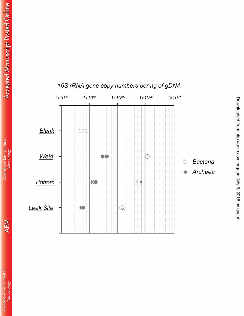
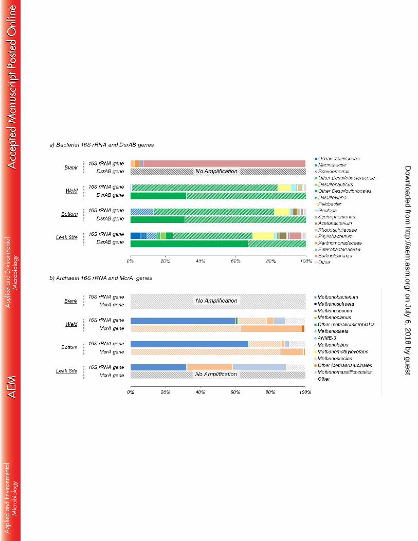
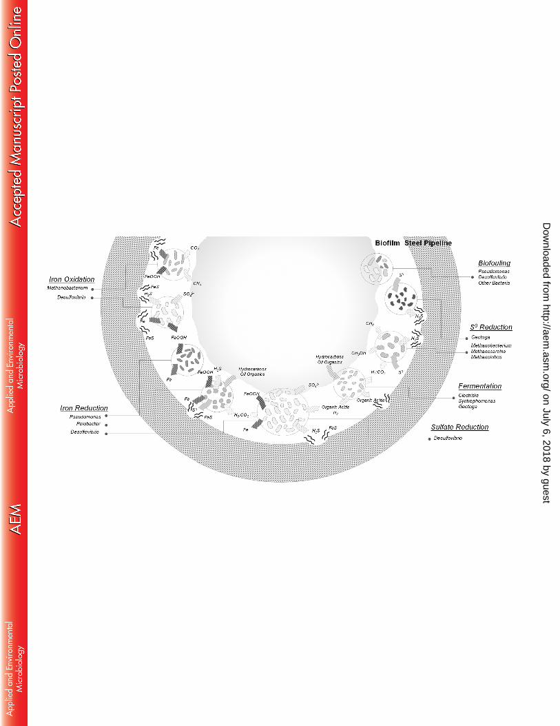

Table 1. PCR primers used for PCR, ARISA and real-time quantitative PCR
Name Function Target group
Sequence (5' - 3') Amplicon size (bp)
Annealing Temp. (°C)
Primer conc. (µM)
Ref.
ITSf ITSreub
ARISA Bacteria GTC-GTA-ACA-AGG-TAG-CCG-TA GCC-AAG-GCA-TCC-ACC
Variable 55 0.5 (Cardinale et al., 2004)
934f 71r
ARISA Archaea AGG-AAT-TGG-CGG-GGG-AGC-A TCG-GYG-CCG-AGC-CGA-GCC-ATC-C
Variable 55 0.5 (Casamayor et al., 2002)
BACT1369F BACT1492R
Q-PCR Bacteria
CGG-TGA-ATA-CGT-TCY-CGG GGW-TAC-CTT-GTT-ACG-ACT-T
142
60
0.6
(Suzuki et al., 2000)
ARC787F ARC1059R
Q-PCR Archaea
ATT-AGA-TAC-CCS-BGT-AGT-CC GCC-ATG-CAC-CWC-CTC-T
273
60
0.5
(Yu et al., 2005)
S-D-Bact-0516-a-S-18 S-D-Bact-0907-a-A-20
Sequencing Bacteria TGC-CAG-CAG-CCG-CGG-TAA CCG-TCA-ATT-CMT-TTG-AGT-TT
420 58 0.5 (Klindwoth et al., 2012)
S-D-Arch-0008-b-S-18 Sequencing Archaea TCY-GGT-TGA-TCC-TGS-CGG 530 58 0.5 (Klinworth et al.,2012)
S-D-Arch-0519-a-A-19 GGT-DTT-ACC-GCG-GCK-GCT-G DSR2060f Sequencing DsrAB CAA-CAT-CGT-YCA-YAC-CCA-GGG 380 50 0.5 (Perry et al.
2014) Dsr4RdegN GTR-TAR-CAG-TTD-CCR-CA MLf Sequencing McrA GGT-GGT-GTM-GGA-TTC-ACA-CAR-
TAY-GCW-ACA-GC 550 55 0.5 (Luton et al.
2002)
MLr TTC-ATT-GCR-TAG-TTW-GGR-TAG-TT Adaptor F Sequencing - TCGTCGGCAGCGTCAGATGTGTATAAG
AGACAG - - - -
Adaptor R GTCTCGTGGGCTCGGAGATGTGTATAAGAGACAG
on July 6, 2018 by guesthttp://aem
.asm.org/
Dow
nloaded from
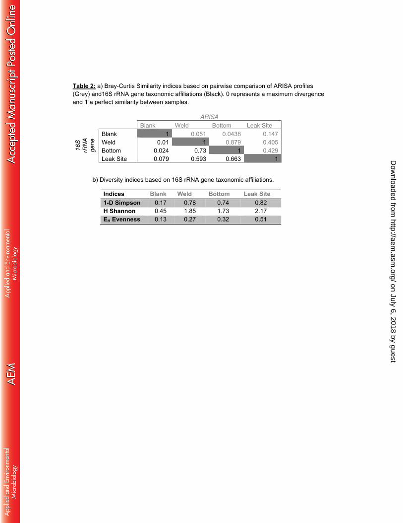
Table 2: a) Bray-Curtis Similarity indices based on pairwise comparison of ARISA profiles (Grey) and16S rRNA gene taxonomic affiliations (Black). 0 represents a maximum divergence and 1 a perfect similarity between samples.
ARISA Blank Weld Bottom Leak Site
16
S
rRN
A
ge
ne
Blank 1 0.051 0.0438 0.147 Weld 0.01 1 0.879 0.405 Bottom 0.024 0.73 1 0.429 Leak Site 0.079 0.593 0.663 1
b) Diversity indices based on 16S rRNA gene taxonomic affiliations.
Indices Blank Weld Bottom Leak Site 1-D Simpson 0.17 0.78 0.74 0.82 H Shannon 0.45 1.85 1.73 2.17 EH Evenness 0.13 0.27 0.32 0.51
on July 6, 2018 by guesthttp://aem
.asm.org/
Dow
nloaded from