complementary and alternative medicine for the treatment
TRANSCRIPT

Complementary and Alternative Medicine for the Treatment of Central Nervous System DisordersGuest Editors: Ching-Liang Hsieh, Lixing Lao, Yi-Wen Lin, and Gerhard Litscher
Evidence-Based Complementary and Alternative Medicine

Complementary and Alternative Medicine forthe Treatment of Central Nervous SystemDisorders

Evidence-Based Complementaryand Alternative Medicine
Complementary and Alternative Medicine forthe Treatment of Central Nervous SystemDisorders
Guest Editors: Ching-Liang Hsieh, Lixing Lao, Yi-Wen Lin,and Gerhard Litscher

Copyright © 2014 Hindawi Publishing Corporation. All rights reserved.
This is a special issue published in “Evidence-Based Complementary and Alternative Medicine.” All articles are open access articlesdistributed under the Creative Commons Attribution License, which permits unrestricted use, distribution, and reproduction in anymedium, provided the original work is properly cited.

Editorial Board
Mahmood Abdulla, MalaysiaJon Adams, AustraliaZuraini Ahmad, MalaysiaUlysses Albuquerque, BrazilGianni Allais, ItalyTerje Alraek, NorwaySouliman Amrani, MoroccoAkshay Anand, IndiaShrikant Anant, USAManuel Arroyo-Morales, SpainSyed Asdaq, Saudi ArabiaSeddigheh Asgary, IranHyunsu Bae, Republic of KoreaLijun Bai, ChinaSandip K. Bandyopadhyay, IndiaSarang Bani, IndiaVassya Bankova, BulgariaWinfried Banzer, GermanyVernon A. Barnes, USASamra Bashir, PakistanJairo Kenupp Bastos, BrazilSujit Basu, USADavid Baxter, New ZealandAndre-Michael Beer, GermanyAlvin J. Beitz, USAYong Boo, Republic of KoreaFrancesca Borrelli, ItalyGloria Brusotti, ItalyIshfaq A. Bukhari, PakistanArndt Bussing, GermanyRainer W. Bussmann, USARaffaele Capasso, ItalyOpher Caspi, IsraelHan Chae, KoreaShun-Wan Chan, Hong KongIl-Moo Chang, Republic of KoreaRajnish Chaturvedi, IndiaChun Tao Che, USAHubiao Chen, Hong KongJian-Guo Chen, ChinaKevin Chen, USATzeng-Ji Chen, TaiwanYunfei Chen, ChinaJuei-Tang Cheng, TaiwanEvan Paul Cherniack, USA
Jen-Hwey Chiu, TaiwanWilliam C. S. Cho, Hong KongJae Youl Cho, KoreaSeung-Hun Cho, Republic of KoreaChee Yan Choo, MalaysiaRyowon Choue, Republic of KoreaShuang-En Chuang, TaiwanJoo-Ho Chung, Republic of KoreaEdwin L. Cooper, USAGregory D. Cramer, USAMeng Cui, ChinaRoberto Cuman, BrazilVincenzo De Feo, ItalyRocıo Vazquez, SpainMartin Descarreaux, USAAlexandra Deters, GermanySiva Durairajan, Hong KongMohamed Eddouks, MoroccoThomas Efferth, GermanyTobias Esch, GermanySaeed Esmaeili-Mahani, IranNianping Feng, ChinaYibin Feng, Hong KongJosue Fernandez-Carnero, SpainJuliano Ferreira, BrazilFabio Firenzuoli, ItalyPeter Fisher, UKW. F. Fong, Hong KongRomain Forestier, FranceJoel J. Gagnier, CanadaJian-Li Gao, ChinaGabino Garrido, ChileMuhammad Ghayur, PakistanAnwarul Hassan Gilani, PakistanMichael Goldstein, USAMahabir P. Gupta, PanamaMitchell Haas, USASvein Haavik, NorwayAbid Hamid, IndiaN. Hanazaki, BrazilK. B. Harikumar, IndiaCory S. Harris, CanadaThierry Hennebelle, FranceSeung-Heon Hong, KoreaMarkus Horneber, Germany
Ching-Liang Hsieh, TaiwanJing Hu, ChinaGan Siew Hua, MalaysiaSheng-Teng Huang, TaiwanBenny Tan Kwong Huat, SingaporeRoman Huber, GermanyAngelo Antonio Izzo, ItalyKong J., USASuresh Jadhav, IndiaKanokwan Jarukamjorn, ThailandYong Jiang, ChinaZheng L. Jiang, ChinaStefanie Joos, GermanySirajudeen K.N.S., MalaysiaZ. Kain, USAOsamu Kanauchi, JapanWenyi Kang, ChinaDae Gill Kang, Republic of KoreaShao-Hsuan Kao, TaiwanKrishna Kaphle, NepalKenji Kawakita, JapanJong Yeol Kim, Republic of KoreaCheorl-Ho Kim, Republic of KoreaYoun Chul Kim, Republic of KoreaYoshiyuki Kimura, JapanJoshua K. Ko, ChinaToshiaki Kogure, JapanNandakumar Krishnadas, IndiaYiu Wa Kwan, Hong KongKuang Chi Lai, TaiwanChing Lan, TaiwanAlfred Langler, GermanyLixing Lao, Hong KongClara Bik-San Lau, Hong KongJang-Hern Lee, Republic of KoreaTat leang Lee, SingaporeMyeong S. Lee, UKChristian Lehmann, CanadaMarco Leonti, ItalyPing-Chung Leung, Hong KongLawrence Leung, CanadaKwok Nam Leung, Hong KongPing Li, ChinaMin Li, ChinaMan Li, China

ChunGuang Li, AustraliaXiu-Min Li, USAShao Li, ChinaYong Hong Liao, ChinaSabina Lim, KoreaBi-Fong Lin, TaiwanWen Chuan Lin, ChinaChristopher G. Lis, USAGerhard Litscher, AustriaKe Liu, ChinaI-Min Liu, TaiwanGaofeng Liu, ChinaYijun Liu, USACun-Zhi Liu, ChinaGail B. Mahady, USAJuraj Majtan, SlovakiaSubhash C. Mandal, IndiaJeanine Marnewick, South AfricaVirginia S. Martino, ArgentinaJames H. McAuley, AustraliaKarin Meissner, USAAndreas Michalsen, GermanyDavid Mischoulon, USASyamMohan, MalaysiaJ. Molnar, HungaryValerio Monteiro-Neto, BrazilH.-I. Moon, Republic of KoreaAlbert Moraska, USAMark Moss, UKYoshiharu Motoo, JapanFrauke Musial, GermanyMinKyun Na, Republic of KoreaRichard L. Nahin, USAVitaly Napadow, USAF. R. F. Nascimento, BrazilS. Nayak, Trinidad And TobagoIsabella Neri, ItalyTelesphore Nguelefack, CameroonMartin Offenbacher, GermanyKi-Wan Oh, Republic of KoreaY. Ohta, JapanOlumayokun A. Olajide, UKThomas Ostermann, GermanyStacey A. Page, CanadaTai-Long Pan, TaiwanBhushan Patwardhan, IndiaBerit Smestad Paulsen, Norway
Andrea Pieroni, ItalyRichard Pietras, USAWaris Qidwai, PakistanXianqin Qu, AustraliaCassandra L. Quave, USARoja Rahimi, IranKhalid Rahman, UKCheppail Ramachandran, USAGamal Ramadan, EgyptKe Ren, USAMan Hee Rhee, Republic of KoreaMee-Ra Rhyu, Republic of KoreaJose Luis Rıos, SpainPaolo Roberti di Sarsina, ItalyBashar Saad, Palestinian AuthoritySumaira Sahreen, PakistanOmar Said, IsraelLuis A. Salazar-Olivo, MexicoMohd. Zaki Salleh, MalaysiaAndreas Sandner-Kiesling, AustriaAdair Santos, BrazilG. Schmeda-Hirschmann, ChileAndrew Scholey, AustraliaVeronique Seidel, UKSenthamil R. Selvan, USATuhinadri Sen, IndiaHongcai Shang, ChinaKaren J. Sherman, USARonald Sherman, USAKuniyoshi Shimizu, JapanKan Shimpo, JapanByung-Cheul Shin, KoreaYukihiro Shoyama, JapanChang Gue Son, KoreaRachid Soulimani, FranceDidier Stien, FranceShan-Yu Su, TaiwanMohd Roslan Sulaiman, MalaysiaVenil N. Sumantran, IndiaJohn R. S. Tabuti, UgandaToku Takahashi, USARabih Talhouk, LebanonWen-Fu Tang, ChinaYuping Tang, ChinaLay Kek Teh, MalaysiaMayankThakur, IndiaMenaka C. Thounaojam, India
Mei Tian, ChinaEvelin Tiralongo, AustraliaS. C. Tjen-A-Looi, USAMichaThl Tomczyk, PolandYao Tong, Hong KongK. V. Trinh, CanadaKarl Wah-Keung Tsim, Hong KongVolkan Tugcu, TurkeyYew-Min Tzeng, TaiwanDawn M. Upchurch, USAMaryna Van de Venter, South AfricaSandy van Vuuren, South AfricaAlfredo Vannacci, ItalyMani Vasudevan, MalaysiaCarlo Ventura, ItalyWagner Vilegas, BrazilPradeep Visen, CanadaAristo Vojdani, USAY. Wang, USAShu-Ming Wang, USAChenchen Wang, USAChong-Zhi Wang, USAKenji Watanabe, JapanJintanaporn Wattanathorn, ThailandWolfgang Weidenhammer, GermanyJenny M. Wilkinson, AustraliaDarren Williams, Republic of KoreaHaruki Yamada, JapanNobuo Yamaguchi, JapanYong-Qing Yang, ChinaJunqing Yang, ChinaLing Yang, ChinaEun Jin Yang, Republic of KoreaXiufen Yang, ChinaKen Yasukawa, JapanMin H. Ye, ChinaM. Yoon, Republic of KoreaJie Yu, ChinaJin-Lan Zhang, ChinaZunjian Zhang, ChinaWei-bo Zhang, ChinaHong Q. Zhang, Hong KongBoli Zhang, ChinaRuixin Zhang, USAHong Zhang, SwedenHaibo Zhu, China
Contents
Complementary and Alternative Medicine for the Treatment of Central Nervous System Disorders,Ching-Liang Hsieh, Lixing Lao, Yi-Wen Lin, and Gerhard LitscherVolume 2014, Article ID 175152, 2 pages
TheAlterations of IL-1Beta, IL-6, and TGF-Beta Levels in Hippocampal CA3 Region of ChronicRestraint Stress Rats after Electroacupuncture (EA) Pretreatment, Tianwei Guo, Zhuo Guo, Xinjing Yang,Lan Sun, Sihan Wang, A. Yingge, Xiaotian He, and Tu YaVolume 2014, Article ID 369158, 7 pages
TheAnxiolytic Effects of Valtrate in Rats Involves Changes of Corticosterone Levels, Shu-Ning Shi,Jin-Li Shi, Yong Liu, Yan-Li Wang, Chun-Guo Wang, Wen-Hui Hou, and Jian-You GuoVolume 2014, Article ID 325948, 8 pages
Antiepileptic Effect of Uncaria rhynchophylla and Rhynchophylline Involved in the Initiation of c-JunN-Terminal Kinase Phosphorylation of MAPK Signal Pathways in Acute Seizures of KainicAcid-Treated Rats, Hsin-Cheng Hsu, Nou-Ying Tang, Chung-Hsiang Liu, and Ching-Liang HsiehVolume 2013, Article ID 961289, 9 pages
Concurrent Use of Hypnotic Drugs and Chinese Herbal MedicineTherapies among Taiwanese Adultswith Insomnia Symptoms: A Population-Based Study, Kuei-Hua Lee, Yueh-Ting Tsai, Jung-Nien Lai,and Shun-Ku LinVolume 2013, Article ID 987862, 8 pages
Rhodiola rosea Impairs Acquisition and Expression of Conditioned Place Preference Induced byCocaine, Federica Titomanlio, Carmen Manzanedo, Marta Rodrıguez-Arias, Laura Mattioli,Marina Perfumi, Jose Minarro, and Marıa A. AguilarVolume 2013, Article ID 697632, 9 pages

EditorialComplementary and Alternative Medicine for theTreatment of Central Nervous System Disorders
Ching-Liang Hsieh,1,2,3 Lixing Lao,4 Yi-Wen Lin,1,5 and Gerhard Litscher6
1 Acupuncture Research Center, China Medical University, Taichung 40402, Taiwan2Graduate Institute of Integrative Medicine, College of Chinese Medicine, China Medical University, Taichung 40402, Taiwan3Department of Chinese Medicine, China Medical University Hospital, Taichung 40402, Taiwan4University of Hong Kong, Hong Kong5 Graduate Institute of Acupuncture Science, College of Chinese Medicine, China Medical University, Taichung 40402, Taiwan6Research Unit for Complementary and Integrative Laser Medicine, Research Unit of Biomedical Engineering in Anesthesiaand Intensive Care Medicine, and TCM Research Center Graz, Medical University of Graz, 8036 Graz, Austria
Correspondence should be addressed to Ching-Liang Hsieh; [email protected]
Received 10 April 2014; Accepted 10 April 2014; Published 27 April 2014
Copyright © 2014 Ching-Liang Hsieh et al. This is an open access article distributed under the Creative Commons AttributionLicense, which permits unrestricted use, distribution, and reproduction in any medium, provided the original work is properlycited.
Central nervous system (CNS) disorders are difficult andcomplicated and cause high costs for clinical therapy andbasic research due to unknown and puzzling mechanisms.The treatment of CNS disorders needs systematic drugs thatcan pass through the brain barrier to target specific recep-tors. Until now, such drugs have severe side effects. Com-plementary and alternative medicine (CAM) has recentlybecome highly recognized as therapeutic medicine andrecommended by the World Health Organization (WHO).Clinical trials, drug development, and basic research of CAMare increased dramatically because of gradual developmentand knowledge. The current special issue is diversified withseveral novel and crucial articles concerning CAM.
Cocaine addiction is amajor economic, social, and healthproblem in developed countries that can influencemany indi-viduals. Development of new drugs to treat cocaine depen-dence is urgent and necessary. Cocaine mainly binds todopamine reuptake transporters resulting in pleasure andaddiction. Rhodiola rosea L. (RHO) is a well-known CAMwith adaptogenic, anxiolytic, antidepressive, and antistressproperties. It can reduce nicotine and morphine withdrawalsymptoms. F. Titomanlio et al. reported that RHO can poten-tiate hyperactivity induced by cocaine. RHO also attenuatedthe acquisition and expression of cocaine-induced condi-tioned place preference.They concluded that RHO is effective
in decreasing the rewarding properties of cocaine but not incocaine-associated reinstatement.
H.-C. Hsu et al. used epileptic rats to evaluate theantiepileptic effect of Uncaria rhynchophylla (UR) and rhyn-chophylline (RP). They injected kainic acid (KA) to induceseizures in SpragueDawley rats.They suggested that pretreat-ment with UR and RP can reliably attenuate seizures accom-panied by reduced c-Jun amino-terminal kinase phosphory-lation (JNKp) of mitogen-activated protein kinase (MAPK)signal pathways in the cerebral cortex and hippocampus.IL-1𝛽, IL-6, and TNF-𝛼 were unaltered, which means thatthe therapeutic effects of UR and RP are based on pJNKactivation during KA-induced seizure processes. Similarly,T.-W. Guo and colleagues indicated that electroacupunc-ture (EA) can protect rats from chronic restraint stress. Inaddition, IL-1𝛽, IL-6, and TGF-𝛽were potentiated in chronicrestraint stress rats and can be alleviated by EA pretreat-ment. The data are crucial that EA can attenuate depressionaccompanied by altering IL-1𝛽, IL-6, and TGF-𝛽 in thehippocampal CA3 region. S.-N. Shi and colleagues reportedthat valtrate, which is a principle compound isolated fromValeriana jatamansi Jones used to treat various mood disor-ders, can reduce depression and simultaneously reduce thecorticosterone level in the rat serum. They conclude thatvaltrate has an anxiolytic effect in behavioral models through
Hindawi Publishing CorporationEvidence-Based Complementary and Alternative MedicineVolume 2014, Article ID 175152, 2 pageshttp://dx.doi.org/10.1155/2014/175152

2 Evidence-Based Complementary and Alternative Medicine
the hypothalamus-pituitary-adrenal axis. The abovemen-tionedmechanisms implied that herbal medicine and EA canactivate similar mechanisms to treat CNS disorders such asdepression and epilepsy.
Furthermore, K.-H. Lee et al. wanted to analyze theconcurrent use of herbal medicine and hypnotic drugs in Tai-wanese insomnia patients. They showed that, among 53,949insomnia sufferers, 83.6% used hypnotic drugs. Jia-Wei-Xiao-Yao-San and Suan-Zao-Ren-Tang were always used,coadministered with hypnotic drugs. They indicated that thehazard ratio of hip fracture for hypnotic-drug users who usedthe herbal medicine was lower than hypnotic-drug only. Theresults are crucial for clinical practice to reduce hip fractureand are beneficial for health and quality of life of patients withinsomnia symptoms.
CAM has wide categories to treat many diseases andsymptoms. In this special issue, diverse CAM therapies aredescribed to treat different CNS disorders such as epilepsy,depression, insomnia, and addiction. This issue is plentifuland strong in CAM therapy with evidence-based medicinefrom basic research to clinical results.
We think that the readers of this special issue will getmany thought-provoking impulses and information.
Ching-Liang HsiehLixing LaoYi-Wen Lin
Gerhard Litscher
Research ArticleThe Alterations of IL-1Beta, IL-6, and TGF-Beta Levels inHippocampal CA3 Region of Chronic Restraint Stress Rats afterElectroacupuncture (EA) Pretreatment
Tianwei Guo,1 Zhuo Guo,1 Xinjing Yang,1 Lan Sun,1 Sihan Wang,1
A. Yingge,2 Xiaotian He,3 and Tu Ya1
1 School of Acupuncture-Moxibustion and Tui Na, Beijing University of Chinese Medicine, Beijing 100029, China2 School of Basic Medical Science, Inner Mongolia Medical University, Hohhot 010110, China3Department of Tradition Chinese Medicine, Sanlitun Health Service Center, Beijing 100027, China
Correspondence should be addressed to Tu Ya; tuya [email protected]
Received 21 November 2013; Revised 25 February 2014; Accepted 3 March 2014; Published 25 March 2014
Academic Editor: Yi-Wen Lin
Copyright © 2014 Tianwei Guo et al. This is an open access article distributed under the Creative Commons Attribution License,which permits unrestricted use, distribution, and reproduction in any medium, provided the original work is properly cited.
Immunological reactions induced by proinflammatory cytokines have been involved in the pathogenesis of depressive disorders.Recent studies showed that Electroacupuncture (EA) was able to reduce depressive symptoms; however, the underlyingmechanismand its potential targets remain unknown. In the present study, we used a 21-day chronic restraint stress rats as a model toinvestigate how EA could alleviate depression. Open field test was carried out to evaluate the depressive symptoms at selectedtime points. At the end of study, immunohistochemistry (IHC) was performed to detect the expressions of IL-1beta, IL-6, andTGF-beta in hippocampal CA3 region. We found that chronic restraint stress significantly decreased behavioral activities, whereasEA stimulation at points Baihui (GV 20) and Yintang (GV 29) showed protective effect during the test period. In addition, theIL-1beta, IL-6, and TGF-beta increased in rats exposed to chronic restraint stress, while EA downregulated the levels of IL-1betaand IL-6.These findings implied that EA pretreatment could alleviate depression throughmodulating IL-1beta and IL-6 expressionlevels in hippocampal CA3 region.
1. Introduction
Depression, with a lifetime prevalence of up to 17%, is theleading cause of disability and ranks the 4th among diseasescontributing to the global burden [1]. Variousmedicine treat-ments including antidepressant medications and psychologytherapies play a pivotal role in depression treatment; however,almost one-fourth of patients are unable to achieve favorableeffects, especially the improvement of somatic symptoms[2]. Thus, seeking an alternative therapy for depression isan urgent issue which is needed to be addressed. Studyhas shown that prevention at early stage appears to be thebest option to minimize the progression of depression [3].In clinical practice, electronic acupuncture (EA) has beenproved to be an effective therapy in treatingmental disorders.Studies have shown that EA can mitigate depression asshown in reduced Hamilton Depression Rating Scale scores
in treated patients. In comparison with antidepressants, EApresented comparable therapeutic effects butwith faster onsetof action and better response rate [4, 5]. Although EA showedpromising effects in alleviating the progression of depression,the underlying mechanism is poorly understood.
Over the past decades, large bodies of evidences have sug-gested that major depression is linked with sign of immuno-logical activation. Specifically, activation of the inflammatoryresponse system (IRS), such as increased production of pro-inflammatory cytokines, is considered to be the key factorfor depression [6]. Both clinical and experimental studiesindicated that increased concentration of certain types ofcytokinemay serve as a leading cause of stress and depression[7]. Dowlati et al. showed that high levels of IL-6 and TNF-alpha were found in depressed patients compared with con-trol subjects [8]. Abbasi showed that antidepressant celecoxib
Hindawi Publishing CorporationEvidence-Based Complementary and Alternative MedicineVolume 2014, Article ID 369158, 7 pageshttp://dx.doi.org/10.1155/2014/369158

2 Evidence-Based Complementary and Alternative Medicine
can reduce HDRS scores as well as IL-6 concentration inpatients with major depressive disorders. In animal study,chronic stress-induced depressive mice showed an increasedIL-1 level in brain tissue [9]. Hippocampus, as a part of limbicsystem, plays an important role in the emotion regulation.Repeated stress causes atrophy of dendrites in hippocampalCA3 region [10]. In addition, CA3 neurons are more vul-nerable to damages compared with dentate granule and CA1neurons [11]. Therefore, hippocampal CA3 region is a crucialpart for observing the physical changes during chronic stress.
Recent studies implied EAmight function viamodulationof nerve-endocrine-immune network [12]. The pathogenesisof depressive symptoms is characterized as a complex of net-work dysfunction in which factors including neurotransmit-ters, hormones, and cytokines, interact intimately. Therefore,a research strategy focusing on nerve-endocrine-immunenetwork might be applied to identify the key player whichinvolved in EA treatment in depression. In the present study,we hypothesized that EA could modulate proinflammatorycytokine levels and thus reduced depression syndrome.
2. Material and Methods
2.1. Animals. A total of 30 specific pathogen-free (SPF)Sprague Dawley rats (260∼280 g) were supplied by theInstitute of Laboratory Animal Sciences, China Academy ofMedical Science, animal license number SCXF (Jing)2009-0017. Animals were housed at (22 ± 2)∘C, 45% humidity,in 12-hour light/dark cycles (light on at 8:00 am), with freeaccess to food and water. The study was performed 3 daysafter environment acclimations of the rats.Theprotocols wereconducted in compliance with the Guidance Suggestions forthe Care and Use of Laboratory Animals formulated by theNational Institute of Health, as well as the 3R principle:Reduction, Replacement, and Refinement. All experimentprocedures were approved by the Animal Care and UseCommittee at Beijing University of Chinese Medicine.
2.2. Groups and Treatment. For control group, no modelinduction and treatment were performed. For model group,chronic stress was conducted for 21 days on a daily basis withmethod described as follows: rats were restrained with self-made cylinder-shaped wire net (20 cm in length and 5 cmin diameter) from 9 am to 3 pm. After restraints, they werereleased for free access to water and food. For EA group, EApretreatment was conducted daily prior to restraint for 21days, restraint method was the same as model group.
2.3. EA Pretreatment. During acupuncture administration,rats were maintained within a cloth bag. Two points wereselected: Baihui (GV20) and Yintang (GV29). GV20 islocated above the apex auriculate, on the midline of thehead. GV29 is located at the middle point between twoeyes [13]. Sterilized disposable stainless steel needles (0.20 ∗25mm, Hua Tuo brand, manufactured by Suzhou medicineCo., Ltd., Suzhou, Jiangsu, China) were inserted obliquely asdeep as 3–5mm for both points. Following the insertions,electrodes were added to the handle of the needles (electric
acupuncture apparatus used: Hans-100A, manufactured byNanjing Jisheng medicine science Co., Ltd., Nanjing, Jiangsu,China). Electricity simulation parameters were 1mA, 2Hz,for 20 minutes.
2.4. Open Field Test. At selected time points: day 0, day 7,day 14, and day 21, the open field test was conducted withmodifications of previous studies [14]. The apparatus, woodin material, was comprised of a square arena 80 × 80 cm with40 cm high wall. It was divided into 25 × 25 equal squareswhich had been drawn in the floor of the arena. A singlerat was gently placed in the center of the floor in orderto explore the arena for 3min. The activity of the rat wasrecorded by a camera installed on top of the lateral high wall.Two observers, blind to the experiment, counted the crossingnumbers (defined as at least three paws in a square) and therearing numbers (defined as the rat standing upright on itshind legs) from amonitor connected to the camerawhichwasset one meter away from the apparatus. After one rat finishedthe test, alcohol was applied to clean the floor to exclude theintervention of odor signals. The body weight was measuredon day 0, day 7, day 14, and day 21 of the experiment.
2.5. Frozen Section and Immunohistochemistry. At the endof the study, rats were deeply anesthetized with 10% chlo-ral hydrate (0.3mL/100 g, i.p.) and perfused with 4∘C 4%paraformaldehyde from left apex. After perfusion, hippocam-pus was harvested and embedded in liquid nitrogen. Forsections preparation, chiasma opticumwas positioned at first;then the tissue was cut into 20𝜇m coronal sections till 2-3mm posterior to chiasma opticum on a sliding microtome(Leica CM1850, German) and stored at −20∘C. Immuno-histochemistry was carried on as previously described [15].The primary antibodies (goat against rat IL-6 IgG, productnumber: SC-1265R; rabbit against rat IL-1beta IgG, productnumber: SC-7884; rabbit against rat TGF-beta IgG, productnumber: SC-146 manufactured by Santa Cruz Biotechnology(Shanghai) Co., Ltd., Shanghai, China) were added and thenincubated over night at 4∘C. Secondary antibodies conjugatedwith horseradish peroxidase (HRP) was added afterward andincubated at 37∘C for 1 hour. HRP substrates were appliedat last for color development. The protein expressions werequantified by Integral Optical Density (IOD) using Image ProPlus 6.0 software. For each rat, 3 to 4 sections were appliedand mean value was obtained to determine the expressionlevel.
2.6. Statistical Analysis. Data were presented as means ±S.E.M. SPSS 20.0 (SPSS Inc, Chicago, USA) was deployed fordata analysis with one-way ANOVA method after the test ofnormal distribution and homogeneity of variance, followedby post hoc multiple comparison. Statistical significance wasset to 𝑃 < 0.05, while highly statistical significance was set to𝑃 < 0.01.
3. Results
3.1. Effects of EA Pretreatment on Body Weight. As shown inFigure 1(a), the body weight increased slowly in model group

Evidence-Based Complementary and Alternative Medicine 3
200
250
300
350
400
Day 0 Day 7 Day 14 Day 21
ControlModelEA
Body
wei
ght
(g) # #
# #
# #
(a)
10
15
20
25
30
35
40
Day0 Day7 Day14 Day21
ControlModelEA
Cros
sing
num
bers
# #
# #
(b)
0
2
4
6
8
10
12
14
16
Day0 Day7 Day14 Day21
ControlModelEA
Rear
ing
num
bers
# # # #
(c)
Figure 1: The effect of electric acupuncture (EA) on body weight and locomotor activity in open field test at selected time points in thefollowing groups (𝑛 = 8 per group): control, model, and EA. (a) Body weight. (b) Crossing numbers in open field test. (c) Rearing numbersin open field test. ##𝑃 < 0.01 as compared with model group.
in contrast with those in control and EA group. 21 days afterinduction, the body weights in control group and EA groupwere significantly higher compared with that in model group(𝑃 < 0.01), whereas no significant differences was foundbetween control group and EA group. This result suggestedthat EA has a protective effect on body weight.
3.2. Effects of EA Pretreatment on Open Field Test. Weused the open field test to evaluate the exploratory andlocomotor activity [16, 17]. As seen in Figure 1(b), modelgroup presented a decline tendency in crossing numbersduring the 21-day restraint stress procedure, while control andEA group showed a rise tendency. 21 days after inductionof the crossing numbers in model group was significantlydecreased in comparison with those in control and EA group
with statistically significant differences (𝑃 < 0.01). Therewas no difference between control and model group (𝑃 >0.05). In addition, restraint stress stimuli remarkably reducedthe rearing numbers in model group compared with thatin control and EA group 21 days after induction, whereasno significant difference was found between control and EAgroup. The results indicated that EA plays a crucial rolein ameliorating stress-impaired exploratory and locomotoractivities.
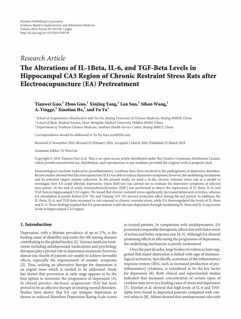
3.3. Effects of EA Pretreatment on IL-1Beta. As shown inFigure 2, the expression of IL-1beta in model group wassignificantly increased compared with that in control group(a) and EA group based on IOD value (𝑃 < 0.01),whereas there was no significantly difference between EA
4 Evidence-Based Complementary and Alternative Medicine
0
20
40
60
80
100
120
ControlModelEA
IOD
val
ue
# ## #
# #
×103
#
#
IL-1𝛽 IL-6 TGF-𝛽
Figure 2: The effect of EA on IL-1beta, IL-6, and TGF-beta proteinexpression in hippocampus (HP) CA3 region in the followinggroups (𝑛 = 8 per group): control, model, and EA. ##
𝑃 < 0.01,#𝑃 < 0.05 as compared with model group.
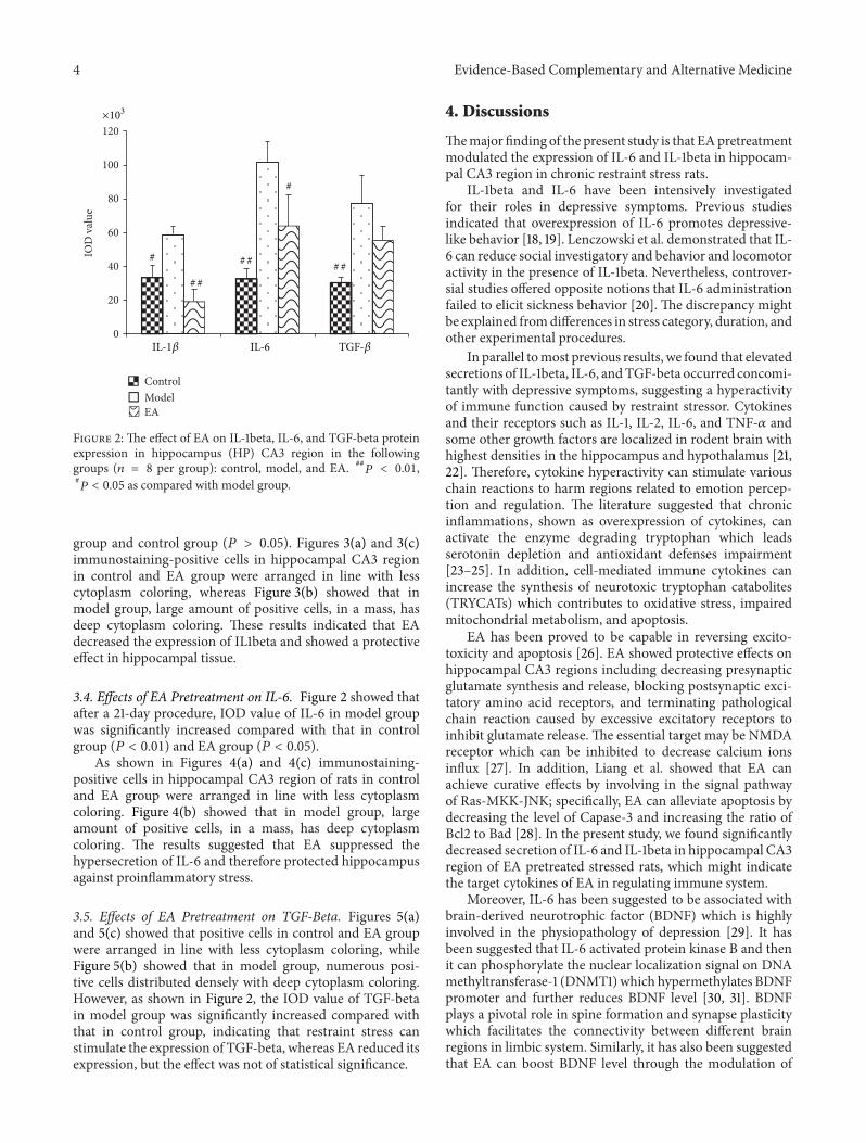
group and control group (𝑃 > 0.05). Figures 3(a) and 3(c)immunostaining-positive cells in hippocampal CA3 regionin control and EA group were arranged in line with lesscytoplasm coloring, whereas Figure 3(b) showed that inmodel group, large amount of positive cells, in a mass, hasdeep cytoplasm coloring. These results indicated that EAdecreased the expression of IL1beta and showed a protectiveeffect in hippocampal tissue.
3.4. Effects of EA Pretreatment on IL-6. Figure 2 showed thatafter a 21-day procedure, IOD value of IL-6 in model groupwas significantly increased compared with that in controlgroup (𝑃 < 0.01) and EA group (𝑃 < 0.05).
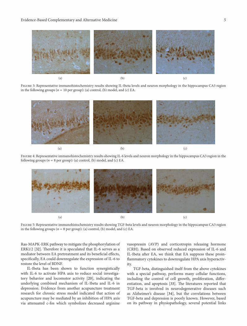
As shown in Figures 4(a) and 4(c) immunostaining-positive cells in hippocampal CA3 region of rats in controland EA group were arranged in line with less cytoplasmcoloring. Figure 4(b) showed that in model group, largeamount of positive cells, in a mass, has deep cytoplasmcoloring. The results suggested that EA suppressed thehypersecretion of IL-6 and therefore protected hippocampusagainst proinflammatory stress.
3.5. Effects of EA Pretreatment on TGF-Beta. Figures 5(a)and 5(c) showed that positive cells in control and EA groupwere arranged in line with less cytoplasm coloring, whileFigure 5(b) showed that in model group, numerous posi-tive cells distributed densely with deep cytoplasm coloring.However, as shown in Figure 2, the IOD value of TGF-betain model group was significantly increased compared withthat in control group, indicating that restraint stress canstimulate the expression of TGF-beta, whereas EA reduced itsexpression, but the effect was not of statistical significance.
4. Discussions
Themajor finding of the present study is that EApretreatmentmodulated the expression of IL-6 and IL-1beta in hippocam-pal CA3 region in chronic restraint stress rats.
IL-1beta and IL-6 have been intensively investigatedfor their roles in depressive symptoms. Previous studiesindicated that overexpression of IL-6 promotes depressive-like behavior [18, 19]. Lenczowski et al. demonstrated that IL-6 can reduce social investigatory and behavior and locomotoractivity in the presence of IL-1beta. Nevertheless, controver-sial studies offered opposite notions that IL-6 administrationfailed to elicit sickness behavior [20]. The discrepancy mightbe explained fromdifferences in stress category, duration, andother experimental procedures.
In parallel tomost previous results, we found that elevatedsecretions of IL-1beta, IL-6, andTGF-beta occurred concomi-tantly with depressive symptoms, suggesting a hyperactivityof immune function caused by restraint stressor. Cytokinesand their receptors such as IL-1, IL-2, IL-6, and TNF-𝛼 andsome other growth factors are localized in rodent brain withhighest densities in the hippocampus and hypothalamus [21,22]. Therefore, cytokine hyperactivity can stimulate variouschain reactions to harm regions related to emotion percep-tion and regulation. The literature suggested that chronicinflammations, shown as overexpression of cytokines, canactivate the enzyme degrading tryptophan which leadsserotonin depletion and antioxidant defenses impairment[23–25]. In addition, cell-mediated immune cytokines canincrease the synthesis of neurotoxic tryptophan catabolites(TRYCATs) which contributes to oxidative stress, impairedmitochondrial metabolism, and apoptosis.
EA has been proved to be capable in reversing excito-toxicity and apoptosis [26]. EA showed protective effects onhippocampal CA3 regions including decreasing presynapticglutamate synthesis and release, blocking postsynaptic exci-tatory amino acid receptors, and terminating pathologicalchain reaction caused by excessive excitatory receptors toinhibit glutamate release. The essential target may be NMDAreceptor which can be inhibited to decrease calcium ionsinflux [27]. In addition, Liang et al. showed that EA canachieve curative effects by involving in the signal pathwayof Ras-MKK-JNK; specifically, EA can alleviate apoptosis bydecreasing the level of Capase-3 and increasing the ratio ofBcl2 to Bad [28]. In the present study, we found significantlydecreased secretion of IL-6 and IL-1beta in hippocampal CA3region of EA pretreated stressed rats, which might indicatethe target cytokines of EA in regulating immune system.
Moreover, IL-6 has been suggested to be associated withbrain-derived neurotrophic factor (BDNF) which is highlyinvolved in the physiopathology of depression [29]. It hasbeen suggested that IL-6 activated protein kinase B and thenit can phosphorylate the nuclear localization signal on DNAmethyltransferase-1 (DNMT1)which hypermethylates BDNFpromoter and further reduces BDNF level [30, 31]. BDNFplays a pivotal role in spine formation and synapse plasticitywhich facilitates the connectivity between different brainregions in limbic system. Similarly, it has also been suggestedthat EA can boost BDNF level through the modulation of
Evidence-Based Complementary and Alternative Medicine 5
(a) (b) (c)
Figure 3: Representative immunohistochemistry results showing IL-1beta levels and neuron morphology in the hippocampus CA3 regionin the following groups (𝑛 = 10 per group): (a) control, (b) model, and (c) EA.
(a) (b) (c)
Figure 4: Representative immunohistochemistry results showing IL-6 levels and neuron morphology in the hippocampus CA3 region in thefollowing groups (𝑛 = 8 per group): (a) control, (b) model, and (c) EA.
(a) (b) (c)
Figure 5: Representative immunohistochemistry results showing TGF-beta levels and neuron morphology in the hippocampus CA3 regionin the following groups (𝑛 = 8 per group): (a) control, (b) model, and (c) EA.
Ras-MAPK-ERK pathway to mitigate the phosphorylation ofERK1/2 [32]. Therefore it is speculated that IL-6 serves as amediator between EA pretreatment and its beneficial effects,specifically, EA could downregulate the expression of IL-6 torestore the level of BDNF.
IL-1beta has been shown to function synergisticallywith IL-6 to activate HPA axis to reduce social investiga-tory behavior and locomotor activity [20], indicating theunderlying combined mechanism of IL-1beta and IL-6 indepression. Evidence from another acupuncture treatmentresearch for chronic stress model indicated that action ofacupuncture may be mediated by an inhibition of HPA axisvia attenuated c-fos which symbolizes decreased arginine
vasopressin (AVP) and corticotropin releasing hormone(CRH). Based on observed reduced expression of IL-6 andIL-1beta after EA, we think that EA suppress these proin-flammatory cytokines to downregulate HPA axis hyperactiv-ity.
TGF-beta, distinguished itself from the above cytokineswith a special pathway, performs many cellular functions,including the control of cell growth, proliferation, differ-entiation, and apoptosis [33]. The literatures reported thatTGF-beta is involved in neurodegenerative diseases suchas Alzheimer’s disease [34], but the correlations betweenTGF-beta and depression is poorly known. However, basedon its pathway in physiopathology, several potential links
6 Evidence-Based Complementary and Alternative Medicine
could be found between TGF-beta and depressive disor-ders. The activation of TGF-beta can activate downstreamMAPK pathway which has been described as to be impli-cated in the expression of BDNF [35]. In addition, ithas been shown that TGF-beta is also involved in Ras-MKK-JNK pathway which is highly correlated to apoptosisand growth arrest, serving as an underlying mechanismof depressive symptoms. Wu demonstrated that EA canboost the expression of Bcl-2 gene to inhibit apoptosisin brain tissue after chronic stress induction [36]. Thuswe hypothesized that EA may exert beneficial effects ondepressive symptoms through a mechanism in which TGF-beta activating Erk1/2 pathway as well as JNK pathway.However, according to our results, TGF-beta level declinedin EA group without statistical significance in comparisonwith model group. The role of TGF-beta in immunologicalactivation and EA prevention warrants further investiga-tions.
In the present study, EA pretreatment was administeredunder a slightly restrained condition. Our previous work(unpublished data) illustrated that no significant differ-ence was observed in serum adrenocorticotropic hormone(ACTH) and corticosterone (CORT) level between the nor-mal rats and normal plus acupuncture rats. It is suggestedthat the acupuncture administration will not induce stressresponse.
According to Traditional Chinese Medicine, Baihui andYintang are points pertaining to Governor Meridian. BasedonMeridian and CollateralTheory, GovernorMeridian is theconvergence of all the Yang meridians; therefore, stimulationon points of Governor Meridian can boost Yang qi of thewhole body to reverse the pathogenesis of depression inwhich it is defined as yang deficiency syndrome. Meanwhile,EA pretreatment design of our study embodies one of themost critical theories in Traditional Chinese Medicine, theprinciple of “treating diseases prior to its onset” whichattaches great significance on disease preventions.
5. Conclusions
In summary, the present study demonstrated that theproinflammatory cytokines IL-1beta, IL-6, and TGF-beta inrats’ hippocampus mediated the onsets of depressive symp-toms after chronic restraint stress inductions. Importantly,our findings suggested that EA can significantly mitigatedeficit behavioral activities elicited by chronic restraint stressthrough a potential mechanism of immunological modula-tion.
Conflict of Interests
The authors declare that there is no conflict of interestsregarding the publication of this paper.
Authors’ Contribution
Tianwei Guo and Zhuo Guo contribute equally to this study.
Acknowledgment
The authors thank Dr. Mongjen Chen for his suggestion inpaper revision.
References
[1] J. Lepine and M. Briley, “The increasing burden of depression,”NeuropsychiatricDisease andTreatment, vol. 7, supplement 1, pp.3–7, 2011.
[2] B.Arroll, S.Macgillivray, S.Ogston et al., “Efficacy and tolerabil-ity of tricyclic antidepressants and SSRIs comparedwith placebofor treatment of depression in primary care: a meta-analysis,”The Annals of Family Medicine, vol. 3, no. 5, pp. 449–456, 2005.
[3] J. P. Docherty, “Barriers to the diagnosis of depression inprimary care,” Journal of Clinical Psychiatry, vol. 58, supplement1, pp. 5–10, 1997.
[4] X. H. Ma, M. Zhang, W. Y. Zhang et al., “Estimation onthe toxticity relieve and effects enhancement in acupuncturecombined for patients with mild and moderate depression,”China Journal of Traditional Chinese Medicine Pharmacy, vol.26, no. 12, pp. 132–135, 2011.
[5] H. Sun, H. Zhao, C. Ma et al., “Effects of electroacupunctureon depression and the production of glial cell line-derivedneurotrophic factor compared with fluoxetine: a randomizedcontrolled pilot study,” The Journal of Alternative and Comple-mentary Medicine, vol. 19, no. 9, pp. 733–739, 2013.
[6] J. K. Kiecolt-Glaser and R. Glaser, “Depression and immunefunction central pathways to morbidity and mortality,” Journalof Psychosomatic Research, vol. 53, no. 4, pp. 873–876, 2002.
[7] T. J. Connor and B. E. Leonard, “Depression, stress andimmunological activation: the role of cytokines in depressivedisorders,” Life Sciences, vol. 62, no. 7, pp. 583–606, 1998.
[8] Y. Dowlati, N. Herrmann,W. Swardfager et al., “Ameta-analysisof cytokines in major depression,” Biological Psychiatry, vol. 67,no. 5, pp. 446–457, 2010.
[9] I. Goshen, T. Kreisel, O. Ben-Menachem-Zidon et al., “Braininterleukin-1 mediates chronic stress-induced depression inmice via adrenocortical activation and hippocampal neuroge-nesis suppression,” Molecular Psychiatry, vol. 13, no. 7, pp. 717–728, 2008.
[10] B. S. McEwen, “Stress and hippocampal plasticity,” AnnualReview of Neuroscience, vol. 22, pp. 105–122, 1999.
[11] Y. Watanabe, E. Gould, and B. S. McEwen, “Stress inducesatrophy of apical dendrites of hippocampal CA3 pyramidalneurons,” Brain Research, vol. 588, no. 2, pp. 341–345, 1992.
[12] J. Y. Wang and S. Q. Pan, “Research progress in mechanismof acupuncture-the relationship between nerve-endocrine-immune network modulation and acupucture treatment,”Anatomy Research, vol. 25, no. 3, pp. 45–48, 2003.
[13] Q. Liu, B. Li, H. Zhu, Y. Wang, J. Yu, and G. Wu, “Glia atrophyin the hippocampus of chronic unpredictable stress-induceddepression model rats is reversed by electroacupuncture treat-ment,” Journal of Affective Disorders, vol. 128, no. 3, pp. 309–313,2011.
[14] J. Lu, J. Liang, and J. R. Wang, “Acupuncture activates ERK-CREB pathway in rats exposed to chronic unpredictablemild stress,” Evidence-Based Complementary and AlternativeMedicine, vol. 2013, Article ID 469765, 7 pages, 2013.
[15] S.W. Lee, Y. Y. Ahn, Y. S. Kim et al., “The immunohistochemicalexpression of STAT3, Bcl-xL, and MMP-2 proteins in colon

Evidence-Based Complementary and Alternative Medicine 7
adenoma and adenocarcinoma,” Gut and Liver, vol. 6, no. 1, pp.45–51, 2012.
[16] R. P. Liu, J. L. Fang, P. J. Rong et al., “Effects of electroacupunc-ture at auricular concha region on the depressive status ofunpredictable chronic mild stress rat models,” Evidence-BasedComplementary and Alternative Medicine, vol. 2013, Article ID789674, 7 pages, 2013.
[17] L. Prut and C. Belzung, “The open field as a paradigm tomeasure the effects of drugs on anxiety-like behaviors: a review,”European Journal of Pharmacology, vol. 463, no. 1–3, pp. 3–33,2003.
[18] B. Sakic, J. Gauldie, J. A. Denburg, and H. Szechtman, “Behav-ioral effects of infectionwith IL-6 adenovector,”Brain, Behavior,and Immunity, vol. 15, no. 1, pp. 25–42, 2001.
[19] B. Sakic, H. Szechtman, T. Braciak, C. Richards, J. Gauldie, andJ. A. Denburg, “Reduced preference for sucrose in autoimmunemice: a possible role of interleukin-6,” Brain Research Bulletin,vol. 44, no. 2, pp. 155–165, 1997.
[20] M. J. P. Lenczowski, R.-M. Bluthe, J. Roth et al., “Centraladministration of rat IL-6 induces HPA activation and fever butnot sickness behavior in rats,” American Journal of Physiology—Regulatory Integrative and Comparative Physiology, vol. 276, no.3, pp. R652–R658, 1999.
[21] B. Schobitz, W. Sutanto, M. P. Carey, F. Holsboer, and E. R.de Kloet, “Endotoxin and interleukin 1 decrease the affinityof hippocampal mineralocorticoid (type I) receptor in parallelto activation of the hypothalamic-pituitary-adrenal axis,” Neu-roendocrinology, vol. 60, no. 2, pp. 124–133, 1994.
[22] S. J. Hopkins and N. J. Rothwell, “Cytokines and the nervoussystem I: expression and recognition,” Trends in Neurosciences,vol. 18, no. 2, pp. 83–88, 1995.
[23] A. Sluzewska, J. Rybakowski, E. Bosmans et al., “Indicators ofimmune activation in major depression,” Psychiatry Research,vol. 64, no. 3, pp. 161–167, 1996.
[24] M. Maes, B. E. Leonard, A. M. Myint, M. Kubera, and R. Verk-erk, “The new “5-HT” hypothesis of depression: cell-mediatedimmune activation induces indoleamine 2,3-diox- ygenase,which leads to lower plasma tryptophan and an increasedsynthesis of detrimental tryptophan catabolites (TRYCATs),both of which contribute to the onset of depression,” Progressin Neuro-Psychopharmacology and Biological Psychiatry, vol. 35,no. 3, pp. 702–721, 2011.
[25] V. Valkanova, K. P. Ebmeier, and C. L. Allan, “CRP, IL-6 and depression: a systematic review and meta-analysis oflongitudinal studies,” Journal of Affective Disorders, vol. 150, no.3, pp. 736–744, 2013.
[26] S. Feng, Q.Wang, H.Wang et al., “Electroacupuncture pretreat-ment ameliorates hypergravity-induced impairment of learningand memory and apoptosis of hippocampal neurons in rats,”Neuroscience Letters, vol. 478, no. 3, pp. 150–155, 2010.
[27] R. X. Shi, Q. Wu, L. N. Qin et al., “The effects of ElectricAcupuncture on body weights and HPA axis of chronic stressrats,” Journal of Clinical Acupuncture and Moxibustion, vol. 23,no. 1, pp. 173–175, 2007.
[28] J. Liang, W. D. Li, Y. P. Wu et al., “Effects of Eliectric Acupunc-ture on hippocampal neuron apoptosis and regeneration ofdepressionmodel rats induced by chronic stress,”China Journalof Traditional ChineseMedicine and Pharmacy, vol. 27, no. 4, pp.54–57, 2012.
[29] R. Yoshimura,W.Umene-Nakano, T.Hoshuyama et al., “Plasmalevels of brain-derived neurotrophic factor and interleukin-6 in
patients with dysthymic disorder: comparison with age- andsex-matched major depressed patients and healthy controls,”Human Psychopharmacology, vol. 25, no. 7-8, pp. 566–569, 2010.
[30] R. P. Sharma, N. Tun, and D. R. Grayson, “Depolarizationinduces downregulation of DNMT1 and DNMT3a in primarycortical cultures,” Epigenetics, vol. 3, no. 2, pp. 74–80, 2008.
[31] D. R. Hodge, E. Cho, T. D. Copeland et al., “IL-6 enhancesthe nuclear translocation of DNA cytosine-5-methyltransferase1 (DNMT1) via phosphorylation of the nuclear localizationsequence by the AKT kinase,”Cancer Genomics and Proteomics,vol. 4, no. 6, pp. 387–398, 2007.
[32] J. Liang, Study on differences of depressive neuron protectivemechanism between Electric Acupuncture and Paroxetine [Ph.D.dissertation], Beijing University of Chinese Medicine, Beijing,China, 2012.
[33] R. Derynck and Y. E. Zhang, “Smad-dependent and Smad-independent pathways in TGF-𝛽 family signalling,”Nature, vol.425, no. 6958, pp. 577–584, 2003.
[34] W. Swardfager, K. Lanctt, L. Rothenburg, A. Wong, J. Cappell,and N. Herrmann, “Ameta-analysis of cytokines in Alzheimer’sdisease,” Biological Psychiatry, vol. 68, no. 10, pp. 930–941, 2010.
[35] L. Attisano and J. L. Wrana, “Signal transduction by the TGF-𝛽superfamily,” Science, vol. 296, no. 5573, pp. 1646–1647, 2002.
[36] Y. P. Wu, Effects of Electric Acupuncture treatment on expres-sion of Bcl-2 GAP-43 in brain of depression rats induced bychronic stress [Ph.D. dissertation], Beijing University of ChineseMedicine, Beijing, China, 2009.

Research ArticleThe Anxiolytic Effects of Valtrate in Rats InvolvesChanges of Corticosterone Levels
Shu-Ning Shi,1 Jin-Li Shi,1 Yong Liu,1 Yan-Li Wang,1 Chun-Guo Wang,1
Wen-Hui Hou,1 and Jian-You Guo2
1 School of Chinese Materia Medica, Beijing University of Chinese Medicine, 6A Wangjing Central South Road, Chaoyang District,Beijing 100102, China
2 Key Laboratory of Mental Health, Institute of Psychology, Chinese Academy of Sciences, 4A Datun Road, Chaoyang District,Beijing 100101, China
Correspondence should be addressed to Jin-Li Shi; [email protected] and Jian-You Guo; [email protected]
Received 22 July 2013; Revised 21 November 2013; Accepted 21 November 2013; Published 20 March 2014
Academic Editor: Gerhard Litscher
Copyright © 2014 Shu-Ning Shi et al. This is an open access article distributed under the Creative Commons Attribution License,which permits unrestricted use, distribution, and reproduction in any medium, provided the original work is properly cited.
Valtrate is a principle compound isolated from Valeriana jatamansi Jones, which is a Traditional Chinese Medicine used to treatvarious mood disorders. The aim of the present study was to investigate the anxiolytic effects of valtrate in rats. The animals wereorally administered valtrate (5, 10, and 20 g/kg daily) for 10 days and exposed to open field test (OFT) and elevated plus-maze(EPM). Then the corticosterone levels in the rat serum were measured by enzyme-linked immunosorbent assay (ELISA). Thevaltrate (10mg/kg, p.o.) exhibited the anxiolytic effect in rats by increasing the time and entry percentage into the open arms in theEPM and the number of central entries in the OFT. Valtrate (10mg/kg, p.o.) significantly reduced the corticosterone level in the ratserum. Taken together, these results suggest that the valtrate has anxiolytic activity in behavioral models that might be mediatedvia the function of hypothalamus-pituitary-adrenal axis.
1. Introduction
Anxiety disorder is a common mental illness on society.Millions of people suffer from amental or behavioral disorder[1]. Previous studies suggest that benzodiazepines are usefulfirst-line agents for most of the anxiety disorders in theworld [2]. However, they may produce fearful side effects; forexample, long-term use of benzodiazepine can cause cogni-tive decline in the elderly [3]. In addition, a lot of patientswith anxiety disorders fail to adequately respond to existingpharmacologic treatments [4].Thus, better antianxiety drugswith greater efficacy and fewer side-effects are needed.
Traditional Chinese prescription has been commonly rec-ognized as a safe and effective prescription in the treatmentof various mood disorders in China [5]. Valeriana jatamansiJones was a famous Traditional Chinese Medicine used totreat anxiety disorders in clinical prescription for many years[6]. Recent study has reported that Valeriana jatamansiJones exerts an anxiolytic effect by improving the frequencyand time percentage of the open arm in the elevated plus
maze [7]. Chemical researches have shown that it includesessential oils, iridoids, and flavonoids compounds [8], but theanxiolytic active components of Valeriana jatamansi Joneshave not been adequately elucidated. Valtrate is a majorcomponent ofValeriana jatamansi Jones and has been shownto have antifungal, antitumor, and cytotoxic activities in earlystudies [9–12]. Currently, valtrate at a high dose has beenfound to have sedative properties by inhibiting spontaneousmotion and increasing the sleeping number induced bypentobarbital sodium in mice [13]. Therefore, these resultsraises the possibility of the anxiolytic effect of valtrate asthe primary antianxiety components in Valeriana jatamansiJones. However, the anxiolytic effect of valtrate and themechanism have not been reported.
Therefore, in the present study, we investigated the anxi-olytic potential of valtrate isolated from Valeriana jatamansiJones in rats. The paradigms we selected here to detect theanxiolytic effect of valtrate are two famous tests of anxiety:the open field test (OFT) and the elevated plus maze test(EPM), which have shown good sensitivity to anxiolytic
Hindawi Publishing CorporationEvidence-Based Complementary and Alternative MedicineVolume 2014, Article ID 325948, 8 pageshttp://dx.doi.org/10.1155/2014/325948
2 Evidence-Based Complementary and Alternative Medicine
drugs.TheEPM is a well-established animalmodel for testinganxiolytic drugs [14] because of its natural stimulus, suchas a fear of a new, bright, and open space and the fear ofbalancing on a relatively narrow raised surface [15]. The OFThas gained popularity as a model of anxiety, which is basedon the rodents’ natural tendency to stay near the perimetersof a novel environment [16] and the aversion of rodents foropen and illuminated spaces [17]. The animals were tested inthe OFT and EPM. After the behavior test, we determinedwhether valtrate altered the serum corticosterone response tostress induced by exposure to the two models.
2. Material and Methods
2.1. Animals. 60 male 8-week-old Sprague-Dawley rats (150–170 g) were obtained from the Laboratory Animal Center ofthe Academy of Military Medical Sciences and used for thisstudy. Each animal was housed in individual cages undercontrolled temperature (22 ± 1∘C) and a 12 h/12 h light/darkcycle (lights on at 07:00 AM–19:00 PM) with free access tofood and water. The experimenter handled the animals dailyto acclimate them to the manipulation. The experimentalprocedures were approved by the Institutional Animal Careand Use Committee of the Institute of Psychology of theChinese Academy of Sciences and in accordance with theNational Institutes of Health Guide for Care and Use ofLaboratory Animals.
2.2. Plant Material and Isolation of Valtrate. Valeriana jata-mansi Rhizoma et Radix was purchased from a commercialsource in Yunnan province, China. The identity of the herbalmedicine was confirmed by Professor Shi Jin-li, a researcherin the Department of Pharmacognosy, Beijing University ofChinese Medicine. Voucher specimens were deposited at theHerbarium of School of Chinese Materia Medica, BeijingUniversity of Chinese Medicine.
Jatamana Valeriana Rhizome was homogenized to coarsepowder (8 kg) and soaked in aqueous ethanol (95%, 12 L,v/v) three times at room temperature, and the combinedalcoholic extract was filtered and evaporated under reducedpressure to yield a residue.The concentrated extract was thensubjected to chromatographic separation on AB-8 macro-porous adsorption resin with 70%, 80%, and 90% EtOH-H2O to give three fractions. Three fractions were subjected
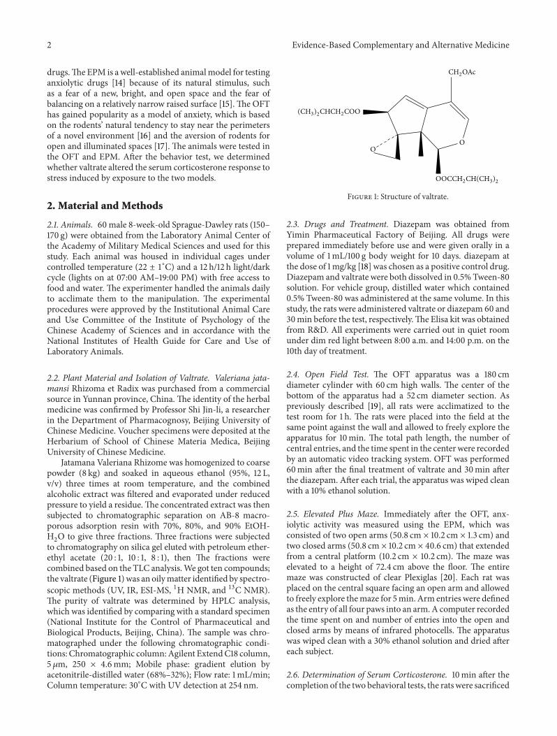
to chromatography on silica gel eluted with petroleum ether-ethyl acetate (20 : 1, 10 : 1, 8 : 1), then The fractions werecombined based on the TLC analysis.We got ten compounds;the valtrate (Figure 1)was an oilymatter identified by spectro-scopic methods (UV, IR, ESI-MS, 1H NMR, and 13C NMR).The purity of valtrate was determined by HPLC analysis,which was identified by comparing with a standard specimen(National Institute for the Control of Pharmaceutical andBiological Products, Beijing, China). The sample was chro-matographed under the following chromatographic condi-tions: Chromatographic column:Agilent ExtendC18 column,5 𝜇m, 250 × 4.6mm; Mobile phase: gradient elution byacetonitrile-distilled water (68%–32%); Flow rate: 1mL/min;Column temperature: 30∘C with UV detection at 254 nm.
(CH3)2CHCH2COO
CH2OAc
OOCCH2CH(CH3)2
OO
Figure 1: Structure of valtrate.
2.3. Drugs and Treatment. Diazepam was obtained fromYimin Pharmaceutical Factory of Beijing. All drugs wereprepared immediately before use and were given orally in avolume of 1mL/100 g body weight for 10 days. diazepam atthe dose of 1mg/kg [18] was chosen as a positive control drug.Diazepam and valtrate were both dissolved in 0.5%Tween-80solution. For vehicle group, distilled water which contained0.5% Tween-80 was administered at the same volume. In thisstudy, the rats were administered valtrate or diazepam 60 and30min before the test, respectively.The Elisa kit was obtainedfrom R&D. All experiments were carried out in quiet roomunder dim red light between 8:00 a.m. and 14:00 p.m. on the10th day of treatment.
2.4. Open Field Test. The OFT apparatus was a 180 cmdiameter cylinder with 60 cm high walls. The center of thebottom of the apparatus had a 52 cm diameter section. Aspreviously described [19], all rats were acclimatized to thetest room for 1 h. The rats were placed into the field at thesame point against the wall and allowed to freely explore theapparatus for 10min. The total path length, the number ofcentral entries, and the time spent in the center were recordedby an automatic video tracking system. OFT was performed60min after the final treatment of valtrate and 30min afterthe diazepam. After each trial, the apparatus was wiped cleanwith a 10% ethanol solution.
2.5. Elevated Plus Maze. Immediately after the OFT, anx-iolytic activity was measured using the EPM, which wasconsisted of two open arms (50.8 cm × 10.2 cm × 1.3 cm) andtwo closed arms (50.8 cm × 10.2 cm × 40.6 cm) that extendedfrom a central platform (10.2 cm × 10.2 cm). The maze waselevated to a height of 72.4 cm above the floor. The entiremaze was constructed of clear Plexiglas [20]. Each rat wasplaced on the central square facing an open arm and allowedto freely explore themaze for 5min. Arm entries were definedas the entry of all four paws into an arm.A computer recordedthe time spent on and number of entries into the open andclosed arms by means of infrared photocells. The apparatuswas wiped clean with a 30% ethanol solution and dried aftereach subject.
2.6. Determination of Serum Corticosterone. 10min after thecompletion of the two behavioral tests, the rats were sacrificed

Evidence-Based Complementary and Alternative Medicine 3
Drug or vehicle treatmentonce a day for 10 days Final administration OFT EPM Blood sample collection
Days
1h or0.5h after
1 2 3 4 5 6 7 8 9 10
Figure 2: Experimental schedule. Experimental schedule, described in Material and Methods section, involved the OFT, EPM test, and thecollection of blood sample.
200
150
100
50
0
0.0 2.5 5.0 7.5 10.0 12.5 15.0 17.5
254nm, 4nm (1.00)
(mAU
)
(min)
(a)
200
150
100
50
0
0.0 2.5 5.0 7.5 10.0 12.5 15.0 17.5
254nm, 4nm (1.00)
(mAU
)
(min)
(b)
Figure 3: HPLC profile of valtrate using acetonitrile-distilled water (68%–32%) at 1mL/min on a Agilent Extend C18 column, 5𝜇m, 250 ×4.6mm, 30∘C with UV detection at 254 nm. (a) The sample of valtrate. (b) The standard specimen of valtrate.
by decapitation; then trunk blood was collected among thefive groups to avoid any substantial time lag in samples collec-tion. Samples were centrifuged at 3000 r⋅min−1 for 15min at4∘C and supernatants were stored at −20∘C until analysis.Thecontent of corticosterone was determined by a commerciallyavailable enzyme-linked immunosorbent assay (ELISA) kitaccording to themanufacturer’s instructions.The absorbanceof each sample was measured at a wavelength of 450 nm andthe results are presented as ng/mL. All procedures of theexperiment were shown in Figure 2.
2.7. Statistical Analysis. The data were expressed as mean ±SEM. The statistical analysis was carried out by one-wayanalysis of variance (ANOVA) following Student-Newman-Keul’s post-hoc test using Prism 5.0 (Graphpad Software,Inc). Probability values lower than 0.05 were consideredstatistically significant.
3. Results
3.1. Assaying of Valtrate by HPLC. The results suggest that thepurity of product can reach 99% (see Figure 3).
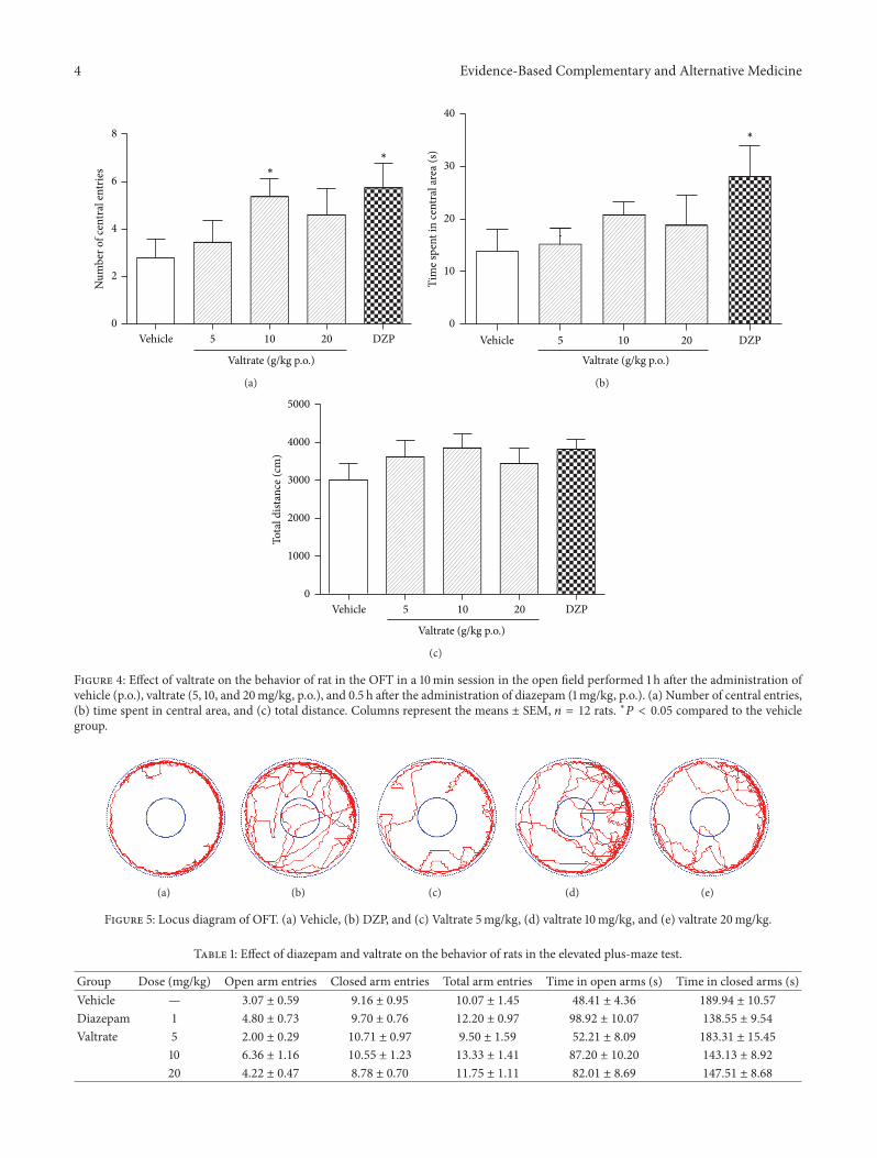
3.2. Effects of Valtrate on Open Field Test in Rats. Theresults for the OFT are shown in Figure 4. Analysesdemonstrated significant effects on number of center entries(𝐹 (4, 55) = 3.541, 𝑃 < 0.05) and time spent in central area(𝐹 (4, 55) = 3.127, 𝑃 < 0.05); further analyses showed thatvaltrate at dose of 10mg/kg significantly increased the entriesin central area (𝑃 < 0.05). Valtrate at the dose of 20mg/kgdid not significantly increase the entries in central area(𝑃 > 0.05). Diazepam significantly increased the number ofcenter entries (𝑃 < 0.05) and the time spent in central area
(𝑃 < 0.05). All of the doses of valtrate did not significantlyincrease the time spent in central area (𝑃 > 0.05). Nodifference in total path length was observed among the fivegroups (𝐹 (4, 55) = 1.207, 𝑃 > 0.05). Locus diagram of openfield test of every group is shown in Figure 5.
3.3. Effects of Valtrate on Elevated Plus Maze in Rats. Asshown in Table 1 and Figure 6, the ANOVA indicated sig-nificant effects on percentage of time spent on the openarm (𝐹 (4, 55) = 7.755, 𝑃 < 0.01) and open arm entries(𝐹 (4, 55) = 6.054, 𝑃 < 0.01). Compared to vehicle group,valtrate at the dose of 10mg/kg significantly increased thepercentage of time spent in the open arms and entry percent-age into the open arms in the elevated plus maze (𝑃 < 0.01;𝑃 < 0.01), and valtrate at the dose of 20mg/kg increased thepercentage of time spent in the open arms of the maze (𝑃 <0.01) but did not increase percentage of open armentries (𝑃 >0.05). Diazepam also significantly increased the percentage oftime spent on open arms (𝑃 < 0.01) and percentage into theopen arms (𝑃 < 0.05). No difference was observed in totalarm entries among groups (𝐹 (4, 55) = 1.042, 𝑃 > 0.05).
3.4. The Level of Serum Corticosterone. As seen in Figure 7,the data show that administration of valtrate at the dose of10mg/kg and 20mg/kg dose reduced the corticosterone level(𝑃 < 0.01, 𝑃 < 0.05). Similarly, serum corticosterone levelsof rats treated with diazepam were lower than those of thevehicle group (𝑃 < 0.01).
4. Discussion
The present study was performed to analyze the behav-ioral effects of anxiolytic valtrate isolated from Valerianajatamansi Jones, using two behavioural measurements of
4 Evidence-Based Complementary and Alternative Medicine
Vehicle 5 10 20 DZP0
2
4
6
8
Valtrate (g/kg p.o.)
Num
ber o
f cen
tral
entr
ies ∗
∗
(a)
Vehicle 5 10 20 DZP0
10
20
30
40
Tim
e spe
nt in
cent
ral a
rea (
s)
Valtrate (g/kg p.o.)
∗
(b)
Vehicle 5 10 20 DZP0
1000
2000
3000
4000
5000
Tota
l dist
ance
(cm
)
Valtrate (g/kg p.o.)
(c)
Figure 4: Effect of valtrate on the behavior of rat in the OFT in a 10min session in the open field performed 1 h after the administration ofvehicle (p.o.), valtrate (5, 10, and 20mg/kg, p.o.), and 0.5 h after the administration of diazepam (1mg/kg, p.o.). (a) Number of central entries,(b) time spent in central area, and (c) total distance. Columns represent the means ± SEM, 𝑛 = 12 rats. ∗𝑃 < 0.05 compared to the vehiclegroup.
(a) (b) (c) (d) (e)
Figure 5: Locus diagram of OFT. (a) Vehicle, (b) DZP, and (c) Valtrate 5mg/kg, (d) valtrate 10mg/kg, and (e) valtrate 20mg/kg.
Table 1: Effect of diazepam and valtrate on the behavior of rats in the elevated plus-maze test.
Group Dose (mg/kg) Open arm entries Closed arm entries Total arm entries Time in open arms (s) Time in closed arms (s)Vehicle — 3.07 ± 0.59 9.16 ± 0.95 10.07 ± 1.45 48.41 ± 4.36 189.94 ± 10.57
Diazepam 1 4.80 ± 0.73 9.70 ± 0.76 12.20 ± 0.97 98.92 ± 10.07 138.55 ± 9.54
Valtrate 5 2.00 ± 0.29 10.71 ± 0.97 9.50 ± 1.59 52.21 ± 8.09 183.31 ± 15.45
10 6.36 ± 1.16 10.55 ± 1.23 13.33 ± 1.41 87.20 ± 10.20 143.13 ± 8.92
20 4.22 ± 0.47 8.78 ± 0.70 11.75 ± 1.11 82.01 ± 8.69 147.51 ± 8.68

Evidence-Based Complementary and Alternative Medicine 5
5 10 20 DZP0
10
20
30
40
50O
pen
arm
entr
ance
(%)
Valtrate (g/kg p.o.)
∗
∗∗
Vehicle
(a)
Vehicle 5 10 20 DZP0
10
20
30
40
50
Tim
e on
open
arm
(%)
∗∗
∗∗
∗∗
Valtrate (g/kg p.o.)
(b)
5 10 20 DZP0
5
10
15
20
Tota
l arm
entr
ies
Vehicle
Valtrate (g/kg p.o.)
(c)
Figure 6: Behavioural performance of rat registered in a 5min session in the EPM performed 1 h after the administration of vehicle (p.o.),valtrate (5, 10, and 20mg/kg, p.o.), and 0.5 h after the administration of diazepam (1mg/kg, p.o.). (a) Percentage of number of entries intothe open arm, (b) percentage of time spent into the open arms, and (c) total arm entries. Columns represent the means ± SEM, 𝑛 = 12 rats.∗𝑃 < 0.05, ∗∗𝑃 < 0.01 compared to the vehicle group.
anxiety, OFT, and EPM. The results showed that valtrateexhibited anxiolytic-like activity and did not induce sedativeside effects. We also found that valtrate could attenuate HPAaxis activity by reducing the corticosterone level.
Valtrate was successfully isolated from subterranean partsof subterranean parts of various Valeriana species for the firsttime byThies [21]. Our laboratory developed a high efficiencyand practicalitymethod for purifyingValtrate fromValerianajatamansi Jones with AB-8 macroporous adsorption resin.The resin yielded the best efficiency when the concentrationof the extraction was 3.5mg/mL, the 70% ethanol acted asthe eluant, and the eluting speed was two column volumesper hour. AB-8 macroporous adsorption resin significantlyincreased the purity of valtrate (99%), with advantage of highabsorption, high elution rate, and low expense.
Hall originally described the OFT for the study ofemotionality in rats [22], which is one of the most popularprocedures in animal psychology and has beenwidely used toassess anxiety, emotionality, or responses to stress in animals[23, 24]; the test is based on the rodents’ natural tendency
to stay near the perimeters of a novel environment and theaversion of rodents for open and illuminated spaces. Thenumber of central entries or the time spent in the center areaserved as indices of anxiety and the distance was consideredan index of locomotor activity [25, 26]. Rats treated withvaltrate at the dose of 10mg/kg significantly increased thenumber of center entries and the total distance was notsignificantly affected. Therefore, valtrate (10mg/kg) has asignificant anxiolytic-like effect in this paradigm.
To further strength these data, we tested the anxiolytic-like effects of these treatments in EPM, Which is a classicalanimal analog for anxiolytic drugs and can play a key rolein the screening of anxiolytic drugs on the central nervoussystem currently [27, 28]. Normally, rodents tend to avoidopen areas of the maze and a preference for sections enclosedby protective walls. Anxiolytic drugs shift the behavioralresponse toward exploration of the open arms [29]. Thepercentage entries into the open arms and time spent inthe open arms are generally used as indices of anxiety anddrugs increasing these measures show anxiolytic properties.

6 Evidence-Based Complementary and Alternative Medicine
5 10 20 DZP0
20
40
60
80C
onte
nts o
f cor
ticos
tero
ne (n
g/m
L)
Valtrate (g/kg p.o.)
∗∗∗
∗∗
Vehicle
Figure 7: Effects of bergamot valtrate (5, 10, and 20mg/kg, p.o.) anddiazepam (1mg/kg, p.o.) compared with vehicle groups on serumcorticosterone after the behavior test. Columns represent the means± SEM, 𝑛 = 12. ∗𝑃 < 0.05, ∗∗𝑃 < 0.01 compared to the vehiclegroup.
The number of entries into the total arms was consideredan index of locomotor activity. In the present study, the ratswere treated with the higher doses of valtrate (10mg/kg and20mg/kg) for 10 days, and anxiety-like behavior in the EPMwas significantly attenuated, without altering the number oftotal arm entries, suggesting that valtrate induces specificanxiolytic-like effects.
The alcohol extract from Valeriana jatamansi Jones (0.3,0.6, 0.9 g/kg) could increase the open entries percent andopen time percent in the EPM [30]. As the content of valtratein alcohol extract from Valeriana jatamansi Jones is about2.87%, 5mg/mL valtrate in rat is equivalent to 0.3 g/mLalcohol extract of Valeriana jatamansi Jones in mice in termsof equal valtrate efficacy. Thus, the doses of valtrate (5, 10,and 20mg/kg) were chosen in this study. We also performedan acute experiment to study the anxiolytic effect of valtrate,and valtrate did not affect the behavior tests in EPM andOFT(data not shown). Therefore, the valtrate was administeredwith ten days. Diazepam is a classical drug to treat anxietyand was chosen as a positive drug in this study. As expected,diazepam had a significant anxiolytic-like effect in both EPMand OFT. The anxiolytic effect of the valtrate (10mg/kg) wasalmost equivalent to that of diazepam (1mg/kg) in EPMand the number of central entries in OFT. Although valtratedid not increase the time spent on the center of the openfield compared to the vehicle group, there were marginalsignificant differences between the valtrate and the vehiclegroup on these tests. In addition, both valtrate (10mg/kg)and diazepam (1mg/kg) could reduce the corticosteronelevels after behavior tests. Therefore, we thought that theanxiolytic effect of valtrate (10mg/kg) was almost equivalentto diazepam (1mg/kg).
The drugs (valtrate or diazepam) in this study wereadministered 60 and 30min before the test; there should bea control group for each drug treatment. We compared thesetwo vehicle groups (administrated 30min and 60min, resp.).
The rats of these groups were exposed to the same procedure.The results suggested that there was no difference betweenthe two groups (data not shown). To avoid too much groupsin the present study, one vehicle group (60min before test)was used as control.
It should be noted that the intermediate dose of valtratewas the most effective in decreasing anxiety-like behaviortests and corticosterone concentrations. One possible reasonmay be that the highest dose of valtrate or its metabolites mayact as an inducer for hepatic microsomal enzyme, which canincrease metabolism of the drug and result in reducing cura-tive effect. In addition, valtrate (10mg/kg and 20mg/kg) wasable to decrease corticosterone concentration (𝑃 < 0.01 and𝑃 < 0.05, resp.), but this effect was not observed in the behav-ior tests. Although valtrate at the dose of 20mg/kg did notincrease percentage of open arm entries (𝑃 = 0.052) and thenumber of central entries (𝑃 = 0.058), there were marginalsignificant differences between the valtrate (20mg/kg) andthe vehicle group on these two tests. Moreover, individualdifferences among rats in the highest dose valtrate (20mg/kg)group are larger than these in intermediate dose valtrate(10mg/kg) group. The same situation was observed in theopen field test. However, valtrate at the dose of 10mg/kg and20mg/kg did not increase the time spent on the center of theopen field compared to the vehicle group (𝑃 = 0.057 and𝑃 = 0.063, resp.). There were marginal significant differencesbetween the valtrate group (10mg/kg or 20mg/kg) and thevehicle group in the time spent in the center area.
Neuroendocrine system plays a key role in the stabilityof the body environment. Hormones change these neurons’network by making change in information and altering theneurotransmitter between cells in cell level, thus affectingthe central nervous system function [31]. It is well knownthat hypothalamic-pituitary-adrenocortical (HPA) axis acti-vation is a key component of the physiological responseto stress and anxiety. The HPA axis is activated by stress;then corticosterone is released from the adrenal gland. Thestress hormone corticosterone was measured to investigatethe response of the HPA axis to valtrate. Research suggeststhat the exposure of rodents to the standard elevated plusmaze activates the HPA axis, leading to an enhancement ofplasma corticosterone [32]. In addition, there was a peakin corticosterone secretion which occurs 5 to 10min afterexposure to two different anxiety/fear tests [33]. It has beenreported that Valeriana jatamansi Jones extract played arole in antianxiety via regulation of the HPA axis [30]. Inthe present study, as the stress hormone corticosterone wasmeasured as the rats were subjected to both EPM and OFtests. These two tests were performed in two adjacent rooms,the delay time was less than one minute, and we thought thatthese procedures might not influence the effectiveness of thedrug or plasma corticosterone concentrations, which werealso used by other researchers [34, 35]. Our data showed thatvaltrate (10mg/kg), a dose which produced anxiolytic activityin the behavioural experiments, attenuated the activity ofHPAaxis by reducing the corticosterone response to the stressof exposure to the elevated plus maze.These findings indicatethat the decreased anxiety-related behaviours may be relatedto the attenuation of HPA axis activity.

Evidence-Based Complementary and Alternative Medicine 7
You et al. had reported the anxiolytic-like effects ofcompound Valeriana jatamansi Jones in mice [36]. Com-pound Valeriana jatamansi Jones is composed of ValerianaeJatamansi Rhizoma et Radix, Ziziphi Spinosae Semen, andAlbiziae Cortex and Junci Medulla (in a ratio of 12 : 9 : 9 : 1).They reported that the compound Valerianae Jatamansi Jonehas anxiolytic effects but no sedative effect at dose of 2.4 and4.8 g/kg. As the content of valtrate in Valeriana jatamansiJones is about 1.8%, 2.4 g and 4.8 g valtrate compound mightcontain valtrate 16.74mg and 33.48mg, respectively. In ourstudy, valtrate at the dose of 10mg/kg and 20mg/kg hasanxiolytic-like effect in rats. Thus, valtrate might be the maincomponent to possess the anxiolytic-like effect of compoundValeriana jatamansi Jones. However, it is very interestingto contrast some pharmacological property of valtrate andValeriana jatamansi Jones (or compoundValeriana jatamansiJones) on behavior and plasma corticosterone. In addition,we did not measure the corticosterone levels in treatednonstressed rats. As the present procedure is characterized bytwo factors (stress and treatment), the corticosterone levels intreated nonstressed rats could strongly improve our presentresults and will be added in the future study.
The EPM test is considered one of the most widelyvalidated tests for assaying new benzodiazepine-like anxi-olytic agents [37]. GABA is the most important inhibitoryneurotransmitter in the human central nervous system. Mostof GABA receptors have separate modulatory sites sensitiveto benzodiazepines. It is well known that the GABAmediatedinhibition of the HPA axis at the level of the paraventricularnucleus of the hypothalamus [38]. As a consequence, wehypothesized that the decreased corticosterone levels byvaltratemay also be related to theGABAergic neurotransmis-sion.
5. Conclusions
In conclusion, the present study indicates that valtrateexhibits anxiolytic-like profiles in the elevated plus maze testand the open field test. Valtrate also attenuated HPA axisactivity by reducing the corticosterone level.
Conflict of Interests
No conflict of financial interests exists.
Acknowledgment
This work was supported by the Key New Drugs Inno-vation project from Ministry of Science and Technology(2012ZX09102201-018) and National Science Foundation ofChina (30800301, 31170992, 31371038).
References
[1] E. H. Reynolds, “Brain and mind: a challenge for WHO,” TheLancet, vol. 361, no. 9373, pp. 1924–1925, 2003.
[2] J. H. Woods, J. L. Katz, and G. Winger, “Benzodiazepines: use,abuse, and consequences,” Pharmacological Reviews, vol. 44, no.2, pp. 155–338, 1992.
[3] S. Paterniti, C. Dufouil, and A. Alperovitch, “Long-term ben-zodiazepine use and cognitive decline in the elderly: theepidemiology of vascular aging study,” Journal of ClinicalPsychopharmacology, vol. 22, no. 3, pp. 285–293, 2002.
[4] L. N. Ravindran andM. B. Stein, “The pharmacologic treatmentof anxiety disorders: a reviewof progress,”The Journal of ClinicalPsychiatry, vol. 71, no. 7, pp. 839–854, 2010.
[5] J.-Y. Guo, C.-C. Han, and Y.-M. Liu, “A contemporary treatmentapproach to both diabetes and depression by cordyceps sinensis,Rich in Vanadium,” Evidence-Based Complementary and Alter-native Medicine, vol. 7, no. 3, pp. 387–389, 2010.
[6] H. Z. Zheng and J. L. Shi, “Anxiolytic-like effects of compoundzhi zhu xiang Capsule about anxiety disorders of learning inclinical,” Modern Journal of Integrated Traditional Chinese andWestern Medicine, vol. 30, pp. 3395–3396, 2012.
[7] Z. Y. Yan, T. E. Zhang, J. Peng, Z. P. Zhang, J. Z. Qin, and C.Chen, “Effect of the extract of Valeriana jatamansi Jones on theethology and neurotransm itter in the brain in the anxietymodelof rat,”Pharmacology andClinics of ChineseMateriaMedica, vol.24, pp. 67–69, 2008.
[8] S.-H. Li and Z.-Y. Yan, “Research of iridoids from Valerianajatamansi Jones,” Chinese Journal of New Drugs, vol. 21, no. 6,pp. 633–637, 2012.
[9] N. Fuzzati, J. L. Wolfender, K. Hostettmann, J. D. Msonthi, S.Mavi, and L. P. Molleyres, “Isolation of antifungal valepotriatesfromValeriana capense and the search for valepotriates in crudeValerianaceae extracts,” Phytochemical Analysis, vol. 7, no. 2, pp.76–85, 1996.
[10] C. Keochanthala-Bounthanh, J. P. Beck, M. Haag-Berrurier,and R. Anton, “Effects of two monoterpene esters, valtrate anddidrovaltrate, isolated from Valeriana wallichii, on the ultra-structure of hepatoma cells in culture,” Phytotherapy Research,vol. 7, no. 2, pp. 124–127, 1993.
[11] C. Keochanthala-Bounthanh,M.Haag-Berrurier, J. P. Beck, andR. Ant on, “Effects of thiol compounds versus the cytotoxicityof valepotriates on cultured hepatoma cells,” PlantaMedica, vol.56, no. 2, pp. 190–192, 1990.
[12] C. Bounthanh, C. Bergmann, J. P. Beck, M. Haag-Berrurier, andR. Anton, “Valepotriates, a new class of cytotoxic and antitumoragents,” Planta Medica, vol. 41, no. 1, pp. 21–28, 1981.
[13] L. Chen, L. P. Kang, L. P. Qin, H. C. Zheng, and C. Guo,“The sedative activity of the Valepotriates in mice,” ChineseTraditional Patent Medicine, vol. 25, no. 8, pp. 663–665, 2003.
[14] G. R. Dawson and M. D. Tricklebank, “Use of the elevatedplus maze in the search for novel anxiolytic agents,” Trends inPharmacological Sciences, vol. 16, no. 2, pp. 33–36, 1995.
[15] M. J. Millan, “The neurobiology and control of anxious states,”Progress in Neurobiology, vol. 70, no. 2, pp. 83–244, 2003.
[16] L. A. Brotto, A. M. Barr, and B. B. Gorzalka, “Sex differencesin forced-swim and open-field test behaviours after chronicadministration of melatonin,” European Journal of Pharmacol-ogy, vol. 402, no. 1-2, pp. 87–93, 2000.
[17] C. Belzung andG.Griebel, “Measuring normal and pathologicalanxiety-like behaviour in mice: a review,” Behavioural BrainResearch, vol. 125, no. 1-2, pp. 141–149, 2001.
[18] Y. L. Wang, J. L. Shi, L. Yong, R. Zhao, Y. J. Zhai, and J. Y. Guo,“Anxiolytic-like effects of compound Zhi Zhu Xiang in rats,”Evidence-Based Complementary and Alternative Medicine, vol.2012, Article ID 701289, 7 pages, 2012.
[19] A. Polissidis, O. Chouliara, A. Galanopoulos et al., “Indi-vidual differences in the effects of cannabinoids on motoractivity, dopaminergic activity and DARPP-32 phosphorylation
8 Evidence-Based Complementary and Alternative Medicine
in distinct regions of the brain,” The International Journal ofNeuropsychopharmacology, vol. 13, no. 9, pp. 1175–1191, 2010.
[20] R. G. Lister, “Ethologically-based animal models of anxietydisorders,” Pharmacology and Therapeutics, vol. 46, no. 3, pp.321–340, 1990.
[21] P. W. Thies, “Die konstitution der valepotriate. Mitteilung uberdie wirkstoffe des baldrians,” Tetrahedron, vol. 24, no. 1, pp. 313–347, 1968.
[22] R. N. Walsh and R. A. Cummins, “The open-field test: a criticalreview,” Psychological Bulletin, vol. 83, no. 3, pp. 482–504, 1976.
[23] F. Sherif, J. Harro, A. El-Hwuegi, and L. Oreland, “Anxiolytic-like effect of the GABA-transaminase inhibitor vigabatrin(gamma-vinyl GABA) on rat exploratory activity,” Pharmacol-ogy Biochemistry and Behavior, vol. 49, no. 4, pp. 801–805, 1994.
[24] H. H. Ang and H. S. Cheang, “Studies on the anxiolytic activityof Eurycoma longifolia Jack roots in mice,” Japanese Journal ofPharmacology, vol. 79, no. 4, pp. 497–500, 1999.
[25] L. Prut and C. Belzung, “The open field as a paradigm tomeasure the effects of drugs on anxiety-like behaviors: a review,”European Journal of Pharmacology, vol. 463, no. 1-3, pp. 3–33,2003.
[26] G. Rentesi, K. Antoniou, M. Marselos, A. Fotopoulos, J. Alboy-charali, and M. Konstandi, “Long-term consequences of earlymaternal deprivation in serotonergic activity andHPA functionin adult rat,” Neuroscience Letters, vol. 480, no. 1, pp. 7–11, 2010.
[27] S. K. Bhattacharya and S. K. Mitra, “Anxiolytic activity of Panaxginseng roots: an experimental study,” Journal of Ethnopharma-cology, vol. 34, no. 1, pp. 87–92, 1991.
[28] R. U. Hasenohrl, C. Nichau, C. Frisch et al., “Anxiolytic-likeeffect of combined extracts of Zingiber officinale and ginkgobiloba in the elevated plus-maze,” Pharmacology Biochemistryand Behavior, vol. 53, no. 2, pp. 271–275, 1996.
[29] J.-Y. Guo, C.-Y. Li, Y.-P. Ruan et al., “Chronic treatmentwith celecoxib reverses chronic unpredictable stress-induceddepressive-like behavior via reducing cyclooxygenase-2 expres-sion in rat brain,” European Journal of Pharmacology, vol. 612,no. 1–3, pp. 54–60, 2009.
[30] Z. Y. Yan, T. E. Zhang, T. Xiao et al., “Anti-anxiety effect of Vale-riana jatamansi Jones extract via regulation of hypothalamus-pituitary-adrenal axies,” Neural Regeneration Research, vol. 14,pp. 1071–1075, 2010.
[31] H. Basedovsky and E. Sorkin, “Network of immune neuroen-docrine interactions,”Clinical & Experimental Immunology, vol.27, no. 1, pp. 1–22, 1977.
[32] R. J. Rodgers, J. Haller, A. Holmes, J. Halasz, T. J. Walton, andP. F. Brain, “Corticosterone response to the plus-maze: highcorrelation with risk assessment in rats and mice,” Physiologyand Behavior, vol. 68, no. 1-2, pp. 47–53, 1999.
[33] V. C. S. Amaral, K. Santos Gomes, and R. L. Nunes-de-Souza,“Increased corticosterone levels in mice subjected to the ratexposure test,” Hormones and Behavior, vol. 57, no. 2, pp. 128–133, 2010.
[34] E. T. Venancio, N. F. M. Rocha, E. R. V. Rios et al., “Anxiolytic-like effects of standardized extract of Justicia pectoralis (SEJP)in mice: involvement of GABA/benzodiazepine in receptor,”Phytotherapy Research, vol. 25, no. 3, pp. 444–450, 2011.
[35] C. J. Miguel, F. R. L. Juan, L. R. P. Maria et al., “The aqueouscrude extract of Montanoa frutescens produces anxiolytic-likeeffects similarly to diazepam in Wistar rats: involvement ofGABAA receptor,” Journal of Ethnopharmacology, vol. 143, no.2, pp. 592–598, 2012.
[36] J. S. You, M. Peng, J. L. Shi et al., “Evaluation of anxiolyticactivity of compound Valeriana jatamansi Jones in mice,” BMCComplementary and Alternative Medicine, vol. 12, p. 223, 2012.
[37] S. Pellow, P. Chopin, S. E. File, and M. Briley, “Validation ofopen:closed arm entries in an elevated plus-maze as a measureof anxiety in the rat,” Journal of Neuroscience Methods, vol. 14,no. 3, pp. 149–167, 1985.
[38] W. E. Cullinan, D. R. Ziegler, and J. P. Herman, “Functional roleof local GABAergic influences on theHPA axis,” Brain Structureand Function, vol. 213, no. 1-2, pp. 63–72, 2008.

Hindawi Publishing CorporationEvidence-Based Complementary and Alternative MedicineVolume 2013, Article ID 961289, 9 pageshttp://dx.doi.org/10.1155/2013/961289
Research ArticleAntiepileptic Effect of Uncaria rhynchophylla andRhynchophylline Involved in the Initiation of c-JunN-Terminal Kinase Phosphorylation of MAPK SignalPathways in Acute Seizures of Kainic Acid-Treated Rats
Hsin-Cheng Hsu,1,2 Nou-Ying Tang,1 Chung-Hsiang Liu,3 and Ching-Liang Hsieh2,4,5
1 Graduate Institute of Chinese Medicine, College of Chinese Medicine, China Medical University, Taichung 40402, Taiwan2Department of Chinese Medicine, China Medical University Hospital, Taichung 40402, Taiwan3Department of Neurology, China Medical University Hospital, Taichung 40402, Taiwan4Acupuncture Research Center, China Medical University, Taichung 40402, Taiwan5 Graduate Institute of Integrated Medicine, College of Chinese Medicine, China Medical University, 91 Hsueh-Shih Road,Taichung 40402, Taiwan
Correspondence should be addressed to Ching-Liang Hsieh; [email protected]
Received 4 October 2013; Accepted 6 November 2013
Academic Editor: Yi-Wen Lin
Copyright © 2013 Hsin-Cheng Hsu et al. This is an open access article distributed under the Creative Commons AttributionLicense, which permits unrestricted use, distribution, and reproduction in any medium, provided the original work is properlycited.
Seizures cause inflammation of the central nervous system.The extent of the inflammation is related to the severity and recurrenceof the seizures. Cell surface receptors are stimulated by stimulators such as kainic acid (KA), which causes intracellular mitogen-activated protein kinase (MAPK) signal pathway transmission to coordinate a response. It is known that Uncaria rhynchophylla(UR) and rhynchophylline (RP) have anticonvulsive effects, although the mechanisms remain unclear. Therefore, the purposeof this study is to develop a novel strategy for treating epilepsy by investigating how UR and RP initiate their anticonvulsivemechanisms. Sprague-Dawley rats were administered KA (12mg/kg, i.p.) to induce seizure before being sacrificed. The brain wasremoved 3 h after KA administration. The results indicate that pretreatment with UR (1.0 g/kg), RP (0.25mg/kg), and valproicacid (VA, 250mg/kg) for 3 d could reduce epileptic seizures and could also reduce the expression of c-Jun aminoterminal kinasephosphorylation (JNKp) of MAPK signal pathways in the cerebral cortex and hippocampus brain tissues. Proinflammatorycytokines interleukin (IL)-1𝛽, IL-6, and tumor necrosis factor-𝛼 remain unchanged, indicating that the anticonvulsive effect ofUR and RP is initially involved in the JNKp MAPK signal pathway during the KA-induced acute seizure period.
1. Introduction
Inflammatory processes play a critical role in neurodegen-erative disorders such as stroke, epilepsy, and Alzheimer’sdisease [1]. Seizure can cause inflammation of the centralnervous system, which can affect the severity and frequencyof recurring seizures [2]; moreover, anti-inflammatory effectssuch as those caused by a ketogenic diet, can produceanticonvulsant effects [2]. Previous research has reportedthat seizure induces the generation of cytokines, such asinterleukin (IL)-1𝛽, IL-6, and tumor necrosis factor (TNF)-𝛼,and that the interactions among these cytokines are related
to the development of epilepsy and play a crucial role inepileptogenesis [3]. Nuclear factor (NF)-𝜅B is a transcriptionfactor that can be activated by TNF-𝛼 and the Fas ligand. NF-𝜅B presents in the cytosol in an inactive form as a 3-subunitcomplex comprising p65/p50 dimers and I𝜅B [4]. NF-𝜅Bis activated by phosphorylation of I𝜅B𝛼 and subsequentlytranslocates into the nucleus where it binds to form NF-𝜅B-DNA [4]. Long-lasting seizures can also cause NF-𝜅Bexpression and translocation [5]. Furthermore, the activationof NF-𝜅B can contribute to susceptibility to seizures [6].The glial NF-𝜅B activation can induce the generation ofproinflammatory cytokines and neurotoxic reactive oxygen

2 Evidence-Based Complementary and Alternative Medicine
species, resulting in neuronal apoptosis [4].Therefore, NF-𝜅Bplays multiple roles in inflammation.
Cells can transmit intracellular signals to coordinatea response to external stimuli [7]. Cell surface receptorssuch as the N-methyl-D-asparate (NMDA) and kainate(non-NMDA) receptors of glutamate play a crucial rolein epilepsy because they can cause activation of mitogen-activated protein kinase (MAPK), which plays a criticalrole in the development of epilepsy [8]. The MAPK signalpathways comprise the following 3 main subfamilies thatare activated by phosphorylation [7]: (1) extracellular signal-regulated protein kinase (ERK); (2) c-Jun aminoterminalkinase (JNK)/stress-activated protein kinase (SAPK); and (3)p38.
Uncaria rhynchophylla (UR) is a traditional Chinese herbthat has been used to treat epileptic seizures for centuries.According to the recording of Bencoogongmu, the UR cantreat vertigo and epileptic seizure [9]. Our previous studyshowed that UR can reduce epileptic seizures [10]. It can alsoreduce microglial activation, neuronal and inducible nitricoxide synthase immunoreactive cells of the hippocampus inkainic acid (KA)-treated rats, and also acts as a neuropro-tector [11]. In addition, our previous research indicated thatUR and its rhynchophylline (RP) component can attenuateJNKp expression and NF-𝜅B activation at 24 hr followingKA-induced epileptic seizures [12]. However, following ourprevious studies [12], further clarification is required toidentify the initiating event during the seizures and whetherthis initiator was possibly related to the development ofepilepsy (epileptogenesis). Therefore, to develop a novelstrategy for treating epilepsy, the purpose of this study isto investigate the anticonvulsive mechanisms of UR and itsRP component during acute seizures. In this study, Sprague-Dawley (SD) rats were pretreated with UR and RP. Becausethe findings of our previous studies [10, 12] showed thatperitoneal administration of KA can cause acute seizures at3 h, in this study the concentrations of the proinflammatorycytokines IL-1𝛽, IL-6, and TNF-𝛼, as well as the MAPKsand NF-𝜅B-activation of brain tissues, were measured at 3 hfollowing KA administration.
2. Material and Methods
2.1. Preparation of UR Extract. Crude UR [Rubiaceae, Un-caria rhyncholhylla (Miq.) jack] was extracted by follow-ing our previous study [12]. The crude UR was extractedtwice (500 g each) with 3.5 and 2.5 L of distilled waterby boiling for 1 h. The extract was filtered twice andfreeze-dried, and the total yield was 63.55 g (12.71%). Thefreeze-dried extract was stored in a refrigerator at 4∘Cand was analyzed using a high-performance liquid chro-matography system (Hitachi Instruments Service Co. Ltd.,Interface D-700, Pump L-7100, UV-Vis Dector L-7420,Ibarki-Ken, Japan) with RP (Matsumura Yakugyo Co., Ltd,Japan) as a standard from Koda Pharmaceutical Company.Because 1 g of the freeze-dried UR extract contained 0.22mgof RP, the dosage of RP employed in this study was0.25mg/kg.
2.2. Animal Models. The adult male SD rats (200–300 g)employed in this study were purchased from Biol-AscoTaiwan Co. Ltd. The rats were housed in the animal centerof China Medical University, under a 12-h light-dark cycle(25∘C ± 1∘C), and provided with food and water. All of theprocedures in the experiments were conducted in accordancewith the Guideline Principles for the Care and Use of Labora-tory Animals.
2.3. Electrode Preparation. The electrodes were prepared 1 wkprior to electroencephalogram (EEG) and electromyogram(EMG) recordings, in accordance with our previous research[12]. The head of the rats was fixed in a stereotactic apparatusunder an anesthetic state (chloral hydrate 400mg/kg, i.p.).Thehair of the rat headwas cut using scissors.Using a surgicalknife, an incision was made on the scalp from the midlineand the skull was exposed. The stainless screw electrodeswere implanted in the bilateral sensorimotor cortices justas locate epidural service as the recordings, and anotherelectrode was located in the frontal sinus as a referencefor the EEG recordings. For the EMG recordings, bipolarelectrode wires were placed around the neck muscle throughthe subcutaneous tissues. Finally, all of the electrodes wereplugged into a relay and then connected to an EEG andEMG recordingmachine (MP100WSW, BIOPACSystem, Inc.Goleta, CA, USA).
2.4. Experimental Procedure. The36 SD rats examined in thisstudy were randomly divided into 6 groups (6 rats/group),detailed as follows: (1) normal group (Group N), phosphate-buffered saline (PBS) solution 1.0mL/kg i.p only; (2) controlgroup (GroupKA), KA (12mg/kg; KingDonCo., Taiwan) i.p.only; (3) PK (Group PK), PBS (1.0mL/kg/day, i.p.) for 2 d, aswell as 15min prior to KA administration; (4) UR (GroupUR), UR (1.0 g/kg/day, i.p.) for 2 d, as well as 15min priorto KA administration; (5) RP (Group RP), RP (0.25mg/kg/d,i.p.) for 2 d, as well as 15min prior to KA administration; and(6) VA (Group VA), VA (250mg/kg/d, i.p.; Sigma USA) for2 d, as well as 15min prior toKA administration.The epilepticseizures were confirmed by behavioral observation and EEGand EMG recordings. The EEG and EMG were recorded15min prior to drug administration until 3 h following KAadministration, and the rats were sacrificed under anesthesia(chloral hydrate, 400mg/kg, i.p.). The rat brain was removedand separated into the cortex and hippocampus regions tomeasure the IL-1𝛽, IL-6, and TNF-𝛼, as well as the JNK, ERK,and p38 of MAPK and NF-𝜅B.
2.5. Rat Brain Tissue Preparation. The brain tissue waswashed twice under cold PBS solution and then dried.Subsequently, 200𝜇L 0.25 × buffer H (HEPES-KOH pH7.9, 100mM; KCl, 250mM; Spermine, 1.5mM; Spermine,5mM; EGTA, 10mM; EDTA, 10mM), 0.5mM DTT, 1 ×protease inhibitor mixture (PMSE, 50mM; Benzamidine,0.1M; leupeptin, 50 𝜇g/mL; pepstatin, 100 𝜇g/mL), and 1× phosphatase inhibitor mixture (okadaic acid, 200 nM;sodium orthovanadate, 20mM; cypermethrin, 8 nM) wereadded. The sample was transferred to a new centrifuge tube,

Evidence-Based Complementary and Alternative Medicine 3
Lt Cx
Rt Cx
EMG
Lt Cx
Rt Cx
EMG
Baseline Wet dog shakes
Facial myoclonia Paw tremor
2 s 200 𝜇
V
Figure 1: KA-induced epileptic seizure and EEG and EMG changes.KA (i.p.) in rats caused 4 types of epileptic seizures: wet dog shakeswith polyspike-like artifacts on the EEG and EMG, facial myocloniawith continuous sharp waves on the EEG, and paw tremor withcontinuous spikes on the EEG. Lt Cx: EEG recorded at left cerebralcortex. Rt Cx: EEG recorded at right cerebral cortex. Baseline: EEGand EMG recordings prior to KA administration.
and 2 × buffer H, 20% glycerol, 0.5mM DTT, 1 × proteininhibitor mixture, and 1 × phosphatase inhibitor were added,the volume of which was identical to that of the braintissue.The samplemixture was homogenised and centrifuged(2500 rpm) at 4∘C for 10min to dislodge the supernatants.The sample was added into an identical volume of low-saltbuffer (1 × buffer H, 20% glycerol, 1mM DTT, 1 × proteaseinhibitor mixture, 1 × phosphate inhibitor mixture) andsubsequently added to a high-salt buffer (1 × buffer H, 1MKCl, 20% glycerol, 1mMDTT, 1 × protease inhibitor mixture,1 × phosphate inhibitor mixture) that was equal to 0.72 × ofthe sample. Next, the sample was extracted at 4∘C for 45min.Finally, the sample was centrifuged (12 000 rpm) at 4∘C for5min, and the supernatants were stored in a refrigerator at−70∘C. The concentration of protein was determined using aBradford assay.
2.6. Enzyme-Linked Immunosorbent Assay (ELISA). The IL-1𝛽 (Bender MedSystems, USA), IL-6 (Bender MedSystems,USA), and TNF-𝛼 (Bender MedSystems, USA) kits wereadded to various concentrations of supernatants for theproteinmarker. Subsequently, a colorimetric method (opticaldensity) was employed using the ELISA reader (DynexMRX,Virginia, USA) to determine the concentration of IL-1𝛽, IL-6,and TNF-𝛼.
2.7. Western Blotting Analysis. Western blotting analysis wasperformed by following our previous study [12]. Briefly,the brain tissue was homogenized in lysis buffer solution
(pH 7.2 Tris-HCl, 50mM; 5% Triton X-100; 1 × proteaseinhibitors). The homogenous brain tissue was centrifuged(12 000 rpm) at 4∘C for 4min, and the supernatants werethen stored in a refrigerator at −70∘C until the AP-1 activitywas determined. The protein extracts (10 𝜇g) were sepa-rated using 10% sodium dodecyl sulfate-polyacrylamide gelelectrophoresis (SDS-PAGE), and the protein bands weresubsequently transferred electrophoretically to nitrocellulosemembranes.Themembranes were blocked in blocking buffersolution (pH 7.6 Tris-HCl, 20mM; NaCl, 140mM; Tween-20,0.1%; skimmilk powder, 5%) and probed with antibodies thatrecognized phosphorylated ERK (ERKp), JNKp, phosphory-lated p38 (p38p), ERK, JNK, or p38.The bound antibody wasdetected using peroxidase-conjugated anti-rabbit antibodyfollowed by chemiluminescence (ECL system, Amersham,Buckinghamshire, UK) and exposure to film. We used thesoftware (AlphaEase FC) to calculate the density of proteinexpression from Western blot assay and then JNK as adenominator and JNKp as numerator. Similarly, ERK and p38are the denominator, and ERKp and p38p are numerator toquantitate the protein express.
2.8. Electrophoretic Mobility Shift Assay. Electrophoreticmobility shift assay (EMSA) was conducted in accordancewith our previous study [12]. The nuclear extract (10 𝜇g forprotein) was incubated in a binding buffer solution withdouble-stranded biotin-labeled oligonucleotide probes at25∘C for 30min and then separated using 6% polyacrylamidegel (40%bis/acryl amide (19 : 1), 1.5mL; 10×TBE, 250𝜇L; 10%APS, 50𝜇L; TEMED, 5𝜇L; DDH
2O, 8.25mL) electrophoresis
(30V) for 2.5–3.0 h. The protein bands were then transferredto nylon membranes for 1 h, which were irradiated in aUV cross-linking apparatus. Subsequently, the membraneswere blocked using a blocking buffer solution (10 × maleicacid buffer, 13mL; 10 × blocking solution, 13mL; DDH
2O,
100mL) and probed using alkaline phosphate-conjugatedstreptavidin. The membrane was washed twice with washingbuffer solutions. The oligonucleotides were detected usingCSPD and exposure to film.
2.9. Statistical Analysis. The data are represented as mean(± standard deviation, SD). The groups were compared byoneway analysis of variance (ANOVA), followed by Tukey’stest. Any 𝑃 values < .05 were considered statistically signifi-cant.
3. Results
3.1. Effect of UR, RP, and VA on the Animal Models of KA-Induced Epileptic Seizures in SD Rats. Peritoneal adminis-tration of KA in the SD rats caused epileptic seizure, andtheir main behaviors included wet dog shakes (WDS) withpolyspike-like artifacts observed on the EEG and EMG, facialmyoclonia (FM) with continuous sharp waves on the EEG,and paw tremor (PT) with continuous EEG spikes (Figure 1).
The WDS counts for Groups KA and PK increasedsignificantly compared with those for Group N (both 𝑃 <.001; Figure 2).These increases were lower in Groups UR, RP,
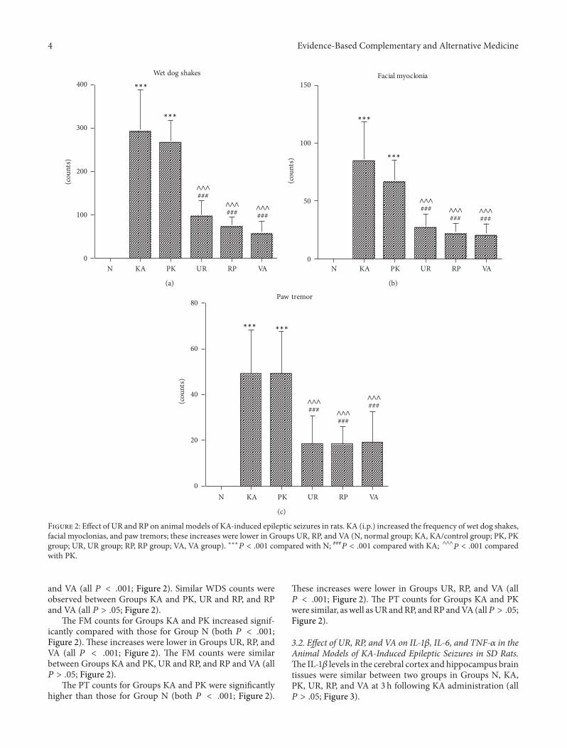
4 Evidence-Based Complementary and Alternative Medicine
0
100
200
300
400
(cou
nts)
Wet dog shakes
N KA PK UR RP VA
∧∧∧###
∧∧∧### ∧∧∧
###
∗∗∗
∗∗∗
(a)(c
ount
s)
N KA PK UR RP VA0
50
100
150Facial myoclonia
∧∧∧### ∧∧∧
###∧∧∧###
∗∗∗
∗∗∗
(b)
(cou
nts)
N KA PK UR RP VA0
20
40
60
80Paw tremor
∧∧∧### ∧∧∧
###
∧∧∧###
∗∗∗ ∗∗∗
(c)
Figure 2: Effect of UR and RP on animal models of KA-induced epileptic seizures in rats. KA (i.p.) increased the frequency of wet dog shakes,facial myoclonias, and paw tremors; these increases were lower in Groups UR, RP, and VA (N, normal group; KA, KA/control group; PK, PKgroup; UR, UR group; RP, RP group; VA, VA group). ∗∗∗𝑃 < .001 compared with N; ###𝑃 < .001 compared with KA; ∧∧∧𝑃 < .001 comparedwith PK.
and VA (all 𝑃 < .001; Figure 2). Similar WDS counts wereobserved between Groups KA and PK, UR and RP, and RPand VA (all 𝑃 > .05; Figure 2).
The FM counts for Groups KA and PK increased signif-icantly compared with those for Group N (both 𝑃 < .001;Figure 2). These increases were lower in Groups UR, RP, andVA (all 𝑃 < .001; Figure 2). The FM counts were similarbetween Groups KA and PK, UR and RP, and RP and VA (all𝑃 > .05; Figure 2).
The PT counts for Groups KA and PK were significantlyhigher than those for Group N (both 𝑃 < .001; Figure 2).
These increases were lower in Groups UR, RP, and VA (all𝑃 < .001; Figure 2). The PT counts for Groups KA and PKwere similar, aswell asURandRP, andRP andVA (all𝑃 > .05;Figure 2).
3.2. Effect of UR, RP, and VA on IL-1𝛽, IL-6, and TNF-𝛼 in theAnimal Models of KA-Induced Epileptic Seizures in SD Rats.The IL-1𝛽 levels in the cerebral cortex and hippocampus braintissues were similar between two groups in Groups N, KA,PK, UR, RP, and VA at 3 h following KA administration (all𝑃 > .05; Figure 3).

Evidence-Based Complementary and Alternative Medicine 5
0
0.5
1
1.5
Cortex HippocampusN KA PK UR RP VA N KA PK UR RP VA
IL-1𝛽
(pg/𝜇
g)
Figure 3: Effect of UR and RP on IL-1𝛽 in KA-induced epilepticseizures in rats. The IL-1𝛽 levels in the cerebral cortex (Cortex) andhippocampus (Hippocampus) brain tissues were similar in GroupsN, KA, PK, UR, RP, and VA (N: normal group; KA: KA/controlgroup; PK: PK group; UR: UR group; RP: RP group; VA: VA group).
0
0.5
1.5
2
∗∗
IL-6
N KA PK UR RP VA N KA PK UR RP VACortex Hippocampus
∗∗∗
∗∗∗∗∗∗
∗∗∗1
(pg/𝜇
g)
Figure 4: Effect of UR and RP on IL-6 in KA-induced epilepticseizures in rats. The IL-6 levels were lower in the cerebral cortex(Cortex) brain tissue for Groups KA, PK, UR, and VA than those inGroup N.The IL-6 levels in the hippocampus (Hippocampus) braintissue for Group UR were lower than those in Group N (N, normalgroup; KA, KA/control group; PK, PK group; UR, UR group; RP, RPgroup; VA, VA group). JNK: ∗P < .05, ∗∗∗P < .001 compared with N.
The IL-6 levels in Group N were significantly higher thanthose in Groups KA, PK, UR, and VA (all 𝑃 < .001; Figure 4).However, the IL-6 levels for Groups N and RP were similar(𝑃 > .05; Figure 4).The IL-6 levels in the cerebral cortex braintissues were similar between two groups in Groups KA, PK,UR, RP, and VA at 3 h following KA administration (all 𝑃 >.05; Figure 4).
The IL-6 levels in Group N were significantly higher thanthose in Group UR (𝑃 < .05; Figure 4). The IL-6 levels inthe hippocampus of brain tissue were similar between twogroups in Groups N, KA, PK, RP, and VA at 3 h after KAadministration (all 𝑃 > .05; Figure 4).
The TNF-𝛼 levels in Group N were significantly higherthan those inGroups PK, UR, andVA (all𝑃 < .001; Figure 5).The TNF-𝛼 levels in Groups N, KA, and RP were similar (all
0
0.2
0.4
0.6
0.8
∗∗∗
∗∗∗
∗∗∗
N KA PK UR RP VA N KA PK UR RP VACortex Hippocampus
TNF-𝛼
(pg/𝜇
g)
Figure 5: Effect of UR and RP on TNF-𝛼 in KA-induced epilepticseizures in rats.TheTNF-𝛼 level in the cerebral cortex (Cortex) braintissue forGroups PK,UR, andVAwere lower than those inGroupN.The TNF-𝛼 levels in the hippocampus (Hippocampus) brain tissuewere similar in Groups N, KA, PK, UR, RP, and VA (N: normalgroup; KA: KA/control group; PK: PK group; UR: UR group; RP:RP group; VA: VA group). ∗∗∗P < .001 compared with N.
𝑃 > .05; Figure 5). The TNF-𝛼 levels in the cerebral cortexbrain tissues were similar between two groups in Groups PK,UR, and VA at 3 h after KA administration (all 𝑃 > .05;Figure 5).
The TNF-𝛼 levels in the hippocampus brain tissues weresimilar between two groups in Groups N, KA, PK, UR, RP,and VA at 3 h following KA administration (all 𝑃 > .05;Figure 5).
3.3. Effect of UR, RP, and VA on JNK, ERK, p38 MAPK, andNF-𝜅B in the Animal Models of KA-Induced Epileptic Seizuresin SD Rats. In the cerebral cortex brain tissues, an increase inthe JNKp expression of MAPK signal pathways was observedin Groups KA and PK, whereas those increases were lower inGroups UR, RP, and VA at 3 h following KA administration(Figure 6).
In the hippocampus brain tissues, an increase in theJNKp expression of MAPK signal pathways was observed inGroups KA and PK, whereas these increases were lower inGroups UR, RP, and VA at 3 h following KA administration(Figure 6).
In both the cerebral cortex and hippocampus braintissues, the ERKp and p38p expressions of MAPK signalpathways were similar in Groups N, KA, PK, UR, RP, and VAat 3 h following KA administration (Figure 6).
Regarding the NF-𝜅B activity, no conclusive evidencewas obtained because of the inconsistent results following 3repeated experiments (Figure 7).
4. Discussion
The results showed that pretreatment using UR, RP, and VAreduced the WDS, FM, and PW counts during the epilepticseizures; indicating that UR, RP, and VA have anticonvulsiveeffects on animal models of KA-induced epileptic seizuresin SD rats; furthermore, these results are in good agreement
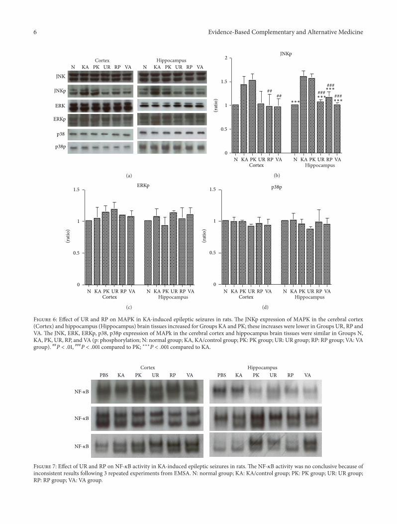
6 Evidence-Based Complementary and Alternative Medicine
NHippocampusCortex
KA PK UR RP VA N KA PK UR RP VA
JNK
JNKp
ERK
ERKp
p38
p38p
(a)
JNKp
(rat
io)
####
∗∗∗∗∗∗
∗∗∗
∗∗∗
###### ###
N KA PK UR RP VA N KA PK UR RP VACortex Hippocampus
0
0.5
1.5
2
1
(b)
ERKp
N KA PK UR RP VA N KA PK UR RP VACortex Hippocampus
(rat
io)
0
0.5
1.5
1
(c)
p38p
N KA PK UR RP VA N KA PK UR RP VACortex Hippocampus
(rat
io)
0
0.5
1.5
1
(d)
Figure 6: Effect of UR and RP on MAPK in KA-induced epileptic seizures in rats. The JNKp expression of MAPK in the cerebral cortex(Cortex) and hippocampus (Hippocampus) brain tissues increased for Groups KA and PK; these increases were lower in Groups UR, RP andVA. The JNK, ERK, ERKp, p38, p38p expression of MAPk in the cerebral cortex and hippocampus brain tissues were similar in Groups N,KA, PK, UR, RP, and VA (p: phosphorylation; N: normal group; KA, KA/control group; PK: PK group; UR: UR group; RP: RP group; VA: VAgroup). ##P < .01, ###P < .001 compared to PK; ∗∗∗P < .001 compared to KA.
PBSHippocampusCortex
KA PK UR RP VA PBS KA PK UR RP VA
NF-𝜅B
NF-𝜅B
NF-𝜅B
Figure 7: Effect of UR and RP on NF-𝜅B activity in KA-induced epileptic seizures in rats. The NF-𝜅B activity was no conclusive because ofinconsistent results following 3 repeated experiments from EMSA. N: normal group; KA: KA/control group; PK: PK group; UR: UR group;RP: RP group; VA: VA group.

Evidence-Based Complementary and Alternative Medicine 7
with those from our previous study [12]. Our previous pro-teomic study showed that under expression of macrophagemigration inhibitory factor (MIF) and cylcophilin proteinsin either the cerebral cortex or hippocampus brain tissuesof KA-treated rats, whereas those under expression couldbe reversed by administering UR or RP as a pretreatment[13]. Treatment with UR can reduce population spikes inhippocampal neuronal cells, and it can also reduce glialproliferation and S100 protein expression [14]; in addition, itcan attenuate mossy fiber sprouting in animal models of KA-induced epileptic seizures in rats [15].These findings indicatethat pretreatment using UR, RP, and VA can suppress thedevelopment of epilepsy.
Brain damage such as that caused by encephalitis inducesinflammatory responses that can promote neuronal excitabil-ity, which contributes to the generation of seizures. Conse-quently, the inflammation becomes exacerbated and results inepileptogenesis [2]. IL-1𝛽 is produced in injured brain tissue,and the bonding of IL-1𝛽 to the IL-1 receptor can initiatethe MAPK signaling pathways and NF-𝜅B activation [16].Our results show that the IL-1𝛽 levels in the cerebral cortexand hippocampus brain tissues were similar between twogroups in Groups N, KA, PK, UR, RP, and VA at 3 h afterKA administration. Pernot et al. [17] reported that the levelsof IL-1𝛽 mRNA increased between 5 and 24 h following KAintrahippocampal injection.
The results of this study show that the IL-6 levels weregreater in Group N than those in Groups KA, PK, UR, andVA, whereas the IL-6 levels in the cerebral cortex brain tissueswere similar between two groups in Groups KA, PK, UR, RP,and VA; however, the IL-6 levels in Group N were greaterthan those in Group UR, whereas the IL-6 levels in thehippocampus brain tissues were similar between two groupsin Groups N, KA, PK, RP, and VA at 3 h following KA admin-istration. Based on these results, we considered whether theIL-6 levels in the cerebral cortex and hippocampus braintissues were unchanged at 3 h following KA administration.Uludag et al. [18] showed that the serum levels of IL-6 andIL-1 receptor antagonist (IL-1Ra) increased at 12 h followingseizures in 23 epilepsy patients. Lehtimaki et al. [19] reportedthat the increased IL-6 levels in the cerebrospinal fluid(CSF) and serum following seizures were greater in recurrentgeneralized tonic-clonic seizure (GTS) patients than thosein either single GTS or prolonged partial seizure patients;therefore, they concluded that seizures could induce cytokineproduction and that the CSF and serum IL-6 levels mightcorrelatewith the severity of a seizure. A recurrent seizure caninduce IL-6 production, which can cause changes in neuronaltissue, resulting in the development of refractory seizures.
The results of this study indicated that the TNF-𝛼 levelsin the cerebral cortex brain tissues for Group N were greaterthan those in Groups PK, UR, and VA, although the TNF-𝛼 levels were similar between two groups in Groups N, KA,and RP, as well as between two groups in Groups PK, UR,and VA; however, the TNF-𝛼 levels in the hippocampus braintissues were similar between two groups in Groups N, KA,PK, UR, RP, and VA at 3 h following KA administration.Accordingly, we considered whether the TNF-𝛼 levels inthe cerebral cortex and hippocampus brain tissues had
not changed at 3 h following KA-induced epileptic seizures.Previous research reported that the TNF-𝛼 mRNA peakedat 6 h following limbic seizure with epilepticus [20]. TNF-𝛼plays dual roles of anti- and proconvulsion, which ismediatedby its neuronal p75 receptor, which can inhibit seizures andexert an anticonvulsive effect. In contrast, another studyreported that TNF-𝛼wasmediated by the p55 receptor, whichacted as a proconvulsant [21]. This discussion shows thatthe levels of IL-1𝛽, IL-6, and TNF-𝛼 did not increase at 3 hfollowing KA administration in this study, indicating thattiming is a critical factor in the production of IL-1𝛽, IL-6,and TNF-𝛼. Our results indicated that UR, RP, and VA couldreduce epileptic seizures, although they did not change thelevels of proinflammatory cytokines IL-1𝛽, IL-6, and TNF-𝛼.Therefore, we infer that the anticonvulsive effect of UR, RP,and VA—at least for proinflammatory cytokines IL-1𝛽, IL-6,and TNF-𝛼—did not play a key role in the KA-induced acuteseizure.
Our results also indicated that the JNKp expression ofthe MAPK signal pathway increased in the cerebral cortexand hippocampus in Groups KA and PK.These increases canbe attenuated by pretreatment using UR, RP, and VA for 3 d.Previous research showed that MAPKs play a critical role incell differentiation, proliferation, and death and is activatedby phosphorylation cascades [22]. KA is an analogue of gluta-mate, and can induce limbic seizures. Furthermore, previousresearch has shown that KA can induce the activation ofJNK and p38, which are both involved in cell death [23]. TheMAPK protein and its active form increase between 30 and60min following pilocarpine-induced epilepticus. Previousresearch hypothesized that this activation could contributeto the mechanisms of acute epileptogenesis and long-lastingchanges of neuropathology [24]. Che et al. [25] reported thatp38 MAPK of brain tissue increased at 4 d following KA-induced seizures in mice and that this p38 MAPK signalpathway plays a crucial role in either neuronal death orreactive gliosis. Previous studies have shown that ERKMAPKplays a critical role in cell death [26] and that the MAPKsignal pathway plays a critical role in the physiological andbiochemical regulation of NMDA receptors [27]. Sokka et al.[28] reported that KA activates non-NMDA receptor andthat induced seizures can cause stress of the endoplasmicreticulum (ER), resulting in the accumulation of unfoldedprotein. Furthermore, this stress of the ER has been shownto cause the activation of JNK, which is similar to cell surfacereceptor reactions to extracellular signal stimulation [29]. KAcan induce the release of glutamate from neuronal cells andcan also induce the ER stress of astrocyte. Chihara et al. [30]reported that the old astrocyte specifically induced substance(OASIS) in astrocyte can respond to the ER stress and that itplayed a protective role inKA-inducedER stress. Lee et al. [31]showed that pretreatment using an alkaloid fraction of URcan block NMDA-induced cytotoxicity, which acts as a neu-roprotector by inhibiting apoptosis. In addition, in this study,the results for the NF-𝜅B levels were inconclusive becauseof the inconsistent results following the 3 experiments; thelevels of NF-𝜅B were the upstream of the pro-inflammatorycytokines IL-1𝛽, IL-6, and TNF-𝛼. Previous studies havereported that the DNA-binding activity of NF-𝜅B increased
8 Evidence-Based Complementary and Alternative Medicine
at 24 h following KA-treatment in rat models [10, 32]. Insummary, we assert that the JNKp MAPK signal pathwayplays a critical role in epileptogenesis at 3 h following KAadministration; therefore, UR, RP, and VA suppression ofthe expression of JNKp MAPk inhibit the development ofepilepsy. Thus, we suggest that the present study is moreadvantage to develop the novel antiepileptic drug than ourprevious study [12]. Because the results indicated that JNKpMAPK signal pathway involve to acute seizure.
Some limitation in the present study is as follows: (1) thedose of UR and RP is difficult to determine due to high dosepossible reduce the effect of UR; (2) although UR is mostcommonly used for the treatment of vertigo and epilepsy inTaiwan and in our clinic and according to our knowledge,the severe side effect and herb-drug interaction were notfound. The further observation and study still remain tobe needed; (3) the present study limits in animal modellevel. Therefore, how to design a randomized, double blind,placebo-controlled clinical trial in epileptic patient is animport issue in the future.
In conclusion, UR, RP, and VA reduce epileptic seizuresand also reduce the JNKp expression of the MAPK signalpathway at 3 h following KA administration, indicating thatthe antiepileptic effect of UR and RP is involved in initiatingthe JNKp MAPK signal pathway.
Conflict of Interests
The authors declare there is no financial or commercialconflict of interests related to this study.
Acknowledgments
This study was supported by the National Science Coun-cil, Taiwan (Grant no. NSC95-2320-B-039-033). It was alsosupported in part by the Taiwanese Department of Health,Clinical Trials and Research Center of Excellence (DOH102-TD-B-111-004).
References
[1] M. Schultzberg, C. Lindberg, A. F. Aronsson, E. Hjorth, S. D.Spulber, and M. Oprica, “Inflammation in the nervous system:physiological and pathophysiological aspects,” Physiology andBehavior, vol. 92, no. 1-2, pp. 121–128, 2007.
[2] A. Vezzani, J. French, T. Bartfai, and T. Z. Baram, “The role ofinflammation in epilepsy,” Nature Reviews Neurology, vol. 7, no.1, pp. 31–40, 2011.
[3] G. Li, S. Bauer, M. Nowak et al., “Cytokines and epilepsy,”Seizure, vol. 20, no. 3, pp. 249–256, 2011.
[4] M. P. Mattson and S. Camandola, “NF-𝜅B in neuronal plasticityand neurodegenerative disorders,”The Journal of Clinical Inves-tigation, vol. 107, no. 3, pp. 247–254, 2001.
[5] M. Lerner-Natoli, P. Montpied, M. Rousset, J. Bockaert, and G.Rondouin, “Sequential expression of surface antigens and tran-scription factor NF𝜅B by hippocampal cells in excitotoxicityand experimental epilepsy,” Epilepsy Research, vol. 41, no. 2, pp.141–154, 2000.
[6] F. D. Lubin, Y. Ren, X. Xu, and A. E. Anderson, “Nuclear factor-𝜅B regulates seizure threshold and gene transcription followingconvulsant stimulation,” Journal of Neurochemistry, vol. 103, no.4, pp. 1381–1395, 2007.
[7] M. J. Robinson and M. H. Cobb, “Mitogen-activated proteinkinase pathways,” Current Opinion in Cell Biology, vol. 9, no. 2,pp. 180–186, 1997.
[8] A. Novelli and R. A. R. Tasker, “Excitatory amino acids inepilepsy: from the clinics to the laboratory,” Amino Acids, vol.32, no. 3, pp. 295–297, 2007.
[9] S. Li, Bencoogongmu, pp. 764-765, Shi-Yi Publisher, Tainan,Taiwan, 2005.
[10] C. Hsieh, M. Chen, T. Li et al., “Anticonvulsant effect ofUncariarhynchophylla (miq) Jack, in rats with kainic acid-inducedepileptic seizure,”American Journal of ChineseMedicine, vol. 27,no. 2, pp. 257–264, 1999.
[11] N. Tang, C. Liu, S. Su et al., “Uncaria rhynchophylla (Miq) Jackplays a role in neuronal protection in kainic acid-treated rats,”American Journal of ChineseMedicine, vol. 38, no. 2, pp. 251–263,2010.
[12] C.Hsieh, T.Ho, S. Su,W. Lo, C. Liu, andN. Tang, “Uncaria rhyn-chophylla and rhynchophylla inhibit c-Jun N-terminal kinasephosphorylation and nuclear factor-𝜅B activity in kainic acid-treated rats,” American Journal of Chinese Medicine, vol. 37, no.2, pp. 351–360, 2009.
[13] W. Lo, F. Tsai, C. Liu et al., “Uncaria rhynchophylla upregulatesthe expression of MIF and cyclophilin A in kainic acid-inducedepilepsy rats: a proteomic analysis,”American Journal of ChineseMedicine, vol. 38, no. 4, pp. 745–759, 2010.
[14] Y. Lin and C. Hsieh, “Oral Uncaria rhynchophylla (UR) reduceskainic acid-induced epileptic seizures and neuronal deathaccompanied by attenuating glial cell proliferation and S100Bproteins in rats,” Journal of Ethnopharmacology, vol. 135, no. 2,pp. 313–320, 2011.
[15] C. Hsieh, C. Liu, Y. Lin, N. Tang, and H. Liu, “Neuroprotectiveeffect ofUncaria rhynchophylla in Kainic acid-induced epilepticseizures by modulating hippocampal mossy fiber sprouting,neuron survival, astrocyte proliferation, and S100b expression,”Evidence-Based Complementary and Alternative Medicine, vol.2012, Article ID 194790, 11 pages, 2012.
[16] S. M. Allan, P. J. Tyrrell, and N. J. Rothwell, “Interleukin-1 andneuronal injury,” Nature Reviews Immunology, vol. 5, no. 8, pp.629–640, 2005.
[17] F. Pernot, C. Heinrich, L. Barbier et al., “Inflammatory changesduring epileptogenesis and spontaneous seizures in a mousemodel of mesiotemporal lobe epilepsy,” Epilepsia, vol. 52, no. 12,pp. 2315–2325, 2011.
[18] I. F. Uludag, S. Bilgin, Y. Zorlu, G. Tuna, and G. Kirkali,“Interleukin-6, interleukin-1 beta and interleukin-1 receptorantagonist levels in epileptic seizures,” Seizure, vol. 22, pp. 457–461, 2013.
[19] K. A. Lehtimaki, T. Keranen, H. Huhtala et al., “Regulation ofIL-6 system in cerebrospinal fluid and serum compartmentsby seizures: the effect of seizure type and duration,” Journal ofNeuroimmunology, vol. 152, no. 1-2, pp. 121–125, 2004.
[20] A. Vezzani, D. Moneta, C. Richichi et al., “Functional roleof inflammatory cytokines and antiinflammatory molecules inseizures and epileptogenesis,” Epilepsia, vol. 43, supplement 5,pp. 30–35, 2002.
[21] S. Balosso, T. Ravizza, C. Perego et al., “Tumor necrosis factor-𝛼inhibits seizures inmice via p75 receptors,”Annals of Neurology,vol. 57, no. 6, pp. 804–812, 2005.

Evidence-Based Complementary and Alternative Medicine 9
[22] G. Pearson, F. Robinson, T. B. Gibson et al., “Mitogen-activatedprotein (MAP) kinase pathways: regulation and physiologicalfunctions,” Endocrine Reviews, vol. 22, no. 2, pp. 153–183, 2001.
[23] S. H. Jeon, Y. S. Kim, C. Bae, and J. Park, “Activation of JNKand p38 in rat hippocampus after kainic acid induced seizure,”Experimental and Molecular Medicine, vol. 32, no. 4, pp. 227–230, 2000.
[24] Y. C. S. Garrido, E. R. G. Sanabria, M. G. Funke, E. A. Cav-alheiro, and M. G. Naffah-Mazzacoratti, “Mitogen-activatedprotein kinase is increased in the limbic structures of therat brain during the early stages of status epilepticus,” BrainResearch Bulletin, vol. 47, no. 3, pp. 223–229, 1998.
[25] Y. Che, Y. Yu, P. Han, and J. Lee, “Delayed induction of p38MAPKs in reactive astrocytes in the brain of mice after KA-induced seizure,”Molecular Brain Research, vol. 94, no. 1-2, pp.157–165, 2001.
[26] S. Subramaniam and K. Unsicker, “ERK and cell death: ERK1/2in neuronal death,” FEBS Journal, vol. 277, no. 1, pp. 22–29, 2010.
[27] J. J. Haddad, “N-methyl-D-aspartate (NMDA) and the regu-lation of mitogen-activated protein kinase (MAPK) signalingpathways: a revolving neurochemical axis for therapeutic inter-vention?” Progress in Neurobiology, vol. 77, no. 4, pp. 252–282,2005.
[28] A. Sokka, N. Putkonen, G.Mudo et al., “Endoplasmic reticulumstress inhibition protects against excitotoxic neuronal injury inthe rat brain,”The Journal of Neuroscience, vol. 27, no. 4, pp. 901–908, 2007.
[29] F. Urano, X. Wang, A. Bertolotti et al., “Coupling of stress inthe ER to activation of JNK protein kinases by transmembraneprotein kinase IRE1,” Science, vol. 287, no. 5453, pp. 664–666,2000.
[30] K.Chihara, A. Saito, T.Murakami et al., “Increased vulnerabilityof hippocampal pyramidal neurons to the toxicity of kainic acidin OASIS-deficient mice,” Journal of Neurochemistry, vol. 110,no. 3, pp. 956–965, 2009.
[31] J. Lee, D. Son, P. Lee et al., “Alkaloid fraction of Uncariarhynchophylla protects against N-methyl-D-aspartate-inducedapoptosis in rat hippocampal slices,” Neuroscience Letters, vol.348, no. 1, pp. 51–55, 2003.
[32] S. J. Won, H. W. Ko, E. Y. Kim et al., “Nuclear factor kappaB-mediated kainate neurotoxicity in the rat and hamster hip-pocampus,” Neuroscience, vol. 94, no. 1, pp. 83–91, 1999.
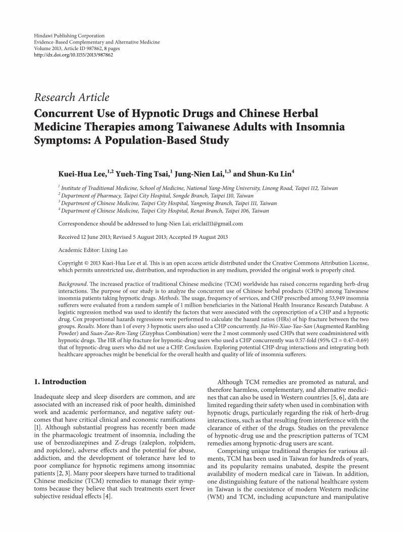
Hindawi Publishing CorporationEvidence-Based Complementary and Alternative MedicineVolume 2013, Article ID 987862, 8 pageshttp://dx.doi.org/10.1155/2013/987862
Research ArticleConcurrent Use of Hypnotic Drugs and Chinese HerbalMedicine Therapies among Taiwanese Adults with InsomniaSymptoms: A Population-Based Study
Kuei-Hua Lee,1,2 Yueh-Ting Tsai,1 Jung-Nien Lai,1,3 and Shun-Ku Lin4
1 Institute of Traditional Medicine, School of Medicine, National Yang-Ming University, Linong Road, Taipei 112, Taiwan2Department of Pharmacy, Taipei City Hospital, Songde Branch, Taipei 110, Taiwan3Department of Chinese Medicine, Taipei City Hospital, Yangming Branch, Taipei 111, Taiwan4Department of Chinese Medicine, Taipei City Hospital, Renai Branch, Taipei 106, Taiwan
Correspondence should be addressed to Jung-Nien Lai; [email protected]
Received 12 June 2013; Revised 5 August 2013; Accepted 19 August 2013
Academic Editor: Lixing Lao
Copyright © 2013 Kuei-Hua Lee et al. This is an open access article distributed under the Creative Commons Attribution License,which permits unrestricted use, distribution, and reproduction in any medium, provided the original work is properly cited.
Background. The increased practice of traditional Chinese medicine (TCM) worldwide has raised concerns regarding herb-druginteractions. The purpose of our study is to analyze the concurrent use of Chinese herbal products (CHPs) among Taiwaneseinsomnia patients taking hypnotic drugs.Methods. The usage, frequency of services, and CHP prescribed among 53,949 insomniasufferers were evaluated from a random sample of 1 million beneficiaries in the National Health Insurance Research Database. Alogistic regression method was used to identify the factors that were associated with the coprescription of a CHP and a hypnoticdrug. Cox proportional hazards regressions were performed to calculate the hazard ratios (HRs) of hip fracture between the twogroups. Results. More than 1 of every 3 hypnotic users also used a CHP concurrently. Jia-Wei-Xiao-Yao-San (Augmented RamblingPowder) and Suan-Zao-Ren-Tang (Zizyphus Combination) were the 2 most commonly used CHPs that were coadministered withhypnotic drugs. The HR of hip fracture for hypnotic-drug users who used a CHP concurrently was 0.57-fold (95% CI = 0.47–0.69)that of hypnotic-drug users who did not use a CHP. Conclusion. Exploring potential CHP-drug interactions and integrating bothhealthcare approaches might be beneficial for the overall health and quality of life of insomnia sufferers.
1. Introduction
Inadequate sleep and sleep disorders are common, and areassociated with an increased risk of poor health, diminishedwork and academic performance, and negative safety out-comes that have critical clinical and economic ramifications[1]. Although substantial progress has recently been madein the pharmacologic treatment of insomnia, including theuse of benzodiazepines and Z-drugs (zaleplon, zolpidem,and zopiclone), adverse effects and the potential for abuse,addiction, and the development of tolerance have led topoor compliance for hypnotic regimens among insomniacpatients [2, 3]. Many poor sleepers have turned to traditionalChinese medicine (TCM) remedies to manage their symp-toms because they believe that such treatments exert fewersubjective residual effects [4].
Although TCM remedies are promoted as natural, andtherefore harmless, complementary, and alternative medici-nes that can also be used inWestern countries [5, 6], data arelimited regarding their safety when used in combination withhypnotic drugs, particularly regarding the risk of herb-druginteractions, such as that resulting from interference with theclearance of either of the drugs. Studies on the prevalenceof hypnotic-drug use and the prescription patterns of TCMremedies among hypnotic-drug users are scant.
Comprising unique traditional therapies for various ail-ments, TCM has been used in Taiwan for hundreds of years,and its popularity remains unabated, despite the presentavailability of modern medical care in Taiwan. In addition,one distinguishing feature of the national healthcare systemin Taiwan is the coexistence of modern Western medicine(WM) and TCM, including acupuncture and manipulative

2 Evidence-Based Complementary and Alternative Medicine
therapies and Chinese herbal products (CHPs), claims forwhich have been covered by the National Health Insurance(NHI) system since 1995 [7].
People in Taiwan are free to choose from care offeredby WM clinics or TCM clinics. With an insured rate of98% to 99%, the random sample that comprises the NHIresearch database (NHIRD) is representative of the generalpopulation of Taiwan and should allow a reasonably accurateassessment of the concurrent usage of TCM and modernmedical resources in Taiwan; therefore, the NHIRD providesan ideal platform for pharmacoepidemiological studies [8].Our study aimed to describe the demographics and patternsof CHP usage among hypnotic-drug users and, by usinga large population-based retrospective database, explorethe risk of hip fracture among a cohort of hypnotic-drugusers prescribed with a CHP concurrently compared withhypnotic-drug users who did not use Chinese herbs. Ourfindings provide evidence-based information for formulatingappropriate management strategies for drug safety and inte-grative medicine.
2. Materials and Methods
2.1. Data Source and Participants. Our study protocols wereapproved by the Institutional ReviewBoard of the CommitteeonChineseMedicine andPharmacy (CCMP),Department ofHealth, Taiwan. Our population-based study retrospectivelyanalyzed the reimbursement records of 1 million NHI ben-eficiaries in the NHIRD that had been previously selected atrandom from the 22million beneficiaries of theNHI to deter-mine the prevalence of concurrent CHP and hypnotic usebetween January 1, 2002, and December 31, 2008, in Taiwan.The electronic records of the NHIRD use beneficiary iden-tification numbers that are encrypted and maintained by theNationalHealth Research Institutes (NHRI) of Taiwan [9, 10].
The NHIRD records contain demographic information,including age and sex, and clinical data, including all recordsof clinical visits and hospitalizations, and all informationregarding prescribed drugs and dosages, including thosefor CHPs. The diagnoses used in the NHIRD are codedaccording to the International Classification of Diseases,Ninth Revision, Clinical Modification (ICD-9-CM) [11].
Because the cost of all prescribed hypnotic drugs isreimbursed by the NHI, they cannot be dispensed at a phar-macy without a physician’s prescription. To construct a fixedcohort, all outpatients with insomnia who used hypnoticdrugs, including benzodiazepines, zolpidem, and zopiclone,were reviewed. For the purpose of studying the use of CHPs,we downloaded the claims forms for reimbursed CHPs fromthe website of the Bureau of National Health Insurance.The corresponding information regarding the CHP was thenobtained from theCCMPwebsite, including the name of eachherb, the proportion of each constituent of the mixture, thedate and period of approval for the drug, theCCMPmanufac-turer code, and the name of the CHPmanufacturer. All CHPswith the same CCMP standard formula are classified in thesame category, regardless of slight variations in the productsamong different CHP manufacturers [12]. For our analysis ofthe demographic and clinical variables, a coprescription of a
CHP and a hypnotic drug (defined as prescriptions for bothissued simultaneously or issued separately with overlappingtreatment periods) was used to determine the use of a CHPand a hypnotic drug on the same day.
The participant selection from the NHIRD was per-formed as shown in Figure 1. We reviewed all beneficiarieswith at least three outpatient visits with any nonorganicinsomnia (ICD-9-CM codes 307.40-42, 307.45-49, 780.50-52,and 780.55-59) and excluded patients who were admittedto hospital and received a hip fracture diagnosis between1999 and 2001 (𝑛 = 138). Patients with hyperinsomnia(ICD-9-CM codes 327, 307.43, 307.44, 780.53, and 780.54) forthe calendar year in which it occurred were excluded. Theprevalent insomnia cases diagnosed before the end of 2001(𝑛 = 9, 242) and patients under 20 years of age (𝑛 = 4, 691)were excluded to ensure that all participants had been newlydiagnosedwith adult insomnia. Patientswith incomplete datafor age or sex (𝑛 = 21) and insomniac patients not takinghypnotic drugs (𝑛 = 10, 628) were also excluded.
2.2. Study Variables. To identify the key factors associatedwith the coadministration of hypnotics and CHP amonginsomnia sufferers (CAHCHP as an acronym for this pop-ulation), we selected the demographic factors according toprevious studies [1, 7, 8]. Patients were classified, basedon age, into one of seven groups as follows: 20–29 years,30–39 years, 40–49 years, 50–59 years, 60–69 years, 70–79 years, and ≥80 years. The geographic areas of Taiwanin which patients resided were classified as one of thefollowing seven regions: Taipei city, Kaohsiung city, Northernregion, Central region, Southern region, Eastern region, andOutlying islands. Patients’ monthly income in New TaiwanDollars (NT$) was categorized as one of the following fourlevels: $0, $1–$19,999, $20,000–$39,999, and ≥$40,000.
The variables for hypnotic-drug use included in ouranalyses were defined according to the specific proprietaryhypnotic preparation used during the study period. We cate-gorized the types of preparation used as follows: long-half-lifebenzodiazepine; short-half-life benzodiazepine; zolpidem;zopiclone; andmixed regimens, including two, three, ormoreof the aforementioned preparations.
To estimate the impact of CHP use on the rate of hipfracture, we selected subjects who were first recorded witha diagnosis of hip fracture (ICD-9-CM codes 820.0–820.9)between January 1, 2002, and December 31, 2008. Further-more, we also analyzed the risk of hip fracture according tothe period of time patients were administered hypnotic drugs(≥30 days and <30 days).
2.3. Statistical Analysis. Data analysis was conducted usingdescriptive statistics, including the prescription rates ofpatients’ concurrent use of a CHP and a sedative hypnoticdrug stratified by age and sex, indications for the prescribedCHP, and the most frequently coprescribed herbal formulasfor treating insomnia. The indications were coded accordingto the ICD-9-CM and grouped into different broader diseasecategories. The ICD-9-CM codes 460–519 were classifiedas diseases of the respiratory system. Codes 780–799 weregrouped as symptoms, signs, and ill-defined conditions, and

Evidence-Based Complementary and Alternative Medicine 3
Number of total insomnia
Number of total insomnia
2002.1.1, n = 9,242
patients, n = 78,624
patients, n = 64,691
Number of total insomnia patients, n = 64,670
Never been prescribed
hypnotic drugs, n = 10,628 hypnotic drugs, n = 54,042
Never used TCM
One million random sample of NHIRDhaving 3 outpatient visits with insomnia diagnosis, n = 78,762
Exclusion of patients having hospital
admission with hip fracture diagnosis
from 1999 to 2001, n = 138
Never used TCM
Never been prescribed
n = 5,848 n = 48,194
n = 4,691
Exclusion of 21 patients with missingdata
Exclusion of patients having outpatientvisits with insomnia diagnosis before
Excluded of patients aged <20,
Figure 1: Flowchart of recruitment of subjects with insomnia from the 1 million random sample of the National Health Insurance ResearchDatabase (NHIRD) from 2002 to 2008 in Taiwan.
codes 520–579 were classified as diseases of the digestive sys-tem. Multiple logistic regression was conducted to evaluatefactors that correlated with CHP use based on the odds ratio(OR) and the 95% confidence interval (CI). Cox proportionalhazards regressions were performed to calculate the hazardratio (HR) of hip fracture among hypnotic-drug users whoused a CHP concurrently and hypnotic-drug users who didnot use Chinese herbs. A significance level of 𝛼 = 0.05was selected. The SAS statistical software, version 9.3 (SASInstitute, Cary,NC,USA), was used for datamanagement andanalyses.
3. Results
The database of outpatient claims contained informationfor 64,670 sleep-disturbance sufferers during the 2002–2008period. A total of 54,042 (83.6%) insomnia patients treatedwith sedative-hypnotic drugs were included in our study.Most of these patients used short-acting benzodiazepines.Most of the sedative-hypnotic users (89.2%) also sought care
from TCM practitioners. Up to 37.0% (𝑛 = 19, 994) of thesedative-hypnotic users received a coprescription for a CHP,resulting in the coadministration of a sedative-hypnotic drugwith a CHP. Such patients who were coadministered the twotypes of drug are hereafter referred to as CAHCHP patients.A total of 19,994 CAHCHP patients were identified in ourinsomnia cohort.
The hypnotic users who did not use a CHP were sig-nificantly older and more likely to be male than were theCAHCHP patients. More CAHCHP patients had incomelevels of $20,000 to $39,999 and above, resided in CentralTaiwan, and used two or more types of sedative-hypnoticdrugs than the sedative-hypnotic users who did not use aCHP.The adjusted ORs (aORs) and 95% CIs calculated usingthe logistic regression model are displayed in Table 1. Afteradjusting for other factors, the aORs of CAHCHP patients(aORs = 2.49) were higher for women than for men (aORs =1.00) except patients aged 80 years and older. Compared withthe patients aged 40–49 years (aORs = 1.00), the aORs of theCAHCHP patients decreased with age. Patients who used at

4 Evidence-Based Complementary and Alternative Medicine
Table 1: Demographic characteristics and results of multiple logistic regression showing the adjusted odds ratio (aOR) and 95% confidenceinterval (CI) of patients with newly diagnosed insomnia ever been prescribed hypnotic drugs from the 1 million random sample of theNational Health Insurance Research Database (NHIRD) from 2002 to 2008 in Taiwan.
Characteristics Total no.Hypnotic userswithout usingChinese medicine,no. (%)
Hypnotic userscoprescribedChinese medicine,no. (%)
Hypnotic users coprescribedChinese medicine/hypnotic userswithout using Chinese medicineOdds ratio (95% CI)
Numbers of hypnotic users among insomniapopulation 54,042 5,848 (10.8) 19,994 (37.0)
SexMale 19,354 3,234 (55.3) 6,350 (31.8) 1.00 (2.34–2.66)Female 34,688 2,614 (44.7) 13,644 (68.2) 2.49
Mean (SD) age at inclusion, years 48.8 (15.4) 55.1 (17.2) 49.9 (14.9)Age groups at inclusion, years no. (%)20∼29 5,616 362 (6.2) 1,631 (8.2) 1.06 (0.93–1.22)
Male 184 (3.0) 477 (5.8)Female 178 (3.2) 1,154 (2.4)
30∼39 11,105 891 (15.2) 3,820 (19.1) 1.03 (0.94–1.14)Male 506 (6.6) 1,184 (13.2)Female 385 (8.6) 2,636 (5.9)
40∼49 13,531 1,193 (20.4) 5,074 (25.4) 1.00Male 675 (8.9) 1,536 (17.7)Female 518 (11.5) 3,538 (7.7)
50∼59 10,637 1,072 (18.3) 4,184 (20.9) 0.85 (0.77–0.93)Male 550 (8.9) 1,118 (15.3)Female 522 (9.4) 3,066 (5.6)
60∼69 6,657 865 (14.8) 2,798 (14.0) 0.76 (0.68–0.84)Male 488 (6.5) 962 (9.2)Female 377 (8.3) 1,836 (4.8)
70∼79 4,787 922 (15.8) 1,951 (9.8) 0.52 (0.46–0.58)Male 545 (6.5) 821 (5.7)Female 377 (9.3) 1,130 (4.1)
Over 80 1,709 543 (9.3) 536 (2.7) 0.24 (0.21–0.28)Male 286 (4.4) 252 (1.4)Female 257 (4.9) 284 (1.3)
$NT/month (Premiums)0 11,542 1,279 (21.9) 4,735 (23.7) 1.001–19,999 27,391 3,268 (55.9) 10,123 (50.6) 0.85 (0.79–0.92)20,000–39,999 9,601 729 (12.5) 3,303 (16.5) 1.13 (1.01–1.26)≥40,000 5,508 572 (9.8) 1,833 (9.2) 1.03 (0.91–1.17)Insured areaTaipei city 9,495 1,159 (19.8) 3,239 (16.2) 1.00Kaohsiung city 3,735 364 (6.2) 1,392 (7.0) 1.46 (1.27–1.68)Northern Taiwan 14,900 1,848 (31.6) 4,929 (24.7) 0.98 (0.90–1.08)Central Taiwan 12,554 824 (14.1) 5,490 (27.5) 2.49 (2.24–2.77)Southern Taiwan 11,910 1,401 (24.0) 4,407 (22.0) 1.28 (1.16–1.41)Eastern Taiwan 1,078 155 (2.7) 439 (2.2) 1.01 (0.82–1.24)Outlying islands 370 97 (1.7) 98 (0.5) 0.36 (0.27–0.49)

Evidence-Based Complementary and Alternative Medicine 5
Table 1: Continued.
Characteristics Total no.Hypnotic userswithout usingChinese medicine,no. (%)
Hypnotic userscoprescribedChinese medicine,no. (%)
Hypnotic users coprescribedChinese medicine/hypnotic userswithout using Chinese medicineOdds ratio (95% CI)
Types and prescription patterns of hypnoticsUsual treatment regimens 16,182 1,352 (23.1) 3,310 (16.6) 1.00
Zolpidem 1,623 350 (6.0) 265 (1.3)Zopiclone 109 16 (0.3) 20 (0.1)BZD-long 8,657 293 (5.0) 1,703 (8.5)BZD-short 5,793 693 (11.9) 1,322 (6.6)
Mixed regimensCo-prescribed two types of hypnotics 18,466 2,168 (37.1) 6,734 (33.6) 1.30 (1.20–1.42)
Zolpidem + Zopiclone 79 20 (0.3) 18 (0.1)Zolpidem + BZD-long 1,796 275 (4.7) 486 (2.4)Zolpidem + BZD-short 4,418 849 (14.5) 1,499 (7.5)Zopiclone + BZD-long 136 25 (0.4) 40 (0.2)Zopiclone + BZD-short 308 63 (1.1) 99 (0.5)BZD-long + BZD-short 11,729 936 (16.0) 4,592 (23.0)
Coprescribed more than two types of hypnotics 19,394 2,328 (39.8) 9,950 (49.8) 1.90 (1.75–2.06)NT$ refers to new Taiwan dollars, of which 1US$ = 30NT$.
least two types of sedative hypnotic drugs (two types: OR =1.30; 95% CI: 1.20–1.42 and ≥ three types: OR = 1.90; 95% CI:1.75–2.06) were more likely to be CHP users than were thepatients who used only one sedative hypnotic drug.
By analyzing the percentage distribution of the 19,994CAHCHP patients, we observed that sleep disturbance(1,450,671 TCM visits) was the most common major diseasecategory for coprescription events, followed by diseases ofthe digestive system (1,349,092 TCM visits) and the diseasesof the respiratory system (1,319,859 TCM visits), as summa-rized in Table 2. As shown in Table 3, zolpidem, alprazolam,lorazepam, estazolam, and fludiazepamwere the five sedativehypnotic drugs that were most frequently coprescribed witha CHP.The details of the most frequently used CHP formulasare shown in Table 3. Suan-Zao-Ren-Tang (Zizyphus Combi-nation) was the most frequently used CHP, followed by Jia-Wei-Xiao-Yao-San (Augmented Rambling Powder) and Tian-Wang-Bu-Xin-Dan (Ginseng and Zizyphus Combination).The average number of days of concurrent use of a sedativehypnotic drug and a CHP was approximately 30 days perCAHCHP patient during the 7-year study period.
The HRs of hip fracture for CAHCHP patients andhypnotic-drug users who did not use Chinese herbs are pre-sented in Table 4. After adjusting for potential confounders,the HR of hip fracture for CAHCHP patient was 0.57-fold(95%CI = 0.47–0.69) that of comparison subjects.We furtheranalyzed the risk of hip fracture according to the lengthof hypnotic treatment. Table 4 shows that, compared withhypnotic-drug users who did not use Chinese herbs, the HRof hip fracture for CAHCHP patients who were prescribedhypnotics for ≥30 days was 0.62 (95% CI = 0.50–0.77) and aslow as 0.33 (95% CI = 0.20–0.54) for CAHCHP patient who
were prescribed hypnotics for <30 days.The incidence rate ofhip fracture among theCAHCHPpatientswas no higher thanthat of hypnotic-drug users who did not use Chinese herbs(≥30-day group: 3.4 versus 6.5 per 100 person-years; <30-daygroup: 2.9 versus 11.1 per 100 person-years).
4. Discussion
According to our review of the literature, this study is thefirst to use a random population-based cohort to documentthe coprescription of hypnotic drugs and CHPs in insom-nia patients in Taiwan. We observed that hypnotic use iscommon among insomnia patients in Taiwan and that ben-zodiazepines were the most frequently prescribed categoryof sedative hypnotic, as shown in Table 1. Previous studieshave reported that patients treatedwith benzodiazepinesmaybe at higher risk for falls and have been associated with anincreased risk of hip fracture. Both physicians and patientsshould be aware of the association between hypnotic useand the potential risk for injury in Taiwan. The possibilityof recall or selection bias can be excluded because weincluded patients who were newly diagnosed with insomniaby qualified conventional physicians during the 2002–2008period from a random sample of the population-based NHIdatabase.
The prevalence of the concurrent use of sedative hyp-notics and CHPs was 37% (Figure 1). The abuse of benzo-diazepines has become a serious problem in North America[13]. The fear of sedative hypnotic side effects or dependencymay motivate patients to seek TCM therapies [2, 3, 14].Although barbiturates have been reported to be the mostprescribed hypnotic in Taiwan, our findings indicate a change
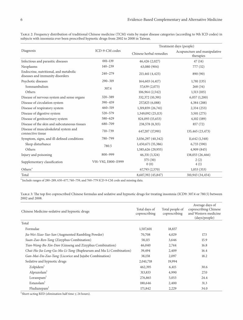
6 Evidence-Based Complementary and Alternative Medicine
Table 2: Frequency distribution of traditional Chinese medicine (TCM) visits by major disease categories (according to 9th ICD codes) insubjects with insomnia ever been prescribed hypnotic drugs from 2002 to 2008 in Taiwan.
Diagnosis ICD-9-CM codesTreatment days (people)
Chinese herbal remedies Acupuncture and manipulativetherapies
Infectious and parasitic diseases 001–139 46,426 (2,027) 47 (14)Neoplasms 140–239 63,080 (904) 777 (32)Endocrine, nutritional, and metabolicdiseases and immunity disorders 240–279 213,461 (4,425) 890 (90)
Psychotic diseases 290–319 164,603 (4,417) 1,781 (135)Somnambulism 307.4 57,639 (2,073) 268 (34)Others 106,964 (2,542) 1,513 (105)
Disease of nervous system and sense organs 320–389 332,372 (10,391) 6,057 (1,200)Disease of circulation system 390–459 257,825 (6,088) 4,384 (288)Disease of respiratory system 460–519 1,319,859 (26,741) 2,334 (253)Disease of digestive system 520–579 1,349,092 (25,113) 3,501 (275)Disease of genitourinary system 580–629 824,093 (15,653) 4,102 (189)Disease of the skin and subcutaneous tissues 680–709 238,578 (8,315) 857 (72)Disease of musculoskeletal system andconnective tissue 710–739 647,207 (17,991) 135,465 (23,473)
Symptom, signs, and ill-defined conditions 780–799 3,036,297 (40,342) 11,642 (1,348)Sleep disturbance 780.5 1,450,671 (35,386) 6,733 (590)Others 1,585,626 (29,955) 4,909 (845)
Injury and poisoning 800–999 46,331 (3,324) 138,053 (26,466)
Supplementary classification V01–V82, E800–E999 375 (30)0 (0)
2 (2)4 (1)
Others∗ 67,793 (2,570) 1,053 (353)Total 8,607,392 (45,847) 310,943 (34,454)∗Include ranges of 280–289, 630–677, 740–759, and 760–779 ICD-9-CM code and missing data.
Table 3: The top five coprescribed Chinese formulas and sedative and hypnotic drugs for treating insomnia (ICD9: 307.4 or 780.5) between2002 and 2008.
Chinese Medicine-sedative and hypnotic drugs Total days ofcoprescribing
Total people ofcoprescribing
Average days ofcoprescribing Chineseand Western medicine
(days/people)Total
Formulae 1,507,601 18,837Jia-Wei-Xiao-Yao-San (Augmented Rambling Powder) 70,708 4,029 17.5Suan-Zao-Ren-Tang (Zizyphus Combination) 58,115 3,646 15.9Tian-Wang-Bu-Xin-Dan (Ginseng and Zizyphus Combination) 46,040 2,744 16.8Chai-Hu-Jia-Long-Gu-Mu-Li-Tang (Bupleurum and Mu Li Combination) 39,494 2,409 16.4Gan-Mai-Da-Zao-Tang (Licorice and Jujube Combination) 38,158 2,097 18.2Sedative and hypnotic drugs 2,041,718 19,994
Zolpidem1 462,395 6,415 30.6Alprazolam1 313,833 4,990 27.0Lorazepam1 276,865 5,053 24.4Estazolam1 180,646 2,400 31.3Fludiazepam1 171,842 2,229 34.0
1Short-acting BZD (elimination half time ≤ 24 hours).
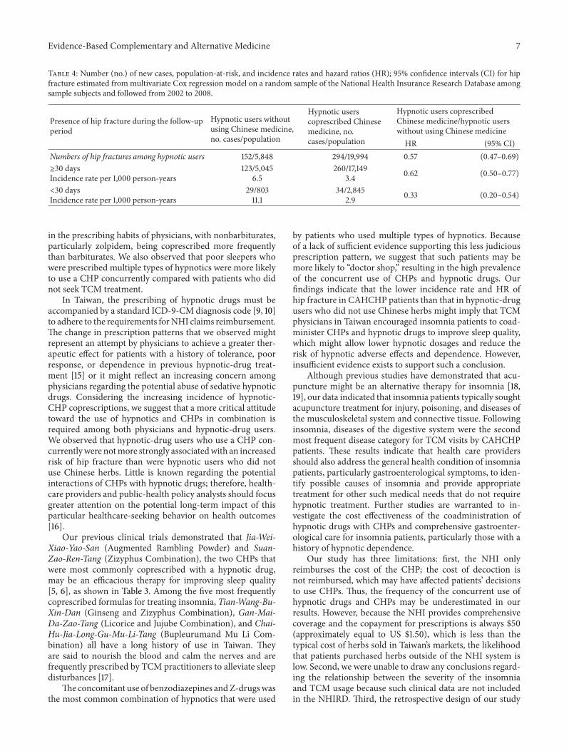
Evidence-Based Complementary and Alternative Medicine 7
Table 4: Number (no.) of new cases, population-at-risk, and incidence rates and hazard ratios (HR); 95% confidence intervals (CI) for hipfracture estimated frommultivariate Cox regression model on a random sample of the National Health Insurance Research Database amongsample subjects and followed from 2002 to 2008.
Presence of hip fracture during the follow-upperiod
Hypnotic users withoutusing Chinese medicine,no. cases/population
Hypnotic userscoprescribed Chinesemedicine, no.cases/population
Hypnotic users coprescribedChinese medicine/hypnotic userswithout using Chinese medicineHR (95% CI)
Numbers of hip fractures among hypnotic users 152/5,848 294/19,994 0.57 (0.47–0.69)≥30 daysIncidence rate per 1,000 person-years
123/5,0456.5
260/17,1493.4 0.62 (0.50–0.77)
<30 daysIncidence rate per 1,000 person-years
29/80311.1
34/2,8452.9 0.33 (0.20–0.54)
in the prescribing habits of physicians, with nonbarbiturates,particularly zolpidem, being coprescribed more frequentlythan barbiturates. We also observed that poor sleepers whowere prescribed multiple types of hypnotics were more likelyto use a CHP concurrently compared with patients who didnot seek TCM treatment.
In Taiwan, the prescribing of hypnotic drugs must beaccompanied by a standard ICD-9-CM diagnosis code [9, 10]to adhere to the requirements forNHI claims reimbursement.The change in prescription patterns that we observed mightrepresent an attempt by physicians to achieve a greater ther-apeutic effect for patients with a history of tolerance, poorresponse, or dependence in previous hypnotic-drug treat-ment [15] or it might reflect an increasing concern amongphysicians regarding the potential abuse of sedative hypnoticdrugs. Considering the increasing incidence of hypnotic-CHP coprescriptions, we suggest that a more critical attitudetoward the use of hypnotics and CHPs in combination isrequired among both physicians and hypnotic-drug users.We observed that hypnotic-drug users who use a CHP con-currentlywere notmore strongly associatedwith an increasedrisk of hip fracture than were hypnotic users who did notuse Chinese herbs. Little is known regarding the potentialinteractions of CHPs with hypnotic drugs; therefore, health-care providers and public-health policy analysts should focusgreater attention on the potential long-term impact of thisparticular healthcare-seeking behavior on health outcomes[16].
Our previous clinical trials demonstrated that Jia-Wei-Xiao-Yao-San (Augmented Rambling Powder) and Suan-Zao-Ren-Tang (Zizyphus Combination), the two CHPs thatwere most commonly coprescribed with a hypnotic drug,may be an efficacious therapy for improving sleep quality[5, 6], as shown in Table 3. Among the five most frequentlycoprescribed formulas for treating insomnia, Tian-Wang-Bu-Xin-Dan (Ginseng and Zizyphus Combination), Gan-Mai-Da-Zao-Tang (Licorice and Jujube Combination), and Chai-Hu-Jia-Long-Gu-Mu-Li-Tang (Bupleurumand Mu Li Com-bination) all have a long history of use in Taiwan. Theyare said to nourish the blood and calm the nerves and arefrequently prescribed by TCM practitioners to alleviate sleepdisturbances [17].
The concomitant use of benzodiazepines andZ-drugswasthe most common combination of hypnotics that were used
by patients who used multiple types of hypnotics. Becauseof a lack of sufficient evidence supporting this less judiciousprescription pattern, we suggest that such patients may bemore likely to “doctor shop,” resulting in the high prevalenceof the concurrent use of CHPs and hypnotic drugs. Ourfindings indicate that the lower incidence rate and HR ofhip fracture in CAHCHP patients than that in hypnotic-drugusers who did not use Chinese herbs might imply that TCMphysicians in Taiwan encouraged insomnia patients to coad-minister CHPs and hypnotic drugs to improve sleep quality,which might allow lower hypnotic dosages and reduce therisk of hypnotic adverse effects and dependence. However,insufficient evidence exists to support such a conclusion.
Although previous studies have demonstrated that acu-puncture might be an alternative therapy for insomnia [18,19], our data indicated that insomnia patients typically soughtacupuncture treatment for injury, poisoning, and diseases ofthe musculoskeletal system and connective tissue. Followinginsomnia, diseases of the digestive system were the secondmost frequent disease category for TCM visits by CAHCHPpatients. These results indicate that health care providersshould also address the general health condition of insomniapatients, particularly gastroenterological symptoms, to iden-tify possible causes of insomnia and provide appropriatetreatment for other such medical needs that do not requirehypnotic treatment. Further studies are warranted to in-vestigate the cost effectiveness of the coadministration ofhypnotic drugs with CHPs and comprehensive gastroenter-ological care for insomnia patients, particularly those with ahistory of hypnotic dependence.
Our study has three limitations: first, the NHI onlyreimburses the cost of the CHP; the cost of decoction isnot reimbursed, which may have affected patients’ decisionsto use CHPs. Thus, the frequency of the concurrent use ofhypnotic drugs and CHPs may be underestimated in ourresults. However, because the NHI provides comprehensivecoverage and the copayment for prescriptions is always $50(approximately equal to US $1.50), which is less than thetypical cost of herbs sold in Taiwan’s markets, the likelihoodthat patients purchased herbs outside of the NHI system islow. Second, we were unable to draw any conclusions regard-ing the relationship between the severity of the insomniaand TCM usage because such clinical data are not includedin the NHIRD. Third, the retrospective design of our study

8 Evidence-Based Complementary and Alternative Medicine
and the lack of a randomized placebo group might diminishthe statistical power of our findings. Thus, our results mustbe interpreted cautiously because we cannot exclude thepossibility of placebo effects.
5. Conclusion
Our findings may have implications for the treatment ofinsomnia patients. Our results suggest that, with equalavailability of conventional medical and TCM care, morethan one-third of the hypnotic-drug users usedCHPs concur-rently for the relief of sleep disturbance and gastroenterolog-ical symptoms. Recognizing the benefits of TCM, exploringpotential interactions and adverse effects, and integratingboth healthcare approaches might be beneficial to the overallhealth and quality of life of insomnia patients, particularlythose with a history of hypnotic dependence. Thus, healthcare providers should proactively explore personalized, opti-mal detoxification for hypnotic dependence while attendingto patients’ psychosocial and physical needs.
Acknowledgments
This research was conducted at the Institute of TraditionalMedicine at the School of Medicine, National Yang-MingUniversity. The authors would like to express their sinceregratitude for the support provided for this project in the formof grants from the CCMP (CCMP95-TP004) and the NHRI(NHRI-EX96-9204PP).
References
[1] 2011 National Institutes of Health Sleep Disorders ResearchPlan, “Research: NIH Sleep Disorders Research Plan,” NationalInstitutes of Health, 2013, http://www.nhlbi.nih.gov/health/prof/sleep/sleep splan.htm.
[2] P.K. Sethi andD.C.Khandelwal, “Zolpidemat supratherapeuticdoses can cause drug abuse, dependence and withdrawalseizure,” Journal of Association of Physicians of India, vol. 53, pp.139–140, 2005.
[3] C. Victorri-Vigneau, E. Dailly, G. Veyrac, and P. Jolliet, “Evi-dence of zolpidem abuse and dependence: results of the FrenchCentre for Evaluation and Information on Pharmacodepen-dence (CEIP) network survey,” British Journal of Clinical Phar-macology, vol. 64, no. 2, pp. 198–209, 2007.
[4] Y. Yuwen, N. N. Shi, L. Y. Wang, Y. M. Xie, X. J. Han,and A. P. Lu, “Development of clinical practice guidelines in11 common diseases with Chinese medicine interventions inChina,” Chinese Journal Integrative Medicine, vol. 18, no. 2, pp.112–119, 2012.
[5] C. H. Yeh, C. K. Arnold, Y. Chen, and J. N. Lai, “Suan Zao RenTang as an original treatment for sleep difficulty in climactericwomen: a prospective clinical observation,” Evidence-basedComplementary and Alternative Medicine, vol. 2011, Article ID673813, 8 pages, 2011.
[6] H. C. Wu, Y. H. Chen, J. N. Lai, J. S. Hwang, and J. D. Wang,“Improving sleep quality in climactericwomenwith insomnia: arandomized, head-to-head trial between Jia-Wei-Shiau-Yau San(JWSYS) and Suan-Zao-Ren Tang (SZRT),” European Journal ofIntegrative Medicine, vol. 3, no. 3, pp. e143–e151, 2011.
[7] L. C. Chang, N. Huang, Y. J. Chou, C. H. Lee, F. Y. Kao, and Y.Huang, “Utilization patterns of Chinese medicine and Westernmedicine under the National Health Insurance Program inTaiwan, a population-based study from 1997 to 2003,” BMCHealth Services Research, vol. 8, p. 170, 2008.
[8] S. C. Hsieh, J. N. Lai, C. F. Lee, F. C. Hu, W. L. Tseng, and J. D.Wang, “The prescribing of Chinese herbal products in Taiwan:a cross-sectional analysis of the national health insurance reim-bursement database,” Pharmacoepidemiology and Drug Safety,vol. 17, no. 6, pp. 609–619, 2008.
[9] Y. C. Lee, Y. T. Huang, Y. W. Tsai et al., “The impact of universalNational Health Insurance on population health: the experienceof Taiwan,” BMC Health Services Research, vol. 10, p. 225, 2010.
[10] N. H. R. Institutes, “National Health Insurance Researchdatabase,” 2013, http://www.nhri.org.tw/nhird/date 01.html#edn1.
[11] C. f. D. a. Prevention, “International Classification of Diseases,Ninth Revision (ICD-9),” http://www.cdc.gov/nchs/icd/icd9.htm.
[12] Department of Chinese Medicine and Pharmacy, Ministry ofHealth andWelfare, Executive Yuan, Taiwan, “List of 100 unifiedformulae,” 2013, http://www.mohw.gov.tw/CHT/DOCMAP/DM1 P.aspx?f list no=758&fod list no=4250&doc no=32620.
[13] M. Warner, L. H. Chen, and D. M. Makuc, “Increase in fatalpoisonings involving opioid analgesics in the United States,1999–2006,” NCHS Data Brief, vol. 81, no. 22, pp. 1–8, 2009.
[14] Y. H. Yang, J. N. Lai, C. H. Lee, J. D. Wang, and P. C.Chen, “Increased risk of hospitalization related tomotor vehicleaccidents among people taking zolpidem: a case-crossoverstudy,” Journal of Epidemiology, vol. 21, no. 1, pp. 37–43, 2011.
[15] M. Calem, J. Bisla, A. Begum et al., “Increased prevalence ofinsomnia and changes in hypnotics use in England over 15years: analysis of the 1993, 2000, and 2007 national psychiatricmorbidity surveys,” Sleep, vol. 35, no. 3, pp. 377–384, 2012.
[16] M. Angell and J. P. Kassirer, “Alternative medicine-the risks ofuntested and unregulated remedies,” New England Journal ofMedicine, vol. 339, no. 12, pp. 839–841, 1998.
[17] S. Hwang, F. Chen, M. Jong et al., “Prescriptions of Chi-nese herbal medicines for insomnia in Taiwan during 2002,”Evidence-based Complementary and Alternative Medicine, vol.2011, Article ID 236341, 9 pages, 2011.
[18] H. Hachul, T. K. Garcia, A. L. Maciel, F. Yagihara, S. Tufik, andL. Bittencourt, “Acupuncture improves sleep in postmenopausein a randomized, double-blind, placebo-controlled study,” Cli-macteric, vol. 16, no. 1, pp. 36–40, 2012.
[19] J. Tu,W. Chung, C. Yang, andD. Tzeng, “A comparison betweenacupuncture versus zolpidem in the treatment of primaryinsomnia,” Asian Journal of Psychiatry, vol. 5, no. 3, pp. 231–235,2012.

Hindawi Publishing CorporationEvidence-Based Complementary and Alternative MedicineVolume 2013, Article ID 697632, 9 pageshttp://dx.doi.org/10.1155/2013/697632
Research ArticleRhodiola rosea Impairs Acquisition and Expression ofConditioned Place Preference Induced by Cocaine
Federica Titomanlio,1 Carmen Manzanedo,2 Marta Rodríguez-Arias,2 Laura Mattioli,1
Marina Perfumi,1 José Miñarro,2 and María A. Aguilar2
1 Pharmacognosy Unit, School of Pharmacy, University of Camerino, Via Madonna delle Carceri 9, 62032 Camerino, Italy2 Unit of Research on Psychobiology of Drug Dependence, Department of Psychobiology, School of Psychology, University of Valencia,Avenida Blasco Ibanez 21, 46010 Valencia, Spain
Correspondence should be addressed to Marıa A. Aguilar; [email protected]
Received 26 June 2013; Revised 17 August 2013; Accepted 26 August 2013
Academic Editor: Yi-Wen Lin
Copyright © 2013 Federica Titomanlio et al. This is an open access article distributed under the Creative Commons AttributionLicense, which permits unrestricted use, distribution, and reproduction in any medium, provided the original work is properlycited.
A novel approach to the treatment of adverse effects of drugs of abuse is one which makes use of natural products. The presentstudy investigated the effect of Rhodiola rosea L. hydroalcoholic extract (RHO) on cocaine-induced hyperactivity and conditionedplace preference (CPP) in mice. In a first experiment, mice received RHO (15, 20 or 25mg/kg, IG), cocaine (25mg/kg, i.p.) (COC),or a combination of both drugs (COC + RHO15, COC + RHO20, and COC + RHO25), and their locomotor activity was evaluated.In a second experiment, the effects of RHO on the acquisition, expression, and reinstatement of cocaine CPP (induced by drugpriming or social defeat stress) were evaluated. RHO alone did not increase activity but potentiated the hyperactivity induced bycocaine. Rhodiola did not induce motivational effects by itself but attenuated the acquisition and expression of cocaine-inducedCPP. Moreover, it was found that RHO did not block reinstatement. The results indicate that RHO is effective in reducing therewarding properties of cocaine but is ineffective in preventing priming or stress-induced cocaine reinstatement. In light of thesefindings, the benefits of Rhodiola rosea L. as a treatment of cocaine addiction would seem to be limited.
1. Introduction
Cocaine addiction has become one of the most serious eco-nomic and health problems of developed societies, as it affectsa great number of individuals [1]. Development of effectivetreatments for cocaine dependence is necessary to reduce itsimpact upon both individual and society. However, currently,there are no approved medications for cocaine dependence,despite numerous studies of pharmacologic agents in bothanimal and human models [2, 3]. A novel approach to thetreatment of adverse effects of drugs of abuse is one whichmakes use of natural products. Recently, there has been anincrease in the number of preclinical models and clinical tri-als addressing the effectiveness of such agents and their activeconstituents [4]. Rhodiola rosea L. (fam. Crassulaceae) is awell-known traditional oriental medicine with adaptogenic,anxiolytic, antidepressive, and antistress properties [5–10].The positive effects of a Rhodiola rosea L. extract (RHO) inpreventing nicotine andmorphinewithdrawal symptoms and
countering the development of dependence on these drugshave recently been demonstrated in animal models [11, 12]. Arecent study demonstrated that the acquisition, expression,and reinstatement of morphine-induced conditioned placepreference (CPP) are blocked by RHO [13].These results sug-gest that RHO is capable of reducing craving and vulnerabilityto relapse and might be an effective natural remedy for thetreatment of opioid addiction [13].
The CPP paradigm has been used to evaluate the reward-ing action of drugs of abuse, including cocaine [14–16], and itcan be used as an animal model of relapse to drug-seekingbehaviour when, after extinction of CPP, exposure to drugpriming or stress induces the reinstatement of CPP [17]. Inprevious studies, we have demonstrated that cocaine inducesCPP in mice at doses between 1 and 50mg/kg [18–22], andthat cocaine priming produces reinstatement of cocaine-induced CPP [18, 19, 21, 22]. Several stressful events alsoinduce reinstatement of cocaine CPP [17, 23–25], includingsocial defeat stress [26].
2 Evidence-Based Complementary and Alternative Medicine
The present study aimed to evaluate the effects of RHOon the motor and rewarding effects of cocaine in the CPPparadigm because RHO appears to modulate the levels andactivities of biogenic monoamines, such as serotonin (5-HT),dopamine (DA), and noradrenaline (NA), in the neural path-ways involved in the regulation of addiction [6, 27–29]. Wehypothesised that the effects of cocaine can be altered byRHOadministration but in a different way to those observed withmorphine, since behavioural and neurobiological differencesbetween opiates and psychostimulants have been reported[30]. Therefore, the objective of the present work was to testthe effectiveness of RHO as a treatment for cocaine addictionin an animal model. We evaluated whether treatment withRHO altered the hyperactivity and rewarding effects ofcocaine (acquisition, expression, and priming- or stress-induced reinstatement of CPP) in mice.
2. Materials and Methods
Subjects. OF1 male mice (Charles River, Barcelona, Spain)arrived at the laboratory at 42 days of age and were housedin groups of four in plastic cages (28 cm × 28 cm × 14.5 cm)for 10 days prior to the initiation of experiments, underthe following conditions: constant temperature (21 ± 2∘C),a light schedule (white lights on: 07.30–19.30 h), and foodand water available ad libitum. Each animal was handledbriefly on the 3 days preceding the initiation of experimentsand acclimatised to intragastric administration. Proceduresinvolving mice and their care were conducted in compliancewith national, regional, and local laws and regulations, whichare in accordance with the European Communities CouncilDirectives of 24 November 1986 (86/609/EEC).
Apparatus. Locomotor activity was automatically measuredby an actimeter (CIBERTEC S.A., Spain) consisting of eightcages (33 × 15 × 13 cm), each with eight infrared lightslocated in a frame around the cage (body level of mice). Thedifferent frames are separated from each other by a distanceof 4 cm and, since they are opaque, prevent animals fromseeing cospecifics. The conditioning place preference appa-ratus consists of four identical Plexiglas place-conditioningboxes. Each box consists of two equally sized compartments(30.7 cm × 31.5 cm × 34.5 cm) separated by a gray centralarea (13.8 cm × 31.5 cm × 34.5 cm). The compartments havedifferent colouredwalls (black versuswhite) and distinct floortextures (smooth in the black compartment and rough in thewhite). Four infrared light beams in each compartment ofthe box and six in the central area allow the recording of theposition of the animal and its crossings from one compart-ment to the other. The equipment was controlled by an IBMPC computer using MONPRE 2Z software (CIBERTEC, SA,Spain).
Drugs. A dried hydroalcoholic extract from the roots ofRhodiola rosea L. (RHO) was employed for the experiments(EPO S.r.l., Milan, Italy; lot number 601252). The HPLCanalysis showed a content of 3% total rosavins, expressed asrosavin, 1% salidroside, and 0.8% tyrosol (for more details see[13]). The extract was dissolved in 1% v/v ethanol solution
and administered by gavage (IG) at doses of 15, 20, and25mg/kg/10mL. The same vehicle (1% v/v ethanol solution)was administered IG to the control groups.The doses of RHOwere selected based on previous studies in which 10, 15, and20mg/kg blocked the effects of nicotine and morphine [11–13]. In the present experiments given that a different drugof abuse was being evaluated, we employed a higher doseof RHO (25mg/kg) and did not employ 10mg/kg (the lesseffective dose in previous studies [11–13]). Cocaine chlorhy-drate (Laboratorios Alcaliber, Madrid, Spain) was dissolvedin saline (NaCl 0.9%) in a volume of 0.01 mL/g and wasadministered intraperitoneally (i.p.) at a dose of 25mg/kg.This dose was selected based on a previous study in which itconsistently induced CPP [18]. Half of this dose was used forcocaine priming (12.5mg/kg), also due to results obtained inthe aforementioned study [18].
Experiment 1. Effects of RHO on cocaine-inducedhyperactivity.
2.1. Procedure and Experimental Design. In this experiment,eight groups of mice were used (𝑛 = 8 per group). Spontane-ous motor activity was recorded for 30 minutes (habituation,0–30min). Afterwards, two groups received a vehicle, twogroups received 15mg/kg of RHO, two groups received20mg/kg of RHO, and the last two groups received 25mg/kgof RHO, and their activity was recorded for 60 minutes inblocks of 30min (31–60, 61–90min). Next, each of the twopreviously named groupswas given either saline (groupsVeh-Sal, RHO15, RHO20, RHO25) or 25mg/kg of cocaine (Veh-Coc, RHO15-Coc, RHO20-Coc, and RHO25-Coc), and theiractivity was registered for a further 60 minutes (91–120, 121–150min).
2.2. Statistical Analysis. Motor activity data registered duringthe total 150min register were analyzed using a mixed anal-ysis of variance (ANOVA) with one “between-subjects” vari-able “treatment” with eight levels (Veh-Sal, RHO15, RHO20,RHO25, Veh-Coc, RHO15 + Coc, RHO20 + Coc, andRHO25 + Coc) and a “within-subjects” variable “time” withfive levels (0–30, 31–60, 61–90, 91–120, and 121–150). All posthoc comparisons were performed with the Bonferroni mul-tiple comparisons test (corrected “alpha” 0.05/40). Calcula-tions were made using the SPSS statistical package 17.0. A 𝑃value of less than 0.05 was considered statistically significant.
Experiment 2. Effects of RHO on the acquisition, expression,and reinstatement of cocaine-induced CPP.
2.3. Procedure and Experimental Design. TheCPP procedure,unbiased in terms of initial spontaneous preference, was per-formed as described previously [18]. In short, in the first phase(preconditioning/pre-C), mice were allowed access to bothcompartments of the apparatus for 15min (900 s) per dayover 2 days. On day 3, the time spent in each compartmentduring a 900 s-period was recorded. Animals showed strongunconditioned aversion (less than 27% of the session time,i.e., 250 s) or preference (more than 73%, i.e., 650 s) for any

Evidence-Based Complementary and Alternative Medicine 3
compartment and were therefore discarded from the rest ofthe experimental procedure. In each group, half the animalsreceived the drug or physiological saline in one compartment,and the other half received it in the other. After assigning ani-mals to the compartments, an analysis of variance (ANOVA)revealed no significant differences between the time spent inthe drug-paired and vehicle-paired compartments during thepreconditioning phase.This is an important step in the exper-imental procedure that avoids any preference bias prior toconditioning. In a second phase (conditioning or acquisition)of 4 days, animals received an injection of physiological salinebefore being confined to the saline-paired compartmentfor 30 minutes, and after an interval of 4 h received aninjection of 25mg/kg of cocaine immediately before beingconfined to the drug-paired compartment for 30 minutes.In this phase, RHO or the vehicle was administered 60minprior to initiation of conditioning (i.e., 60min before thecocaine injection in the corresponding groups). Confinementwas imposed in both cases by closing the guillotine doorthat separated the two compartments. During the thirdphase (postconditioning/post-C), which took place on day 8,the guillotine door separating the two compartments wasremoved and the time spent by the untreated mice ineach compartment during a 900 s observation period wasrecorded. The difference in seconds between the time spentin the drug-paired compartment in the post-C test and thatspent in the pre-C phase is a measure of the degree of rewardinduced by the drug.
In order to evaluate the role of RHO in the acquisitionof cocaine-induced CPP, eight groups of mice (𝑛 = 11–13per group) were assessed. The control group received IGvehicle and one IP injection of saline (Veh+ Sal), three groupsreceived RHO 15, 20 or 25mg/kg plus saline (RHO15, RHO20, and RHO 25), one group received vehicle plus 25mg/kgof cocaine (Veh + Coc), and the last three groups receivedRHO 15, 20, or 25mg/kg plus 25mg/kg of cocaine (RHO15 +Coc, RHO20 + Coc, and RHO25 + Coc).The vehicle or RHOwas administered IG 60min before cocaine or saline, whichwas administered IP immediately prior to four conditioningsessions. The interval of 60min between RHO and cocainewas based on experience gained in previous studies [13], aswas the absence of any interval between cocaine injection andexposure to the drug-paired compartment [18].
In order to evaluate the role of RHO in the expressionof cocaine-induced CPP, four groups of mice (𝑛 = 9-10 pergroup) were conditioned with 25mg/kg of cocaine andreceived the vehicle (Veh) or RHO 15, 20, or 25mg/kg(RHO15, RHO20, and RHO25) IG 60 minutes before post-conditioning test in the colony room.
To evaluate the effects of RHO on reinstatement of theCPP induced by re-exposure to cocaine or social defeat, wefollowed the procedures described previously ([18, 31], resp.).Six groups of mice (𝑛 = 10–13) were conditioned with25mg/kg of cocaine and after the post-C test, animals under-went an extinction session every 72 hours which consisted ofplacing the animals in the apparatus (without the guillotinedoors separating the compartments) for 15 min. No drugswere administered during these sessions. The criterion forconsidering the preference extinguishedwas lack of statistical
significance (according to the Student’s 𝑡-test) between thetime spent by the animals of a given group in the drug-pairedcompartment in the extinction session and in the pre-C ses-sion.Thismeasure was repeated 24 h later in order to confirmthe extinction. After 24 hours of confirmation to extinction,the reinstating effects of cocaine or social stress (alone orwith RHO) were evaluated. Reinstatement tests were thesame as those for post-C (free ambulation for 15min), exceptthat the animals were tested after administrating 12.5mg/kgof cocaine or inflicting social defeat. Three groups received12.5mg/kg of cocaine in the colony room (a neutral placenot previously associated with cocaine) 15min before thereinstatement tests. The first group received cocaine plusvehicle (Coc), and the other two groups received cocaineplus 15 or 20mg/kg of RHO (Coc + RHO15, Coc + RHO20),respectively (vehicle or RHO was given IG 60min before thereinstatement test, i.e., 45min prior to the cocaine IP injec-tion).The other three groups ofmice, exposed to social defeat(SD), received vehicle, 15, or 20mg/kg of RHO (SD, SD +RHO15 and SD+RHO20) IG60min before the reinstatementtest, performed immediately after SD. The social stress wasperformed following the procedure described by Ribeiro DoCouto et al. [31]. It took place in a different room andconsisted of a 10-min agonistic encounter (and 1min ofexploration) in a neutral transparent plastic cage (23 × 13.5× 13 cm) with a defeat result for the experimental mouse.Each experimental mouse was confronted with an aggressiveopponent (of equal age and body weight) with previousfighting experience and had been shown to have a high levelof aggression in previous screening. This procedure can beconsidered a type of social stress (see [31, 32]). Experimen-tal mice presented avoidance/flee and defensive/submissivebehaviours after suffering aggression (threat and attack) froman opponent. The criterion used to define an animal asdefeated was the adopting of a specific posture of defeat, char-acterized by an upright submissive position, limp forepaws,upwardly angled head, and retracted ears [32, 33]. Someanimals excluded from the CPP procedure (𝑛 = 24) wereused as aggressive opponents.They were housed individually,in isolation, in plastic cages (23 × 13.5 × 13 cm) for a monthbefore experiments to induce heightened aggression [32]. Alldefeated mice experienced similar levels of aggression as theopponent displayed attack behaviour as soon as it saw theexperimental mouse (latency < 30 s).
2.4. Statistical Analysis. Data of the acquisition (the timespent in the drug-paired compartment) were analysed witha mixed ANOVA, with “treatment” as a “between-subjects”variablewith eight levels (Veh+ Sal, RHO15, RHO20, RHO25,RHO15 + Coc, RHO20 + Coc, RHO25 + Coc) and “days”as a “within-subjects” variable with two levels (pre-C andpost-C). Data of the expression were analysed with thesame ANOVA, but “treatment” variable has four levels (Veh,RHO15, RHO20, RHO25). Data of the reinstatement wereanalysed with the same ANOVA but, “treatment” has sixlevels (Coc, Coc + RHO15, Coc + RHO20, SD, SD + RHO15and SD + RHO20) and “days” four levels (pre-C, post-C,extinction and reinstatement).

4 Evidence-Based Complementary and Alternative Medicine
0
1000
2000
3000
4000
5000
6000
7000
8000Ac
tivity
coun
ts
Veh-salCoc RHO15
RHO20RHO25
Rhodiola
Cocaine
0–30 31–60 61–90 91–120 121–150Time
RHO15+ CocRHO20 + Coc
RHO25 + Coc
(a)
0100020003000400050006000700080009000
Activ
ity co
unts
Veh-salCoc RHO15
RHO20RHO25
91–120 121–150Time
RHO15+ CocRHO20 + Coc
RHO25 + Coc
∗∗∗∗∗
∗∗
∗∗∗∗
(b)Figure 1: Effects of RHO on cocaine-induced hyperactivity. Mice (𝑛 = 8 per group) were placed in the actimeter for a 30-min adaptationperiod (0–30min). Afterwards, two groups received IG vehicle (Veh-sal and Coc), two groups received IG RHO 15mg/kg (RHO15 + Coc,RHO15), two groups RHO 20mg/kg (RHO20 + Coc, RHO20), and two groups RHO 25mg/kg (RHO25 + Coc, RHO25), and their motoractivity was registered over another hour (31–60, 61–90min). Finally, the first two groups received a IP injection of saline (Veh-Sal) or cocaine25mg/kg (Coc), and the groups treated with RHO received an IP injection of cocaine 25mg/kg (RHO15 + Coc, RHO20 + Coc, and RHO25 +Coc) or saline (RHO 15, RHO20, andRHO25), and theirmotor activitywas registered over a further hour (91–120, 121–150min). (a) representsthe motor activity of all groups over the complete time of testing (0–150min) and shows the time of RHO and cocaine administration in thecorresponding groups. (b) represents the data for the last hour of the test (after cocaine administration to the corresponding groups). Valuesare means ± SEM. ∗𝑃 < 0.05; ∗∗𝑃 < 0.01; significant difference with respect to the values of the control (Veh-sal) group at the same time test.
All post hoc comparisons were performed with the Bon-ferroni multiple comparisons test (corrected “alpha” of 0.05/16 for acquisition data, 0.05/6 for expression data, and 0.05/24for reinstatement data). Calculations were performed usingthe SPSS statistical package 17.0. A 𝑃 value of less than 0.05was considered statistically significant.
3. Results
Experiment 1. The results regarding the effect of RHO oncocaine-induced hyperactivity are represented in Figure 1.The ANOVA showed a significant effect of the variable time(𝐹(4, 224) = 43.358; 𝑃 < 0.001), treatment (𝐹(7, 56) =3.674; 𝑃 < 0.002), and the interaction time × treatment(𝐹(28, 224) = 8.145; 𝑃 < 0.001). The Bonferroni post hoccomparison showed that cocaine increased motor activityin comparison with the vehicle during the 30min after itsadministration (𝑃 < 0.05). The groups treated with cocaineplus RHO also showed an increase in activity with respect tocontrols (𝑃 < 0.001), and the groups RHO15 + Coc andRHO20 + Coc presented more activity than the groupsRHO15 and RHO20, respectively (𝑃 < 0.05 and 𝑃 < 0.001).Between 31 and 60min after cocaine administration, only thegroups treatedwith RHO20 +Coc and RHO25 +Coc showedan increase in activity in comparison to controls (𝑃 < 0.001),and the group RHO20 + Coc also displayed more activitythan RHO20 group (𝑃 < 0.001).
Experiment 2. The results regarding the effect of RHO onacquisition of cocaine-induced CPP are represented in
Figure 2. The ANOVA revealed a significant effect of thevariable days (𝐹(1, 88) = 7.240; 𝑃 < 0.01) and the interactiontreatment × days (𝐹(7, 88) = 4.210; 𝑃 < 0.001). Bonferronipost hoc comparisons revealed that the groups Veh + Coc,RHO15 + Coc, and RHO25 + Coc spent more time in thedrug-paired compartment in post-C than in pre-C (𝑃 < 0.05,𝑃 < 0.001 and 𝑃 < 0.01, resp.). Thus, at the doses used, RHOdid not exert motivational effects, and only that of 20mg/kgwas capable of impairing the acquisition of cocaine-inducedCPP.
The effects of RHO on the expression of cocaine-inducedCPP are represented in Figure 3. The ANOVA revealed asignificant effect of the variable days (𝐹(1, 35) = 33.264; 𝑃 <0.001) and the interaction treatment× days (𝐹(3, 35) = 3.378;𝑃 < 0.05). Bonferroni post hoc comparisons revealed that thegroups receiving vehicle or RHO 20 or 25mg/kg spent moretime in the drug-paired compartment in post-C than in pre-C (𝑃 < 0.001, 𝑃 < 0.001, and 𝑃 < 0.005, resp.). In this way,only the dose of 15mg/kg of RHO blocked the expression ofcocaine-induced CPP.
The effects of RHO on the priming- and social defeat-induced reinstatement of cocaine CPP are represented inFigure 4.TheANOVA revealed a significant effect of the vari-able days (𝐹(3, 59) = 26.101; 𝑃 < 0.001). Bonferroni post hoccomparisons revealed that more time was spent in the drug-paired compartment during post-C and the reinstatementtest than during pre-C or extinction. The variables treatmentand interaction were not significant. These results showthat RHO does not block the reinstatement of cocaine-CPPinduced by priming or stress.
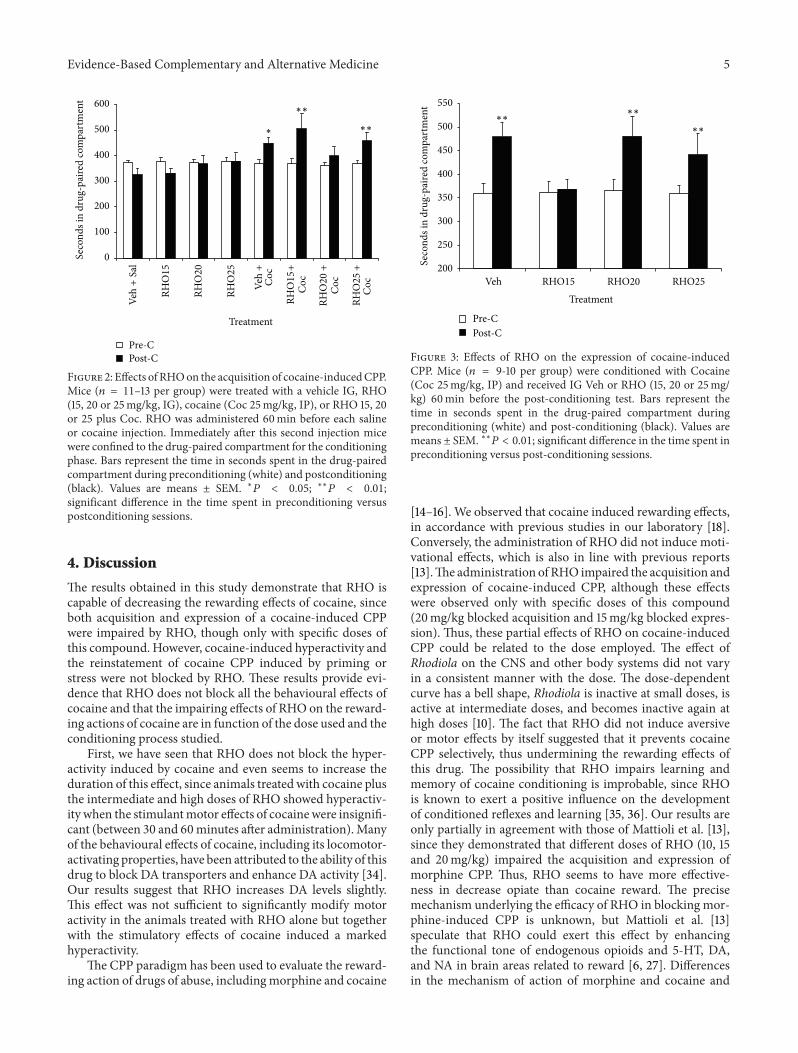
Evidence-Based Complementary and Alternative Medicine 5
0
100
200
300
400
500
600
Pre-CPost-C
Veh+
Sal
RHO
15
RHO
20
RHO
25
Veh+
Coc
RHO
15+
Coc
RHO
20+
Coc
RHO
25+
Coc
Treatment
Seco
nds i
n dr
ug-p
aire
d co
mpa
rtm
ent
∗ ∗∗
∗∗
Figure 2: Effects of RHOon the acquisition of cocaine-inducedCPP.Mice (𝑛 = 11–13 per group) were treated with a vehicle IG, RHO(15, 20 or 25mg/kg, IG), cocaine (Coc 25mg/kg, IP), or RHO 15, 20or 25 plus Coc. RHO was administered 60min before each salineor cocaine injection. Immediately after this second injection micewere confined to the drug-paired compartment for the conditioningphase. Bars represent the time in seconds spent in the drug-pairedcompartment during preconditioning (white) and postconditioning(black). Values are means ± SEM. ∗𝑃 < 0.05; ∗∗𝑃 < 0.01;significant difference in the time spent in preconditioning versuspostconditioning sessions.
4. Discussion
The results obtained in this study demonstrate that RHO iscapable of decreasing the rewarding effects of cocaine, sinceboth acquisition and expression of a cocaine-induced CPPwere impaired by RHO, though only with specific doses ofthis compound. However, cocaine-induced hyperactivity andthe reinstatement of cocaine CPP induced by priming orstress were not blocked by RHO. These results provide evi-dence that RHO does not block all the behavioural effects ofcocaine and that the impairing effects of RHO on the reward-ing actions of cocaine are in function of the dose used and theconditioning process studied.
First, we have seen that RHO does not block the hyper-activity induced by cocaine and even seems to increase theduration of this effect, since animals treated with cocaine plusthe intermediate and high doses of RHO showed hyperactiv-ity when the stimulantmotor effects of cocainewere insignifi-cant (between 30 and 60minutes after administration).Manyof the behavioural effects of cocaine, including its locomotor-activating properties, have been attributed to the ability of thisdrug to block DA transporters and enhance DA activity [34].Our results suggest that RHO increases DA levels slightly.This effect was not sufficient to significantly modify motoractivity in the animals treated with RHO alone but togetherwith the stimulatory effects of cocaine induced a markedhyperactivity.
The CPP paradigm has been used to evaluate the reward-ing action of drugs of abuse, includingmorphine and cocaine
200
250
300
350
400
450
500
550
Veh RHO15 RHO20 RHO25Treatment
Pre-CPost-C
Seco
nds i
n dr
ug-p
aire
d co
mpa
rtm
ent
∗∗
∗∗∗∗
Figure 3: Effects of RHO on the expression of cocaine-inducedCPP. Mice (𝑛 = 9-10 per group) were conditioned with Cocaine(Coc 25mg/kg, IP) and received IG Veh or RHO (15, 20 or 25mg/kg) 60min before the post-conditioning test. Bars represent thetime in seconds spent in the drug-paired compartment duringpreconditioning (white) and post-conditioning (black). Values aremeans ± SEM. ∗∗𝑃 < 0.01; significant difference in the time spent inpreconditioning versus post-conditioning sessions.
[14–16]. We observed that cocaine induced rewarding effects,in accordance with previous studies in our laboratory [18].Conversely, the administration of RHO did not induce moti-vational effects, which is also in line with previous reports[13].The administration of RHO impaired the acquisition andexpression of cocaine-induced CPP, although these effectswere observed only with specific doses of this compound(20mg/kg blocked acquisition and 15mg/kg blocked expres-sion). Thus, these partial effects of RHO on cocaine-inducedCPP could be related to the dose employed. The effect ofRhodiola on the CNS and other body systems did not varyin a consistent manner with the dose. The dose-dependentcurve has a bell shape, Rhodiola is inactive at small doses, isactive at intermediate doses, and becomes inactive again athigh doses [10]. The fact that RHO did not induce aversiveor motor effects by itself suggested that it prevents cocaineCPP selectively, thus undermining the rewarding effects ofthis drug. The possibility that RHO impairs learning andmemory of cocaine conditioning is improbable, since RHOis known to exert a positive influence on the developmentof conditioned reflexes and learning [35, 36]. Our results areonly partially in agreement with those of Mattioli et al. [13],since they demonstrated that different doses of RHO (10, 15and 20mg/kg) impaired the acquisition and expression ofmorphine CPP. Thus, RHO seems to have more effective-ness in decrease opiate than cocaine reward. The precisemechanism underlying the efficacy of RHO in blocking mor-phine-induced CPP is unknown, but Mattioli et al. [13]speculate that RHO could exert this effect by enhancingthe functional tone of endogenous opioids and 5-HT, DA,and NA in brain areas related to reward [6, 27]. Differencesin the mechanism of action of morphine and cocaine and

6 Evidence-Based Complementary and Alternative Medicine
0
100
200
300
400
500
600
700
Treatment
Pre-CPost-C
ExtinctionReinstatement
Coc Coc +RHO15
Coc +RHO20
SD SD +RHO15
SD +RHO20
Seco
nds i
n dr
ug-p
aire
d co
mpa
rtm
ent
∗∗
∗∗
∗∗
∗∗
∗∗
∗∗
∗∗
∗∗
∗∗
∗∗∗∗
∗∗
Figure 4: Effects of RHO on the priming- and stress-inducedreinstatement of cocaine-induced CPP. Six groups of mice (𝑛 = 10–13 for group) were conditioned with cocaine (Coc 25mg/kg, IP),underwent daily extinction sessions until the CPPwas extinguished,and received the following treatments before the reinstatement test:vehicle, RHO 15, or 20mg/kg 45min before 12.5mg/kg IP cocainepriming (Coc, Coc + RHO15, Coc + RHO20); vehicle, RHO 15, or20mg/kg 45min before social defeat exposure (SD, SD + RHO15,SD + RHO20). Bars represent the time in seconds spent in thedrug-paired compartment before conditioning sessions in the pre-C test (white bars), after conditioning sessions in the post-C test(black bars), in the last extinction session (light gray bars), and inthe reinstatement test (dark gray bars). ∗∗𝑃 < 0.001, significantdifference in the time spent in the drug-paired compartment inpreconditioning versus post-conditioning or reinstatement test.
in the behavioural effects of these drugs [30] can explainwhy RHO is less effective to reduce cocaine reward. Cocaineis a psychomotor stimulant that facilitates monoaminergicneurotransmission by binding to DA, 5-HT, and NA trans-porters (DAT, SERT, and NAT) and inhibiting the reuptakeof these monoamines. Cocaine produces reward throughsimultaneous actions at more than one protein site [34, 37].For example, only DAT/SERT knockout mice exhibit impair-ment of cocaine CPP [37] and the contribution of DA, 5-HT,and NA transporters to the rewarding, and aversive effectsof cocaine seem to vary [34]. Activation of the mesolimbicdopaminergic reward circuitry has been proposed as amodality in the long-term treatment of reward deficiencysyndrome and drug addiction disorders [38]. In addition tothe abovementioned dose problem, the fact that RHO impairsacquisition or expression of cocaine CPP only at specificdoses could be related to the effects that this compoundinduces monoamines (DA, 5-HT, and NA) and differentkinds of monoaminergic precursors, receptors, transporters,or degradation enzymes. In such circumstances, impairment
of acquisition/expression of cocaine CPP would be inducedonly by doses of RHO that produce an effective combinationof alterations in the various monoamine proteins involved inthe acquisition and/or expression of the rewarding effects ofcocaine.
Although the positive reinforcing is the main factor inthe acquisition of a drug habit, relapse is the overridingcharacteristic of addiction and the foremost challenge tothe treatment of drug addiction. The CPP paradigm canbe used to model relapse in rodents, and re-exposure todrug or stress after extinction can trigger reinstatement ofCPP [17]. We observed that cocaine CPP is reinstated bycocaine priming and social defeat stress in accordance withprevious studies [18, 26]. However, the administration ofRHOdid not block priming- or stress-induced reinstatement.These results contrast with those obtained by Mattioli et al.[13] who reported that RHO prevents the reinstatement ofmorphine CPP induced by drug re-exposure and restraintstress. Again, these divergent results could be related to thedifferent mechanisms underlying the reinstatement of CPPinduced by morphine or cocaine. It is improbable that theincrease in monoaminergic activity induced by RHO blockspriming-induced reinstatement of cocaine CPP since it hasbeen reported that administration of the DA D1 agonist SKF81297 [39] and the DA enhancer modafinil [40] induces thereinstatement of cocaine CPP. On the other hand, the lack ofeffects of RHO on stress-induced reinstatement of cocaineCPP is surprising, since this compound exerts antistresseffects and interactswith the hypothalamus-pituitary-adrenal(HPA) system, reducing cortisol levels [7–9]. However, it hasbeen reported that corticosterone plays at most a permissiverole in the facilitating effects of social stress on cocaineself-administration in the rat [41] and that stress-inducedreinstatement of cocaine self-administration appears to beindependent of corticosterone [42]. Alternatively, it is possi-ble that a single administration of an acute dose of RHOpriorto reinstatement tests is insufficient to decrease to attenuatethe reinstatement of cocaine CPP.
The doses employed in the present study are in the rangeof those administered to humans for therapeutic purposes,although clinical studies have tended to use higher doses[43, 44]. According to the calculating method described byReagan-Shaw et al. [45], in a human weighing 60 kg, 15mg/kg of RHO corresponds with 72.9mg, 20mg/kg corre-sponds with 97.2mg, and 25mg/kg corresponds with 120mg.Extracts of RHO have already been used in humans withoutsevere adverse consequences, which endorses the safety of thepreparations in question [5, 9, 35, 43, 44, 46–48]. Only a fewmild adverse events have been reported, including headacheor hypersalivation [43]. Moreover, a lack of interaction withother drugs (warfarin and theophylline) has been observed[49]. However, it remains to be determined whether or notRHO interacts with cocaine or other drugs in clinical settings.
Presently, there are no FDA-approved therapies or medi-cations for treating cocaine addiction [2, 3] that can be used asa positive control in the evaluation of RHO’s effects. However,there are recent reports of the positive effects of derivatives ofgenera Stephania and Corydalis in the treatment of cocaineaddiction in animal models. Levo-tetrahydropalmatine has

Evidence-Based Complementary and Alternative Medicine 7
been shown to attenuate cocaine self-administration [50] andreinstatement of extinguished cocaine seeking by cocaine,stress, or drug-associated cues in rats [50, 51]. In light of thisevidence, levo-tetrahydropalmatine has been suggested as apotential new medication for the treatment of cocaine addic-tion [52].
The main limitation of our work lies in the fact that RHOwas administered over a short period of a few days (in fouracquisition sessions) or acutely (in expression and reinstate-ment tests). It is likely that more pronounced effects of RHOwould be observed with a longer period of administration(e.g., during the extinction period). Another limitation of ourapproach is the use of a single animal model to evaluate therewarding effects of cocaine. The CPP paradigm used in thepresent study measures the conditioned rewarding effects ofthe drug while the drug self-administration paradigm mea-sures the primary hedonic properties of the drug [17]. Futureresearch should evaluate the efficacy of RHO in reducing theacquisition of cocaine self-administration and reinstatementof drug seeking after exposure to cocaine priming, drug-conditioned cues, or stress.
In conclusion, we can report that RHO impairs therewarding effects of cocaine, although its effects on acquisi-tion and expression of CPP depend on the dose used.The factthat RHOdoes not affect priming- or drug-induced reinstate-ment of cocaine CPP limits its possible usefulness as a naturaltreatment for cocaine dependence. Nevertheless, though thediscovery of an effective therapy that addresses all aspects ofcocaine addiction continues to elude researchers, the anti-craving effect of RHO shows potential as a component ofcombined therapy.
Conflict of Interests
The authors declare that there is no conflict of interests.
Acknowledgments
This research was funded by the following grants: Ministeriode Economıa y Competitividad, Direccion General de Inves-tigacion (PSI2011-24762), Instituto de Salud “Carlos III” (FIS),RETICS, Red de Trastornos Adictivos (RD06/001/0016), andGeneralitat Valenciana, Conselleria de Educacion (PROME-TEO/2009/072), Spain.
References
[1] UNODC,WorldDrug Report 2011, UnitedNations Publication,Sales No. E.11.XI.10.
[2] D. Shorter and T. R. Kosten, “Novel pharmacotherapeutictreatments for cocaine addiction,” BMCMedicine, vol. 9, article119, 2011.
[3] L. Somaini, C. Donnini, M. A. Raggi et al., “Promising medica-tions for cocaine dependence treatment,”Recent Patents on CNSDrug Discovery, vol. 6, no. 2, pp. 146–160, 2011.
[4] L. Lu, Y. Liu, W. Zhu et al., “Traditional medicine in the treat-ment of drug addiction,” American Journal of Drug and AlcoholAbuse, vol. 35, no. 1, pp. 1–11, 2009.
[5] D. Edwards, A. Heufelder, and A. Zimmermann, “Therapeuticeffects and safety of Rhodiola rosea extract WS 1375 in subjects
with life-stress symptoms—results of an open-label study,”Phytotherapy Research, vol. 26, no. 8, pp. 1220–1225, 2012.
[6] G. S. Kelly, “Rhodiola rosea: a possible plant adaptogen,” Alter-native Medicine Review, vol. 6, no. 3, pp. 293–302, 2001.
[7] L. Mattioli and M. Perfumi, “Rhodiola rosea L. extract reducesstress- and CRF-induced anorexia in rats,” Journal of Psycho-pharmacology, vol. 21, no. 7, pp. 742–750, 2007.
[8] L. Mattioli, C. Funari, and M. Perfumi, “Effects of Rhodiolarosea L. extract on behavioural and physiological alterationsinduced by chronicmild stress in female rats,” Journal of Psycho-pharmacology, vol. 23, no. 2, pp. 130–142, 2009.
[9] A. Panossian, G. Wikman, and J. Sarris, “Rosenroot (Rhodiolarosea): traditional use, chemical composition, pharmacologyand clinical efficacy,” Phytomedicine, vol. 17, no. 7, pp. 481–493,2010.
[10] M. Perfumi and L. Mattioli, “Adaptogenic and central nervoussystem effects of single doses of 3% rosavin and 1% salidrosideRhodiola rosea L. extract in mice,” Phytotherapy Research, vol.21, no. 1, pp. 37–43, 2007.
[11] L. Mattioli and M. Perfumi, “Evaluation of Rhodiola rosea L.extract on affective and physical signs of nicotine withdrawal inmice,” Journal of Psychopharmacology, vol. 25, no. 3, pp. 402–410, 2011.
[12] L. Mattioli and M. Perfumi, “Effects of a Rhodiola rosea L.extract on acquisition and expression of morphine toleranceand dependence in mice,” Journal of Psychopharmacology, vol.25, no. 3, pp. 411–420, 2011.
[13] L.Mattioli, F. Titomanlio, andM. Perfumi, “Effects of aRhodiolarosea L. extract on the acquisition, expression, extinction, andreinstatement of morphine-induced conditioned place prefer-ence in mice,” Psychopharmacology, vol. 221, no. 2, pp. 183–193,2012.
[14] M. T. Bardo, J. K. Rowlett, and M. J. Harris, “Conditioned placepreference using opiate and stimulant drugs: a meta- analysis,”Neuroscience and Biobehavioral Reviews, vol. 19, no. 1, pp. 39–51,1995.
[15] T. M. Tzschentke, “Measuring reward with the conditionedplace preference paradigm: a comprehensive review of drugeffects, recent progress and new issues,” Progress in Neurobiol-ogy, vol. 56, no. 6, pp. 613–672, 1998.
[16] T. M. Tzschentke, “Measuring reward with the conditionedplace preference (CPP) paradigm: update of the last decade,”Addiction Biology, vol. 12, no. 3-4, pp. 227–462, 2007.
[17] M. A. Aguilar, M. Rodrıguez-Arias, and J. Minarro, “Neurobi-ological mechanisms of the reinstatement of drug-conditionedplace preference,”Brain Research Reviews, vol. 59, no. 2, pp. 253–277, 2009.
[18] C. Maldonado, M. Rodrıguez-Arias, A. Castillo, M. A. Aguilar,and J. Minarro, “Gamma-hydroxybutyric acid affects the acqui-sition and reinstatement of cocaine-induced conditioned placepreference inmice,”Behavioural Pharmacology, vol. 17, no. 2, pp.119–131, 2006.
[19] C. Maldonado, M. Rodrıguez-Arias, A. Castillo, M. A. Aguilar,and J. Minarro, “Effect of memantine and CNQX in the acqui-sition, expression and reinstatement of cocaine-induced condi-tioned place preference,”Progress inNeuro-Psychopharmacologyand Biological Psychiatry, vol. 31, no. 4, pp. 932–939, 2007.
[20] C. Manzanedo, M. P. Garcıa-Pardo, M. Rodrıguez-Arias, J.Minarro, and M. A. Aguilar, “Pre-treatment with high dosesof cocaine decreases the reinforcing effects of cocaine in theconditioned place preference paradigm,” Neuroscience Letters,vol. 516, no. 1, pp. 29–33, 2012.
8 Evidence-Based Complementary and Alternative Medicine
[21] B. Ribeiro Do Couto, M. A. Aguilar, J. Lluch, M. Rodrıguez-Arias, and J. Minarro, “Social experiences affect reinstatementof cocaine-induced place preference in mice,” Psychopharma-cology, vol. 207, no. 3, pp. 485–498, 2009.
[22] M. Rodrıguez-Arias, A. Castillo, M. Daza-Losada, M. A. Agui-lar, and J. Minarro, “Effects of extended cocaine conditioning inthe reinstatement of place preference,” Physiology and Behavior,vol. 96, no. 4-5, pp. 620–630, 2009.
[23] J. R. Mantsch, A.Weyer, O. Vranjkovic, C. E. Beyer, D. A. Baker,and H. Caretta, “Involvement of noradrenergic neurotransmis-sion in the stress- but not cocaine-induced reinstatement ofextinguished cocaine-induced conditioned place preference inmice: role for 𝛽-2 adrenergic receptors,” Neuropsychopharma-cology, vol. 35, no. 11, pp. 2165–2178, 2010.
[24] L. K. Vaughn, J. R. Mantsch, O. Vranjkovic et al., “Cannabinoidreceptor involvement in stress-induced cocaine reinstatement:potential interaction with noradrenergic pathways,” Neuro-science, vol. 204, pp. 117–124, 2012.
[25] O. Vranjkovic, S. Hang, D. A. Baker, and J. R. Mantsch, “Betaadrenergic receptor mediation of stress-induced reinstatementof extinguished cocaine-induced conditioned place preferencein mice: roles for beta-1 and beta-2 adrenergic receptors,” Jour-nal of Pharmacology and Experimental Therapeutics, vol. 342,no. 2, pp. 541–551, 2012.
[26] B. B. Land, M. R. Bruchas, S. Schattauer et al., “Activation of thekappa opioid receptor in the dorsal raphe nucleus mediates theaversive effects of stress and reinstates drug seeking,” Proceed-ings of the National Academy of Sciences of the United States ofAmerica, vol. 106, no. 45, pp. 19168–19173, 2009.
[27] Q. G. Chen, Y. S. Zeng, Z. Q. Qu et al., “The effects of Rhodiolarosea extract on 5-HT level, cell proliferation and quantity ofneurons at cerebral hippocampus of depressive rats,” Phytomed-icine, vol. 16, no. 9, pp. 830–838, 2009.
[28] C. German, Z. Ramazanov, and M. Bernard Suarez, Artic Root(Rodiola rosea): The Powerful Ginseng Alternative, KensingtonPublishing, New York, NY, USA, 1999.
[29] D. van Diermen, A. Marston, J. Bravo, M. Reist, P.-A. Carrupt,and K. Hostettmann, “Monoamine oxidase inhibition by Rho-diola rosea L. roots,” Journal of Ethnopharmacology, vol. 122, no.2, pp. 397–401, 2009.
[30] A. Badiani, D. Belin, D. Epstein, D. Calu, and Y. Shaham, “Opi-ate versus psychostimulant addiction: the differences do mat-ter,” Nature Reviews Neuroscience, vol. 12, no. 11, pp. 685–700,2011.
[31] B. Ribeiro Do Couto, M. A. Aguilar, C. Manzanedo, M. Rod-rıguez-Arias, A. Armario, and J. Minarro, “Social stress is aseffective as physical stress in reinstating morphine-inducedplace preference in mice,” Psychopharmacology, vol. 185, no. 4,pp. 459–470, 2006.
[32] M. Rodrıguez-Arias, J. Minarro, M. A. Aguilar, J. Pinazo, and V.M. Simon, “Effects of risperidone and SCH 23390 on isolation-induced aggression in male mice,” European Neuropsychophar-macology, vol. 8, no. 2, pp. 95–103, 1998.
[33] K. A. Miczek, M. L. Thompson, and L. Shuster, “Opioid-likeanalgesia in defeated mice,” Science, vol. 215, no. 4539, pp. 1520–1522, 1982.
[34] G. R. Uhl, F. S. Hall, and I. Sora, “Cocaine, reward, movementand monoamine transporters,” Molecular Psychiatry, vol. 7, no.1, pp. 21–26, 2002.
[35] V. Darbinyan, A. Kteyan, A. Panossian, E. Gabrielian, G.Wikman, and H. Wagner, “Rhodiola rosea in stress induced
fatigue—a double blind cross-over study of a standardizedextract SHR-5 with a repeated low-dose regimen on the mentalperformance of healthy physicians during night duty,” Phy-tomedicine, vol. 7, no. 5, pp. 365–371, 2000.
[36] Z. Q. Qu, Y. Zhou, Y. S. Zeng, Y. Li, and P. Chung, “Pretreat-ment with Rhodiola rosea extract reduces cognitive impair-ment induced by intracerebroventricular streptozotocin in rats:implication of anti-oxidative and neuroprotective effects,” Bio-medical and Environmental Sciences, vol. 22, no. 4, pp. 318–326,2009.
[37] I. Sora, F. S. Hall, A. M. Andrews et al., “Molecular mechanismsof cocaine reward: combined dopamine and serotonin trans-porter knockouts eliminate cocaine place preference,” Proceed-ings of the National Academy of Sciences of the United States ofAmerica, vol. 98, no. 9, pp. 5300–5305, 2001.
[38] K. Blum, A. L. Chuan Chen, T. J. H. Chen et al., “Activationinstead of blocking mesolimbic dopaminergic reward circuitryis a preferred modality in the long term treatment of rewarddeficiency syndrome (RDS): a commentary,”Theoretical Biologyand Medical Modelling, vol. 5, article no. 24, 2008.
[39] D. L. Graham, R. Hoppenot, A. Hendryx, and D. W. Self, “Dif-ferential ability of D1 and D2 dopamine receptor agonists toinduce and modulate expression and reinstatement of cocaineplace preference in rats,” Psychopharmacology, vol. 191, no. 3, pp.719–730, 2007.
[40] R. E. Bernardi, J. R. Lewis, K. M. Lattal, and S. P. Berger,“Modafinil reinstates a cocaine conditioned place preferencefollowing extinction in rats,” Behavioural Brain Research, vol.204, no. 1, pp. 250–253, 2009.
[41] H. E. Covington III and K. A. Miczek, “Intense cocaine self-administration after episodic social defeat stress, but not afteraggressive behavior: dissociation from corticosterone activa-tion,” Psychopharmacology, vol. 183, no. 3, pp. 331–340, 2005.
[42] U. Shalev,M.Marinelli,M.H. Baumann, P.V. Piazza, andY. Sha-ham, “The role of corticosterone in food deprivation-inducedreinstatement of cocaine seeking in the rat,” Psychopharmacol-ogy, vol. 168, no. 1-2, pp. 170–176, 2003.
[43] S. K. Hung, R. Perry, and E. Ernst, “The effectiveness and ef-ficacy of Rhodiola rosea L.: a systematic review of randomizedclinical trials,” Phytomedicine, vol. 18, no. 4, pp. 235–244, 2011.
[44] S. Ishaque, L. Shamseer, C. Bukutu, and S. Vohra, “Rhodiolarosea for physical andmental fatigue: a systematic review,” BMCComplementary and Alternative Medicine, vol. 12, article 70,2012.
[45] S. Reagan-Shaw, M. Nihal, and N. Ahmad, “Dose translationfrom animal to human studies revisited,” The FASEB Journal,vol. 22, no. 3, pp. 659–661, 2008.
[46] E. M. G. Olsson, B. von Scheele, and A. G. Panossian, “Arandomised, double-blind, placebo-controlled, parallel-groupstudy of the standardised extract SHR-5 of the roots of Rhodiolarosea in the treatment of subjects with stress-related fatigue,”Planta Medica, vol. 75, no. 2, pp. 105–112, 2009.
[47] V. Darbinyan, G. Aslanyan, E. Amroyan, E. Gabrielyan, C.Malmstrom, and A. Panossian, “Clinical trial of Rhodiola roseaL. extract SHR-5 in the treatment of mild to moderate depres-sion,” Nordic Journal of Psychiatry, vol. 61, no. 5, pp. 343–348,2007, Erratum in Nordic Journal of Psychiatry, vol. 61, no. 6, pp.503, 2007.
[48] V. Fintelmann and J. Gruenwald, “Efficacy and tolerability ofa Rhodiola rosea extract in adults with physical and cognitivedeficiencies,” Advances in Therapy, vol. 24, no. 4, pp. 929–939,2007.
Evidence-Based Complementary and Alternative Medicine 9
[49] A. Panossian,A.Hovhannisyan,H.Abrahamyan, E.Gabrielyan,and G. Wikman, “Pharmacokinetic and pharmacodynamicstudy of interaction of Rhodiola rosea SHR-5 extract withwarfarin and theophylline in rats,” Phytotherapy Research, vol.23, no. 3, pp. 351–357, 2009.
[50] J. R. Mantsch, S. J. Li, R. Risinger et al., “Levo-tetrahydropal-matine attenuates cocaine self-administration and cocaine-induced reinstatement in rats,” Psychopharmacology, vol. 192,no. 4, pp. 581–591, 2007.
[51] Y. Figueroa-Guzman, C. Mueller, O. Vranjkovic et al., “Oraladministration of levo-tetrahydropalmatine attenuates rein-statement of extinguished cocaine seeking by cocaine, stress ordrug-associated cues in rats,”Drug andAlcohol Dependence, vol.116, no. 1–3, pp. 72–79, 2011.
[52] J. B.Wang and J. R.Mantsch, “L-tetrahydropalamatine: a poten-tial new medication for the treatment of cocaine addiction,”Future Medicinal Chemistry, vol. 4, no. 2, pp. 177–186, 2012.