complementary, alternative, and putative nontroponin biomarkers of acute coronary syndrome: new...
TRANSCRIPT

Rev Esp Cardiol. 2014;67(4):312–320
Update: Acute Coronary Syndromes (III)
Complementary, Alternative, and Putative Nontroponin Biomarkers ofAcute Coronary Syndrome: New Resources for Future Risk AssessmentCalculators
Ronald W. Millarda,* and Michael Tranterb
a Department of Pharmacology & Cell Biophysics, University of Cincinnati College of Medicine, Cincinnati, Ohio, United Statesb Division of Cardiovascular Health and Disease, Department of Internal Medicine, University of Cincinnati College of Medicine, Cincinnati, Ohio, United States
Article history:
Available online 11 March 2014
Keywords:
Inflammation
Platelet activation
Micro-RNAs
Acute coronary syndrome risk stratification
A B S T R A C T
Biomarkers, other than cardiac troponin, with potential sensitivity and selectivity that provide diagnostic
and prognostic insights into the tissue–specific injury processes underlying acute coronary syndrome and
their possible use in risk stratification algorithms are discussed. Such biomarkers may be useful as
complementary or alternative to cardiac troponin (I or T) assays in early diagnosis of acute coronary
syndrome, as well as for monitoring acute coronary syndrome progression and prognosis assessment. The
information included in this article is based on a critical analysis of selected published biomedical literature
accessible through the United States National Library of Medicine’s MEDLINE-PubMed and Scopus search
engines. The majority of articles cited in this review and perspective, except for a few historical publications
as background, were published between January 2000 and December 2013.
� 2014 Sociedad Espanola de Cardiologıa. Published by Elsevier Espana, S.L. All rights reserved.
Biomarcadores no troponınicos, complementarios, alternativos y presuntos, parael sındrome coronario agudo: nuevos recursos para los futuros instrumentos decalculo del riesgo
Palabras clave:
Inflamacion
Activacion plaquetaria
Micro-ARN
Estratificacion del riesgo en el sındrome
coronario agudo
R E S U M E N
En este artıculo se revisan los biomarcadores no troponınicos con posibles sensibilidad y selectividad,
que aportan una perspectiva diagnostica en el sındrome coronario agudo, y su posible uso en los
algoritmos de estratificacion del riesgo. Dichos biomarcadores pueden ser utiles como analisis
complementarios o alternativos a los de troponina cardiaca (I o T) en el diagnostico precoz del sındrome
coronario agudo, ası como para monitorizar su progresion y evaluar el pronostico. La informacion
presentada en este artıculo se basa en un analisis crıtico de una seleccion de la literatura biomedica
disponible a traves de los motores de busqueda Scopus y MEDLINE-PubMed de la National Library of
Medicine de Estados Unidos. La mayor parte de los artıculos citados en este trabajo de revision y
perspectiva, excepto unas pocas publicaciones historicas de referencia, se publico entre enero de 2000 y
diciembre de 2013.
� 2014 Sociedad Espanola de Cardiologıa. Publicado por Elsevier Espana, S.L. Todos los derechos reservados.
Section sponsored by AstraZeneca
Abbreviations
ACS: acute coronary syndrome
BNP: brain natriuretic peptide
CK-MB: creatine kinase–MB fraction
CRP: C-reactive protein
hs-cTn: high-sensitivity cardiac troponin
miRNAs: micro RNAs
MVs: microvesicles
* Corresponding author: Department of Pharmacology & Cell Biophysics,
University of Cincinnati College of Medicine, 231 Albert Sabin Way, ML #575,
P.O. Box 670575, Cincinnati, Ohio 45267-0575, United States.
E-mail address: [email protected] (R.W. Millard).
1885-5857/$ – see front matter � 2014 Sociedad Espanola de Cardiologıa. Published b
http://dx.doi.org/10.1016/j.rec.2013.12.011
INTRODUCTION
This literature review presents a critically selected set ofpublications from our comprehensive search of the publishedbiomedical literature archived by the United States NationalLibrary of Medicine Library (MEDLINE-PubMed) and alternativelyretrieved using the Scopus search engine. For this article, weselected original nonclinical and clinical research, and a repre-sentative sample of historical and recent reviews, that identifyestablished and putative nontroponin biochemical markers (akabiomarkers) of acute coronary syndrome (ACS). This reviewidentifies blood sample biomarkers in current use as complemen-tary or alternative to high-sensitivity cardiac troponin (hs-cTn)assays and emerging tissue and ischemic process-related biomar-kers proposed for use in ACS risk stratification. Clearly, riskstratification of patients screened for possible ACS is performedmost effectively by combining patient history and availablemedical data with electrocardiographic evaluation and serial
y Elsevier Espana, S.L. All rights reserved.
0
2000
2001
2002
2003
2004
2005
2006
2007
2008
2009
2010
2011
2012
2013
10
20
30
40
50
60
70
80
90
Pub
licat
ions
on
risk
stra
tific
atio
n an
d A
CS
, no.
Number of original articles Number of reviews
Number of original articles (without troponin)
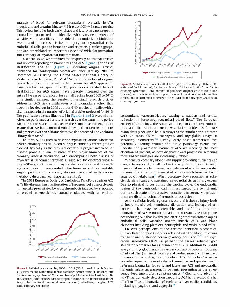
Figure 2. PubMed search results, 2000-2013 (2013 actual through October 31;estimated for 12 months), for the search terms ‘‘risk stratification’’ and ‘‘acutecoronary syndrome’’. Total number of published original articles (solid line,squares), total articles without troponin as one of the biomarkers (dotted line,circles), and total number of review articles (dashed line, triangles). ACS: acute
coronary syndrome.
R.W. Millard, M. Tranter / Rev Esp Cardiol. 2014;67(4):312–320 313
analysis of blood for relevant biomarkers; typically hs-cTn,myoglobin, and creatine kinase–MB fraction (CK-MB) assay results.This review includes both early-phase and late-phase nontroponinbiomarkers purported to identify—with varying degrees ofsensitivity and specificity to reliably detect underlying biologicalevents and processes– ischemic injury to myocardial cells,endothelial cells, plaque formation and eruption, platelet aggrega-tion and other blood cell reporters associated with clot formation,and coronary or myocardial inflammation.
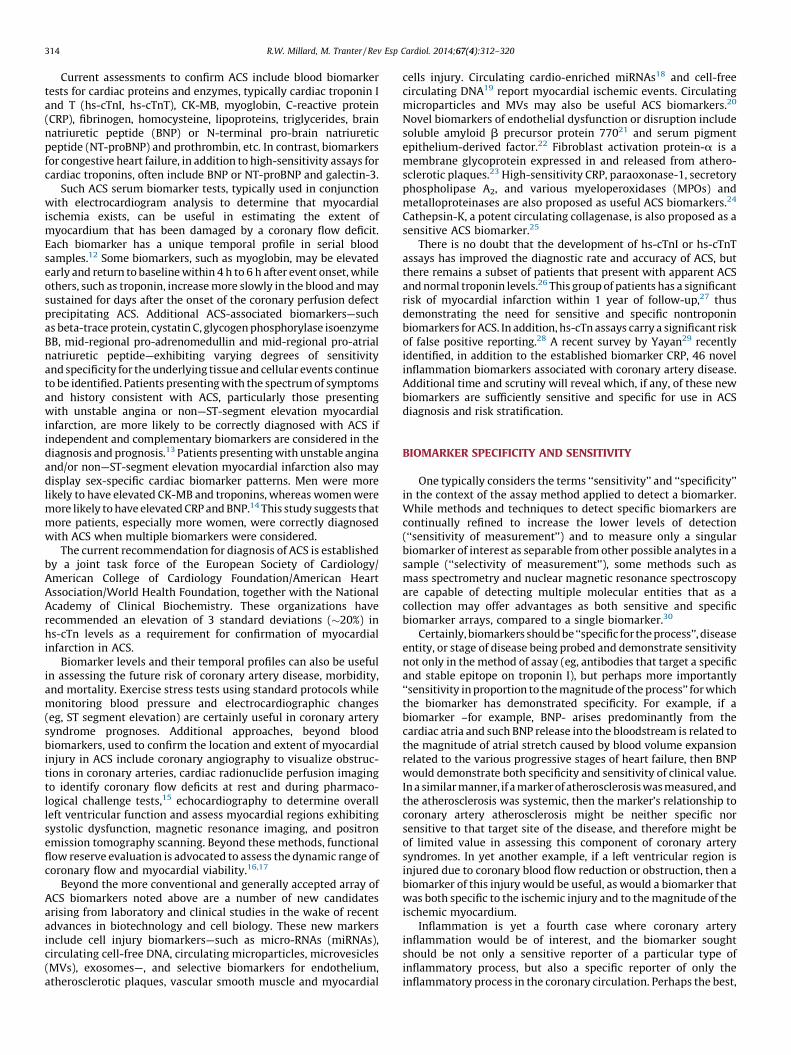
To set the stage, we compiled the frequency of original articlesand reviews reporting on biomarkers and ACS (Figure 1) or on riskstratification and ACS (Figure 2), including original articlespublished for nontroponin biomarkers from January 2000 toDecember 2013 using the United States National Library ofMedicine search engine, PubMed.1 While the number of originalresearch publications reporting biomarkers for ACS appears tohave reached an apex in 2011, publications related to riskstratification for ACS appear have steadily increased over theentire 14-year period, except for a small decline from 2009 to 2012.In a similar manner, the number of original research articlesaddressing ACS risk stratification with biomarkers other thantroponin leveled out in 2009 at around 40 articles annually, with aslight increase in the number of original articles projected for 2013.The publication trends illustrated in Figures 1 and 2 were similarwhen we performed a literature search over the same time periodwith the same search terms, using the Scopus2 search engine. Toassure that we had captured guidelines and consensus opinionsand practices with ACS biomarkers, we also searched The CochraneLibrary database.3
The term ACS is used to describe myriad situations where theheart’s coronary arterial blood supply is suddenly interrupted orblocked, typically as the terminal event of a progressive vasculardisease process in one or more of the major branches of thecoronary arterial circulation. ACS encompasses both classes ofmyocardial ischemia/infarction as assessed by electrocardiogra-phy —ST-segment elevation myocardial infarction and non—ST-segment elevation myocardial infarction— as well as unstableangina pectoris and coronary disease associated with variousmetabolic disorders (eg, diabetes mellitus).
The 2011 European Society of Cardiology Task Force defines ACSas ‘‘a life-threatening manifestation of [progressive] atherosclerosis[. . .] usually precipitated by acute thrombosis induced by a rupturedor eroded atherosclerotic coronary plaque, with or without
0
2000
2001
2002
2003
2004
2005
2006
2007
2008
2009
2010
2011
2012
2013
50
100
150
200
250
300
Pub
licat
ions
on
biom
arke
rs a
nd A
CS
, no.
Number of original articles Number of reviews
Number of original articles (without troponin)
Figure 1. PubMed search results, 2000 to 2013 (2013 actual through October31; estimated for 12 months), for the combined search terms ‘‘biomarker’’ and‘‘acute coronary syndrome’’. Total number of published original articles (solidline, squares), total articles without troponin as one of the biomarkers (dottedline, circles), and total number of review articles (dashed line, triangles). ACS:
acute coronary syndrome.
concomitant vasoconstriction, causing a sudden and criticalreduction in [coronary/myocardial] blood flow.’’ The EuropeanSociety of Cardiology, the American College of Cardiology Founda-tion, and the American Heart Association guidelines for ACSbiomarkers place serial hs-cTn assays as the number one indicator,with CK mass, CK-MB isoenzyme, and myoglobin assays assecondary biomarkers.4,5 Clearly, early onset biomarkers thatpotentially identify cellular and tissue pathology events thatunderlie the progressive nature of ACS are receiving the mostattention at present, as new diagnostic and prognostic screeningtools and technologies are increasingly refined.
Whenever coronary blood flow supply providing nutrients andoxygen to myocardium falls below the required threshold to meetmyocardial metabolic demand, a condition of relative myocardialischemia presents and is associated with a switch from aerobic toanaerobic metabolism.6 When coronary flow reduction is suffi-ciently significant and sustained, myocardial tissue injury occurs.Due to physical forces during the cardiac cycle, the endocardialregion of the ventricular wall is most susceptible to ischemiaduring such acute or progressive reductions in coronary perfusionpressure distal to points of stenosis or occlusion.
At the cellular level, regional myocardial ischemic injury leadsto heart muscle cell membrane disruption and leakage of cellcontents that may be detectable and useful as importantbiomarkers of ACS. A number of additional tissue type disruptionsoccur during ACS that involve pre-existing atherosclerotic plaques,endothelial cells, vascular smooth muscle cells, and bloodelements including platelets, neutrophils and white blood cells.
CK was perhaps one of the earliest identified biochemical(intracellular enzyme) markers released into the blood followingtransient and sustained coronary artery occlusion.7–9 The myo-cardial isoenzyme CK-MB is perhaps the earliest reliable ‘‘goldstandard’’ biomarker for assessment of ACS. In addition to CK-MB,assays for myoglobin and the cardiac contractile protein troponins(cTnI and cTnT) released from injured cardiac muscle cells are usedin combination to diagnose or confirm ACS. Today hs-cTn assaysare relied upon as the most relevant, sensitive, and specific overallreference biomarker for early and late-stage ACS and myocardialischemic injury assessment in patients presenting at the emer-gency department after symptom onset.10 Clearly, the advent ofhs-cTn assays, early-detection capability ACS has elevated the hs-cTn (I or T) as a biomarker of preference over earlier candidates,including myoglobin and copeptin.11
R.W. Millard, M. Tranter / Rev Esp Cardiol. 2014;67(4):312–320314
Current assessments to confirm ACS include blood biomarkertests for cardiac proteins and enzymes, typically cardiac troponin Iand T (hs-cTnI, hs-cTnT), CK-MB, myoglobin, C-reactive protein(CRP), fibrinogen, homocysteine, lipoproteins, triglycerides, brainnatriuretic peptide (BNP) or N-terminal pro-brain natriureticpeptide (NT-proBNP) and prothrombin, etc. In contrast, biomarkersfor congestive heart failure, in addition to high-sensitivity assays forcardiac troponins, often include BNP or NT-proBNP and galectin-3.
Such ACS serum biomarker tests, typically used in conjunctionwith electrocardiogram analysis to determine that myocardialischemia exists, can be useful in estimating the extent ofmyocardium that has been damaged by a coronary flow deficit.Each biomarker has a unique temporal profile in serial bloodsamples.12 Some biomarkers, such as myoglobin, may be elevatedearly and return to baseline within 4 h to 6 h after event onset, whileothers, such as troponin, increase more slowly in the blood and maysustained for days after the onset of the coronary perfusion defectprecipitating ACS. Additional ACS-associated biomarkers—suchas beta-trace protein, cystatin C, glycogen phosphorylase isoenzymeBB, mid-regional pro-adrenomedullin and mid-regional pro-atrialnatriuretic peptide—exhibiting varying degrees of sensitivityand specificity for the underlying tissue and cellular events continueto be identified. Patients presenting with the spectrum of symptomsand history consistent with ACS, particularly those presentingwith unstable angina or non—ST-segment elevation myocardialinfarction, are more likely to be correctly diagnosed with ACS ifindependent and complementary biomarkers are considered in thediagnosis and prognosis.13 Patients presenting with unstable anginaand/or non—ST-segment elevation myocardial infarction also maydisplay sex-specific cardiac biomarker patterns. Men were morelikely to have elevated CK-MB and troponins, whereas women weremore likely to have elevated CRP and BNP.14 This study suggests thatmore patients, especially more women, were correctly diagnosedwith ACS when multiple biomarkers were considered.
The current recommendation for diagnosis of ACS is establishedby a joint task force of the European Society of Cardiology/American College of Cardiology Foundation/American HeartAssociation/World Health Foundation, together with the NationalAcademy of Clinical Biochemistry. These organizations haverecommended an elevation of 3 standard deviations (�20%) inhs-cTn levels as a requirement for confirmation of myocardialinfarction in ACS.
Biomarker levels and their temporal profiles can also be usefulin assessing the future risk of coronary artery disease, morbidity,and mortality. Exercise stress tests using standard protocols whilemonitoring blood pressure and electrocardiographic changes(eg, ST segment elevation) are certainly useful in coronary arterysyndrome prognoses. Additional approaches, beyond bloodbiomarkers, used to confirm the location and extent of myocardialinjury in ACS include coronary angiography to visualize obstruc-tions in coronary arteries, cardiac radionuclide perfusion imagingto identify coronary flow deficits at rest and during pharmaco-logical challenge tests,15 echocardiography to determine overallleft ventricular function and assess myocardial regions exhibitingsystolic dysfunction, magnetic resonance imaging, and positronemission tomography scanning. Beyond these methods, functionalflow reserve evaluation is advocated to assess the dynamic range ofcoronary flow and myocardial viability.16,17
Beyond the more conventional and generally accepted array ofACS biomarkers noted above are a number of new candidatesarising from laboratory and clinical studies in the wake of recentadvances in biotechnology and cell biology. These new markersinclude cell injury biomarkers—such as micro-RNAs (miRNAs),circulating cell-free DNA, circulating microparticles, microvesicles(MVs), exosomes—, and selective biomarkers for endothelium,atherosclerotic plaques, vascular smooth muscle and myocardial
cells injury. Circulating cardio-enriched miRNAs18 and cell-freecirculating DNA19 report myocardial ischemic events. Circulatingmicroparticles and MVs may also be useful ACS biomarkers.20
Novel biomarkers of endothelial dysfunction or disruption includesoluble amyloid b precursor protein 77021 and serum pigmentepithelium-derived factor.22 Fibroblast activation protein-a is amembrane glycoprotein expressed in and released from athero-sclerotic plaques.23 High-sensitivity CRP, paraoxonase-1, secretoryphospholipase A2, and various myeloperoxidases (MPOs) andmetalloproteinases are also proposed as useful ACS biomarkers.24
Cathepsin-K, a potent circulating collagenase, is also proposed as asensitive ACS biomarker.25
There is no doubt that the development of hs-cTnI or hs-cTnTassays has improved the diagnostic rate and accuracy of ACS, butthere remains a subset of patients that present with apparent ACSand normal troponin levels.26 This group of patients has a significantrisk of myocardial infarction within 1 year of follow-up,27 thusdemonstrating the need for sensitive and specific nontroponinbiomarkers for ACS. In addition, hs-cTn assays carry a significant riskof false positive reporting.28 A recent survey by Yayan29 recentlyidentified, in addition to the established biomarker CRP, 46 novelinflammation biomarkers associated with coronary artery disease.Additional time and scrutiny will reveal which, if any, of these newbiomarkers are sufficiently sensitive and specific for use in ACSdiagnosis and risk stratification.
BIOMARKER SPECIFICITY AND SENSITIVITY
One typically considers the terms ‘‘sensitivity’’ and ‘‘specificity’’in the context of the assay method applied to detect a biomarker.While methods and techniques to detect specific biomarkers arecontinually refined to increase the lower levels of detection(‘‘sensitivity of measurement’’) and to measure only a singularbiomarker of interest as separable from other possible analytes in asample (‘‘selectivity of measurement’’), some methods such asmass spectrometry and nuclear magnetic resonance spectroscopyare capable of detecting multiple molecular entities that as acollection may offer advantages as both sensitive and specificbiomarker arrays, compared to a single biomarker.30
Certainly, biomarkers should be ‘‘specific for the process’’, diseaseentity, or stage of disease being probed and demonstrate sensitivitynot only in the method of assay (eg, antibodies that target a specificand stable epitope on troponin I), but perhaps more importantly‘‘sensitivity in proportion to the magnitude of the process’’ for whichthe biomarker has demonstrated specificity. For example, if abiomarker –for example, BNP- arises predominantly from thecardiac atria and such BNP release into the bloodstream is related tothe magnitude of atrial stretch caused by blood volume expansionrelated to the various progressive stages of heart failure, then BNPwould demonstrate both specificity and sensitivity of clinical value.In a similar manner, if a marker of atherosclerosis was measured, andthe atherosclerosis was systemic, then the marker’s relationship tocoronary artery atherosclerosis might be neither specific norsensitive to that target site of the disease, and therefore might beof limited value in assessing this component of coronary arterysyndromes. In yet another example, if a left ventricular region isinjured due to coronary blood flow reduction or obstruction, then abiomarker of this injury would be useful, as would a biomarker thatwas both specific to the ischemic injury and to the magnitude of theischemic myocardium.
Inflammation is yet a fourth case where coronary arteryinflammation would be of interest, and the biomarker soughtshould be not only a sensitive reporter of a particular type ofinflammatory process, but also a specific reporter of only theinflammatory process in the coronary circulation. Perhaps the best,

R.W. Millard, M. Tranter / Rev Esp Cardiol. 2014;67(4):312–320 315
but not always practical, way to improve biomarker specificitywould be to perform sampling or detection at the specific source ofthe disease of interest. This might be achievable by labeling andimaging a particular biomarker at the site of its generation orrelease, and in a more targeted way by obtaining blood samplesfrom supply arteries and draining veins from the tissue or organ ofinterest, thereby improving tissue (ie, locus) specificity. When ACSis the disease of interest, blood samples simultaneously drawnfrom a systemic artery and from the coronary sinus would identifybiomarkers for cardiac-specific events.31
Biomarker signal dilution is a serious challenge when relying onperipheral blood samples to detect biomarkers specific for andsensitive to ACS, and as we previously suggested32 and others30
have proposed, a single biomarker may be neither as specific nor assensitive in ACS prognosis, extent, or progress as would be a set ofbiomarkers that taken together increase both sensitivity andspecificity for assessing the syndrome and perhaps particularevents within the syndrome. This combinatorial approach is takenwhen hs-cTn, myoglobin and CK-MB data are combined to improveACS diagnostic and risk assessment reliability.33–39
MYOCARDIAL CELL ACUTE CORONARY SYNDROMEBIOMARKERS
Biomarkers that are passively released from cardiac myocytesconsequent to ischemic injury are the most commonly relied-uponindicators of ACS and subsequent cell injury. Myoglobin, CK-MB andcardiac troponins are the current conventional biomarkers in thiscategory. In addition, if the mass of left ventricle affected by thecoronary insufficiency is large enough to increase wall stress (dilatethe region during diastole and produce paradoxical bulging duringsystole, then the stretched cardiac myocytes can and will releaseBNP and NT-proBNP.28 Other factors that are released from damagedor dying myocardium include CRP, BNP, ischemic modified albumin,and heart-type fatty acid binding protein.35,40–42
Over the years, a number of previously used biomarkers of ACS —aspartate aminotransaminase, total lactate dehydrogenase, andlactate dehydrogenase isoenzymes—have been abandoned due topoor cardiac injury specificity and because of their ubiquitous tissuedistribution. Early reports identified other cytoplasmic moleculesreleased from transiently or permanently injured myocardial cells.Among these biomarkers was CK and the subsequent identificationof myocardial and brain isoforms.7–9 At present, the hs-cTn assay,based on its superior tissue specificity and improved sensitivity tolow cardiac troponins levels in blood samples, is the standard againstwhich other putative ACS biomarkers are evaluated. The 99thpercentile (3 standard deviations above the normal average value) isused as the decision limit for confirmation of myocardial injury byhs-cTnT, CK-MB mass, and myoglobin.
In addition to the release of cytoplasmic enzymes or proteinproducts into the blood stream, damaged cells may also releasenucleic acids that can be specifically detected as biomarkers.Several recent studies have shown increased serum levels of cell-free circulating DNA following cardiac ischemia, presumablyconsequent to myocyte apoptosis and necrosis.43–45 Increasedserum levels of cell-free circulating DNA have been reported in ACSpatients and were further elevated following ST-segment elevationmyocardial infarction.19,46 However, the utility of cell-freecirculating DNA as a biomarker may be limited by its relativelyshort half-life < 30 min.47 Conversely, while a short half-life maylimit its utility in determining the degree of acute ischemicdamage, its rapid clearance from circulation may render itbeneficial in differentiating between discrete and sustainedischemic events if monitoring and blood sampling occursproximate to coronary events.
INFLAMMATION BIOMARKERS IN ACUTE CORONARYSYNDROME
The previous section described the secretion (passive or active)of biomarkers from injured myocardium following the initiation ofan ischemic event. In contrast to these biomarkers, which areindicative of a prior or ongoing ischemia, the biomarkers discussedin the next two sections (markers of coronary inflammation oratherosclerosis) are more predictive because they are often presentprior to the ischemic damage or myocardial necrosis/apoptosis. Inaddition, coronary inflammation is a primary driving force for thedevelopment and progression of atherosclerosis and, thus,increased inflammation is also a common indicator of coronaryatherosclerosis.
Among mediators and markers of coronary and myocardialinflammation are a number of acute-phase proteins, cytokines,interleukins (IL), and cell adhesion molecules, the foremost ofthese being CRP. Inflammation of and other injury to the vascularendothelium is considered the cardinal event leading, over time, tothe establishment of atherogenic plaque, which may serve as thefocal point for eventual platelet aggregation and thrombosis and/oreruption of the plaque’s protective fibrous cap, for which CRP, aswell as IL-6 and serum amyloid A, is used as the leading biomarkerwithin the ACS paradigm. The release of CRP, detected by a high-sensitivity assay (high-sensitivity CRP), can also produce second-ary effects such as the expression of adhesion molecules, amongother events which themselves may be biomarkers associated withACS. A decade ago, a number of other putative biomarkers for ACSrisk stratification were emerging. These included markers ofplatelet activation, enzymes such as metalloproteinases thatdisrupt an atheroma’s fibrous cap integrity, and MPOs releasedfrom leukocytes activated within the coronary circulation.
The most common and widely used marker of coronaryinflammation that predicts myocardial ischemia risk is CRP.48–52
However, other inflammatory biomarkers, including pentraxin 3,serum amyloid A, fibrinogen, and MPO, have proven to be equallyor more predictive for ACS and subsequent cardiac events.Pentraxin 3, an early-phase ACS response protein that is a morespecific marker of vascular inflammation than is CRP, is released bymultiple cell types, including fibroblasts and dendritic andendothelial cells, in response to primary inflammatory signals.53,54
Serum amyloid A and fibrinogen are also early-phase ACS responseproteins whose serum levels are elevated in response to acuteinflammation.55 Neutrophil cell count and activation are alsomarkers of inflammation, and levels of the neutrophil-derivedlysosomal enzyme MPO can be a sensitive biomarker forinflammation and cardiovascular risk.
First identified in 2003 as a predictive biomarker for ACS in theabsence of myocardial necrosis, MPO can serve as a marker forcoronary inflammation and cardiac risk much earlier thanmyocardial ischemia and infarction biomarkers—myoglobin,troponin, CK-MB, or CRP—in patients evaluated soon after onsetof symptoms.56,57 Recent studies have confirmed the utility of MPOas a biomarker of coronary inflammation and shown that its long-term predictive power is further increased when combined with ahigh-sensitivity CRP assay.48,58
Additional biomarkers have been investigated as potentialmarkers of coronary inflammation and ACS, but their clinicalusefulness as sensitive and specific predictive biomarkers has notyet been determined conclusively. Examples of these potentialbiomarkers include soluble CD40 ligand, tumor necrosis factor-a,IL-10, and IL-18. Both soluble CD40 ligand and tumor necrosisfactor-a are known initiators of pro-inflammatory signaling andare elevated in patients with ACS. However, none of these appearsto be a specific predictor of ACS or of subsequent adverse cardiacevents.59,60 Although IL-18 is a pro-inflammatory cytokine that is

R.W. Millard, M. Tranter / Rev Esp Cardiol. 2014;67(4):312–320316
thought to initiate plaque instability and is elevated in associationwith other established risk factors, it does not independentlypredict ACS or myocardial infarction risk.61
BIOMARKERS OF CORONARY ATHEROSCLEROSIS
The progressive precedent event and process leading to ACS iscoronary arterial atherosclerosis. As such, selective biomarkers canallow detection of rapidly changing events related to athero-sclerotic plaque, including the rupture of its protective fibrous cap.
BIOMARKERS ARISING FROM DAMAGED CORONARYENDOTHELIUM
Integrity of the coronary endothelium is necessary for capillaryhomeostasis and for proper physiological functioning of theunderlying vascular smooth muscle of the coronary arterialsegments leading to the capillaries.62 The endothelium is theblood interface and the first barrier and transducer of thevasomotor effects of microfluidic shear force changes and ofcertain vasoactive agents working through nitric oxide. Injury to orinflammation of the coronary endothelium alters these barrierfunctions, and may lead to the presence of biomarkers and MVs inthe coronary circulation that can be detected in system serumsamples.63–66
BIOMARKERS OF PLATELET ACTIVATION AS PRECURSOR TOCORONARY ARTERIAL THROMBOSIS
Platelet aggregation can occur subsequent to activation of thefibrinogen receptor, glycoprotein IIb/IIIa, and the endothelial celladhesion molecule PECAM-1. Once activated, biomarkers may beshed by platelets as free proteins, miRNAs, or MVs.
CARDIAC-DERIVED MICRO-RNAS AS ACUTE CORONARYSYNDROME BIOMARKERS
MiRNAs are an emerging class of biomarkers that are likely to beactively released by cardiac cells in response to ischemic damage.Changes in miRNA expression within myocytes have been knownfor some time to vary in response to ischemia, and evidence isbuilding that the expression profile of cardiac-derived circulatingmiRNAs represents the environment and conditions of themyocardium. Circulating miRNAs have advantages as biomarkersin that they have a favorable expression time-course (rapid onsetand long-lasting), are stable in circulation and post-isolation, andcan be very specifically detected at very low levels.25 The studiesthat have thus far investigated circulating miRNAs as biomarkersfor ACS and myocardial ischemia/reperfussion injury were recentlyreviewed by Deddens et al.67 Another appeal of miRNAs as ACSbiomarkers is their tissue-specific expression. For example, miR-208 is expressed in a cardiomyocyte-specific manner from anintron in the alpha-MHC gene. Several studies have takenadvantage of this to investigate the secretion of miR-208 as aspecific marker for cardiomyocyte ischemic injury. While not allreports regarding miR-208 as a specific biomarker for ACS arepositive, multiple reports show a rapid increase in circulating miR-208 post-myocardial infarction.68–72 MiR-499 is another miRNAspecific to cardiac muscle that has shown promise as an earlymarker of myocardial infarction.68,73
Perhaps the most promising circulating miRNA biomarker ismiR-1, a muscle-specific biomarker shown by numerous studies tobe elevated following both ST-segment elevation myocardial
infarction and non—ST-segment elevation myocardial infarctioncardiac ischemia.72 In addition to muscle-specific miRNAs such asmiR-1, miR-208, and miR-499, miRNAs from other tissue typessuch as vascular smooth muscle, endothelium, and leukocytes alsorepresent potential biomarkers of components of ACS. Theinvestigation of miRNAs as biomarkers is still novel, with themajority of the publications coming in just the last 3 to 4 years.New miRNAs are still being identified and their use as biomarkershas substantial potential.
CARDIAC–DERIVED MICROVESICLES AS ACUTE CORONARYSYNDROME BIOMARKERS
Some of the biomarkers discussed above are not simplyreleased passively as a result of cellular injury, but rather areactively released as a specific, dynamic, cellular response toischemic stress. Numerous reports have described the release ofextracellular MVs from cardiomyocytes, endothelial cells, andneutrophils. Such MVs may play an active role in the developmentor progression of ACS.65,66,74 The content and release of these MVsappear to be pathology-dependent, further suggesting that theymay serve a specific role in signaling to distant target tissues/cells.64,66,75,76 Secreted MVs are known to contain defined proteincomponents, and at least 3 of these (polygenic immunoglobulinreceptor, cystatin C, and complement factor C5a) may also serve asuseful ACS biomarkers.77
In addition to component proteins, other studies have directlymeasured MVs and the cell types from which they originate andhave implicated endothelial cell-derived and monocyte-derivedMVs as independent predictors of myocardial damage in non—ST-segment elevation myocardial infarction patients.20 CirculatingMVs may represent an entire unique class of distinguishablebiomarkers that can be utilized in combination with each otherdepending on tissue source or content. This is exemplified bystudies that examined multiple distinct MVs populations or theratios of these populations as independent biomarkers.20
Recent discovery of exosomes containing molecules that reportcell-specific information and participate in intercellular commu-nication signaling and delivery of encapsulated materials from onecell to another suggests that the search for ACS biomarkers willcontinue to evolve. In the near future, the analysis of cardiac-originexosome contents and the interpretation of this content profilemay reveal yet another avenue by which early ACS diagnosis,prognosis, and risk stratification will be refined.
NOVEL GENE AND PROTEIN BIOMARKERS FOR ACUTECORONARY SYNDROME DIAGNOSIS AND RISK STRATIFICATION
In a recent report by Silbiger et al,78 whole genomic expressionanalysis using expressed mRNA profiles on gene chip microarraysdetected 549 differentially expressed genes in peripheral blood ofACS patients within the first 48 h. Within these hundreds of genesexpressed during the early onset of ACS, 13 genes were consideredboth technically and biologically validated by real time-PCR, andall were statistically expressed differentially in ACS patients incomparison with control patients in both phases of the study. Theauthors suggest that changes in ALOX15, CA1, and KCNE1 genes mayreflect a ‘‘protective system response’’ following coronary occlu-sion, and that BCL2A and COX7B genes may report apoptosisregulation in endothelial cells and cardiac myocytes. The authorsfurther suggest that 6 genes (AREG, IL18R1, IRS2, MYL4, BCL2L1, andMMP9) may report endothelial and cardiac tissue remodeling afterischemia, and that other combinations among these 13 genes may
R.W. Millard, M. Tranter / Rev Esp Cardiol. 2014;67(4):312–320 317
indicate atherosclerotic plaque progression and rupture, or mayserve as triggers in the inflammatory cascade.
In a similar manner, the IBIS-1 pilot study by Wykrzykowskaet al79 applied microarray technology to correlate coronaryimaging (intracoronary ultrasound imaging and multi-slicecomputed tomography scanning) with circulating biomarkerexpression in patients with the whole spectrum of coronarysyndromes. Elevations were detected in high-sensitivity CRP, IL-6,lipoprotein-associated phospholipase A2 activity, and NT-proBNP,but not in tumor necrosis factor-a or soluble CD40 ligand levels.Anti-apoptotic markers (eg, plasminogen activator inhibitor type1) increased over time. Pro-inflammatory markers and markers oflymphocyte trafficking (eg, C-6 Kine, CTAK) increased initially andthen decreased over time, as did markers of coagulation (eg, D-dimer) and of endothelial shear stress and remodeling (eg,follistatin).
Such gene expression and transcriptional profiling resultssuggest that future studies will identify and refine a critical subsetof gene products as biomarkers for use throughout the ACS time-course for improved patient risk assessment and stratification.Clearly, future efforts will be directed at refining both sensitivityand specificity of these gene biomarkers to gain prognostic value inACS and at identifying which gene biomarkers shed light on tissue-specific events underlying ACS.
MATRIX METABOLOMICS STRATEGIES FOR ACUTE CORONARYSYNDROME DIAGNOSIS AND RISK STRATIFICATION
In recognition that a single biomarker may be insufficientlysensitive or specific for risk stratification of ACS patients, Bodiet al.30,80 reported recently on an improved approach for ACS riskassessment using multiple biochemical markers. It is reasonable toforecast that by applying contemporary analytic methodologiessuch as high-throughput magnetic resonance spectroscopy andmass spectrometry, patient blood samples may be analyzed forACS-related changes using computational algorithms that stratifypatient ACS risk.32 Risk assessment, including risk calculators, oflipids and cardiovascular disease are available throughout much ofEurope and promulgated by British82–84 and Spanish85,86 cardiol-ogy and atherosclerosis professionals. While the search foralgorithms and calculators that integrate biomarker values toassist predictive risk stratification is laudable, unreliable outcomesmay result when outdated biometrics are used to create thecalculation that indicates a need for cholesterol-lowering drugs(statins). Specifically, the use of the new guidelines and riskcalculator recently endorsed and published jointly by theAmerican Heart Association and the American College of Cardiol-ogy81 apparently leads to a substantial overestimation of risk andpotential over-prescribing of this class of medicines to individualpatients who may not truly be at elevated risk for atherosclerosis,which is one component of ACS.
THE FUTURE OF ACUTE CORONARY SYNDROME BIOMARKERS ASAIDS TO EARLY DIAGNOSIS, PROGNOSIS, AND RISKSTRATIFICATION
Sensitive, Cell/Process Specific, and Temporally CorrelatedBiomarkers
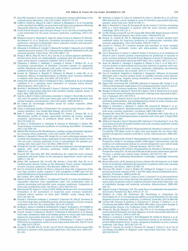
The priorities for biomarker use in ACS risk assessment andstratification require further work to characterize not only thetemporal correlation of each biomarker or assembly of biomarkersto each of the progressive stages of ACS (Figure 3), but also anobjective assessment and verification of each biomarker’s sensitivity
and specificity to an underlying pathology component contributingto ACS itself. The selection of appropriately sensitive biomarkers thatidentify underlying tissue-specific and cell-type-specific changes ina manner that aligns the temporal pathology events referenced toonset of ACS symptoms will be the most useful for accurate diagnosisand risk stratification of such patients. It is our opinion that therapidly expanding wealth of information arising from research innonclinical animal models of ACS and from clinical studies in ACSpatients will soon, after substantial effort, allow the possibility ofselecting the most specific and sensitive biomarkers for each of theunderlying features that present as ACS.
Optimizing Biomarkers for Risk Stratification
Efforts must not only continue to explore and apply newanalytic technologies to identify putative ACS biomarkers, butmust also seek to understand the link to the most specific andsensitive of myriad biomarkers, not simply as correlations to early-or late-stage ACS, but to each of the underlying biologicalprocesses, including the status of coronary atherosclerosis plaques,evidence of platelet aggregation and clot formation, smoothmuscle and endothelial cell health, cardiac myocyte integrity, andperhaps even myocardial sympathetic innervation status.87
The speed and reliability (sensitivity and specificity) ofbiomarker test reporting for patient diagnosis and prognosis, riskstratification, and therapeutic decision-making is increasingly aconversation connecting point-of-care testing with emergingtechnologies and devices, as contrasted to conventional centrallaboratory testing methods and facilities. Most recommended ACSbiomarkers (ie, CK-MB, myoglobin, BNP and/or NT-proBNP, andpossibly excepting hs-cTnI and/or hs-cTnT) can be elevated inconditions other than ACS (eg, hypertension, skeletal muscletrauma, renal failure, primary aldosteronism, and thyroid disease).Therefore, the search continues for unique individual or sets ofbiomarkers that are both sensitive and specific for each of theunderlying pathologies and processes that comprise ACS onset andprogression, as well as those biomarkers that will be most usefuland effective for ACS risk stratification and optimal therapeuticdecisions.
Trends Toward Point-of-care Multiple Biomarker Assays
Among the currently identified biomarkers, there is certainly apotential for redundancy in reporting information related to any ofthe underlying biological events. At some near future date, it islikely that point-of-care, microchip-array, rapid-response assaysystems will integrate the combination of serum protein and genebiomarkers, biochemical enzymes, and cell membrane markers topermit a comprehensive and detailed decision-directed diagnosisof ACS and guide improved risk stratification of ACS patients.88 Thecurrent conversation regarding conventional and novel or emer-ging ACS biomarkers—beyond considerations of laboratorymethod and pathology specificity and sensitivity—includes thetopic of laboratory test turnaround time and the cost/benefit ofdecreasing it with reliable and reproducible testing at the site ofpatient evaluation. Point-of-care testing by primary care physi-cians using a 3-in-1 device capable of rapidly reporting ACS, heartfailure, and thromboembolism biomarkers (cTnT, NT-proBNP, orD-dimer, an indicator of fibrin degradation and coagulationactivation) has been reported to improve differential diagnosisof potential ACS during out-of-hospital visits.89
The value proposition regarding emerging and novel biomar-kers continues to consider whether adding one or more biomarkers(multi-marker approaches) will improve risk stratification andprognostic advantage over current and more conventional

Ischemia-damaged myocardium
Thrombosis
Endothelial injury
Coronary inflammation
Pre-ischemia
Bio
mar
ker
Eve
nt
Thrombus/ischemia Post-ischemia/reperfusion
CRP, PTX3, MPO, MMP, SAA, fibrinogen, sPLA2, PON1, neutrophil-derived microvesicles
sAPP770, PEDF, cathepsin K, endothelial-derived microvesicles. miRNAs
Myoglobin, hs-CK-MB, NT-proBNP, IMA. hs-cTn, H-FABP,cardiac-derived microvesicles, miRNAs, cf-DNA
Platelet-derived microvesicles,miRNAs, homocysteine
FAPα. endothelial-derived microvesicles, miRNAs
Coronary atherosclerosis
Figure 3. Acute coronary syndrome biomarkers associated with the underlying myocardial events. cf-DNA, cell-free circulating DNA; CRP, C-reactive protein; FAP-a, fibroblast activation protein-a; H-FABP, heart-type fatty acid binding protein; hs-CK-MB, high sensitivity-creatine kinase–MB fraction; hs-cTn, high sensitivity-cardiac troponin; IMA, ischemic modified albumin; miRNAs, micro RNAs; MMP, metalloproteinases; MPO, myeloperoxidases; NT-proBNP, N-terminal pro-brainnatriuretic peptide; PEDF, serum pigment epithelium-derived factor; PON1, paraoxonase-1; PTX3, pentraxin 3; SAA, serum amyloid A; sPLA2, secretoryphospholipase A2; sAPP770, soluble amyloid b precursor protein 770.
R.W. Millard, M. Tranter / Rev Esp Cardiol. 2014;67(4):312–320318
stratification strategies.12,90 On this point, an incremental ACSprognostic benefit of combining NT-proBNP and growth differ-entiation factor 15 with the GRACE score plus a corrected hs-cTnTvalue has been demonstrated, considering a combined endpoint of6-month all-cause mortality or nonfatal myocardial infarction.91
By comparison, the 1-year predictive value of a number ofinflammatory and noninflammatory markers assessed in ACSpatients in the SIESTA study showed that only NT-proBNP andfibrinogen were sensitive and specific prognostic biomarkers ofcardiovascular risk (all-cause mortality at 1-year follow up).92 Inthis study, few if any of the 10 examined biomarkers (eg, cystatin,CRP, E-selectin) added information to that provided by conven-tional clinical risk markers; however, more than three fourths ofthe patients in this study were receiving statin (3-hydroxy-3-methylglutaryl coenzyme A reductase inhibitor) treatment knownto exert anti-inflammatory actions, a potential confoundingvariable in the study. Such approaches as those employingpoint-of-care assays and lab-on-a-chip microarrays linked topredictive diagnostic, prognostic, and risk stratification algorithmswill potentially reduce false positive ACS diagnoses and unwar-ranted hospitalizations, and result in health care savings andimprovements in the informed use of therapeutics to the benefit ofpatient health.
ACKNOWLEDGEMENTS
The authors wish to thank Ms. Kristin Luther for her assistancein creating Figure 3.
CONFLICTS OF INTEREST
None declared.
REFERENCES
1. PubMed [accessed 2013 Dec 18]. Available at: www.ncbi.nlm.nih.gov2. Scopus [accessed]. Available at: www.scopus.com3. The Cochrane Library [accessed]. Available at: www.thecochranelibrary.com
4. Hamm CW, Bassand JP, Agewall S, Bax J, Boersma E, Bueno H, et al.; ESCCommittee for Practice Guidelines. ESC Guidelines for the management ofacute coronary syndromes in patients presenting without persistent ST-seg-ment elevation: The Task Force for the management of acute coronary syn-dromes (ACS) in patients presenting without persistent ST-segment elevation ofthe European Society of Cardiology (ESC). Eur Heart J. 2011;32:2999–3054.
5. Cannon CP, Brindis RG, Chaitman BR, Cohen DJ, Cross Jr JT, Drozda Jr JP, et al.American College of Cardiology Foundation/American Heart Association TaskForce on Clinical Data Standards; American College of Emergency Physicians;Emergency Nurses Association; National Association of Emergency MedicalTechnicians; National Association of EMS Physicians; Preventive CardiovascularNurses Association; Society for Cardiovascular Angiography and Interventions;Society of Cardiovascular Patient Care; Society of Thoracic Surgeons. 2013ACCF/AHA key data elements and definitions for measuring the clinical man-agement and outcomes of patients with acute coronary syndromes and cor-onary artery disease: a report of the American College of CardiologyFoundation/American Heart Association Task Force on Clinical Data Standards(Writing Committee to Develop Acute Coronary Syndromes and CoronaryArtery Disease Clinical Data Standards). Circulation. 2013;127:1052–89.
6. Yaniv Y, Juhaszova M, Nuss HB, Wang S, Zorov DB, Lakatta EG, et al. MatchingATP supply and demand in mammalian heart: in vivo, in vitro, and in silicoperspectives. Ann N Y Acad Sci. 2010;1188:133–42.
7. Duma RJ, Seigel AL. Serum creatine phosphokinase in acute myocardial infarc-tion. Arch Intern Med. 1965;115:443–51.
8. Heyndrickx GS, Millard RW, McRitchie RJ, Moroko PR, Vatner SF. Regionalmyocardial functional and electrophysiological alterations after brief coronaryartery occlusion in conscious dogs. J Clin Invest. 1975;56:978–85.
9. Shell WE, Sobel BE. Biochemical markers of ischemic injury. Circulation.1976;53(3 Suppl):I98–106.
10. Hamilton MA, Toner A, Cecconi M. Troponin in critically ill patients. MinervaAnestesiol. 2012;78:1039–45.
11. Lackner KJ. Laboratory diagnostics of myocardial infarction—troponins andbeyond. Clin Chem Lab Med. 2013;51:83–9.
12. Moe KT, Wong P. Current trends in diagnostic biomarkers of acute coronarysyndrome. Ann Acad Med Singapore. 2010;39:210–5.
13. Sabatine MS, Morrow DA, De Lemos JA, Gibson CM, Murphy SA, Rifai N, et al.Multimarker approach to risk stratification in non-ST elevation acute coronarysyndromes: simultaneous assessment of troponin I. C-reactive protein, andB-type natriuretic peptide. Circulation. 2002;105:1760–3.
14. Wiviott SD, Cannon CP, Morrow DA, Murphy SA, Gibson CM, McCabe CH, et al.Differential expression of cardiac biomarkers by gender in patients withunstable angina/non-ST elevation myocardial infarction: a TACTICS-TIMI 18(Treat Angina with Aggrastat and determine Cost of Therapy with an Invasive orConservative Strategy-Thrombolysis in Myocardial Infarction 18) substudy.Circulation. 2004;109:580–6.
15. Radio-pharmaceuticals and tracer kinetics. In: Gerson MC, editor. Cardiacnuclear medicine. 3rd ed. New YorK: McGraw-Hill; 1997. p. 3–28.
16. Roy AS, Banerjee RK, Back LH, Back MR, Khoury S, Millard RW. Delineating theguide-wire flow obstruction effect in assessment of fraction flow reserve andcoronary flow reserve measurements. Am J Physiol Heart Circ Physiol.2005;289:H392–7.
R.W. Millard, M. Tranter / Rev Esp Cardiol. 2014;67(4):312–320 319
17. Kern MJ, Samady H. Current concepts in integrated coronary physiology in thecatheterization laboratory. J Am Coll Cardiol. 2010;55:173–85.
18. Gidlof O, Smith JG, Miyazu K, Gilje P, Spencer A, Blomquist S, et al. Circulatingcardio-enriched microRNAs are associated with long-term prognosis followingmyocardial infarction. BMC Cardiovasc Disord. 2013;13:12.
19. Cui M, Fan M, Wang H, Qin J, Sheng H, Wang Y, et al. Cell-free circulating DNA:a new biomarker for the acute coronary syndrome. Cardiology. 2013;124:76–84.
20. Montoro-Garcıa A, Shantsila E, Tapp LD, Lopez-Cuenca A, Romero AI, Hernan-dez-Romero D, et al. Small-size circulating microparticles in acute coronarysyndromes: relevance to fibrinolytic status, reparative markers and outcomes.Atherosclerosis. 2013;227:313–22.
21. Kitazume S, Yoshihisa A, Yamaki T, Oikawa M, Tachida Y, Ogawa K, et al. Solubleamyloid precursor protein 770 is released from inflamed endothelial cells andactivated platelets. J Biol Chem. 2012;287:40817–25.
22. Wang F, MA X, Zhou M, Pan X, Ni J, Gao M, et al. Serum pigment epithelium-derived factor levels are independently correlated with the presence of cor-onary artery disease. Cardiovasc Diabetol. 2013;12:56–64.
23. Tillmanns J, Widera C, Habbada Y, Galuppo P, Kempf T, Wollert KC, et al.Circulating concentrations of fibroblast activation protein (in apparentlyhealthy individuals and patients with acute coronary syndrome assessed bysandwich ELISA. Int J Cardiol. 2013;168:3926–31.
24. Graner M, Tikkanen E, Rimpila P, Tikkanen H, Ripatti S, Lokki ML, et al.Diagnostic efficacy of myeloperoxidase to identify acute coronary syndromein subjects with chest pain. Ann Med. 2013;45:322–7.
25. Cheng XW, Kikuchi R, Ishii H, Yoshikawa D, Hu L, Takahashi R, et al. Circulatingcathepsin K as a potential novel biomarker of coronary artery disease.Atherosclerosis. 2013;228:211–6.
26. Reichlin T, Hochholzer W, Bassetti S, Steuer S, Stelzig C, Hartwiger S, et al. Earlydiagnosis of myocardial infarction with sensitive cardiac troponin assays. NEngl J Med. 2009;361:858–67.
27. Sanchis J, Bodı V, Nunez J, Bertomeu-Gonzalez V, Gomez C, Bosch MJ, et al. Newrisk score for patients with acute chest pain, non-ST-segment deviation, andnormal troponin concentrations. J Am Coll Cardiol. 2005;46:443–9.
28. De Lemos JA. Increasingly sensitive assays for cardiac troponins. JAMA.2013;309:2262–9.
29. Yayan J. Emerging families of biomarkers for coronary artery disease: inflam-matory mediators. Vasc Health Risk Manag. 2013;9:435–56.
30. Bodi V, Sanchis J, Morales JM, Marrachelli VG, Nunez J, Forteza MJ, et al.Metabolomic profile of human myocardial ischemia by nuclear magneticresonance spectroscopy of peripheral blood serum. J Am Coll Cardiol.2012;59:1629–41.
31. De Rosa S, Fichtlscherer S, Lehmann R, Assmus B, Dimmeler S, Zeiher AM.Transcoronary concentration gradients of circulating microRNAs. Circulation.2011;124:1936–44.
32. Millard RW, Rosevear PR. Metabolomics: seeking a unique biomarker signaturefor coronary artery syndromes. J Am Coll Cardiol. 2012;59:1642–4.
33. Rathore S, Knowles P, Mann APS, Dodds PA. Is it safe to discharge patients fromaccident and emergency using a rapid point of care triple cardiac marker test torule out acute coronary syndrome in low to intermediate risk patients pre-senting with chest pain? Eur J Int Med. 2008;19:537–40.
34. Al-Hadi HA, Fox KA. Cardiac markers in the early diagnosis and management ofpatients with acute coronary syndrome. Sultan Qaboos Univ Med J.2009;9:231–46.
35. Peacock WF, Soto-Ruiz KM. Risk stratification for suspected acute coronarysyndromes and heart failure in the emergency department. Acute Card Care.2009;11:138–45.
36. Kelley WE, Lockwood CM, Cervelli DR, Sterner J, Scott MG, Duh SH, et al.Cardiovascular disease testing on the Dimension VistaW system: biomarkersof acute coronary syndromes. Clin Biochem. 2009;42:1444–51.
37. Kurz K, Giannitsis E, Becker M, Hess G, Zdunck D, Katus HA. Comparison of thenew high sensitive cardiac troponin T with myoglobin, h-FABP and cTnT forearly identification of myocardial necrosis in the acute coronary syndrome. ClinRes Cardiol. 2011;100:209–15.
38. Aldous SJ, Richards M, Cullen L, Troughton R, Than M. A 2-hour thrombolysis inmyocardial infarction score outperforms other risk stratification tools inpatients presenting with possible acute coronary syndromes: Comparison ofchest pain stratification tools. Am Heart J. 2012;164:516–23.
39. Macdonald SPJ, Nagree Y, Fatovich DM, Phillips M, Brown SGA. Serial multiplebiomarkers in the assessment of suspected acute coronary syndrome:multiple infarct markers in chest pain (MIMIC) study. Emerg Med J.2013;30:149–54.
40. Freund Y, Chenevier-Gobeaux C, Leumani F, Claessens YE, Allo JC, Doumenc B,et al. Heart-type fatty acid binding protein and the diagnosis of acute coronarysyndrome in the ED. Am J Emerg Med. 2012;30:1378–84.
41. Jernberg T, James S, Lindahl B, Stridsberg M, Venge P, Wallentin L. NT-ProBNP innon-ST-elevation acute coronary syndrome. J Card Fail. 2005;11(5 Suppl):S54–8.
42. Tsuji R, Tanaka T, Sohmiya K, Hirota Y, Yoshimoto K, Kinoshita K, et al. Humanheart-type cytoplasmic fatty acid-binding protein in serum and urine duringhyperacute myocardial infarction. Int J Cardiol. 1993;41:209–17.
43. Antonatos D, Patsilinakos S, Spanodimos S, Korkonikitas P, Tsigas D. Cell-freeDNA levels as a prognostic marker in acute myocardial infarction. Ann N Y AcadSci. 2006;1075:278–81.
44. Chang CP, Chia RH, Wu TL, Tsa KC, Sun CF, Wu JT. Elevated cell-free serum DNAdetected in patients with myocardial infarction. Clin Chim Acta. 2003;327:95–101.
45. Shimony A, Zahger D, Gilutz H, Goldstein H, Orlov G, Merkin M, et al. Cell freeDNA detected by a novel method in acute ST-elevation myocardial infarctionpatients. Acute Card Care. 2010;12:109–11.
46. Balta S, Demirkol S, Cakar M, Karaman M, Ay SA, Arslan Z. Cell-free circulatingDNA as a novel biomarker in patients with the acute coronary syndrome.Cardiology. 2013;126:122–3.
47. Lo YM, Zhang J, Leung TN, Lau TK, Chang AM, Hjelm NM. Rapid clearance of fetalDNA from maternal plasma. Am J Hum Genet. 1999;64:218–24.
48. Heslop CL, Frolich JJ, Hill JS. Myeloperoxidase and C-reactive protein havecombined utility for long-term prediction of cardiovascular mortality aftercoronary angiography. J Am Coll Cardiol. 2010;55:1102–9.
49. Correia LC, Esteves JP. C-reactive protein and outcomes in acute coronarysyndromes: a systematic review and meta-analysis. Arq Bras Cardiol.2011;97:76–85.
50. Lim P, Moutereau S, Simon T, Gallet R, Probst V, Ferrieres J, et al. Usefulness offetuin-A and C-reactive protein concentrations for prediction of outcome inacute coronary syndrome (from the French Registry of Acute ST-elevation non-ST-elevation myocardial infarction [FAST-MI]). Am J Cardiol. 2013;111:31–7.
51. Sezer M, Akdeniz C, Aslanger E, Kapplan A, Yilmaz A, Guz G, et al. Role of C-reactive protein in determining microvascular function in patients with non-ST-segment elevation acute coronary syndrome undergoing percutaneouscoronary intervention. Am J Cardiol. 2013;111:1734–8.
52. Toss H, Lindahl B, Siegbahn A, Wallentin L. Prognostic influence of increasedfibrinogen and C-reactive protein levels in unstable coronary artery disease.FRISC Study Group. Fragmin during Instability in Coronary Artery Disease.Circulation. 1997;96:4204–10.
53. Eggers KM, Armstrong PW, Califf RM, Johnston N, Simoons ML, Venge P, et al.Clinical and prognostic implications of circulating pentraxin 3 levels in non ST-elevation acute coronary syndrome. Clin Biochem. 2013;46:1655–9.
54. Maugeri N, Rovere-Querini P, Slavich M, Coppi G, Doni A, Bottazzi B, et al. Earlyand transient release of leukocyte pentraxin 3 during acute myocardial infarc-tion. J Immunol. 2011;187:970–9.
55. Song C, Shen Y, Yamen E, Hsu K, Yan W, Witting PK, et al. Serum amyloid A maypotentiate prothrombotic and proinflammatory events in acute coronary syn-dromes. Atherosclerosis. 2009;202:596–604.
56. Baldus S, Heeschen C, Meinertz T, Zeiher AM, Eiserich JP, Munzel T, et al.;CAPTURE Investigators. Myeloperoxidase serum levels predict risk in patientswith acute coronary syndromes. Circulation. 2003;108:1440–5.
57. Brennan ML, Penn MS, Van Lente F, Nambi V, Shishehbor MH, Aviles RJ, et al.Prognostic value of myeloperoxidase in patients with chest pain. N Engl J Med.2003;349:1595–604.
58. Alfakry H, Sinisalo J, Paju S, Nieminin MS, Valtonen V, Tervahartiala T, et al. Theassociation of serum neutrophil markers and acute coronary syndrome. Scand JImmunol. 2012;76:181–7.
59. Jefferis BJ, Whincup PH, Welsh P, Wannamethee SG, Rumley A, Lennon LT, et al.Circulating TNF-alpha levels in older men and women do not show inde-pendent prospective relations with MI or stroke. Atherosclerosis. 2009;205:302–8.
60. Jefferis BJ, Whincup PH, Welsh P, Wannamethee SG, Rumley A, Lawlor DA, et al.Prospective study of circulating soluble CD40 ligand concentrations and theincidence of cardiovascular disease in a nested prospective case-control studyof older men and women. J Thromb Haemost. 2011;9:1452–9.
61. Jefferis BJ, Whincup PH, Welsh P, Wannamethee SG, Rumley A, Ebrahim S, et al.Prospective study of IL-18 and risk of MI and stroke in men and women aged 60-79 years: a nested case-control study. Cytokine. 2013;61:513–20.
62. Aird WC, editor. Endothelial Biomedicine. Cambridge: Cambridge UniversityPress; 2007.
63. Bernal-Mizrachi L, Jy W, Jimenez JJ, Pastor J, Mauro LM, Horstman LL, et al. Highlevels of circulating endothelial microparticles in patients with acute coronarysyndromes. Am Heart J. 2003;145:962–70.
64. Peterson DB, Sander T, Kaul S, Wakim BT, Halligan B, Twigger S, et al. Com-parative proteomic analysis of PAI-1 and TNF-alpha-derived endothelial micro-particles. Proteomics. 2008;8:2430–46.
65. Morel O, Pereira B, Averous G, Faure A, Jesel L, Germain P, et al. Increased levelsof procoagulant tissue factor-bearing microparticles within the occluded cor-onary artery of patients with ST-segment elevation myocardial infarction: roleof endothelial damage and leukocyte activation. Atherosclerosis. 2009;204:636–41.
66. Dignat-George F, Boulanger CM. The many faces of endothelial microparticles.Arterioscler Thromb Vasc Biol. 2010;31:27–33.
67. Deddens JC, Colijn JM, Oerlemans MI, Pasterkamp G, Chamuleau SA,Doevendans PA, et al. Circulating microRNAs as novel biomarkers for the earlydiagnosis of acute coronary syndrome. J Cardiovasc Transl Res. 2013;6:884–98.
68. Corsten MF, Dennert R, Jochems S, Kuznetsova T, Devaux Y, Hofstra L, et al.Circulating MicroRNA-208b and MicroRNA-499 reflect myocardial damage incardiovascular disease. Circ Cardiovasc Genet. 2010;3:499–506.
69. Wang GK, Zhu JQ, Zhang JT, Li Q, He J, Qin YW, et al. Circulating microRNA: anovel potential biomarker for early diagnosis of acute myocardial infarction inhumans. Eur Heart J. 2010;31:659–66.
70. Widera C, Giannitsis E, Kempf T, Korf-Klingebiel M, Fiedler B, Sharma S, et al.Identification of follistatin-like 1 by expression cloning as an activator of thegrowth differentiation factor 15 gene a prognostic biomarker in acute coronarysyndrome. Clin Chem. 2012;58:1233–41.
71. Devaux Y, Vausort M, Goretti E, Nazarov PV, Azuaje F, Gilson G, et al. Use ofcirculating microRNAs to diagnose acute myocardial infarction. Clin Chem.2012;58:559–67.

R.W. Millard, M. Tranter / Rev Esp Cardiol. 2014;67(4):312–320320
72. Oerlemans MI, Mosterd A, Dekker MS, De Very EA, Van Mil A, Pasterkamp G,et al. Early assessment of acute coronary syndromes in the emergency depart-ment: the potential diagnostic value of circulating microRNAs. EMBO Mol Med.2012;4:1176–85.
73. Olivieri F, Antonicelli R, Lorenzi M, D’Alessandra Y, Lazzarini R, Santini G, et al.Diagnostic potential of circulating miR-499-5p in elderly patients with acutenon ST-elevation myocardial infarction. Int J Cardiol. 2013;167:531–6.
74. Antoniak S, Boltzen U, Eisenreich A, Stellbaum C, Poller W, Schultheiss HP, et al.Regulation of cardiomyocyte full-length tissue factor expression and micro-particle release under inflammatory conditions in vitro. J Thromb Haemost.2009;7:871–8.
75. Shantsila E, Kamphuisen PW, Lip GYH. Circulating microparticles in cardiovas-cular disease: implications for atherogenesis and atherothrombosis. J ThrombHaemost. 2010;8:2358–68.
76. Tushuizen ME, Diamant M, Sturk A, Nieuwland R. Cell-derived microparticles inthe pathogenesis of cardiovascular disease: friend or foe? Arterioscl ThrombVasc Biol. 2010;31:4–9.
77. De Hoog VC, Timmers L, Schoneveld AH, Wang JW, Van de Weg SM, Sze SK, et al.Serum extracellular vesicle protein levels are associated with acute coronarysyndrome. Eur Heart J. 2013;2:53–60.
78. Silbiger VN, Luchessi AD, Hirata RD, Lima-Neto LG, Cavichioili D, Carracedo A,et al. Novel genes detected by transcriptional profiling from whole-blood cellsin patients with early onset of acute coronary syndrome. Clin Chim Acta.2013;421:184–90.
79. Wykrzykowska JJ, Garcia-Garcia HM, Goedhary D, Zalewski A, Serruys PW.Differential protein biomarker expression and their time-course in patientswith a spectrum of stable and unstable coronary syndromes in the IntegratedBiomarker and Imaging Study-1 (IBIS-1). Int J Cardiol. 2011;149:10–6.
80. Bodi V, Marrachelli VG, Husser O, Chorro FJ, Vina JR, Monleon D. Metabolomicsin the diagnosis of acute myocardial ischemia. J Cardiovasc Transl Res.2013;6:808–15.
81. Stone NJ, Robinson J, Lichtenstein AH, Bairey Merz CN, Lloyd-Jones DM,Blum CB, et al. 2013 ACC/AHA Guideline on the treatment of blood cholesterolto reduce atherosclerotic cardiovascular risk in adults: a report of the AmericanCollege of Cardiology/American Heart Association task forces on practice guide-lines. J Am Coll Cardiol. 2013. http://dx.doi.org/10.1016/j.jacc.2013.11.002.
82. Cooper A, Nherera L, Calvert N, O’Flynn N, Turnbull N, Robson J, et al. ClinicalGuidelines and Evidence Review for Lipid Modification: cardiovascular risk
assessment and the primary and secondary prevention of cardiovascular dis-ease. London: National Collaborating Centre for Primary Care and Royal Collegeof General Practitioners; 2008.
83. British Cardiac Society; British Hypertension Society; Diabetes UK; HEART UK;Primary Care Cardiovascular Society; Stroke Association. JBS 2: Joint BritishSocieties’. JBS2: Joint British societies’ guidelines on prevention of cardiovas-cular disease in clinical practice. Heart. 2005;91 Suppl 5:v1–52.
84. Heart UK. The Joint British Societies’ Cardiovascular Risk Assessor [accessed2013 Dec 18]. Available at: http://heartuk.org.uk/health-professionals/resources/risk-calculators/jbs2-cv-risk-assessor
85. Brotons C, Lobos JM, Royo-Bordonada MA, Maiques A, De Santiago A,Castellanos A, et al. Implementation of Spanish adaptation of the Europeanguidelines on cardiovascular disease prevention in primary care. BMC FamPract. 2013;14:36.
86. Reiner Z, Catapano AL, De Backer G, Graham I, Taskinen MR, Wiklund O, et al.Guıa de la ESC/EAS sobre el manejo de las dislipemias. Rev Esp Cardiol. 2011;64.1168.e1-60.
87. Goldstein DS. Functional neuroimaging of sympathetic innervation of the heart.Ann N Y Acad Sci. 2004;1018:231–43.
88. Florian-Kujawski M, Hussain W, Chyna B, Kahn S, Hoppensteadt D, Leya F, et al.Biomarker profiling of plasma from acute coronary syndrome patients. Appli-cation of Protein Chip Array analysis. Int Angiol. 2004;23:246–54.
89. Tomonaga Y, Gutzwiller F, Luscher TF, Riesen WF, Hug M, Diemand A, et al.Diagnostic accuracy of point-of-care testing for acute coronary syndromes,heart failure and thromboembolic events in primary care: a cluster-randomisedcontrolled trial. BMC Fam Pract. 2011;12:12–20.
90. Horvath AR, Lord SJ, St John A, Sandberg S, Cobbaert CM, Loenz S, et al. Frombiomarkers to medical tests: The changing landscape of test evaluation. ClinChem Acta. 2014;427:49–57.
91. Widera C, Pencina MJ, Bobadilla M, Reimann I, Guba-Quint A, Marquardt I, et al.Incremental prognosis value of biomarkers beyond the GRACE (Global Registryof Acute Coronary Events) score and high-sensitivity cardiac troponin T in non-ST-elevation acute coronary syndrome. Clin Chem. 2013;59:1497–505.
92. Kaski JC, Fernandez-Berges GJ, Consuegra-Sanchez L, Fernandez JMC,Garcia-Moll X, Mostaza JM, et al. A comparative study of biomarkers for riskprediction in acute coronary syndrome — Results of the SIESTA (systemicinflammation evaluation in non-ST elevation in acute coronary syndrome)study. Atherosclerosis. 2010;212:636–43.