comparison of periapical status and endodontic …
TRANSCRIPT
COMPARISON OF PERIAPICAL STATUS AND ENDODONTIC
TREATMENT BETWEEN TYPE 2 DIABETIC AND NON
DIABETIC INDIVIDUALS
THE TAMILNADU Dr. M.G.R. MEDICAL UNIVERSITY
CONSERVATIVE DENTISTRY AND ENDODONTICS
COMPARISON OF PERIAPICAL STATUS AND ENDODONTIC
TREATMENT BETWEEN TYPE 2 DIABETIC AND NON
DIABETIC INDIVIDUALS – An In Vivo Study
Dissertation submitted to
THE TAMILNADU Dr. M.G.R. MEDICAL UNIVERSITY
In partial fulfillment for the Degree of
MASTER OF DENTAL SURGERY
BRANCH IV
CONSERVATIVE DENTISTRY AND ENDODONTICS
APRIL 2016
COMPARISON OF PERIAPICAL STATUS AND ENDODONTIC
TREATMENT BETWEEN TYPE 2 DIABETIC AND NON
In Vivo Study
THE TAMILNADU Dr. M.G.R. MEDICAL UNIVERSITY
CONSERVATIVE DENTISTRY AND ENDODONTICS
CERTIFICATE
This is to certify that this dissertation titled “COMPARISON OF PERIAPICAL
STATUS AND ENDODONTIC TREATMENT BETWEEN TYPE 2 DIABETIC
AND NON DIABETIC INDIVIDUALS – An In Vivo Study” is a bonafide record of
work done by Dr. Meena under my guidance and to my satisfaction during her
postgraduate study period between 2013 - 2016. This dissertation is submitted to THE
TAMILNADU Dr. M.G.R. MEDICAL UNIVERSITY, in partial fulfillment for the
award of the degree of Master of Dental Surgery in Conservative Dentistry and
Endodontics, Branch IV. It has not been submitted (partial or full) for the award of any
other degree or diploma.
Dr. SUBHA ANIRUDHAN. M.D.S.,
Guide and Reader
Department of Conservative Dentistry and
Endodontics,
Sri Ramakrishna Dental College and
Hospital, Coimbatore.
Dr. V. PRABHAKAR. M.D.S.,
Principal
Professor and Head
Department of Conservative Dentistry
and Endodontics,
Sri Ramakrishna Dental College and
Hospital, Coimbatore
____________________________________
Dr. V. Prabhakar, MDS, Principal,
Sri Ramakrishna Dental College and Hospital,
Coimbatore.
Date:
Place: Coimbatore

ACKNOWLEDGEMENT
I owe an immense debt of gratitude to Dr. V. Prabhakar, M.D.S., Principal,
Professor and Head, Department of Conservative Dentistry and Endodontics, Sri
Ramakrishna Dental College, for his unwavering guidance, immeasurable
encouragement and constant support during my postgraduate tenure.
I express my sincere heartfelt gratitude to Dr. Minu Koshy, M.D.S., Professor,
Department of Conservative Dentistry and Endodontics, Sri Ramakrishna Dental
College, for her valuable guidance that enabled me to comprehend this dissertation and
reach its successful culmination.
I am indebted to my guide Dr. Subha Anirudhan, M.D.S., Reader, Department of
Conservative Dentistry and Endodontics, Sri Ramakrishna Dental College, for her
constant guidance and support which was instrumental in driving me towards achieving
completion of this dissertation. Her dedicated efforts are the backbone behind my
progress.
I also take this opportunity to express my sincere gratitude to Dr. Prabhu, M.D.S.,
Reader, Dr. S. Sudhakar, M.D.S., Senior lecturer, Dr. Sriman Narayanan, M.D.S.,
Senior lecturer Department of Conservative Dentistry and Endodontics, Sri
Ramakrishna Dental College and Hospital, for being a constant source of
encouragement and scholarly support throughout this journey.
It would be unfair of me if I fail to acknowledge the love and encouragement shown to
me by my colleagues Dr. Gayathri and Dr. Mohan Kumar, which helped me to
overcome every pitfall along my path. Their unwavering support and undeniable belief
in my abilities helped me to forge the inspiration and ultimately reach my destination.

I would like to thank my seniors Dr. S. Jayalakshmi, Dr. S. H. Karthick, Dr. A.
Vimal Kumar, my juniors Dr. Devina Dinakar, Dr. C. Keerthana, Dr. Remya for
their guidance and cooperation during the course.
My lists of acknowledgements go meaningless without dedicating and surrendering all
my efforts to my parents and in-laws, husband Dr. Karthick Annamalai, sister and my
lovable son. Words cannot express what they have done for me. Their love, support,
sacrifice and constant encouragement have made me what I am today. I have been
lucky to have the support of a wonderful family, friends and relatives.
Above all, I bow my head to the Almighty!
Dr. Meena

CONTENTS
TITLE PAGE NO
1. Introduction 1
2. Aim and Objective 5
3. Review of Literature 6
4. Materials and Methods 21
5. Results 30
6. Discussion 42
7. Limitation 54
7. Summary and Conclusion 55
9. Bibliography 57

INTRODUCTION

Introduction
1 | P a g e
Diabetes mellitus (DM) is a group of complex multisystem metabolic disorder
caused by a deficiency in insulin secretion. This could be due to pancreatic beta cell
dysfunction (Type I) or insulin resistance in liver and muscle (Type II). Diabetes affects
more than 9% of the adult population and has a dramatic impact on the health care
system through high morbidity and mortality among affected individuals1
India leads the world with the largest number of diabetic subjects, earning the
dubious distinction of being termed the “diabetes capital of the world”1, 2. The
prevalence of diabetes is rapidly rising all over the globe at an alarming rate. The most
disturbing trend is the shift in age of onset of diabetes to a younger age in recent years3.
DM alters many functions of the immune system and is associated with delayed
healing and compromised immune response. This predisposes to chronic inflammation,
progressive tissue breakdown, and diminished tissue repair capacity. Many chronic
macrovascular and microvascular complications of diabetes have been reported in the
literature with only a few reports about oral complications4.
Several soft tissue abnormalities have been reported to be associated with
diabetes mellitus in the oral cavity. These complications which are typically seen in
uncontrolled diabetics include, periodontal diseases (periodontitis and gingivitis);
salivary dysfunction leading to a reduction in salivary flow and changes in saliva
composition, and taste dysfunction. Oral fungal and bacterial infections have also been
reported in patients with diabetes. There are also reports of oral mucosal lesions in the
form of stomatitis, geographic tongue, benign migratory glossitis, fissured tongue,
traumatic ulcer, lichen planus, lichenoid reaction and angular chelitis. In addition,
Introduction
2 | P a g e
delayed mucosal wound healing, mucosal neuro-sensory disorders, dental carries and
tooth loss has been reported in patients with diabetes.4
Diabetes has thus been regarded as a possible disease modifier in the oral
cavity.
Of all the oral manifestations, the association between diabetes mellitus and
periodontal diseases has been studied the most 5-8. Many studies report that diabetes is a
risk factor for gingivitis and periodontitis and it is more severe with poor glycaemic
control(8). The risk of developing periodontitis in patients with diabetes has been
reported to be three times higher than the general population. It has been well
established that periodontal diseases are more common in diabetics. Defects in immune
status, altered bacterial flora, and microvascular disease are the postulated pathogenesis
of diabetic periodontal disease7.
The association between diabetes and endodontic disorders is still not clearly
established. Diabetes has been suggested to influence the development, course, and
response to the treatment of apical periodontitis (AP)9. Apical periodontitis (AP), an
inflammatory process around the apex of the tooth is the primary sequelae to microbial
infection of pulp space of the teeth and is a remarkably wide spread clinical problem
linked with the same systemic disorders associated to periodontal disease10. The results
of studies conducted so far are not conclusive, but suggest an association between DM
and AP 11-13. There is evidence from the literature associating DM with higher
prevalence of AP, greater size of the periapical osteolytic lesions, greater likelihood of
asymptomatic periapical infections, and delay / arrest of periapical repair. The

Introduction
3 | P a g e
prognosis for root filled teeth is worse in diabetics, showing a higher rate of root canal
treatment failure with increased prevalence of persistent chronic apical periodontitis14.
The results of some studies suggest that chronic periapical disease may contribute to
diabetic metabolic dyscontrol15. Moreover it has been found that patients with diabetes
have a reduced likelihood of success of endodontic treatment12. Further prospective in
vivo epidemiological studies are needed to better understand the relationship between
DM and endodontic diseases.
There are not many studies in the literature evaluating the association of type 2
diabetes mellitus with the prevalence of AP and root canal treatment. Most of the
studies done till date, are cross- sectional epidemiological studies16. There is a lack of
Indian data on this aspect. This is surprising, given the high prevalence of Diabetes
Mellitus in India. Moreover, there is a lack of awareness of the oral manifestations in
Diabetes amongst Diabetologists and other health care workers of the Medical
Fraternity. Screening for oral diseases amongst diabetics is not a common practice.
Hence this in vivo study aimed to evaluate the prevalence of Apical
Periodontitis (AP) in patients with type II Diabetes Mellitus, and also compare the
periapical status and endodontic treatment between diabetics and non diabetics in an
urban population from Coimbatore, India.
There are various methods to evaluate the peri apical status of an individual.
These include Periapical radiographs, Subtraction radiography, Orthopantomograms
(OPG), CT scans, Tuned aperture computed tomography (TACT), and Cone beam
computed tomography (CBCT)17. Panoramic radiographs (OPG) offer the advantage of

Introduction
4 | P a g e
a broader coverage of facial bones and teeth, have a very low patient radiation dose,
and are convenient for the patient17. OPG has been used as a screening tool in detecting
apical periodontitis in numerous studies18.

AIM AND OBJECTIVE
Aims and Objectives
5 | P a g e
AIM:
• To evaluate the prevalence of Apical Periodontitis in patients with type II
Diabetes Mellitus aged 40 to 65 years
• To compare the periapical status and endodontic treatment between diabetics
and non diabetics aged between 40 to 65 years, in an urban population from
Coimbatore, India
OBJECTIVES:
• Primary outcome measures
o To study the periapical status in patients with type 2 Diabetes
Mellitus and compare it with non diabetics, in an urban population.
o To study the periapical status of endodontically treated teeth in
patients with and without type 2 diabetes.
• Secondary outcome measures
Effect of glycemic control on periapical status and endodontic treatment
in the diabetic population.

REVIEW OF LITERATURE

Review of Literature
6 | P a g e
Orstavik et al (1986)19
was a pioneer in introducing a scoring system for
registration of apical periodontitis in radiographs is presented. The system was termed
the periapical index (PAI) and provided an ordinal scale of 5 scores ranging from 1
(healthy) to 5 (severe periodontitis with exacerbating features). Its validity was based
on the use of reference radiographs of teeth with verified histological diagnoses.
Results from studies involving 11 observers and 47 selected radiographs documented
that the PAI system was reasonably accurate, reproducible and able to discriminate
between sub-populations. It also allowed for results from different researchers to be
compared. The system has thence been used for the analysis of periapical radiographs
in epidemiological studies, in clinical trials and in retrospective analyses of treatment
results in endodontics.
Rohlin et al (1991) 45 examined the Observer performance in the assessment of
the periapical pathology from panoramic and periapical radiography. Five endodontists,
five general practitioners and five oral radiologists were asked to assess the periapical
status of 117 teeth. The observers assessed the panoramic and periapical radiographs of
the teeth, which were evenly distributed throughout the jaws with a 50% probability
that either an osteolytic or sclerotic lesion was present. When the oral radiologists acted
as observers, the mean p value for periapical radiography was higher than for
panoramic radiography (P<O.OOI), resulting in periapical radiography presenting a
higher overall diagnostic accuracy than panoramic radiography for all observers
(P<O.OI). There was, however, no difference between panoramic and periapical
radiography when the two groups of endodontists and general practitioners acted as
observers. The comparison of the three groups of observers showed no difference
between their diagnostic accuracy when assessing panoramic radiographs. With

Review of Literature
7 | P a g e
periapical radiography, the oral radiologists demonstrated a higher diagnostic accuracy
than the endodontists (P<0.05).
Molander et al (1993)44 compared panoramic and intraoral radiographs from
400 consecutive patients for their ability to demonstrate periapical pathology and
caries. Periapical osteolytic and sclerotic lesions as well as approximal caries were
recorded independently by two observers. They concluded that panoramic and intraoral
radiography perform equally well as diagnostic tools for the detection of periapical
lesions, although the results are not identical. They also stated that panoramic
radiograph had a very small radiation dose.
Sikri Vimal et al (1996)20
analyzed the effect of quality of root filling and the
coronal restoration on the radiographic periapical status of endodontically treated teeth.
The results showed a strong association between treatment and the presence or absence
of periapical inflammation. It also confirmed that good endodontic filling and good
coronal restoration are the most effective. It was also observed that good endodontic
filling with poor coronal restoration gave better peri – radicular response at follow up
of 1 year, when compared to poor endodontic filling with good coronal restoration. The
study clearly indicated that endodontic treatment of good quality offered better
prognosis. However, with the placement of good quality coronal restoration, the results
were more encouraging.
Kohsaka et al (1996)40
investigated the periapical tissue histologically after putpal
exposure in diabetic rats. It was found that diabetes increased the severity of periapical lesions
in experimental rats. Inflammation in the apical periodontal ligament, root resorption, and
Review of Literature
8 | P a g e
alveolar bone resorption were greater in diabetic rats than in controls. Also, histometrically,
vertical length, horizontal length, and area of periapical ligament in diabetic rats were larger (p
< 0.01) than those in control rats. The histometrical study revealed that, in experimental rats,
the lesion in the periapical area was significantly larger than in controls. Alveolar bone
resorption and inflammation in the apical periodontal membrane in the diabetic group were
observed to be more severe than those activities in the non diabetic group.
Britto et al (2003)18
investigated the prevalence of radiographic periradicular
radiolucencies in endodontically treated and untreated teeth in patients with and
without diabetes.. They found that individuals with type 2 diabetes who had endodontic
treatments were more likely to have residual lesions after treatment. The authors
concluded that type 2 diabetes are associated with an increased risk of periradicular
tissues to odontogenic pathogens
Fouad (2003)21
reviewed the literature on the pathogenesis, progression, and
healing of endodontic pathosis in diabetic patients. The natural history of endodontic
infections and endodontic treatment outcome in diabetics was addressed in this review
article. The results showed that diabetics with preoperative perireadicular lesions had a
significantly lower chance of successful outcome at two years compared with
nondiabetics. They also observed that F. nucleatum, P. micros, and Streptococcus spp.
were the most prevalent of the microorganisms examined. P. endodontalis and P.
gingivalis were more prevalent among diabetics.
Bender and Bender (2003)37 evaluated the oral manifestations of diabetes mellitus,
with particular attention to the dental pulp. In a study involving 252 diabetics with poor

Review of Literature
9 | P a g e
glycemic control, a high rate of asymptomatic tooth infection was found. Inflammatory
reactions were greater in diabetic states, and the increased local inflammation caused an
intensification of diabetes with a rise in blood glucose, placing the patient in an uncontrolled
diabetic state. This often requires an increase in insulin dosage or therapeutic adjustment.
Removal of the inflammatory state in the periodontium created a need for a lesser amount of
insulin for glycemic control. Thus, it is essential to remove all infections including those of the
dental pulp. When diabetes mellitus is under therapeutic control, periapical and other lesions
healed as readily as in nondiabetics.
Segura-Egea et al (2004)22
investigated the quality of root fillings and coronal
restorations and their association with periapical status in an adult Spanish population.
They concluded that the incidence of AP in root filled teeth was high. Adequate root
fillings and coronal restorations were associated with a lower incidence of AP; an
adequate root filling had a more substantial impact on the outcome of treatment than
the quality of the coronal restoration.
Siqueira et al (2005)23
did a cross sectional study to determine the prevalence
of periradicular lesions in root-filled teeth from an urban adult Brazilian population.
They investigated the quality of root canal fillings and coronal restorations and their
association with the periradicular status of these teeth. Their results revealed a high
prevalence of periradicular lesions in root-filled teeth, which was comparable to that
reported in other methodologically compatible studies from diverse geographical
locations. In addition, even though the coronal restoration had a significant impact on
the periradicular health, the quality of the root canal filling was found to be the most
critical factor in this regard.
Review of Literature
10 | P a g e
Segura-Egea et al (2005)13
studied prevalence of Apical Periodontitis(AP) in
patients with and without type 2 diabetes mellitus in a retrospective cohort study. The
authors evaluated 38 subjects with diabetes and 32 control subjects and found that
apical periodontitis was present in at least one tooth in 81.3% of diabetic patients and in
58% of the control subjects. They concluded that Type 2 diabetes mellitus is
significantly associated with an increased prevalence of AP.
Iwama A et al (2006)24investigated the relationship between type 2 diabetes
mellitus and anaerobic bacteria detected in infected root canals. They also performed a
chemotaxis assay using polymorphonuclear leukocytes from type 2 diabetic rats to
evaluate the status of the host defence system. The results showed that the rate of
obligate anaerobic bacteria detected in the infected root canal of rats with type 2
diabetes mellitus was significantly higher than that for the normal rats. They also
observed that the chemotactic response of the polymorphonuclear leukocytes from the
diabetic rats was significantly lower than that of the control rats, and the number of
leukocytes was lower in the diabetic group. These results suggested that the metabolic
conditions produced by type 2 diabetes mellitus in rats might lower the general host
resistance against bacterial infections.
Estrela et al (2008) 46 assessed the accuracy of imaging methods for detection
of apical periodontitis (AP). Imaging records from a consecutive sample of 888
imaging exams of patients with endodontic infection (1508 teeth), including cone beam
computed tomography (CBCT) and panoramic and periapical radiographs, were
selected. Sensitivity, specificity, predictive values, and accuracy of periapical and
panoramic radiographs were calculated. Prevalence of AP was significantly higher with

Review of Literature
11 | P a g e
CBCT. Overall sensitivity was 0.55 and 0.28 for periapical and panoramic radiographs,
respectively. The authors concluded that AP was correctly identified with conventional
methods when showed advanced status. CBCT was proved to be accurate to identify
AP.
Stuart Garber et al (2009) 38
studied the effect of hyperglycemia on pulpal healing in
exposed rat pulps capped with mineral trioxide aggregate. The results showed that teeth with a
complete dentin bridge exhibited no inflammation of the pulpal tissue, whereas teeth with an
incomplete or no bridge showed variable degrees of inflammation. Furthermore, pulps in the
diabetic rats were significantly more inflamed after the pulp-capping procedure (p_0.005) when
compared with the nondiabetic rats. This study showed that Sprague-Dawley rats with diabetes
mellitus did not respond to pulp capping procedures as well as normal rats. Hyperglycemia
clearly inhibits macrophage function including chemotaxis, phagocytosis, and bacterial killing.
The resulting inflammatory state produces an unfavorable environment for angiogenesis,
cellular proliferation, and wound healing, functions critical for the healing of dental pulp.
Additionally, chronic hyperglycemia results in protein glycation, thereby impeding
transcapillary diffusion of nutrients and waste products with a resultant impairment of normal
healing. Impaired wound healing can lead to chronic irritation of a dental pulp on exposure.
This phenomenon should be kept in mind when patients with diabetes mellitus are treated with
vital pulp therapy.
Tavares et al (2009)16
did a cross-sectional study determined the prevalence of
apical periodontitis in 1035 root canal–treated teeth from adult French patients and
investigated the influence of the quality of canal fillings and coronal restorations on the
periradicular status. Periapical radiographs were used for analyses, and teeth were
classified as healthy or diseased according to the periapical index scoring system.
Review of Literature
12 | P a g e
Overall, the prevalence of apical periodontitis in root canal–treated teeth was 33%.
Only 19% of the teeth had endodontic treatments rated as adequate. The success rate
(number of healthy teeth) for cases with adequate endodontic treatment was 91%,
which was significantly higher when compared with teeth with inadequate treatment
(61%). Teeth with adequate restorations had significantly decreased prevalence of
apical periodontitis (29%) as compared with teeth with inadequate restorations (41%).
The combination of adequate endodontic treatment and adequate restorations yielded
the highest success rate (93.5%). The quality of the endodontic treatment was the most
important factor for success, although the quality of the coronal restoration also
influenced the treatment outcome.
Santos et al (2010)17
radiographically evaluated the relationship between the
quality parameters of root canal fillings (apical extension, homogeneity, and taper) and
periapical status. The results showed that there was a relationship between high
standard of quality of the root fillings and high proportion of periapical radiographic
normality. Significant changes in periapical status after endodontic treatment occured
in the first-year follow-up with high predictability. Homogeneity presented as the least
sensitive parameter of quality compared with taper and apical extension, possibly as a
result of the high prevalence of the ideal condition, which ranged from 89.3%–97%. An
altered taper was the main radiographic parameter associated with periapical lesions
after 4- to 7-year follow-up period. Moreover, preoperative periapical lesions and the
altered taper condition increased the chance of maintenance or the development of
periapical lesions during the follow-up period. This study did not demonstrate a
relationship between groups of teeth or the occurrence of complicating factors during
the endodontic treatment and postoperative periapical status.
Review of Literature
13 | P a g e
Yingying Su et al (2010)42
investigated whether vitamin D intake assisted in
improving the outcome of endodontic treatment for diabetic patients. They concluded
that adjuvant therapy of vitamin D in diabetics resulted in an increase in the successful
outcome of endodontic treatment for those patients.
Lopez-Lopez et al (2011)12
investigated radiographically the prevalence of
apical periodontitis (AP) and endodontic treatment in a sample of adult type II diabetic
patients and control subjects. In this cross-sectional study, the radiographic records of
50 adult patients reporting a history of well-controlled type 2 diabetes mellitus (DM)
(study group) and 50 age- and sex-matched subjects who reported no history of DM
(control group) were examined. Periapical status of all teeth was assessed using the
periapical index score. The results showed that in adult patients, type II DM is
significantly associated with an increased prevalence of AP and endodontic treatment.
Maskari et al (2011)9 studied oral manifestations and complications of diabetes
mellitus. They observed that several soft tissue abnormalities are reported to be
associated with diabetes mellitus in the oral cavity. It was identified that diabetics with
poor glycemic control are more prone to recurrent bacterial infections. The authors
proposed that diabetic oral complications need to be identified and included in the
ultimate care of diabetes. They also noted that chronic oral complications in patients
with diabetes adversely affected the blood glucose control. The need for regular follow
up of patients with diabetes mellitus by dentists was emphazised, and the major role
that dentists should play in recognizing the signs and symptoms of diabetes and their
oral complications was highlighted.

Review of Literature
14 | P a g e
Gündüz et al (2011)33did a cross sectional study to determine the prevalence of
periapical lesions in root canal-treated teeth in a rural, male, adult population. They also
investigated the influence of the quality of root canal fillings on prevalence of
periapical lesions. The overall success rate of root canal treatment was 32.1%. The
success rates of adequate root canal treatment were significantly higher than inadequate
root canal treatment, regardless of the quality or presence of the coronal restoration (P
< .001). In addition, the success rate of inadequate root canal treatment was also
significantly affected by the quality of coronal restorations. The authors concluded that
the quality of the root canal treatment was a key factor for prognosis with or without
adequate coronal restoration.
Marotta et al (2012)11evaluated the prevalence of apical periodontitis
(AP) and endodontic treatment in type 2 diabetic individuals as compared with
nondiabetics from an adult Brazilian population using Full-mouth radiographs from 30
type 2 diabetic and 60 age- and sex-matched nondiabetic individuals. They found that
AP was significantly more present in teeth from diabetic individuals than in
nondiabetic controls .They concluded that AP was significantly more prevalent in
untreated teeth from type 2 diabetics. They proposed that diabetes may serve as a
disease modifier of AP, suggesting that diabetic individual can be more prone to
develop primary disease. Interestingly, the findings did not confirm that diabetes may
influence the response to root canal treatment since treated teeth had no increased
prevalence of AP when compared with controls.
Review of Literature
15 | P a g e
Christine Peters et al (2012)41
studied the various imaging techniques to detect
periapical changes. According to the authors, clinical examination, radiographic images
taken with intraoral and extraoral techniques, radiographic subtraction techniques,
ultrasound, MRI, tuned aperture computed tomography (TACT), computed tomography
(CT) and Cone beam computed tomography CBCT. They concluded that assessment of
endodontic treatment efficacy using 3D imaging from small field-of-view CBCT units
held promise. Though two-dimensional periapical radiographs had a low predictive
value to distinguish between periapical disease and health, panoramic radiographs
served as a good screening tool and provided overview images for teeth, TMJ, sinuses,
nasal cavity, maxilla and mandible.
Nayak et al (2013)25
reviewed the evidence from the literature associating
diabetes mellitus with higher prevalence of AP, greater size of the periapical osteolytic
lesions, greater likelihood of asymptomatic periapical infections, and delay / arrest of
periapical repair. According to the authors, the prognosis for root filled teeth is worse in
diabetics, showing a higher rate of root canal treatment failure with increased
prevalence of persistent chronic apical periodontitis.
Chakravarthy (2013)14
assessed the literature on DM and its implication on
pulp and periapical diseases, and their treatment outcome. Their research showed an
increased prevalence of periapical lesions in diabetics, with decreased success rate of
endodontic treatment. A reciprocal relationship existed between glycaemic control and
chronic periapical lesions
Review of Literature
16 | P a g e
Kaya et al (2013)30
investigated the oral health (with regard to the periapical
status, quality of root fillings and coronal restorations) in an urban adult Turkish
subpopulation using digital panoramic radiographs. The prevalence of apical
periodontitis was 0.4% in root-filled teeth and 0.8% in teeth without root fillings. The
presence of apical periodontitis was significantly correlated with inadequate coronal
restorations and root canal fillings. They concluded that tooth type, quality and type of
coronal restorations, and length and homogeneity of root fillings significantly affected
the periapical status.
Jersa and Rita (2013)34
assessed the prevalence of apical periodontitis and
quality of root canal fillings in an adult Riga subpopulation. The technical quality of
root fillings was evaluated in terms of length in relation to the root apex and lateral
adaptation to the canal wall. The periapical status was assessed using the PAI index.
There was a statistically significant relationship between quality of root canal fillings
and apical periodontitis(p<0.0001). In teeth with complete fillings only 15% were with
apical periodontitis, but apical periodontitis were detected in 342 teeth (35%) with
incomplete root fillings. The prevalence of apical periodontitis was also high in this
selected group of patients (72%). The results of this study indicated a high prevalence
of apical periodontitis and low quality of root fillings in a selected adult Riga
population
Rafael Astolphi et al (2013)36
evaluated the effect of periapical lesions (PLs) on insulin
signaling and insulin sensitivity in rats. The rats with PLs showed higher plasmatic TNF-α,
lower constant rate for glucose disappearance values, and reduced pp185 tyrosine
phosphorylation status but no change in serine phosphorylation status in white adipose tissue
Review of Literature
17 | P a g e
after insulin stimulation. They concluded that periapical lesions can cause alterations to both
insulin signaling and insulin sensitivity, probably because of elevation of plasmatic TNF-α. The
results from this study emphasize the importance of the prevention of local inflammatory
diseases, such as periapical lesions, with regard to the prevention of insulin resistance.
Lima et al (2013)39
studied the effect of diabetes mellitus on the pulp and periapical
tissue. They stated that endodontic treatment of diabetic patients with root canal infections is
related to a decrease in success, and these patients may have increased flare-ups. They must
have endodontic treatment based on careful assessments and effective antimicrobial regimens
of the root canal. Root canal treatment in patients with diabetes mellitus should be performed
using controlled strategies to prevent dissemination of microorganisms through the use of
intracanal disinfectants and decontamination prior to crown-down instrumentation. It is also
important that prior to dental treatment, glycaemic control has to be established or the
procedure has to be subject to medical clearance. The relationship between poorly controlled
diabetes and periapical lesions remains unclear. Molecular knowledge of periapical lesions,
microorganisms and the immunoinflammatory response, could better guide efficient endodontic
treatment and offer new therapeutic directions for diabetic patients.
Abbas Mesgarani et al (2014)26 evaluated the frequency of periradicular lesions
in diabetic patients in Babol, North of Iran. The duration of the diagnosis of diabetes (>
48 months was called long term and < 48 months short term) was taken as the quality
of control of their diabetes. This study threw light on the frequency of periradicular
lesions and the duration / quality of control of diabetes. It was observed that the
frequency of periradicular lesions in diabetic patients was higher in long-term diabetic
patients than the short-term diabetic patients.
Review of Literature
18 | P a g e
Ferreira et al (2014)27evaluated the influence of diabetes mellitus on the
periapical tissues and the success of endodontic treatment in those patients. The results
of the study were inconclusive regarding the increasing prevalence of apical
periodontitis and diabetes mellitus. Regarding the evaluation of the success of
endodontic treatment, it was found that the success rate amongst diabetic patients was
lower, though it was not statistically significant. However the study was limited by the
small sample size of only 32 patients. The authors also emphasized on the need for
further studies to assess the prevalence of apical periodontitis and progression in
patients with diabetes mellitus.
Hebling et al (2014)28studied the prevalence and frequency of apical
periodontitis and root fillings in 450 institutionalized Brazilian elderly individuals.
They observed a significant correlation between the presence of periapical pathology
and inadequate root-filled teeth. Inadequate root-filled teeth were associated with an
increased prevalence of apical periodontitis in these subjects. The authors opined that
this fact may result in increased endodontic retreatment needs for this population.
Gundappa et al (2014)29
did a cross-sectional study to evaluate clinico-
radiographically the prevalence of Apical Periodontitis (AP) in non- treated &
endodontically treated teeth in a general population. A total of 503 new patients, aged
25-50 years were included in the study. All participants underwent
Orthopantomograph (OPG) followed by Intra Oral Periapical Radiograph (IOPAR) of
the diseased teeth. Periapical status of diseased teeth was assessed, using Peri Apical
Index (PAI) score. The results showed that the prevalence of apical periodontitis in
India is more as compared to other populations across the world. More number of
Review of Literature
19 | P a g e
patients had untreated teeth with apical periodontitis. Apical Periodontitis was more
commonly seen in older age group (41-50years) as compared to younger age group in
both non- treated and treated groups.
Fabricio et al (2014)31
investigated the relationship between root fillings and
the presence of apical periodontitis. Among the teeth with apical periodontitis, 32.3%
had adequate endodontic fillings and 51.6% had inadequate fillings. There was a
significant correlation between the quality of endodontic fillings that were considered
adequate and lower frequency of apical periodontitis in this population. There was a
wide diversity of criteria for the analysis of the quality of root filled teeth and the
periapical status.
Cintra et al (2014)35
measured glycosylated haemoglobin (HbA1c) in diabetic rats as a
means of investigating apical periodontitis and periodontal disease for their effects on both
blood glucose concentrations and long-term glycaemic control. The inflammatory infiltrate and
alveolar bone resorption were more severe in diabetic rats (P < 0.05). Diabetic rats exhibited
higher levels of HbA1c independent of apical periodontitis or periodontal disease (P < 0.05).
However, the presence of oral infections in diabetic rats was associated with increased blood
glucose concentrations (P < 0.05). The authors concluded that oral infections affect glycaemic
conditions in diabetic rats and increase HbA1c levels in normoglycaemic or diabetic rats.
Segura et al (2015)32
reviewed the literature on the association between
endodontic variables and systemic health (especially diabetes mellitus and smoking
habits). According to the authors, the results of the studies conducted so far were not
conclusive, but suggested an association between Diabetes Mellitus and apical
Review of Literature
20 | P a g e
periodontitis. Diabetes Mellitus was associated with a higher prevalence of apical
periodontitis, greater size of periapical osteolytic lesions, greater likelihood of
asymptomatic periapical infections and delay/arrest of periapical repair. The prognosis
for root filled teeth was worse in diabetics, with a higher rate of root canal treatment
failure with increased prevalence of persistent chronic apical periodontitis. On the other
hand, chronic periapical disease may contribute to diabetic metabolic dyscontrol.
Prospective epidemiological studies are needed to better understand the relationship
between Diabetes Mellitus and periapical inflammation.
Sanchez et al(2015)43
studied the association between the prevalence of apical
periodontitis and the glycemic control of type 2 diabetic patients. In a cross – sectional
study, they examined the radiographic records of 83 type 2 diabetic patients. Glycemic
control was assessed by the mean glycated hemoglobin levels (HbA1C). Apical
periodontitis was assessed using the Periapical Index score (PAI). Based on the HbA1C
levels, diabetics were classified as well controlled (<6.5) or poorly controlled (>6.5).
The results revealed a significantly higher prevalence of AP in type 2 diabetics with
poor glycemic control (p 0.03). However, there was no significant association between
glycemic control and root canal treatment, and endodontic failure. The authors
concluded that there was a definite relationship between glycemic control and
periapical inflammation in diabetic patients.

MATERIALS AND METHODS

Materials and Methods
21 | P a g e
Individuals seeking routine dental care and attending the department of oral
medicine of Sri Ramakrishna Dental College, Coimbatore were included in the study.
The study group was composed of 40 type 2 diabetic individuals (23 males, 17
females), with ages ranging from 40 years to 65 years (mean, 51±8 years). Controls
were age and sex matched for diabetics so that there were 2 non diabetics for each
diabetic individual. Ages for the 80 non diabetics (42 males, 38 females) ranged from
40 to 65 years (mean, 50±8 years).
The Hospital scientific committee and the Institution Ethics committee
approved the study, and all the patients gave written informed consent.
Periapical and endodontic status were diagnosed on the basis of examination of
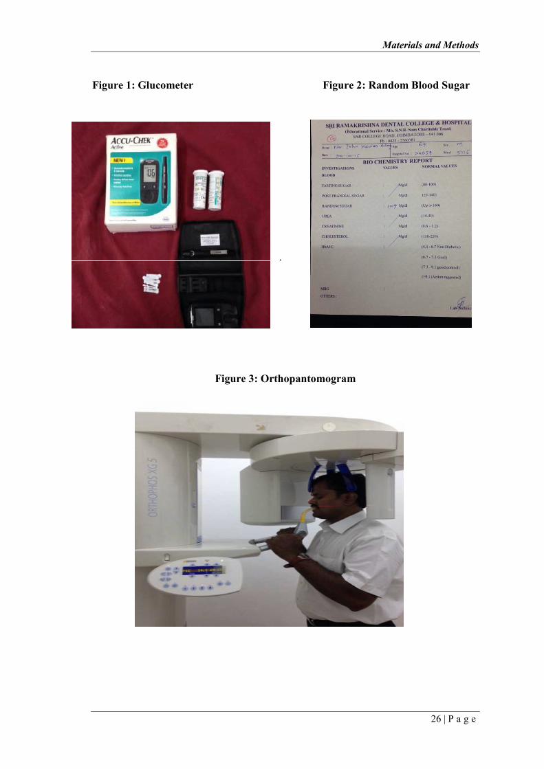
digital panoramic radiographs of the jaws. Two trained radiographic technician using a
digital orthopantomograph machine (Orthhophos XG5, Sirona) took the panoramic
radiographs. All films were Konica exposed for 1.1 s with 50 kV and long cone
(Figures 1,2 &3). All teeth, excluding third molars, were recorded. Patients with total
number of teeth less than 14 were excluded. Grossly decayed teeth were considered as
absent. Teeth were categorized as root filled teeth if they had been filled with a
radiopaque material in the root canal(s). The following information was recorded on a
structured form for each subject
1. The number of teeth present
2. The number and location of teeth without root fillings (untreated teeth)
having identifiable periapical lesions
3. The number and location of root-filled teeth
Materials and Methods
22 | P a g e
4. The number and location of root filled teeth having identifiable periapical
lesions.
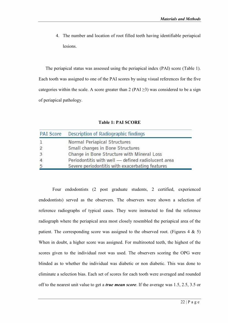
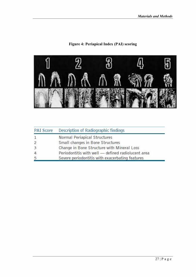
The periapical status was assessed using the periapical index (PAI) score (Table 1).
Each tooth was assigned to one of the PAI scores by using visual references for the five
categories within the scale. A score greater than 2 (PAI ≥3) was considered to be a sign
of periapical pathology.
Table 1: PAI SCORE
Four endodontists (2 post graduate students, 2 certified, experienced
endodontists) served as the observers. The observers were shown a selection of
reference radiographs of typical cases. They were instructed to find the reference
radiograph where the periapical area most closely resembled the periapical area of the
patient. The corresponding score was assigned to the observed root. (Figures 4 & 5)
When in doubt, a higher score was assigned. For multirooted teeth, the highest of the
scores given to the individual root was used. The observers scoring the OPG were
blinded as to whether the individual was diabetic or non diabetic. This was done to
eliminate a selection bias. Each set of scores for each tooth were averaged and rounded
off to the nearest unit value to get a true mean score. If the average was 1.5, 2.5, 3.5 or
Materials and Methods
23 | P a g e
4.5, it was rounded off to the unit value closest to the most experienced observer's
score.
The total number of teeth with apical periodontitis between both the groups was
compared and tabulated.
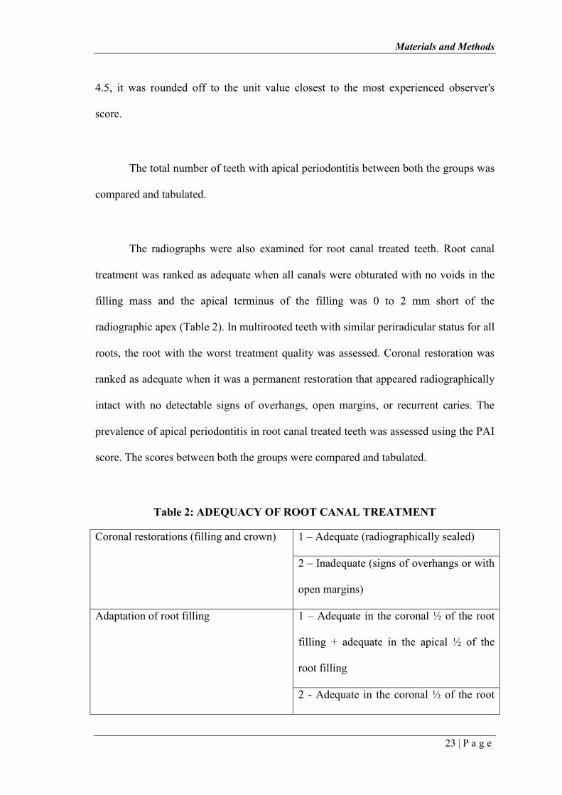
The radiographs were also examined for root canal treated teeth. Root canal
treatment was ranked as adequate when all canals were obturated with no voids in the
filling mass and the apical terminus of the filling was 0 to 2 mm short of the
radiographic apex (Table 2). In multirooted teeth with similar periradicular status for all
roots, the root with the worst treatment quality was assessed. Coronal restoration was
ranked as adequate when it was a permanent restoration that appeared radiographically
intact with no detectable signs of overhangs, open margins, or recurrent caries. The
prevalence of apical periodontitis in root canal treated teeth was assessed using the PAI
score. The scores between both the groups were compared and tabulated.
Table 2: ADEQUACY OF ROOT CANAL TREATMENT
Coronal restorations (filling and crown)
1 – Adequate (radiographically sealed)
2 – Inadequate (signs of overhangs or with
open margins)
Adaptation of root filling
1 – Adequate in the coronal ½ of the root
filling + adequate in the apical ½ of the
root filling
2 - Adequate in the coronal ½ of the root

Materials and Methods
24 | P a g e
filling + Inadequate in the apical ½ of the
root filling
3 - Inadequate in the coronal ½ of the root
filling + adequate in the apical ½ of the
root filling
4 - Inadequate in the coronal ½ of the root
filling + Inadequate in the apical ½ of the
root filling
Length of root filling 1 - Root filling ending ≤ 3 mm from
radiographic apex
2 - Root filling ending ≥ 3 mm from
radiographic apex
3 - Pulpotomy, material seen only in the
pulp chamber
4 - Flush, root filling ending at the
radiographic apex
5 - Over-filling, root filling material seen
in the periapical area
Materials and Methods
25 | P a g e
Table 3: SCORING FOR ROOT CANAL TREATED TEETH
1. Adaptation of root filling to canal walls: adequate if no voids were present in
the root filling;
Score 1 = Adequate
Scores 2, 3 and 4 = Inadequate
2. Length of root filling: adequate if ending ≤ 3 mmfrom, or flush with, the
radiographic apex
Score 1 and 4 = Adequate
Score 2, 3 and 5 = Inadequate
In the diabetic group, the HbA1c levels were recorded as a proof of the
glycemic control status, and an attempt was made to find out if the level of glycemic
control had any effect on the prevalence of apical periodontitis.
Data were statistically analyzed to evaluate the significance in the differences
between type 2 diabetic individuals and controls using the Wilcoxon signed rank and
McNemar tests when the individual was the unit of analysis, whereas the chi-square test
with Yates correction was used when tooth was the unit of analysis.
Materials and Methods
26 | P a g e
Figure 1: Glucometer Figure 2: Random Blood Sugar
.
Figure 3: Orthopantomogram
Materials and Methods
27 | P a g e
Figure 4: Periapical Index (PAI) scoring
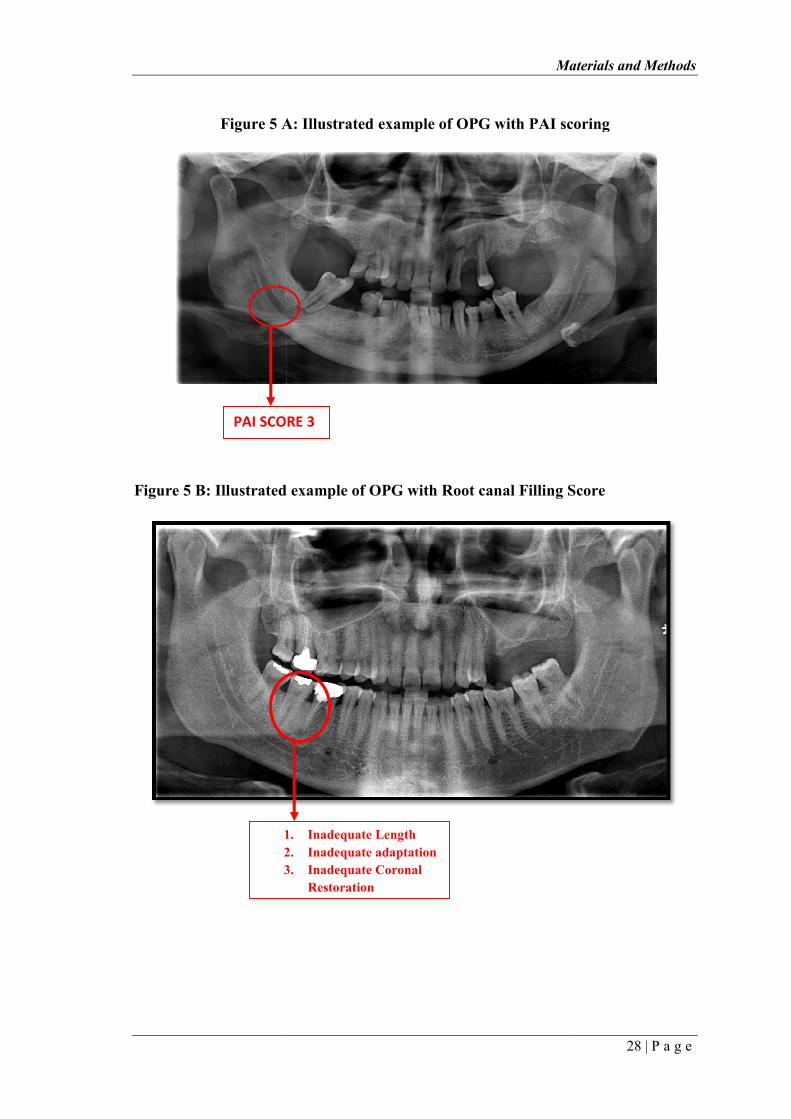
Figure 5 A
Figure 5 B: Illustrated example of OPG with Root canal
PAI SCORE 3
1.
2.
3.
Materials and Methods
A: Illustrated example of OPG with PAI scoring
Figure 5 B: Illustrated example of OPG with Root canal Filling Score
PAI SCORE 3
1. Inadequate Length
2. Inadequate adaptation
3. Inadequate Coronal
Restoration
Materials and Methods
28 | P a g e
: Illustrated example of OPG with PAI scoring
Filling Score

Figure 6 – schematic representation of the methodology of the study
Materials and Methods
schematic representation of the methodology of the study
Materials and Methods
29 | P a g e
schematic representation of the methodology of the study

RESULTS
STUDY GROUP
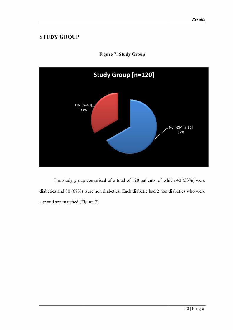
The study group comprised of a total of 120 patients, of which 40 (33%) were
diabetics and 80 (67%) were non diabetics. Each diabetic had 2 non diabetics who we
age and sex matched (Figure 7
DM [n=40]
33%
Figure 7: Study Group
The study group comprised of a total of 120 patients, of which 40 (33%) were
diabetics and 80 (67%) were non diabetics. Each diabetic had 2 non diabetics who we
age and sex matched (Figure 7)
DM [n=40]
33%
Study Group [n=120]
Results
30 | P a g e
The study group comprised of a total of 120 patients, of which 40 (33%) were
diabetics and 80 (67%) were non diabetics. Each diabetic had 2 non diabetics who were
Non-DM[n=80]
67%
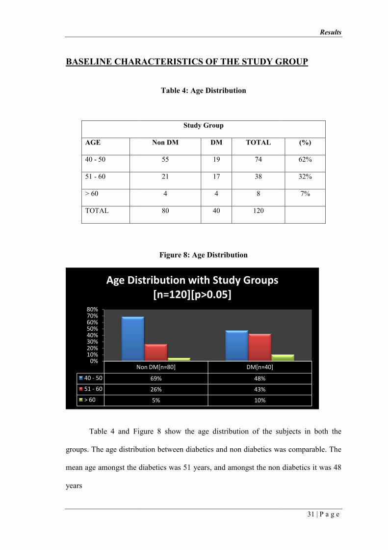
BASELINE CHARACTERISTICS OF THE STUDY
AGE
40 - 50
51 - 60
> 60
TOTAL
Table 4 and Figure 8
groups. The age distribution between diabetics and non diabetics was comparable. The
mean age amongst the diabetics was 51 years, and amongst the non diabetics it was 48
years
Non DM[n=80]
40 - 50
51 - 60
> 60
0%10%20%30%40%50%60%70%80%
Age Distribution with Study Groups
BASELINE CHARACTERISTICS OF THE STUDY GROUP
Table 4: Age Distribution
Study Group
Non DM DM TOTAL
55 19 74
21 17 38
4 4 8
80 40 120
Figure 8: Age Distribution
and Figure 8 show the age distribution of the subjects in both the
groups. The age distribution between diabetics and non diabetics was comparable. The
mean age amongst the diabetics was 51 years, and amongst the non diabetics it was 48
Non DM[n=80] DM[n=40]
69% 48%
26% 43%
5% 10%
Age Distribution with Study Groups
[n=120][p>0.05]
Results
31 | P a g e
GROUP
(%)
62%
32%
7%
show the age distribution of the subjects in both the
groups. The age distribution between diabetics and non diabetics was comparable. The
mean age amongst the diabetics was 51 years, and amongst the non diabetics it was 48
Age Distribution with Study Groups
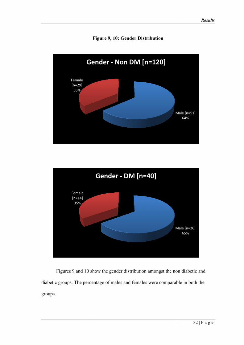
Figures 9 and 10
diabetic groups. The percentage of males and females were comparable in both the
groups.
Female
[n=29]
36%
Gender
Female
[n=14]
35%
Figure 9, 10: Gender Distribution
10 show the gender distribution amongst the non diabetic and
diabetic groups. The percentage of males and females were comparable in both the
Male [n=51]
Gender - Non DM [n=120]
Male [n=26]
Gender - DM [n=40]
Results
32 | P a g e
show the gender distribution amongst the non diabetic and
diabetic groups. The percentage of males and females were comparable in both the
Male [n=51]
64%
Male [n=26]
65%

Results
33 | P a g e
Table 5: Mean number of teeth per person in both the groups
Mean Teeth per person
Study Mean
SD
95% CI for Mean
Minimum Maximum Sig
Group Teeth Lower Upper
Non DM 25.3 3.4 24.5 26.0 12 28
DM 25.6 2.4 24.8 26.4 19 28 >0.05
Total 25.4 3.1 24.8 25.9 12 28
Figure 11: Mean number of teeth per person in both the groups
As seen in Table 5 and Figure 11, the mean number of teeth per person in the
diabetic group was 25.6 ±2.4, and in the non diabetic group it was 25.3 ±3.4.
With AP
Without AP
0%
20%
40%
60%
80%
100%
Association of AP Teeth with study
Groups [N=120][p<0.05]
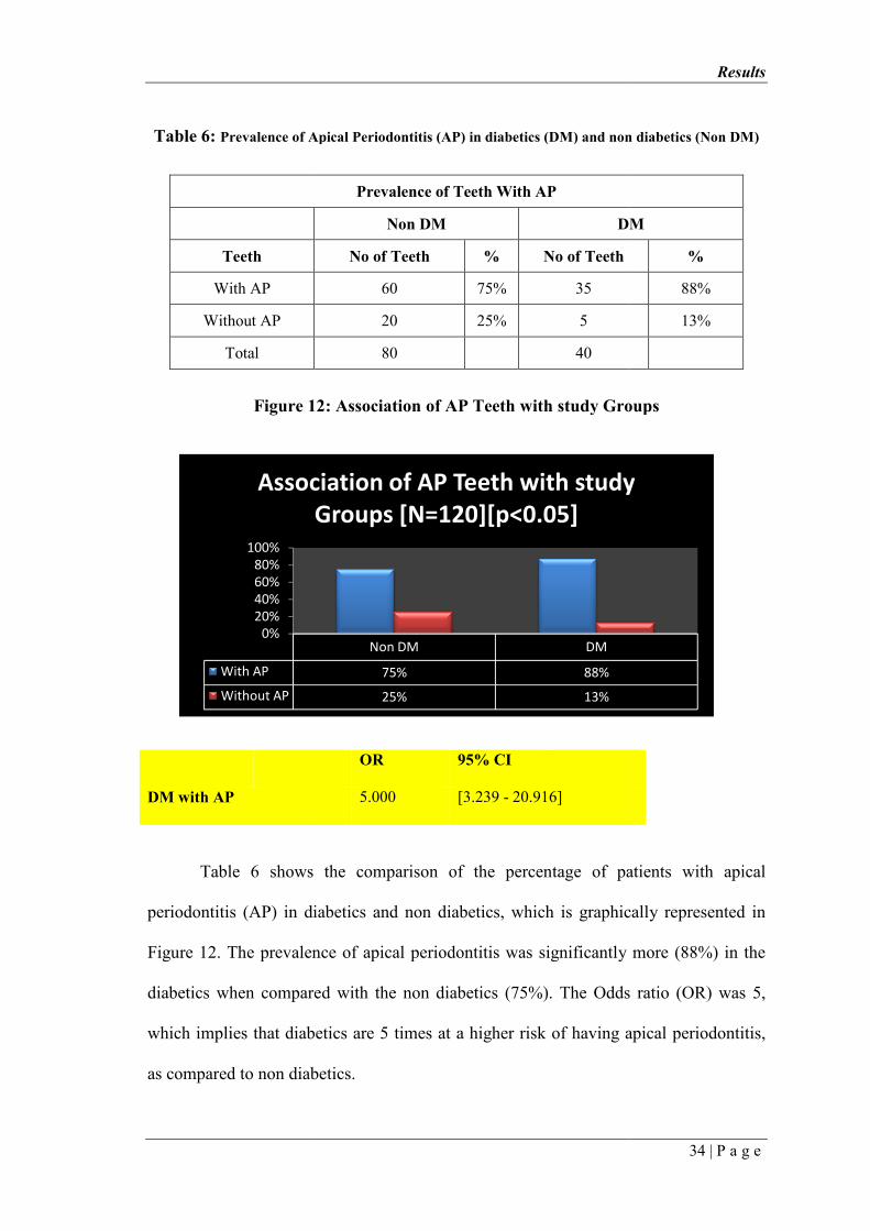
Table 6: Prevalence of Apical Periodontitis (AP) in diabetics (DM) and non
Teeth
With AP
Without AP
Total
Figure 1
Table 6 shows the comparison of the percentage of patients with apical
periodontitis (AP) in diabetics and non diabetics, which is gra
Figure 12. The prevalence of apical periodontitis was significantly more (88%) in the
diabetics when compared with the non diabetics (75%). The Odds ratio (OR) was 5,
which implies that diabetics are 5 times at a higher risk of having apical periodontitis,
as compared to non diabetics.
DM with AP
Non DM DM
75% 88%
25% 13%
Association of AP Teeth with study
Groups [N=120][p<0.05]
Prevalence of Apical Periodontitis (AP) in diabetics (DM) and non
Prevalence of Teeth With AP
Non DM DM
No of Teeth % No of Teeth
60 75% 35
20 25% 5
80
40
12: Association of AP Teeth with study Group
shows the comparison of the percentage of patients with apical
periodontitis (AP) in diabetics and non diabetics, which is graphically represented in
. The prevalence of apical periodontitis was significantly more (88%) in the
diabetics when compared with the non diabetics (75%). The Odds ratio (OR) was 5,
which implies that diabetics are 5 times at a higher risk of having apical periodontitis,
red to non diabetics.
OR 95% CI
5.000 [3.239 - 20.916]
Results
34 | P a g e
Association of AP Teeth with study
Prevalence of Apical Periodontitis (AP) in diabetics (DM) and non diabetics (Non DM)
DM
%
88%
13%
Association of AP Teeth with study Groups
shows the comparison of the percentage of patients with apical
hically represented in
. The prevalence of apical periodontitis was significantly more (88%) in the
diabetics when compared with the non diabetics (75%). The Odds ratio (OR) was 5,
which implies that diabetics are 5 times at a higher risk of having apical periodontitis,
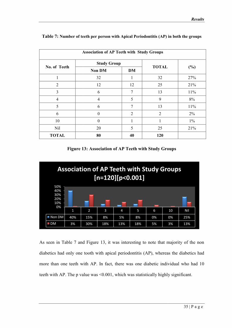
Table 7: Number of teeth per person with Apical Periodontitis (AP) in both the groups
Association of AP Teeth with Study Groups
No. of Teeth
1
2
3
4
5
6
10
Nil
TOTAL
Figure 13
As seen in Table 7 and Figure 13
diabetics had only one tooth with apical periodontitis (AP), whereas the diabetics had
more than one teeth with AP. In fact, there was one diabetic individual who had 10
teeth with AP. The p value was <
1
Non DM 40%
DM 3%
0%10%20%30%40%50%
Association of AP Teeth with Study Groups
Number of teeth per person with Apical Periodontitis (AP) in both the groups
Association of AP Teeth with Study Groups
Study Group TOTAL
Non DM DM
32 1 32
12 12 25
6 7 13
4 5 9
6 7 13
0 2 2
0 1 1
20 5 25
80 40 120
13: Association of AP Teeth with Study Groups
and Figure 13, it was interesting to note that majority of the non
diabetics had only one tooth with apical periodontitis (AP), whereas the diabetics had
more than one teeth with AP. In fact, there was one diabetic individual who had 10
teeth with AP. The p value was <0.001, which was statistically highly significant.
2 3 4 5 6
15% 8% 5% 8% 0%
30% 18% 13% 18% 5%
Association of AP Teeth with Study Groups
[n=120][p<0.001]
Results
35 | P a g e
Number of teeth per person with Apical Periodontitis (AP) in both the groups
(%)
27%
21%
11%
8%
11%
2%
1%
21%
Association of AP Teeth with Study Groups
, it was interesting to note that majority of the non
diabetics had only one tooth with apical periodontitis (AP), whereas the diabetics had
more than one teeth with AP. In fact, there was one diabetic individual who had 10
0.001, which was statistically highly significant.
10 Nil
0% 25%
3% 13%
Association of AP Teeth with Study Groups

Table 8: Prevalence of Root canal Treatment (RCT) between both the groups
RCT
No of Teeth
Done
Not Done
Total
Table 8 and Figure 14
both the groups. The total number
diabetics had root canal treated teeth, compared to 51% of non d
diabetics had a higher incidence of non treated teeth, this difference was minimal and
not statistically significant. (p >0.05)
Done
Not Done
0%
10%
20%
30%
40%
50%
60%
Prevalence of RCT [N=120][p>0.05]
: Prevalence of Root canal Treatment (RCT) between both the groups
Prevalence of RCT
Non DM DM
No of Teeth % No of Teeth
41 51% 22
39 49% 18
80 40
Figure 14: Prevalence of RCT
and Figure 14 represent the prevalence of root canal treatment between
both the groups. The total number of Root canal treated teeth was
diabetics had root canal treated teeth, compared to 51% of non diabetics. Though non
diabetics had a higher incidence of non treated teeth, this difference was minimal and
not statistically significant. (p >0.05)
Non DM DM
51% 55%
49% 45%
Prevalence of RCT [N=120][p>0.05]
Results
36 | P a g e
: Prevalence of Root canal Treatment (RCT) between both the groups
DM
%
55%
45%
represent the prevalence of root canal treatment between
of Root canal treated teeth was 63. 55% of the
iabetics. Though non
diabetics had a higher incidence of non treated teeth, this difference was minimal and
Prevalence of RCT [N=120][p>0.05]
Table 9: Prevalence of AP in RCT treated teeth in diabetics and non diabetics
Figure 15
59%
AP in RCT treated teeth
RCT treated teeth with AP
RCT treated teeth with AP
RCT treated teeth without AP
: Prevalence of AP in RCT treated teeth in diabetics and non diabetics
Figure 15: AP in RCT treated teeth - Diabetics
41%
AP in RCT treated teeth - Diabetics (n - 22)
RCT treated teeth with AP RCT treated teeth without AP
Diabetic (n – 22)
Non diabetic (n – 41)
Total
9 (41%) 12 (29%) 21
13 (59%) 29(71%) 42
Results
37 | P a g e
: Prevalence of AP in RCT treated teeth in diabetics and non diabetics
Diabetics
RCT treated teeth without AP
p value
<0.05
<0.05

Figure
Table 9 shows the prevalence of apical periodontitis (AP) in root canal treated
teeth amongst diabetics and non diabetics, which is grap
15 and Figure 16 respectively. 41% of diabetic root canal treated teeth had apical
periodontitis, whereas only 29% of non diabetic root canal treated teeth had AP. This
difference was statistically significant with a p value of <0.05.
AP in RCT treated teeth
RCT treated teeth with AP
Figure 16: AP in RCT treated teeth - Non diabetic
shows the prevalence of apical periodontitis (AP) in root canal treated
teeth amongst diabetics and non diabetics, which is graphically represented in Figure
respectively. 41% of diabetic root canal treated teeth had apical
whereas only 29% of non diabetic root canal treated teeth had AP. This
difference was statistically significant with a p value of <0.05.
29%
71%
AP in RCT treated teeth - Non diabetic (n - 41)
RCT treated teeth with AP RCT treated teeth without AP
Results
38 | P a g e
Non diabetic
shows the prevalence of apical periodontitis (AP) in root canal treated
hically represented in Figure
respectively. 41% of diabetic root canal treated teeth had apical
whereas only 29% of non diabetic root canal treated teeth had AP. This
RCT treated teeth without AP
Results
39 | P a g e
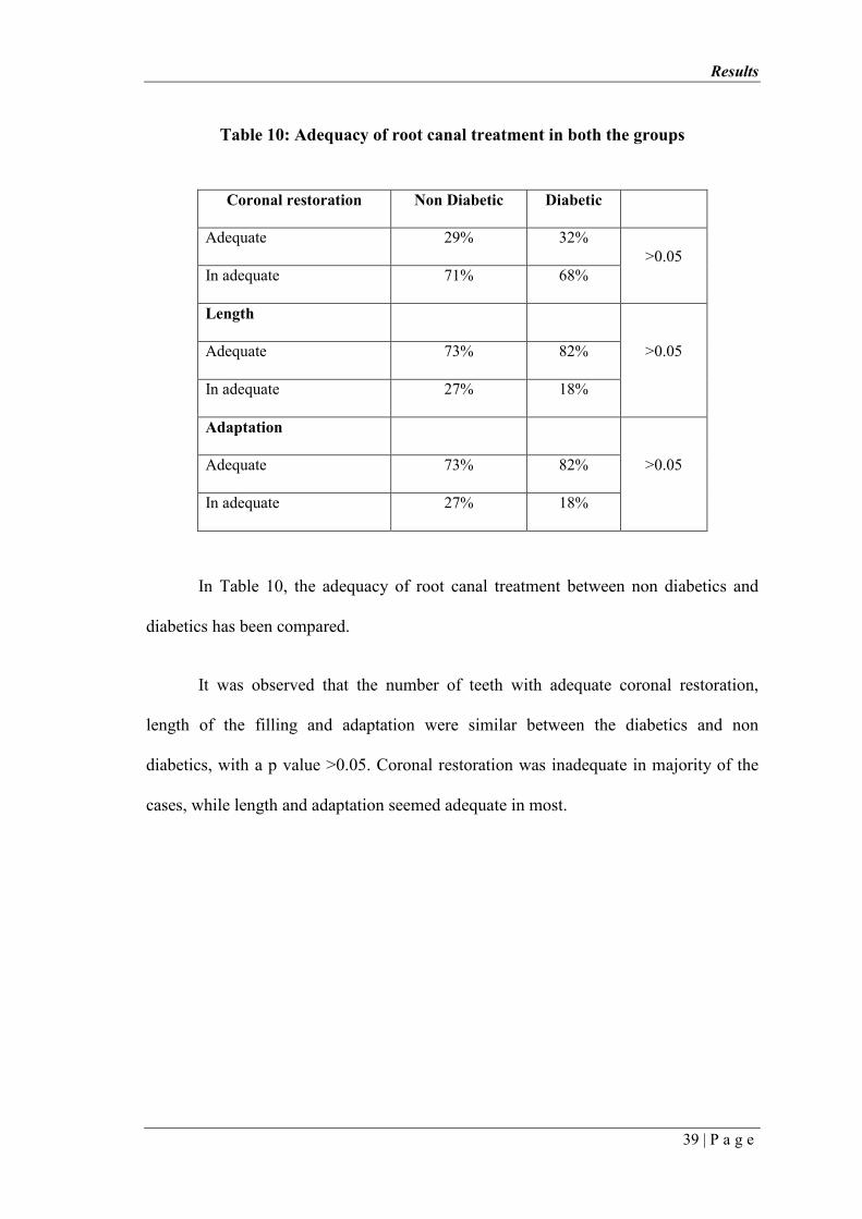
Table 10: Adequacy of root canal treatment in both the groups
In Table 10, the adequacy of root canal treatment between non diabetics and
diabetics has been compared.
It was observed that the number of teeth with adequate coronal restoration,
length of the filling and adaptation were similar between the diabetics and non
diabetics, with a p value >0.05. Coronal restoration was inadequate in majority of the
cases, while length and adaptation seemed adequate in most.
Coronal restoration Non Diabetic Diabetic
Adequate 29% 32%
>0.05 In adequate 71% 68%
Length
>0.05 Adequate 73% 82%
In adequate 27% 18%
Adaptation
>0.05 Adequate 73% 82%
In adequate 27% 18%
Results
40 | P a g e
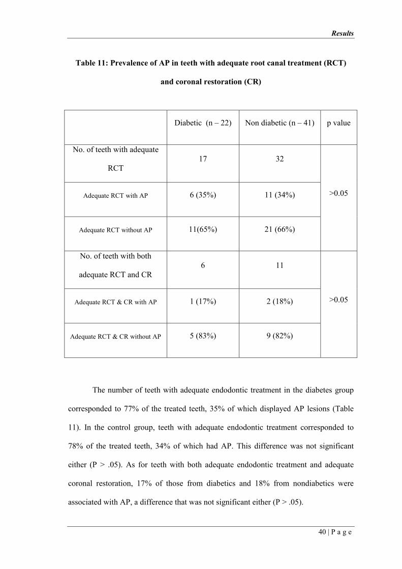
Table 11: Prevalence of AP in teeth with adequate root canal treatment (RCT)
and coronal restoration (CR)
The number of teeth with adequate endodontic treatment in the diabetes group
corresponded to 77% of the treated teeth, 35% of which displayed AP lesions (Table
11). In the control group, teeth with adequate endodontic treatment corresponded to
78% of the treated teeth, 34% of which had AP. This difference was not significant
either (P > .05). As for teeth with both adequate endodontic treatment and adequate
coronal restoration, 17% of those from diabetics and 18% from nondiabetics were
associated with AP, a difference that was not significant either (P > .05).
Diabetic (n – 22) Non diabetic (n – 41) p value
No. of teeth with adequate
RCT 17 32
>0.05 Adequate RCT with AP 6 (35%) 11 (34%)
Adequate RCT without AP 11(65%) 21 (66%)
No. of teeth with both
adequate RCT and CR 6 11
>0.05 Adequate RCT & CR with AP 1 (17%) 2 (18%)
Adequate RCT & CR without AP 5 (83%) 9 (82%)
Results
41 | P a g e
< 6.5 > 6.5
with AP[n=35] 20% 80%
without AP[n=5] 60% 40%
0%
20%
40%
60%
80%
100%
Association of AP teeth with HbA1C of DM
Group [N=40][p<0.05]
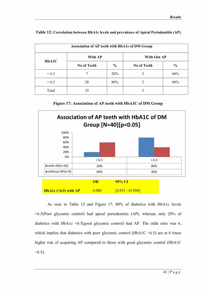
Table 12: Correlation between HbA1c levels and prevalence of Apical Periodontitis (AP)
Association of AP teeth with HbA1c of DM Group
HbA1C With AP With Out AP
No of Teeth % No of Teeth %
< 6.5 7 20% 3 60%
> 6.5 28 80% 2 40%
Total 35
5
Figure 17: Association of AP teeth with HbA1C of DM Group
As seen in Table 12 and Figure 17, 80% of diabetics with HbA1c levels
>6.5(Poor glycemic control) had apical periodontitis (AP), whereas only 20% of
diabetics with HbA1c <6.5(good glycemic control) had AP. The odds ratio was 6,
which implies that diabetics with poor glycemic control (HbA1C >6.5) are at 6 times
higher risk of acquiring AP compared to those with good glycemic control (HbA1C
<6.5).
OR 95% CI
HbA1c (>6.5) with AP 6.000 [0.835 - 43.094]

DISCUSSION

Discussion
42 | P a g e
Endodontic infection and periodontal disease are very common conditions
worldwide. Results from numerous studies have suggested links between periodontal
disease and diabetes, but endodontic disease has not been studied extensively in this
regard. The possible connection between chronic oral inflammatory conditions such as
chronic apical periodontitis and systemic health is one of the most interesting areas
currently being studied by the medical and dental scientific community. As there are so
far very few studies in the literature reporting on diabetes as a disease modifier in
endodontics, this cross-sectional study was conducted to investigate the prevalence of
AP and endodontic treatment in type 2 diabetic individuals.
A cross-sectional design was used to include a large number of individuals. The
subjects included in this study were adult patients attending dental service of the Dental
College for the first time. The recruitment of subjects was the same as those used by
other authors (Kirkevang et al. 200047
, Britto et al.200318
, Fouad & Burleson 200321).
Both the study and the control groups consisted of more men than women;
however, epidemiological studies have reported that sex had no effect on the presence
of AP or the frequency of endodontic treatment (Ørstavik et al. 198619
, Jime´nez-
Pinzo´n et al. 200448). There was no significant difference in age between both groups.
Both the groups were age and sex matched. Matching individuals by sex and age was
performed with the purpose of reducing the interference of these variables on the final
outcome.
Discussion
43 | P a g e
In an attempt to circumvent possible biases, all the individuals participating in the study
were attending the Dental college for the first time and basically pertained to the same
socioeconomic status.
Orthopantomogram (OPG) was used to evaluate the presence of AP in our
study. Previous studies have also used OPG (Imfeld 199149
, Kirkevang et al.200150
,
Boucher et al. 200251
, Britto et al. 200318
, Kirkevang & Wenzel 200352). Moreover,
the Periapical Index score (PAI) used for scoring periapical status was first described
for periapical radiographs (Ørstavik et al. 198619) and has been widely used in the
literature (Eriksen et al. 199553
, Marques et al. 199854
, Sidaravicius et al. 199955
,
Kirkevang et al. 2001, Boucher et al. 2002, Kirkevang & Wenzel 2003, Segura-Egea
et al. 200422). In our study, all teeth, excluding third molars, were recorded. Patients
with total number of teeth less than 14 were excluded. Grossly decayed teeth were
considered as absent. Thus, the results reproduced the periapical status of the subjects.
Other authors, in similar studies, have excluded teeth with absent or defective coronal
restorations, teeth with their periradicular tissues near radiolucent anatomic structures,
or root filled teeth with inadequate root canal treatment (Britto et al. 200318
). However,
these exclusions may alter the results and prevent the determination of the real
periapical status of the subjects.
The average number of teeth was similar in diabetic patients (25.6±2.4) and non
diabetic controls (25.3±2.4). This is in contrast to what was observed by Lopez et al12
in a study done in Spain in 2011. The author observed that the average number of teeth
was significantly lower in diabetic patients (21.9 ± 6.4 and) than in control subjects
(24.6 ± 3.8). It was proposed that Diabetes Mellitus, especially when poorly controlled,
Discussion
44 | P a g e
was associated with significant tooth loss because of the increased incidence and
severity of caries and the aggressive forms of periodontal disease associated with
diabetes. However, in another study done by Falk et al. (198956
), there was no
significant difference in the number of teeth between diabetic and non diabetic subjects.
One possible explanation for this could be that we had excluded grossly decayed teeth
and patients with less than 14 teeth. Thus the influence of periodontal disease has been
minimized in our study.
Endodontology includes pulp and periapical biology and pathology. Whilst the
initial diagnoses and the difficulties associated with treatment may be related to the
state of the pulp, the ultimate biological aim of this treatment is no longer the
preservation of the pulp, but the prevention and elimination of infection in the root
canal system to prevent or cure apical periodontitis (AP)32. Apical periodontitis, an
inflammatory process around the apex of a root, is primarily a sequel to microbial
infection of the pulp space. The infectious etiology of AP and the main role of
microbial factors in the initiation, development and persistence of the condition have
been widely documented, and it can be considered as a disease of bacterial infection.
Apical periodontitis is a remarkably prevalent condition, especially in a country
like India25, 57. The prevalence of AP is as high as 61% of individuals and 2.8–4.2% of
the teeth, as noted by Segura et al, 201532. Inadequate aseptic control, poor access
cavity design, missed canals, insufficient instrumentation and leaking temporary or
permanent restorations are common problems that may lead to persistent AP.
Discussion
45 | P a g e
Even though periapical infections cause a number of local tissue responses with
the purpose of limiting the spread of the infectious elements, AP may not exclusively
be a local phenomenon. The interaction between the lipopolysaccharide (LPS) from
anaerobic gram-negative bacteria causing AP with Toll-like receptor 4 (TLR4) on
macrophages and neutrophils activates the broad axis of innate immunity, up-regulating
pro-inflammatory cytokines such as IL-1b, IL-6, IL-8, TNF-a and prostaglandin E2
(PGE2). These cytokines may be released into the systemic circulation inducing or
perpetuating an elevated chronic systemic inflammatory status. Although there is no
conclusive scientific evidence indicating that an infected root canal may act as a focus
of infection to distant body sites (except for systemically compromised patients), the
opposite has not been proven either, that is, there is no clear evidence showing that
endodontic infections are an isolated event with no effect on the rest of the body.
In the two last decades, ‘periodontal medicine’ has developed as a distinct area
that focuses on the relationship between periodontal disease (PD) and systemic
diseases58. Several epidemiological studies have found associations between systemic
health and PD. Thus, PD has been associated with diabetes mellitus (DM), coronary
heart disease (CHD) and acute myocardial infarction (AMI), preterm-low birthweight,
respiratory diseases, osteoporosis in post-menopause women, metabolic syndrome and
early loss of memory and capacity for calculation. The evidence of the association
between PD and systemic diseases has focused attention on the diagnosis and treatment
of PD, improving, consequently, the patient’s oral and systemic health. (Figure 18)
Chronic periodontal and endodontic inflammatory processes have three
important similarities:
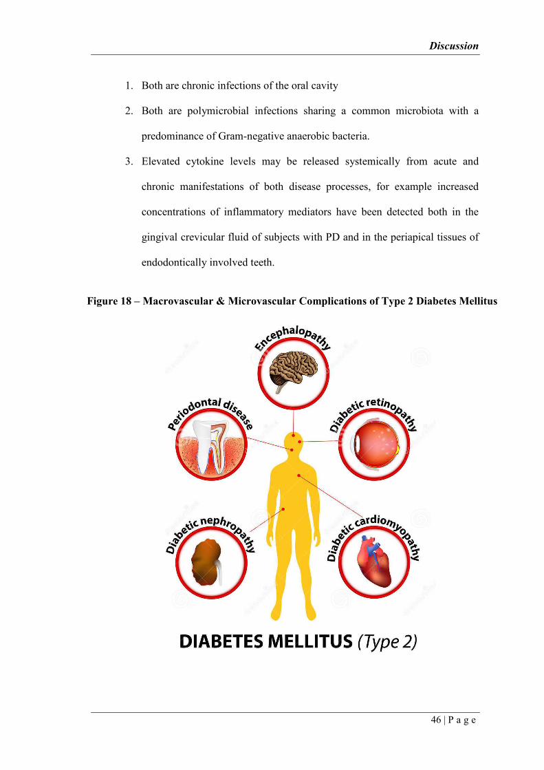
Discussion
46 | P a g e
1. Both are chronic infections of the oral cavity
2. Both are polymicrobial infections sharing a common microbiota with a
predominance of Gram-negative anaerobic bacteria.
3. Elevated cytokine levels may be released systemically from acute and
chronic manifestations of both disease processes, for example increased
concentrations of inflammatory mediators have been detected both in the
gingival crevicular fluid of subjects with PD and in the periapical tissues of
endodontically involved teeth.
Figure 18 – Macrovascular & Microvascular Complications of Type 2 Diabetes Mellitus
Discussion
47 | P a g e
Likewise, one might assume that AP is associated with the same systemic
disorders that are associated with PD. Therefore, ‘endodontic medicine’ should be
developed following the same path as ‘periodontal medicine’: evaluating the
association between endodontic and systemic diseases. However, the influence that
chronic periapical processes could produce on highly prevalent systemic diseases, such
as diabetes and congenital heart disease has been poorly studied. The lack of scientific
studies on this topic might be masking the potential risk of retaining teeth with chronic
AP and the real importance and health advantages of endodontic treatments to patients,
doctors and dentists. Pro-inflammatory status and impaired immune response
associated with systemic diseases can affect the reparative response of the dental pulp
and periapical healing, influencing the two main endodontic variables: the prevalence
of AP and the frequency of RCT.
Diabetes mellitus is a clinically and genetically heterogeneous group of
disorders affecting the metabolism of carbohydrates, lipids and proteins, in which
hyperglycaemia is a main feature (Expert Committee on the Diagnosis and
Classification of Diabetes Mellitus 200059
). These disorders are due to a deficiency in
insulin secretion caused by pancreatic b-cell dysfunction and/or insulin resistance in
liver and muscle. Diabetes affects more than 9% of the adult population, and its high
morbidity and mortality amongst affected individuals has a substantial impact on
national healthcare systems. Age-adjusted and country-adjusted prevalence of Type 2
diabetes mellitus (T2DM) in 11 European countries in 2004 was 10.2% in men and
8.5% in women (Espelt et al. 201360
). India leads the world with largest number of
diabetic subjects earning the term “DIABETES CAPITAL OF THE WORLD.”

Discussion
48 | P a g e
According to Indian council of medical research data published in 20061, prevalence of
type 2 diabetes in India is 40.9 million and is expected to rise to 69.9 million by 2025.
Glycated haemoglobin (HbA1c) has been used as a ‘gold standard’ for mean
glycaemia and as a measure of risk for the development of DM complications (Expert
Committee on the Diagnosis and Classification of Diabetes Mellitus 2000)58. The
American Association of Clinical Endocrinologists (AACE)61 considers HbA1c levels
≤6.5% as a goal for optimal glycaemic control in diabetic patients. Type 1 diabetes
results from cellular-mediated autoimmune destruction of pancreatic b-cells, which
usually leads to total loss of insulin secretion; in contrast, type 2 diabetes is caused by
resistance to insulin combined with a failure to produce enough additional insulin to
compensate for the resistance. Many studies have shown that inflammation plays a very
important role in the pathogenesis of T2DM.
In animal studies, histological and histometrical changes in pulpal and
periapical tissues after pulpal exposure in streptozotocin-induced diabetic rats have
been studied24, 38. It was observed that more pronounced periapical inflammation and
larger periapical lesions were seen in diabetic rats compared with controls. The effect
of hyperglycaemia on pulp healing in exposed rat pulps capped with Mineral Trioxide
Aggregate (MTA) has also been investigated. There was an inverse association between
dentine bridge formation and inflammatory cell infiltration: dentine bridge formation
was inhibited in diabetic rats and more inflammation was observed in these pulps. It
has also been shown that oral infections affect glycaemic conditions in diabetic rats and
increase HbA1c levels in normoglycaemic and diabetic rats.
Discussion
49 | P a g e
Several clinical and epidemiological studies carried out in humans have analysed the
association between endodontic variables and DM11, 12. The main endodontic variables
analysed in these studies are as follows:
1. The prevalence of AP
2. The prevalence of RCT and
3. The outcome of RCT, assessed as the percentage of RFT with or without PLs,
or as the prevalence of tooth extraction after nonsurgical RCT (NSRCT).
The results of studies conducted so far are inconclusive, but suggest an
association between DM and a higher prevalence of AP.
In the present study, diabetic patients showed a higher prevalence of AP (88%)
compared with age- and sex-matched control subjects (75%; p <0.05). This is in
accordance with previous reports by Segura et al.200513
, Bender et al 200337
, Lopez et
al 201112. On the contrary, Britto et al (2003)
18 in a similar study design, investigated
the prevalence of radiographic periradicular radiolucencies in root-filled teeth and
untreated teeth in patients with and without diabetes and found no significant
differences in the prevalence of AP between diabetics and controls. However, these
investigators excluded teeth with absent or defective coronal restorations, teeth with
their periradicular tissues near radiolucent anatomic structures, and root-filled teeth
with inadequate root canal treatment. Because of this, their results do not reflect the real
periapical status of the subjects studied, and the comparison between both groups
cannot produce definite conclusions. The number of diabetics and control subjects in

Discussion
50 | P a g e
our study (40 and 80 respectively) were more than the number of subjects included in
the previous studies. All the studies that have been quoted above, have been done in the
Western world. Our study is one of the first in the Indian population.
On comparing the number of patients having one tooth with AP and those
having more than one teeth with AP, in the present study it was observed that 34 out of
40 diabetics had more than one teeth with AP, whereas 32 out of 80 non diabetics had
only 1 tooth with AP. This difference was statistically highly significant with a p value
of < 0.001. Marotta Patricia et al (201211
) found no significant differences when the
analysis involved either the number of patients with at least 1 AP lesion or the mean
number of lesions per individual. But the sample size in their study was too small (30
diabetics and 30 non diabetics). Our study observed results similar to Lopez et al
(2011)12
and Segura et al (2005)13.
The prevalence of root canal treatment was similar in both the groups, and there
were no significant differences between the percentages of diabetic and nondiabetic
individuals. It is worth pointing out that this factor involves other variables, such as the
accessibility of patients to dental care. As reported earlier, we tried to avoid this bias by
including only individuals belonging to a similar socioeconomic status and attending
our dental college for the first time.
The prevalence of AP in root canal–treated teeth may be suggestive of the
success rate of the treatment although data should be viewed with care because of the
cross-sectional nature of the study. In our study, 41% of diabetic root canal treated
Discussion
51 | P a g e
teeth, had AP compared to only 29% of non diabetic root canal treated teeth with AP
(p <0.05). Foud et al (2003)21
concluded that patients with diabetes displayed a
reduced success rate in endodontic treatments of teeth with preoperative AP. Britto et
al (2003)18 also observed similar findings. It has been hypothesized that diabetes may
increase the rate of endodontic failure and persistent AP, and this statement is
supported by the results of the present study. There is scientific evidence to
demonstrate a poorer prognosis for root filled teeth (RFT) in diabetics. Thus, diabetic
patients have delayed periapical repair and greater likelihood of RFT loss.
Metabolic control of diabetes mellitus and apical periodontitis
The lack of control in DM could delay healing of PLs and increase their size,
despite having received endodontic treatment. In well-controlled diabetic patients PLs
healed as readily as in nondiabetics. In our study, we determined the metabolic control
of DM by measuring glycated haemoglobin levels and classifying diabetic patients as
well-controlled (HbA1c < 6.5%) or poor-controlled (HbA1c > 6.5%). 80% of diabetics
with poor glycemic control had apical periodontitis whereas only 20 % of diabetics
with good glycemic control had AP ( p <0.001).
It is also interesting to note that chronic periapical disease may contribute to
diabetic metabolic dyscontrol. There are a few studies which have shown better
glycemic control after treatment of the periapical disease. Bender et al (2003)37
reported that removal of the periapical inflammatory state usually creates a need for a
lesser amount of insulin for diabetic control. Thus, achieving a good glycemic control
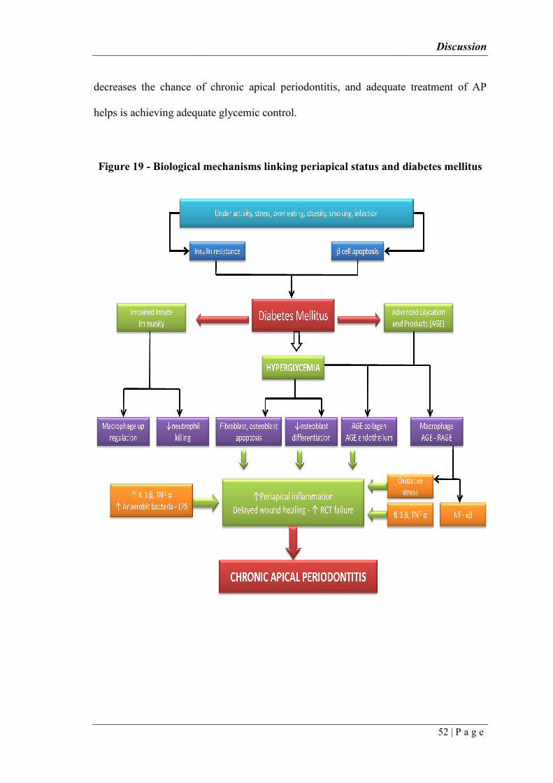
decreases the chance of chronic apical periodontitis, and adequate treatment of AP
helps is achieving adequate glycemic control.
Figure 19 - Biological mechanisms linking
decreases the chance of chronic apical periodontitis, and adequate treatment of AP
helps is achieving adequate glycemic control.
Biological mechanisms linking periapical status and diabetes mellitus
Discussion
52 | P a g e
decreases the chance of chronic apical periodontitis, and adequate treatment of AP
periapical status and diabetes mellitus
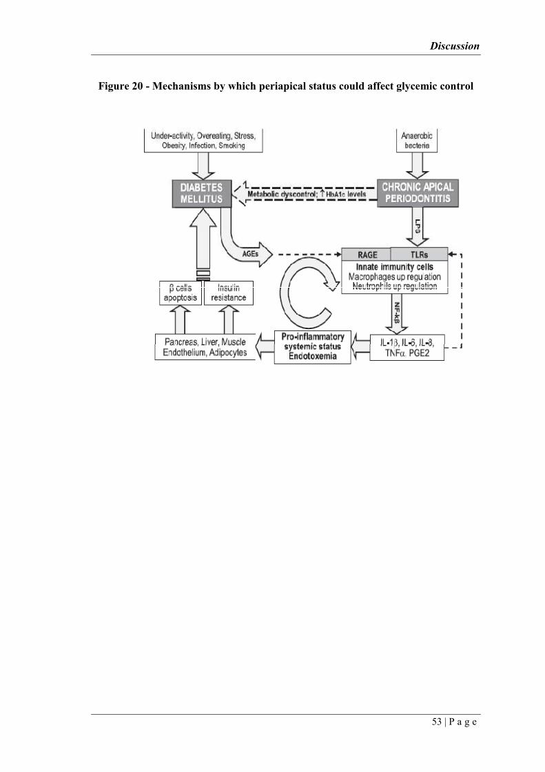
Discussion
53 | P a g e
Figure 20 - Mechanisms by which periapical status could affect glycemic control

Limitation
54 | P a g e
1. Being a cross sectional study, one of the major limitation is the fact that the
mean time since completion of endodontic treatment is unknown.
2. The number of teeth assessed for adequacy of endodontic treatment was too
small.

SUMMARY AND CONCLUSION
Summary and Conclusion
55 | P a g e
The aim of this cross-sectional study was to compare the prevalence of apical
periodontitis and endodontic treatment between patients with type 2 diabetes and those
without diabetes. 40 diabetics and 80 non diabetics (age and sex matched), with ages
ranging from 40 to 65 years, who were attending the oral medicine department for
routine dental care, were included in the study. All the subjects in both the groups,
underwent a digital panoramic radiograph of the jaws to evaluate the periapical and
endodontic status. Grossly decayed teeth were considered as absent, patients with total
number of teeth less than 14 were excluded and the third molars were also excluded.
Periapical index (PAI) score was used to assess the periapical status. A score greater
than 2 (PAI ≥3) was considered to be a sign of periapical pathology. Four observers
who were blinded, assessed the radiographs independently and the true mean score
was calculated. The prevalence of apical periodontitis in root canal treated and
untreated teeth, between both the groups were compared and tabulated. Glycated
hemoglobin (HbA1c) levels were measured in diabetics to assess their glycemic
control. An attempt was made to find out if the level of glycemic control had any effect
on the prevalence of apical periodontitis. Random blood sugar (RBS) levels were
measured for the non diabetic group, to pick up undiagnosed type 2 diabetes mellitus.
Data were statistically analyzed to evaluate the significance in the differences
between both the groups. Wilcoxon signed rank test and Mc Nemar tests were used
when the individual was the unit of analysis, whereas the chi – square test with Yates
correction was used when the tooth was the unit of analysis.
Summary and Conclusion
56 | P a g e
The results showed a higher prevalence of apical periodontitis in patients with
type 2 diabetes mellitus. However, considering the limitations of cross-sectional
studies, larger prospective clinical studies are further needed to confirm this
association. As diabetes is one of the most prevalent conditions in medically
compromised patients seeking dental treatment, dentists should be aware of the
possible relationship between endodontic infections and diabetes mellitus. It is very
vital for dental clinicians to understand the disease process of type 2 diabetes, its effect
on pulp and periapical diseases, and treatment outcome to provide competent
endodontic treatment to patients with type 2 diabetes mellitus.
Within the limitations of the study, it can be concluded that apical periodontitis
was significantly more prevalent in teeth from type 2 diabetic individuals. This higher
prevalence was observed in root canal treated and untreated teeth, with decreased
success rate of endodontic treatment. These findings suggest that diabetes can serve as
a disease modifier of apical periodontitis, which implies that individuals with diabetes
may be more prone to develop primary apical periodontitis. Even in diabetics, those
with good glycemic control, are likely to have a lesser risk of acquiring apical
periodontitis.

BIBLIOGRAPHY
Bibliograph
57 | P a g e
1. Mohan V, Sandeep S, Deepa R, Shah B, Varghese C. Epidemiology of type 2
diabetes: Indian scenario. The Indian journal of medical research.
2007;125(3):217-30.
2. Kaveeshwar SA, Cornwall J. The current state of diabetes mellitus in India. The
Australasian medical journal. 2014;7(1):45.
3. Whiting DR, Guariguata L, Weil C, Shaw J. IDF diabetes atlas: global estimates
of the prevalence of diabetes for 2011 and 2030. Diabetes research and clinical
practice. 2011;94(3):311-21.
4. McKenna SJ. Dental management of patients with diabetes. Dental Clinics of
North America. 2006;50(4):591-606.
5. Stewart JE, Wager KA, Friedlander AH, Zadeh HH. The effect of periodontal
treatment on glycemic control in patients with type 2 diabetes mellitus. Journal
of Clinical Periodontology. 2001;28(4):306-10.
6. Loe H. Periodontal disease: the sixth complication of diabetes mellitus.
Diabetes care. 1993;16(1):329-34.
7. Tsai C, Hayes C, Taylor GW. Glycemic control of type 2 diabetes and severe
periodontal disease in the US adult population. Community dentistry and oral
epidemiology. 2002;30(3):182-92.
8. Grossi SG, Genco RJ. Periodontal Disease and Diabetes Mellitus: A Two-Way
Relationship*. Annals of periodontology. 1998;3(1):51-61.
9. Al-Maskari AY, Al-Maskari MY, Al-Sudairy S. Oral manifestations and
complications of diabetes mellitus: a review. Sultan Qaboos University Medical
Journal. 2011;11(2):179.
10. Nair PNR. On the causes of persistent apical periodontitis: a review.
International Endodontic Journal. 2006;39(4):249-81.

Bibliograph
58 | P a g e
11. Marotta PS, Fontes TV, Armada L, Lima KC, Ras IN, Siqueira JF. Type 2
diabetes mellitus and the prevalence of apical periodontitis and endodontic
treatment in an adult Brazilian population. Journal of endodontics.
2012;38(3):297-300.
12. Lopez - Lopez J, Jan-Salas E, Estrugo-Devesa A, Velasco-Ortega E, Martn-
Gonzalez J, Segura-Egea JJ. Periapical and endodontic status of type 2 diabetic
patients in Catalonia, Spain: a cross-sectional study. Journal of endodontics.
2011;37(5):598-601.
13. Segura Egea JJ, Jimnez Pinzn A, Ros Santos JV, Velasco Ortega E,
Cisneros Cabello R, Poyato Ferrera M. High prevalence of apical periodontitis
amongst type 2 diabetic patients. International endodontic journal.
2005;38(8):564-9.
14. Chakravarthy PV. Diabetes mellitus: An endodontic perspective. European
Journal of General Dentistry. 2013;2(3):241.
15. Vernillo AT. Diabetes mellitus: relevance to dental treatment. Oral Surgery,
Oral Medicine, Oral Pathology, Oral Radiology, and Endodontology.
2001;91(3):263-70.
16. Tavares PBL, Bonte E, Boukpessi T, Siqueira JF, Lasfargues J-J. Prevalence of
apical periodontitis in root canal treated teeth from an urban French population:
influence of the quality of root canal fillings and coronal restorations. Journal of
endodontics. 2009;35(6):810-3.
17. Santos SMC, Soares JA, Costa GM, Brito-Junior M, Moreira AN, de Magalhes.
Radiographic parameters of quality of root canal fillings and periapical status: a
retrospective cohort study. Journal of endodontics. 2010;36(12):1932-7.
Bibliograph
59 | P a g e
18. Britto LR, Katz J, Guelmann M, Heft M. Periradicular radiographic assessment
in diabetic and control individuals. Oral Surgery, Oral Medicine, Oral
Pathology, Oral Radiology, and Endodontology. 2003;96(4):449-52.
19. Orstavik D, Kerekes K, Eriksen HM. The periapical index: a scoring system for
radiographic assessment of apical periodontitis. Dental Traumatology.
1986;2(1):20-34.
20. Vimal S, Poonam S. Periapical status of endodontically treated teeth-A
qualitative analysis. Endodontology. 1996;8.
21. Fouad AF. Diabetes mellitus as a modulating factor of endodontic infections.
Journal of Dental Education. 2003;67(4):459-67.
22. Segura Egea JJ, Jimnez, Pinz A, Poyato Ferrera M, Velasco Ortega E,
Ros Santos JV. Periapical status and quality of root fillings and coronal
restorations in an adult Spanish population. International Endodontic Journal.
2004;37(8):525-30.
23. Siqueira JF, Ras IN, Alves FvRF, Campos LsC. Periradicular status related to
the quality of coronal restorations and root canal fillings in a Brazilian
population. Oral Surgery, Oral Medicine, Oral Pathology, Oral Radiology, and
Endodontology. 2005;100(3):369-74.
24. Iwama A, Morimoto T, Tsuji M, Nakamura K, Higuchi N, Imaizumi I, et al.
Increased number of anaerobic bacteria in the infected root canal in type 2
diabetic rats. Oral Surgery, Oral Medicine, Oral Pathology, Oral Radiology, and
Endodontology. 2006;101(5):681-6.
25. Nayak M, Kotigadde S, Shetty H, Ramya MK. Diabetes mellitus & apical
periodontitis. Journal of Endodontology. 2013;24(2):103-8.
Bibliograph
60 | P a g e
26. Mesgarani A, Haghanifar S, Eshkevari N, Ehsani M, Khafri S, Nafarzade S, et
al. Frequency of odontogenic periradicular lesions in diabetic patients. Caspian
journal of internal medicine. 2014;5(1):22.
27. Ferreira MM, Carrilho E, Carrilho F. Diabetes mellitus and its influence on the
success of endodontic treatment: a retrospective clinical study. Acta Médica
Portuguesa. 2014;27(1):15-22.
28. Hebling E, Coutinho LA, Ferraz CCzR, Cunha FL, Queluz DdP. Periapical
Status and Prevalence of Endodontic Treatment in Institutionalized Elderly.
Brazilian dental journal. 2014;25(2):123-8.
29. Gundappa M, Khaneja K, Bansal R, Agarwal A, Gumber R, Khoriya S.
Radiographic Evaluation of Periapical Status of Endodontically Treated and
Non Treated Teeth in Indian Population“A Cross Sectional Study. Advisory
Board Prof Rakesh Kumar Mudgal. 2014:46.
30. Ureyen Kaya B, Kececi AD, Guldas HE, Orhan H. A retrospective radiographic
study of coronal-periapical status and root canal filling quality in a selected
adult Turkish population. Medical Principles and Practice. 2013;22(4):334-9.
31. Rutz da Silva F, Padilha EZ, Candido VrS, Cavassim R, Pereira AC, Hebling E.
Relationship between quality of root canal obturation and periapical lesion in
elderly patients: a systematic review. Gerodontology. 2014.
32. Segura Egea JJ, Martana Gonza¡lez J, Castellanosa Cosano L. Endodontic
medicine: connections between apical periodontitis and systemic diseases.
International endodontic journal. 2015;48(10):933-51.
33. Gunduz K, Avsever H, Orhan K, Demirkaya K. Cross-sectional evaluation of
the periapical status as related to quality of root canal fillings and coronal

Bibliograph
61 | P a g e
restorations in a rural adult male population of Turkey. BMC Oral Health.
2011;11(1):20.
34. Jersa I, Kundzina R. Periapical status and quality of root fillings in a selected
adult Riga population. Stomatologija. 2013;15(3):73-7.
35. Cintra LTA, Samuel RO, Facundo ACS, Prieto AKC, Sumida DH, Bomfim
SRM, et al. Relationships between oral infections and blood glucose
concentrations or HbA1c levels in normal and diabetic rats. International
endodontic journal. 2014;47(3):228-37.
36. Astolphi RD, Curbete MM, Colombo NH, Shirakashi DJ, Chiba FY, Prieto
AKC, et al. Periapical lesions decrease insulin signal and cause insulin
resistance. Journal of endodontics. 2013;39(5):648-52.
37. Bender IB, Bender AB. Diabetes mellitus and the dental pulp. Journal of
endodontics. 2003;29(6):383-9.
38. Garber SE, Shabahang S, Escher AP, Torabinejad M. The effect of
hyperglycemia on pulpal healing in rats. Journal of endodontics. 2009;35(1):
60-2.
39. Lima SMF, Grisi DC, Kogawa EM, Franco OL, Peixoto VC, Gonsalves Junior
JF, et al. Diabetes mellitus and inflammatory pulpal and periapical disease: a
review. International endodontic journal. 2013;46(8):700-9.
40. Kohsaka T, Kumazawa M, Yamasaki M, Nakamur H. Periapical lesions in rats
with streptozotocin-induced diabetes. Journal of endodontics. 1996;22(8):
418-21.
41. Peters CI, Peters OA. Cone beam computed tomography and other imaging
techniques in the determination of periapical healing. Endodontic Topics.
2012;26(1):57-75.
Bibliograph
62 | P a g e
42. Su Y, Ye L. Can vitamin D intake assist in improving the outcome of
endodontic treatment for diabetic patients? Medical hypotheses.
2010;74(4):673-5.
43. Sanchez-Dominguez B, Lopez-Lopez J, Jana-Salas E, Castellanos-Cosano L,
Velasco-Ortega E, Segura-Egea JJ. Glycated Hemoglobin Levels and
Prevalence of Apical Periodontitis in Type 2 Diabetic Patients. Journal of
endodontics. 2015;41(5):601-6.
44. Molander B, Ahlqwist M, Gandahl HG, Hollender L. Comparison of panoramic
and intraoral radiography for the diagnosis of caries and periapical pathology.
Dentomaxillofacial Radiology. 1993;22(1):28-32.
45. Rohlin M, Kullendorff B, Ahlqwist M, Stenstram B. Observer performance in
the assessment of periapical pathology: a comparison of panoramic with
periapical radiography. Dentomaxillofacial Radiology. 1991;20(3):127-31.
46. Estrela C, Bueno MR, Leles CuR, Azevedo B, Azevedo JR. Accuracy of cone
beam computed tomography and panoramic and periapical radiography for
detection of apical periodontitis. Journal of endodontics. 2008;34(3):273-9.
47. Kirkevang LL, Vath M, Harsted Bindslev P, Bahrami G, Wenzel A. Risk
factors for developing apical periodontitis in a general population. International
endodontic journal. 2007;40(4):290-9.
48. Jime Pinzo A, Segura Egea JJ, Poyato Ferrera M, Velasco Ortega E,
Ros Santos JV. Prevalence of apical periodontitis and frequency of root filled
teeth in an adult Spanish population. International endodontic journal.
2004;37(3):167-73.
49. Imfeld TN. Prevalence and quality of endodontic treatment in an elderly urban
population of Switzerland. Journal of Endodontics. 1991;17(12):604-7.
Bibliograph
63 | P a g e
50. Kirkevang LL, Harsted Bindslev P, Orstavik D, Wenzel A. Frequency and
distribution of endodontically treated teeth and apical periodontitis in an urban
Danish population. International Endodontic Journal. 2001;34(3):198-205.
51. Boucher Y, Matossian L, Rilliard F, Machtou P. Radiographic evaluation of the
prevalence and technical quality of root canal treatment in a French
subpopulation. International Endodontic Journal. 2002;35(3):229-38.
52. Kirkevang LL, Wenzel A. Risk indicators for apical periodontitis. Community
Dentistry and Oral Epidemiology. 2003;31(1):59-67.
53. Eriksen HM, Berset GP, Hansen BF, Bjertness E. Changes in endodontic status
among 35 year olds in Oslo, Norway. International Endodontic Journal.
1995;28(3):129-32.
54. Marques MD, Moreira B, Eriksen HM. Prevalence of apical periodontitis and
results of endodontic treatment in an adult, Portuguese population. International
Endodontic Journal. 1998;31(3):161-5.
55. Sidaravicius B, Aleksejuniene J, Eriksen HM. Endodontic treatment and
prevalence of apical periodontitis in an adult population of Vilnius, Lithuania.
Dental Traumatology. 1999;15(5):210-5.
56. Falk H, Hugoson A, Thorstensson H. Number of teeth, prevalence of caries and
periapical lesions in insulin†dependent diabetics. European Journal of Oral
Sciences. 1989;97(3):198-206.
57. Archana D, Gopikrishna V, Gutmann JL, Savadamoorthi KS, Kumar ARP,
Narayanan LL. Prevalence of periradicular radiolucencies and its association
with the quality of root canal procedures and coronal restorations in an adult
urban Indian population. Journal of conservative dentistry: JCD. 2015;18(1):34.

Bibliograph
64 | P a g e
58. Chapple ILC, Genco R. Diabetes and periodontal diseases: consensus report of
the Joint EFP/AAP Workshop on Periodontitis and Systemic Diseases. Journal
of clinical periodontology. 2013;40(s14):S106-S12.
59. Kahn R. Follow-up report on the diagnosis of diabetes mellitus: The expert
committee on the diagnosis and classifications of diabetes mellitus*. Diabetes
care. 2003;26(11):3160.
60. Espelt A, Borrell C, Paloncia L, Goday A, Spadea T, Gnavi R, et al.
Socioeconomic inequalities in the incidence and prevalence of type 2 diabetes
mellitus in Europe. Gaceta Sanitaria. 2013;27(6):494-501.
61. Rodbard H, Jellinger P, Davidson J, Einhorn D, Garber A, Grunberger G, et al.
Statement by an American Association of Clinical Endocrinologists/American
College of Endocrinology consensus panel on type 2 diabetes mellitus: an
algorithm for glycemic control. Endocrine practice. 2009;15(6):540-59.